organization of the core structure of the …organization of the core structure of the postsynaptic...
TRANSCRIPT
Organization of the core structure of thepostsynaptic densityXiaobing Chen*, Christine Winters*, Rita Azzam†, Xiang Li‡, James A. Galbraith*, Richard D. Leapman§,and Thomas S. Reese*¶
*Laboratory of Neurobiology and †Electron Microscopy Facility, National Institute of Neurological Disorders and Stroke, National Institutes of Health,Bethesda, MD 20892; §Laboratory of Bioengineering and Physical Science, National Institute of Biomedical Imaging and BioEngineering, NationalInstitutes of Health, Bethesda, MD 20892; and ‡Department of Physiology and Cell Biophysics, Columbia University, New York, NY 10032
Contributed by Thomas S. Reese, January 28, 2008 (sent for review December 19, 2007)
Much is known about the composition and function of the postsyn-aptic density (PSD), but less is known about its molecular organi-zation. We use EM tomography to delineate the organization ofPSDs at glutamatergic synapses in rat hippocampal cultures. Thecore of the PSD is dominated by vertically oriented filaments, andImmunoGold labeling shows that PSD-95 is a component of thesefilaments. Vertical filaments contact two types of transmembranestructures whose sizes and positions match those of glutamatereceptors and intermesh with two types of horizontally orientedfilaments lying 10–20 nm from the postsynaptic membrane. Thelonger horizontal filaments link adjacent NMDAR-type structures,whereas the smaller filaments link both NMDA- and AMPAR-typestructures. The orthogonal, interlinked scaffold of filaments at thecore of the PSD provides a structural basis for understandingdynamic aspects of postsynaptic function.
EM tomography � high-pressure freezing � hippocampal neuron � PSD-95
The postsynaptic density (PSD), a macromolecular signalingassembly embedded in the postsynaptic membrane (PSM) of
neurons, contains receptors, scaffold molecules, and cytoskeletalelements and is the primary postsynaptic site for signal trans-duction and signal processing (1–3). PSDs are identified by EMas a band of electron-dense material (4) 20–30 nm thick and 300nm long. The PSDs at excitatory synapses contain glutamatereceptors of the NMDA and AMPA type. Recycling of AMPAreceptors at the PSD accounts for dynamic changes in synaptictransmission (5).
Prompted by success with EM tomography on presynapticstructures at the frog neuromuscular junction (6), we adaptedmethods to determine the superamolecular structure of the PSD.The PSD, however, is a much larger molecular machine that mayinclude several hundred proteins (7–9).
PSDs in dendritic spines from unstimulated hippocampalcultures were prepared for tomography by high-pressure freez-ing and freeze substitution. We started by identifying structurescontaining PSD-95, a member of the family of membrane-associated guanylate kinase (MAGUK) proteins composed ofPSD-95, PSD-93, SAP97, and SAP102 (10), which have manyknown binding partners and large numbers of copies in PSDs(11). Next, we determined the relationships of major transmem-brane structures and transverse elements inside the PSD tostructures containing PSD-95. A picture emerges, in whichvertically oriented filaments containing PSD-95 family memberslink glutamate receptors with other scaffolding molecules toestablish an orthogonal matrix at the core of the PSD, by whichwe mean the organization of the scaffolding proteins concen-trated near the postsynaptic membrane. This picture provides astructural basis for further understanding many aspects of PSDfunction.
ResultsTomography of PSDs. Eight dendritic spines free from ice damage[supporting information (SI) Fig. 6 and SI Methods] and with
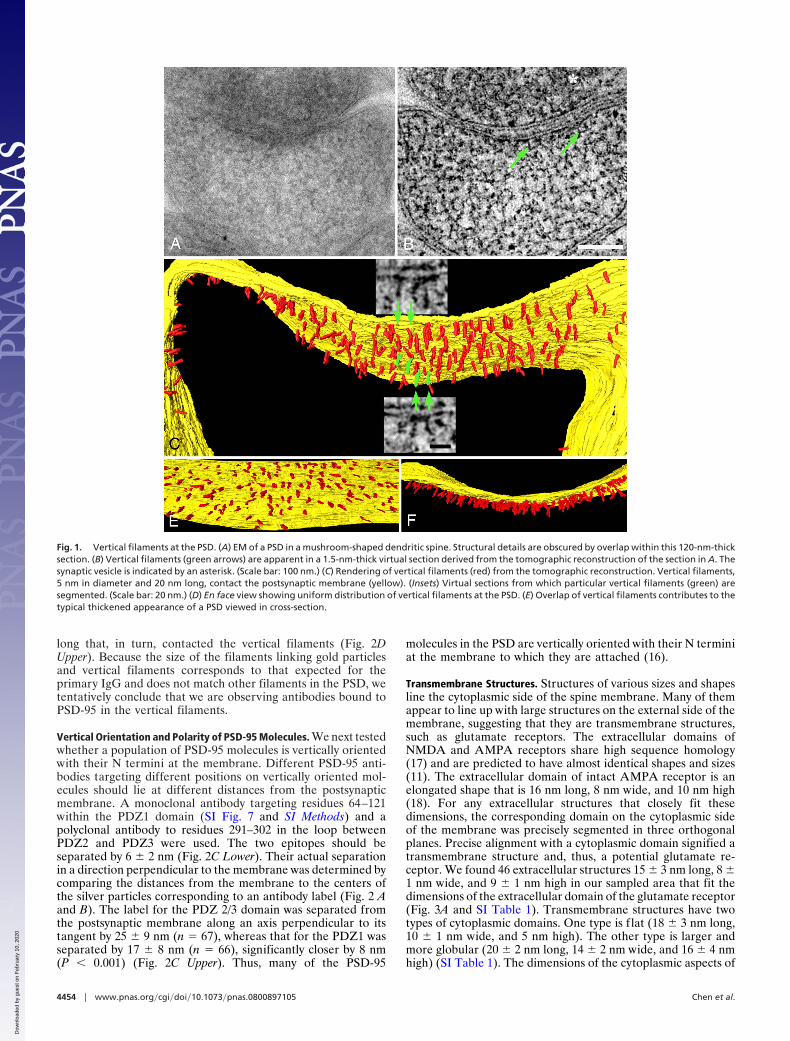
cross-sectioned PSDs were selected for EM tomography. One PSDfrom the reconstruction of a mushroom-shaped spine (SI Methods)was selected for extensive segmentation, rendering, and structuralanalysis (Fig. 1 A and B). Structures as small as 3–4 nm in diameterwithin 1- to 1.5-nm-thick virtual sections can be segmented. Di-mensions of structures such as actin filaments (7 nm wide) (data notshown), synaptic vesicles (40 nm in diameter), and synaptic cleft (25nm wide) are in close agreement with dimensions determined bycryo-EM and other methods (12–14).
Vertical Filaments. Filaments segmented in a series of virtualsections were classified on the basis of their location, shape, anddimensions. A large class of membrane-associated filaments atthe PSD is nearly straight and vertically oriented with respect tothe postsynaptic membrane (Fig. 1 B and C and SI Movie 1). Werefer to filaments of this type as vertical filaments. Verticalfilaments are typically 5 nm in diameter (4.9 � 0.2 nm, n � 22)and 20 nm long (21 � 3 nm, range 16–25 nm, n � 38). Verticalfilaments within the PSD are uniformly spaced, with a nearestneighbor distance of 13.4 � 2.8 nm (n � 31) (Fig. 1D). Thethicket of vertical filaments gives rise to the typical denseappearance that is characteristic of PSDs in standard EMcross-sectional views (Fig. 1E). Vertical filaments are ubiquitousin reconstructions of PSDs, even in places where other structuralelements are absent. Based on their density, there would be�400 vertical filaments in a 400-nm-diameter (0.16-�m2) PSD.
The dimensions of vertical filaments, and their associations withthe postsynaptic membrane, suggest that they belong to the PSD-95family of MAGUK proteins (10). These family members sharemany structural similarities, permitting the dimensions of a genericfamily member to be estimated by combining the known sizes oftheir domains (20 nm long, 4 nm in diameter) (SI Methods).Dimensions of fully extended PSD-95 and SAP97 molecules (16–22nm long and 6 nm in diameter) by single-particle EM (15) alsomatch to those of vertical filaments.
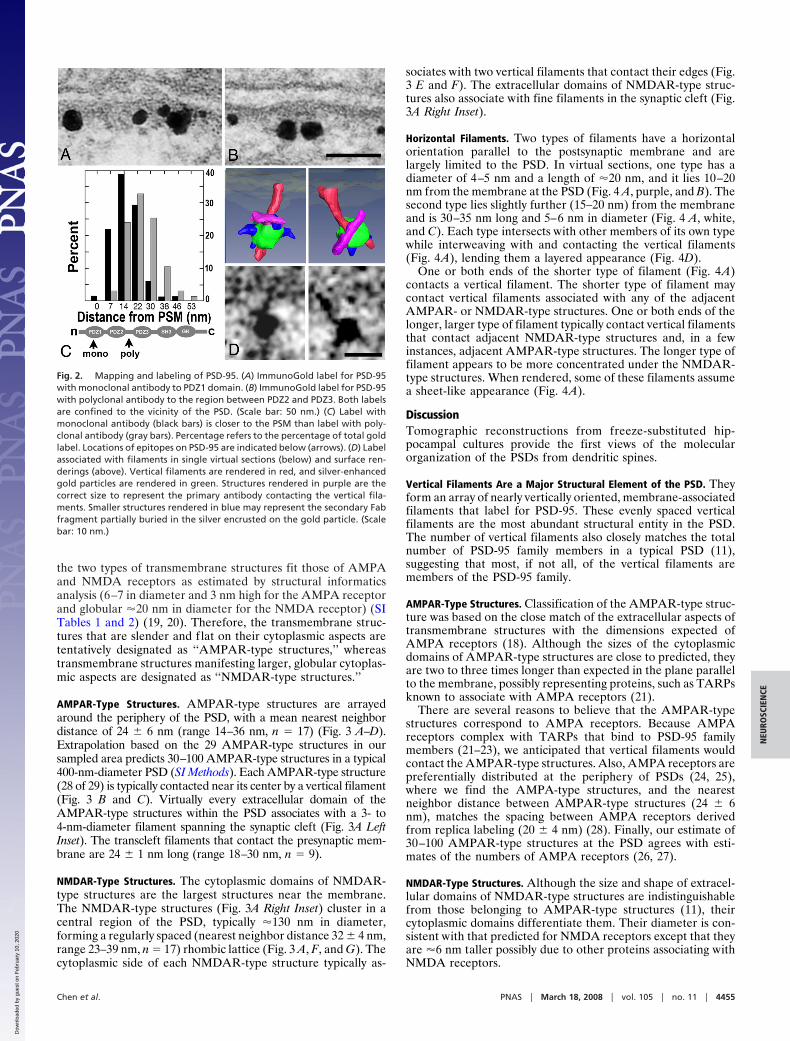
Vertical Filaments Label for PSD-95. Conventional immuno-EMshows that the antibodies to PSD-95 specifically label PSDs (Fig.2 A and B). When parallel labeling experiments are followed byfreeze substitution and tomography, electron-dense particlescorresponding to silver-enhanced Nanogold are located nearvertical filaments (n � 9) (Fig. 2D Lower). We expected that thesecondary antibody on the Nanogold would be enveloped insilver; in the four instances that were rendered, structuresembedded in the silver grain contacted filaments up to 15 nm
Author contributions: X.C., R.D.L., and T.S.R. designed research; X.C., C.W., R.A., J.A.G.,R.D.L., and T.S.R. performed research; X.L. contributed new reagents/analytic tools; X.C.analyzed data; and X.C. and T.S.R. wrote the paper.
The authors declare no conflict of interest.
¶To whom correspondence should be addressed. E-mail: [email protected].
This article contains supporting information online at www.pnas.org/cgi/content/full/0800897105/DC1.
© 2008 by The National Academy of Sciences of the USA
www.pnas.org�cgi�doi�10.1073�pnas.0800897105 PNAS � March 18, 2008 � vol. 105 � no. 11 � 4453–4458
NEU
ROSC
IEN
CE
Dow
nloa
ded
by g
uest
on
Feb
ruar
y 10
, 202
0 D
ownl
oade
d by
gue
st o
n F
ebru
ary
10, 2
020
Dow
nloa
ded
by g
uest
on
Feb
ruar
y 10
, 202
0
long that, in turn, contacted the vertical filaments (Fig. 2DUpper). Because the size of the filaments linking gold particlesand vertical filaments corresponds to that expected for theprimary IgG and does not match other filaments in the PSD, wetentatively conclude that we are observing antibodies bound toPSD-95 in the vertical filaments.
Vertical Orientation and Polarity of PSD-95 Molecules. We next testedwhether a population of PSD-95 molecules is vertically orientedwith their N termini at the membrane. Different PSD-95 anti-bodies targeting different positions on vertically oriented mol-ecules should lie at different distances from the postsynapticmembrane. A monoclonal antibody targeting residues 64–121within the PDZ1 domain (SI Fig. 7 and SI Methods) and apolyclonal antibody to residues 291–302 in the loop betweenPDZ2 and PDZ3 were used. The two epitopes should beseparated by 6 � 2 nm (Fig. 2C Lower). Their actual separationin a direction perpendicular to the membrane was determined bycomparing the distances from the membrane to the centers ofthe silver particles corresponding to an antibody label (Fig. 2 Aand B). The label for the PDZ 2/3 domain was separated fromthe postsynaptic membrane along an axis perpendicular to itstangent by 25 � 9 nm (n � 67), whereas that for the PDZ1 wasseparated by 17 � 8 nm (n � 66), significantly closer by 8 nm(P � 0.001) (Fig. 2C Upper). Thus, many of the PSD-95
molecules in the PSD are vertically oriented with their N terminiat the membrane to which they are attached (16).
Transmembrane Structures. Structures of various sizes and shapesline the cytoplasmic side of the spine membrane. Many of themappear to line up with large structures on the external side of themembrane, suggesting that they are transmembrane structures,such as glutamate receptors. The extracellular domains ofNMDA and AMPA receptors share high sequence homology(17) and are predicted to have almost identical shapes and sizes(11). The extracellular domain of intact AMPA receptor is anelongated shape that is 16 nm long, 8 nm wide, and 10 nm high(18). For any extracellular structures that closely fit thesedimensions, the corresponding domain on the cytoplasmic sideof the membrane was precisely segmented in three orthogonalplanes. Precise alignment with a cytoplasmic domain signified atransmembrane structure and, thus, a potential glutamate re-ceptor. We found 46 extracellular structures 15 � 3 nm long, 8 �1 nm wide, and 9 � 1 nm high in our sampled area that fit thedimensions of the extracellular domain of the glutamate receptor(Fig. 3A and SI Table 1). Transmembrane structures have twotypes of cytoplasmic domains. One type is f lat (18 � 3 nm long,10 � 1 nm wide, and 5 nm high). The other type is larger andmore globular (20 � 2 nm long, 14 � 2 nm wide, and 16 � 4 nmhigh) (SI Table 1). The dimensions of the cytoplasmic aspects of
Fig. 1. Vertical filaments at the PSD. (A) EM of a PSD in a mushroom-shaped dendritic spine. Structural details are obscured by overlap within this 120-nm-thicksection. (B) Vertical filaments (green arrows) are apparent in a 1.5-nm-thick virtual section derived from the tomographic reconstruction of the section in A. Thesynaptic vesicle is indicated by an asterisk. (Scale bar: 100 nm.) (C) Rendering of vertical filaments (red) from the tomographic reconstruction. Vertical filaments,5 nm in diameter and 20 nm long, contact the postsynaptic membrane (yellow). (Insets) Virtual sections from which particular vertical filaments (green) aresegmented. (Scale bar: 20 nm.) (D) En face view showing uniform distribution of vertical filaments at the PSD. (E) Overlap of vertical filaments contributes to thetypical thickened appearance of a PSD viewed in cross-section.
4454 � www.pnas.org�cgi�doi�10.1073�pnas.0800897105 Chen et al.
Dow
nloa
ded
by g
uest
on
Feb
ruar
y 10
, 202
0
the two types of transmembrane structures fit those of AMPAand NMDA receptors as estimated by structural informaticsanalysis (6–7 in diameter and 3 nm high for the AMPA receptorand globular �20 nm in diameter for the NMDA receptor) (SITables 1 and 2) (19, 20). Therefore, the transmembrane struc-tures that are slender and flat on their cytoplasmic aspects aretentatively designated as ‘‘AMPAR-type structures,’’ whereastransmembrane structures manifesting larger, globular cytoplas-mic aspects are designated as ‘‘NMDAR-type structures.’’
AMPAR-Type Structures. AMPAR-type structures are arrayedaround the periphery of the PSD, with a mean nearest neighbordistance of 24 � 6 nm (range 14–36 nm, n � 17) (Fig. 3 A–D).Extrapolation based on the 29 AMPAR-type structures in oursampled area predicts 30–100 AMPAR-type structures in a typical400-nm-diameter PSD (SI Methods). Each AMPAR-type structure(28 of 29) is typically contacted near its center by a vertical filament(Fig. 3 B and C). Virtually every extracellular domain of theAMPAR-type structures within the PSD associates with a 3- to4-nm-diameter filament spanning the synaptic cleft (Fig. 3A LeftInset). The transcleft filaments that contact the presynaptic mem-brane are 24 � 1 nm long (range 18–30 nm, n � 9).
NMDAR-Type Structures. The cytoplasmic domains of NMDAR-type structures are the largest structures near the membrane.The NMDAR-type structures (Fig. 3A Right Inset) cluster in acentral region of the PSD, typically �130 nm in diameter,forming a regularly spaced (nearest neighbor distance 32 � 4 nm,range 23–39 nm, n � 17) rhombic lattice (Fig. 3 A, F, and G). Thecytoplasmic side of each NMDAR-type structure typically as-
sociates with two vertical filaments that contact their edges (Fig.3 E and F). The extracellular domains of NMDAR-type struc-tures also associate with fine filaments in the synaptic cleft (Fig.3A Right Inset).
Horizontal Filaments. Two types of filaments have a horizontalorientation parallel to the postsynaptic membrane and arelargely limited to the PSD. In virtual sections, one type has adiameter of 4–5 nm and a length of �20 nm, and it lies 10–20nm from the membrane at the PSD (Fig. 4 A, purple, and B). Thesecond type lies slightly further (15–20 nm) from the membraneand is 30–35 nm long and 5–6 nm in diameter (Fig. 4 A, white,and C). Each type intersects with other members of its own typewhile interweaving with and contacting the vertical filaments(Fig. 4A), lending them a layered appearance (Fig. 4D).
One or both ends of the shorter type of filament (Fig. 4A)contacts a vertical filament. The shorter type of filament maycontact vertical filaments associated with any of the adjacentAMPAR- or NMDAR-type structures. One or both ends of thelonger, larger type of filament typically contact vertical filamentsthat contact adjacent NMDAR-type structures and, in a fewinstances, adjacent AMPAR-type structures. The longer type offilament appears to be more concentrated under the NMDAR-type structures. When rendered, some of these filaments assumea sheet-like appearance (Fig. 4A).
DiscussionTomographic reconstructions from freeze-substituted hip-pocampal cultures provide the first views of the molecularorganization of the PSDs from dendritic spines.
Vertical Filaments Are a Major Structural Element of the PSD. Theyform an array of nearly vertically oriented, membrane-associatedfilaments that label for PSD-95. These evenly spaced verticalfilaments are the most abundant structural entity in the PSD.The number of vertical filaments also closely matches the totalnumber of PSD-95 family members in a typical PSD (11),suggesting that most, if not all, of the vertical filaments aremembers of the PSD-95 family.
AMPAR-Type Structures. Classification of the AMPAR-type struc-ture was based on the close match of the extracellular aspects oftransmembrane structures with the dimensions expected ofAMPA receptors (18). Although the sizes of the cytoplasmicdomains of AMPAR-type structures are close to predicted, theyare two to three times longer than expected in the plane parallelto the membrane, possibly representing proteins, such as TARPsknown to associate with AMPA receptors (21).
There are several reasons to believe that the AMPAR-typestructures correspond to AMPA receptors. Because AMPAreceptors complex with TARPs that bind to PSD-95 familymembers (21–23), we anticipated that vertical filaments wouldcontact the AMPAR-type structures. Also, AMPA receptors arepreferentially distributed at the periphery of PSDs (24, 25),where we find the AMPA-type structures, and the nearestneighbor distance between AMPAR-type structures (24 � 6nm), matches the spacing between AMPA receptors derivedfrom replica labeling (20 � 4 nm) (28). Finally, our estimate of30–100 AMPAR-type structures at the PSD agrees with esti-mates of the numbers of AMPA receptors (26, 27).
NMDAR-Type Structures. Although the size and shape of extracel-lular domains of NMDAR-type structures are indistinguishablefrom those belonging to AMPAR-type structures (11), theircytoplasmic domains differentiate them. Their diameter is con-sistent with that predicted for NMDA receptors except that theyare �6 nm taller possibly due to other proteins associating withNMDA receptors.
Fig. 2. Mapping and labeling of PSD-95. (A) ImmunoGold label for PSD-95with monoclonal antibody to PDZ1 domain. (B) ImmunoGold label for PSD-95with polyclonal antibody to the region between PDZ2 and PDZ3. Both labelsare confined to the vicinity of the PSD. (Scale bar: 50 nm.) (C) Label withmonoclonal antibody (black bars) is closer to the PSM than label with poly-clonal antibody (gray bars). Percentage refers to the percentage of total goldlabel. Locations of epitopes on PSD-95 are indicated below (arrows). (D) Labelassociated with filaments in single virtual sections (below) and surface ren-derings (above). Vertical filaments are rendered in red, and silver-enhancedgold particles are rendered in green. Structures rendered in purple are thecorrect size to represent the primary antibody contacting the vertical fila-ments. Smaller structures rendered in blue may represent the secondary Fabfragment partially buried in the silver encrusted on the gold particle. (Scalebar: 10 nm.)
Chen et al. PNAS � March 18, 2008 � vol. 105 � no. 11 � 4455
NEU
ROSC
IEN
CE
Dow
nloa
ded
by g
uest
on
Feb
ruar
y 10
, 202
0

Each large cytoplasmic domain of the NMDA-type structuretypically associates with two PSD-95 containing vertical fila-ments around its rim. Indeed, it is known that PSD-95 binds toNR2 (28, 29) and that each NMDA receptor includes two copiesof NR2 (17). Almost all of the NMDAR-type structures clusterat the center of the PSD, where they are arranged in a rhombiclattice, allowing for four to five NMDAR-type structures ar-ranged per lattice line. This arrangement would allow for 16–25NMDA receptors in the central cluster of a typical PSD, whichis consistent with previous estimates of 20–30 NMDA receptors(11, 30).
Extracellular domains of NMDAR- and AMPAR-type struc-tures associate with transcleft filaments, which are likely to beadhesion molecules (31, 32). Their molecular identities requirefurther investigation.
Two Types of Horizontal Filament Link Vertical Filaments. The shorter(�20 nm long) type of horizontal filament typically associateswith vertical filaments contacting either NMDAR- or AMPAR-type structures as if linking adjacent receptors via the verticalfilaments. The lengths of these links would explain the �20-nmnearest neighbor distance among AMPAR-type structures. Thelonger (�30 nm) type of horizontal filament associates withvertical filaments contacting both AMPAR- and NMDAR-typestructures, but they predominately link adjacent NMDAR-typestructures, which might explain their �30-nm spacing. The twotypes of horizontal filaments lie in slightly separate layers,lending the PSD a laminar organization (33, 34).
Because PSD-95 family members have many potential bindingpartners, the molecular identities of the horizontal filaments thatthey contact may be complex, even including a few multimerizedPSD-95 family members (35). GKAP/SAPAP is likely to be partof the horizontal filament system because it binds to the GKdomain of PSD-95. There are �150 copies of GKAP/SAPAP ina PSD (11), where it exists in 95- and 130-kDa forms (36). Thesemolecular mass are in line with those expected of 5-nm filamentseither the same length (20 nm) or 1.5 times the length (30 nm)of the vertical filaments. Another prevalent scaffolding mole-cule, Shank, binds to GKAP/SAPAP, so the sheet-like structuresformed by horizontal filaments may include both GKAP andShank (37). Indeed, Shank has a SAM domain that can poly-merize into sheet-like structures (38).
Organization of the Core of the PSD. PSD-95 has been regarded asa central player in the core organization of the PSD due to itsprevalence in the PSD and its multiple binding sites for receptorsand other scaffolding molecules. Our findings that a large array ofvertically oriented filaments dominates the core structure of thePSD, and that the vertical filaments include PSD-95 in an extendedconfiguration, show how PSD-95 and related family members couldlink other core components of the PSD into a coherent structure,as schematized in Fig. 5. The vertical orientation of PSD-95 puts itsPDZ domains near the membrane in positions to bind directly, orthrough intermediate proteins, to glutamate receptors and deploysthe SH3-GK domains further from the membrane, where they cancontact a family of horizontally oriented filaments, including
Fig. 3. Transmembrane structures at the PSD. (A) Cytoplasmic surface of the membrane (yellow) at the PSD, showing AMPAR-type (blue) and NMDAR-typecytoplasmic domains (cyan), and vertical filaments (red). (Left Insets) AMPAR-type structures in virtual sections from the tomographic reconstruction. Arrowpoints to a transcleft filament. (Right Insets) NMDAR-type structures. Arrow points to a transcleft filament, and double arrow (lower right) indicates the extentof synaptic cleft. (Scale bar: 20 nm.) (B–D) AMPAR-type structures shown in cross-section (B, extracellular domain green), en face from inside the spine (C), anden face from outside the spine (D). Cytoplasmic domains of AMPAR-type structures are contacted by vertical filaments (C). (E–G) NMDAR-type structures shownin cross-section (E, extracellular domain gold), en face from inside the spine (F), and en face from outside the spine (G). Cytoplasmic domains of NMDAR-typestructures are contacted by one or two vertical filaments (F).
4456 � www.pnas.org�cgi�doi�10.1073�pnas.0800897105 Chen et al.
Dow
nloa
ded
by g
uest
on
Feb
ruar
y 10
, 202
0

GKAP and, perhaps, Shank. The vertical orientation and extendedshape assumed by PSD-95 family members lying in the core of thePSD thus puts them in a position to form a stable orthogonalstructure in the core of the PSD-linking receptors and otherscaffolding molecules.
The lability of AMPA receptors relative to NMDA receptorsis thought to make a contribution to synaptic plasticity (5).Details of the orthogonal arrangement of core components showhow PSD-95 family members might stabilize the NMDA recep-tors arrayed in the center of the PSD while allowing AMPAreceptors arrayed around the periphery to turn over morequickly. The longer type of horizontal filament is the right lengthto link vertical filaments from adjacent NMDA receptors andfurther stabilize them at their characteristic spacing of �30 nmin the center of the PSD. The shorter type of horizontal filamentsare the right length to link vertical filaments attached to adjacentAMPA receptors and stabilize them at their spacing of �20 nmat the periphery of the PSD, and link AMPA to NMDAreceptors at their interface. Thus, the horizontal filaments closethe loop to create a framework on which to organize and stabilize
NMDA and AMPA receptors beyond that realized by theirindividual associations with vertical filaments.
The arrangement of regular-spaced vertical filaments andglutamate receptors nevertheless leaves open the possibility thatreceptors could be added or deleted at the edges of the PSD. Alarge number of vertical filaments do not associate with gluta-mate receptor structures, and these filaments could bind addi-tional AMPA receptors arriving at the edge of the PSD or acceptreceptors into the horizontal filament meshwork if they werealready attached to a vertical filament (39). Further recruitmentof AMPA receptors into the PSD may depend on the enlistmentof PSD-95 family members into the vertical filament scaffold atthe periphery of the PSD as the PSD increases in diameter (24,26, 40, 41), whereas reduction in PSD-95 would lead to the lossof peripheral AMPA receptors (42–44).
MethodsCultured Hippocampal Neurons. Dissociated rat hippocampal neurons (E20)were plated onto glia in a Bal-Tec 3-mm gold specimen chamber and main-tained for 3 weeks in 10% CO2 (45). Neurons on the specimen carrier weremonitored by reflection microscopy.
Freeze Substitution. Cultures were frozen at 2100 Bar with a Bal-Tec HPM 010machine (Techno Trade) in 124 mM NaCl, 2 mM KCl, 1.24 mM KH2PO4, 1.3 mMMgCl2, 2.5 mM CaCl2, 30 mM glucose, 25 mM Hepes, and 0.5% ovalbumin (pH7.4; osmolarity of 325). Samples were covered with hexadecane before freez-ing. Specimen carriers were left on frozen saturated uranyl acetate and 2%acrolein in HPLC-grade acetone at �160°C for 15 min in an AFS Leica unit,ramped from �160 to �90°C in 14 h, held at �90°C for 8 h, ramped to �60°in 6 h, held for 12 h, and infiltrated in Lowicryl HM20 resin in acetone. Lowicrylwas polymerized by UV at �50°C. Acrolein helped stabilize membrane struc-ture, and uranyl acetate gave a fine-grained stain that extended evenlythrough sections and did not overstain. Sections �100–200 nm thick were cuten face and mounted on Formvar/carbon-coated grids with 10-nm goldparticles applied to both sides as fiducial markers.
EM Tomography. PSDs at mature synapses in areas free of ice crystal damagewere photographed in an FEI Tecnai 300-kV electron microscope with afield-emission gun at a dose of �300 electrons per nm2 per image. Series wereacquired in two axes at tilt increments of 2° from �74° to �74°. Pixel sizes were0.48–0.75 nm (2,048 � 2,048 image). Series were reconstructed, and thedual-axis 3D volumes merged with IMOD (46) at a typical alignment error of�0.3 pixels. The 3D volume (tomogram) was analyzed with EM3D (6), seg-
Fig. 4. Filament network at the PSD. (A) Meshwork of horizontal filaments at the core of the PSD. The shorter type (purple) is 4–5 nm in diameter and �20nm long, and the longer type (white) is 5–6 nm in diameter and 30–35 nm long. The asterisks indicate sheet-like structures. (B and C) Shorter (B, arrow) and longer(C, arrowhead) types of horizontal filament in virtual sections. (Scale bar: 20 nm.) (D) Cross-sectional view of the PSD in A showing layering of horizontal filaments.The shorter filaments (B, purple) lie somewhat closer to the postsynaptic membrane than the longer filaments (C, white).
Fig. 5. Core structure of the PSD based on tomographic reconstructions.Components and spacings between components are drawn approximately toscale. Dominant structure is an array of vertical filaments containing PSD-95(red). NMDAR-type structures, differentiated by their large cytoplasmic ex-tensions (cyan), concentrate in the center of the PSD. AMPAR-type structures,differentiated by their flattened cytoplasmic aspects (blue), surround theNMDAR structures. Virtually all NMDAR- and AMPAR-type structures arecontacted by vertical filaments. Short horizontal filaments (purple) link ver-tical filaments associated with both NMDAR- and AMPAR-type structures.Longer horizontal filaments (white) concentrated under the NMDAR-typestructures cross-link the vertical filament meshwork.
Chen et al. PNAS � March 18, 2008 � vol. 105 � no. 11 � 4457
NEU
ROSC
IEN
CE
Dow
nloa
ded
by g
uest
on
Feb
ruar
y 10
, 202
0
mented semiautomatically, and surface-rendered with Amira (Mercury Com-puter Systems).
In Amira, the magic wand tool was used to determine the density of part of astructure, and then the whole structure was automatically outlined. Ambiguitiesin outlining density in one cross-sectional view were resolved by segmenting thesame structure in two other views. Objects were segmented one by one, andobjects were grouped into classes, allowing relationships between these objects,individually and as a class, to be defined. All measurements were performedeither in EM3D or with ImageJ (http://rsb.info.nih.gov/ij) in snap shots of surface-rendered objects obtained from Amira. All data are reported as mean � SD.
Antibodies and Immunocytochemistry. Monoclonal antibody to PSD-95 is fromAffinity BioReagents (MA1–046). A polyclonal antibody to residues 290–307designed by Ayse Dosemeci was made by New England Peptide. For details onmapping the epitope recognized by ABR PSD-95 antibody, see SI Methods. Forconventional ImmunoGold labeling, cells were fixed in 4% paraformaldehydefor 45 min, incubated with the primary antibody for 1 h, secondary antibody-conjugated to 1.4-nm Nanogold (47), and silver enhanced (HQ kit; Nano-probes). For tomography, cultures were fixed and immunolabeled with thepolyclonal PSD-95 antibody and Nanogold second antibody as described
above, and silver enhanced for 6–8 min, followed by the high-pressurefreezing, freeze substitution, and embedding protocol used to prepares cul-tures for tomography. When primary antibody was eliminated from theprotocol, no specific labeling was observed.
Protein Structural Informatics. Differentiation between structured and na-tively unstructured protein conformations was based on primary sequence(20). Estimates of the sizes of protein domains were done as described in ref.19. For further details, see SI Methods.
ACKNOWLEDGMENTS. We thank Jung-Hwa Tao-Cheng (National Institute ofNeurological Disorders and Stroke, Bethesda) for electron micrographs; Vir-ginia Crocker for immunolabeling; Bechara Kachar for valuable suggestionsabout freeze substitution; John Lisman, Wayne Albers, Carolyn Smith, andAyse Dosemeci for valuable comments on the manuscript; John Chludzinskifor help with figures; the EM3D (Stanford University, Stanford, CA) team; andthe IMOD team (University of Colorado, Boulder, CO) for software support.This work was supported by National Institute of Neurological Disorders andStroke and National Institute of Biomedical Imaging and BioEngineeringIntramural Research Programs.
1. Siekevitz P (1985) The postsynaptic density: A possible role in long-lasting effects in thecentral nervous system. Proc Natl Acad Sci USA 82:3494–3498.
2. Ziff EB (1997) Enlightening the postsynaptic density. Neuron 19:1163–1174.3. Kennedy MB (2000) Signal-processing machines at the postsynaptic density. Science
290:750–754.4. Palay SL (1956) Synapses in the central nervous system. J Biophys Biochem Cytol
2:193–202.5. Malinow R, Malenka RC (2002) AMPA receptor trafficking and synaptic plasticity. Annu
Rev Neurosci 25:103–126.6. Harlow ML, Ress D, Stoschek A, Marshall RM, McMahan UJ (2001) The architecture of
active zone material at the frog’s neuromuscular junction. Nature 409:479–484.7. Chen X, et al. (2005) Mass of the postsynaptic density and enumeration of three key
molecules. Proc Natl Acad Sci USA 102:11551–11556.8. Cheng D, et al. (2006) Relative and absolute quantification of postsynaptic density pro-
teome isolated from rat forebrain and cerebellum. Mol Cell Proteomics 5:1158–1170.9. Dosemeci A, et al. (2007) Composition of the synaptic PSD-95 complex. Mol Cell
Proteomics 6:1749–1760.10. Kim E, Sheng M (2004) PDZ domain proteins of synapses. Nat Rev Neurosci 5:771–781.11. Sheng M, Hoogenraad CC (2007) The postsynaptic architecture of excitatory synapses:
A more quantitative view. Ann Rev Biochem 76:823–847.12. Volkmann N, et al. (2001) Structure of Arp2/3 complex in its activated state and in actin
filament branch junctions. Science 293:2456–2459.13. Zuber B, Nikonenko I, Klauser P, Muller D, Dubochet J (2005) The mammalian central
nervous synaptic cleft contains a high density of periodically organized complexes. ProcNatl Acad Sci USA 102:19192–19197.
14. Lucic V, Yang T, Schweikert G, Forster F, Baumeister W (2005) Morphological charac-terization of molecular complexes present in the synaptic cleft. Structure (London)13:423–434.
15. Nakagawa T, et al. (2004) Quaternary structure, protein dynamics, and synapticfunction of SAP97 controlled by L27 domain interactions. Neuron 44:453–467.
16. Craven SE, El-Husseini AE, Bredt DS (1999) Synaptic targeting of the postsynapticdensity protein PSD-95 mediated by lipid and protein motifs. Neuron 22:497–509.
17. Mayer ML (2006) Glutamate receptors at atomic resolution. Nature 440:456–462.18. Nakagawa T, Cheng Y, Ramm E, Sheng M, Walz T (2005) Structure and different
conformational states of native AMPA receptor complexes. Nature 433:545–549.19. Uversky VN (2002) What does it mean to be natively unfolded? Eur J Biochem 269:2–12.20. Dosztanyi Z, Csizmok V, Tompa P, Simon I (2005) The pairwise energy content esti-
mated from amino acid composition discriminates between folded and intrinsicallyunstructured proteins. J Mol Biol 347:827–839.
21. Nicoll RA, Tomita S, Bredt DS (2006) Auxiliary subunits assist AMPA-type glutamatereceptors. Science 311:1253–1256.
22. Chen L, et al. (2000) Stargazin regulates synaptic targeting of AMPA receptors by twodistinct mechanisms. Nature 408:936–943.
23. Schnell E, Karimzadegan S, Chen L, Bredt DS, Nicoll RA (2002) Direct interactionsbetween PSD-95 and stargazin control synaptic AMPA receptor number. Proc NatlAcad Sci USA 99:13902–13907.
24. Takumi Y, Ramirez-Leon V, Laake P, Rinvik E, Ottersen OP (1999) Different modes ofexpression of AMPA and NMDA receptors in hippocampal synapses. Nat Neurosci2:618–624.
25. Kharazia VN, Weinberg RJ (1997) Tangential synaptic distribution of NMDA and AMPAreceptors in rat neocortex. Neurosci Lett 238:41–44.
26. Matsuzaki MHN, Ellis-Davies GC, Kasai H (2004) Structural basis of long-term potenti-ation in single dendritic spines. Nature 429:761–766.
27. Nusser Z, et al. (1998) Cell type and pathway dependence of synaptic AMPA receptornumber and variability in the hippocampus. Neuron 21:545–559.
28. Kornau HC, Schenker LT, Kennedy MB, Seeburg PH (1995) Domain interaction betweenNMDA receptor subunits and the postsynaptic density protein PSD-95. Science269:1737–1740.
29. Niethammer M, Kim E, Sheng M (1996) Interaction between the C terminus of NMDAreceptor subunits and multiple members of the PSD-95 family of membrane-associatedguanylate kinases. J Neurosci 16:2157–2163.
30. Nimchinsky EA, Yasuda R, Oertner TG, Svoboda K (2004) The number of glutamatereceptors opened by synaptic stimulation in single hippocampal spines. J Neurosci24:2054–2064.
31. Saglietti L, et al. (2007) Extracellular interactions between GluR2 and N-cadherin inspine regulation. Neuron 54:461–477.
32. Wang CY, et al. (2006) A novel family of adhesion-like molecules that interacts with theNMDA receptor. J Neurosci 26:2174–2183.
33. Petersen JD, et al. (2003) Distribution of postsynaptic density (PSD)-95 and Ca2�/cal-modulin-dependent protein kinase II at the PSD. J Neurosci 23:11270–11278.
34. Valtschanoff JG, Weinberg RJ (2001) Laminar organization of the NMDA receptorcomplex within the postsynaptic density. J Neurosci 21:1211–1217.
35. Kim E, Cho KO, Rothschild A, Sheng M (1996) Heteromultimerization and NMDAreceptor-clustering activity of Chapsyn-110, a member of the PSD-95 family of proteins.Neuron 17:103–113.
36. Kim E, et al. (1997) GKAP, a novel synaptic protein that interacts with the guanylatekinase-like domain of the PSD-95/SAP90 family of channel clustering molecules. J CellBiol 136:669–678.
37. Naisbitt S, et al. (1999) Shank, a novel family of postsynaptic density proteins that bindsto the NMDA receptor/PSD-95/GKAP complex and cortactin. Neuron 23:569–582.
38. Baron MK, et al. (2006) An architectural framework that may lie at the core of thepostsynaptic density. Science 311:531–535.
39. Bats C, Groc L, Choquet D (2007) The interaction between Stargazin and PSD-95regulates AMPA receptor surface trafficking. Neuron 53:719–734.
40. El-Husseini AE, Schnell E, Chetkovich DM, Nicoll RA, Bredt DS (2000) PSD-95 involve-ment in maturation of excitatory synapses. Science 290:1364–1368.
41. Harris KM, Stevens JK (1989) Dendritic spines of CA 1 pyramidal cells in the rathippocampus: serial electron microscopy with reference to their biophysical charac-teristics. J Neurosci 9:2982–2997.
42. El-Husseini A-D, et al. (2002) Synaptic strength regulated by palmitate cycling onPSD-95. Cell 108:849–863.
43. Colledge M, et al. (2003) Ubiquitination regulates PSD-95 degradation and AMPAreceptor surface expression. Neuron 40:595–607.
44. Elias GM, et al. (2006) Synapse-specific and developmentally regulated targeting ofAMPA receptors by a family of MAGUK scaffolding proteins. Neuron 52:307–320.
45. Mayer ML, Vyklicky L, Jr, Westbrook GL (1989) Modulation of excitatory amino acidreceptors by group IIB metal cations in cultured mouse hippocampal neurones.J Physiol 415:329–350.
46. Kremer JR, Mastronarde DN, McIntosh JR (1996) Computer visualization of three-dimensional image data using IMOD. J Struct Biol 116:71–76.
47. Dosemeci A, et al. (2001) Glutamate-induced transient modification of the postsyn-aptic density. Proc Natl Acad Sci USA 98:10428–10432.
4458 � www.pnas.org�cgi�doi�10.1073�pnas.0800897105 Chen et al.
Dow
nloa
ded
by g
uest
on
Feb
ruar
y 10
, 202
0
Editorial Expression of Concern and Corrections.
EDITORIAL EXPRESSION OF CONCERN. PNAS is publishing an Edito-rial Expression of Concern regarding the following three articles:
(i) PHYSIOLOGY. ‘‘NCX-1000, a NO-releasing derivative of ur-sodeoxycholic acid, selectively delivers NO to the liver andprotects against development of portal hypertension,’’ by Ste-fano Fiorucci, Elisabetta Antonelli, Olivia Morelli, AndreaMencarelli, Alessandro Casini, Tommaso Mello, Barbara Palaz-zetti, Dominique Tallet, Piero del Soldato, and Antonio Morelli,which appeared in issue 15, July 17, 2001, of Proc Natl Acad SciUSA (98:8897–8902; first published July 10, 2001; 10.1073�pnas.151136298);
(ii) PHARMACOLOGY. ‘‘NCX-1015, a nitric-oxide derivative ofprednisolone, enhances regulatory T cells in the lamina propriaand protects against 2,4,6-trinitrobenzene sulfonic acid-inducedcolitis in mice,’’ by Stefano Fiorucci, Elisabetta Antonelli,Eleonora Distrutti, Piero Del Soldato, Roderick J. Flower, MarkJ. Paul Clark, Antonio Morelli, Mauro Perretti, and Louis J.Ignarro, which appeared in issue 24, November 26, 2002, of ProcNatl Acad Sci USA (99:15770–15775; first published November11, 2002; 10.1073�pnas.232583599); and
(iii) MEDICAL SCIENCES. ‘‘A �-oxidation-resistant lipoxin A4analog treats hapten-induced colitis by attenuating inflamma-tion and immune dysfunction,’’ by Stefano Fiorucci, John L.Wallace, Andrea Mencarelli, Eleonora Distrutti, GiovanniRizzo, Silvana Farneti, Antonio Morelli, Jih-Lie Tseng, BabuSuramanyam, William J. Guilford, and John F. Parkinson, whichappeared in issue 44, November 2, 2004, of Proc Natl Acad SciUSA (101:15736–15741; first published October 25, 2004;10.1073�pnas.0404722101).
The editors wish to note that a reader has raised questionsabout the apparent duplication in the use of certain figures in theforegoing articles. We have been informed by the University ofPerugia, Italy, of an ongoing review conducted by an inquirycommittee at the university. We are awaiting the findings of thecommittee to determine the appropriate next steps.
Randy Schekman, Editor-in-Chief
www.pnas.org�cgi�doi�10.1073�pnas.0803890105
PLANT BIOLOGY. For the article ‘‘Salt tolerance of Arabidopsisthaliana requires maturation of N-glycosylated proteins in theGolgi apparatus,’’ by Jae Sook Kang, Julia Frank, Chang HoKang, Hiroyuki Kajiura, Meenu Vikram, Akihiro Ueda, SewonKim, Jeong Dong Bahk, Barbara Triplett, Kazuhito Fujiyama,Sang Yeol Lee, Antje von Schaewen, and Hisashi Koiwa, whichappeared in issue 15, April 15, 2008, of Proc Natl Acad Sci USA(105:5933–5938; first published April 11, 2008; 10.1073�pnas.0800237105), the authors note that the affiliation forKazuhito Fujiyama should have appeared as Osaka University.The corrected author line, the affiliation line, and a relatedfootnote appear below.
Jae Sook Kang*, Julia Frank†, Chang Ho Kang*‡, HiroyukiKajiura§, Meenu Vikram‡, Akihiro Ueda‡, Sewon Kim*,Jeong Dong Bahk*, Barbara Triplett¶, Kazuhito Fujiyama§,Sang Yeol Lee*�, Antje von Schaewen†�, and HisashiKoiwa‡�
*Brain Korea-21, Division of Applied Life Science and EnvironmentalBiotechnology National Core Research Center, Graduate School ofGyeongsang National University, Jinju 660-701, Korea; †MolekularePhysiologie der Pflanzen, Institut fur Botanik, WestfalischeWilhelms-Universitat Munster, Schlossgarten 3, 48149 Munster, Germany;‡Vegetable and Fruit Improvement Center, Department of HorticulturalSciences, and Molecular and Environmental Plant Science Program, TexasA&M University, College Station, TX 77843-2133; §International Center forBiotechnology, Osaka University, 2-1 Yamada-oka, Suita, Osaka 565-0871,Japan; and ¶Cotton Fiber Bioscience Research Unit, Southern RegionalResearch Center, Agricultural Research Service, United States Departmentof Agriculture, 1100 Robert E. Lee Boulevard, New Orleans, LA 70124
�To whom correspondence may be addressed. E-mail: [email protected],[email protected], or [email protected].
www.pnas.org�cgi�doi�10.1073�pnas.0803745105
NEUROSCIENCE. For the article ‘‘Organization of the core structureof the postsynaptic density,’’ by Xiaobing Chen, Christine Win-ters, Rita Azzam, Xiang Li, James A. Galbraith, Richard D.Leapman, and Thomas S. Reese, which appeared in issue 11,March 18, 2008, of Proc Natl Acad Sci USA (105:4453–4458; firstpublished March 7, 2008; 10.1073�pnas.0800897105), the au-thors note that on page 4455, right column, in ‘‘AMPAR-TypeStructures,’’ paragraph 2, line 10, the phrase ‘‘from replicalabeling (20 � 4 nm) (28)’’ should instead read: ‘‘from replicalabeling (20 � 4 nm) (48).’’ The authors also wish to add thefollowing reference citation:
48. Tanaka J, et al. (2005) Number and density of AMPA receptors in single synapses inimmature cerebellum. J Neurosci 25:799–807.
www.pnas.org�cgi�doi�10.1073�pnas.0803900105
www.pnas.org PNAS � June 3, 2008 � vol. 105 � no. 22 � 7893
EXPR
ESSI
ON
OF
CON
CERN
CORR
ECTI
ON