organisation of care for adults with a rare or complex cancer - kce
TRANSCRIPT

2014 www.kce.fgov.be
ADDENDUM TO KCE REPORT 219
ORGANISATION OF CARE FOR ADULTS WITH A RARE OR COMPLEX CANCER – CONCRETE PROPOSALS FOR 14 CANCER TYPES

2014 www.kce.fgov.be
ADDENDUM TO KCE REPORT 219 HEALTH SERVICES RESEARCH
ORGANISATION OF CARE FOR ADULTS WITH A RARE OR COMPLEX CANCER – CONCRETE PROPOSALS FOR 14 CANCER TYPES

COLOPHON Title : Organisation of care for adults with a rare or complex cancer – Concrete proposals for 14 cancer types
Coordinators of the working groups: Frederic Amant (UZ Leuven), Ahmad Awada (Institut Bordet), Ivan Borbath (Cliniques universitaires Saint-Luc), Tom Boterberg (UZ Gent), Lieve Brochez (UZ Gent), Dominique Bron (Institut Bordet), Giuseppe Costante (Institut Bordet), Karen Geboes (UZ Gent), Marc Hamoir (Cliniques universitaires Saint-Luc), Alex Kartheuser (Cliniques universitaires Saint-Luc), Jan Lerut (Cliniques universitaires Saint-Luc), Toni Lerut (UZ Leuven), Philippe Nafteux (UZ Leuven), Eric Van Cutsem (UZ Leuven), Jan Van Meerbeeck (UZ Antwerpen), Ignace Vergote (UZ Leuven)
Project coordinator and Senior supervisor:
Sabine Stordeur (KCE)
Reported interests: Participation in scientific or experimental research as an initiator, principal investigator or researcher: Jan Lerut Grants, fees or funds for a member of staff or another form of compensation for the execution of research: Dominique Bron (ALGENE, JANSSEN, GSK) Payments to lecture, training remuneration, subsidised travel or payment for participation at a conference: Ahmad Awada, Karen Geboes, Jan Lerut, Jan Van Meerbeeck Presidency or accountable function within an institution, association, department or other entity on which the results of this report could have an impact: Marc Hamoir (Secrétaire du groupe FNRS tête et cou, Membre du CA de la Fondation contre le Cancer)
Layout : Ine Verhulst
Publication date 10 February 2014
Domain: Health Services Research (HSR)
MeSH : Rare diseases; Oncology Service, Hospital; Cancer Care Facilities; Centralized Hospital Services; Referral and Consultation
NLM Classification : QZ23-24
Language : English

Format : Adobe® PDF™ (A4)
Legal depot : D/2014/10.273/22
How to refer to this document ? Organisation of care for adults with a rare or complex cancer – Concrete proposals for 14 cancer types. Health Services Research (HSR) Brussels: Belgian Health Care Knowledge Centre (KCE). 2014. KCE Reports 219. D/2014/10.273/22.
This document is available on the website of the Belgian Health Care Knowledge Centre


ADDENDUM PROPOSALS TABLE OF CONTENTS
REFERENCE CENTRES FOR RARE/COMPLEX CANCERS: CONCRETE PROPOSITIONS
Introduction and objectives ..................................................................................................................... 1 Methodology ........................................................................................................................................... 1
RARE HAEMATOLOGICAL CANCERS PREFERRED MODEL OF CARE AND CRITERIA FOR REFERENCE CENTRES
A. Type of cancer ................................................................................................................................... 1 B. Short description of the cancer .......................................................................................................... 2 C. Model of care pathway suggested for adult patients with rare haematological cancers ................... 2 D. Phase(s) of the clinical pathway for which Reference Centres are required ..................................... 3 E. General and specific criteria for Reference Centres .......................................................................... 6
CANCERS OF THE CENTRAL NERVOUS SYSTEM PREFERRED MODEL OF CARE AND CRITERIA FOR REFERENCE CENTRES
A. Type of cancer ................................................................................................................................... 1 B. Short description of the cancer .......................................................................................................... 1 C. Model of care pathway suggested for adult patients with brain tumours .......................................... 1 D. Phase(s) of the clinical pathway for which Reference Centres are required ..................................... 2 E. General and specific criteria for Reference Centres .......................................................................... 4
CANCERS OF THE HEAD AND NECK 8 PREFERRED MODEL OF CARE AND CRITERIA FOR REFERENCE CENTRES
A. Sub-localisations of head & neck cancer ........................................................................................... 1 B. Short description of head & neck cancer ........................................................................................... 1 C. Model of care pathway suggested for adult patients with head and neck cancers ........................... 3 D. Phase(s) of the clinical pathway for which Reference Centres are required ..................................... 4 E. General and specific criteria for Reference Centres .......................................................................... 6
CANCERS OF THE ENDOCRINE ORGANS – ENDOCRINE NEOPLASMS PREFERRED MODEL OF CARE AND CRITERIA FOR REFERENCE CENTRES

PREFERRED MODEL OF CARE AND CRITERIA FOR REFERENCE CENTRESA. Types of cancer ................................................................................................................................ 1 B. Short description of the cancers ........................................................................................................ 1 C. Model of care pathway suggested for adult patients with rare endocrine neoplasms ....................... 4 D. Phase(s) of the clinical pathway for which Reference Centres are required ..................................... 4 E. General and specific criteria for Reference Centres ........................................................................ 13
CANCERS OF THE ENDOCRINE ORGANS – RARE THYROID CANCERS PREFERRED MODEL OF CARE AND CRITERIA FOR REFERENCE CENTRES
A. Type of cancer ................................................................................................................................... 1 B. Short description of the cancers ........................................................................................................ 1 C. Model of care pathway suggested for adult patients with rare forms of thyroid cancer ................... 3 D. Phase(s) of the clinical pathway for which Reference Centres are required ..................................... 4 E. General and specific criteria for Reference Centres ........................................................................ 11
NEUROENDOCRINE TUMOURS (NETS) PREFERRED MODEL OF CARE AND CRITERIA FOR REFERENCE CENTRES
A. Type of cancer ................................................................................................................................... 1 B. Short description of the cancer .......................................................................................................... 1 C. Model of care pathway suggested for adult patients with neuroendocrine tumours ......................... 2 D. Phase(s) of the clinical pathway for which Reference Centres are required ..................................... 2 E. General and specific criteria for Reference Centres .......................................................................... 9
MALIGNANT PLEURAL MESOTHELIOMA PREFERRED MODEL OF CARE AND CRITERIA FOR REFERENCE CENTRES
A. Type of cancer ................................................................................................................................... 1 B. Short description of the cancer .......................................................................................................... 1 C. Model of care pathway suggested for adult patients with MPM ........................................................ 2 D. Phase(s) of the clinical pathway for which Reference Centres are required ..................................... 2 E. General and specific minimal criteria for Reference Centres ............................................................ 4
RARE CANCERS OF THE FEMALE GENITAL SYSTEM PREFERRED MODEL OF CARE AND CRITERIA FOR REFERENCE CENTRES
A. Type of cancers ................................................................................................................................. 1
B. Short description of the cancers ........................................................................................................ 1 C. Model of care pathway suggested for adult patients with female genital cancer .............................. 3 D. Phase(s) of the clinical pathway for which Reference Centres are required ..................................... 6 E. General and specific criteria for Reference Centres .......................................................................... 9
CANCERS OCCURRING DURING PREGNANCY PREFERRED MODELS OF CARE AND CRITERIA FOR REFERENCE CENTRES
A. Type of cancer ................................................................................................................................... 1 B. Short description of the cancer .......................................................................................................... 1 C. Model of care pathway suggested for pregnant women with a cancer ............................................. 2 D. Phase(s) of the clinical pathway for which Reference Centres are required ..................................... 2 E. General and specific criteria for Reference Centres .......................................................................... 5
CANCERS OF THE OESOPHAGUS PREFERRED MODEL OF CARE AND CRITERIA FOR REFERENCE CENTRES
A. Type of cancer ................................................................................................................................... 1 B. Short description of the cancer .......................................................................................................... 1 C. Model of care pathway suggested for adult patients with an oesophageal cancer ........................... 1 D. Phase(s) of the clinical pathway for which Reference Centres are required ..................................... 2 E. General and specific criteria for Reference Centres .......................................................................... 4
CANCERS OF THE PANCREAS PREFERRED MODEL OF CARE AND CRITERIA FOR REFERENCE CENTRES
A. Type of cancer ................................................................................................................................... 1 B. Short description of the cancer .......................................................................................................... 1 C. Model of care pathway suggested for adult patients with pancreatic cancer .................................... 2 D. Phase(s) of the clinical pathway for which Reference Centres are required ..................................... 3 E. General and specific criteria for Reference Centres (Level 2+3) ...................................................... 6
RARE HEPATO-BILIARY CANCERS PREFERRED MODEL OF CARE AND CRITERIA FOR REFERENCE CENTRES
A. Type of cancer ................................................................................................................................... 1 B. Short description of the cancer .......................................................................................................... 1 C. Model of care pathway suggested for adult patients with hepato-biliary cancer ............................... 2

D. Phases of the clinical pathway for which Reference Centres are required ....................................... 3 E. General and specific criteria for Reference Centres .......................................................................... 6
CANCERS OF THE PERITONEUM – CARCINOMATOSIS PREFERRED MODEL OF CARE AND CRITERIA FOR REFERENCE CENTRES
A. Type of cancer ................................................................................................................................... 1 B. Short description of the cancer .......................................................................................................... 1 C. Model of care pathway suggested for adult patients with colorectal peritoneal carcinomatosis ....... 1 D. Phase(s) of the clinical pathway for which Reference Centres are required ..................................... 1 E. General and specific criteria for Reference Centres .......................................................................... 3
CANCERS OF THE PERITONEUM – PSEUDOMYXOMA PREFERRED MODEL OF CARE AND CRITERIA FOR REFERENCE CENTRES
A. Type of cancer ................................................................................................................................... 1 B. Short description of the cancer .......................................................................................................... 1 C. Model of care pathway suggested for adult patients with peritoneal pseudomyxoma ...................... 1 D. Phase(s) of the clinical pathway for which Reference Centres are required ..................................... 1 E. General and specific criteria for Reference Centres .......................................................................... 3
CANCERS OF THE PERITONEUM – PERITONEAL MESOTHELIOMA PREFERRED MODEL OF CARE AND CRITERIA FOR REFERENCE CENTRES
A. Type of cancer ................................................................................................................................... 1 B. Short description of the cancer .......................................................................................................... 1 C. Model of care pathway suggested for adult patients with peritoneal mesothelioma ......................... 1 D. Phase(s) of the clinical pathway for which Reference Centres are required ..................................... 2 E. General and specific criteria for Reference Centres .......................................................................... 4
FAMILIAL ADENOMATOUS POLYPOSIS (COLORECTAL CANCER) PREFERRED MODEL OF CARE AND CRITERIA FOR REFERENCE CENTRES
A. Type of cancer ................................................................................................................................... 1 B. Short description of the cancer .......................................................................................................... 1 C. Model of care pathway suggested for adult patients with Familial Adenomatous Polyposis (FAP) .. 1 D. Phase(s) of the clinical pathway for which Reference Centres are required ..................................... 2 E. General and specific criteria for Reference Centres .......................................................................... 6

RARE MALIGNANT SKIN TUMOURS PREFERRED MODEL OF CARE AND CRITERIA FOR REFERENCE CENTRES
A. Types of cancer ................................................................................................................................. 1 B. Short description of the skin cancer types ......................................................................................... 1 C. Model of care pathway suggested for adult patients with skin cancers ............................................ 2 D. Phase(s) of the clinical pathway for which Reference Centres are required ..................................... 2 E. General and specific criteria for Reference Centres .......................................................................... 3


REFERENCE CENTRES FOR RARE/COMPLEX CANCERS: CONCRETE PROPOSALS


KCE Report 219 Addendum Rare/complex cancers – concrete proposals 1
Introduction and objectives An important task assigned to the KCE by the Minister was to propose new concepts for the organisation of care for adult patients with rare cancers and cancers that require complex care. Instead of limiting our report with the analysis of Belgian data to define rare cancers and the illustration of healthcare services for patients with rare/complex cancers implemented in other countries, we have decided to follow a more innovative and ambitious approach. For this specific purpose, several multidisciplinary working groups were constituted to propose concrete recommendations for the organisation of care for patients with rare/complex cancers, adapted to the Belgian context.
Methodology Initiation of the project In June and July 2013, we launched a first invitation to medical experts involved in the management of rare/complex cancer patients to collaborate. The objectives of the study were presented, as in the following scheme, and suggestions were asked for the last empty box:
Identify the strategies to improve the quality of care for patients with rare/complex cancers
Reduce the delay of diagnosis and decrease the number of misdiagnoses
Ensure care is delivered according to EBM standards Ensure complex treatments are performed by experienced professionals
Stimulate the development of multidisciplinary environments
Ensure access to innovative treatments (in Belgium or abroad) Identify and concentrate expertise
Create links between experts and between centres
Create processes for referral
... ? Due to the summer season, first meetings were held at several occasions and locations. These meetings aimed to evaluate the acceptability and the feasibility of our approach, and to assess the medical experts’ interest in collaboration in this project. In addition, we intended to delineate a list of cancer groups - based on rarity or complexity of the management – for which concrete proposals for an improved organisation of care could be elaborated. For this purpose the following definitions for rarity and complexity were applied:

2 Rare/complex cancers – concrete proposals KCE Report 219 Addendum
A cancer is considered rare when it affects less than 6 new adult patients/100 000 adult inhabitants/year (based on the RARECARE categorisation).
A cancer requiring complex care is defined as
a cancer on a very specific and extremely difficult to reach anatomic localisation (for instance a brain tumour or an ocular tumour),
a cancer occurring during a specific condition (for instance a cancer occurring during pregnancy),
a cancer requiring a high level of expertise, because of its diagnosis and/or treatment (for instance soft tissue sarcoma, oesophageal cancer),
a cancer requiring very high-tech or costly technical infrastructure (for instance HIPEC treatment for tumours of the peritoneum).
Wherever in the text the term “rare/complex cancer/tumour” is used, we refer to these definitions. Based on these criteria, the epidemiological (incidence) data for Belgium, the experience from other European countries, the feasibility within a very limited time frame and the availability of medical experts, resulted in the following list of rare and/or complex cancer types for which proposals for an improved organisation of care were further elaborated:1
Table 1 – List of rare and /or complex cancer types for which proposals were elaborated Rare haematological cancers Rare cancers of the female genital system Cancers of the head and neck Cancers of the oesophagus Cancers of the pancreas and hepatobiliary tract Malignant skin tumours Cancers of the Central Nervous System Rare cancers of the endocrine organs (thyroid) Cancers of male genital system (testis, penis) Neuroendocrine tumours (NETS) Malignant mesotheliomas Cancers occurring during pregnancy Cancers of the Peritoneum Familial adenomatous polyposis (colorectal cancer)

KCE Report 219 Addendum Rare/complex cancers – concrete proposals 3
The proposals were formulated by 14 multidisciplinary working groups, which involved 220 clinical experts from about 30 different university and non-university hospitals, from different ideological backgrounds, from Flanders, Brussels and Wallonia. In the future, similar work should be done for other rare and complex cancer types (e.g. cancer of the thymus, renal cancer, soft tissue and bone sarcomas, complex lung surgery...) that could not be covered within the frame of the present KCE report due to time constraints.
Working process The trajectory of the multidisciplinary working groups involved three main steps: Step 1 - Installing a multidisciplinary (medical oncology, surgery, pathology, radiotherapy, medical imaging, nuclear medicine...) working group, with clinical experts and pathologists with specific interest, clinical experience and/or subspecialty training in rare or complex cancer concerned, from different hospitals (university and non-university), from different ideological backgrounds and from across the country. Although the coordinators of the groups were asked to involve university as well as non-university affiliated experts, the majority of participants were
affiliated to university hospitals. Apparently it was not evident for some groups to get non-university affiliated colleagues involved (e.g. lack of time, lack of expertise, lack of interest).
Once the group was composed, its members designated the working group coordinator. The complete composition of the working groups is reported in the proposals, which are added to the scientific report as addendum and can be found on the KCE website. Step 2 - Identifying the cancer subtypes and the phases of the clinical pathway that require a management in Reference Centres. Whenever possible, the RARECARE definition and typology (layer 1 and layer 2) were applied. For some working groups (e.g. cancers occurring during pregnancy, familial adenomatous polyposis), it turned out difficult to follow this methodology. The coordinators provided a precise description of the included cases. Step 3 - Defining detailed eligibility criteria for a Reference Centre to be certified as such. Each group was asked to develop a detailed proposal for an improved organisation of care for the cancer type it was assigned. They were explicitly asked to start from the patient’s perspective. An important message shared with all coordinators was that they should avoid any monopoly by university hospitals. In addition, they should not define the number of hospitals to be recognised as Reference Centres. The starting point was the Royal Decree of 21st March 2003 that defines criteria for oncology care programmes (i.e. criteria to offer more advanced diagnostic options as well as various therapeutic possibilities). The working groups were asked to define criteria supplementary to those stipulated in the Royal Decree on oncology care. The supplementary criteria should ensure that recognised Reference Centres truly apply a multidisciplinary approach and acquire and maintain high expertise on the rare cancers they are recognised for. To support the working groups, eligibility criteria for (rare or complex cancers) Reference Centres applied in other countries (e.g. SONCOS criteria, BCBSA criteria, OECI criteria, NHS contracts for UK) were provided. It was mentioned clearly that those documents could be used as a starting point for discussions and that the content not necessarily corresponded to the views of the KCE team. The working groups worked autonomously but reported the progress of their activities on a regular basis to the KCE team. A comprehensive template was sent to all coordinators to structure the reflections and to ensure the homogeneity of the proposals. The template comprised the following main topics:

4 Rare/complex cancers – concrete proposals KCE Report 219 Addendum
Short description of this cancer type (epidemiology, aggressiveness, prognosis, symptoms, ...)
For which phase of the clinical pathway are Reference Centres required for patients with this cancer? (diagnosis, treatment, follow-up, ...)
Ideally, which model has to be applied for the organisation of care?
Model 1: Reference Centres exclusively (from diagnosis to follow-up). Once a patient is suspected of the cancer, he/she should be referred to a Reference Centre. A network with other Reference centres or with specific experts working in other centres is encouraged.
Model 2: Shared care between Reference Centres and local hospitals. For example, the first contact is taken with a Reference Centre (diagnostic step and MOC), then the patient can be referred back to the referring hospital (for treatment, palliative care, follow-up).
Model 3: Alternative, proposed by the working group
Detailed list of specific criteria (in addition to those required by the oncology care programme) that have to be fulfilled by a hospital that would like to be recognized as Reference Centre: human resources and dedicated team, multidisciplinary management, required facilities and equipment, patient centred care, minimal volume of patients, quality assurance research and other scientific activities, teaching and dissemination.
The actual work of the 14 different multidisciplinary working groups was performed from September to December 2013. Each working group adopted its own work methodology (e.g. face to face discussions, teleconference, e-mail discussions) and formulated proposals according to its own insights and methods. Draft versions of the proposals were regularly reviewed by the KCE team. During four feedback meetings with all working group coordinators and the KCE team, practical aspects, difficulties and controversial issues were discussed in plenum.


RARE HAEMATOLOGICAL CANCERS PREFERRED MODEL OF CARE AND CRITERIA FOR REFERENCE CENTRES

Coordinator: Dominique Bron (Haematologist, Institut Jules Bordet)
Authors : Yves BEGUIN (Hematologist [Acute Myeloid Leukemia, Myeloproliferative Neoplasms], ULg); Dominique BRON (Haematologist, Institut Jules Bordet); Michel DELFORGE (Hematologist [Multiple Myeloma, Myelodysplastic syndromes], KUL); Fritz OFFNER (Hematologist [Follicular Lymphoma], UZGent); Rik SCHOTS (Hematologist [Multiple Myeloma], VUB); Wilfried SCHROYENS (Hematologist [Non Hodgkin’s Lymphoma], UZ Antwerpen); Achiel VAN HOOF (Hematologist [Mantle Cell NHL], AZ St Jan); Gregor VERHOEF (Hematologist [Large Cell NHL], KUL).
Reviewers Tom BOTERBERG (Radiotherapist, UZGent); Pascale DEPAEPE (Pathologist, AZ St Jan); Pierre HEIMANN (Geneticist and molecular biology, ULB); Kristoff MUYLLE (Nuclearist, ULB); Ivan THEATE (Pathologist, UCL); Thomas TOUSSEYN (Pathologist, KUL); Peter VANDENBERGHE (Cytogenetics and Molecular biology, KUL); Pascal WOLTER (Oncologist/Dermatologist, KUL)
Disclaimer : The coordinators of the working groups and all the authors listed by chapter have worked autonomously under the supervision of the KCE team. The KCE experts are not co-authors of these proposals and did not necessarily validate their content.
Hospitals with which coordinators and authors of these proposals are affiliated are not de facto considered Reference Centres. Similarly, Belgian hospitals that are not represented in these proposals are not de facto considered Peripheral Centres.
These proposals were not submitted to the external validators. This addendum only exists in English. No French or Dutch translation was done. Finally, the report to which this addendum refers has been approved by common assent by the
Executive Board. Copyright : KCE reports are published under a “by/nc/nd” Creative Commons Licence
http://kce.fgov.be/content/about-copyrights-for-kce-reports.
This section is part of a whole document (KCE Report 219 Addendum) available on the website of the Belgian Health Care Knowledge Centre


KCE Report 219 Addendum Rare/complex cancers – concrete proposals 1
PREFERRED MODEL OF CARE AND CRITERIA FOR REFERENCE CENTRES A. Type of cancer Rare haematological malignancies are arbitrarily defined as < 6/100 000 habitants and cover thus all malignant hemopathies excepted diffuse large B cell lymphomas, follicular non Hodgkin lymphomas, chronic lymphocytic leukemias and multiple myeloma.
Rare lymphoid malignancies (LM) include 1. All other Mature B cell neoplasms 2. All diseases among the mature T and NK cell neoplasms 3. All diseases among the precursor lymphoid B and T neoplasms 4. Hodgkin lymphoma 5. All diseases among the hystiocytic and dendritic cell neoplasms 6. Post transplants lymphoproliferative disorders 7. AL amyloidosis
Rare myeloid malignancies (MM) are defined as 1. All diseases among the myeloproliferative Neoplasms 2. All diseases among the myeloid and lymphoid neoplasms with eosinophilia 3. All diseases among the myelodysplastic/Myeloproliferative Neoplasms 4. All diseases among the Myelodysplastic syndromes 5. Acute Myeloid leukemias 6. Acute Leukemias with ambiguous lineage Cutaneous T-cell lymphomas are discussed in another document “the organisation of care for patients with primary cutaneuous lymphomas” (cf Working group Skin Tumours).

2 Rare/complex cancers – concrete proposals KCE Report 219 Addendum
B. Short description of the cancer New statistics on malignant hemopathies have recently been collected by the National Cancer Registry and haematological cancers represent – in 2010 - 5 885 new cases, meaning 10% of all malignancies. Lymphoid malignancies and myeloid represent respectively 70% (4 000 cases) and 30% (1 800 cases). These disorders (annexe 1) represent a real challenge for haematoma-oncologists, not only in terms of pathological diagnoses (now very complex with the integration of morphological, phenotypical, cytogenetical and molecular data) but also in terms of imaging (requiring true experts in the interpretation of 18FDG-PET/CT scan) and in terms of therapeutic approaches (including chemotherapy, radiotherapy, immunotherapy, targeted therapy, transplantation and the management of curable diseases in older patients). These malignant hemopathies require today a comprehensive approach by a multidisciplinary team to guarantee to the patient the optimal healthcare, the access to the most modern therapies and thus the best overall survival.
C. Model of care pathway suggested for adult patients with rare haematological cancers Model of care pathway Preferred models
1. Model 1: Reference Centres exclusively (from diagnosis to follow-up). Once there is a suspicion of the cancer type described in A or the cancer type described in A has been diagnosed, the patient should be referred to a Reference Centre. A network with other Reference centres or with specific experts working in other centres is encouraged.
# 3 - # 7 # 12 - # 13
N= +/- 700 cases /yr
2. Model 2 : The Reference Centre is reviewing the pathological diagnosis, the imaging and a decision is taken by the Reference MOC. In (Phone/videoconf) relationship with the Peripheral Centre referring the patient for advice. The treatment and the follow-up are then taken in charge by the haematologist of the patient.
3. Model 3: The Reference Centre is reviewing the pathological diagnoses only. Beside true rare malignancies requiring a specific program for their management, we would like to stress that the most frequent lymphomas such as “diffuse large B cell” or “follicular” lymphomas include many “borderline” cases requiring a specific expertise in morphology and molecular pathology, in order to identify rare entities requiring more aggressive or more specific treatments. That’s the reason why we have proposed a third categorie “C” where all lymphomas are reviewed by a panel of haemato-pathologists as proposed in the KCE pathological WG for haematological malignancies. Imaging, MOC and therapeutical approaches are performed by the haematologist of the patient in their own centre with a multidisciplinary team.
# 1 – 2 – 4 – 5 – 6 –
8 – 9 – 10 – 11
N=+/-2 500 cases /yr
Diffuse large B cell and follicular lymphomas

KCE Report 219 Addendum Rare/complex cancers – concrete proposals 3
D. Phase(s) of the clinical pathway for which Reference Centres are required Phase of the Clinical Pathway
Reference Centre Peripheral Centre
Comprehensive AP diagnosis
#1 – 13 + diffuse large B cell and follicular lymphomas
multiple myeloma, chronic lymphocytic leukemias
(n ≈1 500 pts/yr)
Diagnostic confirmation (with medical imaging)
# 1 to 13 diffuse large B cell and follicular lymphomas, multiple myeloma, chronic lymphocytic
leukemias
MOC*
# 1 to 13 diffuse large B cell and follicular lymphomas, multiple myeloma, chronic lymphocytic
leukemias
Therapeutic modalities
- chemotherapy # 3 – 7 - 12 - 13 All others
- radiation therapy # 4 All others
- targeted therapy # 3 – 7 - 12 - 13 All others
transplantation # 3 – 7 - 12 - 13 (or when indicated)
Allotransplant JACIEa centres
Autotransplant JACIE Centres
Follow-up
# 3 - 7 - 12 - 13 # 1 – 2 – 4 – 5 – 6 –8 – 9 – 10 # 11
a JACIE: The Joint Accreditation Committee-ISCT (Europe) & EBMT is a non-profit body established in 1998 for the purposes of assessment and accreditation in the field
of haematopoietic stem cell (HSC) transplantation. JACIE's primary aim is to promote high quality patient care and laboratory performance in haematopoietic stem cell collection, processing and transplantation centres through an internationally recognised system of accreditation. As of January 2013, 9 centres were JACIE accreditated and some others are in the process in Belgium.

4 Rare/complex cancers – concrete proposals KCE Report 219 Addendum
Multidisciplinary Oncological Consult (MOC*) in a Reference Centre includes : 1. Three full time equivalent (FTE) clinical hematologists (+ contact by teleconference with the local haematologist) 2. A radiotherapist with expertise in onco-hematology 3. A lab hematologist with expertise in marrow cytology and flow cytometry 4. A hemato-pathologist (recognized by peers as such) 5. A nuclearist working in a department accredited for PET/CT) (present or “web-based imaging platform” during MOC) 6. A geneticist working in an accredited cytogenetics department (available by teleconference during the MOC) 7. A clinical biologist or pathologist or geneticist for molecular hematological analyses (available by teleconference during the MOC) 8. An expert in allogenic transplantation from a JACIE accredited Centre (present during the MOC) 9. A program for clinical research (ongoing Ethics committee-approved clinical trials and data nurse unit)
Comprehensive AP review (Central Review) by hemato-pathologist in a Reference Centre is justified by 1. Complexity of morphology, phenotype and molecular data in most of the rare disorders. 2. Facilities and equipment required to perform new cytogenetics/molecular tests 3. Expertise required both to perform the cell or tissue sampling and to interpret the results 4. A network of pathologists with specific interest in haemato-pathology from different institutions must be put into place to allow interactive discussion.
Practical organisation has been described by the WG on pathology with special emphasis for “hematopathology” with T. Tousseyn, P. de Paepe and Y. Theate (annex 2)
Diagnostic confirmation has to be performed in a Reference Centre because of 1. Complexity of the histology and sophisticated new molecular techniques that are mandatory 2. Complexity of the diagnosis in terms of morphology, phenotype, cytogenetics and molecular techniques 3. Facilities and equipment are required: morphology, flow cytometry 4. Expertise required both to interpret the results of imaging techniques such as 18FDG-PET/CT (a reference nuclear department should fulfil the national
criteria of AR 2007.03.04 or European accreditation) 5. Complexity in integrating clinical data, imaging (18FDG-PET/CT…), morphology, phenotype, cytogenetics and molecular techniques to provide an
accurate diagnosis.
Centralized MOC and treatment decision in a Reference Centre is justified by 1. Complexity in integrating clinical data, imaging (PET/CT…), morphology, phenotype, cytogenetics and molecular techniques to provide an accurate
diagnosis with appropriate prognostic stratification.

KCE Report 219 Addendum Rare/complex cancers – concrete proposals 5
2. Complexity in integrating all therapeutic approaches (chemotherapy, immunotherapy, targeted therapy, transplantation, radiotherapy…) to determine the best treatment plan for each patient, requiring a true multidisciplinary approach (Cf section 1)
3. To guaranty to the patient the optimal approach in a rare disease 4. To provide access to new drugs (in clinical trial, if available in Belgium)
Therapeutic modalities
Model 2 and 3: A hospital with a program in oncology is able to administer chemotherapy or biotherapy A Model 1 reference centre for treatment is only required for rare diseases reported in # 3, # 7, #12, #13. Because of :
o Complexity of the chemotherapy and transplantation, new therapeutic strategies (immunotherapy, vaccines, targeted treatments...), techniques to spare organs and preserve function (severe neutropenia), identification of bacterial, fungal, viral ....pathogens
o Facilities and equipment required such as a “Sterile Unit” o Care covered 7 days/week for clinical, radiological and biological procedures, for the treatment of chemotherapy-induced neutropenia or other side
effects o A team with at least 3 FTE experts in onco-hematology to guaranty interactions and coverage of holidays or meeting periods o A team with specifically trained nurses and paramedical familiar with neutropenic and immunosuppressed patients o Full supportive care services available on-site (therapeutic apheresis, neurology, pain clinic, palliative care, …) and/or through formal collaborations
(sperm storage, ovary cryopreservation...) o An allogeneic JACIE-accredited centre for all allogeneic transplants
In Hodgkin’s Lymphoma, a “reference radiotherapy service” should include: at least 2 linear accelerators with on board imaging, CT-simulation, appropriate immobilisation devices, treatment planning system allowing IMRT and/or IMAT, access to nuclear imaging for fusion. Radiotherapy should be restricted to one site, with at least 3 FTE radiation oncologists, of whom at least 1 has specific expertise in treating haematological diseases. According to protocols using involved field or node radiotherapy, experience with these techniques is required (e.g. by earlier participation to trials).
There are two special other situations: o For Total Body Irradiation (TBI), we refer to the requirements for transplantation centres. o For total skin irradiation, there are only a few centres offering this technique in Belgium.
Follow-up: only in Reference Centre for # 3, # 7, # 12, # 13 because of : 1. Close knowledge of the patient’s diagnosis, treatment pathway, history of comorbidities and post-therapeutic complications, personality and environment 2. Complexity of the surveillance 3. Medical expertise required in short term and long term Side effects of new drugs

6 Rare/complex cancers – concrete proposals KCE Report 219 Addendum
E. General and specific criteria for Reference Centres A Reference Centre is defined by specific requirements in terms of human resources and infrastructure 1. Three full time equivalent (FTE) clinical hematologists 2. A radiotherapist with expertise in onco-hematology 3. Two lab hematologists with expertise in marrow cytology and flow cytometry 4. A hemato-pathologist (recognized by peers as such) 5. A nuclearist working in a department with national or European accreditation for PET/CT (present or linked through a “web-based imaging platform”
during the MOC) 6. A close collaboration with a geneticist working in an accredited cytogenetics department (available by teleconference during the MOC) 7. A clinical biologist or pathologist for molecular hematological analyses (available by teleconference during the MOC) 8. An expert in allogenic transplantation from a JACIE accredited Centre (present during the MOC) 9. A department equipped for medical and nursing care of immunosuppressed hematological patients (« sterile unit ») 10. A program for clinical research (ongoing Ethics committee-approved clinical trials and data nurse unit) 11. A nurse’s team qualified in oncology and with continuous training 12. An ICU equipped for immunosuppressed patients 13. A laboratory equipped for the diagnosis of bacterial, fungal, viral infections in immunosuppressed patients 14. A blood bank timely providing blood product support, including irradiated products and aphaeresis platelet transfusions 15. A “reference radiotherapy service” for treatment of Hodgkin’s Lymphoma and Total Body Irradiation (TBI) 16. A blood bank timely providing blood product support, including irradiated products and apheresis platelet transfusions 17. Services performing urgent dialysis and therapeutic apheresis (plasmapheresis and cytapheresis)
Other requirements 1. Reasonable waiting and throughput times with regard to first outpatients’ visit, admission, and tests/treatment 2. Continuity of care (care covered 7 days a week by specialised staff, agreements concerning the continuity of care...) 3. Support services for the patient (identification of a care coordinator, support for patient's information, link with patient's associations, specific website for
patients / professionals...) 4. National and international networking with other Reference Centres (appropriate arrangements for referrals within Belgium and from/to other EU countries
if applicable) 5. Shared care: formal links with other hospitals, specialists and general practitioners (Consideration of E-Health solutions -e.g. shared case management
systems, expert systems for tele-expertise and shared repository of cases-).

KCE Report 219 Addendum Rare/complex cancers – concrete proposals 7
6. Organisation of collaborations to assure the continuity of care between childhood, adolescence and adulthood, if relevant. 7. Organisation of collaborations to assure the continuity of care between all stages of the disease.
Minimal volume of patients No consensus could be obtained because of the rarity of most of these entities.
Quality Assurance : A reference centre has Written procedures (SOP) for 1. Diagnostic and treatment guidelines in malignant hemopathies 2. Quality indicators and quality objectives in terms of structure, treatments, outcome 3. Exhaustive and reliable information sent to National Cancer Registry 4. Compliance with existing guidelines and documentation of deviations from guidelines 5. Annual activity report ensuring transparency (e.g. number of new patients / type of cancer, diagnostic, treatment and outcome data, specific protocols for
reporting and recording complications..)
Research and other scientific activities A Reference Centre has to demonstrate: 1. Involvement in clinical studies (RCTs, Cohort studies, translational studies) 2. Publications in peer-reviewed journals, grants, ... 3. Link with a tumour biobank 4. Development of clinical practice guidelines for diagnosis , work up and treatment.
Educational activities should include initial and continuous medical training of physicians and “paramedics”, organization of scientific meetings,... is an additional value but not mandatory to be a Reference Centre.

8 Rare/complex cancers – concrete proposals KCE Report 219 Addendum
Additional comments The current management of rare haematological cancers in Belgium may be quite different from the model that is proposed here. A transition period of 3 to 5 years seems reasonable to allow adapting the organisation of care to deal with rare haematological cancers according to this model. It is equally important that after this period, the model is re-evaluated and necessary adaptations are discussed again. Reference centres (or rather reference networks) will need to arrange themselves according to the requirements mentioned above. Peripheral centres will need to set up collaborations with reference centres to offer the patients efficient and dedicated care in a reasonable time frame. In order to increase the chances that this model can be successfully implemented, it is highly recommended that some incentive or other form of facilitation is created to encourage referral and collaboration between peripheral and reference centres and/or between different sites of reference networks. Finally, before such model is implemented, this proposal has to be thoroughly discussed with the Belgian Hematological Society. Members of the BHS recognize that a comprehensive reorganization of the hemato-oncology in Belgium with the ultimate goal to improve care for patients is a valid goal. The BHS board’s point of view is the following: 1. We recognize that the organization of the hemato-oncology in Belgium can be improved to the benefit of the patients. This relates to the complexity of
diagnosis and treatment and to the increasing subclasses and different stages of the diseases which makes them all rare enough to be the subject of a multidisciplinary approach including experts in all diagnostic and therapeutic approaches.
2. We feel that such reorganization including the definition of rare entities as well as the definition and function of reference centres requires more time and discussion and must be based on a general consensus involving university as well as non-university centres.
3. The BHS is the only organization entitled to represent the field of hematology in Belgium and should be involved in any initiative with potential major impact on the practice of hematology in Belgium.
4. Only recognized clinical hematologists should be entitled to take care of hemato-oncological diseases according to the definitions of the officially defined competence.
5. The pathology review is crucial in our practice and should be a priority. 6. A good spirit of collaboration and confidence between all stakeholders in the hemato-oncology should be preserved with respect for the specific and
complementary functions of both university and non-university centres.

KCE Report 219 Addendum Rare/complex cancers – concrete proposals 9
ANNEXE #1 : WHO Classification of Tumours of Haematopoietic and Lymphoid Tissues. Swerdlow SH, et al., Lyon, France: IARC Press; 2008.

10 Rare/complex cancers – concrete proposals KCE Report 219 Addendum

KCE Report 219 Addendum Rare/complex cancers – concrete proposals 11

12 Rare/complex cancers – concrete proposals KCE Report 219 Addendum
ANNEXE #2: Establishment of a Reference platform for HematoPathological review
Current situation and Rationale for the establishment of a reference platform for diagnostic review: Hematopathology, and specifically the diagnosis of Lymphoma, is becoming more and more complex due to the continuous update in lymphoma classification and the need for integration of the morphology with prognostic biomarkers and molecular tests. As this is the case in other organ systems, it becomes more and more difficult for general pathologists to stay updated with all these new findings. As treatment options fully rely on histopathological diagnosis (‘no meat, no treat’), this calls for the establishment of a reference platform for Hematopathology consisting of a group of pathologists with an expertise in hematopathology. The choice for therapeutic agents are more and more personalized depending on the lymphoma subtype and the expression of predictive biomarkers. Suboptimal therapy regimens based upon an incomplete or wrong diagnosis are ineffective and very expensive. At this moment all pathology labs in Belgium are allowed to make the diagnosis of lymphoma, without systematic revision of the quality of diagnosis or the quality of the immunohistochemical or molecular tests. There is neither the practice of standardized reporting, nor a consensus of which prognostic biomarkers should be reported. This makes comparing diagnoses between different labs very difficult. Another problem is that for a center that has no systematic load of lymphoma cases it is not cost-effective to have all necessary antibodies or molecular techniques available. Second opinions/revisions of diagnosis are most frequently done in current practice when patients are referred between different centers for treatment or between colleagues when in doubt. There is no RIZIV/INAMI financing available for this kind of revision. We propose that one national reference platform is established consisting of an independent technical unit on the one hand and a panel of experts in hematopathology (max 10) for systematic central review of all cases on the other hand. Both technical aspects and central review and reporting need to be standardized. Ideally, an independent technical unit is responsible for the registration of cases, preparation of H&E slides, predefined panels of immunohistochemical stainings, in situ hybridization, scanning of slides, distribution of scanned slides to the reference pathologists, database management and archiving of reviewed cases. The preparation of slides and the immunohistochemical procedures are done according to validated protocols in uniform and standardized conditions. All lymphoma cases are anonymized and distributed randomly and in a digital fashion to one of the reference pathologists, evenly distributed on a yearly basis between the different reference pathologists. They review and sign out cases on a daily basis. Cases on which there is a disagreement and difficult cases are sent to other experts (national or international if necessary) and can be discussed in interexpert meetings. The review is done according to standardized protocols that are established by the group of reference pathologists. Feedback will be provided to the referring pathologist to improve primary diagnostics. This will improve quality of diagnosis and eventually reduce the costs for the Health Care System. After revision of diagnosis, patients can be referred for treatment to the original regional center, or to a more specialized (academic) center.

KCE Report 219 Addendum Rare/complex cancers – concrete proposals 13
Requirements:
Development of a financing system for the national technical unit (reference laboratory) Development of a financing system for expert panel revisions and interexpert meetings. Development of a digital pathology platform for interexpert consultation. Supporting the datamanagement for the generation of database and providing
digital storage capacity.
Future Role for any laboratory in lymphoma diagnostics:
Preparation of tissue blocks. Initial review of slides and pathological report to clinician. If biopsy suspicious for lymphoma, immediate transfer of tissue blocks (FFPE/frozen) to the technical unit.of the reference platform
Future role for the Reference platform in lymphoma diagnostics:
+ Setup of guidelines for diagnosis, reporting (IHC stainings, prognostic biomarkers) of Lymphoma cases. + Daily diagnosis (fine tuning/Revision) of all lymphoma cases sent by all Laboratories. + Interexpert second review of unusual cases (preferably using telepathology): once-twice monthly + Improving diagnostic quality by inter-expert discussion and feedback to referring pathologists + Setup of integrated Lymphoma Database/Registry (incl pathology, cytogenetics and molecular analysis) cfr GELA
Selection Procedure for Hematopathology Platform experts (max 10): Requirements for central technical unit 1. Required facilities and equipment
o Independent laboratory (not connected to an existing hospital/ general histopathology lab) o Fully equipped laboratory for Histopathology, with access to all necessary antibodies for IHC (subtyping and prognostic biomarkers)and ISH o Fully equipped laboratory for Molecular Pathology, including PCR, EBER ISH o Facilities for Interexpert Consultation : Multiheaded microscopes; Digital Pathology Platform for tele-expertise and shared repository of cases o ( Collaboration with a reference laboratory for Human Genetics for FISH, karyotyping)

14 Rare/complex cancers – concrete proposals KCE Report 219 Addendum
2. Quality Assurance o Accreditation By “Commission d'Agréation pour les Laboratoires d'Anatomie Pathologique”/ “Erkenningscommissie Pathologische Anatomie” o BELAC accreditation of Cytogenetics and Molecular Tests o Exhaustive and reliable information sent to Cancer Registry o Compliance with existing guidelines and documentation of deviations from guidelines o Involvement in quality initiatives (e.g. benchmarking, participation in ringtesting)
Requirements for reference pathologist: Any pathologist (both academic and regional) can apply for participation in the review panel. Selection by Commission of peers, based upon the requirement criteria below. 1. General requirements:
o M.D. o Specialist in Surgical Pathology, with Special Training in Hematopathology (based upon fellowships, courses, research experience, publication
records, …) o Minimal 3 years of Experience in Hematopathology o Participation in Multidisciplinary management, incl MOC
2. Research and other scientific activities o Involvement in translational studies (optional: agreements to be made between the reference pathologists) o Publications in peer-reviewed journals, grants, ... (optional: agreements to be made between the reference pathologists) o Link with a tumour bank (optional) o Setup of clinicopathological database o Development of clinical practice guidelines for diagnosis and reporting
3. Patient centred care o SOP for throughput times for primary diagnosis and revision/second opinion: 7-10 working days o Agreements concerning the Continuity of care (between the central technical unit and referring pathologist, between referring pathologist and expert
pathologist and between the Reference pathologists and the central technical unit) o National and international networking with other Pathology Reference Centres (appropriate arrangements for referrals within Belgium and from/to
other EU countries if applicable)
Pascale De Paepe, MD PhD & Thomas Tousseyn, MD PhD


CANCERS OF THE CENTRAL NERVOUS SYSTEM PREFERRED MODEL OF CARE AND CRITERIA FOR REFERENCE CENTRES

Coordinator: Tom Boterberg (radiation oncology, Ghent University Hospital)
Authors : Jean-Francois Baurain (University Hospital St.-Luc Brussels, medical oncology), Tom Boterberg (Ghent University Hospital, radiation oncology), Paul Clement (University Hospital Leuven, medical oncology), Jean-François Daisne (Clinique & Maternité Ste-Elisabeth Namur, radiation oncology), Philippe David (Erasme Hospital Brussels, radiology), Steven De Vleeschouwer (University Hospital Leuven, neurosurgery), Catherine Godfraind (University Hospital St.-Luc Brussels, pathology), Ingeborg Goethals (Ghent University Hospital, nuclear medicine), Martin Lammens (University Hospital Antwerp, pathology), Florence Lefranc (Erasme Hospital Brussels, neurosurgery), Nick Liefhooghe (Groeninge Hospital Kortrijk, radiation oncology), Paul Meijnders (Middelheim Hospital Antwerp, radiation oncology), Micheline Mouchamps (Citadelle Hospital Liège, neurosurgery), Bart Neyns (University Hospital Brussels, medical oncology), Laurette Renard (University Hospital St.-Luc Brussels, radiation oncology), Isabelle Salmon (Université libre de Bruxelles, Hospital Erasme, pathology), Frank Van Calenbergh (University Hospital Leuven, neurosurgery), Ludo Vanopdenbosch (Hospital St.-Jan Brugge, neurology), Frank Weyns (Hospital East Limburg Genk, neurosurgery), Nicolas Whenham (Hospital St.-Pierre Ottignies, medical oncology)
Disclaimer : The coordinators of the working groups and all the authors listed by chapter have worked autonomously under the supervision of the KCE team. The KCE experts are not co-authors of these proposals and did not necessarily validate their content.
Hospitals with which coordinators and authors of these proposals are affiliated are not de facto considered Reference Centres. Similarly, Belgian hospitals that are not represented in these proposals are not de facto considered Peripheral Centres.
These proposals were not submitted to the external validators. This addendum only exists in English. No French or Dutch translation was done. Finally, the report to which this addendum refers has been approved by common assent by the
Executive Board. Copyright : KCE reports are published under a “by/nc/nd” Creative Commons Licence
http://kce.fgov.be/content/about-copyrights-for-kce-reports.
This section is part of a whole document (KCE Report 219 Addendum) available on the website of the Belgian Health Care Knowledge Centre


KCE Report 219 Addendum Rare/complex cancers – concrete proposals 1
PREFERRED MODEL OF CARE AND CRITERIA FOR REFERENCE CENTRES A. Type of cancer Group 1: ependymoma, medulloblastoma, embryonal tumours of Central Nervous System (CNS), choroid plexus carcinoma, atypical and malignant meningioma, paraganglioma, nerve (sheath) tumours, intradural spinal tumours, skull base tumours Group 2: astrocytoma (all grades, including glioblastoma), oligodendroglioma, oligo-astrocytoma
B. Short description of the cancer Although somewhat artificial, the division in two groups tries to take into account both the incidence or need for complex care and all types of treatment that may be required for this disease. The tumours of group 1 are rare to extremely rare with less than 30 cases of each type per year in Belgium. The tumours of group 2 are more frequent, though at an incidence of around 6 per 100 000. Additionally, most of the tumours of group 1 also require multidisciplinary high level care and therefore qualify to be treated in reference centres only. Most of both types of tumours occur in the brain (astrocytoma, oligodendroglioma, oligo-astrocytoma, medulloblastoma, embryonal tumours, choroid plexus carcinoma), some can occur both in the brain and spine (ependymoma, atypical and malignant meningioma). Intradural spinal tumours obviously only occur in and around the spinal cord, paragangliomas and nerve (sheath) tumours can occur at different sites and skull base tumours are a mixture of different types of histopathology, but are all located in or at the base of the skull. In contrast to other types of cancer, distant metastases almost never develop in these tumours, except for malignant nerve (sheath) tumours. So-called drop metastases along the spinal cord, however, are frequently seen in medulloblastoma, ependymoma and embryonic tumours. The neurological symptoms are mainly dependent on the location where these tumours develop. Although some tumours of group 1 can be very aggressive at presentation (like metastatic embryonal tumours), when appropriately treated, their prognosis is usually good, in contrast to the high grade gliomas like glioblastoma or anaplastic astrocytoma, which represent the majority of tumours of group 2. There has been some improvement in the treatment results of high grade tumours in the last decennium, but long term survival (i.e. 5 years or longer) is still exceptional and below 5%, especially if one includes all cases and not only trial-eligible patients.
C. Model of care pathway suggested for adult patients with brain tumours Model of care pathway Preferred model
1. Model 1: Reference Centres exclusively (from diagnosis to follow-up). Once there is a suspicion of the brain cancer or the brain cancer has been diagnosed, the patient should be referred to a Reference Centre. A network with other Reference centres or with specific experts working in other centres is encouraged.
2. Model 2: Shared care between Reference Centres and peripheral hospitals. Part of the care pathway is performed in the Reference Centre and for another part of the care pathway the patient is referred (back) to the peripheral centre.
X

2 Rare/complex cancers – concrete proposals KCE Report 219 Addendum
D. Phase(s) of the clinical pathway for which Reference Centres are required Phase of the Clinical Pathway Reference Centre Peripheral Centre
1. MOC Group 1 and 2 2a. Diagnostic confirmation (AP) Group 1 and difficult or unclear cases from
group 2 Group 2
2b. Diagnostic confirmation (anatomical imaging) Group 1 and 2 2c. Diagnostic confirmation (functional and metabolic imaging)
Group 1 and 2
3. Comprehensive AP diagnosis, including molecular pathology and genetics
Group 1 and 2
4. Therapeutic modalities Surgery Group 1 and cases from group 2 requiring
special expertise due to location of tumour Group 2
Radiotherapy Group 1 and difficult cases from group 2 Group 2 Chemotherapy Group 1, clinical trials for group 1 and 2 Group 2 5. Follow-up Difficult cases from group 1 and 2, patients in
clinical trials Group 1 and 2
6. Medical genetics counselling Specific tumours from group 1
Multidisciplinary Oncological Consult: Reference Centre All newly diagnosed patients with a (suspected) tumour of group 1 or 2 are to be discussed at the MOC. The initial MOC can take place at a Peripheral Centre, but has to be confirmed or advised by a second opinion MOC at a Reference Centre and may need to take place after initial surgery in some cases. The MOC at the Reference Centre should take place at least once a week and involve all disciplines that are needed to come to a final diagnostic and/or treatment plan: pathology, neuro-radiology, nuclear medicine, oncology, radiation oncology, neurosurgery, neurology, all with sufficient expertise in diagnosis and therapy of brain and nerve tumours. The physician in charge of the patient, if not one of the before mentioned, equally has to attend the MOC meeting. It is indispensable that all are present at the same time, although teleconference techniques may not require the physical presence of each. The presence of the general practitioner and other specialists involved in the diagnosis and treatment of the tumours in group 1 and 2, like medical geneticists, is highly recommended.

KCE Report 219 Addendum Rare/complex cancers – concrete proposals 3
Diagnostic confirmation: Reference Centre The techniques mentioned require specific expertise and equipment of which availability is limited to Reference Centres, allowing enough expertise and cost-effectiveness. 1. Complexity and new approaches: functional imaging (functional MRI, tractography), metabolic imaging (PET-scan with specific tracers) to allow image-
guided biopsy 2. Facilities and equipment required: dedicated MRI, PET-scan 3. Professional expertise required both to perform the diagnostic procedure and to interpret the results: dedicated neuro-radiologists and nuclear medicine
specialists
Comprehensive AP diagnosis: Reference Laboratory The techniques mentioned require specific expertise and equipment of which availability is limited to Reference Centres, allowing enough expertise and cost-effectiveness. The requirements for AP reference laboratories are described in more detail in the model developed by the separate pathology group of this rare tumours project (see synthesis). 1. Complexity and new approaches: immunohistochemistry with rarely used antibodies, molecular pathology, genetic analysis 2. Facilities and equipment required, use of new technology to predict a tumour's aggressiveness or its response to certain forms of therapy, as well as to
identify genetic abnormalities in some tumours: dedicated immunohistochemistry equipment, genetic analysis including FISH, chromosomal analysis, genetic sequencing or any other innovative technique
3. Expertise required both to perform the cell or tissue sampling and to interpret the results: dedicated neuropathologists, geneticists, molecular pathologists. Easy access to a dedicated neuro-radiologist is also highly recommended since for accurate diagnosis of some tumours, confrontation between radiology and pathology is mandatory.
Therapeutic modalities: Hospital with a program in oncology or Reference Centre according to the group of tumours and therapeutic modality (see table) 1. Complexity and expertise required: as described in table 2. Facilities and equipment required: as described below 3. Para-medical expertise required: clinical nurse specialists, dieticians, physiotherapists, psychologists, dedicated neuropsychologists both for
neurocognitive testing as for psychosocial rehabilitation
Follow-up: Hospital with a program in oncology or Reference Centre according to the group of tumours and specific situation (see table) 1. Complexity: in most cases, follow-up can be done at a Peripheral Centre. In case of doubt about the evolution on imaging or suspicion of relapse, the
patient should be discussed again at the MOC at the Reference Centre. 2. Facilities and equipment required: similar to those described at diagnosis 3. Medical expertise required: in case of suspicion of progression or relapse, similar to the situation at diagnosis
4 Rare/complex cancers – concrete proposals KCE Report 219 Addendum
4. Para-medical expertise required: clinical nurse specialists, dieticians, physiotherapists, psychologists, dedicated neuropsychologists both for neurocognitive testing as for psychosocial rehabilitation
E. General and specific criteria for Reference Centres Human Resources and dedicated team
Specialized medical staff: in a reference centre, for every involved medical specialty, at least one specialist should focus on brain tumours and should be identifiable as an expert in brain tumours for that specific medical specialty. This expert should take the responsibility for all brain tumour issues related to his/her specialty including clinical, scientific and educational activities, quality issues and quality assurance and all patient centred aspects. This expert should be present at the MOC.
There should be at least 2 neurosurgeons who spend at least 50% of their activities in neuro-oncological surgery and are regularly involved in dedicated specialty clinics caring for brain and nerve tumour patients.
The neuro-radiologist(s) should spend at least 50% of his activities in the practice of neuro-radiology. The neuro-pathologist(s) should be an accredited pathologist who has specialist expertise in neuro-oncology, and takes part in the national Quality
Assurance programme. The neurologist(s) should have expertise in neuro-oncology, epilepsy, neuro-rehabilitation and care of patients with neurological consequences of a CNS
tumour and/or of its treatment. There should be at least 3 FTE radiation oncologists, of whom at least 1 has specific expertise in treating brain and nerve tumours. The radiotherapy
centre should fulfil the national requirements regarding number of radiographers and medical physicists. There should be at least 3 FTE medical oncologists, of whom at least 1 has specific expertise in treating brain and nerve tumours. In addition to the medical specialties described above that participate to the MOC, at least one of each of the following should be available:
anaesthesiologists, intensive care specialists, pain clinic specialists, medical geneticists, rehabilitation specialists, palliative care specialists. They should all have expertise in treating brain tumour patients and spend a specified time for the care of CNS tumour patients.
The paramedical members of the multidisciplinary management group include: clinical nurse specialists, dieticians, physiotherapists, psychologists, dedicated neuropsychologists both for neurocognitive testing as for psychosocial rehabilitation, ward nursing, speech and language therapists, palliative care nurses, occupational therapists, community palliative nursing, data management nurses, a MOC coordinator/secretary. All should have specialist knowledge of CNS tumours and spend a specified time for the care of CNS tumour patients.
The multidisciplinary management (including all medical and paramedical specialties) should be adequately described: procedures for MOC, description of specialists involved in the MOC, necessary audiovisual facilities to discuss diagnostic and examination results during a MOC session, documentation of discussion process and results.

KCE Report 219 Addendum Rare/complex cancers – concrete proposals 5
Required facilities and equipment
Surgery: all equipment and facilities (intra-operative monitoring etc.) for major neurosurgery and related anaesthesiology infrastructure. Surgery has to be performed in a single, one campus located facility
Dedicated intensive care unit, allowing clustering of neurosurgery patients Dedicated neurosurgical ward, allowing clustering of neurosurgery patients Radiotherapy: at least 2 linear accelerators with on board imaging, CT-simulation, appropriate immobilisation devices, IMRT, access to MRI and nuclear
imaging for fusion, dedicated stereotactic radiosurgery equipment and treatment planning software. Radiotherapy should be restricted to one site. The centre should take part in the national quality assurance programme.
Chemotherapy: sufficient equipment to allow in and outpatient treatment as well as clinical trials. Interventional radiology and all imaging modalities: as described above Reference laboratory for pathology: as described above Fully integrated electronic medical file
Patient centred care
A reference centre should offer emergency management (including emergency surgical procedures, imaging and pathological diagnosis) 24/24 h and 7/7 d. A Reference Centre should be able to offer a new non-urgent patient a consultation for intake within one week after referral.
Continuity of care: care should be covered 7 days a week by specialised staff. Agreements concerning the continuity of care should be written. Any centre not being able to offer emergency management and/or continuity of care should make arrangements with a Reference or Peripheral Centre to
refer brain tumour patients at any time. It is important that patients are supported from diagnosis through the entire pathway with appropriate neuro-rehabilitation support. Rehabilitation care
pathways provide a model for this support and cover the acute, community and primary care settings. There should be appropriate assessment of patients’ rehabilitative needs across the pathway and the provider must ensure that high quality neuro-rehabilitation is provided.
Support services for the patient include: identification of a care coordinator/clinical nurse specialist, support for patient's information, link with patient's associations, specific website for patients / professionals, ...
The general practitioner should be involved as much as possible. Supportive and palliative care: patients who require palliative care will be referred to a palliative care team in the hospital and the team will be involved
early to liaise directly with the community palliative services. Specialist palliative care advice should be available on a 24 hour, seven days a week basis. National and international networking with other Reference Centres with appropriate arrangements for referrals within Belgium and from/to other EU
countries if applicable is highly recommended. Shared care: formal links with other hospitals, specialists and general practitioners should be made, considering E-Health solutions e.g. shared case
management systems, expert systems for tele-expertise and shared repository of cases.

6 Rare/complex cancers – concrete proposals KCE Report 219 Addendum
Organisation of collaborations to assure the continuity of care between childhood, adolescence and adulthood, as some brain tumour patients may be treated as a child, but will need continued follow-up as an adult.
Minimal volume of patients There are no evidence-based data on a minimal volume of patients treated which may influence quality of care for brain tumours. At least for surgery it is not possible to define a sharp threshold in mortality outcome. Using the Nationwide Inpatient Sample hospital discharge database for the years 1988 to 2000, which represented 20% of inpatient admissions to non-federal U.S. hospitals, investigators found that large-volume hospitals had lower in-hospital mortality rates after craniotomies for primary brain tumors (odds ratio [OR] = 0.75 for a tenfold higher caseload; 95% confidence interval [CI], 0.62–0.90). Centres with 5 or less procedures per year have a mortality rate of 4.5%, while this is 1% in centres treating 42 patients per year. Moreover, not only the surgical volume is important, ICU expertise is equally important and volume figures should always be linked to quality and quality assurance. Based on these figures, the group suggests a minimum number of around 40 patients per year to be treated in Reference Centres
Quality Assurance
Exhaustive and reliable information sent to Cancer Registry Development of clinical guidelines for diagnosis and management with regular updates Compliance with existing guidelines and documentation of deviations from guidelines Involvement in quality initiatives (e.g. benchmarking, audits) Annual activity report ensuring transparency detailing the number of new patients / type of cancer per year; diagnostic, treatment and outcome data;
specific protocols for reporting and recording complications Capacity to propose quality indicators (structure, process, outcome)
Research and other scientific activities
Involvement in national and international clinical studies (RCTs, cohort studies, translational studies) in the primary management of CNS tumour patients and at relapse.
Publications in peer-reviewed journals and grants obtained for scientific and/or clinical research projects by at least three of the members of the reference centre are highly recommended.
Link with a tumour bank
Educational activities: Teaching and dissemination of expertise
Involvement in training and continuous education programs with at least the organisation of an annual or preferably more frequently training or educational programme for medical specialists in training, medical specialists, general practitioners, nurses, supportive disciplines together or for a more specified audience, but always with emphasis on a multidisciplinary approach.
Organisation of and/or communications at national and international scientific congresses by at least three of the members of the reference centre are highly recommended.

KCE Report 219 Addendum Rare/complex cancers – concrete proposals 7
Additional comments The current management of brain tumours in Belgium may be quite different from the model that is proposed here. Implementation of this model is not possible as of tomorrow. A transition period of 3 to 5 years seems reasonable to allow adapting the organisation of care to deal with brain tumours according to this model. It is equally important that after this period, the model is re-evaluated and necessary adaptations are discussed again. Reference centres (or rather reference networks) will need to arrange themselves according to the requirements mentioned above. Peripheral centres will need to set up collaborations with reference centres to offer the patients efficient and dedicated care in a reasonable time frame. Finally, in order to increase the chances that this model can be successfully implemented, it is highly recommended that some incentive or other form of facilitation is created to encourage referral and collaboration between peripheral and reference centres and/or between different sites of reference networks. The training programme of some specialties will need to be adapted to increase the familiarity with brain tumours and their management in order to form dedicated brain tumour specialists. It is beyond the scope of this project to define the criteria for this. Finally, this model will also have financial implications. Especially, for pathology and radiology, it seems mandatory to compensate the additional workload and deployment of personnel and equipment, which is currently not reimbursed, or only to a very limited extent. The same is true for the quality assurance programme described in E.5. which is hardly possible with the current financial and human resources.
CANCERS OF THE HEAD AND NECK PREFERRED MODEL OF CARE AND CRITERIA FOR REFERENCE CENTRES
Coordinator: Marc Hamoir (Otolaryngology, Head & neck surgery, Cliniques universitaires St Luc)
Authors : Marc Hamoir (Otolaryngology, Head & neck surgery, Cliniques universitaires St Luc), Johan Abeloos (MKA Maxillofacial surgery, AZ St Jan Brugge), Guy Andry (Head and neck surgery, Institut Jules Bordet ULB), Philippe Deron (Otolaryngology, Head and neck surgery, UZ Gent), Fréderic Duprez (Radiotherapy, UZ Gent), Olivier Lenssens (MKA Oral & maxillofacial surgery, ZNA Middelheim), Max Lonneux (Nuclear medicine, CHIREC E. Cavell, Bruxelles), Etienne Marbaix (Pathology, Cliniques universitaires St Luc), Jean-Pascal Machiels (Medical Oncology, Cliniques universitaires St Luc), Sandra Nuyts (Radiotherapy Oncology, UZ Leuven), Carl Van Laer (Otolaryngology-Head & neck surgery, UZA), Vincent Vander Poorten (ORL Head & neck surgery, UZ Leuven), Jan Vermorken (Medical Oncology, UZA), Birgit Weynand (Pathology, CHU Mont Godinne), Vincent Grégoire (Radiotherapy-oncology, Cliniques universitaires St Luc)
Disclaimer : The coordinators of the working groups and all the authors listed by chapter have worked autonomously under the supervision of the KCE team. The KCE experts are not co-authors of these proposals and did not necessarily validate their content.
Hospitals with which coordinators and authors of these proposals are affiliated are not de facto considered Reference Centres. Similarly, Belgian hospitals that are not represented in these proposals are not de facto considered Peripheral Centres.
These proposals were not submitted to the external validators. This addendum only exists in English. No French or Dutch translation was done. Finally, the report to which this addendum refers has been approved by common assent by the
Executive Board. Copyright : KCE reports are published under a “by/nc/nd” Creative Commons Licence
http://kce.fgov.be/content/about-copyrights-for-kce-reports.
This section is part of a whole document (KCE Report 219 Addendum) available on the website of the Belgian Health Care Knowledge Centre


KCE Report 219 Addendum Rare/complex cancers – concrete proposals 1
PREFERRED MODEL OF CARE AND CRITERIA FOR REFERENCE CENTRES A. Sub-localisations of head & neck cancer Oral cavity, oropharynx, larynx, hypopharynx, nasopharynx, nasal cavity & paranasal sinus, middle ear, skull base, salivary glands
B. Short description of head & neck cancer Head and neck cancer refers to a group of biologically diverse cancers that start in the Upper Aerodigestive Tract (UAT), including oral cavity, larynx, oropharynx, hypopharynx, and very rare tumours arising in nasal cavity and paranasal sinus, nasopharynx, middle ear, salivary glands and skull base. In addition to those tumours, managed by a head & neck multidisciplinary team, thyroid gland cancer, sarcomas of the head and neck and advanced skin cancer adjacent to the ear or the nose are frequently managed by the same team. Mean age at diagnosis is 62 years in males and 63 years in females. The majority of head and neck cancers of the upper aerodigestive tract is squamous cell carcinomas (SCC) and is associated with a history of smoking and alcohol use. This is not the case for cancers of the paranasal sinuses or salivary gland. In addition, tumours of the nose or paranasal sinuses have been linked with occupational and chemical exposures. Infection with human papilloma virus (HPV) is now also accepted as a contributing risk factor for the development of oropharyngeal cancers. Head & neck cancer occurs preferentially in males (male/female ratio in Belgium: 3.7). In 2010, there were 2 395 newly diagnosed head and neck cancers in Belgium. The overall cumulative incidence rate for head & neck cancer is around 21 per 100 000 population. This includes cancers of the oral cavity (682 cases, 6.2 per 100 000 population) and lip (61 cases, 0.6 per 100 000 population, larynx (676 cases, 6.1 per 100 000 population), oropharynx (503 cases, 4.5 per 100 000 population), hypopharynx (219 cases, 2 per 100 000 population), nasopharynx (58 cases, 0.5 per 100 000 population), paranasal sinuses, nasal cavity and middle ear (121 cases, 1.1 per 100 000 population), skull base cancers form a subset of very rare head & neck cancers arising from the paranasal sinuses or adjacent to the temporal bone, and salivary glands (135 cases, 1.2 per 100 000 population). In Belgium, the 5-year relative survival rate is 50% and 57%, in males and females respectively. Most deaths related to head and neck cancer occur within the first three years after diagnosis (3-year relative survival of 58.7% in males and 63.7% in females). However, beyond the 5-year period, relative survival further decreases to reach about 39.5% in males and 48.1% in females at 10 years after diagnosis. Patients diagnosed with head and neck cancer are at high risk of developing second primary tumours that can impair their chances of survival (especially tobacco and alcohol related cancers in the upper aero-digestive tract). Clinical stage is of utmost importance to select initial treatment and serve as a prognostic factor for survival. Whereas the 5-year relative survival for early-stage tumours without lymph node invasion (Stage I) is good (82.4% in males and 77.5% in females), more locally and/or regionally advanced disease have a poorer prognosis (5-year relative survival for stage IV: 31% in males and 36% in females). Cancer of the oral cavity and the lips represents approximately 30% of all cancers of the head & neck. In Belgium, 743 new patients with cancer of the oral cavity and lips (682 cancer of the oral cavity and 61 cancer of the lips) were diagnosed in 2010, i.e. 32% of all cancer of the UAT. Oral cancer has the highest incidence of the head and neck cancers. Like other cancers of the UAT, it is more common in men than in women. Approximately 90% of oral cancers are SCCs arising from the lining of the mouth. The most frequently invaded subsites are the tongue and the floor of the mouth. The most common symptom of oral cavity cancer is a persistent sore or lump on the lip or in the mouth, but there may also be pain and/or a lump in the neck. Other symptoms are a white or red patch on the gums, tongue or lining of the mouth, and unusual bleeding, pain or numbness in the mouth. As the oral cavity is extremely rich in lymphatics, cancers originating from this region often present with invaded lymph nodes. Occult metastases have been demonstrated in up to 20 – 44% of patients with oral cavity SCCs whose neck is classified N0. In Belgium, the 5-year relative survival is 51.5% for males and 60.1% for females.

2 Rare/complex cancers – concrete proposals KCE Report 219 Addendum
Cancer of the hypopharynx represents approximately 7-8% of all cancers of the upper aerodigestive tract. In Belgium, 219 new patients with hypopharyngeal cancers were diagnosed in 2010, i.e. 9% of all cancer of the UAT. Most of them (75%) are localized in the pyriform sinus, whereas the remaining 25% occurred in other hypopharyngeal sites (posterior pharyngeal wall and post cricoid). The male/female (M/F) ratio is 5 in Belgium. Patients are typically 55–70-year old men, heavy smokers and drinkers. Human papilloma virus (HPV) is implicated to a much lower extent than in oropharynx and oral cancers. Swallowing difficulties and ear pain are common symptoms and hoarseness is not uncommon. Hypopharynx cancers often spread to the lymph nodes of the neck, and this is often the first sign of the disease at the time of diagnosis. The management of hypopharyngeal squamous cell carcinoma remains difficult. Most patients have advanced loco-regional disease at the time of diagnosis. When oncologically suitable, treatment selection should favour laryngeal preservation approaches either surgically or non-surgically to improve the quality of life without compromising locoregional control and survival. Advanced hypopharyngeal cancers have still a dismal prognosis. The frequency of distant metastases is the highest of all subsites of head and neck cancer. During follow-up, 25% of patients locoregionally controlled will develop distant metastases, mainly in the lungs and, to a lower extent, to the liver and the bones. Despite a good local control rate, most patients succumb to distant metastases, intercurrent diseases, or second primaries. Overall 5-year survival rate is approximately 30%. When the 5-year survival rate with early lesions is about 50–60%, in advanced stage, survival drops to 25–35% at 5 years. Cancer of the nasopharynx: in Belgium, 58 new patients with nasopharyngeal cancers were diagnosed in 2010, i.e. 2.5 % of all head and neck cancer. Nasopharynx cancer (NPC) occurs in children and adults and is most common in males. It differs significantly from other cancers of the head and neck in its occurrence, causes, clinical behaviour, and treatment. NPC is uncommon in Belgium and European countries, representing less than 1 case per 100 000 in most populations. The World Health Organization classifies nasopharyngeal carcinoma in three types. Type 1 (I) is squamous cell carcinoma. Type 2a (II) is keratinizing undifferentiated carcinoma. Type 2b (III) is nonkeratinizing undifferentiated carcinoma (WHO Classification. Head and Neck Tumors, 2005). Type 2b (III) nonkeratinizing undifferentiated form also known as lymphoepithelioma is most common, and is most strongly associated with EBV infection of the cancerous cells. Most nasopharyngeal cancer is treated by radiotherapy alone or a combination of chemotherapy and radiotherapy. Early disease limited to nasopharyngeal can be cured in large majority. Patients with more advanced disease have a higher risk to develop distant metastases during the first years following treatment. In Belgium, the 5-year relative survival is 60% for males and 72.9% for females. Cancer of the nasal cavity, middle ear and paranasal sinus: in 2010, 121 cases of nasal cavity, middle ear (42 cases) and accessory paranasal sinus (79 cases) were diagnosed in Belgium, i.e. together 5.2% of all head and neck cancer. The 5-year relative survival of patients with cancer of the nasal cavity/middle ear is 64% for males and 53.4% for females. The 5-year relative survival of patients with cancer of the accessory sinuses is 53.1% for males and 37.8% for females. Cancer of the skull base: Cancers invading the skull base form a subset of rare head and neck cancers. Skull base cancers either arise from the accessory sinuses (anterior skull base) or originate in adjacent soft tissue adjacent to extend into the temporal bone (lateral skull base). The incidence of skull base cancers in Belgium is not known. Because most of those arise from the paranasal sinuses or are adjacent to the middle ear, the number of skull base cancers is obviously included in the overall number of those cancers.

KCE Report 219 Addendum Rare/complex cancers – concrete proposals 3
Cancers of the salivary glands: In Belgium, 135 new patients with cancer of the salivary glands were diagnosed in 2010, i.e. 5.8% of all cancer of the head & neck. They comprise a group of more than 35 morphologically different neoplasms with various natural courses requiring specific treatment approaches depending on the pathology. Salivary neoplasms are typically divided into two groups: those arising in the major salivary glands (parotid, submandibular and sublingual gland) and those arising in the minor salivary glands lining the oral cavity, the pharynx, the larynx, the nasal cavity and the paranasal sinuses. Most of them occur in the parotid gland. Surgery is the mainstay of treatment for salivary gland tumors. The difficulty in achieving a proper diagnosis by clinical and radiological parameters implies that surgical removal should be performed with adequate margins. Adjuvant external radiation is indicated for malignant tumors with high-risk features. Because not infrequently, salivary gland tumours are diagnosed as cancer only after surgery, some patients may undergo surgical excision by surgeons not familiar with the management of salivary gland cancers. The prognosis in females is better than in males with a 5-year relative survival of 71.3% and 59.4%, respectively.
C. Model of care pathway suggested for adult patients with head and neck cancers Model of care pathway Preferred model
1. Model 1: Reference Centres exclusively (from diagnosis to follow-up). Once there is a strong suspicion (based on physical examination including fibre optic examination) of one of the cancer type described in A or the cancer type described in A has been diagnosed, the patient should be referred to a Reference Centre. A network with other Reference centres or with specific experts working in other centres is encouraged.
2. Model 2: Shared care between Reference Centres and peripheral hospitals. Part of the care pathway is performed in the Reference Centre and for another part of the care pathway, the patient is referred (back) to the regional hospital
3. Model 3: Alternative proposed by the working group. When the diagnosis is suspected, based on physical examination including fibre optic examination, a specific expert working in a peripheral centre is allowed to ask for specific imaging and/or to perform direct endoscopy with biopsy for diagnostic confirmation before referral to the Reference Centre. These examinations need to be performed with the same quality as required in Reference Centres (e.g. drawings and pictures of the tumour during endoscopy, pathologic report, MRI with diffusion-weighted imaging, CT with contrast injection and narrow slices of 1-2mm, Whole-body PET-CT + dedicated H&N sequences (full-resolution, arms along the body, head fixed + contrast-enhanced CT). If not properly performed, these examinations will have to be repeated in the Reference Centres with additional cost and loss of time as consequences. National guidelines for Head and Neck cancer diagnosis procedures need to be published and followed in this field. It should be emphasized that, most of the time, a new endoscopy of the UAT under general anaesthesia will have to be performed in the Reference Centre to evaluate the extent of the tumour, in order to accurately assess the resectability of the tumour, the surgical procedure when surgery is considered or the delineation of the clinical target volume (CTV) if radiotherapy is selected for treatment. Regarding the follow-up, patients may be followed alternatively in the Reference Centre and in the Peripheral Centre
X

4 Rare/complex cancers – concrete proposals KCE Report 219 Addendum
D. Phase(s) of the clinical pathway for which Reference Centres are required Phase of the Clinical Pathway Reference Centre Peripheral centre
1. COM/MOC X 2. Diagnostic confirmation X X (see model 3) 3. Comprehensive AP diagnosis X 4. Therapeutic modalities X Surgery X
Radiotherapy X
Chemotherapy/ Targeted therapy… X 5. Follow-up X X (see model 3)
Multidisciplinary Oncological Consultation: Reference Centre Necessity to gather all the experts involved in specialities dealing with head and neck cancer: diagnosis and treatment. Accurate staging of the tumour. Experience and expertise in management of head & neck cancer.
Diagnostic confirmation: Reference Centre Complexity and new approaches are optimally performed in reference centres by a multidisciplinary team gathering experts (e.g. pathologist, radiologist, nuclear medicine, head & neck surgeons, radiation oncologist, medical oncologist) dedicated to head & neck cancer either exclusively or with a major part of their working time (> 50% FTE for the physicians in charge of the treatment). Together, these experts typically manage a large number of patients per year (minimal number of 100 new cancers of the upper aerodigestive tract (UAT) and salivary glands per year).
Comprehensive AP diagnosis: Reference Laboratory The diagnosis of squamous cell carcinoma (SCC) can be typically performed in most non-reference pathology laboratories. There is however increasing evidence of routine use of immunohistochemistry (for instance: p16 expression,…) and molecular biology in the diagnosis requiring laboratories with adequate facilities and equipment as well as expertise in interpretation of the results. For molecular analysis, reference laboratories exist to which the samples may be referred to. Most of these analyses can be done on formalin fixed paraffin embedded (FFPE) tissue. Once a patient with a diagnosis of head & neck SCC (performed in a “non reference” laboratory) is referred to a reference centre for work-up completion and treatment, if no additional biopsies need to be performed in the reference centre, upon request from the reference centre, pathology specimens must be sent for revision to the reference laboratory for diagnosis confirmation. Slices and/or blocks should be sent by the non-reference laboratory to the reference centre. Whenever possible, a sample of tumour should be frozen in order to avoid having to redo a biopsy if a non-fixed tumour sample is required. Every uncommon tumour diagnosis beside classical SCC should be reviewed by an expert from a Reference laboratory. Biobanking should be advised with a sufficient tumour load.

KCE Report 219 Addendum Rare/complex cancers – concrete proposals 5
Therapeutic modalities: Reference Centre
Complexity of treatments: Treatment of head and neck cancer is a very challenging issue. Optimal locoregional control with organ preservation is the main issue. The Reference Centre should propose a panel of treatments aimed to spare organs and preserve function: when oncologically suitable, surgery, radiotherapy alone or combined with chemotherapy or targeted therapies should be favoured. For advanced local disease not suitable for nonsurgical approaches, the surgical team must be able to perform extended resection (e.g. transmandibular oropharyngectomy +/- mandibular bone resection, total laryngopharyngectomy +/- oesophagectomy) and reconstruction using distant pedicled flaps, microvascularized flaps (e.g. mandibular reconstruction using microvascularized composite free flaps, reconstruction of the oesophagus including gastric pull up/colon transposition, after pharyngolaryngectomy and oesophagectomy and speech rehabilitation afterwards). In addition, handling the toxicities caused by the therapy is often challenging. For instance, avoiding treatment interruptions of radiotherapy by good supportive therapy, prevention and management of complications following chemotherapy and targeted therapies to avoid interruptions of treatment or reduction of doses as well as fatal complications.
Equipment and facilities: Regarding radiotherapy, IMRT or comparable (e.g. rotational therapy) is now a standard in the treatment of head & neck cancers and should be available for head-and-neck cancer in the Reference Centre. Because concomitant chemoradiation is a standard of treatment in many advanced head and cancers, it is obvious that these treatments should be administered in the same institution.
Expertise required to perform the treatment: Surgery for head and neck cancer requires an expertise in head and neck surgical oncology, conservation surgery, reconstructive surgery and salvage surgery particularly. The selection and delineation of the Gross Tumour Volume (GTV), Clinical Target Volume (CTV) and Organs at Risk (OAR) require a specific expertise in radiation oncology in head & neck cancer as well as the use and knowledge of possible pitfalls of IMRT to treat these patients. A substantial proportion of patients needs combined modality treatment for which cytotoxic or non-cytotoxic medication is required. Integration of this in the whole treatment program in the most optimal manner requires medical expertise having knowledge of the specific needs and limitations of treating such head and neck cancer patients. This overall expertise cannot be maintained if the Centre does not treat at least 100 new cases of cancer of the UAD and salivary glands per year.
Para-medical expertise required: Clinical nurse specialist (Onco-coach/ CSO specifically dedicated to head & neck cancer patients), nutritionists, dieticians, speech therapists specifically dedicated to head & neck cancer patients, psycho-oncologist specifically dedicated to head & neck cancer patients, nursing staff with specific expertise in the management of head & neck cancer patients (management of postoperative course after major head & neck surgical procedures, management of complications, …)
Follow-up: Reference Centre in collaboration with peripheral centre with a program in oncology Because the patients are treated in Reference centres exclusively, the follow-up in those same centres is totally justified. According to the expertise of some Peripheral Centres accredited for a program in oncology, an alternated follow-up is a good option and even preferable in patients not living in close proximity of the Reference Centre but only when a specific expert is available in the peripheral centre. Typically, the specific expert is an Otolaryngologist working in a Peripheral Centre and referring the patients to the Reference Centre after diagnosis of the tumour. S/he is able to perform outpatient fibre optic examination of the UAT. In case of suspicion of recurrence, s/he should inform the Reference Centre immediately. According to the discussion, the patient is sent to the Reference Centre or some procedures (e.g. direct endoscopy, CT,…) are performed in the peripheral centre before referral to the Reference Centre. In any case, evidence of loco-regional recurrence and/or metastatic disease must be demonstrated by biopsy and pathologic confirmation, unless impossibility to sampling tumour tissue but clinical evidence of recurrence/metastasis.
6 Rare/complex cancers – concrete proposals KCE Report 219 Addendum
E. General and specific criteria for Reference Centres Human Resources and dedicated team The Reference Centre in Head & Neck Cancer is requested to treat a minimum number of new cases of cancer localized in the upper aero-digestive tract (oral cavity, larynx, oropharynx, hypopharynx, nasopharynx, nose and paranasal sinus) and salivary gland cancer per annum. Specifically the Reference Center in head & neck cancer should provide high quality holistic care delivered through a multidisciplinary team, with a special interest in head and neck: Surgeons: at least 3 dedicated surgeons, board certified in Otolaryngology, Maxillo-Facial surgery, Head and Neck surgery, or Plastic and Reconstructive
surgery. They have a surgical expertise in oncologic surgery for head and neck tumours, including neck dissection, tumour resection by open and transoral approaches, reconstruction by local and distant flaps. At least 1 surgeon in the team should have an expertise in reconstruction with microvascularized flaps (+ 1 back up, when the surgeon with expertise in microvascular surgery is not available). The management of skull base tumours may require the collaboration of 1 surgeon board certified in Neurosurgery, mostly devoted to surgery of brain/skull base tumours (+ 1 back up when the neurosurgeon with specific expertise is not available). Those surgeons should be able to demonstrate an expertise in head and neck surgical oncology, validated by fellowships in head & neck surgical oncology, scientific presentations, publications and presence in head-and-neck oncological symposia. The professional activity of these physicians is exclusively or mostly devoted to head & neck tumours patients (work up, follow-up) and surgery of head & neck tumours.
Radiation oncologist. At least 1 Radiation Oncologist (+ 1 back up, when the radiation oncologist devoted to H&N cancer is not available) with a special interest in the management of head & neck cancer. All or most of the professional activity of this Radiation Oncologist should be devoted to care of patients with head and neck cancer and s/he should be able to demonstrate an expertise in head and neck radiotherapy, validated by fellowships in head & neck radiotherapy, scientific presentations, abstracts on own data on national or international symposia/meetings, publications and presence in head-and-neck oncological symposia/meetings. When there is only 1 Radiation Oncologist in the multidisciplinary group, one Radiation oncologist member of the department of Radiation Oncology is designated as “back up”, in case of unavailability and should ensure the continuity of care with the same quality level.
Medical Oncologist. At least 1 Medical Oncologist (+ 1 back up, when the medical oncologist devoted to HN cancer is not available) with a special interest in the management of head & neck cancer. All or most of the professional activity of this Medical Oncologist should be devoted to care of patients with head and neck cancer and she/he should be able to demonstrate an expertise in head and neck oncology, validated by fellowships, scientific presentations, abstracts on own data on national or international symposia/meetings, publications and presence in head-and-neck oncological symposia/meetings. When there is only 1 Medical Oncologist in the multidisciplinary group, one Medical oncologist member of the department of Medical Oncology is designated as “back up”, in case of unavailability and should ensure the continuity of care with the same quality level.
At least 1 Pathologist with special interest and expertise in pathology of head and neck tumours and expertise in histopathology and cytopathology. When there is only 1 Pathologist in the multidisciplinary group, one Pathologist member of the department of Pathology is designated as “back up”, in case of unavailability and should ensure the continuity of care with the same quality level.
At least 1 Radiologist with special interest and expertise in imaging of head and neck tumours, combining, at least, expertise in CT, MRI and ultrasonography (US) of the head and neck. When there is only 1 Radiologist in the multidisciplinary group, one Radiologist member of the department of Radiology is designated as “back up”, in case of unavailability and should ensure the continuity of care with the same quality level.

KCE Report 219 Addendum Rare/complex cancers – concrete proposals 7
1 Nuclear medicine specialist with interest and expertise in PET imaging of head and neck tumours. When there is only 1 Nuclear medicine specialist in the multidisciplinary group, one Nuclear medicine specialist, member of the department of Nuclear medicine, is designated as “back up”, in case of unavailability and should ensure the continuity of care with the same quality level.
At least 1 oral and maxillofacial surgeon (with 1 back up in case of unavailability), 1 restorative dentist and 1 maxillofacial prosthodontist on-site for the assessment of dental and periodontal status and treatment (restorative and or extractions) before radiotherapy. Proposal of all the ways to avoid osteoradionecrosis. The maxillofacial prosthodontist can help in reconstructive procedures.
One nutritionist - dietician on-site, with 1 back up in case of unavailability. At least one speech therapist on-site specifically dedicated to head & neck cancer patients, with 1 back up in case of unavailability. At least one psycho-oncologist on-site specifically dedicated to head & neck cancer patients, with 1 back up in case of unavailability. One social worker one-site specifically dedicated to head & neck cancer patients, with 1 back up in case of unavailability. Clinical nurse specialist (Onco-coach/ CSO) specifically dedicated to head & neck cancer patients, with 1 back up in case of unavailability. Nursing staff with specific expertise in the management of head & neck cancer patients (management of postoperative course after major head & neck
surgical procedures, management of complications, …).
Required facilities and equipment
Radiological, pathological and diagnostic facilities to effectively diagnose, classify and stage the condition prior to planning treatment. The accessibility to up-to-date CT, MRI and PET-CT equipment should be guaranteed since they are the core of the initial workup and follow-up of the
patients. It is of utmost importance to have as participants of the multidisciplinary team the diagnostic radiologist specialists in imaging of head & neck and nuclear
medicine physician expert in head & neck tumours to allow a permanent discussion with the members of the group involved in the treatment as imaging require an interpretation according to the input provided by clinicians.
Pathology department on-site, with daily access for frozen section assessment during surgical procedures. Radiotherapy department on the same site or close proximity of the hospital where surgery is performed. It is acknowledged that IMRT is currently the standard treatment. It is also acknowledged that when the treatment requires a combination of chemotherapy
and radiotherapy, chemotherapy should be administered in the centre where the radiotherapy department is located. When there is no Radiation Oncology department in the Reference Centre, patients must be sent to a Radiation Oncology Department having experience in treating head and neck cancer patients, e.g. typically treating more than 75 patients a year. In this setting, a formal partnership agreement needs to be reached between the Reference Centre and the Radiation Oncology Department. In the same setting, the Radiation oncologist in charge of H&N cancer patients must be physically present to participate to the multidisciplinary board meeting of the Reference Centre where the charts of the patients are discussed.
Cytotoxic chemotherapy and biological therapies must be provided in a specific unit with nurses specialized in oncology and under the surveillance of physician specialized in medical oncology and with proven expertise in treating head and neck cancer patients. Because concomitant chemoradiation is currently a standard in the treatment of advanced head and neck cancer (as primary treatment and as adjuvant treatment postoperatively), the facilities for chemotherapy and radiotherapy administration must be on the same site. The medical oncologist(s) should have access to an outpatient ambulatory

8 Rare/complex cancers – concrete proposals KCE Report 219 Addendum
centre to deliver chemotherapy but also to a regular dedicated oncology hospitalization unit, specialized in cancer patients management, to take care of the potential complications and to take care of the supportive treatment. One medical oncologist must be on call twenty-four hours a day and have access to an emergency room and intensive care unit.
High quality speech and oral rehabilitation with “home” staff. Outpatient facility dedicated to head & neck cancer patients to allow short time and long term multidisciplinary surveillance after definitive treatment.
Patient centred care
Typically, the patients referred to the Reference Centre should have a first outpatient visit within 2 weeks following call for appointment. The work up required to stage the tumour should be performed within the 3 next weeks following the first outpatient visit. After treatment decision discussed in the weekly multidisciplinary team meeting, the treatment should be initiated within the 3 weeks following this meeting. Typically, the delay between the first consultation in the Reference Centre and treatment starting should not exceed 6 weeks.
Continuity of care (care covered 7 days a week by specialised staff) Support services for the patient (care coordinator: nurse coordinator in oncology – CSO/ Oncocoach dedicated to head & neck cancer patients, psycho-
oncologist…) Collaboration to assure the continuity of care with a unit specialized in palliative care
Minimal volume of patients The Reference Centre deals with all Head & neck Cancer. Today, only the centres treating a minimum of 100 new cases of head & neck cancer localized in the UAT (oral cavity, larynx, oropharynx, hypopharynx, nasopharynx, nose/middle ear and paranasal sinus), skull base, salivary gland, sarcomas of the head and neck and advanced head and neck skin cancer per annum should be accepted. This selection criterion should be based on the 2 or 3 years preceding the edition of this document. Clearly, centres with less than 50 new cases/year should no longer treat patients with head and neck cancer. As ideally, each Reference Centre in Head & Neck Cancer should treat a minimum of 200 new cases a year, the selected Reference Centres treating more than 100 patients/year but less than 200 patients/year must reach the required number after a transition period of 5 years. Because it is expected that all small centres will not be allowed to treat head & neck cancer patients, the optimal number of >200 new patients/year should be easily reached by the selected Reference Centres if the rules are strictly respected.
Quality Assurance
An annual activity report is required, including transparency (number of new patients / type of cancer; diagnostic, treatment and outcome data; specific protocols for reporting and recording complications...).
Continuous monitoring of risk and governance to ensure that clinical treatment is safe and effective. Development of clinical guidelines for diagnosis and care with regular updates. Compliance with existing guidelines and documentation of deviations from guidelines. Capacity to propose quality indicators (structure, process, outcomes). Short time and long term surveillance after definitive treatment.

KCE Report 219 Addendum Rare/complex cancers – concrete proposals 9
Obligatory registration of all head & neck cancers (incidence and follow-up data) to the National Cancer Registry – preferably the extended version such as already available for the subsite “oropharynx”.
Research and other scientific activities
Current involvement in multicentre clinical protocols (RCTs, cohort studies, translational studies) Publications in peer-reviewed journals: at least 10 peer-reviewed publications during the last 10 years, by at least 3 members of the Reference Centre Publications of outcome data of the Reference Centre (see quality assurance) Link with a tumour bank
Educational activities: Teaching and dissemination
Involvement in training and continuous education programs (annual or multi-annual training / educational programme for physicians, nurses, supportive disciplines)
Organisation / communication in scientific congresses, by at least 3 members of the Reference Centre.

CANCERS OF THE ENDOCRINE ORGANS – ENDOCRINE NEOPLASMS PREFERRED MODEL OF CARE AND CRITERIA FOR REFERENCE CENTRES

Coordinators: Giuseppe COSTANTE (Endocrinology, Institut Jules Bordet), Ahmad AWADA (Medical oncology, Institut Jules Bordet).
Authors : Giuseppe COSTANTE (Endocrinology, Institut Jules Bordet), Kris POPPE (Endocrinology, VUB), Laurent VROONEN (Endocrinology, CHU Liège), Bruno LAPAUW (Endocrinology, UZ Gent), Isabelle SALMON (Pathology, Erasme), Guy ANDRY (Head and neck surgery, Institut Jules Bordet), Ahmad AWADA (Medical oncology, Institut Jules Bordet).
Disclaimer : The coordinators of the working groups and all the authors listed by chapter have worked autonomously under the supervision of the KCE team. The KCE experts are not co-authors of these proposals and did not necessarily validate their content.
Hospitals with which coordinators and authors of these proposals are affiliated are not de facto considered Reference Centres. Similarly, Belgian hospitals that are not represented in these proposals are not de facto considered Peripheral Centres.
These proposals were not submitted to the external validators. This addendum only exists in English. No French or Dutch translation was done. Finally, the report to which this addendum refers has been approved by common assent by the
Executive Board.
Copyright : KCE reports are published under a “by/nc/nd” Creative Commons Licence http://kce.fgov.be/content/about-copyrights-for-kce-reports.
This section is part of a whole document (KCE Report 219 Addendum) available on the website of the Belgian Health Care Knowledge Centre


KCE Report 219 Addendum Rare/complex cancers – concrete proposals 1
PREFERRED MODEL OF CARE AND CRITERIA FOR REFERENCE CENTRES A. Types of cancer Rare endocrine neoplasms include Adrenocortical Carcinoma (ACC) Parathyroid Carcinoma (PCA) (Malignant) pheochromocytoma / paraganglioma Pituitary tumours (carcinoma and invasive carcinoma) Neuroendocrine tumours: Glucagonoma Neuroendocrine tumours: Insulinoma Familial forms: Multiple Endocrine Neosplasia Type 1 (MEN 1) Familial forms: Multiple Endocrine Neosplasia Type 2 (MEN 2), Familial Medullary Thyroid Cancer (FMTC), malignant pheochromocytoma
B. Short description of the cancers Cancers of endocrine glands are very rare neoplasms. A general characteristic of these cancers is that they usually present with symptoms of excess of the specific hormone secreted by the involved gland and are resistant to radiotherapy and DNA-damaging chemotherapies. For these reasons, early diagnosis and radical surgery are crucial and the prognosis is usually poor in the case of persistent/recurrent disease.
Adrenocortical Carcinoma The incidence of adrenocortical carcinoma (ACC) in Belgium is approximately 30 cases/year. AAC presentation is rather heterogeneous and the prognosis is usually poor. In approximately 60% of cases, patients present symptoms of adrenal steroid hormone excess and rapidly progressing Cushing’s disease, with or without virilization. The remaining 40% are hormonally inactive AAC. Survival is dependent on stage at presentation. Thus, early diagnosis is crucial. The reported 5-year survival rate in stage IV patients is approximately 10%. The management of patients with ACC requires a multidisciplinary approach. After diagnosis, the complete surgical resection is the overriding goal. Adjuvant medical therapy or irradiation should also be considered, to improve the outcome. Systemic therapies for advanced ACC are limited. New targeted treatments are under investigation and a few trials on experimental drugs are in progress.
Parathyroid Carcinoma The incidence of parathyroid carcinoma (PCA) in Belgium is 3 PCA cases/year. Usually, PCA arises in the normal parathyroid gland locations within the central neck, but it may develop in ectopic glands in other neck or mediastinal locations. The clinical course of PCA is not readily predictable, often quite indolent. PCA may infiltrate locally and/or metastasize distantly, with a specific predilection toward the thyroid and the lungs, but other organs can be involved with metastases. Most patients with PCA present with severe hyperparathyroidism characterized by very high levels of PTH, which ultimately result in severe hypercalcemia. Extensive surgical resection is the treatment of choice in patients with PCA limited to the neck. Recurrence of PCA occurs about 30%–50% of
2 Rare/complex cancers – concrete proposals KCE Report 219 Addendum
the time, even after extensive surgical resection. The average time between initial surgery and first recurrence is 3 years, and once it occurs cure is rare. The median survival time following first recurrence is 28 months.
(Malignant ) pheochromocytoma / paraganglioma Both pheochromocytoma (arising from the adrenal medulla) and paragangliomata (arising from sympathetic ganglia) are catecholamine-secreting tumours. Although probably underestimated, annual incidence of pheochromocytoma is estimated at 0.8 per 100 000 person years. Most of these tumours are benign, however 10% of pheochromocytomas are malignant (as defined by the presence of metastases) whereas up to 25% of paragangliomas are malignant. Malignant properties of the primary tumour cannot be derived from histopathological examination of initially resected tissue and the occurrence of metastases can be delayed for as long as 20 years after initial diagnosis and treatment, therefore, lifelong surveillance is warranted. A diagnosis of malignancy can only be made by identifying tumour deposits in tissues that do not normally contain chromaffin cells. Actually, in 30-40% of all pheochromocytomas and in an even higher percentage of paragangliomas an underlying genetic mutation can be found. This is of importance, as some genetic mutations give risk to malignant disease. Prognosis of malignant pheo/paragangliomas is highly variable, with five-year survival rates ranging from 12-84%, depending on primary tumour site and site of metastases.
Pituitary tumours (carcinoma and invasive carcinoma) Although pituitary adenomas are relatively frequent (prevalence: 1/1 000), prevalence of pituitary carcinomas is rare (0.2% of pituitary symptomatic tumours). According to the World Health Organization classification of pituitary tumours, only those with systemic metastasis must be considered as carcinomas. However, locally invasive and aggressive pituitary tumours (without evident metastases) have bad prognosis. Eighty per cent of carcinomas are functional, producing ACTH and PRL in most cases (42% and 33% of cases respectively). At diagnosis, tumours are usually more than 1cm, with evident tumour syndrome (headache, chiasma compression, diplopia). Importantly, pituitary tumours can be observed in the context of Multiple Endocrine Neoplasia type 1.
Neuroendocrine tumours: Glucagonoma Glugagonomas are tumours arising from pancreatic alpha cells. They are rare and estimated to represent 7% of all pancreatic neuro-endocrine tumours (pNET). Glucagonomas have a propensity to metastasize (mainly to the liver) and are therefore considered malignant. Metastases are often already present at the time of diagnosis. After diagnosis, complete surgical resection is indicated, since it offers the chance of complete cure. For patients deemed inoperable, external beam radiotherapy might confer symptomatic palliation and slow down local progression. In case of metastatic disease, resection of the primary tumour might still be considered to alleviate symptoms. In addition, treatment with somatostatin analogs often improves symptoms but there is insufficient data to support antitumoural activity of somatostatin analogs on metastatic glucagonoma. Other possible therapies can either be focused on the liver (surgery, chemo-embolization, ablation) or be systemic chemotherapy and targeted therapy. However, although the above mentioned treatments are considered to prolong survival, cure is generally considered rare once the tumour is metastatic. Overall 5-year survival ranges from 36-77%.

KCE Report 219 Addendum Rare/complex cancers – concrete proposals 3
Neuroendocrine tumours: Insulinoma Insulinoma are tumours arising from pancreatic beta cells. They are rare and the annual incidence is estimated at 0.4 per 100 000/year. Excessive and inappropriate secretion of insulin by these tumours leads to hypoglycaemia, causing severe neuroglycopenic symptoms and may occasionally lead to irreversible neurologic damage or death. Insulinoma are mostly unifocal, but can be multifocal or malignant. Multiple insulinoma or metastatic disease may occur in patients with MEN1 syndrome. After diagnosis and localisation, complete surgical resection is indicated since it offers the chance of complete cure. After successful surgery, the overall survival rate is similar to that of the normal population. However, recurrence rate ranges from 5 to more than 20%, being higher in those patients known with MEN 1, therefore follow-up is warranted.
Multiple Endocrine Neoplasia Type 1 (MEN 1) MEN1 disease is a rare autosomal dominant hereditary cancer syndrome presenting tumours of the parathyroid glands, endocrine pancreas and anterior pituitary. Other tumour types have also been described (adrenal tumours, thymus or pulmonary carcinoid tumours) and more than 20 various combinations of tumours have already been reported in the literature. This syndrome is characterised by a very high penetrance and an equal sex distribution and occurs in approximately one in 30 000 individuals. It affects people between 8 and 80 years old. Tumours in MEN1 disease occur as follow: parathyroid (95%), pancreatic islets (30% - 80%) and anterior pituitary tumours (15 - 90%). Other tumours are rare. Hyperparathyroidism is the most common and usually the first clinical manifestation of MEN1 while gastrinoma and carcinoids represent the most frequent causes of mortality. MEN1 gene is located on chromosome 11q13 and encodes menin, a 610 amino acid nuclear protein that play a crucial role in DNA transcription and replication. Mutations of MEN1 gene lead to a loss of function, suggesting a role as a tumour suppressor gene. Mortality is mainly the consequence of neuroendocrine tumour of the gastrointestinal tractus and tumour of the thymus. Morbidity results from the consequences of hyperparathyroidism (osteopenia, renal lithiasis, pancreatitis), of pancreatic tumour (insulinoma, glucagonoma) or other gastrointestinal tumour (VIPoma,...). Pituitary tumours in MEN1 are mainly prolactinoma and more resistant to conventional therapies than in sporadic disease.
Multiple endocrine neoplasia type 2 (MEN 2), Familial Medullary Thyroid Cancer (FMTC) MEN 2 is a genetic syndrome inherited as an autosomal dominant trait, with age-related penetrance. There are two distinct clinical syndromes named MEN 2A and MEN 2B. MEN 2A is characterized by medullary thyroid carcinoma (MTC) (95-100%), pheochromocytoma (50%) and parathyroid hyperplasia or adenoma (25%). Variants of MEN 2A include Familial MTC (FMTC), characterized by the presence of MTC alone in at least four members of affected families, MEN 2A associated with cutaneous lichen amyloidosis and MEN 2A associated with Hirschsprung disease. MEN type 2B consists of MTC, pheochromocytoma, a marfanoid habitus with prominent lips and tongue nodules, mucosal ganglioneuromatosis of the gut, and diffuse neuromegaly. There is significant morbidity and mortality associated with both MTC and an undiagnosed pheochromocytoma. Before the introduction of biochemical and genetic testing, sudden death, probably due to pheochromcytoma occurred in MEN 2 families at a rate almost equivalent or higher to death from MTC. The susceptibility gene for MEN 2A is the REarranged during Transfection (RET) proto-oncogene. A single activating mutation is considered sufficient to induce neoplastic transformation and the presence of a germ-line RET point mutation occurs in more than 90% of MEN 2 patients. At present, RET mutation analysis represents a paradigm in medical genetics for its tremendous diagnostic and therapeutic implications. In fact, genetic screening of patients at risk

4 Rare/complex cancers – concrete proposals KCE Report 219 Addendum
gives the opportunity to identify gene carriers, with sensitivity and specificity close to 100%. Once the mutation carrier status has been identified, appropriate clinical interventions can be planned in a prophylactic attempt, prior to neoplastic progression, or at an early pre-symptomatic stage, for optimal cure.
C. Model of care pathway suggested for adult patients with rare endocrine neoplasms Model of care pathway Preferred model
1. Model 1: Reference Centres exclusively (from diagnosis to follow-up). Once there is a suspicion of the cancer type described in A or the cancer type described in A has been diagnosed, the patient should be referred to a Reference Centre. A network with other Reference centres or with specific experts working in other centres is encouraged.
X
2. Model 2: Shared care between Reference Centres and peripheral hospitals. Part of the care pathway (e.g. diagnosis, MOC, surgical treatment, ..) is performed in the Reference Centre and for another part of the care pathway (e.g. chemotherapy, radiation therapy, follow-up, ..) the patient is referred (back) to the regional hospital
D. Phase(s) of the clinical pathway for which Reference Centres are required Phase of the Clinical Pathway Reference Centre Peripheral centre
MOC X Diagnostic confirmation (AP and/or medical imaging) X Comprehensive AP diagnosis X Genetic counselling for familial forms X Therapeutic modalities X - Surgery X
- Radiotherapy X
- Chemotherapy/Targeted Therapy X
Follow-up X

KCE Report 219 Addendum Rare/complex cancers – concrete proposals 5
Multidisciplinary Oncological Consult: Reference Centre 1. Adrenocortical Carcinoma
ACC is an aggressive and rare malignancy with limited therapeutic options. The access to advanced imaging techniques, the availability of expert surgeons, the possibility to manage complex chemotherapy protocols and the participation to collaborative international networks, represent the only possibility to offer affected patients adequate therapeutic perspectives.
2. Parathyroid Carcinoma PCA is a malignancy with limited therapeutic options. In a relevant number of cases the diagnosis is post-surgical, at the time of pathological examination.
3. (Malignant) pheochromocytoma / paraganglioma The access to advanced imaging techniques, the availability of expert surgeons, the possibility to manage complex chemotherapy protocols and the participation to collaborative international networks, represent the only possibility to offer affected patients adequate therapeutic perspectives.
4. Pituitary tumours (carcinoma and invasive carcinoma) Management of pituitary tumours requires easy access to advanced nuclear imaging techniques, experienced neurosurgeons, endocrinologists and oncologists. Due to the rarity of the disease, collaborative international network is mandatory as it can give access to adequate therapeutic options.
5. Glucagonoma The access to advanced imaging techniques, the availability of expert surgeons, the possibility to manage complex chemotherapy protocols and the participation to collaborative international networks, represent the only possibility to offer affected patients adequate therapeutic perspectives.
6. Insulinoma The access to advanced imaging techniques, the availability of interventional radiologists, expert surgeons, the possibility to manage complex chemotherapy protocols and the participation to collaborative international networks, represent the only possibility to offer affected patients adequate therapeutic perspectives.
7. Multiple Endocrine Neoplasia Type 1 (MEN 1) and type 2 (MEN2). As MEN diseases affect various organs, they require an extremely complex multidisciplinary approach. The affected patients need genetic counselling and molecular biology tests must be performed in patients and their relatives to adequately identify subjects at risk and to plan appropriate prophylactic treatment.

6 Rare/complex cancers – concrete proposals KCE Report 219 Addendum
Diagnostic confirmation: Reference Centre Complexity and new approaches
Adrenocortical Carcinoma A careful endocrine workup and the modern cross sectional imaging techniques are able to identify preoperatively an adrenal mass as ACC. Both size and appearance of an adrenal mass on computerized tomography (CT), magnetic resonance imaging (MRI), and more recently 18F-fluorodeoxyglucose positron emission tomography (FDG-PET) represent useful tools to distinguish between benign and malignant lesions. Imaging is also important for staging. If a surgical approach is not feasible and the diagnosis cannot be otherwise established before starting medical therapy, a biopsy should be performed.
Parathyroid Carcinoma Most patients with PCA present with severe hyperparathyroidism characterized by very high levels of PTH, which ultimately result in severe hypercalcemia. Ultrasonography is useful to localize parathyroid tumours, and may help to differentiate PCA from parathyroid adenoma. Technetium-99 (99 Tc)-sestamibi scanning is an unreliable tool for differentiating between adenoma and carcinoma. Generally, biopsy (including fine-needle aspiration cytology) of PCA is unnecessary and should be avoided in resectable cases.
(Malignant) pheochromocytoma / paraganglioma Given that malignant pheochromocytomas/paragangliomas might be difficult to localize, diagnostic procedures should be performed in a centre with highly experienced personnel with access to high-resolution computerized tomography (CT), magnetic resonance imaging (MRI), and nuclear imaging (FDG-PET, MIBG-SPECT, somatostatin receptor -PET). Imaging is also important for staging. If a surgical approach is not feasible and the diagnosis cannot be otherwise established before starting medical therapy, a biopsy should only be performed after thorough medical preparation.
Pituitary tumours (carcinoma and invasive carcinoma) Pituitary carcinomas mainly produce ACTH and PRL resulting in Cushing syndrome in the first case and in clinical specific symptoms in the latter. Cushing syndrome is characterized by all manifestations of a cortisol excess. In rare cases, the diagnosis is easy due to the association of evident clinical symptoms (hypertension, hirsutism, diabetes, truncular obesity with muscular amyotrophy, easy bruising, purple stretch marks). More frequently, the diagnosis of Cushing syndrome is difficult and requires repetitive laboratory tests (urinary cortisol and derivatives levels, response to dynamic tests, midnight salivary cortisol levels). PRL levels can be falsely low because of the so called "hook effect" resulting in misdiagnosis or falsely high by the presence of macroprolactinemia. Recent studies reported the importance of evaluating genetics in aggressive pituitary disease. AIP and MEN 1 mutation have already been reported. Diagnosis of pituitary carcinoma relies on the presence of metastases. However, confrontation of imaging, nuclear medicine techniques and histopathological findings can predict bad prognosis in patients with pituitary tumours. In consequence, in absence of evident metastases, diagnosis is based on a multiple approach, requiring experience in all departments concerned.

KCE Report 219 Addendum Rare/complex cancers – concrete proposals 7
Glucagonoma Given that glucagonomas are often already metastasized at presentation, diagnostic procedures should be performed in a centre with highly experienced personnel with access to high-resolution computerized tomography (CT), magnetic resonance imaging (MRI), endoscopic ultrasonography and nuclear imaging (FDG-PET, somatostatin receptor-PET). Imaging is also important for staging. If a surgical approach is not feasible and the diagnosis cannot be otherwise established before starting medical therapy, a biopsy should be performed.
Insulinoma Insulinomas are often difficult to diagnose and localize, multiple diagnostic procedures should be performed. These will include: dedicated clinical laboratory, advanced imaging techniques such as high-resolution computerized tomography (CT), magnetic resonance imaging (MRI), endoscopic ultrasonography, interventional radiology and nuclear imaging (FDG-PET, somatostatin receptor-PET). Imaging is also important for staging. If a surgical approach is not feasible and the diagnosis cannot be otherwise established before starting medical therapy, a biopsy should be performed.
Multiple Endocrine Neosplasia Type 1 (MEN 1) MEN1 disease request easy access to physician specialists (endocrinologist, gastroenterologist, surgeon, neurosurgeon), advanced medical imaging technique (MRI, PET scan). Evaluation of gastrointestinal tract tumours require echoendoscopy and experienced gastroenterologist.
MEN 2, familial medullary Thyroid Cancer (FMTC) Because of both the high penetrance (>90% of MEN carriers) and its earlier occurrence, MTC is often the first manifestation of both MEN 2A and 2B. At variance with sporadic MTC, which presents as a solitary thyroid nodule, hereditary MTC is more often multicentric and bilateral. The tumour is usually aggressive and more frequently spreads to the regional lymph nodes and subsequently to the liver, lung, bone, and brain. In MEN 2A, which accounts for 75% of MEN 2 families, pheochromocytoma is the second manifestation in order of frequency (50%). It is more often unilateral at presentation but a contralateral pheochromocytoma may by observed within10 years. With a lesser frequency, hyperparathyroidism and other neuroendocrine tumours may be associated. In MEN-2B, there are additional extra features. Neuromas may be present. Concerning MTC, the de novo diagnosis of familial forms can be performed through the same protocol of sporadic MTC. The clinical suspicion of a pheochromocytoma relies on clinical findings, namely severe hypertension. The suspicion of pheocromocytoma should be confirmed by the measurement of plasma-free or urinary-fractionated metanephrines or both. MIBG scintigraphy, Computed tomography scanning and magnetic resonance imaging are employed for the localization of pheochromocytomas. According to the latest guidelines released by the European Society of Medical Oncology (ESMO), when pheocromocytoma is proven, either Dopa/Dopamine-PET or FDG-PET is necessary. The possibility of concomitant hyperparathyroidism should be verified by evaluation of Phosphorum/Calcium balance and parathormone measurement. In most cases, it is due to a parathyroid adenoma wich shoud be identified by Ultrasound and parathyroid scintigraphy. When either MTC or pheocromocytoma are diagnosed in apparently sporadic form, the possibility of a familial form should always be considered. It is now recommended that each patient with apparently sporadic MTC should be tested for germline RET mutations, even though the likelihood to find a RET mutation ranges 1-7% only. Similarly, possible hereditary aetiology (occurring in 5-15% of cases) should also be systematically considered in apparently sporadic pheochromocytomas. In these patients, germline analysis may include succinate dehydrogenase B, D, C SDHB,SDHD, SDHC), von Hippel-Lindau (VHL), VHDL and neurofibromin 1 (NF1), in addition to RET gene. At variance with sporadic MTC and pheochromocytoma, RET analysis is not indicated in sporadic hyperparathyroidism, while in the familial form, screening for MEN 1 is considered more appropriate.

8 Rare/complex cancers – concrete proposals KCE Report 219 Addendum
Comprehensive AP diagnosis: Reference Laboratory With regard to histopathological or cytological diagnosis, the methodology developed by the pathology working group within the framework of the KCE project « organisation of care for rare cancers » will be followed. The second opinion (with ancillary techniques) will be organized as proposed by the methodology developed by the pathology working group within the framework of the KCE project « organization of care for rare cancers
Therapeutic modalities: Reference Centre Complexity
Adrenocortical Carcinoma o Complete tumour removal represents a critical point for initial treatment. o Replacement therapy with hydrocortisone will be needed. o An adjuvant treatment with mitotane, irradiation of the tumour bed, cytotoxic agents or combinations should be offered. o For recurrent AAC, reintervention is recommended, whenever possible. o Systemic therapy should be evaluated in patients not qualifying for a localized treatment. o Novel approaches should be considered. Trials are ongoing on targeted therapies such as anti-EGF-receptor anti-VEGF-TKI anti-IGF2-receptor etc.
Parathyroid Carcinoma o The recommended initial procedure is en bloc removal of the affected parathyroid including ipsilateral thyroid isthmo-lobectomy, tracheal
skeletonization, and excision of any adherent muscle. o Radiotherapy has been often considered ineffective for the treatment of PCA. o Systemic therapy could be evaluated in recurring patients not qualifying for a localized treatment. o Novel approaches should be considered.
(Malignant) pheochromocytoma / paraganglioma In case of malignant pheochromocytomas/paragangliomas, surgery should be the first treatment of choice. If metastases are considered not to be completely resectable, surgical debulking therapy should be discussed. After surgery, or if surgery is not feasible, external beam radiotherapy, chemotherapy or peptide receptor radionuclide therapy should be offered. Novel approaches should be considered. Trials are ongoing on targeted therapies such as thyrosine kinase inhibitors or mTOR-inhibitors.
Pituitary tumours (carcinoma and invasive carcinoma) o Treatment of pituitary carcinoma requires multimodal approach (neurosurgery, medical treatment, chemotherapy, radiotherapy). o Experienced neurosurgeon should perform, when possible, total resection of the pituitary cancer. When total resection is not possible, debulking
should be performed to avoid consequence of the tumour volume (as optic chiasma compression). Moreover, it can allow to improve medical treatment efficacy.

KCE Report 219 Addendum Rare/complex cancers – concrete proposals 9
o Medical treatment is mainly based on somatostatin analogs and cabergoline, aiming to normalize hormonal levels and to reduce tumour volume. o Chemotherapy consists of different combinations of lomustine and 5 fluorouracil in mild cases and of cisplatine and etoposide in severe cases. o Advances in radiotherapy (cyberknife, gamma knife) allow a reduction of the total dose delivered, with a reduction of adverse effects frequently
associated with conventional radiotherapy (stroke, secondary tumour, panhypopituitarism). o Recently, interest was given to temozolomide as this alkylating agent was efficient in controlling pituitary carcinomas..
Glucagonoma Pancreatic surgery for glucagonoma should only be performed by an experienced surgeon with the goal to maximize residual pancreatic tissue and the same applies for surgery of liver metastases. Additionally, since surgery for liver metastases should be weighed against other forms of local hepatic treatment (ablation, embolisation, SIRT), the discussion concerning this final decision should be done in a centre with sound experience in these techniques. Enrollment in clinical trials should be possible in the treating centre.
Insulinoma Pancreatic surgery for insulinoma should only be performed by an experienced surgeon, with the goal to maximize residual pancreatic tissue. The same applies to surgery of liver metastases. Additionally, since surgery for liver metastases should be weighed against other forms of local hepatic treatment (ablation, embolisation, SIRT), the discussion concerning this final decision should be done in a centre with sound experience in these techniques. Access to clinical trials should be possible in the treating centre.
Multiple Endocrine Neoplasia Type 1 (MEN 1) As no preventive surgical approach can be performed, frequent evaluation during follow-up is mandatory. In case of severe hyperparathyroidism, resection of all parathyroid with brachial reimplantation is usually performed.
MEN 2, familial medullary Thyroid Cancer (FMTC) o The difficulties of sporadic MTC treatment apply also to MTC in the familial settings. o The diagnosis of pheocromocytoma implies surgical treatment. Adrenalectomy should be performed prior to any other surgery, it may be performed
by laparoscopic approach in most cases. o Surgical approach for hyperparathyroidism implies the same difficulties as for parathyroid cancer. o A major issue in the management of MEN 2 and FMTC relates to the ascertainment of gene carriers, in order to plan appropriate prophylactic
treatment for family members. Because potentially lethal manifestations of the disease, particularly MTC, may occur as early as the age of 6, the identification of the carriers before onset of symptoms is crucial in the management of such families. Thus, genetic counselling represents a main complement to specific treatment option for patients with clinically established disease.

10 Rare/complex cancers – concrete proposals KCE Report 219 Addendum
8. Expertise required to perform the treatment Adrenocortical Carcinoma
o Adrenalectomy should be performed by surgeons experienced in “en bloc resection” of invaded organs and lymphadenectomy. Laparoscopic adrenalectomy for ACC could be considered only in patients included in adequately designed prospective trials.
o For recurrent AAC, reintervention should be evaluated only if complete resection is feasible. In this respect, the experience of the surgical team is crucial for optimal outcome.
o The only approved drug for AAC is mitotane, which presents significant systemic toxicity. These effects are mainly gastrointestinal or involve the central nervous system. Mitotane has a narrow therapeutic window, and adverse effects occur frequently and are often dose limiting. Due to the long half-life of mitotane, blood levels and adverse effects usually increase over time, even if the dose remains unchanged. Due to its adrenolytic activity, mitotane treatment induces adrenal insufficiency. Replacement therapy with hydrocortisone will require unusually high doses due to the effect of mitotane on corticosteroids metabolism. Flucortisone may be required. The centre should have access to the monitoring of treatment with the determination of blood mitotane levels.
o The combination chemotherapy of etoposide, doxorubicin, cisplatin and mitotane (EDP-M) is an alternative. o Radiotherapy has been often considered ineffective for the treatment of ACC. o Due to the high rate of failure of currently available treatments, the centre should have access to international networks and clinical trials on
experimental treatments. Parathyroid Carcinoma
Since the extent of tumour resection is critical to minimize the risk of recurrence, and surgery is presently the unique possible approach for recurrent PCA, the experience of the surgical team is crucial for optimal outcome. Due to the high rate of failure of currently available treatments in recurrent PCA, the centre should have access to international networks and clinical trials on experimental treatments.
(Malignant) pheochromocytoma / paraganglioma Complete tumour removal should be performed by surgeons experienced in “en bloc resection” of invaded organs and lymphadenectomy. Laparoscopic adrenalectomy is the surgical approach of choice. However, metastatic disease is often difficult to remove at laparoscopy. In case of chemotherapy, the treatment centre should have experience with cyclophosphamide, dacarbazine, vincristine, and doxorubicin.
Pituitary tumours (carcinoma and invasive carcinoma) Experienced neurosurgeon team. Experienced endocrinologists and oncologists to manage specific medical treatment and explore adverse effects of such therapeutics (pituitary carcinoma and invasive carcinoma)
Glucagonoma Complete tumour removal should be performed by surgeons experienced in “en bloc resection” of invaded organs and lymphadenectomy. Laparoscopic versus open surgical approach depends on the localization and extent of disease, as well as on the presence of resectable liver metastases. In case of treatment choice for chemotherapy, the treatment centre should have experience with streptozocin and doxorubicin as well as with temozolomide.

KCE Report 219 Addendum Rare/complex cancers – concrete proposals 11
Insulinoma Pancreatic surgery for insulinoma should only be performed by an experienced surgeon with the goal to maximize residual pancreatic tissue and the same applies for surgery of liver metastases. Surgery for liver metastases should be weighed against other forms of local hepatic treatment (ablation, embolisation, SIRT), the discussion concerning this final decision should be done in a centre with sound experience in these techniques. Enrollment in clinical trials should be possible in the treating centre.
Multiple Endocrine Neosplasia Type 1 (MEN 1) Experienced abdominal surgeons and endocrine surgeons.
MEN 2, familial medullary Thyroid Cancer (FMTC) Concerning neck surgery, the technical complexity of the surgical approach described for MTC and parathyroid carcinoma in sporadic forms applies to the familial settings. In patients with a single pheochromocytoma, unilateral adrenalectomy is indicated. In patients with bilateral pheochromocytomas, the risk of Addisonian crisis associated with bilateral adrenalectomy is particularly high. For this reason, preoperative and postoperative corticosteroid coverage under close monitoring is necessary. Procedures such as subtotal adrenalectomy, to preserve adrenocortical function, are under investigation in order to reduce substantial morbidity and occasional mortality associated with bilateral adrenalectomy for pheochromocytoma. Due to the low frequency of the disease, such procedures should be performed in multicentric controlled clinical trials.
Follow-up: Reference Centre Complexity
Adrenocortical Carcinoma No international recommendations on follow-up are available, except the ESMO guidelines, which were formulated on the basis of personal experience and consensus among panellists rather than on solid evidence. For patients with complete resection of the tumour, regular follow up will include abdominal CT/MRI, thoracic CT and hormone monitoring. In the case of locally advanced or metastatic disease, closer monitoring is required 8 every 12 weeks or less depending on type of treatment) and time to progression represents a major primary endpoint.
Parathyroid Carcinoma Close follow-up is required during the first 2 years from initial surgery, because of the higher rate of recurrence during this period. Recurrence can, however, take place up to 15 years after surgery. Thus, long-term follow-up is recommended.
(Malignant) pheochromocytoma / paraganglioma Since hormonal control of persistent disease is crucial for quality of life but also to prevent hypertensive crises, regular biochemical evaluation is key in follow-up of hormonally active tumours. In both hormonally active and non-active tumours, treatment effect should be assessed by both conventional (CT, MRI) as well as functional imaging (MIBG, PET). Follow-up interval obviously depends on progression rate of the disease, ranging from re-evaluation every 2 to 6 months. Treatment effect of external radiotherapy is known to be rather slow and even if successful; regression of the tumoral mass is seldom due to fibrosis.
Pituitary tumours (carcinoma and invasive carcinoma)

12 Rare/complex cancers – concrete proposals KCE Report 219 Addendum
Experience in adverse effects of treatment is mandatory. Glucagonoma and insulinoma
Since hormonal control of persistent disease is crucial for quality of life, regular biochemical evaluation is key in follow-up of hormonally active tumours. Follow-up interval of conventional imaging obviously depends on progression rate of the disease, ranging from re-evaluation every 2 to 6 months.
Multiple Endocrine Neoplasia Type 1 (MEN 1) Frequent evaluation should be performed (at least 1/year) in each patient with biological tests, MRI, Octreoscan.
MEN 2, familial medullary Thyroid Cancer (FMTC) malignant pheocromocytoma Patient follow up relies on the procedures necessary for the various manifestations (MTC, Pheocromocytoma hyperparathyroidism), described in the relative sections of this document. A major issue in the management of MEN 2 relates to the ascertainment of gene carriers. In this respect, a timely and appropriate diagnosis in the patients presenting with one of the MEN 2 related tumours is extremely important. Because of both the high penetrance (90% of MEN2 carriers) and its earlier occurrence, MTC is often the first manifestation of both MEN 2A and 2B. After genetic analysis has been widely applied, the management of MEN 2 patients has registered a tremendous improvement. In fact, the mortality from hereditary MTC was reduced to less than 5% and the rate of death from cardiovascular events related to pheochromocytoma decreased to even a greater extent. The risk that offspring of MEN 2 affected subjects will eventually develop clinically relevant disease approximates 35%. Moreover, potentially lethal manifestations of the disease, particularly MTC, may occur as early as the age of 6. For these reasons, the identification of the carriers before onset of symptoms is crucial in the management of such families. About 90% of children managed in this way exhibit evidence of long-term cure. If no mutations are found after full sequencing, then the risk for a familial case is low because 98% of mutations are identifiable. Most mutation-negative patients are managed on a case-by-case basis because data in this area are evolving. Continued follow-up of all affected or gene “positive” individuals should include annual screening for both medullary cancer by basal or stimulated calcitonin and pheochromocytoma by standard biochemical and imaging techniques.

KCE Report 219 Addendum Rare/complex cancers – concrete proposals 13
E. General and specific criteria for Reference Centres Human Resources and dedicated team Due to the heterogeneity of affected organs and the peculiarity of initial manifestations (i.e. the hormone excess syndrome) the presence of an endocrinology team with specific experience endocrine tumours is a pre-requisite for timely and correct identification of affected patients. In addition, considering the high prevalence of endocrine complications occurring during treatment, the team should be familiar with the endocrine peri-operative and post-surgical management of such conditions, For the medical staff, the following competences are necessary:
o Endocrinologist with specific competence in endocrine oncology o Nuclear Medicine Specialist dedicated to radioisotope therapy o Oncologist o Surgeon (ORL-head-neck, thoracic, liver, pancreas, urologist, neurosurgeon) o Chemotherapist o Radiotherapist o Radiologist o Interventional imaging specialist o Neuroradiologist o Pathologist o Care coordinator o Gastroenterologist o Clinical Genetics Specialist
For the para-medical staff, the following competences are necessary: o Psychologists o Reference nurses o Clinical research unit (nursing and administrative support for patient management in clinical trials)

14 Rare/complex cancers – concrete proposals KCE Report 219 Addendum
The MOC of the reference centre should fulfill all the following conditions 1. all required disciplines should be represented by at least one specialist involved in endocrine oncology; 2. the coordinator of the MOC should have a documented experience in endocrine cancer management, demonstrated by the following:
o clinical research in endocrine oncology; o hospital-based clinical activity
3. the pathologist should have an accomplished experience for revision of histopathological or cytological diagnosis, according to the criteria established by the pathology working group, within the framework of the KCE project « organization of care for rare cancers ».
The list of specialists included in the MOC and the fulfillment of the required conditions should be revised at least every 4 years.
Required facilities and equipment
Advanced Imaging techniques o Endoscopic ultrasonography o Scintigraphy o SPECT/CT o PET / MIBG / somatostatin receptor scintigraphy o OCTREOSCAN
Gammaknife, Cyberknife Ultrasound-guided Fine Needle Aspiration Cytology Clinical Laboratory Laboratory for pathology Radiology/Interventional Radiofrequency Radiotherapy Medical Oncology unit Molecular genetic testing For familial forms: A number of techniques have been described to search for RET mutations in the blood or tissue of patients. These include direct DNA
sequencing, analysis of restriction sites introduced or deleted by a mutation, and gel shift analysis (single-strand conformation polymorphism analysis or denaturing gradient gel electrophoresis).
Electronic medical record Facilities for videoconference

KCE Report 219 Addendum Rare/complex cancers – concrete proposals 15
Patient centred care
Waiting time with regard to first outpatients’ visit, admission, and tests should not exceed 15 working days Continuity of care for critical patients should be covered 24 h a day 7 days a week by specialised staff Support should be provided through a the Oncology care program The centre should be able to offer support services (identification of a care coordinator, support for patient's information, link with patient's associations,
specific website for patients / professionals, information on accessibility to clinical trials) to patients requiring complex or innovative treatments The centre should be involved in National and international networking
Minimal volume of patients Due to the rarity of the disease, a minimal number of patients cannot be established. It seems reasonable that the number would be established after a 5 year observation of the actual cases treated in each centre fulfilling all other criteria indicated in the present document.
Quality Assurance
Capacity to propose quality indicators (structure, process, outcomes) Exhaustive and reliable information sent to Cancer Registry. The MOC decisions should be recorded according to the standard requirements of the
oncology care programme. Compliance with existing guidelines should be ensured. For all those conditions for which sharp indications on the international guidelines are not
available, it is recommended that each Reference centre should previously establish a policy concerning the preferred treatment modalities according to the various clinical, staging and grading conditions. This policy should be revised on a regular basis (3-5 years). If the Reference Centre is recognized as a network, the policy must be agreed between the different units, approved by their Institutional Ethical Committees and published on the Institutional website.
Involvement in quality initiatives (e.g. benchmarking) Annual activity should be reported and the data published on the Institutional website (e.g. number of new patients / type of cancer; diagnostic, treatment
and outcome data; specific protocols for reporting and recording complications...)
Research and other scientific activities
Access to clinical trials Link with a tumour bank Participation to national and international networks Case reports publication Clinical research in the endocrine oncology field

16 Rare/complex cancers – concrete proposals KCE Report 219 Addendum
Educational activities: Teaching and dissemination
Involvement in training and continuous education programs (annual or multi-annual training / educational programme for physicians, nurses, supportive disciplines)
Organisation / communication in international and national scientific congresses


CANCERS OF THE ENDOCRINE ORGANS – RARE THYROID CANCERS PREFERRED MODEL OF CARE AND CRITERIA FOR REFERENCE CENTRES

Coordinator: Giuseppe COSTANTE (Endocrinology, Institut Jules Bordet), Ahmad AWADA (Medical oncology, Institut Jules Bordet).
Authors : Giuseppe COSTANTE (Endocrinology, Institut Jules Bordet), Kris POPPE (Endocrinology, VUB), Laurent VROONEN (Endocrinology, CHU Liège), Bruno LAPAUW (Endocrinology, UZ Gent), Isabelle SALMON (Pathology, Erasme), Guy ANDRY (Head and neck surgery, Institut Jules Bordet), Ahmad AWADA (Medical oncology, Institut Jules Bordet).
Disclaimer : The coordinators of the working groups and all the authors listed by chapter have worked autonomously under the supervision of the KCE team. The KCE experts are not co-authors of these proposals and did not necessarily validate their content.
Hospitals with which coordinators and authors of these proposals are affiliated are not de facto considered Reference Centres. Similarly, Belgian hospitals that are not represented in these proposals are not de facto considered Peripheral Centres.
These proposals were not submitted to the external validators. This addendum only exists in English. No French or Dutch translation was done. Finally, the report to which this addendum refers has been approved by common assent by the
Executive Board.
Copyright : KCE reports are published under a “by/nc/nd” Creative Commons Licence http://kce.fgov.be/content/about-copyrights-for-kce-reports.
This section is part of a whole document (KCE Report 219 Addendum) available on the website of the Belgian Health Care Knowledge Centre


KCE Report 219 Addendum Rare/complex cancers – concrete proposals 1
PREFERRED MODEL OF CARE AND CRITERIA FOR REFERENCE CENTRES A. Type of cancer Rare forms of thyroid cancers (intermediate or high risk differentiated thyroid cancers, anaplastic thyroid cancer, medullary thyroid carcinoma)
B. Short description of the cancers They may arise from follicular or parafollicular cells. Thyroid cancers of follicular origin are Differentiated Thyroid cancers (DTC) and Anaplastic Thyroid Cancer (ATC). Medullary Thyroid Carcinoma (MTC) derives from parafollicular cells. DTC comprises papillary thyroid cancer (PTC) and follicular thyroid cancer (FTC). PTC is far more common than FTC. Thyroid cancers represent the most rapidly growing category of cancer in both sexes. The present incidence in Belgium is approximately 700 cases/year. As mortality rates from DTC are very low, the 2006 European Thyroid Association Consensus and the 2009 American Thyroid Association guidelines for the management of DTC proposed to classify DTCs according to the probability of recurrence: high, intermediate, or low on the basis of pTNM parameters plus other types of information that are generally available shortly after the initial surgery.
Low risk DTC (classical variant of Papillary Thyroid Cancer T1-3N0M0, “minimally invasive” follicular thyroid carcinoma) Approximately 70-80% of DTCs are small (≤10mm), localized, asymptomatic low risk forms. A relevant proportion is discovered incidentally in patients sent to surgery for other thyroid disease, in the absence of preoperative suspicion of malignancy or during a neck imaging procedure performed for other reasons. Low risk DTCs include: classical variant of PTC T1-3N0M0, the so-called “minimally invasive” follicular thyroid carcinoma (FTC) and FTC without vascular invasion.
Intermediate risk Differentiated Thyroid Cancer (DTC) A considerable proportion (15-25%) of DTC carries an intermediate risk of recurrence. Although the mortality rate is relatively low (less than 3%), recurrence occurs in more than 20 % of this class of tumours, with a strong impact in terms of quality of life and cost of required treatment. Intermediate risk DTC is defined as follows: T1-3N1M0 classical PTCs, all T1-3N0-1M0 aggressive variants of PTC (insular, tall cell, trabecular, Hurtle cell, diffuse sclerosing), T1-3N0-1M0 follicular thyroid carcinoma, Hurtle cell carcinoma.
High risk Differentiated Thyroid Cancer (DTC) Five to ten per cent of DTCs carry a high risk (approximately 70%) of recurrence. DTCs are defined at high risk when one of the following conditions would apply: presence of distant metastases, incomplete tumour resection, macroscopic invasion of perithyroidal soft tissues, serum Thyroglobulin out of proportion as compared to post-treatment scan. Distant metastases occur at presentation in less than 5% of DTC patients. Recurrent disease occurs in 10–15%. When distant metastases occur, radioactive iodine treatment is effective in approximately 1/3 of cases. Approximately 1/3 of patients with distant metastases do not respond to this treatment because the tumour has lost the ability of iodine uptake. Another 25-35 % of cases are defined radioactive iodine “resistant” (i.e. the iodine uptake is conserved, but the administration of therapeutic doses is ineffective with respect to tumour progression). In these 2 latter instances, the mortality rate is 58-75% at 10 years. Persistent/recurrent disease has a strong impact on the quality of life of affected patients.

2 Rare/complex cancers – concrete proposals KCE Report 219 Addendum
Anaplastic Thyroid Cancer (ATC) Anaplastic thyroid carcinoma (ATC) is one of the most aggressive cancers in humans. It represents 2% of all thyroid carcinomas and is associated with a rapidly lethal clinical course, accounting for 14–39% of all thyroid carcinoma deaths. ATCs are often observed in patients with longstanding goitre or incompletely treated DTCs (both papillary and follicular). Due to the rarity of ATC and its aggressive nature, the prognosis is poor and it is difficult to predict patient outcome and to adequately assess the response to therapeutic approaches. In ATC patients, invasion of surrounding tissues and distant metastases rapidly occur. The median survival is usually less than 6 months and the 1 year survival rate is less than 20%. Poor prognostic factors are male sex, age >60 years, and the presence of extra-thyroidal extension. The best chance of improved survival and long-term control is dependent on complete surgical resection. This is possible only in few cases, while most patients have unresectable cancer at presentation. In a relevant number of ATC patients, death is related to local invasion and mainly occurs from upper airway respiratory failure. For this reason, if the patient can tolerate it, aggressive local therapy is recommended. If safely achievable, surgical treatment should always be considered. Surgery is not to be considered only when distant metastases of the ATC, determining life-threatening conditions, are present. Since ATCs do not concentrate radioiodine, external beam radiotherapy represents a mainstay for control of local invasion and may also be used as neoadjuvant treatment for disease control prior to surgery. Systemic chemotherapy can be administered as adjuvant treatment. Nevertheless, most ATCs are resistant to chemotherapy and radiotherapy.
Medullary Thyroid Cancer (MTC) Medullary thyroid carcinoma (MTC) derives from parafollicular or C cells, producing calcitonin (CT) and accounts for 5–10% of all thyroid cancers. Its incidence in Belgium is approximately 40 cases/year. It is a rather aggressive cancer, with an average 10 year mortality rate of 40. Distant metastases are the main cause of MTC-related death. They are often multiple and occur simultaneously in several organs (liver, lungs and bones). After the discovery of distant metastases, the 5 year survival is <30%. MTC prognosis is influenced by several factors, including male gender, age at diagnosis, tumour size, extra-thyroid and vascular invasion and the initial disease extent, including lymph node and distant metastases. MTC may occur in sporadic (75% of cases) or hereditary (25% of cases) form. In the familial form, some genotypes are associated with increased aggressiveness. Sporadic MTC can occur at any age, with a peak between the fourth and sixth decades of life. Lymph node metastases are initially present in approximately 50% patients and distant metastases at presentation occur in about 20% of cases. Approximately 5% of apparently sporadic MTC have a germline mutation of the RET proto-oncogene and are, therefore, familial forms. The diagnosis of MTC has a number of important implications: a careful evaluation of disease extent should be performed, the possible association of other tumours, particularly parathyroid adenoma and/or pheochromocytoma should be screened and a direct genetic analysis of the RET proto-oncogene is also required, to ascertain whether MTC is sporadic or hereditary.

KCE Report 219 Addendum Rare/complex cancers – concrete proposals 3
C. Model of care pathway suggested for adult patients with rare forms of thyroid cancer Model of care pathway Preferred model
1. Model 1: Reference Centres exclusively (from diagnosis to follow-up). Once there is a suspicion of the cancer type described in A or the cancer type described in A has been diagnosed, the patient should be referred to a Reference Centre. A network with other Reference centres or with specific experts working in other centres is encouraged.
X
(high risk DTC, ATC, MTC)
2. Model 2: Shared care between Reference Centres and peripheral hospitals. The patients can have initial treatment and be followed up in the Peripheral Centres. The second opinion on histopathology should be performed in the Reference Centre. The decision to administer radioactive ablation therapy should be agreed between the Peripheral MOC and the MOC in the Reference Centre. Recurrence or relapse should be addressed to Reference centre.
X
(low risk DTC)
3. Model 3: Alternative model proposed by the working group. Ideally, a network between several Peripheral Centres and a Reference Centre should aggregate in a two level structure, with more sophisticated procedures and pathological examination performed at the Reference Centre. The decision concerning both the administration of radioactive ablation therapy and the dose to be administered should be made within the MOC in the Reference Centre. The follow up is performed in Reference Centres for the first 3 years, The patients that do not present evidence of persistence/relapse can be readdressed to Peripheral Centres for further follow up.
X
(intermediate risk DTC )

4 Rare/complex cancers – concrete proposals KCE Report 219 Addendum
D. Phase(s) of the clinical pathway for which Reference Centres are required Phase of the Clinical Pathway Reference Centre Peripheral centre Low risk DTC MOC X Diagnostic confirmation X AP: second reading of AP slices may be necessary X Surgery X Radioactive iodine ablation therapy X Follow-up X Recurrence X Intermediate risk DTC MOC X 1st Surgery X 1st Surgery in case of cN1 or T3 X Reintervention X Radioactive iodine ablation therapy X Further Radioactive iodine treatment X Initial follow-up X Long term: follow up in absence of disease X High risk DTC, ATC, MTC MOC X Diagnostic confirmation X 1st Surgery X High risk DTC, Radioactive iodine treatment X Radiotherapy X ATC, Medical oncology treatment X Medical Oncology standard treatments and experimental treatments X Follow-up X

KCE Report 219 Addendum Rare/complex cancers – concrete proposals 5
Multidisciplinary Oncological Consult: Reference Centre for intermediate or high risk DTC, ATC, MTC
Diagnostic confirmation: Reference Centre for intermediate or high risk DTC, ATC, MTC 1. Complexity and new approaches Differentiated Thyroid Cancer (DTC)
Most patients with DTC present with thyroid nodular disease and are usually managed pre-operatively by endocrinologists or surgeons. Ultrasonography by a dedicated radiologist is indicated to localize and correctly describe suspicious nodules. Radioactive scanning is an unreliable tool for differentiating between adenoma and carcinoma, but may be of use to exclude benign autonomous nodules. Fine-needle aspiration cytology is the most reliable tool for preoperative diagnosis of thyroid cancer. The use of preoperative US guided FNA should be encouraged. Approximately 15-20% of thyroid nodules present with “indeterminate” cytology (i.e. follicular lesion). In such instance, molecular diagnostic testing could be of help but is not yet routinely used. Only post-operative histology provides the conclusive diagnosis. It is, therefore, advised that intraoperative histopathology should be performed in such cases. Furthermore, due to the critical role of radical surgery and pathological evaluation for risk assignment, and for adequate definition of further treatment and follow up it is recommended that peripheral hospitals would interact with reference centres.
Low risk DTC Classical PTC (stages T1-T3N0M0) and “minimally invasive” FTC do not mandatorily require a reference centre for management. A second reading of AP slices may be necessary to confirm “minimally invasive” FTC and in selected PTC cases, to exclude more aggressive variants of PTC.
Intermediate risk DTC Diagnosis and initial treatment of intermediate risk DTC patients do not necessarily require reference centres. Due to the critical role of surgical pathology for risk assignment, all aggressive variants of PTC (insular, tall cell, trabecular, Hurtle cell, diffuse sclerosing) follicular thyroid carcinoma, Hurtle cell carcinoma should undergo second reading of slices at the reference center. Post-surgical management of intermediate risk patients requires a multidisciplinary approach which includes ultrasonography, thyroglobulin measurement, scintigraphy and in some instances SPECT/CT and/or PET imaging. Disease persistence/relapse in the neck should be confirmed by fine needle aspiration biopsy for cytological and/or biochemical confirmation (i.e. Thyroglobulin measurement in the wash out fluid and/or molecular biology procedures). A second reading of AP slices may be necessary for selected PTC cases, to exclude more aggressive variants of PTC and for FTC confirmation.
High risk DTC The assignment to the high risk group of DTC patients requires the appropriate consideration of elements provided by the pathological examination at the first surgery or by imaging/biochemical findings at initial treatment or subsequent follow up. Therefore, it is possible that patients who received initial treatment in peripheral centres will be belong to the high risk category. It is crucial, at this stage, to perform all procedures necessary to determine the exact extent of the disease and to identify the optimal therapeutic approach. Presently, the achievement of these goals requires a multidisciplinary effort which includes ultrasonography, thyroglobulin measurement, scintigraphy and in some instances SPECT/CT and/or PET imaging. Disease persistence/relapse in the neck should be confirmed by fine needle aspiration biopsy for cytological and biochemical confirmation (i.e. Thyroglobulin measurement in the wash out fluid and/or molecular biology procedure).
Anaplastic Thyroid Cancer (ATC) ATC usually present as a rapidly growing thyroid mass. Ultrasonography is useful to localize suspicious nodules and tumour extension, but most often other conventional imaging (CT, MRI) is needed due to extrathyroidal extension. Generally, fine-needle aspiration cytology (FNA) is the most reliable tool

6 Rare/complex cancers – concrete proposals KCE Report 219 Addendum
for preoperative diagnosis of ATC. Depending on the morphologic pattern, the differential diagnosis may be difficult and in several instances the material obtained at FNA may not be adequate for diagnosis. In such instances, the need for core biopsy or open biopsy should be evaluated. Intraoperative pathology consultation should be performed in those patients in whom the diagnosis could not be anticipated preoperatively.
Medullary Thyroid Cancer (MTC) MTC usually presents as a palpable thyroid nodule. Ultrasonography and scintigraphy may help in selecting suspicious nodules. Unlikely with DTC, for which fine-needle aspiration cytology is the most reliable tool for preoperative diagnosis, this technique is less sensitive (<50%) for MTC diagnosis. Virtually all MTC patients have elevated basal circulating calcitonin (CT) levels and plasma CT is a very sensitive marker of C cell disease. Unfortunately, when CT is employed for universal screening of thyroid nodules, the specificity of increased basal CT levels is rather low and further stimulation testing (pentagastrin, i.v. calcium) are necessary to confirm MTC diagnosis. No clear-cut threshold values for basal and stimulated CT levels are presently available. CT measurement in the wash out fluid of fine-needle aspiration is an important complement to cytology for both pre-surgical diagnosis and detection of local recurrence of MTC. Carcinogenic antigen (CEA) presents a high specificity for MTC diagnosis and follow-up and can be used as a complement to CT for MTC diagnosis and follow up. Although the great majority of MTCs are sporadic, the possibility of a familial form should always be considered. Thus, all newly diagnosed cases should be tested for germline RET mutations. When a RET mutation is identified, the patient should be referred for genetic counselling in a Reference Centre for the management of familial endocrine tumours.
Comprehensive AP diagnosis: for low, intermediate or high risk DTC, ATC, MTC 1. Complexity and new approaches:
The second opinion (with ancillary techniques) will be organized as proposed by the methodology developed by the pathology working group within the framework of the KCE project « organization of care for rare cancers ».
2. Facilities and equipment required: Immunohistochemistry, molecular biology
Therapeutic modalities: Reference Centre for intermediate or high risk DTC, ATC, MTC 1. Complexity of treatment:
The treatment of most DTC patients is based on surgery, radioactive iodine and thyroid hormone therapy. Low risk DTC
o In both T1N0M0 classical PTC and “minimally invasive” FTC patients, thyroidectomy is considered curative and no further treatment options need to be considered in addition to surgery. The surgery can be performed in a peripheral center.
o Thyroid hormone doses at non suppressive TSH levels is adequate. o Radioiodine ablation should be evaluated for PTCs T1b-T3 patients on an individual basis. Following a MOC decision agreed with the reference
centre, a few patients with “minimally” invasive FTC can also be considered for radioiodine ablation. Since both the indication whether to refer for

KCE Report 219 Addendum Rare/complex cancers – concrete proposals 7
ablation and the optimal activity to be administered are not conclusively been established, it is advised that this treatment option should be discussed in the peripheral MOC and submitted for advice to the MOC of a Reference centre.
o Recurrence occurs in approximately 3% of cases and carries virtually no risk for mortality. o In case of recurrent disease, the model 3 proposed for intermediate risk patients should be considered and it is recommended that re-intervention
should be performed in reference centres. Intermediate risk DTC
For patients initially diagnosed in peripheral centres, the preferred treatment should be evaluated on an individual basis and agreed between the MOC of both the peripheral and Reference centres. For cN1-T3 PTC patients, it is recommended that the surgical treatment should be performed in the Reference centre. Thyroid hormone doses at suppressive TSH levels should be considered. Radioiodine ablation should be evaluated. All patients with persistence/relapse or rare histological variants should be initially addressed to Reference Centres.
High risk o Patients with preoperatively already evidence for high risk disease as defined by gross extrathyroidal extension or multiple lymph node involvement
should undergo surgery in the reference centre, by definition need postoperative I131 ablation, and should be treated with levothyroxine at thyrotropin (TSH)-suppressive doses. Selected patients might also need external beam radiation therapy postsurgically.
o Patients with recurrent or persistent disease may require additional surgery, radioiodine, and in some cases external beam radiation therapy. In case external beam RT is indicated, IMRT should be used. Complete remission in such patients may be observed in approximately 2/3 of cases.
o Distant metastases should be treated with administration of high activities of radioiodine if uptake is present, as needed. o Among the patients for whom the radioiodine treatment may be considered, a complete remission may occur in only one-third of cases. Importantly,
approximately 1/3 patients, who present radioiodine uptake in their metastases do not respond to this treatment and further administration is useless. In some instances, other local treatment modalities for the distant metastases (surgery, external beam radiation therapy, radiofrequency, cement injection, or embolization) should be considered.
o In recent years, new targeted therapies with multikinase inhibitors have become available and need to be considered in case of clearly progressive disease. Trials are ongoing on targeted therapies.
Anaplastic Thyroid Cancer (ATC) o Optimal preoperative imaging studies should be performed to determine resectability. Thyroidectomy represents the initial treatment of ATC and
current guidelines recommend surgery to be always performed unless surgical inrresectability or life threatening conditions due to distant metastases are present. Total or near-total thyroidectomy with dissection of the central and lateral neck lymph node compartments should be the preferred approach in resectable ATCs. In patients with diffuse metastatic disease, resection of the primary tumour for palliation to prevent airway or esophageal obstruction should be evaluated.
o In patients with extrathyroidal invasion, the aim of surgery is to achieve grossly negative margins (R1 resection). o In some patients, isthmectomy or debulking of the pretracheal tumor may be necessary prior to perform tracheostomy.

8 Rare/complex cancers – concrete proposals KCE Report 219 Addendum
o High dose radiotherapy should be administered. o Interventional radiological approaches may be required. o Radio-sensitizing and adjuvant chemotherapy, in addition to surgery and radiotherapy may allow improved ATC outcome. o Chemotherapy with cisplatin and taxane based chemotherapy may give some benefit to patients. o Concerning the optimal sequence of radiation and systemic therapy, no definitive data are presently available. o The combination of radiotherapy and cytotoxic chemotherapy should be considered in good performance status patients. However, often a purely
supportive treatment is the best therapeutic choice. Medullary Thyroid Cancer (MTC)
o Surgery is the cornerstone of treatment of MTC and should always be performed in a reference centre. Thyroidectomy represents the initial treatment of MTC and current guidelines recommend prophylactic central neck dissection in case of cN0 disease. Therapeutic neck dissection should be extended to the lateral compartment in the presence of suspect lymph nodes. In patients with locally invasive MTC involving the trachea and/or the esophagus, more extensive surgery may be necessary (debulking, laryngectomy, esophagectomy, laryngopharyngectomy). Of importance, biochemical cure is obtained only in 75–90% of patients without lymph node involvement. The rate of cure in patients with lymph node metastases or large primary tumours is definitely low (20–30%) and virtually null (4%) in patients with more than 10 metastatic lymph nodes. External radiation therapy may be selectively used as adjuvant treatment after surgery, especially in case of suboptimal resection.
o External radiation therapy to the neck and mediastinum should be considered in inoperable patients or in patients with persistent disease and might be of benefit for the treatment of symptomatic distant metastases, particularly for bone metastasis.
o Bone surgery may be required in patients with orthopaedic or neurological complications. Surgical treatment may also be useful in some instance for brain, lung or liver of metastases.
o Embolization could be the treatment of choice in some patients with bone or liver metastases. o Traditional chemotherapy with several different approaches has been used with disappointing results. o In recent years the multikinase inhibitors have provided promising results and should nowadays be considered in case clearly progressive disease. A
novel approach using octreotide-targeted isotope administration has provided promising results. 2. Expertise required to perform the treatment: Intermediate risk and High risk DTC
Re-intervention to remove tumour recurrence in the neck requires a high surgical expertise and it is advised that it should be performed in Reference centres. In some instances, it could be useful to perform surgery after radioiodine administration, under guidance of a probe for radioactive iodine, which facilitates the identification of metastatic foci.
Anaplastic Thyroid Cancer (ATC) Surgery for anaplastic thyroid cancer is extremely complex and carries high risk of complication/ death. Thus, high expertise in head and neck surgery is necessary. Post-operative management demand access to intensive care and palliation therapy units. The decisions concerning radiotherapy and chemotherapy required highly qualified specialists.

KCE Report 219 Addendum Rare/complex cancers – concrete proposals 9
Medullary Thyroid Cancer (MTC) The rate and extent of persistent disease is strongly influenced by the experience of the surgical team. Moreover, the rate of surgical complications (laryngeal nerve palsy, and hypoparathyroidism) increases with the wider extent of neck dissection.
Follow-up: Reference Centre for intermediate or high risk DTC, ATC, MTC 1. Low risk DTC
The follow up relies on neck ultrasonography and laboratory monitoring of serum Thyroglobulin and thyroid function tests. In a limited number of patients (e.g. in case of Tg antibodies), Whole Body Scintigraphy with diagnostic dose of radioiodine may be required. The follow up can be performed in hospitals with a program in oncology.
2. Intermediate risk DTC The follow up is mainly aimed at the identification of persistence/relapse and to the choice of appropriate treatment. It relies on neck ultrasonography, laboratory monitoring of serum Thyroglobulin under suppression therapy and stimulated by either thyroid hormone withdrawal or exogenous TSH administration and in selected cases Whole body Scintigraphy either with diagnostic or therapeutic radioactive iodine doses, according to the individual patient’s needs, as defined at the MOC session of the reference centre. The patients should be examined at least once a year in the Reference centre for the first three years. The patients will undergo evaluation for delayed risk assessment after this period of follow up. All those patients that after three years of follow up do not present evidence of persistence/recurrence can be reclassified as low risk and have further follow- up in the peripheral centre. The identification of persistence/relapse requires further treatment, which could be either reiteration of surgery or additional radioiodine therapy. In such instance, the patients will be treated as high risk DTC patients for whom treatment and further follow up will be performed in Reference centres only, according to the proposed Model 1.
3. High risk DTC o The follow up is mainly aimed at monitoring the effectiveness of treatment on persistence/relapse and/or distant metastases. It relies on neck
ultrasonography, laboratory monitoring of serum Thyroglobulin under suppression therapy and stimulated by either thyroid hormone withdrawal or exogenous TSH administration and in selected cases Whole body Scintigraphy either with diagnostic or therapeutic radioactive iodine doses. In addition, Contrast-enhanced computed tomography may help to detect lesions in the brain, neck, chest, and abdomen (triple-phase scanning is required for liver studies). Magnetic resonance imaging is more appropriate for the detection of brain, liver, and bone lesions. Bone scintigraphy may be useful in case of skeletal lesions.
o 18FDG–PET is indicated for the identification of occult metastases in patients with elevated Tg levels and negative wholebody o scans. It is also of pivotal importance to identify patients unlikely to respond to RAI therapy or patients with distant metastases at a highest risk for
cancer-related mortality. o The responses to systemic or local treatment should be carefully documented in order to timely address the patient to innovative treatment options.
10 Rare/complex cancers – concrete proposals KCE Report 219 Addendum
4. Anaplastic Thyroid Cancer (ATC) o ATC is a very aggressive rapidly life- threatening cancer. The emergency relates to both the threat to the airway and oesophagus and the risk of
metastatic spread. The survival of patients with metastatic ATC is very short, with negligible cure prospects. o Patients with ATC require close monitoring of the airway during radiation therapy. o Maintenance of nutrition by PEG/feeding tube can be required in patients with oesophageal invasion. Enteral nutrition may also be necessary prior to
chemotherapy and radiation therapy. o An extremely important issue relates to the potential toxicity of chemotherapy, namely neutropenia with consequent complicating infection. This risk is
heightened by the concomitant use of chemotherapy and radiation. o In case of rapid progression under chemo/radiotherapy, a timely decision to supportive treatment is crucial.
5. Medullary Thyroid Cancer (MTC) o Patients should receive thyroxine replacement therapy without need for TSH suppression. o Appropriate follow up requires an accurate definition of the individual patient risk, which relies on postoperative staging according to the updated
(2002) American Joint Committee on Cancer (AJCC)/International Union Against Cancer (UICC) staging system and on other prognostic factors such as age and the postoperative CT and CEA levels. Of importance, the CT and CEA doubling time have been shown reliable predictors of survival. Persistently elevated CT levels indicate the need for identifying the site of residual disease. Tumour localization can be pursued by ultrasonography, CT or MRI of the neck, chest and liver, and bone scintigraphy. FDG-PET has provided disappointing results, but the recent implementation of somatostatin receptor-PEThas provided improvement of disease detection.
o When possible, surgery is the optimal treatment for local and regional recurrences. The extent of surgery should take into account the type of previous surgical procedures, the site and the nature of the recurrence. When tumour markers remain elevated and distant metastases cannot be identified, external radiation therapy to the neck and mediastinum may be indicated after surgery.
o The management of metastatic disease should first be aimed to both removal of the metastatic mass and symptoms relief. Palliative surgical procedures should be evaluated on an individual basis.
o In case of symptomatic progressive disease, multikinase inhibitors and other innovative therapies should be considered. o Innovative treatments using octreotide-targeted isotopes are also under investigation.

KCE Report 219 Addendum Rare/complex cancers – concrete proposals 11
E. General and specific criteria for Reference Centres The MOC of the reference centre Because there is a rather wide grey zone where no clear cut indication for optimal approach is provided in the existing guidelines, it is recommended that each Reference centre should previously establish criteria concerning the preferred treatment modalities according to the various clinical, staging and grading conditions. These criteria should be revised on a 3 year basis, taking into account the progress in current practice international guidelines. The MOC of the reference centre should fulfil all these conditions: 1. At least the following disciplines should be represented: Endocrinology, Nuclear Medicine, Radiology, Surgery (ORL-head-neck, thoracic), Medical
Oncology, Radiotherapy, Pathology; a Care coordinator should also be included; 2. It is recommended that all specialists participating to the MOC are specifically involved in the management of thyroid cancer patient; 3. The coordinator of the MOC should have a documented experience in thyroid cancer management and members of the MOC should have documented
research activity in this field; 4. The pathologist should have an accomplished experience for revision of histopathological or cytological diagnosis, according to the criteria established by
the pathology working group, within the framework of the KCE project « organization of care for rare cancers ». The list of specialists included in the MOC and the fulfilment of the required conditions should be revised at least every 4 years.
Human Resources and dedicated team 1. Medical expertise Endocrinologist experienced in thyroid cancer management Nuclear Medicine Specialist with experience in radioisotope therapy Radiologist with experience in head-neck sonography FNA sampling expert Surgeon (ORL-head-neck, thoracic, bone, liver) Medical oncology specialist Radiation oncologist with experience in highly conformal external beam radiotherapy Pathologist Gastroenterologist (for ATC) 2. Paramedical expertise required: Care coordinator Psychologists Reference nurses

12 Rare/complex cancers – concrete proposals KCE Report 219 Addendum
Speech therapist Clinical research unit (nursing and administrative support for patient management in clinical trials)
Required facilities and equipment
Electronic medical record Facilities for videoconference Advanced Imaging techniques
o Ultrasonography o Scintigraphy o SPECT/CT o PET
Isolation room for radioactive Iodine treatment (for DTCs requiring radioiodine ablation therapy) Pathology lab for Fine Needle Aspiration Cytology and histology Clinical Laboratory Radiology/Interventional Radiofrequency Radiotherapy-oncology equipped to perform IMRT Palliative care unit (for ATC)
Patient centred care
Waiting time with regard to first outpatients’ visit, admission, and tests should not exceed 15 working days Continuity of care for critical patients should be covered 24 h a day 7 days a week by specialised staff, Support should be provided through a the Oncology care program The centre should be able to offer support services (identification of a care coordinator, support for patient's information, link with patient's associations,
specific website for patients / professionals, information on accessibility to clinical trials) to patients requiring complex or innovative treatments, The centre should be involved in National and international networking (Belgian Cancer Register, EORTC, European Thyroid Association Cancer
Research Network, etc.) The centre should have access to clinical trials

KCE Report 219 Addendum Rare/complex cancers – concrete proposals 13
Minimal volume of patients 1. surgery : Initial
o Low risk DTC: 12/year o Intermediate risk DTC: 4 patients/year o High risk DTC : 2 patients/year o Medullary Thyroid Cancer (MTC): 2 patients/year o Anaplastic thyroid cancer: Due to the rarity of the disease, it cannot be provided a yearly based volume of patients
complex surgical interventions: 10 or more /year for thyroid cancers; o medical management: 25 or more complex (i.e. metastatic disease, persistence/recurrence) thyroid cancer patients; o radioiosotopic treatment procedures: 40 or more/year. o The minimal number of cases/year will be re-evaluated according to the actual distribution of observed cases countrywide over a 4 year period.
Quality Assurance
Capacity to propose quality indicators (structure, process, outcomes) Exhaustive and reliable information sent to Cancer Registry and the MOC decisions should be recorded according to the standard requirements of the
oncology care programme. Compliance with existing guidelines should be ensured. For all those conditions for which sharp indications on the international guidelines are not
available, it is recommended that each Reference centre , by preference in consensus with other Reference centres, should previously establish a policy concerning the preferred treatment modalities according to the various clinical, staging and grading conditions. This policy should be revised on a regular basis (3-5 years). If the Reference centre is recognized as a network, the policy must be agreed between the different units, approved by their Institutional Ethical Committees and published on the Institutional website.
Involvement in quality initiatives (e.g. benchmarking) Annual activity should be reported and the data published on the Institutional website (e.g. number of new patients / type of cancer; diagnostic, treatment
and outcome data; specific protocols for reporting and recording complications...)

14 Rare/complex cancers – concrete proposals KCE Report 219 Addendum
Research and other scientific activities
Access to clinical trials for experimental drugs. Link with a tumour bank Participation to national and international networks (European Thyroid Association Cancer Research Network, IAES) Case reports publication Clinical and basic research in the Thyroid field
Educational activities: Teaching and dissemination
Involvement in training and continuous education programs (annual or multi-annual training / educational programme for physicians, nurses, supportive disciplines)
Organisation / communication in international and national (Belgian Thyroid Club) scientific congresses

NEUROENDOCRINE TUMOURS (NETS) PREFERRED MODEL OF CARE AND CRITERIA FOR REFERENCE CENTRES

Coordinators: Karen Geboes (Digestive Oncology/Gastroenterology, UZ Gent) and Eric Van Cutsem (Digestive Oncology/Gastroenterology, UZ Leuven)
Authors : Karen Geboes (Digestive Oncology/Gastroenterology, UZ Gent) and Eric Van Cutsem (Digestive Oncology/Gastroenterology, UZ Leuven), Raymond Aerts (Abdominal and Hepatobiliary Surgery, UZ Leuven), Ivan Borbath (Digestive Oncology/Gastroenterology, Cliniques universitaires Saint-Luc), Saskia Carton (Digestive Oncology/Gastroenterology, AZ St Maarten, Mechelen), Karin Dahan (Medical Genetics, Loverval, Charleroi), Etienne Danse (Radiology, Cliniques universitaires Saint-Luc), Thierry Delaunoit (Digestive Oncology/Gastroenterology, Jolimont), Pieter Demetter (Pathology, Hôpital Erasme ULB), Christophe Deroose (Nuclear Medicine, UZ Leuven), Damien Dresse (Surgery, CHR Citadelle, Liege), Patrick Flamen (Nuclear Medicine, Hôpital Bordet, Brussels), Kristiaan Nackaerts (Respiratory Oncology/Pneumology, UZ Leuven), Bart Op De Beeck (Radiology, UZ Antwerpen), Geert Roeyen (Abdominal and Hepatobiliary Surgery, UZ Antwerpen), Frank Timmermans (Cardiology, UZ Gent), Brigitte Velkeniers (Endocrinology, UZ Brussel), Chris Verslype (Digestive Oncology/Gastroenterology UZ Leuven)
Reviewers An Lievrouw (Psychologist, UZ Gent), Mieke Maesschalck (Digestive Oncology Nurse, UZ Gent)
Disclaimer : The coordinators of the working groups and all the authors listed by chapter have worked autonomously under the supervision of the KCE team. The KCE experts are not co-authors of these proposals and did not necessarily validate their content.
Hospitals with which coordinators and authors of these proposals are affiliated are not de facto considered Reference Centres. Similarly, Belgian hospitals that are not represented in these proposals are not de facto considered Peripheral Centres.
These proposals were not submitted to the external validators. This addendum only exists in English. No French or Dutch translation was done. Finally, the report to which this addendum refers has been approved by common assent by the
Executive Board.
Copyright : KCE reports are published under a “by/nc/nd” Creative Commons Licence http://kce.fgov.be/content/about-copyrights-for-kce-reports.
This section is part of a whole document (KCE Report 219 Addendum) available on the website of the Belgian Health Care Knowledge Centre


KCE Report 219 Addendum Rare/complex cancers – concrete proposals 1
PREFERRED MODEL OF CARE AND CRITERIA FOR REFERENCE CENTRES A. Type of cancer Neuroendocrine tumours (NETs) consist of a spectrum of malignancies that can arise from neuroendocrine cells throughout the body (1). Historically, there are large differences in terminology, grading and staging systems of these tumours. Today the International Union Against Cancer (UICC), the American Joint Cancer Committee (AJCC) and the World Health Organization (WHO) substantially endorsed the ENETS proposal. A common language is at the basis of this internationally accepted classification. Its simple rules are: 1. The adjective ‘neuroendocrine’ is defined to specifically connote this neoplastic disease, recognizing the expression of neuroendocrine markers in tumour
cells; 2. The word ‘neoplasm’ is defined to embrace the whole family of low-, intermediate- and high-grade tumours (neuroendocrine neoplasm, NEN); 3. The term ‘tumour’ (neuroendocrine tumour, NET) is meant for low- to intermediate-grade neoplasms, as previously defined either ‘carcinoid’ or ‘atypical
carcinoid’; 4. The word ‘carcinoma’ (neuroendocrine carcinoma, NEC) is meant only for high-grade neoplasms, as previously defined poorly differentiated carcinomas. This terminology is adopted by the ENETS 2011 Guidelines (2). This proposal considers all gastroenteropancreatic neoplasms (GEP-NENs), including goblet cell carcinomas of the addendum and all neuroendocrine tumours of the lung, i.e. low- to intermediate-grade neoplasms: typical and atypical bronchial carcinoids, as well as large cell neuroendocrine carcinoma (‘LCNEC’), but NOT small cell lung carcinoma (SCLC) (3). This proposal does not consider the following neoplasms: Merkel cell tumours (i.e. neuroendocrine neoplasm of the skin) or endocrine tumours, i.e. tumours that generate steroid hormones (feochromocytoma, adrenal tumours) or thyroid cancer and paraganglioma. NENs can be a part of genetic syndromes, such as Multiple Endocrine Neoplasia type 1 or 2 and Von Hippel Lindau disease. Therefore this proposal includes a chapter on genetic counselling.
B. Short description of the cancer Neuroendocrine neoplasms can occur throughout the body. For this reason and because of the different terminologies used in the past, it is difficult to obtain clear epidemiological data. An analysis of the American SEER database in 2008 noted a (29 year limited duration) prevalence of 35/100 000. There was an increase in incidence over time, likely caused by improvements in classification of these tumours. The total number of patients is probably underestimated in the Cancer Registry since only patients with malignant NENs are included. Data on many small, benign appearing tumours are likely excluded from the registries. Histological evidence of invasion of a basement membrane defines malignant behaviour for most epithelial malignancies. The definition of malignant behaviour for NEN however, is more complex. These tumours are characterized by their ability to produce and secrete (glyco)-peptide hormones and biogenic amines. Therefore, these tumours can cause characteristic hormonal syndromes. The tumours are called neuroendocrine neoplasm because of the marker proteins that they share with the neural cell system. These markers are synaptophysin and neuron-specific enolase. Other markers that also recognise the neuroendocrine phenotype are the chromogranins A, B and C.

2 Rare/complex cancers – concrete proposals KCE Report 219 Addendum
Most NENs are more indolent than other epithelial malignancies. Their prognosis largely depends on the site of the primary tumour, the stage of the disease and the grade (ki67 index or mitotic count, Rindi grade, cfr. Addendum 1). However, NENs can be aggressive and resistant to therapy (1, 2, 4, 5).
C. Model of care pathway suggested for adult patients with neuroendocrine tumours Model of care pathway Preferred model
1. Model 1: Reference Centres exclusively (from diagnosis to follow-up). Once there is a suspicion of NETs or NET has been diagnosed, the patient should be referred to a Reference Centre. A network with other Reference centres or with specific experts working in other centres is encouraged.
4. Model 2: Shared care between Reference Centres and peripheral hospitals. Part of the care pathway is performed in the Reference Centre and for another part of the care pathway the patient is referred (back) to the regional hospital
X
D. Phase(s) of the clinical pathway for which Reference Centres are required We propose a model with 3 types of centres or 3 types of care: the peripheral centre/hospital with a program in oncology the reference centre/network that has certain minimum requirements to fulfil (cfr. E) expert centres that possess experience in certain rare indications or treatment techniques (cfr. E). A centre can be an expert centre for transplantation, but
not for peptide receptor radionuclide therapy (PRRT) or vice versa. Therefore we cannot propose a structure in 3 layers. Every reference centre however, has to identify the expert centre(s) it collaborates with for each specific kind of expert care, i.e. genetic counselling, peptide receptor radionuclide therapy (PRRT), transplantation, surgery for intra-abdominal recurrence, specific pulmonary surgical interventions (sleeve resections for centrally-located endobronchial carcinoid tumours, complete mediastinal lymph node resection), cardiac surgery.

KCE Report 219 Addendum Rare/complex cancers – concrete proposals 3
Phase of the Clinical Pathway Reference Centre Peripheral centre
MOC X (all patients) X (first discussion) Diagnostic confirmation (AP and/or medical imaging)
X (both for AP and medical imaging: review in the reference centre is necessary, but additional techniques
may be performed in the peripheral centre if it is equipped to do so)
X
Comprehensive AP diagnosis X Therapeutic modalities surgery X (complex surgery, e.g. pancreatic surgery, planned
surgery on patients with hormonal hypersecretion syndromes should be performed in a Reference Centre
– liver transplantation, specific pulmonary surgical interventions and surgery for intra-abdominal
recurrence should be performed in expert centres)
X
treatment of hormonal syndromes X X
follow-up and treatment of cardiological complications
X (with cardiac valve surgery in an expert centre)
medical treatment X (discussion of treatment and medical treatment in clinical trials)
X (medical treatment guided by oncologist or gastroenterologist/pulmonologist with a
certificate showing specific competence in oncology)
embolisation and/or PRRT X ((embolisation in Reference Centres, PRRT in expert centres)
diagnosis of a genetic syndrome and counselling
X (identification of patients that need genetic counselling will be performed in Reference Centres; diagnosis and counselling will be performed in expert
centres)
Follow-up
of a lesion before surgery/treatment X X
after treatment X X Relapse X X
4 Rare/complex cancers – concrete proposals KCE Report 219 Addendum
Multidisciplinary Oncological Consult (MOC) Patients will often be identified in a hospital with a program in oncology and thus will be discussed in the local MOC. However, all patients will have to be presented at MOC in the Reference Centre. The use of TELE-MOC would be of particular interest to achieve this goal. Presentation at MOC in Reference Centres is necessary to identify the following situations: Patients who might require additional surgery (on the basis of the pathology report or a review of the blocks), e.g. NET of the addendum or rectum Patients who might need better/additional imaging Patients with functional hypersecretion syndromes that need follow-up by experienced endocrinologists Patients at risk for developing carcinoid heart disease that need specific follow-up and possibly early surgical intervention Patients with a pancreatic NET, with liver-limited disease or with a specific presentation, e.g. an intra-abdominal recurrence that is eligible for surgery by
an experienced surgeon Patients in whom treatment with targeted therapy or treatment in clinical trials can be considered, i.e. all metastatic patients who progress on
somatostatin-analogues Patients who are eligible for treatment with embolisation techniques, Selective internal radiation therapy (SIRT) or peptide receptor radionuclide therapy
(PRRT) within or out of clinical trials Patients who need genetic counselling
Diagnostic confirmation Review in the Reference Centre is necessary, but additional techniques may be performed in the peripheral centre if it is equipped to do so Complexity and new approaches
Pathology review: The pathology of all NENs should be reviewed by a reference pathologist, linked to one of the expert centres for the following reasons: all neuroendocrine neoplasms should be classified according to a standardized approach including a specific TNM staging and grading of the tumour including ki67 index and/or mitotic count (Rindi grade, cfr addendum1). The tumour grade gives very important prognostic information. Depending on the method (eyeballing versus counting) used and the area inspected (random or a hot spot in the tumour), different results may be presented (6). In specific cases, such as a low-, or intermediate-grade NEN of the addendum, additional surgery may be required after appendectomy. This decision is based on size, but also location, extent into the meso-addendum and other criteria that are often underreported in pathology reports (7). Similar arguments hold true for rectal NEN (8). Radiology review: The choice of imaging procedures varies considerably depending on the patient’s tumour status at presentation. For instance, an image during IV contrast enhancement in the late arterial phase is needed in order to diagnose well-vascularised liver metastases. Sometimes, an additional MRI or nuclear medicine procedures may be required (9). Endoscopic ultrasound (EUS), endoscopic bronchial ultrasound (EBUS) and interpretation of cytology: Sometimes a diagnosis is based on the appearance of a lesion at endoscopic ultrasound and cytology acquired through fine needle aspiration. o Facilities and equipment required
A Reference Centre has to be equipped with a 1.5 tesla MRI at least.

KCE Report 219 Addendum Rare/complex cancers – concrete proposals 5
CT scans for follow-up can be performed in peripheral centres, but have to be performed including scanning during IV contrast enhancement in the late arterial phase. Octreoscan and bone scan can also be performed in peripheral centres, but should only be performed in centres equipped with SPECT/CT. Reference Centres do not require a FDG-PET/CT or 68Ga-DOTA-Peptide PET/CT on site. However, they need to have easy access to both imaging modalities. FDG-PET/CT centres are limited in Belgium. 68Ga-DOTA-Peptide PET/CT requires an onsite gallium-68 generator and dedicated expert personnel (e.g. a radiopharmacist). There are advantages of scale if patients are centralized for this indication, such as better use of the generator and better use of a very short-lived product.
o Professional expertise required both to perform the diagnostic procedure and to interpret the results: the initial diagnosis and imaging can be made in the peripheral centre, but specific expertise is necessary to supervise the quality and completeness of diagnostic work-up, especially in specific cases, mentioned under 2a. A second reading of the pathology specimen or the radiological images may be needed; therefore a Reference Centre should have at least 2 expert pathologists. The pathologists have to be experienced in reading cytology specimens. Additional imaging will sometimes prove to be necessary and depending on what is needed, will be performed in the peripheral centre or in the Reference Centre. A Reference Centre needs a team of radiologists with expertise in MRI (with knowledge on specific imaging issues in NEN and targeted therapies; there are currently no existing specific criteria for subspecialisation in radiology) and a specialist in nuclear medicine (who has easy access on a routine basis to 68Ga-DOTA-Peptide PET/CT and/or octreoscan SPECT/CT in the Reference Centre – and who has knowledge of the indications for PRRT and easy access to an expert centre for PRRT) who are able to give advice on the necessary further examinations. A Reference Centre needs a gastroenterologist that performs endoscopic ultrasound (EUS) with specific expertise (the criteria for this training are under discussion in the training centres for gastroenterology). A Reference Centre needs a pulmonologist that performs endobronchial ultrasound (EBUS-TBNA) and invasive endobronchial treatment.
Comprehensive AP diagnosis The working group refers to the structure as proposed by the pathology laboratories (see synthesis).
Therapeutic modalities Surgery: Hospital with a program in oncology or Reference Centre, depending on the specific type of surgery. Complexity, new therapeutic strategies
Surgery that has to be performed in Reference Centres Patients with a pancreatic NET will need to have a surgery performed by an experienced pancreatic- hepatobiliary surgeon, with expertise in enucleation techniques (10). The criteria for the subspecialisations in surgery are under discussion (see also criteria for Whipple resections in working group on pancreatic cancer). Specific care is necessary in patients with hormonal hypersecretion syndromes, to provide appropriate treatment and to avoid sometimes life-threatening crises that may occur during manipulation (10). Anaesthesiologists should be aware of these risks and the interventions required. Patients with liver-limited disease should also be discussed with an experienced hepatobiliary surgeon, working in the reference centre.

6 Rare/complex cancers – concrete proposals KCE Report 219 Addendum
Surgery that has to be performed in Expert Centres Transplantation may be an option in specific cases (11). Patients that have an intra-abdominal recurrence need care in an Expert Centre by a surgeon with experience in NEN and debulking surgery. Specific pulmonary interventions (sleeve resections for centrally-located endobronchial carcinoid tumours, complete mediastinal lymph node resection), need to be performed in expert centres (12). Surgery that can be performed in Peripheral Centres Surgery for all indications not specified above can be performed in peripheral centres. As for the discussion on additional surgery in appendiceal or rectal NEN (cfr.2a): the surgical procedure that has to be performed does not require specific expertise and therefore can be performed in the peripheral centre. o Facilities and equipment required: For a Reference Centre, specifically trained surgeons, appropriate intensive care staffing and specialists in
interventional radiology (for CT-guided punctures and angiography) are necessary on a 24/7 base. o Expertise required to perform the treatment: A Reference Centre needs an experienced pancreatic-hepatobiliary surgeon, with expertise in
enucleation techniques (10). The criteria for the subspecialisations in surgery are under discussion (see also criteria for Whipple resections in working group on pancreatic cancer). A Reference Centre also needs anaesthesiologists that are aware of risks in patients with hormonal hypersecretion syndromes.
o Paramedical expertise required: psychologists, nurses and dieticians with experience in this field of surgery and this type of tumour.
Treatment of hormonal syndromes: Reference Centre
Complexity, new therapeutic strategies: in the past, patients frequently died from the untreated effects of the hormone excess state, therefore it is important that this is controlled. It can be accomplished in most cases by using a combination of medical, surgical and radiological approaches. Localisation of the primary tumour can prove to be difficult and sometimes requires selective angiography and embolisation techniques, functional localisation methods, nuclear imaging, endoscopic ultrasound or various intraoperative localization methods (13). Patients with a carcinoid syndrome that can be treated with somatostatine analogues can receive their treatment in a peripheral centre in close collaboration with the Reference Centre.
Facilities and equipment required: A Reference Centre has to be equipped with a 1.5 tesla MRI at least. Every Reference Centre has to have easy access to FDG-PET/CT and 68Ga-DOTA-Peptide PET/CT.
Expertise required to perform the treatment: o a team of endocrinologists with specific expertise in hormonal hypersecretion and paraneoplastic syndromes, o surgeons that have experience in localizing small tumours in the duodenum and the pancreas, o anaesthesiologists who know how to handle a carcinoid crisis, o gastroenterologists with experience in EUS (cfr. 2c), o a gastroenterologist or pneumologist with a specific competence in oncology or a medical oncologist with experience in GI oncology and a vast
experience in NET, o radiologists and experts in nuclear medicine (cfr. 2c).

KCE Report 219 Addendum Rare/complex cancers – concrete proposals 7
Paramedical expertise required: dieticians and psychologists with knowledge on hormonal hypersecretion syndromes Follow-up and treatment of cardiological complications: follow-up should be performed in Reference Centres, surgical treatment should be performed in Expert Centres Complexity, new therapeutic strategies: Carcinoid heart disease has an important impact on the prognosis of these patients. Early diagnosis and
treatment is mandatory in each patient with a carcinoid syndrome (14). Expertise required to perform the treatment: Follow-up should be organized by cardiologists in Reference Centres. Personal experience with at least 200
echocardiographies per year is recommended for those evaluating patients with carcinoid heart (14). Surgical valve replacement should be performed in a very limited number of centres that acquire specific expertise (is more difficult than valve replacement in non-carcinoid patients) (15). A Reference Centre needs to have easy access to an Expert Centre that performs surgical valve replacement in this specific situation.
Medical treatment: Reference Centre or Peripheral Centre All treatment options should be discussed in the Reference Centre at every progression. The medical treatment itself however, can be provided in a hospital with a program in oncology or a Reference Centre.
Complexity, new therapeutic strategies: All possible treatment options, including surgery, PRRT and medical treatment have to be discussed at every progression (16). Specific radiologic evaluation for response evaluation may be necessary (9). For all these reasons, patients have to be discussed in the Reference Centre upon suspicion of progression. Patients that are eligible for medical treatment in clinical trials have to be identified. Patients should be encouraged to participate to trials in Reference Centres, whenever appropriate. Medical treatment outside of clinical trials can be provided in a hospital with a program in oncology, provided the medication is prescribed by an oncologist or a gastroenterologist or pulmonologist who obtained a certificate showing specific expertise in oncology. Medical treatment involves the use of chemotherapy or cytotoxic drugs and also newer molecular targeted agents including those that targeted angiogenesis and the mammalian target of rapamycin (mTOR) pathway (17, 18). Most of these newer agents are oral drugs.There is ample evidence in literature – although mostly in other tumour types - that patient outcome on treatment with tyrosine kinase inhibitors and other oral drugs is largely influenced by patient compliance and handling of side effects (19).
Paramedical expertise required: Because of the existing evidence in literature that patient outcome on treatment with tyrosine kinase inhibitors and other oral drugs is largely influenced by patient compliance and handling of side effects (19), Reference Centres are encouraged to work within a multidisciplinary team, including nurses and/or pharmacists with knowledge of side-effects, drug-interactions and specific importance of compliance (20). A multidisciplinary team should also include psychologists and dieticians. Being diagnosed with cancer can be very stressful, and even overwhelming, for both the patient and the family. The quality of life (physical, emotional, social and cognitive and role functioning) of both patient and close family is under a lot of strain. This all seems to be even more true for those patients being diagnosed with a rare type of cancer (21). Patients with NET describe long term effects of their treatment, both physically and mentally. The uncertain and chronicle nature of the disease, makes coping extra difficult (22, 23). Multidisciplinary cancer clinics focusing on a particular cancer type, will more effectively enlarge knowledge, efficiency and patient outcomes, even on a psychological level (e.g. better coping thanks to better support) (24, 25).
8 Rare/complex cancers – concrete proposals KCE Report 219 Addendum
Embolisation and/or PRRT: Reference Centre and Expert Centre for PRRT Complexity, new therapeutic strategies: The use of embolisation techniques should be evaluated in patients with liver metastases (10). All possible
treatment options, including surgery, (chemo-)embolisation (26), selective internal radiation therapy (SIRT) (27), PRRT and medical treatment have to be discussed at every progression (16).
Facilities and equipment required: easy access to an expert centre for PRRT (28), a unit for interventional radiology. Expertise required to perform the treatment: interventional radiologists, specialists in nuclear medicine: follow-up after embolisation or PRRT has to be
performed in Reference Centres because of the specific changes that may take place after these interventions (29, 30). Diagnosis of a genetic syndrome and counselling: Expert Centre
Patients that need further work-up for diagnosis of a genetic syndrome and counselling will be identified through discussions at MOC in the Reference Centres. The patients that need to be referred are for instance patients at risk for MEN-1. Multiple endocrine neoplasia type I (MEN1) is an autosomal dominant cancer syndrome affecting primarily parathyroid, enteropancreatic, endocrine and pituitary tissues. Less commonly associated tumours include carcinoids, lipomatous tumors, angiofibromas, thyroid adenomas and adrenocortical adenomas. Disease-specific mortality in MEN1 is arising largely from the effect of malignant carcinoid and pancreatic islets tumors. MEN1 is caused by germline mutations in MEN1, a tumour suppressor gene encoding a nuclear protein named menin. MEN1 accounts for ~ 10% of patients with parathyroid adenomas occurring before the age of 30 years (or multigland parathyroid disease). In clinical practice, screening for MEN1 should be undertaken in any patient <30 years of age with one MEN1-associated tumours or in any patient with 2 or more tumors.
Complexity, new therapeutic strategies: In about 70% of MEN1 cases, truncating germline mutations in the MEN1 are found, depending on the range of molecular techniques applied in the analysis. Approximately, 25% are nonsense mutations, approximately 40% are frameshift deletions or insertions, 5% are in-frame deletions or insertions, and 10% are splice site mutations. Techniques relying on PCR amplification, including sequencing, will miss a significant minority of them (deletions and rearrangements that involve the PCR primer binding sites are found in about 10% of the patients). Therefore, the optimal mutation detection strategy should include a combination of the traditional PCR-based methods (like sequencing) and a method to detect large deletions and rearrangements (such as MLPA, multiplex-ligation dependent probe amplification).
Facilities and equipment required: Genetic counseling of MEN1 patients and their family members should be performed in one of the 8 centres for Human Genetics by a multidisciplinary team including clinical geneticists and psychosocial support. It can occur in several contexts: at the time of diagnosis of MEN1, at the time a MEN1 patient is considering reproductive options, at the time the MEN1 patient is having his or her children to be screened, and at the time that an at-risk person is considering genetic testing. Genetic testing of MEN1 is available in different Centres of Human Genetics in compliance with International Organization for Standardization (ISO) 15189, becoming in 2014 an obligation for assuring laboratory quality. The laboratory offering the molecular test should be equipped to detect point mutations as well as intragenic rearrangements.
Paramedical expertise required: Psychosocial support in a context of genetic disease.

KCE Report 219 Addendum Rare/complex cancers – concrete proposals 9
Follow-up: Reference Centre or Peripheral Centre Elements for follow-up have been discussed in the previous paragraphs (e.g. 2a, 4: chapter on medical treatment) and depending on the situation will be possible in Peripheral Centres (hospitals with a program in oncology) and/or Reference Centres. Follow-up after embolisation or PRRT has to be performed in Reference Centres because of the specific changes that may take place after these interventions (29, 30)
Relapse: Reference Centre or Expert Centre Patients with relapse will have to be discussed in Reference Centres, in order to choose the correct imaging modality and/or medical or surgical treatment, as stated in the previous paragraphs. Depending on the site of relapse, treatment can be performed in a hospital with a program in oncology (e.g. as stated in 4: chapter on medical treatment), a Reference Centre (embolisation, pancreatic surgery,…) or an expert centre (intra-abdominal recurrence, transplantation, PRRT,…)
E. General and specific criteria for Reference Centres Human Resources and dedicated team A Reference Centre will need: At least 2 expert pathologists (with knowledge on cytology, the importance of ki67 staining and molecular technology), A team of radiologists with expertise in MRI (with knowledge on specific imaging issues in NEN and targeted therapies; there are currently no existing
specific criteria for subspecialisation in radiology), An expert in nuclear medicine (who has easy access on a routine basis to 68Ga-DOTA-Peptide PET/CT and/or octreoscan SPECT/CT in the Reference
Centre itself – and who has knowledge of the indications for PRRT and easy access to an expert centre for PRRT), A dedicated oncologist or a gastroenterologist/pulmonologist who obtained a certificate showing specific expertise in oncology, with experience in NET,
targeted therapies,… A team of endocrinologists with experience in hormonal hypersecretion and paraneoplastic syndromes, An interventional radiologist, A gastroenterologist with expertise in EUS (the criteria for this training are under discussion in the training centres for gastroenterology), A pulmonologist who performs endobronchial ultrasound (EBUS-TBNA) and invasive endobronchial treatment, A dedicated cardiologist with expertise in follow-up of carcinoid heart disease and easy access to an expert centre that performs surgical valve
replacement in this specific situation, A surgeon with experience in hepatobiliary and liver surgery (the criteria for this expertise are under discussion in other reference groups) and has easy
access on a routine basis to a centre for liver transplantation, Anaesthesiologists with knowledge on how to handle a carcinoid crisis,

10 Rare/complex cancers – concrete proposals KCE Report 219 Addendum
Some elements really need a very specific expertise and are therefore not necessary in Reference Centres. However, EXPERT centres that are specialised in these treatments have to be identified: Genetic counselling PRRT Transplantation Cardiac surgery Specific pulmonary surgical interventions Expert surgery for intra-abdominal recurrence
Required facilities and equipment
A pathology lab that is equipped for cytology, ki67 staining and molecular technology, A reference centre has to be equipped with a 1.5 tesla MRI at least, A department of nuclear medicine with easy access on a routine basis to 68Ga-DOTA-Peptide PET/CT and/or octreoscan SPECT/CT in the reference
centre, An unit for interventional radiology that provides 24/7 care, An ICU with experience in handling patients after hepatobiliary and liver surgery, Peroperative ultrasound.
Patient centred care
Waiting and throughput times: 2 weeks to first visit Support services for the patient: oncologic nurse coordinator National and international networking with other Reference Centres: Clear links between the Reference Centres and the expert centres have to be
defined, communication through tele-MOC or other teleconferences will be necessary
Minimal volume of patients No clear numbers exist in literature. In order to be certified as a Reference Centre for ENETS (European Neuroendocrine Tumor Society), a centre has to have at least 80-100 new patients a year. This figure is deemed too high. In some countries a cut-off of 25-35 patients is cited. It is estimated that there are around 400 new patients per year in Belgium, although we do not have real precise data on the incidence/prevalence of NEN in Belgium (including the ‘more benign’ lesions). We propose that a minimum number of 30 individual new patients (incidence) has to be discussed and seen in a Reference Centre at an annual basis - and at least half of this number of patients has to be treated in the Reference Centre itself.

KCE Report 219 Addendum Rare/complex cancers – concrete proposals 11
Quality Assurance
Capacity to propose quality indicators : a Reference Centre should be able to provide procedures for specific situations in all types of NEN A Reference Centre has to provide the cancer registry exhaustive and reliable information A Reference Centre has to work in compliance with the existing ENETS guidelines and keep documentation of deviations Involvement in quality initiatives: a Reference Centre should strongly recommend patients to participate in the DNET-registry A Reference Centre should provide a report every 3-5 year ensuring transparency: patient numbers, treatment and outcome data, records of collaboration
and continuous education of the medical professionals involved in patient care
Research and other scientific activities
Involvement in clinical studies (RCTs, Cohort studies, translational studies), participation rate in clinical trials Publications in peer-reviewed journals, grants, ... Possibility of access to a tumour bank and/or blood bank has to be encouraged Development of clinical practice guidelines for diagnosis and care
Educational activities: Teaching and dissemination
Involvement in training and continuous education programs has to be reported every 3-5 year Organisation / communication in scientific congresses
Additional comments Funding for second opinion by pathologists and radiologists and specialists in nuclear medicine has to be provided. Funding for tele-MOC is necessary in order to avoid unnecessary trips by multiple health care professionals in Reference and Expert Centres.
References 1. Yao J, Hassan M, Phan A, Dagohoy C, Leary C, Mares J, Abdalla E, Fleming J, Vauthey JN, Rashid A, Evans D. One hundred years after ‘carcinoid’:
Epidemiology of and prognostic factors for Neuroendocrine Tumours in 35825 cases in the United States. J Clin Oncol 2008; 26: 3063-3072 2. Salazar R, Wiedenmann B, Rindi G, Ruszniewski P. ENETS 2011 Consensus Guidelines for the management of patients with digestive Neuroendocrine
Tumours: an update. Neuroendocrinology 2012; 95: 71-73 3. Tsuta K, Raso M, Kalhor N, Liu D, Wistuba I, Moran C. Histological features of low- and intermediate-grade neuroendocrine carcinoma (typical and
atypical carcinoid tumors) of the lung. Lung Cancer 2011; 71: 34-41. 4. Klöppel G. Classification and pathology of gastroenteropancreatic neuroendocrine neoplasms. Endocrine-Related Cancer 2011; 18: S1-16

12 Rare/complex cancers – concrete proposals KCE Report 219 Addendum
5. Klöppel G, Couvelard A, Perren A, Komminoth P, McNicol AM, Nilsson O, Scarpa A, Scoazec JY, Wiedenmann B, Papotti M, Rindi G, Plöckinger U et al. ENETS Consensus Guidelines for the standards of care in neuroendocrine tumours: towards a standardized approach to the diagnosis of gastroenteropancreatic neuroendocrine tumours and their prognostic stratification. Neuroendocrinology 2009; 90: 162-166.
6. Adsay V. Ki67 labeling index in neuroendocrine tumours of the gastrointestinal and pancreaticobiliary tract. To count or not to count is not the question but rather how to count. Am J Surg Pathol 2012; 36 (12): 1743-1746.
7. Pape UF, Perren A, Niederle B, Gross D, Gress T, Costa F, Arnold R, Denecke T, Plöckinger U, Salazar R, Grossman A et al. ENETS consensus guidelines for the management of patients with neuroendocrine neoplasms from the jejuno-ileum and the addendum including goblet cell carcinomas. Neuroendocrinology 2012; 95: 135-156.
8. Ramage J, Goretzki P, Manfredi R, Komminoth P, Ferone D, Hyrdel R, Kaltsas G, Kelestimur F, Kvols L, Scoazec JY, Garcia M, Caplin M et al. Consensus guidelines for the management of patients with digestive neuroendocrine tumours: well-differentiated colon and rectum tumour/carcinoma. Neuroendocrinology 2008; 87: 31-39
9. Sundin A, Vullierme MP, Kaltsas G, Plöckinger U et al. ENETS consensus guidelines for the standards of care in neuroendocrine tumours: radiological examinations. Neuroendocrinology 2009; 90: 167-183.
10. Akerström G, Falconi M, Kianmanesh R, Ruszniewski P, Plöckinger U et al. ENETS consensus guidelines for the standards of care in neuroendocrine tumours: pre- and perioperative therapy in patients with neuroendocrine tumours. Neuroendocrinology 2009; 90: 203-208
11. Steinmüller T, Kianmanesh R, Falconi M, Scarpa A, Taal B, Kwekkeboom D, Lopes J, Perren A, Nikou G, Yao J, Delle Fave G, O’Toole D et al. Consensus guidelines for the management of patients with liver metastases from digestive (neuro)endocrine tumours: foregut, midgut, hindgut and unknown primary. Neuroendocrinology 2008; 87: 47-62.
12. Detterbeck FC. Management of carcinoid tumors. Ann Thorac Surg 2010; 89: 998-1005. 13. Jensen R, Cadiot G, Brandi M, de Herder W, Kaltsas G, Komminoth P, Scoazec JY, Salazar R, Sauvanet A, Kianmanesh R et al. ENETS consensus
guidelines for the management of patients with digestive neuroendocrine neoplasms: functional pancreatic endocrine tumour syndromes. Neuroendocrinology 2012: 95: 98-119
14. Plöckinger U, Gustafsson B, Ivan D, Szpak W, Davar J et al. ENETS consensus guidelines for the standards of care in neuroendocrine tumours: echocardiography. Neuroendocrinology 2009; 90: 190-193
15. Mokhles P, van Herwerden L, de Jong P, de Herder W, Siregar S, Constantinescu A, van Domburg R, Roos-Hesselink J. Carcinoid heart disease: outcomes after surgical valve replacement. Eur J Cardiothorac Surg 2012; 41(6): 1278-83
16. Singh S, Law C. Multidisciplinary reference centres: the care of neuroendocrine tumors. J Oncol Pract. 2010; 6(6): e11–e16. 17. Bertino E, Confer P, Colonna J, Ross P, Otterson G.Pulmonary Neuroendocrine/Carcinoid tumors. Cancer 2009; 115: 4434-41. 18. Sharma J, Duque M, Saif M. Emerging therapies and latest development in the treatment of unresectable pancreatic neuroendocrine tumors: an update
for clinicians. Therap Adv Gastroenterol2013 Nov;6(6):474-90. 19. O’Dwyer M, Atallah E. Practical considerations for the management of patients in the tyrosine kinase inhibitor era. Semin Hematol 2009; 46 (2): S16-21 20. Al-Barrak J, Cheung WY. Adherence to imatinib therapy in gastrointestinal stromal tumours and chronic myeloid leukemia. Support Care Cancer 2013,
2013: 2351-7.

KCE Report 219 Addendum Rare/complex cancers – concrete proposals 13
21. Griffiths, J, Willard, C, Burgess, A, Amir, Z, Luker, K. Meeting the ongoing needs of survivors of rarer cancers. European Journal of Oncology Nursing 2007; 11: 434-441.
22. Feinberg, Y, Law, C, Singh, S, Wright, F C. Patient experiences of having a neuroendocrine tumour: A qualitative study. European Journal of Oncology Nursing 2013; 17: 541-545.
23. Pezzilli, R, Campana, D, Morselli-Labate, A, Fabbri, M, Brocchi, E, Tomassetti, P. Patient-reported outcomes in subjects with neuroendocrine tumors of the pancreas. World Journal of Gastroenterology 2009; 15: 5067-5073.
24. Gutierrez, JC, Perez, EA, Moffat, FL, Livingstone, AS, Franceschi, D, Koniaris, LG. Should soft tissue sarcomas be treated at high-volume centers? An analysis of 4205 patients. Annals of Surgery 2007; 245: 952-958.
25. Modlin, IM, Oberg, K, Chung, DC, Jensen, RT, de Herder, WW, Thakker, RV. Gastroenteropancreatic neuroendocrine tumours. Lancet Oncology 2008; 9: 61-72.
26. Yang T, Chua T, Morris D. Radioembolization and chemoembolization for unresectable neuroendocrine liver metastases - a systematic review. Surg Oncol. 2012 ;21(4):299-308.
27. Ahmadzadehfar H, Biersack H, Ezziddin S. Radioembolisation of liver tumors with yttrium-90 microspheres. Semin Nucl Med. 2010 Mar;40(2):105-21. 28. Kwekkeboom D,Krenning E, Lebtahi R, Komminoth P, Kos-Kudła B, de Herder W, Plöckinger U. ENETS Consensus Guidelines for the Standards of Care
in Neuroendocrine Tumors: Peptide Receptor Radionuclide Therapy with Radiolabeled Somatostatin Analogs. Neuroendocrinology 2009;90:220–226 29. Gowdra Halappa V, Corona-Villalobos C, Bonekamp S, Li Z, Reyes D, Cosgrove D, Pawlik T, Diaz L, Bhagat N, Eng J, Geschwind J, Kamel I.
Neuroendocrine liver metastases treated by using intraarterial therapy: volumetric functional imaging biomarkers of early tumor respons and survival. Radiology. 2013; 266(2):502-13.
30. Ozao-Choy J, Friedman M, Kim A, Wachsman A, Wolin E, Yu R, Nissen N, Colquhoun S. Radioembolisation for treatment of liver metastases from neuroendocrine tumors: correlation with imaging and biomarkers. Pancreas. 2013;42(2):358-60.
Addendum 1 In addition to the site-specific TNM classifications, a three-tiered grading system of GEP-NENs based on mitotic count and ki67 index and a standardised diagnostic procedure were suggested. This grading system is often referred to as Rindi grade, after the original author. At pathological examination, one has to count the number of mitoses per 10 high power fields (HPF) (= 2 mm2, one has to count at least 40 fields at 40x magnification in the areas of the tumour with highest mitotic density) and/or out of 2000 tumour cells the % of cells that stain for ki67, i.e. a MIB1 antibody (a marker for proliferation). Once again the counting has to be performed in the areas of the tumour with the highest nuclear labelling. Grade 1 NETs have a mitotic count <2/10 HPF and a ki67 index ≤2. Grade 2 NETs have a mitotic count in between 2 and 20 and a ki67 index in between 3 and 20. Grade 3 NENs have a mitotic count > 2/10 HPF and a ki67 index above 20.

MALIGNANT PLEURAL MESOTHELIOMA PREFERRED MODEL OF CARE AND CRITERIA FOR REFERENCE CENTRES

Coordinator: Jan van Meerbeeck (Thoracic Oncology, MOCA, UZ Antwerpen)
Authors: Jan van Meerbeeck (Thoracic Oncology, MOCA, UZ Antwerpen), Kurt Tournoy (Respiratory Medicine/Thoracic Oncology, OLV-Aalst), Yolande Lievens (Radiation Oncology, UZ Gent), Myriam Remmelink (Surgical Pathology, Hôpital Erasme ULB), Kristiaan Nackaerts (Pneumology/Respiratory Oncology, UZ Leuven), Philippe Nafteux (Thoracic Surgery, UZ Leuven), Thierry Berghmans (Medical Oncology, Institut Jules Bordet), Paul De Vuyst, (Pneumology, Hôpital Erasme ULB), Paul van Schil (Thoracic Surgery, UZ Antwerpen), Thierry Pieters (Pneumology, Cliniques universitaires Saint-Luc)
Reviewers: Sylvie Lambin (Thoracic Oncology Nurse, Cliniques universitaires Saint-Luc), Marianne Fontaine (Physiotherapist, UZ Leuven), Esther Vandebosch (Psychologist, UZ Leuven), Patricia Eraets (Head Nurse, UZ Leuven), Nathalie Leclerq (Thoracic Oncology Nurse, Institut Jules Bordet), Yves Libert (Psychologist, Institut Jules Bordet), Sarah Houtman (Registered Thoracic Oncology Nurse, OLV Aalst), Ellen Troch (Clinical Psychologist, OLV-Aalst), Sisca Kohl (Oncology Nurse, UZ Antwerpen)
Disclaimer : The coordinators of the working groups and all the authors listed by chapter have worked autonomously under the supervision of the KCE team. The KCE experts are not co-authors of these proposals and did not necessarily validate their content.
Hospitals with which coordinators and authors of these proposals are affiliated are not de facto considered Reference Centres. Similarly, Belgian hospitals that are not represented in these proposals are not de facto considered Peripheral Centres.
These proposals were not submitted to the external validators. This addendum only exists in English. No French or Dutch translation was done. Finally, the report to which this addendum refers has been approved by common assent by the
Executive Board.
Copyright : KCE reports are published under a “by/nc/nd” Creative Commons Licence http://kce.fgov.be/content/about-copyrights-for-kce-reports.
This section is part of a whole document (KCE Report 219 Addendum) available on the website of the Belgian Health Care Knowledge Centre


KCE Report 219 Addendum Rare/complex cancers – concrete proposals 1
PREFERRED MODEL OF CARE AND CRITERIA FOR REFERENCE CENTRES A. Type of cancer Mesothelioma of the pleura and the pericard
B. Short description of the cancer The crude incident rates of malignant pleural and pericardial mesothelioma (MPM) in Belgium are 2.49 per 100 000 for all cases, 4.15 per 100 000 males and 0.90 per 100 000 females. The mesothelioma incidence has substantially increased over the last 20 years to 273 incident cases in 2011b, and will peak around 2020, with an ensuing smooth descending slope, reflecting the persistence of the main carcinogen –asbestos– in the environment. Presenting symptoms of mesothelioma are aspecific: chest pain due to chest wall involvement and/or dyspnoea due to pleural or pericardial effusion, the latter with tamponade of the heart. Although a diagnosis can be suspected on pleural or pericardial fluid cytology, a formal diagnosis and subtyping requires a tissue biopsy, typically obtained via thoracoscopy or transthoracic fine needle biopsy. The disease is considered almost universally fatal. Overall survival is dismal with < 5% of patients alive 5 years after diagnosis and even fewer disease free at that moment. Survival has however, been improving –partly by an earlier diagnosis– and for patients diagnosed in 2005-2009 the one year relative survival estimates were 44.5% in males and 49.5% in females. Whereas complete resection is controversial, a minority of patients (<10%) might benefit from cytoreductive or debulking surgery by either extrapleural pneumonectomy or pleurectomy/decortication as part of a multimodality treatment protocol. Both procedures are complex and require expertise and a dedicated care pathway. MPM is a highly symptomatic cancer and access to specialist palliative interventions will form an important part of any high quality service.
b http://www.kankerregister.org/default.aspx?url=Statistieken_tabellen_jaarbasis

2 Rare/complex cancers – concrete proposals KCE Report 219 Addendum
C. Model of care pathway suggested for adult patients with MPM Model of care pathway: A stepped care design
Step A: Regional MPM reference centres will be installed, where treatment with radical intent and clinical trials are centralized. Treatment with radical intent includes any attempt at radical resection and/or definitive radiotherapy of the primary tumour, whether or not experimental. At (suspected) diagnosis, all fit patients should be discussed with the specialist mesothelioma multidisciplinary team from the nearest malignant pleural mesothelioma centre in order to select those patients for referral for these treatments. Step B: Radical surgery of any kind will be concentrated in appropriate thoracic surgical reference centres. Although such a surgical reference centre has to be located in a MPM reference centre’s institution, the inverse does not apply; a MPM reference centre should not necessarily qualify as surgical reference centre. In the latter case, MPM and surgical reference centres function however, as close partners; for peri-operative hemi-thorax radiotherapy, e.g. in the context of a treatment with radical intent, the radiation and surgical oncologists partner up in the same MPM centre (see further). Step C: Palliative therapy, including standard palliative chemotherapy or radiotherapy for symptom control, can be done in the peripheral centre, provided that the treatment is coordinated by a multidisciplinary team, including a pulmonologist with oncological competence and/or a medical oncologist, a clinical nurse specialist, a psychologist with a specific training in psycho-oncology and/or in palliative care, a pain specialist, all in close collaboration with primary care and palliative care services.
D. Phase(s) of the clinical pathway for which Reference Centres are required Phase of the Clinical Pathway MPM Reference Centre Peripheral Centre 0 Application for compensation to Fund of Occupational Diseases or
Asbestos Fund according to Belgian legislation X
1 MOC (at diagnosis) X: 2nd opinion MOC X 2 Diagnostic confirmation National Mesothelioma Panel 3a Diagnostic procedures i.c. thoracoscopy X 3b Invasive staging procedures X 4a Treatment with palliative intent, including standard chemotherapy,
radiotherapy, pleurodesis X
4b Treatment with radical intent, inclusive any attempt of multimodality treatment including extended surgery
X (in collaboration with surgical reference centre)
4c Clinical intervention study, for the duration of the trial X 5 Follow-up (x for 4b/4c) X for 4a 6 At relapse X: 2nd opinion MOC

KCE Report 219 Addendum Rare/complex cancers – concrete proposals 3
Multidisciplinary Oncological Consult (MOC): Peripheral Centre and Reference Centre Eligibility for surgery and for clinical trials - with either systemic treatment and/or combined modality- requires a specific knowledge of staging, inclusion and exclusion criteria. This is done as a second opinion-MOC, either by the physical presence of the referring physician at the reference centre’s MOC, by tele-consulting (tele-MOC) or after viewing the patient at a specialised consultation in the reference centre.
Diagnostic confirmation: Reference Panel European guidelinesc : An independent expert panel should be asked to confirm the diagnosis particularly in clinical trials, or in any case where there is doubt about the diagnosis. Belgian regulation: National pathology panel review required for compensation by Asbestos Fund or Occupational Diseases Fund
Comprehensive anatomo-pathological diagnosis The diagnostic procedures leading to a diagnosis of MPM should be available and possible in every general hospital. It is noted that preference should be given to a histological diagnosis. Invasive staging in case of treatment with radical intent (e.g. mediastinoscopy or laparoscopy) should be done in the Reference Centre. Endoscopic Ultrasound (EBUS/EUS) can be performed in a Peripheral Centre, provided it is performed by an experienced pulmonologist and cytology is reviewed .
Therapeutic modalities: Reference Centre European guidelinesd advocate to perform: extended surgery only in selected patients by experienced thoracic surgeons in the context of a multidisciplinary team and preferably as part of a clinical
trial of multimodality treatment; postoperative Intensity Modulated thoracic Radiotherapy (IMRT) in specialised centres only; clinical trials in mesothelioma by multidisciplinary teams with a profound knowledge of staging and response evaluation; It was felt by the experts that extended resections in mesothelioma are not the remit of surgical departments without extensive thoracic surgical expertise.
Follow-up: Reference Centre Only for those patients qualifying for treatment with radical intent (late toxicity) or for the duration of clinical trials. At relapse, fit patients should be presented at the MOC of the MPM Reference Centre in order to select and refer for participation in clinical trials. Eligibility for clinical trials requires a specific knowledge of inclusion and exclusion criteria. This is done as a second opinion-MOC, either by the physical presence of the
c Scherpereel A, Astoul P, Baas P, Berghmans T, Clayson H, de Vuyst P, et al. Guidelines of the European Respiratory Society and the European Society of Thoracic
Surgeons for the management of malignant pleural mesothelioma. Eur Respir J 2010; 35: 479–95 d Stahel RA, Weder W, Lievens Y, Felip E. On behalf of the ESMO Guidelines Working Group. Malignant pleural mesothelioma: ESMO Clinical Practice Guidelines for
diagnosis, treatment and follow-up. Annals of Oncology 2010; 1 (Supplement 5): v126–a8

4 Rare/complex cancers – concrete proposals KCE Report 219 Addendum
referring physician at the reference centre’s MOC, by tele-consulting (tele-MOC) or after viewing the patient at a specialised consultation in the reference centre.
E. General and specific minimal criteria for Reference Centres Human Resources and dedicated team Specialized staff: members of the specialist mesothelioma multidisciplinary team include the same range of professionals as the lung cancer multidisciplinary team. Each of the following specialities should be represented by at least 1 member: Pulmonologist with oncological competence and/or a medical oncologist with a special expertise in mesothelioma, taking managerial responsibility for the
service as a whole; Thoracic surgeon with experience in the management of pleural disease including mesothelioma, working within the mesothelioma Reference Centre or in
a partnered up thoracic surgical unit (see further); Radiation Oncologist with a special interest in thoracic oncology and experience in mesothelioma, working within the mesothelioma Reference Centre or
in a partnered up radiotherapy department; Pathologist with experience in mesothelioma diagnosis; Nuclear medicine physician with expertise in thoracic oncology; Radiologist with thoracic expertise; A clinical nurse specialist, linked to the National Cancer Plan programme, with specialised knowledge of lung cancer and mesothelioma; Pain specialist with close links with the palliative care team. Multidisciplinary management All new ‘fit’ patients are routinely presented at a specialized thoracic oncology MOC; Results of MOC are documented according to the standard requirements of the oncology care programme and Cancer Registry; Patients have access to a psychologist with a specific training in the psycho-oncology field, and provided through the oncology care program. If necessary
a (liaison) psychiatrist can be consulted; Patients have access to dieticians, physiotherapists, social workers, provided through the oncology care programme; Adequate and sufficient support to provide the administrative coordination of the multidisciplinary team and the registration of outcome data and provided
through the oncology care programme. Appropriate funding for keeping database and personnel to collect and send quality indicators and required treatment information to the Belgian Cancer Registry

KCE Report 219 Addendum Rare/complex cancers – concrete proposals 5
Required facilities and equipment
Adequate meetings of the National Mesothelioma Panel require an appropriate facility for tele-pathology for participants; Surgery: a thoracic surgery reference department is defined as proposed by the Belgian criteria for coordinating training centres in thoracic surgerye,
either in house or partnered up to the MPM reference centre. A formal collaboration between thoracic surgeon and radiation oncologist is required with regard to postoperative radiotherapy planning;
Radiotherapy: o planning systems that allow image fusion of different data sets as well as advanced dose computation algorithms (type B algorithms); o linear accelerators at least capable of Intensity Modulated Radio Therapy (IMRT) and Image Guided Radiotherapy (IGRT) (volumetric imaging, cone-
beam CT scan);f o motion management techniques are highly advisable.
Chemotherapy is carried out by appropriate specialists and is compliant with local and national quality assurance regulations for chemotherapy administration and acute oncology;
Imaging: all appropriate imaging inclusive dedicated PET-CT facility and image-guided biopsy modalities are available to patients in a timely manner; Laboratory for pathology has access to a range of appropriate immune-histochemical stainings; Specialist palliative care including treatment of refractory pain and dyspnoea; Dedicated MOC-room with simultaneous projection of imaging and patient data and optional tele-MOC facilities; Access to tumour bank; Access to an oncological rehabilitation programme; Facilities for clinical trial conduct and support, including research nurse and data manager involved in oncology trials according to existing standards
(International Conference on Harmonisation Good Clinical Practice (IHC-GCP).
e See addendum A f IMRT is a high precision form of radiotherapy. It conforms the shape and dose of the radiation precisely to the volume of tumour tissue that needs to be treated; Image
Guided Radiotherapy (IGRT) is any imaging at pre-treatment and delivery, the result of which is acted upon, that improves or verifies the accuracy of radiotherapy. IGRT encompasses the whole range from simple visual field alignment checks, through to the more complex volumetric imaging that allows direct visualization of the target volume and surrounding anatomy.

6 Rare/complex cancers – concrete proposals KCE Report 219 Addendum
Patient centred care
Presence of comprehensive institutional Standard Operating Procedures detailing diagnostic, therapeutic management, continuity of care and follow-up of (suspected) mesothelioma patients;
Core services cover continuity of care 24/7 by specialised staff in agreement with the reference centre’s emergency department and house duty call rules. This applies for services provided by the departments of pulmonology, thoracic surgery, radiology, oncology and pain specialist;
Support services for the patient are available through the Oncology care programme; National and international networking with other national and international Reference Centres for second opinion or specific indications which require
further centralisation of expertise, e.g. cordotomy, pleural IMRT, experimental targeted treatment, referral to phase 1 clinical trials; Tele-MOC facilities with other hospitals and specialists in order to discuss eligibility for referral.
Minimal volume of patients
For MPM reference centres: after a run-in period of 5 years, an average caseload of at least 20 patients with mesothelioma per year, referred for either diagnosis, treatment or second opinion. Less than half of these should consist of second opinions, referred without further treatment in the reference centre.
For surgical reference centres: as radical surgery is not a standard therapeutic approach for MPM, it is impossible to propose a minimal number of patients defining a reference surgery centre for radical mesothelioma surgery. Up to 10% of MPM patients are expected to receive radical surgery treatment, which corresponds to ± 30 patients /year in Belgiumg. We propose that a surgical reference centre for radical/ extended mesothelioma surgery is defined as a coordinating training centre for thoracic surgery (as defined by the Belgicum Collegium Chirurgicum (addendum A)) and is handling - after a run-in period of 5 years - at least 5 patients per year by any kind of radical surgery. In case a surgeon trained in such a reference surgery centre aims to develop a programme for radical MPM surgery in another MPM reference centre, the same criteria have to be fulfilled to consider the reference centre as a new reference surgical centre.
g Damhuis R, Khakwani A, de Schutter H, van Meerbeeck J, Rich A, Burgers J. International comparison of treatment and survival for pleural mesothelioma, combined
analysis of 9.014 patients from Belgium, the Netherlands and England. In press

KCE Report 219 Addendum Rare/complex cancers – concrete proposals 7
Quality Assurance
Diagnosis of the local pathologist should be confirmed within reasonable delay (< 2 weeks) by the specialist mesothelioma panel of pathologists. This frequency of meetings will require the financing of the installation of tele-pathology (see above).
Annual activity report on number of new patients, diagnostic, treatment and outcome data. Capacity to propose quality indicators (structure, process, outcomes)
o Structure indicator: composition of the multidisciplinary team and technical determinants of the centre o Process indicators Threshold
1. fraction of referred patients seen within 2 weeks of referral 95% 2. fraction of referred patients discussed at MOC of Reference Centre 100% 3. fraction of patients starting tumour directed treatment within 1 month after the MOC where
the therapeutic decision was proposed 85%
Outcome indicators 4. 30-day mortality of radical surgery (average on a 3 year base) <7%
90-day mortality of radical surgery (average on a 3 year base) <15% 5. 1-year and 5-year survival rate (to be provided by the Belgian Cancer Registry)
Research and other scientific activities
Participation to clinical trials in which patients with mesothelioma can be recruited, including local, national and international, observational, translational and interventional studies of any phase
Medical team members versed in clinical management of patients have proficiency in mesothelioma care and in clinical trial conduct (GCP accreditation) Link with a tumour bank Quality indicators
o fraction of referred patients are enrolled in a study over a period of 3 years 10% o fraction of operated patients having their tissue banked and linked with clinical data 80%

8 Rare/complex cancers – concrete proposals KCE Report 219 Addendum
Educational activities: Teaching and dissemination
Involvement in training and continuous education programmes (annual or multi-annual training / educational programme for physicians, nurses, supportive disciplines) is encouraged.
Organisation / communication in scientific congresses Organisational strategy to prevent burn-out or emotional fatigue and care for moral distress in caregivers (e.g. through meetings, intervision, training,
coaching, ...)
Additional comments 1. As expertise is linked to experts, Reference Centres should be audited every 5 years for their performance based on the proposed quality indicators. 2. As knowledge about mesothelioma is rapidly evolving, the criteria for Reference Centres should be re-evaluated at least every 5 years, preferably with the
aid of the KCE. 3. The instalment of Reference Centres for mesothelioma is conditional of the official recognition procedure of the titles of pulmonologist with oncological
competenceh and of general surgeon with thoracic surgical competence. 4. Care should be taken that the financing of the Reference Centres is appropriate and takes into account the multitude of extra tasks required.
Addendum A: Minimal criteria to fulfill to be recognized as Coordinating Training Centre for Thoracic Surgery, as proposed by the Belgicum Collegium Chirurgicum 1. A centre dealing with all fields of General Thoracic Surgery, including Thoracic Oncology. 2. At least 75 major thoracic surgery operations per year should be performed in the centre. 3. At least two staff surgeons should be appointed full time in the centre, both bearing the title of Specific Competence in Thoracic Surgery and dedicating at
least 50% of their activities to General Thoracic Surgery. One staff surgeon should have the degree of surgery for at least 8 years, the second for at least five years.
4. The centre should be held responsible for keeping records and patients files according to the at the moment accepted quality norms. 5. The centre should be responsible for the organization of training programs in Thoracic Surgery. 6. At least every 3 months, staff meeting for medical and paramedical staff should be organized. 7. Internal quality controls should be organized.
h Federale Overheidsdienst Volksgezondheid, Veiligheid van de Voedselketen en Leefmilieu. Ministerieel besluit tot vaststelling van de bijzondere criteria voor de
erkenning van geneesheren-specialisten, houders van de bijzondere beroepstitel in de medische oncologie en van de bijzondere beroepsbekwaamheid in de oncologie evenals van stagemeesters en stagediensten voor deze disciplines en deze bijzondere beroepsbekwaamheid. Belgisch Staatsblad N. 2007 — 2308 [C − 2007/22836]/ Service Public fédéral Santé Publique, Sécurité de la Chaîne alimentaire et Environnement. Arrêté ministériel fixant les critères spéciaux d’agréation des médecins spécialistes porteurs du titre professionnel particulier en oncologie médicale et de la qualification professionnelle particulière en oncologie ainsi que des maîtres de stage et des services de stage pour cette spécialité et cette qualification professionnelle particulière. Moniteur Belge N. 2007 — 2308 [C − 2007/22836].


RARE CANCERS OF THE FEMALE GENITAL SYSTEM PREFERRED MODEL OF CARE AND CRITERIA FOR REFERENCE CENTRES

Coordinator: Ignace Vergote (Gynaecological Oncology, KULeuven)
Authors : I Vergote (Gynaecological Oncology, KULeuven), JF Baurain (Medical Oncology, Cliniques universitaires Saint-Luc), C Colpaert (Pathology, GZA en UZA), K De Rop (Gynaecological Oncology, Imelda ZH Bonheiden), D Debruyne (Gynaecological Oncology, Groeninge ZH Kortrijk), F Goffin (Gynaecological Oncology, La Citadelle Liège), M Huizing (Medical Oncology, UAntwerpen), J Kerger (Medical Oncology, Institut Jules Bordet), F Kridelka (Gynaecological Oncology, CHU Liège), A Logmans (Gynaecological Oncology, Sint-Lucas, Gent), P Moerman (Pathology, KULeuven), G Orye (Gynaecological Oncology, Jessa ZH Hasselt), P Scalliet (Radiotherapy-Oncology, Cliniques universitaires Saint-Luc), P Simon (Gynaecological Oncology, Erasme, ULB, Bruxelles), W Tjalma (Gynaecological Oncology, UAntwerpen), E Van Limbergen (Radiotherapy, KULeuven), G Verhulst (Gynaecology, Stedelijk ZH Aalst), P Vuylsteke (Medical Oncology, CSME Namur)
Disclaimer : The coordinators of the working groups and all the authors listed by chapter have worked autonomously under the supervision of the KCE team. The KCE experts are not co-authors of these proposals and did not necessarily validate their content.
Hospitals with which coordinators and authors of these proposals are affiliated are not de facto considered Reference Centres. Similarly, Belgian hospitals that are not represented in these proposals are not de facto considered Peripheral Centres.
These proposals were not submitted to the external validators. This addendum only exists in English. No French or Dutch translation was done. Finally, the report to which this addendum refers has been approved by common assent by the
Executive Board.
Copyright : KCE reports are published under a “by/nc/nd” Creative Commons Licence http://kce.fgov.be/content/about-copyrights-for-kce-reports.
This section is part of a whole document (KCE Report 219 Addendum) available on the website of the Belgian Health Care Knowledge Centre


KCE Report 219 Addendum Rare/complex cancers – concrete proposals 1
PREFERRED MODEL OF CARE AND CRITERIA FOR REFERENCE CENTRES A. Type of cancers 1. Cancer of the vulva 2. Cancer of the vagina 3. Cervical cancer 4. Endometrial cancer 5. Sarcomas of the uterus 6. Mixed mullerian malignant tumours (MMMT) of the female genital organs 7. Gestational trophoblastic disease 8. Malignant epithelial tumours of the ovary, fallopian tubes or peritoneum 9. Non epithelial tumours of ovary, fallopian tube
B. Short description of the cancers Cancer of the vulva Cancer of the vulva is rare with an incidence of about 200 cases per year in Belgium. There are 2 types of vulvar squamous cancer. The first type is HPV associated, often multifocal and occurs typically between 40 and 60 years old. The second type of squamous cell carcinoma is often related to lichen sclerosus et atrophicus and occurs typically in older patients (70 years or older). Besides these 2 types there are also more rare types such as e.g. Paget’s disease of the vulva, melanoma of the vulva. The therapy is primarily surgical in most patients and consists of wide local excision (or hemi- or radical vulvectomy) with inguino-femoral lymph node dissection in patients with a squamous cell carcinoma infiltrating at least 1 mm. During the last decade a sentinel procedure has become the standard of care in patients with squamous cell carcinoma of the vulva that is unifocal and smaller than 4 cm in diameter. Radiotherapy is indicated in some patients with resection margins that are not free or in patients with metastatic inguino-femoral lymph nodes.
Cancer of the vagina Cancer of the vagina is rare with an incidence of about 25 cases per year in Belgium, usually of the squamous cell carcinoma type. Treatment is similar to the treatment of vulvar cancer for cancer located at the outer third of the vagina, and similar to the treatment of cervical cancer for cancers located at the inner two thirds of the vagina. However, due to the anatomical limitations surgical therapy is more difficult and most often radiotherapy with eventually brachytherapy is the preferred treatment.

2 Rare/complex cancers – concrete proposals KCE Report 219 Addendum
Cervical cancer Cervical cancer has an incidence of about 600 patients per year. The incidence has been decreasing the last decade mainly due to better cytological screening of the patients. Cervical cancer is almost always caused by HPV infection and it is expected that the current number of cervical cancers will decrease to about 20-30% of the current number of patients if all women are vaccinated with the currently available vaccines against HPV 16 and 18. Cervical cancer presents often with as first symptom postcoital bleeding. Cervical cancers in low stage are typically treated with radical hysterectomy (Wertheim-Meigs procedure) or radiochemotherapy in inoperable patients. In stage Ib2 – IVa radiochemotherapy with brachytherapy is globally the standard of care. However, some patients with Stage Ib2-IIb might also be treated with neoadjuvant chemotherapy followed by Wertheim-Meigs procedure. The Wertheim-Meigs procedure is performed in Belgium for cervical cancer in only about 200 cases per year.
Endometrial cancer Endometrial cancer is the most frequent pelvic gynaecological cancer with an incidence of about 1 300 patients per year. There are 2 main types of endometrial cancer: Type I : endometrioid endometrial cancer; Type II: includes serous, clear cell and undifferentiated endometrial cancer. Endometrial cancer presents often with as first symptom postmenopausal bleeding. In low risk endometrial cancer (defined e.g. according to the Mayo Clinic criteria as type I, FIGO stage I,G1-2 smaller than 2 cm and infiltrating < half of the myometrial thickness or type 1, FIGO stage I, G1 > 2cm with < 1/3 myometrial infiltration) can be treated with simple hysterectomy, bilateral salpingo-oophorectomy and peritoneal cytology. In the other operable endometrial intermediate/high risk cancers, usually a pelvic (and eventually para-aortic) lymphadenectomy is recommended.
Sarcomas of the uterus Sarcomas of the uterus are rare with an incidence in Belgium of about 60 cases per year. The aggressiveness varies from low grade to high grade tumours. The therapy varies substantially according to this grading and also according to the histological type (leiomyosarcoma, endometrial stromal sarcoma, …).
Mixed mullerian malignant tumours (MMMT) MMMT’s are rare with an incidence in Belgium of about 90 cases per year. These tumours are of epithelial origin but contain as stromal (malignant) components. The prognosis is poor. They usually metastasize as the poorly differentiated pure epithelial tumours and are also treated as the poorly differentiated pure epithelial tumours. This group should include not only MMMT’s of the uterus but also of the cervix and ovaries.
Gestational trophoblastic disease Gestational trophoblastic disease comprises partial and complete moles, invasive molar pregnancy, choriocarcinoma, placental site trophoblastic tumours and epitheloid trophoblastic tumours. The incidence of gestational trophoblastic disease is estimated at about 200 cases per year in Belgium. However, malignant gestational neoplasias are very rare with an incidence of about 20 patients per year in Belgium. The number of patients registered according to the Belgian Cancer Registry is highly underestimated because there is often no histological proof of malignancy. Partial moles and complete moles become malignant in about 1% and 10-15%, respectively. For this reason, it is of utmost importance that also partial and complete moles are followed adequately with weekly serum HCG values.

KCE Report 219 Addendum Rare/complex cancers – concrete proposals 3
The therapy of gestational trophoblastic neoplasia and persistent gestational trophoblastic disease consists mainly of chemotherapy, but includes sometimes also surgery and radiotherapy.
Malignant epithelial tumours of the ovary, fallopian tubes or peritoneum Malignant invasive epithelial ovarian (including fallopian tube and peritoneal cancer) has an incidence of about 900 cases per year in Belgium. The type is typically diagnosed at a late stage and has a 5-year survival of less than 50%. Ovarian cancer is characterized as the “silent killer” due to the lack of symptoms leading to the diagnosis and the high mortality. Early stage patients often need comprehensive staging including pelvic and para-aortic lymphadenectomy. The debulking surgery performed in ovarian cancer is regarded as the most challenging surgery in gynaecological cancer. Also after neoadjuvant chemotherapy the surgery remains very complicated. The selection of patients for primary or interval debulking surgery is crucial for the prognosis of the patients. Sometimes the diagnosis of a pelvic mass suspicious of an ovarian cancer is missed. However, recent algorithms with expert gynaecological ultrasonography or Magnetic Resonance of the pelvis have been able to characterize ovarian tumours better than in the past.
Non epithelial tumours of ovary, fallopian tube Non-epithelial ovarian malignancies are rare (about 40 cases per year in Belgium) and have a different occurrence, behaviour and management compared with epithelial ovarian cancers. There are many different types and subgroups; the therapy is different according to each subgroup. The most frequent groups are germ cell and sex cord stromal tumours of the ovaries.
C. Model of care pathway suggested for adult patients with female genital cancer Model of care pathway Preferred model
Shared care between Reference Centres and peripheral hospitals. Part of the care pathway is performed in the Reference Centre and for another part of the care pathway, the patient is referred (back) to the peripheral hospital
Reference Centre For all female genital cancers discussed in the proposal MOC/COM (can be at the Peripheral Centre with at least one person present of the Reference
Centre) Operative staging including pelvic and/or para-aortic lymphadenectomy Surgery and radiotherapy of all recurrences (chemotherapy can be performed at the Peripheral
Centre – after MOC/COM as described above) For cancer of the vulva MOC/COM after excision or biopsy Surgery of patients with a depth of infiltration of ≥ 1mm Radiotherapy

4 Rare/complex cancers – concrete proposals KCE Report 219 Addendum
Management of all patients with non-squamous carcinoma of the vulva (including Paget‘s disease ) For cancer of the vagina Management of all patients with vaginal cancer Radiotherapy For cervical cancer Management of patients with cervical carcinoma stage Ib or higher Management of patients with non-squamous or non-adenocarcinomas cancers For endometrial cancer
Management of patients with non-endometrioid cancers of the corpus of the uteri For sarcomas of the uterus If strong suspicion of sarcoma of the uterus preoperatively, patients should be referred to the
Reference Centre All patients with suspicion of sarcoma of the uterus should be discussed at the COM/MOC
preoperatively and postoperatively with at least one member present of the Reference Centre. For mixed mullerian malignant tumours (MMMT) Management of patients with MMMT, whatever the genital organ involved For gestational trophoblastic disease All moles (including partial and complete moles, and all persistent trophoblastic diseases) should be
registered in the Belgian Trophoblastic Register and discussed at the MOC/COM in the presence of at least one member of the Reference centre.
All following patients should be treated at a limited number of (e.g. 3) Reference Centres in Belgium: o All patients with at the time of first line chemotherapy a WHO-score of 6 or higher o All patients needing second line chemotherapy o All patients with histological proven choriocarcinoma, placental site trophoblastic tumour or
epitheloid trophoblastic tumour at the time of diagnosis For malignant epithelial tumours of the ovary, fallopian tubes or peritoneum (all high risk stage I and higher) MOC discussion with at least one representative of the Reference Centre of all patients with
suspicious pelvic masses All patients needing operative staging including lymphadenectomy should be referred to the
Reference Centre. This means that all high-risk stage I invasive epithelial ovarian cancers and all

KCE Report 219 Addendum Rare/complex cancers – concrete proposals 5
patients with stage II or higher should be referred for surgery to the Reference Centre. Radiotherapy Chemotherapy can be performed at the Peripheral Centre after discussion with the Reference Centre For non-epithelial tumours of ovary, fallopian tube MOC discussion with at least one representative of the Reference Centre of all patients with
suspicious pelvic masses All patients with suspicion of non-epithelial ovarian cancer (e.g. increased serum tumour markers, or
ultrasound or MRI suggestive of non-epithelial ovarian cancer) should be referred for surgery to the Reference Centre.
Radiotherapy First-line chemotherapy can be performed at the Peripheral Centre after discussion with the
Reference Centre; later lines of chemotherapy at Reference Centre. Peripheral Centre Clinical staging of gynaecological caners Surgery of squamous vulvar cancer infiltrating less than 1 mm, squamous or adenocarcinoma of the
cervix stage Ia, low risk clinical stage I endometrioid endometrial cancer not needing lymphadenectomy, low risk epithelial ovarian carcinoma stage I not needing lymphadenectomy.
Chemotherapy in Peripheral Centre after discussion with the Reference Centre of endometrioid endometrial carcinoma, epithelial ovarian carcinoma, first-line non-epithelial ovarian carcinoma, cervical squamous or adenocarcinoma, vulvar squamous carcinoma and first-line low risk gestational trophoblastic disease.
Follow-up

6 Rare/complex cancers – concrete proposals KCE Report 219 Addendum
D. Phase(s) of the clinical pathway for which Reference Centres are required Phase of the Clinical Pathway Reference Centre Peripheral centre
MOC/COM All gynaecological cancers should be discussed preoperatively, postoperatively and at recurrence in a MOC/COM in the presence of at least one representative of the Reference Centre. The Reference Centre defines the optimal practical modalities of the MOC/COM
All gynaecological cancers should be discussed preoperatively, postoperatively and at recurrence on a MOC/COM in the presence of at least one representative of the Reference Centre. The Reference Centre defines the optimal practical modalities of the MOC/COM
Diagnostic confirmation (AP) For the following cancers:
Cancer of the vulva / cancer of the vagina / MMMT / gestational trophoblastic disease / non-epithelial ovarian cancers: Cervical cancer: non-squamous and non-adenocarcinomas Endometrial cancers: non-endometrioid carcinomas.
For the other situations
Therapeutic modalities For all female genital cancers All patients who need operative staging
including pelvic and/or para-aortic lymphadenectomy
Treatment of all recurrences For cancer of the vulva For surgery all patients with a depth of
infiltration of 1mm or more should be referred for surgery at the Reference Centre
For radiotherapy the patient should be referred to a Reference Centre
All patients with non-squamous carcinomas (including Paget‘s disease) of the vulva should be referred to the Reference Centre
Where needed, chemotherapy can be performed at the Peripheral Centre after discussion with the Reference Centre
KCE Report 219 Addendum Rare/complex cancers – concrete proposals 7
For cancer of the vagina Radiotherapy and surgery For cervical cancer All patients with cervical carcinoma stage Ib or
higher All non-squamous or non-adenocarcinomas For endometrial cancer
All non-endometrioid cancers of the corpus of the uteri
For gestational trophoblastic disease All following patients should be treated at a limited number of (e.g. 3) Reference Centres in Belgium: All patients with at the time of first line
chemotherapy a WHO-score of 6 or higher All patients needing second line chemotherapy All patients with histological proven
choriocarcinoma, placental site trophoblastic tumour or epitheloid trophoblastic tumour at the time of diagnosis
For sarcomas of the uterus If strong suspicion of sarcoma of the uterus
preoperatively, patients should be referred to the Reference centre
For mixed mullerian malignant tumours (MMMT) Management of patients with MMMT,
whatever the genital organ involved For malignant epithelial tumours of the ovary, fallopian tubes or peritoneum (all high risk stage I and higher) All patients needing operative staging
including lymphadenectomy should be referred to the reference center. This means

8 Rare/complex cancers – concrete proposals KCE Report 219 Addendum
that all high-risk stage I invasive epithelial ovarian cancers and all patients with stage II or higher should be referred for surgery to the reference center.
For non-epithelial tumours of ovary, fallopian tube All patients with suspicion of non-epithelial
ovarian cancer (e.g. increased serum tumor markers, or ultrasound or MRI suggestive of non-epithelial ovarian cancer) should be referred for surgery to the reference center.
First-line chemotherapy can be performed at the local hospital after discussion with the reference center; later lines of chemotherapy at reference center
Radiotherapy All patients needing radiotherapy including brachytherapy
Follow-up X
Multidisciplinary Oncological Consult It is preferable to have all cases discussed on a MOC/COM in a Reference Centre before start of any therapy in order to select the correct patients for the correct management (including surgical staging procedure).
Therapeutic modalities
Complexity: o Cancer of the vulva: Due to the low incidence and the surgical and radiotherapeutical expertise needed for the management of cancer of the vulva, it
is recommended that the surgery and radiotherapy are performed in a Reference Centre o Cervical cancer: High enough experience and training to perform Wertheim–Meigs procedure in an adequate way. New techniques allowing complete
surgical treatment by endoscopic techniques (laparoscopic and/or robot-assisted) in patients needing Wertheim-Meigs procedure or endoscopic staging (para-aortic staging in Stage Ib2-Iva) are recommended. Nerve-sparing surgery and fertility sparing management needs also expertise of the Reference Centre.
o Endometrial cancer: New surgical techniques allowing complete surgical staging by endoscopic techniques (laparoscopic and/or robot-assisted) in patients with clinically FIGO stage I or II disease; adequate lymphadenectomies
o Sarcomas of the uterus: Due to the low incidence and the aggressive nature of uterine sarcomas it is recommended that the primary diagnosis and surgeries are performed at the Reference Centres.

KCE Report 219 Addendum Rare/complex cancers – concrete proposals 9
o MMMT: Due to the low incidence and the aggressive nature of MMMT, it is recommended that the primary diagnosis and surgeries are performed at the Reference Centres
o Gestational trophoblastic diseases: Due to the very low incidence of gestational trophoblastic neoplasia and persistent gestional trophoblastic disease few doctors have adequate experience in the disease. The prognosis for most patients is excellent if the therapy is performed in or under the guidance of specialized centresi.
o Malignant epithelial tumours of the ovary, fallopian tubes or peritoneum: Due to the aggressive nature and surgical complexity of ovarian cancer it is recommended that the primary diagnosis and surgeries are performed at the reference centers, in patients as stated above.
E. General and specific criteria for Reference Centres Criteria to be met within 3 years
Human Resources and dedicated team
Specialized staff: at least 2 gynaecological oncologists, 2 medical oncologists specialized in gynaecological cancer (if gynaecological oncologists is not experienced in systemic treatment of gynaecological cancers), 2 radiation-oncologists specialized in gynaecological cancer including brachytherapy, 1 radiologist, 1 nuclear medicine specialist, 1 pathologist specialized in gynaecological cancer. Extended multidisciplinary team consists of psychologist/psychiatrist/counsellor with experience in cancer and psychosexual problems, cancer genetic specialist, social worker, doctor specialized in palliative care, doctor specialized in reproductive medicine.
Multidisciplinary management with specialized staff; necessary audiovisual facilities to discuss diagnostic and examination results during a MOC session, documentation of discussion process and results
Required facilities and equipment
Surgery: access to endoscopy (eventually robot-assisted) to perform complete staging in specific patients (e.g. for cervical cancer: FIGO stage IB2-IVa; for endometrial cancer: FIGO stage I).
Radiotherapy: access to brachytherapy and IMRT Chemotherapy Department for Reproductive Medicine with facilities for IVF needed for patients with ovarian cancer, below 40 years old. Availability of intra-operative frozen section Lymphedema Centre
i Seckl MJ, Sebire NJ, Berkowitz RS. Gestational trophoblastic disease. The Lancet. 2010;376(9742):717-29 / Lybol C, Westerdijk K, Sweep FC, Ottevanger PB,
Massuger LF, Thomas CM. Human chorionic gonadotropin (hCG) regression nomograms for patients with high-risk gestational trophoblastic neoplasia treated with EMA/CO (etoposide, methotrexate, actinomycin D, cyclophosphamide and vincristine) chemotherapy. Ann Oncol. 2012 Nov;23(11):2903-6.

10 Rare/complex cancers – concrete proposals KCE Report 219 Addendum
Patient centred care
Waiting and throughput times (maximum waiting and throughput times for patients with regard to first outpatients’ visit, admission, and tests/treatment): to be determined later.
Continuity of care: care covered 7 days a week by specialised staff, agreements concerning the continuity of care... Strongly recommended: expertise in pain control, fertility issues, practical and social support, nutritional and dietetic support, physiotherapy and
occupational therapy, psychological support and psychosexual counselling, lymphedema
Minimal volume of patients: For all gynaecological cancers, at least 60 invasive cervical, uterine and ovarian malignancies per year (mean/year over the last 3 years), and in addition for: Cancer of the vulva: at least 5/year (mean/year over the last 3 years). Cancer of the vagina: at least 3/year (mean/year over the last 3 years). Sarcomas of the uterus: at least 5/year (mean/year over the last 3 years). Gestational trophoblastic diseases: at least 3/year needing high-dose or second-line chemotherapy (mean/year over the last 3 years). Non-epithelial ovarian cancer: at least 3/year (mean/year over the last 3 years). Mixed Malignant Mullerian Tumours: at least 5/year (mean/year over the last 3 years).
Quality Assurance
Quality indicators for radical (Wertheim-Meigs) hysterectomy (see Verleye et al. Ann Oncol 2009)
Quality indicator Accepted standard – Mean over a period of 3 years
Structure * Number of radical hysterectomies by surgeon per year. * Number of radical hysterectomies by institution per year.
≥ 10 ≥ 20
Outcome * 5-year survival of cervical cancer patients having received radical hysterectomy (FIGO stage I – IIa) * Percentage of cervical cancer patients suffering pelvic recurrence after radical hysterectomy for cervical cancer. * Percentage of patients having short-term complications after radical hysterectomy. - post-operative mortality - post-operative haemorrhage - urinary tract injury
≥ 80% ≤ 15% ≤ 1% ≤ 1%

KCE Report 219 Addendum Rare/complex cancers – concrete proposals 11
- bowel obstruction - deep venous thrombosis * Percentage of patients having long term complications after radical hysterectomy. symptomatic lymphocysts ureteral stenosis incisional hernia fistula requiring surgery (vesico-, uretero- or recto-vaginal) * Percentage of radical hysterectomy specimens with tumour positive resection margins.
≤ 1% ≤ 1% ≤ 3% ≤ 5% ≤ 3% ≤ 3% ≤ 3% ≤ 5%
Process * Percentage of surgery reports that contain information on mode of access, radicality of the different steps of the operation and completeness of lymphadenectomy. * Percentage of pelvic lymphadenectomy specimens that contain more than 11 examined lymph nodes. * Percentage of pelvic lymphadenectomy specimens that contain at least 1 examined lymph node in each common iliac, external and internal iliac and obturator area. * Percentage of radical hysterectomies without peritoneal closure and retroperitoneal drainage. * Percentage of patients undergoing radical hysterectomy who receive adequate administration of peri-operative antibiotics. * Percentage of patients starting normal diet on day 1 after a radical hysterectomy.
≥95% ≥ 90% ≥ 95% ≥ 95%
≥ 95% ≥ 90%

12 Rare/complex cancers – concrete proposals KCE Report 219 Addendum
Quality indicators for endometrial cancer (see Werbrouck et al. Gynecol Oncol 2013):
Indicators Mean proportion over a period of 3 years
Proportion of patients with at least once MOC/COM during management 98%
Proportion of operated patients with preoperative biopsy 98%
Proportion of patients with clinical stage I undergoing surgery including at least a Total Hysterectomy 99%
Proportion of patients undergoing surgery for whom histological type according to WHO classification is available 98%
Proportion of patients who had pelvic lymphadenectomy for whom the number of lymph nodes removed is specified 98%
Proportion of operated patients receiving subsequent adjuvant therapy with a maximum waiting time between MOC/COM and first radiotherapy or chemotherapy of less than 45 days
95%
Proportion of patients who received external radiotherapy as adjuvant treatment for whom the technique used was IMRT or 3DCRT
98%
Proportion of patients who received postoperative adjuvant chemotherapy for whom the regimen included platinum based drugs 98%
Proportion of patients operated who died with 30 days after the operation < 2 %
Proportion of patients with endometrial carcinoma clinical stage I who were operated endoscopically (laparoscopy or robotically) 75%
Proportion of patients with clinical stage I and grade 3 tumours who had at least a pelvic lymphadenectomy 75%
Proportion of patients with endometrial carcinoma undergoing surgery for whom myometrial invasion is semi-quantitatively or quantitatively reported
99%
Proportion of patients undergoing surgery for whom grade (1/2/3 or type II) is reported 95%

KCE Report 219 Addendum Rare/complex cancers – concrete proposals 13
Quality indicators for staging laparotomy for invasive ovarian cancer grossly confined to the pelvis (Verleye L et al 2008 Eur J Cancer):
Indicators Mean proportion over a period of 3 years
Proportion of patients with a suspicious ovarian mass undergoing staging laparotomy within 1 month after decision to treat or documented clinical or patient-related reason for delay
95%
Proportion of performed staging laparotomies for an ovarian mass suspected to be malignant performed through a vertical incision
95%
Proportion of performed staging laparotomies in which all of the following procedures are included: total hysterectomy, bilateral salpingo-oophorectomy, cytology of the peritoneal cavity, infracolic omentectomy, random peritoneal biopsies and systematic pelvic and para-aortic lymphadenectomy if medium or high risk features
95%
Proportion of surgery reports with documented presence or absence of cyst rupture before or during surgery 95%
Proportion of surgery reports with documented presence or absence of dense adhesions Proportion of dense adhesions biopsied
95%
Proposed EORTC–GCG quality indicators for primary debulking surgery in stage III–IV epithelial ovarian cancer. (Verleye L et al 2008 Eur J Cancer):
Indicators Mean proportion over a period of 3 years
Proportion of patients with advanced-stage ovarian cancer undergoing debulking laparotomy within 31 days after decision to treat or documented clinical or patient-related reason for delay
95%
Proportion of patients undergoing debulking surgery with the spread of disease fully assessed for operability at the start of surgery and initial findings documented in the operation notes
95%
Proportion of debulking operations including a hysterectomy, bilateral salpingo-oophorectomy and infracolic omentectomy when the surgeon considers optimal debulking feasible
95%
Proportion of debulking operations for advanced ovarian cancer at the end of which complete cytoreduction, defined as no macroscopic residual disease at the end of the operation, was achieved
50%
Proportion of debulking operations for which the size and location of residual disease at the end of the operation is documented in the operation notes
95%

14 Rare/complex cancers – concrete proposals KCE Report 219 Addendum
Exhaustive and reliable information sent to Cancer Registry. Additionally, all moles (including partial and complete moles, and all persistent trophoblastic diseases) should be registered in the Belgian Trophoblastic Register.
Compliance with existing guidelines of the College of Oncology or other International Guidelines and documentation of deviations from guidelines Involvement in quality initiatives (e.g. benchmarking) Annual activity report ensuring transparency (e.g. number of new patients / type of cancer; diagnostic, treatment and outcome data; specific protocols for
reporting and recording complications...)
Research and other scientific activities
Involvement in clinical studies (RCTs, cohort studies, translational studies) on gynaecological cancers Publications in peer-reviewed journals on gynaecological cancers Link with a tumour bank for frozen cancer tissues Development of clinical practice guidelines for diagnosis and care
Educational activities: Teaching and dissemination
Involvement in training and continuous education programs (annual or multi-annual training / educational programme for physicians, nurses, supportive disciplines)
Organisation / communication in scientific congresses
Additional comments
Proportion of patients with FIGO Stage Ib undergoing radical hysterectomy treated endoscopically: at least 50% Proportion of patients with FIGO Stage Ib < 2cm undergoing nerve sparing radical hysterectomy: at least 90% Based on the recommendations above we recommend that: There will be a supplementary specific fee
o for the delegates of reference centres to attend the MOC/COM’s at the peripheral centre, be it in person or via web-conference o for second opinions performed at the reference centre by pathologists for review of the tumour slides, for radiologists and specialists in nuclear
medicine for review of the images and for clinicians for evaluating the patients in an outpatient clinic, as these actions are all labour intensive. The success of the instalment of reference centres for gynaecological cancers will depend on the official recognition of all specialities and titles as
mentioned under E.1. We suggest that the criteria to be met are discussed each 5 years. It is recommended that patients with the pathological diagnosis of choriocarcinoma, placental site trophoblastic tumour, or epitheloid trophoblastic tumour,
or a WHO score at the time of diagnose of 6 or higher or need for second line chemotherapy are referred to a limited number of reference centres (e.g. 3).

KCE Report 219 Addendum Rare/complex cancers – concrete proposals 15
Additional references 1. Robert E. Bristow, et al, High-Volume Ovarian Cancer Care: Survival Impact and Disparities in Access for Advanced-Stage Disease. Gynecol Oncol
2013. 2. Jeff F. Lina et al. Impact of Facility Volume on Therapy and Survival for Locally-Advanced Cervical Cancer. Gynecol Oncol 2013.
CANCERS OCCURRING DURING PREGNANCY PREFERRED MODELS OF CARE AND CRITERIA FOR REFERENCE CENTRES

Coordinator: Frédéric Amant (Gynecologic Oncology and Multidisciplinary Breast Centre, UZ Leuven & KU Leuven)
Authors : Sevilay Altintas (Medical Oncology, UZA & Universiteit Antwerpen), Frédéric Amant (Gynecologic Oncology and Multidisciplinary Breast Centre, UZ Leuven & KU Leuven), Frédéric Goffin (Gynecologic Oncology, CHU Liège & Université de Liège), Joseph Kerger (Medical Oncology, Jules Bordet Institute), Mina Mhallem (Obstetrics, UCL Bruxelles), Lucien Noens (Hematology, UZ Gent & Universiteit Gent), Kristel Van Calsteren (Perinatology and pharmacology UZ Leuven & KU Leuven), Timour Willems (Gynecology & Obstetrics, Grand Hôpital de Charleroi)
Disclaimer : The coordinators of the working groups and all the authors listed by chapter have worked autonomously under the supervision of the KCE team. The KCE experts are not co-authors of these proposals and did not necessarily validate their content.
Hospitals with which coordinators and authors of these proposals are affiliated are not de facto considered Reference Centres. Similarly, Belgian hospitals that are not represented in these proposals are not de facto considered Peripheral Centres.
These proposals were not submitted to the external validators. This addendum only exists in English. No French or Dutch translation was done. Finally, the report to which this addendum refers has been approved by common assent by the
Executive Board.
Copyright : KCE reports are published under a “by/nc/nd” Creative Commons Licence http://kce.fgov.be/content/about-copyrights-for-kce-reports.
This section is part of a whole document (KCE Report 219 Addendum) available on the website of the Belgian Health Care Knowledge Centre


KCE Report 219 Addendum Rare/complex cancers – concrete proposals 1
PREFERRED MODELS OF CARE AND CRITERIA FOR REFERENCE CENTRES A. Type of cancer All types of malignancy diagnosed in pregnant women, including pre-invasive disease during pregnancy are referred to. The different types of cancer which can be diagnosed in pregnant women are similar to the cancer types in non-pregnant young women. Pregnancy does not predispose to any particular cancer type. Most frequently, breast cancer, hematological cancers and melanoma’s are diagnosed during pregnancy. Other rare types of cancer (e.g. sarcoma, thyroid, lung and tongue cancer) can also be diagnosed during pregnancy. For pre-invasive disease, we mainly refer to ductal carcinoma in situ (DCIS) of the breast, lobular cancer in situ (LCIS of the breast), cervical intraepithelial neoplasia (CIN), vaginal intraepithelial neoplasia (VAIN), and vulvar intraepithelial neoplasia (VIN). The motivation to include pre-invasive disease is that a proper diagnosis and exclusion of a malignancy may request sufficient expertise. Patients diagnosed with cancer postpartum (thus after delivery) are not included in this proposal.
B. Short description of the cancer Cancer during pregnancy has an estimated incidence of one per 1 000 to 2 000 pregnancies. This means that we estimate approximately 60-120 cases in Belgium on a yearly basis. As women delay the timing of their pregnancies to a later age, there is a higher risk to develop cancer during pregnancy. Treating a pregnant woman with cancer requires a delicate balancing between maternal benefit and foetal risk. Historically, and also intuitively, cancer treatment was not started during pregnancy, because of fear for foetal safety. However, prolonged treatment delay may worsen the mother’s chances for survival. On the other hand, iatrogenic induction of preterm labour in an attempt to start treatment postpartum may impair foetal outcome, as prematurity is associated with higher risks of short and long term morbidity and also mortality. Oncologic treatment during pregnancy has been proven feasible in several case series, with maximal consideration for foetal safety and well-being. With adequate staging and treatment during pregnancy, standard treatment should be aimed for, meanwhile awaiting foetal maturity and aiming for term delivery. Optimal treatment during pregnancy requires constant communication between oncologist and perinatologist (and also other specialists) to maintain the balance between mother and unborn child: cancer treatment for the mother, without jeopardizing foetal safety. The maternal prognosis is only described for the most common cancers. It appears that for breast cancer the maternal prognosis is comparable to non pregnant women with breast cancer. We hypothesise that also for other cancer types the prognosis is comparable provided that the same treatment is applied.

2 Rare/complex cancers – concrete proposals KCE Report 219 Addendum
C. Model of care pathway suggested for pregnant women with a cancer Model of care pathway Preferred model
1. Model 1: Reference Centres exclusively (from diagnosis to follow-up). Once there is a suspicion of a cancer or a cancer has been diagnosed, the patient should be referred to a Reference Centre. A network with other Reference centres or with specific experts working in other centres is encouraged.
For all invasive cancer types during pregnancy.
2. Model 2: Shared care between Reference Centres and peripheral hospitals. Part of the care pathway is performed in the Reference Centre and for another part of the care pathway, the patient is referred (back) to the peripheral hospital.
For all pre-invasive diseases during pregnancy.
D. Phase(s) of the clinical pathway for which Reference Centres are required D.1. Pre-invasive disease For pre-invasive disease (ductal carcinoma in situ (DCIS) of the breast, lobular cancer in situ (LCIS of the breast), cervical intraepithelial neoplasia (CIN), vaginal intraepithelial neoplasia (VAIN), and vulvar intraepithelial neoplasia (VIN)), treatment can be postponed until after delivery. Therefore, the discussion on the treatment should be discussed in the COM/MOC of the reference centre. For cervical intraepithelial neoplasia a colposcopy by an expert from the reference centre is mandatory. The subsequent follow-up during pregnancy can in most cases be performed in the peripheral centre. However, the advice of the COM/MOC should be followed in case a closer follow-up is needed. These diseases are not interfering with the pregnancy evolution and a natural term delivery is possible.
D.2. Invasive disease In the following, we discuss more in detail the proposal for invasive cancer during pregnancy.
Phase of the Clinical Pathway Reference Centre A and B Peripheral centre
1. COM/MOC X 2. Diagnostic confirmation X 3. Comprehensive AP diagnosis X 4. Therapeutic modalities X
Follow-up of pregnancy (US) X (X)
Delivery X (X)
5. Follow-up after pregnancy Oncological, mother Pediatric
X X
(X)

KCE Report 219 Addendum Rare/complex cancers – concrete proposals 3
Multidisciplinary Oncological Consult: Reference Centre Although precise figures are lacking, we estimate that 60-120 (1 in 1 000-2 000 pregnancies and approximately 120 000 births per year) cases of cancer in pregnancy are diagnosed in Belgium. Given a complex situation, a multidisciplinary discussion with the oncologist (medical, radiation and surgical), obstetrician, neonatologist and psychologist is mandatory. The complexity cannot be underestimated and therefore all steps in the diagnostic procedure and interpretation, therapeutic modalities and follow-up need to take place in centre that is used to deal with the complexity.
Diagnostic confirmation: Reference Centre In case of doubt and when a biopsy without any particular harm can be taken to make a diagnosis, this can be done in the peripheral centre. However, in any case, an expert organ pathologist should evaluate the slides. This, not only to confirm the diagnosis, but also to exclude pre-invasive disease. So, where possible, the biopsy is best taken in a reference centre. Especially, when the clinical diagnosis is very suspect or when major surgery is needed to make a diagnosis (for example, a laparotomy or laparoscopy for an ovarian cyst/mass), the patient is referred to the reference centre. It is important to perform a full staging with the lowest possible foetal exposure dose of radiation. Before any staging procedure is performed, a multidisciplinary meeting should define which examinations are needed during pregnancy and which will alter the treatment. Different examinations (and thus cumulation of foetal radiation exposure) that inform on the same organ site should be avoided. This requires a clear view that needs to be established during a first COM/MOC. During these discussions, new approaches in diagnostics can be discussed and applied where possible or needed. A second COM/MOC aims to discuss the treatment modalities. Complexity and new approaches: The pregnancy complicates imaging of breast and abdomen, necessitating sufficient expertise from the radiologist.
Expertise is mainly defined as working in a large volume radiological unit. Facilities and equipment required: ultrasonography, Rx, MRI scan, PET, diffusion MRI Professional expertise required both to perform the diagnostic procedure and to interpret the results: breast tissue, haematological parameters, renal and
liver metabolism change in pregnancy and levels of tumour markers change during pregnancy. These physiologic changes are likely to disturb the results and the results need to be interpreted taking the pregnancy into consideration. Clinicians need to be aware of the impact of the pregnant state on the results. In some cases, for example magnetic resonance imaging, the influence of pregnancy is unknown and caution is needed. Organ specialists are required to treat these patients. The medical and surgical oncology expertise thus is dependent on the organ that is involved. Per organ one reference specialist per centre is suggested. For example, if 4 specialists treat breast cancer in one centre, one of these 4 should see all pregnant breast cancer patients. In addition, one physicist per centre should calculate the expected foetal radiation exposure. Similarly, among obstetricians, one expert obstetrician should be appointed who is the reference clinician for all cancer types.
Comprehensive pathological diagnosis: Reference Pathology Laboratory Apart from confirming a diagnosis, the pathologist should make sure the patient does not suffer from pre-invasive disease. The discrimination between invasive and pre-invasive disease is of paramount importance, especially during pregnancy.

4 Rare/complex cancers – concrete proposals KCE Report 219 Addendum
Therapeutic modalities: Reference Centre Cervical cancer is the most challenging situation since the pregnant organ itself is involved. Therefore we suggest that treatment of cervical cancer should be strictly centralised in one of the centres specialised in cancer treatment during pregnancy. Cervical cancer during pregnancy is diagnosed in approximately 6-12 patients per year in Belgium. The same is true for ovarian cancer. Individualisation according to tumour type is therefore of paramount importance and needs to be discussed and decided among reference centres. Referral from one reference centre to another should be possible for all cancers but for cervical and ovarian cancer in particular. The follow-up of the pregnancy should be organised by one of the reference centres and can be done in collaboration with peripheral centres. The advised place of delivery depends on the gestational age at that time, the timing in the oncological treatment, whether or not complications occurred. The decision on the place of delivery should be taken in the reference centre. Expertise required to perform the treatment : As we stated above, individualisation is mandatory since the type of surgery is more standard in some cases
(e.g. breast, thyroid, melanoma...) when compared to other situations (e.g. cervical cancer). However, complexity is determined by the different pharmacokinetics in pregnancy, the foetal dose calculation of radiotherapy, high risk obstetrics / foeto-maternal medicine and high care neonatology, and the combination of these suggest that overall, treatment is best planned and performed in a reference centre.
Para-medical expertise required: a reference centre is more likely to provide a specialised social assistant and psychologist. These are very important when cancer is diagnosed during pregnancy. The complex situation and the uncertainty about the oncological and obstetrical outcome put a large amount of stress on both the patient and her partner. A permanent support is therefore mandatory.
Follow-up: Collaboration between a peripheral centre with a program in oncology and a Reference Centre
Complexity: Children who were exposed to antenatal chemo and/or radiotherapy need careful follow-up in order to assess the impact of treatment on their health. In particular, their growth, cardiac function (especially after exposure to anthracyclines), neurocognitive outcome, fertility and secondary cancers need to be documented. We propose that all these children are investigated according to the same protocol in order to document these health aspects. The results of these examinations will inform parents and the pooled analysis on the long term will inform future parents and clinicians.
We do believe that mothers can be followed in the peripheral centre, however in close collaboration with the reference centre. Follow-up data from the reference centre need to be transferred to the reference centre. If a mother wants to become pregnant again later, the safety should be discussed with the reference centre.
Facilities and equipment required: Pediatricians, psychologists and echocardiographists can examine these children using a clinical examination, sonar and a test battery for the neurocognitive outcome. Especially the latter lacks in routine practice and therefore the follow-up is best done in the reference centre.
Medical expertise required: A paediatrician with expertise in the neurocognitive development is best placed to interprete the results. Para-medical expertise required: psychological support should be offered, both for the mother and father.

KCE Report 219 Addendum Rare/complex cancers – concrete proposals 5
E. General and specific criteria for Reference Centres Human Resources and dedicated team The experienced and multidisciplinary setting, inclusive perinatologists and obstetricians, is the essential element here. Also the habit to have the interdisciplinary discussions is pivotal. Given the low frequency of the co-incidence of cancer, a strong centralisation only will allow some teams to have a better experience. Teams should include medical oncologists, radiation oncologists, ‘organ surgeons’, obstetricians, fertility specialist, psychologists, and a nurse specialised in pregnant cancer women. Given the particular situation, all should have sufficient expertise in their field. At two occasions a COM/MOC should be done. The first COM/MOC is organised to confirm diagnosis and to define the staging strategy. The second COM/MOC should be planned when treatment modalities are decided upon. Per reference centre, one specialist of the team (less important which specialty), should be appointed who co-ordinates the cancer in pregnancy program.
Required facilities and equipment The required facilities refer to those used for the standard cancer care. The reason for sending patients to a reference centre is the multidisciplinary setting and expertise, but not the particular facilities and equipment.
Patient centred care Given the stressful situation and high level of uncertainty for pregnant cancer patients, they should be seen within one week after their announcement. They should be seen by the reference clinicians (organ specialized oncologist and obstetrician) dedicated to the cancer in pregnancy program. Already at the first consultation, a psychologist or specialized nurse should be available to support the patient and her partner.
Minimal volume of patients In order to allow centres to gain and maintain the experience, sufficient centralisation is needed. Given an estimated incidence of 60-120 cases a year in Belgium, we do believe that no more than 6 centres should diagnose and treat pregnant cancer patients. We make a distinction between A and B centres and propose 1 A centre and 5 B centres. Both A and B centres can diagnose and treat pregnant cancer patients. However, the diagnosis and treatment of all patients is discussed with the A centre which is a national centre that coordinates the B centres that are dispersed throughout the country. At least one consultation of a new pregnant patient in the A centre is mandatory. Reference centres should be chosen based on available multidisciplinary teams, their expertise, motivation and interest in the management of cancer during pregnancy. Apart from expertise and numbers, the importance of motivation and interest in the field of cancer and pregnancy is underscored. The selection of the A centre is based on its current experience in the diagnosis and treatment of pregnant cancer patients. In addition, centre A should be actively involved in the writing of international consensus statements that are published. Also, centre A should conduct research within the field of cancer during pregnancy, both on a national and international level. This system for A and B centres should be evaluated after 5 years and adapted where necessary. We do believe this proposal is much better than the alternative that consist to impose a minimal yearly caseload of 20 cases by centre in order to built sufficient expertise. Given the low incidence of 60-120 patients per year, 3-6 centres would reach this minimal requirement in Belgium. This number per centre can be evaluated after 5 years and the number of centres needs to be adapted according to the actual numbers (since the number of 60-120 is only a very rough estimate).

6 Rare/complex cancers – concrete proposals KCE Report 219 Addendum
Quality Assurance The reference centre should treat the pregnant patients according to guidelines where available. Registration in the national cancer registry is mandatory. Quality assurance can be assessed both on the short and long term. On the short term surgical and obstetrical complications should be reported. Also the perinatal assessment of the child fits well within the short term outcome. On the long term, the maternal prognosis and development of the child can be assessed as a parameter of long term quality. We do believe that the co-incidence of pregnant cancer patients is that uncommon, that each patient should be referred to one of the 6 reference centres that are dispersed throughout the country. All pregnant cancer patients seen in a reference centre B should be discussed with the reference A centre. Diagnostic steps and treatment modalities should be agreed on. Where needed, a patient is referred from reference centre B to reference centre A for treatment. In order to improve the national collaboration among the 6 reference centres, a national work group should be established with at least one representative of each reference centre (A and B). Collaboration should be discussed and can be adapted according to experience. An annual meeting should be planned, resulting in an annual activity report. Yearly updates on the maternal and pediatric outcome should be part of the follow-up registry.
Research and other scientific activities As the incidence is low, treating clinicians should do a maximal effort to study the problem. Apart from the clinical approach, they should be involved in research programs on cancer in pregnancy. This will finally allow us to learn and draw conclusions. This information will be beneficial for future patients of course. Given the low incidence, the research efforts should be embedded in an international setting. Clinicians should not only add clinical data, though also imaging studies, biopsies,… where needed. Participation to existing international initiatives is strongly recommended in order to collect clinical data for further research. Such an international task force for all cancers in pregnancy is currenty running in the European Society of Gynaecological oncology (ESGO). Practice guidelines are developed within the network of this task force, and should be applied among reference centres.
Educational activities: Teaching and dissemination Treating clinicians should be able to teach the approach in practice to peers, nurses and students. They should be recognised as experts in the field through their participation in the above mentioned task force, panel of specialists involved in the construction of guidelines and participation to the international registry.


CANCERS OF THE OESOPHAGUS PREFERRED MODEL OF CARE AND CRITERIA FOR REFERENCE CENTRES

Coordinators: Toni Lerut (Surgery, UZ Leuven) and Philippe Nafteux (Surgery, UZ Leuven)
Authors : Toni Lerut (Surgery, UZ Leuven), Philippe Nafteux (Surgery, UZ Leuven), Tom Boterberg (Radiation oncology, Universiteit Gent), Paul Leyman (Surgery, GZA), Donald Claeys (Surgery, AZ Maria Middelares Gent), Jean-Marie Collard (Surgery, UCL), Claude Cuvelier (Pathology, UZ Gent), Pieter Demetter (Pathology, Hôpital Erasme), Pierre Deprez (Gastroenterology, UCL), Dirk De Vriendt (Surgery, AZ Groeninge), Karin Haustermans (Radiation oncology, UZ Leuven), Anne Jouret-Mourin (Pathology, UCL), Jan Lesaffer (Surgery, AZ Sint-Jan Brugge), Piet Pattyn (Gastrointestinal Surgery, Universiteit Gent), Marc Peeters (Gastroenterology, UZA), Hans Prenen (Digestive oncology, UZ Leuven), Xavier Sagaert (Pathology, UZ Leuven), Daniel Van Daele (Gastroenterology, CHU Liège), Tom Van der Vurst (Surgery, CHR Namur), Jean-Luc Van Laethem (Gastroenterology, ULB), Yves Van Molhem (Surgery, OLVZ Aalst), Joseph M. Weerts (Surgery, CHC Liège)
Reviewers : M. Fontaine (Physiotherapist, UZ Leuven); S. Wouters (Dietician, UZ Leuven); E. Vos (Social Worker, UZ Leuven); K. Luyten (Onconurse, Radiation Oncology, UZ Leuven); H. Waelkens (Psychologist, UZ Leuven); S. De Keersmaecker (Onconurse, Digestive oncology, UZ Antwerp); J. Moons (Database manager and Nurse surgery, UZ Leuven); H. Vandeweeghe (Head Nurse, Departement Thoracic Surgery, UZ Leuven).
Disclaimer : The coordinators of the working groups and all the authors listed by chapter have worked autonomously under the supervision of the KCE team. The KCE experts are not co-authors of these proposals and did not necessarily validate their content.
Hospitals with which coordinators and authors of these proposals are affiliated are not de facto considered Reference Centres. Similarly, Belgian hospitals that are not represented in these proposals are not de facto considered Peripheral Centres.
These proposals were not submitted to the external validators. This addendum only exists in English. No French or Dutch translation was done. Finally, the report to which this addendum refers has been approved by common assent by the
Executive Board.
Copyright : KCE reports are published under a “by/nc/nd” Creative Commons Licence http://kce.fgov.be/content/about-copyrights-for-kce-reports.
This section is part of a whole document (KCE Report 219 Addendum) available on the website of the Belgian Health Care Knowledge Centre


KCE Report 219 Addendum Rare/complex cancers – concrete proposals 1
PREFERRED MODEL OF CARE AND CRITERIA FOR REFERENCE CENTRES A. Type of cancer Cancer of the Oesophagus (this definition includes gastro-oesophageal junction cancer according to the TNM staging manual 7th edition)
B. Short description of the cancer In Belgium, cancer of the oesophagus and gastro-oesophageal junction is diagnosed in approximately 1 200 patients per year. In 2010, the age-adjusted incidence rate of oesophageal cancer was 10.4 per 100 000 person years in males, and 2.9 per 100 000 person years in females (Belgian Cancer Registry, 2012). Its incidence has increased substantially over the last 3 decades and is expected to continue to rise in particular for adenocarcinoma, given the well-known association with Barrett metaplasia. Presenting symptoms are rather aspecific and late in the development of the disease. As a result, the disease is often diagnosed in an advanced stage. Five-year overall survival of all diagnosed patients is low, around 10-15%. However, more recently improved diagnostic and staging techniques, including advanced endoluminal procedures, have shown a positive impact on the therapeutic strategies and related outcome. Surgery is the mainstay of the treatment with curative option. Acknowledged as one of the most complex surgeries, oesophageal surgery is increasingly performed in a setting of multimodality therapy, i.e. in combination with chemotherapy +/- radiotherapy. Also in the palliative setting, progress has been made with an increasing beneficial impact of endoluminal interventional techniques +/- systemic therapy and/or radiotherapy. As a result, cancer of the oesophagus and gastro-oesophageal junction requires expertise and a dedicated care pathway to provide the highest quality of care to the patients.
C. Model of care pathway suggested for adult patients with an oesophageal cancer Model of care pathway Preferred model
1. Model 1: Reference Centres exclusively (from diagnosis to follow-up). Once there is a suspicion of the oesophageal cancer A or oesophageal cancer has been diagnosed, the patient should be referred to a Reference Centre. A network with other Reference centres or with specific experts working in other centres is encouraged.
2. Model 2: Shared care between Reference Centres and peripheral hospitals. Part of the care pathway is performed in the Reference Centre and for another part of the care pathway, the patient is referred (back) to the peripheral hospital.
X

2 Rare/complex cancers – concrete proposals KCE Report 219 Addendum
D. Phase(s) of the clinical pathway for which Reference Centres are required
Multidisciplinary Oncological Consult All newly diagnosed patients with oesophageal Cancer (OC) are to be discussed at MOC. This MOC can take place at a Peripheral Centre but in such case has to be linked to a second opinion MOC at RC. At this second opinion MOC, all involved care disciplines have to take part i.e. pathologists, imaging specialists, endoscopists/gastroenterologists, oncologists, radiation oncologists, surgeons, all with sufficient expertise in diagnosis and therapy of OC. Paramedical professionals involved in the diagnostic and therapeutic work-up are encouraged to attend the MOC.
Diagnostic confirmation and staging
Complexity and new approaches o Can be performed in a Peripheral Centre: i.e. endoscopy and biopsy, CT, PET/CT, echography, barium swallow. Digitalised imaging that can be
edited has to be available at RC second opinion MOC. When deemed necessary, a diagnostic test can be repeated at RC. o To be performed in RC only: new technologies and techniques such as endoluminal manipulation/instrumentation (e.g. Endoscopic mucosal resection
–EMR– and endoscopic submucosal dissection –ESD–) and interventional imaging (e.g. specific CT guided diagnostic procedures, EUS guided FNA, specific surgical diagnostic procedures).
Facilities and equipment required o Endoluminal equipment o Specialised imaging techniques
Professional expertise required both to perform the diagnostic procedure and to interpret the results: o Familiarity with endoluminal /instrumentation – radiological interventional techniques, diagnostic surgeries with specific reference to OC
Comprehensive diagnosis and staging
Specimens when deemed necesary (e.g. high grade dysplasia, pT1a – EMR/ESD) have to be reviewed by an expert pathologist in RC Use of emerging new technologies and techniques (e.g. EMR/ESD, EUS guided FNA)
Phase of the Clinical Pathway Reference Centre Peripheral centre
MOC X X
Diagnostic confirmation and clinical TNM staging X X
Therapeutic modalities X **
Follow-up X X

KCE Report 219 Addendum Rare/complex cancers – concrete proposals 3
Therapeutic modalities
Complexity: o Sufficient experience can only be obtained if therapy is centralised in RC: organ saving techniques (e.g. EMR/ESD), new surgical techniques (e.g.
vagal sparing oesophagectomy), new protocols using e.g. existing therapeutic modalities, second line chemotherapy, experimental chemotherapeutic agents and/or biologicals, clinical trials.
o **Procedures classified as local care may be delivered locally but only after discussion at the multidisciplinary team in RC and subject to agreement in network clinical guidelines.
Facilities and equipment required: o RC has to be a one single, one campus located o Radiation oncology: 2 linear accelerators ranging from 6 – 15 Mv with on board imaging; appropriate immobilisation devices, IMRT, brachytherapy,
PET/CT o Dedicated Intensive Therapy Unit o Patients are to be clustered in an identifiable OC surgical dedicated sector, including surgical ward, and looked after by a OC specialised dedicated
multidisciplinary medical and paramedical team o Simultaneous combination therapies e.g. chemotherapy + concurrent radiotherapy are to be performed in the same centre.
Expertise required to perform the treatment o Sufficient experience can only be obtained if therapy is centralised in RC.
Para-medical expertise required: o Dedicated paramedical team including nursing in different areas of diagnostic & therapeutic aspects, physiotherapists, speech therapists, dieticians,
onconurses, data managers, psychologists...
Follow-up
Specific situations require referral to RC (e.g. follow-up of clinical trials, surgical complications, recurrent disease (new MOC mandatory), EMR/ESD follow-up, nutritional issues, toxicity and side effects of multimodal therapy.

4 Rare/complex cancers – concrete proposals KCE Report 219 Addendum
E. General and specific criteria for Reference Centres Human Resources and dedicated team
Specialized staff Involved specialties are: radiology, nuclear medicine, pathology, endoscopy and interventional endoscopy, radiation oncology, medical/GI oncology, surgery, anaesthesiology and intensive care, pain clinic. Within each involved specialty, at least one colleague with a special focus on oesophageal cancer has to be identified as an expert in oesophageal cancer. This colleague is taking on the responsibility for all oesophageal cancer issues related to his/her specialty including scientific and educational activities, quality issues and quality assurance, patient centred aspects, and is representing the involved specialty at the second opinion MOC.
Multidisciplinary management
Besides the involved medical specialties, multidisciplinarity includes a wide spectrum of paramedical professions, e.g. dieticians, physiotherapists, speech therapists, dosimetrists, physicists, psychologists, onconurses, lab technicians, data nurses and data managers, nursing staff and technologists in different diagnostic and therapeutic segments. All of these professions should designate within their group individuals with a special focus on oesophageal cancer related issues.
Required facilities and equipment
Options for multidisciplinary consultation (both inpatient and outpatient) should be available Audiovisual equipment (e.g. webcam, tele-pathology) is essential IT infrastructure for adequate data registration Fully integrated electronic medical file Surgery: infrastructure for major thoraco/abdominal surgery and related anaesthesiology infrastructure on one campus located facility Dedicated Intensive Therapy Unit: patients are to be clustered in a identifiable oesophageal cancer surgery dedicated sector and looked after by EC
specialised dedicated nursing staff and intensivists On the ward, in particular the surgical ward, the patients are to be clustered in a identifiable oesophageal cancer surgery dedicated sector and looked after
by a in oesophageal cancer specialised dedicated nursing staff Radiotherapy: 2 linear accelerators ranging from 6 – 15 Mv with on board imaging; appropriate immobilisation devices, IMRT, brachytherapy. Restrict
radiotherapy to recognised centres (no referral to satellite centres). Combination therapy (e.g. concurrent radiochemotherapy) has to be administered in one single centre
Chemotherapy: Restrict chemotherapy to recognised centres for oncologic care Interventional imaging PET/CT scan, MRI, High resolution CT Collaboration with a reference laboratory for pathology: access to tumour bank, access to molecular biology techniques/technologies

KCE Report 219 Addendum Rare/complex cancers – concrete proposals 5
Other: access to rehabilitation programs Facilities to organise and conduct high quality clinical trials
Patient centred care
Time line: Maximum 2 weeks from index endoscopy to 1st MOC Maximum 2 weeks between 1st and second RC MOC Maximum 2 months between diagnosis and initiation of therapy as decided at the RC MOC Maximum 4 weeks between RC MOC and initiation of therapy
o General practitioner to be involved in every major step of the diagnostic and therapeutic process o Continuity of care (care covered 7 days a week by specialised staff, agreements concerning the continuity of care...) o Support services for the patient (identification of a care coordinator, support for patient information (e.g. informative flyers on cancer of the
oesophagus) o Shared care: formal links with collaborative centres (Consideration of E-Health solutions, e.g. shared case management systems, expert systems for
tele-expertise and shared repository of cases) o For the patient: freedom of choice of his/her treating specialist team and this without any negative financial impact for the patient
Minimal volume of patients
The work-up towards minimum volumes is time-consuming and is to be seen as a proactive process as it will require the set-up of effective collaboration between the clinical network centres. It is assumed that this process will require a time frame of at least 3 years
At that time point the minimum hospital volume (taking into account the actual numbers of newly diagnosed patients in Belgium) is 50 new patients/year as registered in the RC MOC
Within the same context, minimum volume of oesophagectomies for oesophageal cancer is 12/year Volume will be linked to quality and quality assurance aspects in order to obtain the accreditation of RC (see point 5) These volume criteria are to be re-evaluated and adjusted after another time slot of 3 years
Quality Assurance
All new patients presenting with oesophageal cancer have to be registered in a registry that will be peer-reviewed and centrally controlled Annual activity report ensuring transparency: i.e. containing information on number of new diagnoses, type and location of tumour, comprehensive
diagnosis and MOC report, therapeutic protocol and strategy Capacity to propose quality indicators (structure, process, outcomes)
o Measurement of quality indicators proposed by the KCE report 200

6 Rare/complex cancers – concrete proposals KCE Report 219 Addendum
o Curative resection rates for oesophageal cancer o Outcome: 90 days mortality after surgery <5% o Measurement of 1 and 5-year survival outcomes after surgery or other types of radical treatment, adjusted for the case-mix o Number of second opinions o Specific protocols for reporting and recording complications
Exhaustive and reliable information sent to Belgian Cancer Registry Compliance with existing guidelines and documentation of deviations from guidelines Involvement in quality initiatives (e.g. benchmarking)
Research and other scientific activities
Involvement in clinical studies (RCTs, cohort studies, translational studies), participation rate in clinical trials Publications in peer-reviewed journals Link with a tumour bank Development of clinical practice guidelines for diagnosis and care
Educational activities: Teaching and dissemination
Interdisciplinary training Training of future experts in oesophageal cancer should involve a dedicated period in a high volume oesophageal cancer centre. In particular for
surgeons, one year in a centre with a high volume i.e. >30 oesophagectomies/year Tutorship Training personnel, specialists Organisation / communication in scientific congresses
Additional comments All above requires an engagement from the responsible health care authorities to provide sufficient funding. Especially, funding for second opinion in expert pathology and radiology is mandatory as well as the funding of the organisational aspects (e.g. the mandatory second opinion MOC, the logistics related to the influx of an increased volume in the RC).


CANCERS OF THE PANCREAS PREFERRED MODEL OF CARE AND CRITERIA FOR REFERENCE CENTRES

Coordinator: Ivan Borbath (GI-Oncology, Cliniques universitaires Saint-Luc)
Authors : Raymond Aerts (HBP surgery, UZ Leuven), Monia Bali (HBP radiology, ULB Erasme), Ivan Borbath (GI-Oncology, Cliniques universitaires Saint-Luc),Tom Boterberg (HBP radiotherapy, UZ Gent), Alexandra Dili (HBP surgery, Entité Jolimontoise), Catherine Hubert (HBP surgery, Cliniques universitaires Saint-Luc ), Anne Hoorens (HBP pathology, UZ Brussel), Geert Roeyen (HBP surgery, UZ Antwerpen), Eric Van Cutsem (GI-Oncology, UZ Leuven), Bert Van den Bossche (HBP surgery, ASZ Aalst), Daniel Van Daele (GI-Oncology, CHU Liège), Jean-Luc Van Laethem (GI-Oncology, ULB Erasme).
Reviewers: Ms Claire Averty (psychologist, Centre du Cancer, Cliniques universitaires Saint-Luc), Marianne Demorcy (onco-coach, Centre du Cancer, Cliniques universitaires Saint-Luc), Mrs Hadi Waelkens (psychologist of the Leuven Cancer Institute, Universitair Ziekenhuis Leuven).
Disclaimer : The coordinators of the working groups and all the authors listed by chapter have worked autonomously under the supervision of the KCE team. The KCE experts are not co-authors of these proposals and did not necessarily validate their content.
Hospitals with which coordinators and authors of these proposals are affiliated are not de facto considered Reference Centres. Similarly, Belgian hospitals that are not represented in these proposals are not de facto considered Peripheral Centres.
These proposals were not submitted to the external validators. This addendum only exists in English. No French or Dutch translation was done. Finally, the report to which this addendum refers has been approved by common assent by the
Executive Board.
Copyright : KCE reports are published under a “by/nc/nd” Creative Commons Licence http://kce.fgov.be/content/about-copyrights-for-kce-reports.
This section is part of a whole document (KCE Report 219 Addendum) available on the website of the Belgian Health Care Knowledge Centre


KCE Report 219 Addendum Rare/complex cancers – concrete proposals 1
PREFERRED MODEL OF CARE AND CRITERIA FOR REFERENCE CENTRES A. Type of cancer Peri-ampullary and pancreatic cancers
B. Short description of the cancer Peri-ampullary and pancreatic cancers are linked with a very poor prognosis, due to an aggressive biology, poor response to therapy, late diagnosis and no efficient screening program established. Their incidence is 13/100 000 in Belgium. Surgery combined with chemotherapy is the only curative therapy, but is only possible in 10-15% of the patients. For the locally advanced (35%) and metastatic (50%) patients, chemotherapy is the only efficient therapy, but remains palliative. Surgery is highly complex, linked with significant morbidity/mortality in non-expert hands. Furthermore, the accurate staging of the non-metastatic patients also needs expertise, so as not to consider for useless surgery those patients with locally advanced disease, but also not to overstage the patients that could be cured by surgery/chemotherapy. Multidisciplinarity is thus crucial in the diagnostic but also therapeutic stage, as peri-operative chemotherapy/chemo-radiotherapy combinations need to be accurately designed at the time of diagnosis.

2 Rare/complex cancers – concrete proposals KCE Report 219 Addendum
C. Model of care pathway suggested for adult patients with pancreatic cancer Model of care pathway Preferred model
1. Model 1: Reference Centres exclusively (from diagnosis to follow-up). Once there is a suspicion of the pancreatic cancer or this cancer has been diagnosed, the patient should be referred to a Reference Centre. A network with other Reference centres or with specific experts working in other centres is encouraged.
2. Model 2: Shared care between Reference Centres and peripheral hospitals. Part of the care pathway is performed in the Reference Centre and for another part of the care pathway, the patient is referred (back) to the peripheral hospital.
3. Model 3: Level 3: high reference centre Pathology to be treated at this level: all pancreatic tumours with vascular reconstruction (portal vein, superior mesenteric vein,
inferior vena cava, all surgery in patients ASA ≥3) Minimal requirements: single balloon ERCP and interventional ERCP (Endoscopic retrograde cholangio-pancreatography),
expertise in vascular reconstruction techniques + minimal requirements of Level 2 Level 2: reference centre Pathology to be treated at this level: surgery in ASA 2 patients, with no vascular involvement and no risk for organ failure. Minimal requirements: availability and knowledge of per-operative ultrasound, of ERCP and endoscopic ultrasound, interventional
imaging (CT-guided punctures and angiography on 24/7 base), and being part of a team comprising 2 surgeons performing a minimum of 10 pancreas resections for cancer per year at one hospital site, as well as experienced GI-oncologists, radiation oncologists, pathologists and radiologists, defined as below.
Level 1: peripheral centre No pancreatic surgery, only diagnosis (imaging) and follow-up
X

KCE Report 219 Addendum Rare/complex cancers – concrete proposals 3
D. Phase(s) of the clinical pathway for which Reference Centres are required Phase of the Clinical Pathway Reference Centre (Level 2+3) Peripheral centre (Level 1)
1. MOC For non metastatic cancer
For metastatic cancer, based on current systemic treatments; subject to change with advances in
treatment
2. Diagnostic confirmation Molecular diagnosis X
3. Comprehensive AP diagnosis X X
4. Therapeutic modalities:
Interventional therapies (radiology, gastroenterology; ERCP; EUS;..)
X
Surgery Level 3 Level 2
5. Follow-up X
Multidisciplinary Oncological Consult (COM/MOC) Based on the stage, patients would be discussed in Peripheral Centre or Reference Centre, according to above defined model: Metastatic: Level 1 Non-metastatic: Level 2 and 3
Diagnostic and staging confirmation
Pathology
Diagnosis Preoperative diagnosis is usually made on endoscopic ultrasound-guided fine needle aspiration cytology (EUS-FNA). Preoperative diagnosis can be made in all centres (Level 1, Level 2, Level 3) if pathologists have developed in their laboratory a method for collection of suitable material for ancillary immunocytochemical stains (e.g. preparation of cell blocks). Moreover, if pathologists feel uncertain about their diagnosis, the material can be sent to a pathologist with special experience in this topic for second opinion.
Evaluation after resection Appropriate handling of the resection specimen is possible in each laboratory. However, for rare tumour types, Level 2 centres will not have all immunohistochemical stains needed for differential diagnosis. It is advised that centres that do not have all antibodies required (as stated in the literature) send their cases for second opinion to a Level 3 centre with special experience in this type of pathology.

4 Rare/complex cancers – concrete proposals KCE Report 219 Addendum
Pathology criteria to ask a second opinion
Adenocarcinoma with variants of pancreas Does not need review in Level 2+3
Squamous cell carcinoma with variants of pancreas
Does not need review in Level 2+3
Carcinoma with osteoclast-like giant cells of pancreas
Does not need review in Level 2+3
Acinar cell carcinoma of pancreas and Solid pseudopapillary carc of pancreas
The distinction between acinar cell carcinomas, solid pseudopapillary tumours and neuroendocrine tumours cannot be made without ancillary immunohistochemical stains. There are however some pitfalls in interpretation of these immunohistochemical stains. It thus seems appropriate that they are analysed in a Level 3 centre OR by an expert panel.
Mucinous cystadenocarcinoma of the pancreas Does not need review in Level 2+3 (tumours with similar characteristics occur in the ovary and are well known by pathologists, as they are less rare)
Intraductal papillary muc carc invas of pancreas Does not need review in Level 2+3 In case the pathologist is not familiar with this type of tumour s/he can send it to a pathologist with more experience in this type of pathology for second opinion, especially in case there is doubt about the fact that there is invasion
Serous cystadeocarcinoma of pancreas Does not need review in Level 2+3 In case the pathologist is not familiar with this type of tumour s/he can send it to a pathologist with more experience in this type of pathology for second opinion
Staging
The therapeutic strategy has to be planned in a reference centre (Level 2+3) for resectable and borderline resectable tumours (according to the KCE report 105A and NCCN guidelines, www.nccn.org). Concerning imaging, assess the quality of imaging, and if quality criteria are not met, repeat.
Facilities and equipment required Level 1 of the model Professional expertise required both to perform the diagnostic procedure and to interpret the results Level 1+2+3 of the model
KCE Report 219 Addendum Rare/complex cancers – concrete proposals 5
Radiology Multi-detector Computed Tomography (MD-CT) and/or Magnetic Resonance (MR) with Magnetic Resonance CholangioPancreatography (MRCP) are required for pancreatic and periampullary cancer detection and staging. These cross-section imaging modalities should be performed before any endoscopic procedures to avoid the lack of useful diagnostic imaging features and also eventual post-procedural pancreatitis that may limit the ability to visualise the tumour and the interface between the tumour and the vessels.
Comprehensive AP diagnosis
Complexity and new approaches: Level 1 of the model Facilities and equipment required, use of new technology to predict a tumour's aggressiveness or its response to certain forms of therapy, as well as to
identify genetic abnormalities in some tumours: Currently, this does not apply to pancreas cancer. In the future, this will be performed in BELAC certified laboratories.
Expertise required both to perform the cell or tissue sampling and to interpret the results: According to the model proposed, this will be performed in Level 2+3 centres. Pancreatic Cancer Pathology Reports should include all information necessary to provide quality patient care. Cancer Protocols and checklists are for example provided by the College of American Pathologists (CAP) at no charge, and are available on the CAP website. The protocols consist of cancer case summaries accompanied by background documentation.
Therapeutic modalities
Complexity, new therapeutic strategies: Level 2+3; this is performed in centres doing research in the field. Clinical trials are performed in Level 2+3 Facilities and equipment required: according to our model, Level 2+3 for surgery, Level 1 for chemotherapy. Expertise required to perform the treatment: according to our model, Level 2+3 for surgery Para-medical expertise required: Clinical nurse specialist (Onco-coach specifically dedicated to pancreas cancer patients), nutritionists, dieticians,
psychologists specifically dedicated to pancreas cancer patients and introduced early in the care pathway, nursing staff with specific expertise in the management of operated pancreas cancer patients: Level 2+3
Follow-up
Level 1 centre, based on current treatment options for recurrence.

6 Rare/complex cancers – concrete proposals KCE Report 219 Addendum
E. General and specific criteria for Reference Centres (Level 2+3) Human Resources and dedicated team
Specialized staff (number, qualification, experience…):
Surgeon: Part of a team of 2 surgeons performing a minimum of 10 pancreas resections for cancer per year on one hospital site. Radiologist: At least two radiologists with main activity (> 50% of work time) in abdominal imaging (CT/MR) and in particular with dedicated interest in
pancreatic diseases, including cancer. Gastroenterologist with endoscopic expertise:
o Part of a team of 2 gastroenterologists performing ≥100 Endoscopic Retrograde Cholangio-Pancreatography (ERCP)/year and ≥ 50 Endoscopic ultrasound (EUS)/year, on one hospital site.
o One Interventional radiologist, for the treatment of surgical complications (bleeding, collections) Pathologist GI oncologist: Part of a team of 2 GI oncologists seeing a minimum of 40 patients with pancreatic cancer/year on one hospital site. Radiotherapist Level 2+3 centres should have easy access to or collaborate with a team of at least 3 radiation oncologists, of which 1 radiation oncologist with
experience in the treatment of digestive cancer, including concomitant chemotherapy, on one hospital site. One nutritionist - dietician on-site. At least one psychologist on-site specifically dedicated to the support of pancreas cancer patients and their families. Clinical nurse specialist (Onco-coach/ CSO) specifically dedicated to pancreas cancer patients. Nursing staff with specific expertise in the management of pancreas cancer patients (management of postoperative course after major pancreatic surgical
procedures, management of complications…).
Multidisciplinary management: Specialists required for a reference (Level 2+3) MOC: pathologist, gastroenterologist, GI-oncologist, radiologist with specific expertise in pancreas liver MRI/CT, nuclear medicine specialist, general surgeon with interest in HPB surgery, radiotherapist. Among the members, 3 specialists with proven experience based upon publications or study involvement or lectures during symposia and knowledge of guidelines.

KCE Report 219 Addendum Rare/complex cancers – concrete proposals 7
Required facilities and equipment
Surgery: See proposed model; i.e. Level 2+3: o Expertise how to perform vascular reconstructions in HPB surgery: Level 3 only o Minimal invasive surgery: Level 2+3
Radiotherapy: Intensity modulated radiotherapy and/or intensity modulated arc therapy should be available; also the possibility to fuse planning images with FDG-PET images is recommended: Level 2+3
Interventional imaging: CT-guided punctures and angiography on 24/7 base, Level 2+3 Collaboration with a reference laboratory for molecular diagnosis
Patient centred care
Waiting and throughput times: Maximum 1 week to first visit Continuity of care (care covered 7 days a week by specialised staff, agreements concerning the continuity of care...) Support services for the patient (care coordinator: nurse coordinator in oncology – CSO/ Oncocoach dedicated to pancreas cancer patients,
Psychologist…) National and international networking with other Reference Centres (appropriate arrangements for referrals within Belgium and from/to other EU countries
if applicable) Shared care: formal links with other hospitals, specialists and general practitioners (Consideration of E-Health solutions -e.g. shared case management
systems, expert systems for tele-expertise and shared repository of cases-).
Minimal volume of patients
40 patients/centre/year for oncosurgical approach (including metastatic) Level 2+3 At least 10 pancreatic resections for cancer per year for a team of 2 surgeons at 1 hospital site, Level 2+3
Quality Assurance
Capacity to propose quality indicators (structure, process, outcomes) Exhaustive and reliable information sent to Cancer Registry Compliance with existing guidelines and documentation of deviations from guidelines (e.g. NCCN guidelines, KCE guidelines) Involvement in quality initiatives (e.g. benchmarking) MOC Annual activity report ensuring transparency: advised but not required.

8 Rare/complex cancers – concrete proposals KCE Report 219 Addendum
Research and other scientific activities
Involvement in clinical studies (RCTs, cohort studies, translational studies), participation rate in clinical trials Publications in peer-reviewed journals Link with a tumour bank Development of clinical practice guidelines for diagnosis and care (e.g. Manual in oncology)
Educational activities: Teaching and dissemination
Involvement in training and continuous education programs (annual or multi-annual training / educational programme for physicians, nurses, supportive disciplines)
Organisation / communication in scientific congresses
Additional comments: Funding for second opinion in expert pathology and radiology is MANDATORY.


RARE HEPATO-BILIARY CANCERS PREFERRED MODEL OF CARE AND CRITERIA FOR REFERENCE CENTRES

Coordinator: Jan Lerut (HB surgery/transplantation, Cliniques universitaires Saint-Luc)
Authors : Jan Lerut (HB surgery/transplantation, Cliniques universitaires Saint-Luc), Chris Verslype (GI oncology, UZ Leuven), Hans Van Vlierberghe (Hepatology, UZ Gent), Valerio Lucidi, (HBP surgery, ULB Erasme), Roberto Troisi (HBP surgery, UZ Gent), Olivier Detry (HBP surgery/liver transplantation, CHU Liège), Bert Van den Bossche (HBP surgery, ASZ Aalst), Pierre Deprez (Interventional endoscopy, Cliniques universitaires Saint-Luc), Pierre Goffette (Interventional radiology, Cliniques universitaires Saint-Luc), Vincent Vandecaveye (HBP radiology, UZ Leuven), Christine Sempoux (Pathology, Cliniques universitaires Saint-Luc)
Disclaimer : The coordinators of the working groups and all the authors listed by chapter have worked autonomously under the supervision of the KCE team. The KCE experts are not co-authors of these proposals and did not necessarily validate their content.
Hospitals with which coordinators and authors of these proposals are affiliated are not de facto considered Reference Centres. Similarly, Belgian hospitals that are not represented in these proposals are not de facto considered Peripheral Centres.
These proposals were not submitted to the external validators. This addendum only exists in English. No French or Dutch translation was done. Finally, the report to which this addendum refers has been approved by common assent by the
Executive Board.
Copyright : KCE reports are published under a “by/nc/nd” Creative Commons Licence http://kce.fgov.be/content/about-copyrights-for-kce-reports.
This section is part of a whole document (KCE Report 219 Addendum) available on the website of the Belgian Health Care Knowledge Centre


KCE Report 219 Addendum Rare/complex cancers – concrete proposals 1
PREFERRED MODEL OF CARE AND CRITERIA FOR REFERENCE CENTRES A. Type of cancer Hepatocellular, intrahepatic and proximal bile duct cholangiocellular cancer, gallbladder cancer, vascular tumours
B. Short description of the cancer Hepatocellular cancer (HCC) HCC ranks as the fifth most common cancer worldwide and the third most common cause of cancer mortality. Together with intrahepatic cholangiocellular cancer (IH-CCC) about 500 new cases are diagnosed every year in Belgium. Extrahepatic bile duct and gallbladder tumours are less frequent; their yearly incidence in Belgium is around 300. The difficulty in relation to the treatment of these tumours mostly relies to the fact that the patient presents two diseases, the tumour and the very frequently present underlying liver disease. Most primary liver tumours (up to 95%) are diagnosed in patients presenting an underlying liver disease mostly due to HCV and HBV infection, alcoholic (ALD) and non-alcoholic (non-alcoholic fatty liver disease NAFLD or, nonalcoholic steatohepatitis - NASH) liver disease. At diagnosis, approximately 70% of patients are ineligible for curative surgery due to tumour extent and/or poor hepatic function. Surgery, either as partial or total hepatectomy, represents the only possible curative treatment. Different loco-regional therapies (LRT) such as percutaneous ethanol injection (PEI), radiofrequency (RF), trans-arterial (chemo-)embolization (TA(C)E) or radio-embolization (TARE) can stabilize or control the disease offering up to 50% three-years survival. LRT and/or interventional radiologic procedures aiming at raising the functional liver mass may enable patients to be brought back towards a resectable tumour status. In case of advanced and metastatic disease, target therapy using sorafenib (currently the only licensed drug) can prolong survival by some months.
Cholangiocellular cancer (CCC) CCC can be present as an intrahepatic tumour mass or as an infiltrative process of the bile ducts. Tumours of the upper third of the bile duct are most frequent; when invading the primary biliary confluence they are termed Klatskin tumours. Chronic inflammation as seen in sclerosing cholangitis, lithiasis and fluke infestation is a frequent underlying cause of the disease. The surgical treatment as well as prognosis of intrahepatic (IH-CCC) and proximal bile ducts tumours (EH-CCC) depends not only on the resectability but also on the residual liver function. Experience of the surgical team (R0 resection) supported by a specialized multidisciplinary hepato-biliary team is the main determinant in order to obtain good results, especially in case of the EH-CCC. Indications for liver resection and transplantation have nowadays been very well established leading to five-years survival rates of up to 70% for HCC and 50% for CC. In some cases in which tumour extent and following surgical strategy could lead to an insufficient functional residual liver mass, interventional radiological procedures (such as portal and hepatic vein embolization, chemoembolization and biliary drainage) may allow to downgrade tumour burden and upgrade liver mass, allowing thereby to perform successful surgery. If surgery is contra-indicated, different LRTs may still offer good results usually up to 3 years survival. Experience of the surgical team is of paramount importance to select patient for curative resections.

2 Rare/complex cancers – concrete proposals KCE Report 219 Addendum
Vascular tumours Vascular tumours are diagnosed very rare. Haemangiosarcoma is an extremely aggressive tumour for which no effective treatment is nowadays available; hepatic epitheloid haemangioendothelioma (HEHE) in contrary can be cured by liver transplantation, even in case of limited extra-hepatic localisation 5-years survival rates of 80% can be obtained.
C. Model of care pathway suggested for adult patients with hepato-biliary cancer Model of care pathway Preferred model
1. Model 1: Reference Centres exclusively (from diagnosis to follow-up). Once there is a suspicion of a cancer or a cancer has been diagnosed, the patient should be referred to a Reference Centre. A network with other Reference centres or with specific experts working in other centres is encouraged.
2. Model 2: Shared care between Reference Centres (RC) and Peripheral Centres (PC). Part of the care pathway is performed in the Reference Centre and for another part of the care pathway, the patient is referred (back) to the peripheral hospital
X

KCE Report 219 Addendum Rare/complex cancers – concrete proposals 3
D. Phases of the clinical pathway for which Reference Centres are required Phase of the Clinical Pathway Reference Centre – RC Peripheral Centre – PC
COM/MOC All newly diagnosed patients have to be discussed at COM/MOC consisting of surgeon, radiologist, pathologist, (interventional) endoscopist, interventional radiologist, radiotherapist, nuclear medicine physician and digestive oncologist, all experienced in the field of HB oncology.
All newly diagnosed patients have to be discussed at COM/MOC consisting of surgeon, hepatogastroenterologist, radiologist, pathologist, (interventional) endoscopist and digestive oncologist and, if available, radiotherapist and nuclear medicine physician, all experienced in the field of HB oncology.
Diagnostic confirmation and staging
Review of diagnosis and staging quality of PC at a second opinion (tele-)COM/MOC.
The PC COM/MOC must be linked to the RC COM/MOC in order to confirm diagnosis and staging and to decide about the therapeutic strategy as well as modality.
Comprehensive diagnosis State of the art diagnostic performance of imaging and pathology updated to the newest developments and driven by a dedicated medical and surgical HB team.
Therapeutic modalities The complete spectrum of all therapeutic modalities to be considered following interaction between all experienced caregivers in order to optimize patient care.
The spectrum of therapeutic modalities to be discussed with the RC in order to optimize patient care and to choose those treatments that can be done at the PC.
Decision on surgical strategies including (complex) hepato-biliary surgery or liver transplantation.
Non complex hepatobiliary surgery (segmentectomy, atypical and wedge resections; laparoscopic radiofrequency)
Loco-regional or systemic therapy Interventional endoscopy and/or radiology
Loco-regional or systemic therapy Interventional endoscopy and/or radiology on the condition that necessary expertise is available
Follow-up (FU) Routine oncologic and hepatologic FU Routine oncologic and hepatologic FU
Follow-up related to downstaging program using (combination of) locoregional therapies in view of eventual transplantation or increasing resectability rate using interventional radiology
The spectrum of locoregional therapies to be discussed with the RC in order to optimize patient care and to choose those treatments that can be done at the PC.

4 Rare/complex cancers – concrete proposals KCE Report 219 Addendum
Multidisciplinary Oncological Consult (COM/MOC): Reference Centre or Peripheral centre with a program in oncology All newly diagnosed patients with HB cancer must be discussed at COM/MOC. The quality of diagnosis and tumour staging done at the PC must be reviewed by the RC [second opinion (tele-)MOC]. As most patients will present with an underlying hepato-biliary disease, interactivity between all involved caregivers experienced in HB oncology and hepatology is necessary in order to choose within the large spectrum of modalities not only the best therapy but also the one best adapted to the frequently present underlying liver disease. This is especially important when decisions about loco-regional or systemic neo-adjuvant and/or adjuvant therapy and/or complex surgical strategies, including liver transplantation, have to be taken. Indeed patients presenting with a similar tumour burden may have completely different treatment options depending on the underlying liver disease. Guaranteeing a continuity of the therapeutic ‘strategy’ is of paramount importance in these pathologies. The dedication of the medical and surgical HB teams will continuously trigger the experience of the departments of imaging and pathology, especially when implementing the more and more frequently applied neo-adjuvant and adjuvant treatments.
Diagnostic confirmation: Reference Centre
Complexity and new approaches: Expert treatment of HB tumours more and more relies on state of the art imaging techniques (3 phase contrast CT or MRI; PET-scan; scintigraphy), refined pathology and complex hepato-biliary surgery. These ‘moving’ fields are of much importance for accurate tumour staging and surgical R0 procedures. In case of combined radiological and endoscopic diagnostic and therapeutic interventions, integration of expertise is needed.
Facilities and equipment required: State of the art units of interventional radiology, endoscopy and intensive care with experience in treatment of liver failure.
Comprehensive diagnosis and staging: Reference Centre or peripheral pathology lab Diagnosis of HB malignancies can be made in all centres if the pathologist has in his/her laboratory the necessary immune-histochemical (IHC) stains. In case of doubt or of a missing IHC test, the material can be sent for second opinion to a reference pathologist. In addition, in HB malignancies, biopsies will most often be done because imaging is not conclusive. These cases are the difficult ones and need to be sent directly to a reference centre. Review of PC diagnosis by ‘tele-pathology’ is eventually to be considered. Mixed HCC-CCC, differential diagnosis between well-differentiated HCC and (atypical) adenoma in ‘normal’ livers, or with dysplastic nodules in the cirrhotic liver, differential diagnosis in vascular tumours have to be seen by a pathologist from RC. After resection, appropriate handling of the surgical specimen can be done in all pathology laboratories, providing that they follow the guidelines from the literature and use the required IHC tests. Again, in case of doubt or missing IHC tests, referral to centre with special experience is advised. The pathology report has to indicate all the necessary information needed for appropriate patient care and for this purpose, the use of standardized report forms is recommended. Tumour gene profiling and additional particular IHC tests will become more and more necessary to fine tune not only in diagnosis but also to assess prognosis and tailor future systemic treatments. This will be more available in RC with the support of the other members of the HB group. There is mostly no need for RC pathology to confirm diagnoses (see AASLD guidelines - American Association for the Study of Liver Diseases - and EASL guidelines - European Association for the Study of the Liver).

KCE Report 219 Addendum Rare/complex cancers – concrete proposals 5
Therapeutic modalities: Collaboration between a peripheral centre with a program in oncology and a Reference Centre The evaluation of both tumour (size, number, biology, staging, grading) and underlying liver function is necessary to offer state of the art treatment in HB oncology. Different experts (hepatologist, gastro-enterologist spending at least half of their professional activity in management of liver diseases, oncologist, radiologist, surgeon with expertise in extended liver surgical procedures, interventional radiologist, intensive care physician, anaesthesiologist, interventional endoscopist, transplantation surgeon/physician) are necessary to support the treatment choice. Complex endoscopic and radiologic procedures, liver parenchyma sparing surgery, especially in cirrhotic patients, complex liver surgery for biliary tract tumours and liver transplantation must be centralised in RC. Non complex hepato-biliary surgery can be performed in PC by surgeons trained in HPB surgery and in close interaction with the RC (consultant). New protocols or clinical trials can be set up in collaboration with PC. To streamline the model 2 of collaboration between RC and PC, uniform and standardized referral forms are necessary. Expertise required to perform the treatment
o Endoscopy, radiology, hepato-biliary surgery and oncology; o Surgeons with experience in complex hepatobiliary surgery and liver transplantation when required; o Loco-regional treatment (intra-arterial chemotherapy or radio-nucleid treatment): the team needs experience on how to avoid and to treat liver failure
as well as to recognize and treat infectious complications (sepsis and abscess formation) properly; o Systemic treatment: expertise necessary in order to handle side effects and to maintain proper dosing of drugs in liver patients; o Proper imaging interpretation necessary using standardized (m-RECIST) criteria or comparable methods to evaluate treatment response.
Follow-up: Collaboration between a Peripheral Centre with a program in oncology and a Reference Centre Follow-up can be done in PC using a standardized, continuously re-evaluated, pathway care (EBM guidelines to be followed). State of the art imaging has to be performed following a well-defined scheme related to definitive (pathologic) tumour staging. This can only be done in PC if adequate infrastructure is available as well as expertise in interventional endoscopy and radiology, surgery and oncology. The paramedical expertise required consists of dedicated nursing team, oncologic nurse specialist, nutritionist team, data nurses and data manager. The evolution must be re-discussed at regular times and after each adverse event or major change in patient condition with RC during COM/MOC (e.g. by tele-MOC). Every case of local or extra-hepatic tumour recurrence after interventional radiology as well as after resection surgery (especially in view of ‘rescue’ transplantation) and transplantation (especially in view of emerging adjuvant target therapies) is to be re-discussed at RC COM/MOC. Follow-up of liver transplantation can be done in collaboration between RC and PC. Follow-up of clinical trials should be concentrated in RC.

6 Rare/complex cancers – concrete proposals KCE Report 219 Addendum
E. General and specific criteria for Reference Centres Human Resources and dedicated team Different studies in USA, and UK (high versus low volume), Germany (MM or ‘mindest-mengen’), France and The Netherlands (‘normen’) showed that concentration of patients in HB oncology and transplantation favoured outcome. This is surely true for liver resection and transplantation but also seems to be of value for loco-regional (TACE, PEI, RF…) and even systemic treatments (e.g. sorafenib). In a RC, two (or more) experts in the field of HB oncology have to be identified (cfr. Mayo model). One medical doctor should be the reference person in his/her field of expertise. S/he is responsible for all HB issues related to this expertise including scientific and educational activities, quality control and continuity of care. Under his/her guidance, the guidelines in relation to HB oncology should be updated to the most recent (EBM) knowledge and progress reports. The RC must have COM/MOC consisting of specialists in all related fields of HB oncology. The reference medical doctor per speciality also represents his speciality at the second opinion COM/MOC. The set-up of an audio-visual infrastructure in order to allow not only a ‘tele-MOC’ but also to set up a reference network is desirable. MOC meetings are to be held weekly. Morbidity-mortality MOC conferences have to be organized every six months (HB-MoMo-MOC), involving referring specialists from peripheral centres. The team also consists of: nutritional team (Total Enteral Nutrition team); physiotherapy team; oncologic psychologist; data nurses and manager; specialized nursing team; oncologic nurse specialist. HB oncologic nurse specialist is necessary in order to interconnect the medico-surgical teams and the paramedical teams and the patient and his family.

KCE Report 219 Addendum Rare/complex cancers – concrete proposals 7
Required facilities and equipment The RC has to be on one single hospital site. Up-to-date interventional endoscopic and radiological facilities and intensive care unit with experience in treatment of liver failure are required. Following specialized units must be present: dedicated and advanced state of art diagnostic using standardized reporting forms; state of the art pathology department using standardized reporting forms (see PROCARE project); dedicated and advanced state of the art interventional radiology; dedicated interventional endoscopy unit; nuclear medicine; hepato-(pancreato) biliary surgical ward. Complex surgical procedures, including extended liver surgery to be performed in one single facility; liver transplantation unit or structured collaboration with centre performing liver transplantation; intensive care with experience in treatment of liver failure or collaboration with ICU experienced in liver disease or liver failure; anaesthesiology with experience in liver disease management; hepatology or hepato-gastro-enterology (unit); radiotherapy; pain clinic; HB oncology and gastro-intestinal outpatient clinic; access to rehabilitation program; infrastructure to conduct/participate in high quality clinical trials; set up of specialized database. A 24/7 service must be available for all involved caregivers.
Patient centred care An effective and time limited care pathway is very important in HB oncology. Factors related to tumour evolution, time and liver insufficiency indeed can progress very rapidly making any curative treatment impossible. The optimal time-line is proposed as follows: Waiting time for first outpatient clinic visit is preferably one week; Time-span necessary for diagnosis and staging is preferably three weeks; Time-span to 1st COM/MOC and 2nd COM/MOC (RC) is maximally four weeks;

8 Rare/complex cancers – concrete proposals KCE Report 219 Addendum
Time-span between first visit and start of treatment is maximally six weeks; Deviation of proposed time-line is possible in particular cases; documented justification is necessary. In order to respect such time-line, collaboration with other expert centres should be made possible. After the MOC discussion, the general practitioner has to be informed of the therapeutic plan. The nurse specialist plays an important role in the transmission of information about diagnostic and therapeutic timeline and plan. Information flyers for patient and families and support service for patient and families are to be developed.
Minimal volume of patients In Belgium, during the period 2004-2010, a yearly average of 520 new cases of epithelial tumours of liver and intrahepatic bile tract was recorded by the Belgian Cancer Registry. A RC should treat 50 and a PC should treat 12 (one monthly) new (and unique) patients yearly. In this context, surgical expertise is very important. Following literature data, the number of patients necessary to a centre to be recognized as a RC is however very variable. The number of 24 new patients has been advanced to distinguish high volume from low volume surgical centres (see references in addendum). It is proposed that during a period of three successive years, 12 liver surgeries will have to be performed yearly (one monthly) in order to keep sufficient expertise. Surgical treatment of the less frequent biliary tract cancers should be concentrated in some RC having a particular experience with this pathology. Volume must be linked to quality and outcome after defined periods of 3 years in order to remain recognized as RC.
Quality Assurance
All new patients have to be included in the RC and PC HB-Onco registries; Collaboration with existing Belgian Cancer Registry; The COM/MOC representatives have to adapt guidelines to most recent knowledge in the field (see for example EASL and AASLD guidelines); An annual detailed report including activity, tumour types, diagnostic and therapeutic guidelines, 3 months mortality and long-term (one and five-years)
outcome is mandatory. Survival rates should be specified for all different (loco-regional and systemic) treatments as well as one and five-year survival after liver transplantation and mortality on liver transplantation waiting list;
Use of and adherence to standardized imaging and pathology reports; Two-yearly Mortality-Morbidity HB oncology MOC meetings; Evaluation of the number of second opinions given by RC COM/MOC.

KCE Report 219 Addendum Rare/complex cancers – concrete proposals 9
Research and other scientific activities
Involvement in clinical studies (RCTs, cohort studies, translational studies); Publications in peer-reviewed national and international journals; Presentations at local, national and international conferences; Research projects and/ or grants; Mandatory link with a tumour bank; Development of clinical practice guidelines for diagnosis and care; Standard operating procedures should be available to all members of the involved teams and caregivers.
Educational activities: Teaching and dissemination
Guaranteeing continuity of expertise in the RC by training of young staff members in expert high-volume centres; Regular scientific meetings for all members of the team highlighting progresses, new diagnostic and therapeutic modalities in general or in particular fields
of the domain (role of coordinator COM/MOC); Regular meetings related to HB oncology for paramedical teams; Participation to postgraduate courses; Information and involvement of patient groups
Addendum. Documents consulted in relation to volume in HB surgery 1. Fong Y, Gonen M, Rubin D, Radzyner M, Brennan MF. Long-term survival is superior after resection for cancer in high-volume centers. Ann Surg.
2005;242(4):540-4; discussion 544-547. 2. Tol JA, van Gulik TM, Busch OR, Gouma DJ.Centralization of highly complex low-volume procedures in upper gastrointestinal surgery. A summary of
systematic reviews and meta-analyses. Dig Surg. 2012;29(5):374-383 3. SONCOS Multidisciplinaire Normen Oncologische Zorg In Nederland. Naar een nog betere kwaliteit van behandeling van patiënten met kanker - 2013 4. NHS Standard Contract Hepatobiliary and Pancreas version 2012/2013 and AUGIS (Association Of Upper Gastrointestinal Surgeons Of Great Britain
And Ireland) Guidance on minimum surgeon volumes; http://www.augis.org/pdf/reports/AUGIS_recommendations_on_Minimum_Volumes.pdf 2010

CANCERS OF THE PERITONEUM – CARCINOMATOSIS PREFERRED MODEL OF CARE AND CRITERIA FOR REFERENCE CENTRES

Coordinator: Alex Kartheuser (Colorectal Surgery, Cliniques universitaires St-Luc, UCL)
Authors : Alex Kartheuser (Colorectal Surgery, Cliniques universitaires St-Luc, UCL), Noella Bletard (Pathology, CHU – ULg), Wim Ceelen (Surgery, UZ Gent), Carla Coimbra (Colorectal Surgery, CHU – ULg), Etienne Danse (Radiology, Cliniques universitaires St-Luc, UCL), André d’Hoore (Colorectal Surgery, UZ Leuven), Alexandra Dili (Surgery, Entité Jolimontoise), Bertrand Filleul (Oncology, Entité Jolimontoise), Gabriel Liberale (Surgery, Bordet – ULB), Anne Mourin (Pathology, Cliniques universitaires St-Luc, UCL), Laura Polastro (Oncology, Bordet – ULB), Eric Van Cutsem (Gastroenterology / Oncology, UZ Leuven), Kurt Van der Speeten (Colorectal Surgery, ZOL Genk), Jaarke Vannoote (Gastroenterology, ZOL Genk).
Reviewers : Collard Annabelle (Psychologist, Bordet – ULB), Eva Pape (Oncology Nurse Specialist, UZ Gent)
Disclaimer : The coordinators of the working groups and all the authors listed by chapter have worked autonomously under the supervision of the KCE team. The KCE experts are not co-authors of these proposals and did not necessarily validate their content.
Hospitals with which coordinators and authors of these proposals are affiliated are not de facto considered Reference Centres. Similarly, Belgian hospitals that are not represented in these proposals are not de facto considered Peripheral Centres.
These proposals were not submitted to the external validators. This addendum only exists in English. No French or Dutch translation was done. Finally, the report to which this addendum refers has been approved by common assent by the
Executive Board.
Copyright : KCE reports are published under a “by/nc/nd” Creative Commons Licence http://kce.fgov.be/content/about-copyrights-for-kce-reports.
This section is part of a whole document (KCE Report 219 Addendum) available on the website of the Belgian Health Care Knowledge Centre


KCE Report 219 Addendum Rare/complex cancers – concrete proposals 1
PREFERRED MODEL OF CARE AND CRITERIA FOR REFERENCE CENTRES A. Type of cancer Peritoneal metastases from adenocarcinoma of the colon-rectum. Rare peritoneal tumours.
B. Short description of the cancer Colorectal cancer (CRC) is one of the commonest causes of cancer related death in developed countries. Peritoneal carcinomatosis of colorectal origin affects 13% of patients with colorectal adenocarcinoma. Systemic chemotherapy offers no long term survival with median survival around 15.2 months in a recent trial (CAIRO II). Prognosis is very poor with poor quality of life of patients in their terminal stages of disease. The combination of cytoreductive surgery and Hyperthermic Intraperitoneal Peroperative Chemotherapy (HIPEC) has resulted in encouraging results both in Phase II and III trials.
C. Model of care pathway suggested for adult patients with colorectal peritoneal carcinomatosis Model of care pathway Preferred model
1. Model 1: Reference Centres exclusively (from diagnosis to follow-up). Once there is a suspicion of colorectal peritoneal carcinomatosis or colorectal peritoneal carcinomatosis has been diagnosed, the patient should be referred to a Reference Centre. A network with other Reference centres or with specific experts working in other centres is encouraged.
2. Model 2: Shared care between Reference Centres and peripheral hospitals. Part of the care pathway is performed in the Reference Centre and for another part of the care pathway, the patient is referred (back) to the peripheral hospital
X
D. Phase(s) of the clinical pathway for which Reference Centres are required Phase of the Clinical Pathway Reference Centre Peripheral centre
MOC X X
Diagnostic confirmation (AP and/or medical imaging)
X X
Comprehensive AP diagnosis X
Therapeutic modalities X
Follow-up X X
Multidisciplinary Oncological Consult Reference centre. Expert team should establish the indication for surgery or palliative chemotherapy based on diagnostic information provided by the Peripheral or Reference Centre. All patients should be discussed in MOC in a Reference Centre.

2 Rare/complex cancers – concrete proposals KCE Report 219 Addendum
Diagnostic confirmation Diagnostic confirmation is in the scope of general as well as expert radiological centres if there are radiologists having an abdominal subspecialisation
Complexity and new approaches: Standard diagnostic modalities needed o Based first on high quality CT and/or high quality MRI o PET-CT strongly recommended
Facilities and equipment required o Imaging (CT, MRI, PET), surgery (laparoscopy, laparotomy), pathology
Professional expertise required both to perform the diagnostic procedure and to interpret the results o Abdominal surgeon, radiologist, nuclear physician, pathologist
Comprehensive AP diagnosis
Complexity and new approaches o Standard pathologic modalities (light microscopy, immunohistology, molecular biology)
Facilities and equipment required o Standard pathology and molecular biology lab
Expertise required both to perform the cell or tissue sampling and to interpret the results o Pathologist with expertise in colorectal cancer
Therapeutic modalities:
Complexity: Need for trained team in surgical approach Facilities and equipment required: Surgical suites, need for HIPEC equipment (CE agreement), ICU, chemotherapy unit Expertise required to perform the treatment: Surgeon with established training in cytoreductive surgery and HIPEC, Anesthesiologist and ICU physicians
with experience in intra-operative and peri-operative care of HIPEC patients Para-medical expertise required:
o operating room and ICU nurses with experience in HIPEC o perfusionist o stoma nurses o specialists in psycho-oncology o physiotherapist o nutritionist

KCE Report 219 Addendum Rare/complex cancers – concrete proposals 3
Follow-up
Complexity: Standard follow-up Facilities and equipment required: Standard follow-up modalities (lab, CT...) Medical expertise required: Medical oncologist or GI oncologist Para-medical expertise required: Standard follow-up
E. General and specific criteria for Reference Centres Human Resources and dedicated team
Surgical team, anesthesiologist, ICU physician, pathologist, GI oncologist, radiologist Nutrition team Stoma nurses Support team HIPEC trained operating room and ICU nurses, perfusionist Multidisciplinary management per guidance of the MOC.
Required facilities and equipment
Surgery o CE approved chemo-perfusion apparatus; adequate protocols and organization in place to allow intra-operative administration of chemotherapy o The team should have access to a CE certified perfusion machine. Use of ‘custom made’ solutions (without formal certification) should probably be
discouraged. o The team should implement a formal safety procedure including written guidelines (handbook), training of staff, and communication with local
workplace safety representatives regarding safe handling of cytotoxic drugs in the operating room and in the postoperative phase. When using open abdomen perfusion, adequate care should be taken to protect the operating room environment.
Radiotherapy Chemotherapy
o HIPEC perfusion machine, pharmacy accredited for chemotherapy preparation Interventional imaging
o Both diagnostic and therapeutic interventional radiology Intensive care unit
4 Rare/complex cancers – concrete proposals KCE Report 219 Addendum
Patient centred care
Waiting and throughout times: Review of referred patient within two weeks (including review of imagery, pathology and MOC). Operating waiting list under 6 weeks.
Continuity of care: At least one surgeon experienced with peri-operative complications and care of HIPEC patients on call at all time. Support services for the patient: HIPEC care coordinator, structured patient information (website, brochures, etc….) National and international networking with other Reference Centres: Dedicated referral pattern and protocol for external (national and international)
patients.
Minimal volume of patients
Number of patients o 10 patients per year. o at least 50 HIPEC performed in the last 5 years for all types of indications.
Number of second opinions (annual volume of referrals and second opinions): 10 per year
Quality Assurance
Compulsory prospective registration of quality indicators (indications, treatment, incidents and complications, re-interventions, 30-day mortality, 1, 3 and 5 year survival, permanent stoma rate)
Compulsory registration with the Cancer Registry Compliance with existing guidelines and documentation of deviations from guidelines: Implementation of KCE guidelines on HIPEC Involvement in quality initiatives (e.g. benchmarking) Annual activity report ensuring transparency (e.g. number of new patients / type of cancer; diagnostic, treatment and outcome data; specific protocols for
reporting and recording complications...) o All teams should keep a prospective database of all procedures including indications, complications (Dindo Clavien system), and outcome. Ideally,
this should be a shared web based database. For Belgian patients, reimbursement should probably be conditional on delivery of a minimal clinical dataset, or on providing data to a central, government organized database.

KCE Report 219 Addendum Rare/complex cancers – concrete proposals 5
Research and other scientific activities Referral centre should be able to demonstrate participation in international research protocols Involvement in clinical studies (RCTs, Cohort studies, translational studies) in the field of carcinomatosis or HIPEC Active participation in national and international scientific and educational efforts Publications in peer-reviewed journals: At least one peer reviewed, PubMed cited publication in the field of carcinomatosis and/or HIPEC Compulsory link with a tumour bank Compulsory development of clinical practice guidelines for diagnosis and care
Educational activities: Teaching and dissemination Established active participation in scientific and educational efforts in the HIPEC field

CANCERS OF THE PERITONEUM – PSEUDOMYXOMA PREFERRED MODEL OF CARE AND CRITERIA FOR REFERENCE CENTRES

Coordinator: Alex Kartheuser (Colorectal Surgery, Cliniques universitaires St-Luc, UCL)
Authors : Alex Kartheuser (Colorectal Surgery, Cliniques universitaires St-Luc, UCL), Noella Bletard (Pathology, CHU Liège – ULg), Wim Ceelen (Surgery, UZ Gent), Carla Coimbra (Colorectal Surgery, CHU Liège – ULg), Etienne Danse (Radiology, Cliniques universitaires St-Luc, UCL), André d’Hoore (Colorectal Surgery, UZ Leuven), Alexandra Dili (Surgery, Entité Jolimontoise), Bertrand Filleul (Oncology, Entité Jolimontoise), Gabriel Liberale (Surgery, Bordet – ULB), Anne Mourin (Pathology, Cliniques universitaires St-Luc, UCL), Laura Polastro (Oncology, Bordet – ULB), Eric Van Cutsem (Gastroenterology / Oncology, UZ Leuven), Kurt Van der Speeten (Colorectal Surgery, ZOL Genk), Jaarke Vannoote (Gastroenterology, ZOL Genk).
Reviewers: Collard Annabelle (Psychologist, Bordet – ULB), Eva Pape (Oncology Nurse Specialist, UZ Gent).
Disclaimer : The coordinators of the working groups and all the authors listed by chapter have worked autonomously under the supervision of the KCE team. The KCE experts are not co-authors of these proposals and did not necessarily validate their content.
Hospitals with which coordinators and authors of these proposals are affiliated are not de facto considered Reference Centres. Similarly, Belgian hospitals that are not represented in these proposals are not de facto considered Peripheral Centres.
These proposals were not submitted to the external validators. This addendum only exists in English. No French or Dutch translation was done. Finally, the report to which this addendum refers has been approved by common assent by the
Executive Board.
Copyright : KCE reports are published under a “by/nc/nd” Creative Commons Licence http://kce.fgov.be/content/about-copyrights-for-kce-reports.
This section is part of a whole document (KCE Report 219 Addendum) available on the website of the Belgian Health Care Knowledge Centre


KCE Report 219 Addendum Rare/complex cancers – concrete proposals 1
PREFERRED MODEL OF CARE AND CRITERIA FOR REFERENCE CENTRES A. Type of cancer Pseudomyxoma is one of the most occurring among the rare peritoneal tumours and by definition always discovered at a stage of peritoneal dissemination.
B. Short description of the cancer Pseudomyxomea peritonei (PMP) refers to the accumulation of mucin and mucinous epithelial cell on the peritoneal surface. Peritoneal spread occurs most often in association with Low grade Appendiceal Mucinous Neoplasm (LAMN) and Mucinous Adenocarcinoma (MAA) (see the description of these lesions in the addendum). It is a clinically descriptive term and not a pathological diagnosis in itself. Most of the cases are due to the appendiceal tumour (LAMN or MAA). Exceptional cases of pseudomyxoma have been reported in association with mucinous carcinomas at other sites (colorectum, pancreas, urachus, stomach, and gallbladder). Rarely, an appendiceal-type mucinous tumour arises in an ovarian teratoma.
C. Model of care pathway suggested for adult patients with peritoneal pseudomyxoma Model of care pathway Preferred model
1. Model 1: Reference Centres exclusively (from diagnosis to follow-up). Once there is a suspicion of peritoneal pseudomyxoma or peritoneal pseudomyxoma has been diagnosed, the patient should be referred to a Reference Centre. A network with other Reference centres or with specific experts working in other centres is encouraged.
X
2. Model 2: Shared care between Reference Centres and peripheral hospitals. Part of the care pathway (e.g. diagnosis, MOC, surgical treatment, ..) is performed in the Reference Centre and for another part of the care pathway (e.g. chemotherapy, radiation therapy, follow-up, ..) the patient is referred (back) to the regional hospital.
D. Phase(s) of the clinical pathway for which Reference Centres are required Phase of the Clinical Pathway Reference Centre Peripheral centre
1 MOC X 2 Diagnostic confirmation X 3 Comprehensive AP diagnosis X 4 Therapeutic modalities X 5 Follow-up X

2 Rare/complex cancers – concrete proposals KCE Report 219 Addendum
Multidisciplinary Oncological Consult Reference Centre. The team of the Reference Centre establishes the indication for surgery (complete cyto-reduction with HIPEC) or other treatment.
Diagnostic confirmation
Complexity and new approaches A consensus about Appendiceal Mucinous Tumour and Pseudomyxoma is necessary. Terminology and classification based on WHO 2010 are proposed (see addendum).
Facilities and equipment required o Diagnostic tests may include CT scans, and the evaluation of tumour markers: Carcinoembryonic antigen (CEA), Cancer Antigen (CA-125 and CA-
19.9). In most cases, colonoscopy is not a suitable diagnostic tool because cancer originating from the addendum spreads within the abdominal cavity and implants grow on the outside, not the inside of the colon (however, trans-serosal spread inside the colon is occasionally reported). PET scans are used for higher-grade cancers, but are not reliable for low-grade malignancy. New MRI procedures are being developed for disease monitoring, but standard MRIs are not typically used as a diagnostic tool. Diagnosis is confirmed through pathology.
o Diagnosis of PMP often requires laparotomy or laparoscopy. o A pseudomyxoma peritonei centre should have access to a full range of general surgical and general medical back-up services on a 24 hour basis
including an intensive therapy unit, specialist respiratory, renal, gastro-enterological and microbiological expertise. Professional expertise required both to perform the diagnostic procedure and to interpret the results
A team of experts: abdominal surgeon, medical or digestive oncologist, radiologist, pathologist and nuclear physician is necessary.
Comprehensive AP diagnosis: See addendum
Therapeutic modalities: Reference Centre
Complexity, new therapeutic strategies: Need for trained team in surgical approach, i.e. cyto-reductive surgery and HIPEC. Facilities and equipment required:
o Need for HIPEC o Intensive care-chemotherapy unit-surgical suites
Expertise required to perform the treatment: o Surgeon with established training in cyto-reductive surgery and HIPEC, Anesthesiologist and ICU physicians with experience in intra-operative and
peri-operative care of HIPEC patients Para-medical expertise required:
o Stoma nurses o ICU nurses and operating room nurses with experience with HIPEC o Specialists in psycho-oncology

KCE Report 219 Addendum Rare/complex cancers – concrete proposals 3
o Social workers o Paramedic support (physiotherapist, nutritionist,…) o Staff for chemotherapy, pharmacy, radiology
Follow-up: Reference Centre
Complexity: o Need for standard follow-up methods and physician with an expertise in this type of cancer
Facilities and equipment required: o Standard follow-up modalities : CT-scan, lab, MRI, PET scan
Medical expertise required: o Experts in GI oncology or medical oncology
Para-medical expertise required: o Standard follow-up
E. General and specific criteria for Reference Centres Human Resources and dedicated team
Surgical team, anesthesiologist, ICU physician, pathologist, GI oncologist, radiologist Nutrition team Stoma nurses Support team HIPEC trained OR and ICU nurses, perfusionist Multidisciplinary management (including, doctors, nurses, dieticians, physiotherapists, psychologists,…): Per guidance of the MOC
Required facilities and equipment
Surgery o CE approved chemoperfusion apparatus; adequate protocols and organization in place to allow intra-operative administration of chemotherapy o The team should have access to a CE certified perfusion machine. Use of ‘custom made’ solutions (without formal certification) should probably be
discouraged. o The team should implement a formal safety procedure including written guidelines (handbook), training of staff, and communication with local
workplace safety representatives regarding safe handling of cytotoxic drugs in the OR and in the postoperative phase. When using open abdomen perfusion, adequate care should be taken to protect the OR environment.

4 Rare/complex cancers – concrete proposals KCE Report 219 Addendum
Radiotherapy Chemotherapy
HIPEC perfusion machine, pharmacy accredited for chemotherapy preparation Interventional imaging
Both diagnostic and therapeutic interventional radiology Collaboration with a reference laboratory for pathology
To facilitate the collaboration between the different laboratories and to improve the delay of answers, the Working Group recommends using Telepathology
Intensive Care Unit
Patient centred care
Waiting and throughout times: Review of referred patient within two weeks (including review of imagery, pathology and MOC). Operating waiting list under 6 weeks.
Continuity of care: At least one surgeon experienced with peri-operative complications and care of HIPEC patients on call at all time Support services for the patient: HIPEC care coordinator, structured patient information (website, brochures, etc….) National and international networking with other Reference Centres: Dedicated referral pattern and protocol for external (national and international)
patients.
Minimal volume of patients
Number of patients admitted/diagnosed, surgically/medically treated: o 2 per year. o at least 50 HIPEC in the last 5 years for all indications.
Number of second opinions (annual volume of referrals and second opinions): 5 per year
Quality Assurance
Compulsory prospective registration of quality indicators (indications treatment, incidents and complications, re-interventions, 30-day mortality, 1-, 3- and 5-year survival, permanent stoma rate)
Compulsory registration with the Cancer Registry Compliance with existing guidelines and documentation of deviations from guidelines: Implementation of KCE guidelines on HIPEC Involvement in quality initiatives (e.g. benchmarking) Annual activity report ensuring transparency: Compulsory

KCE Report 219 Addendum Rare/complex cancers – concrete proposals 5
All teams should keep a prospective database of all procedures including indications, complications (Dindo Clavien system) and outcome. Ideally, this should be a shared web based database. For Belgian patients, reimbursement should probably be conditional on delivery of a minimal clinical dataset, or on providing data to a central, government organized database.
Research and other scientific activities: Referral centre should be able to demonstrate participation in international research protocols
Involvement in clinical studies (RCTs, cohort studies, translational studies), participation rate in clinical trials. Expert or Reference Centres should demonstrate involvement in clinical and/or translational research in the field of carcinomatosis or HIPEC.
Publications in peer-reviewed journals: At least one peer reviewed, PubMed cited publication in the field of carcinomatosis and/or HIPEC Compulsory Link with a tumour bank Compulsory Development of clinical practice guidelines for diagnosis and care
Educational activities: Teaching and dissemination Established active participation in scientific and educational efforts in the HIPEC field
ADDENDUM A consensus about Appendiceal Mucinous Tumour and Pseudomyxoma is necessary. Terminology and classification based on WHO 2010 are proposed Appendiceal tumours 1. LOW GRADE APPENDICEAL MUCINOUS NEOPLASM (M-8480/1)
o low grade cytologic atypia – villous or flat mucinous epithelium proliferation o undulating epithelium (small papillary excrescences) o penetrate into or through appendiceal wall o lesions in which breach of the appendiceal wall cannot be evaluated o included the “old term” of mucinous tumours of uncertain malignant potential (any mucin outside the appendice or extruding on to the serosal
surface, pools of acellular mucin in the wall) o the majority of the lesion consist of extracellular mucin o can spread to the peritoneal cavity and correspond to well differentiated mucinous adenocarcinoma (G1) in PP
NB: term adenomucinosis should be avoided adenoma is a “benign" (intra-mucosal) lesion: tubular - villous adenoma or sessile serrated adenoma
o the muscularis mucosae is clearly intact o do not infiltrate the appendiceal wall o not present on the serosa or in extra appendiceal mucin

6 Rare/complex cancers – concrete proposals KCE Report 219 Addendum
o if there any doubt, use low grade appendiceal mucinous neoplasm Cystadenoma: purely descriptive term – do not imply a specific lesion Mucocele: distended organ (retention cyst) resulting from inflammation or post inflammation; gross description – not a pathological diagnosis.
2. HIGH GRADE MUCINOUS OR INVASIVE ADENOCARCINOMA (M-8480/3) o high grade cytologic atypia (G2-G3) – nuclear stratification, vesicular nuclei, prominent nucleoli, mitotic activity o destructive invasion in the wall NB: term mucinous cystadenocarcinoma should be avoided
Pseudomyxoma peritoneal clinically descriptive term pathological diagnosis should indicate the histological type and grade of the neoplasm and its origin if possible 1. MUCINOUS LOW GRADE ADENOCARCINOMA:
o low cellularity (epithelium less than 10% of the surface) o epithelium non stratified, focally proliferative, cuboidal, few mitoses o well differentiated mucinous adenocarcinoma
NB: If mucin is acellular, tissue sampling must be wide Term disseminated peritoneal adenomucinosis should be avoided
2. HIGH GRADE MUCINOUS ADENOCARCINOMA o high cellularity o poorly or moderately differentiated o numerous mitosis o cribriform structures , signet ring cells …
RECOMMENDATIONS When pseudomyxoma peritonei: Appendice must be submitted entirely for microscopic evaluation Origin should be known : use IHC (CK 20, CDX2, MUC 2, CK7) Extravasations of acellular mucin rarely result in recurrence. however extra appendiceal mucin must be completely examined microscopically to confirm
absence of cells


CANCERS OF THE PERITONEUM – PERITONEAL MESOTHELIOMA PREFERRED MODEL OF CARE AND CRITERIA FOR REFERENCE CENTRES

Coordinator: Alex Kartheuser (Colorectal Surgery, Cliniques universitaires St-Luc, UCL)
Authors : Alex Kartheuser (Colorectal Surgery, Cliniques universitaires St-Luc, UCL), Noella Bletard (Pathology, CHU – ULg), Wim Ceelen (Surgery, UZ Gent), Carla Coimbra (Colorectal Surgery, CHU – ULg), Etienne Danse (Radiology, Cliniques universitaires St-Luc, UCL), André d’Hoore (Colorectal Surgery, UZ Leuven), Alexandra Dili (Surgery, Entité Jolimontoise), Bertrand Filleul (Oncology, Entité Jolimontoise), Gabriel Liberale (Surgery, Bordet – ULB), Anne Mourin (Pathology, Cliniques universitaires St-Luc, UCL), Laura Polastro (Oncology, Bordet – ULB), Eric Van Cutsem (Gastroenterology / Oncology, UZ Leuven), Kurt Van der Speeten (Colorectal Surgery, ZOL Genk), Jaarke Vannoote (Gastroenterology, ZOL Genk).
Reviewers Collard Annabelle (Psychologist, Bordet – ULB), Eva Pape (Oncology Nurse Specialist, UZ Gent).
Disclaimer : The coordinators of the working groups and all the authors listed by chapter have worked autonomously under the supervision of the KCE team. The KCE experts are not co-authors of these proposals and did not necessarily validate their content.
Hospitals with which coordinators and authors of these proposals are affiliated are not de facto considered Reference Centres. Similarly, Belgian hospitals that are not represented in these proposals are not de facto considered Peripheral Centres.
These proposals were not submitted to the external validators. This addendum only exists in English. No French or Dutch translation was done. Finally, the report to which this addendum refers has been approved by common assent by the
Executive Board.
Copyright : KCE reports are published under a “by/nc/nd” Creative Commons Licence http://kce.fgov.be/content/about-copyrights-for-kce-reports.
This section is part of a whole document (KCE Report 219 Addendum) available on the website of the Belgian Health Care Knowledge Centre


KCE Report 219 Addendum Rare/complex cancers – concrete proposals 1
PREFERRED MODEL OF CARE AND CRITERIA FOR REFERENCE CENTRES A. Type of cancer Malignant mesothelioma is a highly lethal malignancy of the serosal membranes of the pleura, peritoneum, and pericardium. The peritoneum is the second most frequent site of origin of mesothelioma, following the pleura. For example, Malignant Peritoneal Mesothelioma represents 26% of Rare Peritoneal Tumours in the French “RENAPE” Registry.
B. Short description of the cancer Malignant Peritoneal Mesothelioma is a rare malignancy. In France the number of new cases of Malignant Mesothelioma (MM) is about 800-1 200 each year. Malignant Peritoneal Mesothelioma (MPeM) corresponds approximately to 10% of all Malignant Mesothelioma. MPeM is confıned to the serosal surface of the peritoneal cavity with unequivocal stromal invasion. MPeM may be associated with more prolonged, heavy asbestos exposure than pleural MM. Other potential causes of MPeM include thorotrastj, erionitek, therapeutic radiation, familial Mediterranean fever, and other causes of chronic peritonitis. There is greater variability in the survival of patients with MPeM compared to those with pleural MM. Patients with MPeM are significantly younger than those with pleural MM.
C. Model of care pathway suggested for adult patients with peritoneal mesothelioma Model of care pathway Preferred model
1. Model 1: Reference Centres exclusively (from diagnosis to follow-up). Once there is a suspicion of peritoneal mesothelioma or peritoneal mesothelioma has been diagnosed, the patient should be referred to a Reference Centre. A network with other Reference centres or with specific experts working in other centres is encouraged.
X
2. Model 2: Shared care between Reference Centres and peripheral hospitals. Part of the care pathway (e.g. diagnosis, MOC, surgical treatment, ..) is performed in the Reference Centre and for another part of the care pathway (e.g. chemo therapy, radiation therapy, follow-up, ..) the patient is referred (back) to the regional hospital.
j Thorotrast is a suspension containing particles of the radioactive compound thorium dioxide, ThO2, that was used as a radiocontrast agent in medical radiography in the
1930s and 1940s. k Erionite is a natural fibrous mineral usually found in volcanic ash. Some properties of erionite are similar to the properties of asbestos.

2 Rare/complex cancers – concrete proposals KCE Report 219 Addendum
D. Phase(s) of the clinical pathway for which Reference Centres are required Phase of the Clinical Pathway Reference Centre Peripheral centre
1 MOC X
2 Diagnostic confirmation (AP and/or medical imaging) X
3 Comprehensive AP diagnosis X
4 Therapeutic modalities X
5 Follow-up X
Multidisciplinary Oncological Consult Reference centre. The team of the expert centre establishes the indication for surgery (complete cyto-reduction with HIPEC) or other treatment
Diagnostic confirmation
Complexity and new approaches o The first step in diagnosing peritoneal mesothelioma is a physical exam and patient history: work history and potential mesothelioma risk factors. o The second step is imaging of the abdomen: CT (or computed axial tomography) scan, or MRI may be performed. o The third step is biopsy to confirm the diagnosis. A consensus about terminology and classification is proposed in the addendum
Facilities and equipment required o Abdominal computed tomography o Laparoscopic biopsy of the omentum and peritoneum is often needed to confirm the diagnosis of malignant peritoneal mesothelioma. o Panel of immunohistochemical staining is necessary for the diagnosis of mesothelioma peritonei (At least two positive mesothelial markers and Two
negative stains to exclude other diagnosis) o A mesothelioma peritonei centre should have access to a full range of general surgical and general medical back-up services on a 24 hour basis
including an intensive therapy unit, specialist respiratory, renal, gastro-enterological and microbiological expertise. Professional expertise required both to perform the diagnostic procedure and to interpret the results: A team of experts in abdominal surgery, medical or digestive oncology, radiology, pathology and nuclear medicine is necessary.

KCE Report 219 Addendum Rare/complex cancers – concrete proposals 3
Therapeutic modalities: Reference centre
Complexity, new therapeutic strategies: Need for trained team in surgical approach, i.e. cyto-reductive surgery and HIPEC. Facilities and equipment required:
o Need for HIPEC o Intensive care-chemotherapy unit-surgical suites
Expertise required to perform the treatment: o Surgeon with established training in cyto-reductive surgery and HIPEC, Anesthesiologist and ICU physicians with experience in intra-operative and
peri-operative care of HIPEC patients Para-medical expertise required:
o Stoma nurses o ICU nurses and operating room nurses with experience with HIPEC o Specialist in psycho-oncology o Social workers o Paramedic support o Staff for chemotherapy, pharmacy, radiology
Follow-up: Reference Centre
Complexity: o Need for standard follow-up methods physician with a expertise in this type of cancer
Facilities and equipment required: o Standard follow up modalities : CT-scan, lab, MRI, PET scan
Medical expertise required: o Experts in GI oncology or medical oncology
Para-medical expertise required: o Standard follow-up

4 Rare/complex cancers – concrete proposals KCE Report 219 Addendum
E. General and specific criteria for Reference Centres Human Resources and dedicated team
Surgical team, anesthesiologist, ICU physician, pathologist, GI oncologist, radiologist Nutrition team Stoma nurses Support team HIPEC trained OR and ICU nurses, perfusionist Multidisciplinary management (including, doctors, nurses, dieticians, physiotherapists, psychologists,…): Per guidance of the MOC
Required facilities and equipment
Surgery o CE approved chemoperfusion apparatus; adequate protocols and organization in place to allow intra-operative administration of chemotherapy o The team should have access to a CE certified perfusion machine. Use of ‘custom made’ solutions (without formal certification) should probably be
discouraged. o The team should implement a formal safety procedure including written guidelines (handbook), training of staff, and communication with local
workplace safety representatives regarding safe handling of cytotoxic drugs in the OR and in the postoperative phase. When using open abdomen perfusion, adequate care should be taken to protect the OR environment.
Radiotherapy Chemotherapy
HIPEC perfusion machine, pharmacy accredited for chemotherapy preparation Interventional imaging
Both diagnostic and therapeutic interventional radiology Collaboration with a reference laboratory for pathology
To facilitate the collaboration between the different laboratories and to improve the delay of answers, the Working Group recommends using Telepathology
Intensive Care Unit

KCE Report 219 Addendum Rare/complex cancers – concrete proposals 5
Patient centred care
Waiting and throughout times: Review of referred patient within two weeks (including review of imagery, pathology and MOC). Operating waiting list under 6 weeks.
Continuity of care: At least one surgeon experienced with peri-operative complications and care of HIPEC patients on call at all time Support services for the patient: HIPEC care coordinator, structured patient information (website, brochures, etc….) National and international networking with other Reference Centres: Dedicated referral pattern and protocol for external (national and international)
patients.
Minimal volume of patients
Number of patients admitted/diagnosed, surgically/medically treated: o 2 per year. o at least 50 HIPEC in the last 5 years for all indications.
Number of second opinions (annual volume of referrals and second opinions): 5 per year
Quality Assurance
Compulsory prospective registration of quality indicators (indications treatment, incidents and complications, re-interventions, 30-day mortality, 1-, 3- and 5-year survival, permanent stoma rate)
Compulsory registration with the Cancer Registry Compliance with existing guidelines and documentation of deviations from guidelines: Implementation of KCE guidelines on HIPEC Involvement in quality initiatives (e.g. benchmarking) Annual activity report ensuring transparency: Compulsory
All teams should keep a prospective database of all procedures including indications, complications (Dindo Clavien system) and outcome. Ideally, this should be a shared web based database. For Belgian patients, reimbursement should probably be conditional on delivery of a minimal clinical dataset, or on providing data to a central, government organized database.
Research and other scientific activities: Referral centre should be able to demonstrate participation in international research protocols
Involvement in clinical studies (RCTs, cohort studies, translational studies), participation rate in clinical trials. Expert or Reference Centres should demonstrate involvement in clinical and/or translational research in the field of carcinomatosis or HIPEC.
Publications in peer-reviewed journals: At least one peer reviewed, PubMed cited publication in the field of carcinomatosis and/or HIPEC Compulsory Link with a tumour bank Compulsory Development of clinical practice guidelines for diagnosis and care

6 Rare/complex cancers – concrete proposals KCE Report 219 Addendum
Educational activities: Teaching and dissemination Established active participation in scientific and educational efforts in the HIPEC field
ADDENDUM: CLASSIFICATION (WHO 2004) Epithelioid mesothelioma (M-9052/3) Sarcomatoid mesothelioma (M-9051/3) Desmoplastic mesothelioma (M-9051/3) Biphasic mesothelioma (M-9053/3) NB: Borderline malignant potential: Well-differentiated papillary mesothelioma (M-9052/1) Benign multicystic mesothelioma - More common in the peritoneum than in the pleura - More frequent in women - Are not attributed to asbestos - Rare cases of transformation to malignant mesothelioma (probably misdiagnosis) - Indolent behavior but locally recurrent


FAMILIAL ADENOMATOUS POLYPOSIS (COLORECTAL CANCER) PREFERRED MODEL OF CARE AND CRITERIA FOR REFERENCE CENTRES
Coordinator: Alex Kartheuser (Colorectal Surgery, Cliniques universitaires St-Luc, UCL)
Authors : Alex Kartheuser (Colorectal Surgery, Cliniques universitaires St-Luc, UCL), Kathleen Claes (Genetics, UZ Gent), Carla Coimbra (Colorectal Surgery, CHU – Ulg), Karin Dahan (Genetics, Centre de Génétique, IPG), Jacques De Grève (Oncology, UZ Brussel), Pieter Demetter (Pathology, Erasme – ULB), André d’Hoore (Colorectal Surgery, UZ Leuven), Denis Franchimont (Gastroenterology, Erasme - ULB), Ilse Hoffman (Pediatric Gastroenterology, UZ Leuven – KUL), Elisabeth Macken (Gastroenterology, UZ Antwerpen), Piet Pattyn (Surgery, UZ Gent), Christine Sempoux (Pathology, Cliniques universitaires St-Luc, UCL), Xavier Stephenne (Pediatric Gastroenterology, Cliniques universitaires St-Luc, UCL), Eric Van Cutsem (Gastroenterology / Oncology, UZ Leuven), Jean Van de Stadt (Colorectal Surgery, Erasme – ULB).
Reviewers: An Lievrouw (Psychologist, Digestive Oncology, UZ Gent), Myriam Renson (Data Manager, Familial Adenomatous Polyposis Association – FAPA), Eva Pape (Nurse-consultant, Digestive Oncology, UZ-Gent), Dominique Van Paemel (Social Worker, Centre de Génétique, IPG).
Disclaimer : The coordinators of the working groups and all the authors listed by chapter have worked autonomously under the supervision of the KCE team. The KCE experts are not co-authors of these proposals and did not necessarily validate their content.
Hospitals with which coordinators and authors of these proposals are affiliated are not de facto considered Reference Centres. Similarly, Belgian hospitals that are not represented in these proposals are not de facto considered Peripheral Centres.
These proposals were not submitted to the external validators. This addendum only exists in English. No French or Dutch translation was done. Finally, the report to which this addendum refers has been approved by common assent by the
Executive Board.
Copyright : KCE reports are published under a “by/nc/nd” Creative Commons Licence http://kce.fgov.be/content/about-copyrights-for-kce-reports.
This section is part of a whole document (KCE Report 219 Addendum) available on the website of the Belgian Health Care Knowledge Centre

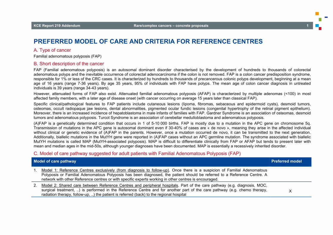
KCE Report 219 Addendum Rare/complex cancers – concrete proposals 1
PREFERRED MODEL OF CARE AND CRITERIA FOR REFERENCE CENTRES A. Type of cancer Familial adenomatous polyposis (FAP)
B. Short description of the cancer FAP (Familial adenomatous polyposis) is an autosomal dominant disorder characterised by the development of hundreds to thousands of colorectal adenomatous polyps and the inevitable occurrence of colorectal adenocarcinoma if the colon is not removed. FAP is a colon cancer predisposition syndrome, responsible for 1% or less of the CRC cases. It is characterized by hundreds to thousands of precancerous colonic polyps development, beginning at a mean age of 16 years (range 7-36 years). By age 35 years, 95% of individuals with FAP have polyps. The mean age of colon cancer diagnosis in untreated individuals is 39 years (range 34-43 years). However, attenuated forms of FAP also exist. Attenuated familial adenomatous polyposis (AFAP) is characterized by multiple adenomas (<100) in most affected family members, with a later age of disease onset (with cancer occurring on average 15 years later than classical FAP). Specific clinical/pathological features to FAP patients include cutaneous lesions (lipoma, fibromas, sebaceous and epidermoid cysts), desmoid tumors, osteomas, occult radiopaque jaw lesions, dental abnormalities, pigmented ocular fundic lesions (congenital hypertrophy of the retinal pigment epithelium). Moreover, there is an increased incidence of hepatoblastoma in male infants of families with FAP. Gardner Syndrome is an association of osteomas, desmoid tumors and adenomatous polyposis. Turcot Syndrome is an association of cerebellar medulloblastoma and adenomatous polyposis. (A)FAP is a genetically determined condition that occurs in 1 of 5-10 000 births. FAP is mostly due to a mutation in the APC gene on chromosome 5q. Transmission of mutations in the APC gene is autosomal dominant even if 30-40% of cases are « de novo », meaning they arise in the affected individual without clinical or genetic evidence of (A)FAP in the parents. However, once a mutation occurred de novo, it can be transmitted to the next generation. Additionally, biallelic mutations in the MutYH gene were reported in (A)FAP cases without an APC germline mutation. The syndrome associated with biallelic MutYH mutations is called MAP (MutYH-associated polyposis). MAP is difficult to differentiate clinically from FAP or AFAP but tends to present later with mean and median ages in the mid-50s, although younger diagnoses have been documented. MAP is essentially a recessively inherited disorder.
C. Model of care pathway suggested for adult patients with Familial Adenomatous Polyposis (FAP) Model of care pathway Preferred model
1. Model 1: Reference Centres exclusively (from diagnosis to follow-up). Once there is a suspicion of Familial Adenomatous Polyposis or Familial Adenomatous Polyposis has been diagnosed, the patient should be referred to a Reference Centre. A network with other Reference centres or with specific experts working in other centres is encouraged.
2. Model 2: Shared care between Reference Centres and peripheral hospitals. Part of the care pathway (e.g. diagnosis, MOC, surgical treatment, ..) is performed in the Reference Centre and for another part of the care pathway (e.g. chemo therapy, radiation therapy, follow-up, ..) the patient is referred (back) to the regional hospital
X

2 Rare/complex cancers – concrete proposals KCE Report 219 Addendum
D. Phase(s) of the clinical pathway for which Reference Centres are required All FAP patients should be discussed and examined in Reference Centres, who can let part of the follow-up be done in a ‘peripheral centre’ according to the guidelines. Reference Centres are those who have access to specific expertise to guide all the aspects of the disease: Genetics, Gastroenterology (endoscopy), Surgery, Imaging, Pathology, Paediatric Gastroenterologist, Oncology Some specific interventions, such as restorative total proctocolectomy with pouch construction and ileal pouch-anal anastomosis should be done in a Reference Centre.
Phase of the Clinical Pathway Reference Centre Peripheral centre
1 MOC X
2 Diagnostic confirmation (AP and/or medical imaging) X
3 Comprehensive AP diagnosis X
4 Therapeutic modalities X
5 Follow-up X X
Multidisciplinary Oncological Consult Genetic counselling of FAP patients and their family members should be performed in a centre for human genetics or in a familial cancer clinic (including oncologist, geneticist,...) and can occur in several contexts: at the time of diagnosis of FAP, at the time a FAP patient is considering reproductive options, at the time the FAP patient is having his or her children to be screened, and at the time that an at-risk person is considering genetic testing. For MutYH: mutation analysis of the partner of a MAP patient or MutYH heterozygous carrier is recommended in case parents wish prenatal testing.
Diagnostic confirmation The diagnosis of attenuated FAP (AFAP) is suspected when more than 10 adenomas are found in one patient during colonoscopy. If more than 100 adenomas are found, clinical diagnosis of FAP can be made. Genetic testing is required for definitive diagnosis in following cases: 1. > 100 colorectal adenomas 2. First-degree relatives (10 yr or older) of patients with FAP 3. > 20 cumulative colorectal adenomas (suspected attenuated FAP) 4. First-degree relatives (10 yr or older) of patients with attenuated FAP

KCE Report 219 Addendum Rare/complex cancers – concrete proposals 3
Clinical genetic testing is available for the APC and the MutYH gene. The finding of germline mutations in these genes has important implications for the patient and his/her relatives. Individuals who inherit a deleterious APC mutation (dominant) have a very high likelihood of manifesting colonic adenomas; penetrance has been estimated to be 50% by the age of 15 years and 95% by the age of 35 years. The medical goal in patients with FAP is to prevent colon cancer. If a mutation is found, all first degree relatives should undergo genetic testing. Genetic testing should be offered to at risk children from the age of 10
years. If gene carrier status is confirmed, full colonoscopy and an upper endoscopic exam should be performed. If no mutation is found, all at risk relatives should undergo endoscopic screening. Multidisciplinary consult between different experts (pediatric gastroenterologists, gastroenterologists, surgeons, geneticists…) is necessary. The work-out of these patients and family members should be done in a centre with expertise in pediatric gastroenterology, in upper and lower gastrointestinal endoscopy (the first with a side-viewing endoscope), and in restorative total proctocolectomy and ileal pouch-anal anastomosis. Complexity and new approaches
Endoscopy, Pathology, Genetics and Molecular Biology Facilities and equipment required
Endoscopy, Genetic Counselling, Molecular Biology Laboratory Professional expertise required both to perform the diagnostic procedure and to interpret the results
Expert GI-Endoscopist Histological confirmation of polyps as adenomas is required to distinguish FAP from other forms of polyposis, including hamartomatous polyposis, lymphoid hyperplasia and lymphomatous polyposis. Histological confirmation of a diagnosis of hepatoblastoma is required before treatment. Because of the rarity of this type of liver tumour and the need for special immunohistochemistry, diagnosis has to be made, or confirmed, in a Reference Centre and this will commonly be asked by the clinical team of the Reference Centre before making a therapeutic decision. As a result of the characterization of the causative gene (APC or MutYH), predictive genetic testing can be offered to family members in FAP kindreds. Multidisciplinary consultation including clinical geneticists and psychosocial supports (available in the 8 Centres of Human Genetics) may help at risk individuals who will choose genetic testing to understand the implication of the test for the patient and his/her relatives.

4 Rare/complex cancers – concrete proposals KCE Report 219 Addendum
Comprehensive AP diagnosis
Complexity and new approaches In about 70-90% of FAP cases and families truncating germline mutations in the APC gene are found, depending on the range of molecular techniques applied in the analysis. Techniques relying on PCR amplification, including sequencing, will miss a significant minority of mutations (deletions and rearrangements that involve the PCR primer binding sites are found in about 10% of the patients). Therefore, the optimal mutation detection strategy should include a combination of the traditional PCR-based methods (like sequencing) and a method to detect large deletions and rearrangements (such as MLPA, multiplex-ligation dependent probe amplification). If no mutation in the APC gene is shown and in case of a recessive inheritance pattern, mutation analysis of the MutYH gene is performed. In about 20% of the patients with adenomatous polyposis without an APC mutation, biallelic MutYH mutations are identified (representing about 1.4% of all adenomatous polyposis patients). Genetic testing of both MutYH and APC genes are available in different Centres of Human Genetics in Belgium.
Facilities and equipment required Genetic counselling of FAP patients and their family members should be performed in a centre for human genetics or in a familial cancer clinic and the possibility for psychosocial support. The laboratory offering the molecular test should be equipped to detect point mutations as well as intra-genic rearrangements.
Expertise required both to perform the cell or tissue sampling and to interpret the results Sampling of the surgical specimens and microscopic analysis require, at least for hepatoblastoma, a reference pathologist whose laboratory will have the necessary immunohistochemical tools. Again, histological analysis of the surgical specimen has to distinguish FAP from other type of polyposis. A diagnosis of adenomas can be made by a general centre but the other types of polyps are less frequent and may require a reference pathologist.
Therapeutic modalities
Complexity, new therapeutic strategies FAP is a monogenic disorder due, for the most part, to mutations within the APC gene. At present time, there is no gene targeted therapies for this monogenic disease. Thus, there is not per se a curative treatment. FAP leads to colorectal cancer and other extra-colonic cancers (see above) and benign tumours (see above; i.e. osteomas). Each cancer or benign tumours must be managed by each organ-related medical specialities within the frame of a multi-disciplinary approach specific to FAP patients. Specific management of FAP patients includes prevention and surveillance of FAP patients and families (Diagnosis relies on positive APC gene testing or, in 20% of patients which are negative for APC gene testing, FAP positive clinical and endoscopic criteria) and prophylactic surgery. Prevention and surveillance of FAP patients relies on National and International recommendations. There are not only general recommendations for all FAP patients, but also specific recommendations for families presenting with a high prevalence of extra-colonic cancer such as hepatoblastoma or Turcot Syndrome. Patients with positive FAP clinical and endoscopic criteria who are negative for gene testing should be considered at very high risk patients for CRC and extra-colonic patients and should be enrolled in the same screening and surveillance program than FAP patients with positive gene testing. Many FAP are actually attenuated FAP (AFAP) (diagnosis suspicion should be raised if patient presents with 20 consecutive adenomas on surveillance colonoscopies)

KCE Report 219 Addendum Rare/complex cancers – concrete proposals 5
FAP can be diagnosed before cancer occurs or at the time of diagnosis of cancer. When FAP patients present with cancer, medical and surgical management of FAP-related cancers remain similar to medical and surgical management of sporadic cancer, although more extensive surgery is often recommended, because of high risk of metachronous cancer. In FAP patients, colonoscopic screening should start at the age of 10 if the mutation in the APC gene is present. The goal is to detect dysplasia. Definitive prophylactic surgical treatment is usually recommended at the end of puberty, before the risk of developing colorectal cancer. The classical procedure required for this syndrome is a total proctocolectomy, excising the entire colon and rectum in order to completely eliminate the inevitable risk of colorectal cancer. The reconstruction is done by performing an ileo-anal anastomosis with the construction of an ileal pouch, usually a J-pouch, which is then sutured to the anal canal. In order to avoid the consequences of pelvic complications of this procedure, as anastomotic fistula, a protective and temporary ileostomy may be constructed. Laparoscopic procedures should be offered whenever possible. Closure of the ileostomy is then performed 8-10 weeks later. In the attenuated form of the syndrome (AFAP), the rectum may be preserved from polyps and the risk of cancer is lower and delayed. In this setting, a less aggressive surgical procedure may be discussed. A total colectomy, leaving the rectum in place, with an ileo-rectal anastomosis is then performed. The main advantages of this second procedure are the avoidance of a temporary ileostomy placement, a better functional outcome and less effect on future fertility. This option requires a surveillance of the remaining rectum by a lifelong 6-months follow-up rectoscopy.
Facilities and equipment required Appropriate management of FAP patients for the prevention and surveillance of gastro-intestinal (GI-) cancer requires expertise in diagnostic and therapeutic endoscopy. The recognition and management of GI FAP lesions requires a high –volume of FAP patients. For example, in the upper GI tract, the Spiegelman classification of duodenal adenoma that eventually guides the endoscopic or surgical management of duodenal adenoma is sometimes difficult to establish. Another example for the lower GI tract is the appropriate evaluation of patients for prophylactic surgery. All classical FAP patients should undergo total coloprotectomy with ileo-anal anastomosis. However, some FAP patients eligible for prophylactic surgery should be evaluated for colectomy with ileo-rectal anastomosis (rectal sparing surgery). Upper GI endoscopic ultrasound (EUS), Endoscopic Retrograde Cholangio-Pancreatography (ERCP), Lateral Duodenoscopy, Capsule Endoscopy and Double-Balloon Endoscopy should be available in tertiary centres dedicated to the management of FAP patients.
Expertise required to perform the treatment Expertise in endoscopic polypectomy and mucosectomy of duodenal adenomas, and more specifically, expertise and experience in the endoscopic removal of the ampulla (Vater papilla) (Endoscopic ampullectomy) is highly recommended. Surgical team with expertise in laparoscopic and open colorectal surgery, especially with expertise in total proctocolectomy with ileo-anal pouch reconstructive surgery.
Para-medical expertise required As major partners involved in genetic counseling, trained psychologists, nurses and social workers are recommended to communicate with families facing or experiencing a genetic diagnosis (parents' concerns for their children, age-appropriate language, childhood testing). Dietician and stoma nurse are also required.

6 Rare/complex cancers – concrete proposals KCE Report 219 Addendum
Follow-up Patients with FAP are at risk for extracolonic malignancies, including duodenal ampullary cancer, thyroid carcinoma, hepatoblastoma and central nervous system tumours. Desmoid tumours affect 10-20% of patients with FAP and are an important cause of morbidity and mortality because, due to prophylactic colectomy, life expectancy of patients with FAP is increasing.
Follow-up of patients after total proctocolectomy :
Upper gastrointestinal screening with both end-viewing and side-viewing instruments, because a side-viewing endoscope is better suited for visualization of the papilla. The Spigelman classification of duodenal polyposis should be used to determine the surveillance interval.
Polypectomy of duodenal polyps (if indicated) and ampullectomy or biopsy of an abnormal papilla should be performed. Yearly surveillance with lower endoscopy is recommended because patients who have undergone total proctocolectomy remain at risk for the
development of adenomas in the ileal pouch. Palpation of the thyroid is recommended yearly. Children at risk for FAP should be examined every 6 months No surveillance is recommended for desmoid tumors. If indicated chemoprevention with celecoxib should be started.
Follow-up of gene carriers or at risk family members with uninformative genetic testing :
Surveillance of the colon with colonoscopy should be performed Surveillance for extra-intestinal tumors should be carried out as described previously.
Follow-up should be done in a centre with expertise in pediatric gastroenterology, in lower gastrointestinal endoscopy and in upper GI endoscopy with a side-viewing endoscope. The gastroenterologist should be aware of the possibility of extra-intestinal tumours and the existence of chemoprevention. Preferably the same physician should see the patient to notice the evolution of the lesions in time.
E. General and specific criteria for Reference Centres Human Resources and dedicated team Multidisciplinary team of experts: Gastroenterologists-endoscopists, paediatric gastroenterologist with experience in FAP, radiologists, geneticists, pathologists, colorectal surgeons, gastroenterologist who obtained a certificate showing specific expertise in oncology or a medical oncologist experienced in the treatment of GI cancer, intensive care doctors and nurses, stoma nurses, psychologists, dieticians.
Required facilities and equipment GI-endoscopy, abdominal and GI Radiology and interventional Radiology, Pathology, Genetics, Genetic counselling, Molecular Biology, Surgery, ICU, Chemotherapy facilities, Paediatric Gastroenterologist with experience in FAP.

KCE Report 219 Addendum Rare/complex cancers – concrete proposals 7
Patient centred care Inclusion of all FAP patients and their families in the FAPA Registry (Familial Adenomatous Polyposis Association). The FAPA is member of the InSight Group (International Society for Gastrointestinal Hereditary Tumours) and participates regularly to international meetings for FAP and Lynch registries. FAPA has also collaborations with STOET (Stichting Opsporing Erfelijke Tumoren) in the Netherlands.
Minimal volume of patients Centres of excellence for FAP should have experience with laparoscopic restorative proctocolectomy and ileoanal pouch anastomosis. As decision making regarding surgical procedure (laparoscopic ileo-anal pouch anastomosis versus laparoscopic ileo-rectal anastomosis) is complex in a young population (desmoids, fecundity, lifetime cancer risk), this should be done in a Reference Centre. More than 10 ileo-anal pouches should be performed annually and experience of at least 50 ileo-anal pouch reconstructions should have been documented in a Reference Centre for FAP and UC (Ulcerative Colitis) indications combined. Centres should be accredited also for pancreas surgery (duodenal polyps) and oncologic surgery for desmoid tumours.
Quality Assurance The physician should be able to determine in conjunction with a multidisciplinary team the preferred therapy, should have knowledge and experience of FAP and extra-intestinal tumours. A minimum number of procedures to achieve and maintain competence is necessary. Physicians should adhere to the guidelines and a database (number of new patients, endoscopic investigations, surgery, follow-up) should be sent to the FAPA Registry. Adherence to quality control in endoscopy (evaluation of number of procedures, completeness, adverse drug reactions, complications,…) is necessary, just as surgical expertise and adherence to quality control in surgery (number of procedures, outcome,…). Genetic testing laboratories provide major medical services to clinicians requesting a test, patients from whom the sample was collected, or referral laboratories. Therefore, the accreditation in compliance with International Organization for Standardization (ISO) 15189 becomes in Belgium and for the beginning of 2014 an obligation for assuring laboratory quality in the Centres of Human Genetics.
Research and other scientific activities Each Centre of Human Genetics has developed close relationships with a biobank, especially those funded by the "Plan Cancer/Kankerplan" available in the several academic and non-academic hospitals.
Educational activities: Teaching and dissemination One of the objectives of FAPA is to sensitize patients as well as doctors regarding FAP and Lynch Syndrome to increase the knowledge about these 2 syndromes improving the detection of at risk persons and the follow-up of the patients. This is done by the distribution of info brochures, the organization of info days for the patients, personal contacts (via mail or phone contact), info stands at medical congresses, sending of Newsletters and distribution of guidelines for professionals… These educational activities of the FAPA are shared by Reference Centres.

RARE MALIGNANT SKIN TUMOURS PREFERRED MODEL OF CARE AND CRITERIA FOR REFERENCE CENTRES

Coordinator: Brochez Lieve (Dermatology, UZ Gent)
Authors : For melanoma, epithelioma and other rare skin tumours: Brochez Lieve (Dermatology, UZ Gent), Awada Ahmad (Medical Oncology, Bordet Institute), Baurain Jean-François (Medical Oncology, UCL), Buyse Veronique (Medical Oncology, AZ Groeninge, Kortrijk), Del Marmol Veronique (Dermatology, ULB), Garmyn Marjan (Dermatology, UZ Leuven), Kerger Joseph (Medical Oncology, Bordet Institute), Krüse Vibeke (Medical Oncology, UZ Gent), Neyns Bart (Medical Oncology, UZ Brussel), Nikkels Arjen (Dermatology, CHU Liège), Roche Nathalie (Reconstructive surgery, UZ Gent), Wolter Pascal (Medical Oncology, UZ Leuven), Sales François (Surgery, Bordet Institute), Snauwaert Johan (Dermatology, Brasschaat), Specenier Pol (Medical Oncology, UZ Antwerpen), Stas Marguerite (Surgery, UZ Leuven), Temmerman Linda (Dermatology, AZ Maria Middelares Gent), Van Eeckhout Pascal (Pathology, UCL), Vossaert Katrien (Dermatology, Maldegem). For primary cutaneous lymphomas: Brochez Lieve (Dermatology, UZ Gent), Achten Ruth (Pathology, UZ Antwerpen), André Josette (Dermatology, CHU St Pierre Bruxelles), Bervoets An (Dermatology, UZ Antwerpen), Boterberg Tom (Radiotherapie, UZ Gent), De Raeve Hendrik (Pathology, OLV Aalst), Fink Wolfram (CHU Brugmann, Bruxelles), Haspeslagh Marc (Dermatopathology, Ardooie), Hauben Esther (Pathology, UZ Leuven), Lambein Kathleen (Pathology, UZ Gent), Matthieu Lucretia (Dermatology, UZ Antwerpen), Sass Ursula (CHU St Pierre Bruxelles), Schroyens Wilfried (Hematology, UZ Antwerpen), Van Den Neste Eric (Hematology, UCL), Van Gestel Dirk (Radiotherapy, UZ Antwerpen), Wolter Pascal (Medical Oncology, UZ Leuven)
Disclaimer : The coordinators of the working groups and all the authors listed by chapter have worked autonomously under the supervision of the KCE team. The KCE experts are not co-authors of these proposals and did not necessarily validate their content.
Hospitals with which coordinators and authors of these proposals are affiliated are not de facto considered Reference Centres. Similarly, Belgian hospitals that are not represented in these proposals are not de facto considered Peripheral Centres.
These proposals were not submitted to the external validators. This addendum only exists in English. No French or Dutch translation was done. Finally, the report to which this addendum refers has been approved by common assent by the
Executive Board. Copyright : KCE reports are published under a “by/nc/nd” Creative Commons Licence
http://kce.fgov.be/content/about-copyrights-for-kce-reports.
This section is part of a whole document (KCE Report 219 Addendum) available on the website of the Belgian Health Care Knowledge Centre


KCE Report 219 Addendum Rare/complex cancers – concrete proposals 1
PREFERRED MODEL OF CARE AND CRITERIA FOR REFERENCE CENTRES A. Types of cancer 1. Melanoma stage III & IV 2. Epitheliomas (basal cell and squamous cell carcinoma) that are:
o locally advanced and inoperable o stage III-IV
3. Other rare skin tumours. These include Merkel cell tumour, angiosarcoma, dermatofibrosarcoma protuberans, atypical fibroxanthoma, malignant fibrous histiocytoma, leiomyosarcoma, Kaposi sarcoma
4. Primary cutaneous lymphomas
B. Short description of the skin cancer types 1) Melanoma is the most aggressive skin cancer arising from malignant transformation in the pigmented cells of the skin. It is currently the fifth most incident cancer in females and affects a relative young population (premature morbidity and mortality). Melanoma stage III is a melanoma that has metastasized to regional lymph nodes or has developed in-transit metastases or satellites. There is no evidence of distant metastasis. Stage III disease is considered to be intermediate to high-risk for local recurrence or distant metastasis depending on specific sub-classification. Five-year overall survival varies between 26.7 to 69.5%. Melanoma stage IV is a melanoma that has metastasized to distant organs. Five-year overall survival is around 9.5%. 2) Epitheliomas are the most frequent types of skin cancer and even the most frequent cancer in Caucasians. It is expected that 1 in 6 people with this skin type will develop non-melanoma skin cancer during their life. In the majority of cases the treatment is simple and straightforward and the cure rate is very high. In a very small subgroup, the treatment can be less straightforward (locally advanced inoperable tumours) and/or these cancers can behave more aggressively (regional or systemic metastasis). 3) The other skin cancers are very rare (some dozens per year for Belgium, unfortunately no reliable figures available) 4) Primary cutaneous lymphomas are a group of rare lymphomas presenting in the skin without any evidence of extracutaneous disease at time of diagnosis. There is an estimated incidence of 1/100 000/year. There are different subgroups with different disease course and aggressiveness/prognosis. It is important to note that primary cutaneous lymphoma often has a completely different clinical behaviour and prognosis in comparison to their histologically similar systemic counterparts. For that reason the most recent classification systems (WHO, EORTC, …) include primary cutaneous lymphomas as separate entities. A large part of the cutaneous lymphomas have an excellent prognosis and remain restricted to the skin. These need a totally different approach than the haematological lymphomas and usually their basic treatment is mainly skin directed. The diagnosis can be difficult and often needs integration of the clinical picture, the histopathological, immunophenotypic and sometimes molecular data.

2 Rare/complex cancers – concrete proposals KCE Report 219 Addendum
C. Model of care pathway suggested for adult patients with skin cancers Model of care pathway Preferred model
1. Model 1: Reference Centres exclusively (from diagnosis to follow-up). Once there is a suspicion of skin tumour or a skin tumour has been diagnosed, the patient should be referred to a Reference Centre. A network with other Reference centres or with specific experts working in other centres is encouraged.
2. Model 2: Shared care between Reference Centres and peripheral hospitals. Part of the care pathway is performed in the Reference Centre and for another part of the care pathway the patient is referred (back) to the regional hospital or private practice.
X
D. Phase(s) of the clinical pathway for which Reference Centres are required Phase of the Clinical Pathway Reference Centre Peripheral centre
MOC X
Diagnostic confirmation X X
Comprehensive AP diagnosis X X
Therapeutic modalities X X
Follow-up X X
In case of relapse or progression: MOC
X
Multidisciplinary Oncological Consult All patients with a disease condition referred under A should have a specialized MOC in a Reference Centre to give advice on the management strategies needed / possible in the specific patient case. This is done either by the physical presence of the referring physician at the Reference Centre’s MOC, by tele-consulting (tele-MOC) or after viewing the patient at a specialized consultation in the Reference Centre. The Reference Centre will give a management advice (specialized written MOC report) within 2 weeks of first announcement of the patient file. The referring doctor is responsible for passing all available information to the Reference Centre at the moment of referral for advice. This MOC report should be sent to the referring doctor, the general practitioner of the patient and the patient himself. This MOC report should include standard of care procedures as well as possible clinical trials, including trials conducted in other centres. For the skin cancer types designated under A type 3 (other rare skin tumours) the specialized written MOC report has to document that the management plan is evidence-based (publications/reviews in peer-reviewed journals) or why it deviates from the guidelines suggested in literature.

KCE Report 219 Addendum Rare/complex cancers – concrete proposals 3
The communication of this report to the patient can be done by the referring physician. Depending on the treatment chosen the referring physician can choose to start up the treatment himself or refer the patient for treatment to the Reference Centre (e.g. for study protocol that is not available in the referring centre). Treatment should be started preferably within 31 days of the report. In case of disease progression or relapse, a second opinion MOC has to be planned in a Reference Centre.
Diagnostic confirmation In Reference Centre if advised by specialized written MOC report and agreed by referring centre. For skin cancer types as designated under A type 3 (other rare skin tumours) and type 4 (primary cutaneous lymphoma) it is advisable to have diagnostic confirmation in the Reference Centre.
Comprehensive AP diagnosis In Reference Centre if advised by specialized written MOC report. A Reference Centre also must have the possibility to send the histological slides to specific reference pathology laboratories (national or international) that can help to establish the diagnosis.
Therapeutic modalities In Reference Centre if advised by specialized written MOC report.
Follow-up In Reference Centre if advised by specialized written MOC report.
E. General and specific criteria for Reference Centres Human Resources and dedicated team The multidisciplinary team at the Reference Centre should have the following members: Dermatologists: minimum of 2 dermatologists, covering a major interest in skin cancer, including skin cancer surgery and cutaneous lymphoma Medical oncologists/haematologists: minimum 2 medical oncologists (or haematologists in case of skin cancer type designated under A point 4 (primary
cutaneous lymphoma)), covering a major interest in skin cancer. Specialist reconstructive surgeons: minimum of 2 surgeons with a designated interest and training in skin cancer surgery. Surgeons undertaking block
dissections must perform at least 10 block dissections per year. Pathologist: minimum 1 pathologist with special interest in dermato-pathology Radiotherapist with knowledge of the latest radiotherapy (RT) techniques including intensity modulated radiotherapy (IMRT), rotational IMRT, stereotactic
RT, 3D-image guided RT(IGRT), particle therapy, total skin RT. Dedicated nurse / psychologist Palliative support team Cosmetic camouflage service advisers

4 Rare/complex cancers – concrete proposals KCE Report 219 Addendum
Liaison psychiatrist The presence of at least one member of each specialty during the specialized MOC is advised.
Required facilities and equipment The Reference Centre should also be able to offer: Molecular analysis important for diagnostic or treatment work-up: detection of tumour mutations, detection of clonality Imaging: all appropriate imaging including PET-CT facility and image-guided biopsy modalities are available to patients in a timely manner Specific surgical techniques such as sentinel node biopsy, block dissections (minimum of 10 per surgeon per year), Mohs surgery State of the art radiotherapy: electron therapy, 3D-conformal RT, IMRT, rotational IMRT, stereotactic RT, 3D IGRT Information leaflets for the patients with information on the disease, its management and the specific treatment options proposed to the patient Facilities for clinical trial conduct and support, including research nurse and data manager involved in oncology trials according to existing standards
(International Conference on Harmonisation Good Clinical Practice (IHC-GCP) Facility of prosthetics and orthotics
Patient centred care
National and international networking with other Reference Centres for second opinion or referral of patients for other options not available at the Reference Centre (specific clinical trials, specialized surgical procedures, specialized radiation therapies)
There is a national cutaneous lymphoma working group where primary cutaneous lymphomas that are difficult to diagnose or treat can be discussed.
Minimal volume of patients
A Reference Centre should manage (actually diagnose and/or treat outside second opinion in specialized MOC conditions) at least 5% of all patient subtypes registered by the National Cancer Registry. As some skin cancer subtypes are most probably underestimated, estimations could be made by the Cancer Registry based on international data.
Surgeons undertaking block dissections must perform at least 10 block dissections per year.
Quality Assurance
Identification of a care program with standardized working procedures and clear, up-to-date guidelines for stage III and IV melanoma patients at the Reference Centre. For the skin cancer types designated under A type 3 (other rare skin tumours) the specialized written MOC report has to document that the management plan is evidence-based (publications/reviews in peer-reviewed journals) or why it deviates from the guidelines suggested in literature.
A specialized written MOC report should be available within 2 weeks of first announcement in 90% of the referrals. This report should be sent to the referring physician, the general practitioner and the patient himself. In case of delay of the report, the reason for delay must be clearly documented.
The referring physician should report back to the Reference Centre the treatment that has been chosen based on the specialized MOC report after discussion with the patient. S/he also reports if and when the treatment will be started at the referring centre or if the patient will be referred back to the

KCE Report 219 Addendum Rare/complex cancers – concrete proposals 5
Reference Centre for treatment. A treatment should be initiated within 31 days of the written report, either in or out the Reference Centre in 85% of the cases. In case of delay of the start of treatment (> 31 days), the reason for delay must be clearly documented by the referring physician or the Reference Centre in case the patient was sent back for treatment.
The Reference Centre must report all specialized MOCs to the National Cancer Registry. There will be an annual report by the Reference Centre with the number of all specialized MOCs and their through-put times (time to MOC report, time to
start of the treatment).
Research and other scientific activities For a Reference Centre for skin cancer, research activities resulting in publication in peer-reviewed journals (reported in pubmed) are encouraged.
Educational activities: Teaching and dissemination All members of the multidisciplinary team of the Reference Centre must attend national and international meetings with skin cancer-specific topics at least once a year e.g. ASCO, ESMO, EADO, EORTC melanoma / cutaneous lymphoma subgroup meeting, visit to international excellence centres, .... Membership of at least one member of the multidisciplinary team to national or international scientific skin cancer organizations is encouraged. The Reference Centre itself is involved in training and continuous education programs for physicians, nurses, supportive disciplines and communication in scientific congresses.
Additional comments
In order to make this operable, the working group believes that a quality control program will be necessary as well as additional financial support for these dedicated MOC.
The members of the working group think it would be advisable to have a transition period of 1 to 2 years.
