organic cation transporters in health and disease
TRANSCRIPT

1521-0081/72/1/253–319$35.00 https://doi.org/10.1124/pr.118.015578PHARMACOLOGICAL REVIEWS Pharmacol Rev 72:253–319, January 2020Copyright © 2019 by The American Society for Pharmacology and Experimental Therapeutics
ASSOCIATE EDITOR: LYNETTE C. DAWS
Organic Cation Transporters in Health and DiseaseHermann Koepsell
Institute of Anatomy and Cell Biology and Department of Molecular Plant Physiology and Biophysics, Julius-von-Sachs-Institute,University of Würzburg, Würzburg, Germany
Abstract . . . . . . . . . . . . . . . . . . . . . . . . . . . . . . . . . . . . . . . . . . . . . . . . . . . . . . . . . . . . . . . . . . . . . . . . . . . . . . . . . . . 255I. Introduction . . . . . . . . . . . . . . . . . . . . . . . . . . . . . . . . . . . . . . . . . . . . . . . . . . . . . . . . . . . . . . . . . . . . . . . . . . . . . . . 255II. Cloning, Chromosomal Location, Functional Properties, and Regulation of Human
Organic Cation Transporters . . . . . . . . . . . . . . . . . . . . . . . . . . . . . . . . . . . . . . . . . . . . . . . . . . . . . . . . . . . . . . . 257A. Human Organic Cation Transporter 1 (SLC22A1). . . . . . . . . . . . . . . . . . . . . . . . . . . . . . . . . . . . . . . 257B. Human Organic Cation Transporter 2 (SLC22A2). . . . . . . . . . . . . . . . . . . . . . . . . . . . . . . . . . . . . . . 258C. Human Organic Cation Transporter 3 (SLC22A3). . . . . . . . . . . . . . . . . . . . . . . . . . . . . . . . . . . . . . . 260D. Human Novel Organic Cation Transporter 1 (SLC22A4) . . . . . . . . . . . . . . . . . . . . . . . . . . . . . . . . 260E. Human Novel Organic Cation Transporter 2 (SLC22A5) . . . . . . . . . . . . . . . . . . . . . . . . . . . . . . . . 264F. Human Multidrug and Toxin Exclusion 1 (SLC47A1) . . . . . . . . . . . . . . . . . . . . . . . . . . . . . . . . . . . 264G. Human Multidrug and Toxin Exclusion 2, Human Multidrug and Toxin Exclusion
Kidney-Specific 2 (SLC47A2) . . . . . . . . . . . . . . . . . . . . . . . . . . . . . . . . . . . . . . . . . . . . . . . . . . . . . . . . . . . 265III. Structure–Function Relationships in Organic Cation Transporters . . . . . . . . . . . . . . . . . . . . . . . . . . 265
A. Primary Structures and Membrane Topology of Human Organic Cation Transporters . . . 265B. Solved Crystal Structures of Transporters of the Major Facilitator Superfamily and
Multidrug and Toxin Exclusion Family . . . . . . . . . . . . . . . . . . . . . . . . . . . . . . . . . . . . . . . . . . . . . . . . . 265C. Homology Three-Dimensional Models of Mammalian Organic Cation Transporters . . . . . . 266D. In-Depth Functional Characterization of Organic Cation Transporter 1 and Organic
Cation Transporter 2. . . . . . . . . . . . . . . . . . . . . . . . . . . . . . . . . . . . . . . . . . . . . . . . . . . . . . . . . . . . . . . . . . . 267E. Mutagenesis Studies in Rat Organic Cation Transporter 1, Rat Organic Cation
Transporter 2, and Human Organic Cation Transporter 2. . . . . . . . . . . . . . . . . . . . . . . . . . . . . . . 268F. Mutagenesis Studies in Human Novel Organic Cation Transporter 1 and Human
Novel Organic Cation Transporter 2 . . . . . . . . . . . . . . . . . . . . . . . . . . . . . . . . . . . . . . . . . . . . . . . . . . . . 272G. Mutagenesis in Human Multidrug and Toxin Exclusion 1 and Human Multidrug and
Toxin Exclusion Kidney-Specific 2 . . . . . . . . . . . . . . . . . . . . . . . . . . . . . . . . . . . . . . . . . . . . . . . . . . . . . . 272IV. Expression and Locations of Human Organic Cation Transporters in Cells, Tissues,
Organs, and Tumors . . . . . . . . . . . . . . . . . . . . . . . . . . . . . . . . . . . . . . . . . . . . . . . . . . . . . . . . . . . . . . . . . . . . . . . 272A. Expression and Membrane Locations of Human Organic Cation Transporter 1,
Human Organic Cation Transporter 2, and Human Organic Cation Transporter 3 . . . . . . . 272B. Expression and Membrane Locations of Human Novel Organic Cation Transporter 1
and Human Novel Organic Cation Transporter 2 . . . . . . . . . . . . . . . . . . . . . . . . . . . . . . . . . . . . . . . 273C. Expression and Membrane Locations of Human Multidrug and Toxin Exclusion 1 and
Human Multidrug and Toxin Exclusion Kidney-Specific 2 . . . . . . . . . . . . . . . . . . . . . . . . . . . . . . . 275D. Transporters That Mediate Organic Cation Transport in Small Intestine . . . . . . . . . . . . . . . . 275E. Transporters That Mediate Organic Cation Transport in Liver . . . . . . . . . . . . . . . . . . . . . . . . . . 275F. Transporters that Mediate Organic Cation Transport in Kidney. . . . . . . . . . . . . . . . . . . . . . . . . 276G. Transporters that Mediate Organic Cation Transport in Lung. . . . . . . . . . . . . . . . . . . . . . . . . . . 276H. Transporters that Mediate Organic Cation Transport in Brain . . . . . . . . . . . . . . . . . . . . . . . . . . 276I. Organic Cation Transporters That Are Expressed in Tumors. . . . . . . . . . . . . . . . . . . . . . . . . . . . 277
V. Model Substrates and Endogenous Compounds That Are Transported by Human OrganicCation Transporters . . . . . . . . . . . . . . . . . . . . . . . . . . . . . . . . . . . . . . . . . . . . . . . . . . . . . . . . . . . . . . . . . . . . . . . 278
Address correspondence to: Dr. Hermann Koepsell, Institute of Anatomy and Cell Biology, University of Würzburg, Koellikerstr. 6,97070 Würzburg, Germany. E-mail: [email protected]
https://doi.org/10.1124/pr.118.015578.
253
by guest on February 10, 2022
Dow
nloaded from

A. Model Substrates of Human Organic Cation Transporters. . . . . . . . . . . . . . . . . . . . . . . . . . . . . . . 278B. Neurotransmitters and Neuromodulators That Are Transported by Human Organic
Cation Transporters. . . . . . . . . . . . . . . . . . . . . . . . . . . . . . . . . . . . . . . . . . . . . . . . . . . . . . . . . . . . . . . . . . . . 278C. Metabolites and Food Components That Are Transported by Human Organic Cation
Transporters . . . . . . . . . . . . . . . . . . . . . . . . . . . . . . . . . . . . . . . . . . . . . . . . . . . . . . . . . . . . . . . . . . . . . . . . . . . 279VI. Toxins That Are Transported by Human Organic Cation Transporters . . . . . . . . . . . . . . . . . . . . . . 280VII. Drugs That Are Substrates and/or Inhibitors of Human Organic Cation Transporters . . . . . . . 281
A. General Considerations . . . . . . . . . . . . . . . . . . . . . . . . . . . . . . . . . . . . . . . . . . . . . . . . . . . . . . . . . . . . . . . . 281B. Antidiabetic Drugs . . . . . . . . . . . . . . . . . . . . . . . . . . . . . . . . . . . . . . . . . . . . . . . . . . . . . . . . . . . . . . . . . . . . . 282C. Antineoplastic Drugs. . . . . . . . . . . . . . . . . . . . . . . . . . . . . . . . . . . . . . . . . . . . . . . . . . . . . . . . . . . . . . . . . . . 282D. Antiallergic Drugs . . . . . . . . . . . . . . . . . . . . . . . . . . . . . . . . . . . . . . . . . . . . . . . . . . . . . . . . . . . . . . . . . . . . . 282E. Antiarrhythmic, Antihypertensive, and Hypertensive Drugs. . . . . . . . . . . . . . . . . . . . . . . . . . . . . 282F. Antiemetic and Antimigraine Drugs . . . . . . . . . . . . . . . . . . . . . . . . . . . . . . . . . . . . . . . . . . . . . . . . . . . . 283G. Psychotropic and Analgetic Drugs . . . . . . . . . . . . . . . . . . . . . . . . . . . . . . . . . . . . . . . . . . . . . . . . . . . . . . 283H. Antibiotic Drugs and Antiseptic Substances . . . . . . . . . . . . . . . . . . . . . . . . . . . . . . . . . . . . . . . . . . . . 283I. Antifungal, Antiparasitic, and Antiviral Drugs. . . . . . . . . . . . . . . . . . . . . . . . . . . . . . . . . . . . . . . . . . 283
VIII. Genetic Variants in Human Organic Cation Transporters. . . . . . . . . . . . . . . . . . . . . . . . . . . . . . . . . . . 283A. Genetic Variants in Human Organic Cation Transporter 1 . . . . . . . . . . . . . . . . . . . . . . . . . . . . . . 283B. Genetic Variants in Human Organic Cation Transporter 2 . . . . . . . . . . . . . . . . . . . . . . . . . . . . . . 286C. Genetic Variants in Human Organic Cation Transporter 3 . . . . . . . . . . . . . . . . . . . . . . . . . . . . . . 286D. Genetic Variants in Human Novel Organic Cation Transporter 1. . . . . . . . . . . . . . . . . . . . . . . . 286E. Genetic Variants in Human Novel Organic Cation Transporter 2. . . . . . . . . . . . . . . . . . . . . . . . 287F. Genetic Variants in Human Multidrug and Toxin Exclusion 1. . . . . . . . . . . . . . . . . . . . . . . . . . . 287G. Genetic Variants in Human Multidrug and Toxin Exclusion 2 Kidney-Specific . . . . . . . . . . . 287
IX. Effects of Genetic Variants in Organic Cation Transporters on Drug Treatment . . . . . . . . . . . . . 287A. Treatment of Type 2 Diabetes with Metformin. . . . . . . . . . . . . . . . . . . . . . . . . . . . . . . . . . . . . . . . . . 287B. Antineoplastic Treatment with Cisplatin . . . . . . . . . . . . . . . . . . . . . . . . . . . . . . . . . . . . . . . . . . . . . . . 289C. Treatment of Myeloid Leukemia with Imatinib . . . . . . . . . . . . . . . . . . . . . . . . . . . . . . . . . . . . . . . . . 290D. Analgetic Treatment with Tramadol . . . . . . . . . . . . . . . . . . . . . . . . . . . . . . . . . . . . . . . . . . . . . . . . . . . . 290E. Pain-Relieving Treatment with Morphine and Antitussive Treatment with Codeine . . . . . 291F. Treatment of Side Effects of Chemotherapy with Tropisetron . . . . . . . . . . . . . . . . . . . . . . . . . . . 291G. Treatment of Migraine with Sumatriptan. . . . . . . . . . . . . . . . . . . . . . . . . . . . . . . . . . . . . . . . . . . . . . . 291H. Treatment of Asthma with Fenoterol . . . . . . . . . . . . . . . . . . . . . . . . . . . . . . . . . . . . . . . . . . . . . . . . . . . 292I. Treatment of Malaria with Proguanil . . . . . . . . . . . . . . . . . . . . . . . . . . . . . . . . . . . . . . . . . . . . . . . . . . . 292
X. Drug–Drug Interactions of Substrates and Inhibitors at Organic Cation Transporters . . . . . . . 292A. General Considerations . . . . . . . . . . . . . . . . . . . . . . . . . . . . . . . . . . . . . . . . . . . . . . . . . . . . . . . . . . . . . . . . 292B. Interactions of Drugs during Treatment of Type 2 Diabetes with Metformin . . . . . . . . . . . . . 296
ABBREVIATIONS: 3D, three-dimensional; ADE, adverse drug effect; AMPK, AMP-stimulated protein kinase; AP, area postrema; AP-1,activating protein-1; Ap2-rep, activating protein-2 repressor; ASP, 4-(4-(dimethylamino)styryl)-N-methylpyridinium; BBB, blood brain bar-rier; BBM, brush–border membrane; CaM, calmodulin; CD, Crohn’s disease; CGI, CpG islands; CHT, choline transporter; CKD, chronickidney disease; CML, chronic myeloic leukemia; CNT, concentrative nucleoside transporter; CTL, choline-like transporter; DA, dopamine;DAT, DA transporter; EMA, European Medicines Agency; FDA, Food and Drug Administration; FST, forced-swim test; GFP, green fluorescentprotein; GFR, glomerular filtration rate; GWAS, genome-wide association studies; Hb1Ac, hemoglobin A1c; HEK, human embryonic kidney;hMATE, human MATE; hMATE-K, human MATE-K; hMDR1, human multidrug resistance protein 1; HNF, hepatic nuclear factor; hOCT,human OCT; hTHTR, human THTR; IBD, inflammatory bowel disease; IRIP, ischemia/reperfusion-inducible protein; KD, dissociation constant;Km, Michaelis Menten constant; KO, knockout; LAPTM4A, lysosmal-associated protein 4a; LDL, low-density lipoprotein; maleimide-PEO2-biotin,(1)-biotinyl-3-maleimidopropionamidyl-3,6-dioxaoctanediamine; MAF, major allele frequency; MATE, multidrug and toxin exclusion; MATE-K,MATE kidney-specific; METH, methamphetamine; MFS, major facilitator superfamily; MLL1, mixed-lineage-leucemia 1; mOCT, mouse OCT;mOCTN, mouse OCTN; MPP, 1-methyl-4-phenylpyridinium; MTSET, 2-(trimethylammonium)ethyl methanethiosulfonate; MZ1, myeloid zincfinger 1; NE, norepinephrine; NET, NE transporter; NME, new molecular entity; OAT, organic anion transporter; OCD, obsessive-compulsivedisorder; OCT, organic cation transporter; OCTN, novel OCT; OGTT, oral glucose tolerance test; PET, positron emission tomography; PfMATE,MATE from Pyrococcus furiosus; PI3K, phosphatidylinositol-3-kinase; PKA, protein kinase A; PKC, protein kinase C; PKG, protein kinase G;PMAT, monoamine transporter; PPAR, peroxisome proliferator-activated receptor; RA, rheumatoid arthritis; rOCT, rat OCT; RUNX1, runt-related transcription factor 1; SCD, primary systemic carnitine deficiency; Scr, serum creatinine; SE, serotonin; SERT, SE transporter; SFO,subfornical organ; SGLT, sodium-D-glucose cotransporter; SNV, single-nucleotide variant; Sp1, specifity protein 1; T1D, type 1 diabetes; T2D,type 2 diabetes; TBuA, tetrabutylammonium; TEA, tetraethylammonium; THTR, thiamine transporter; TMAO, trimethylamine N-oxide; TMH,transmembrane helix; TMRM, tetramethylrhodamine-6-maleimide; TPeA, tetrapentylammonium; UC, ulcerative colitis; USF, upstream bindingstimulating factor; WAT, white adipose tissue; WT, wild-type.
254 Koepsell

C. Interactions of Drugs during Treatment of Cancer with Cisplatin . . . . . . . . . . . . . . . . . . . . . . . 297D. Decrease of Renal Drug Clearance by Cimetidine . . . . . . . . . . . . . . . . . . . . . . . . . . . . . . . . . . . . . . . 297
XI. Diseases That Are Associated with Genetic Variants in Organic Cation Transporters. . . . . . . . 298A. General Considerations . . . . . . . . . . . . . . . . . . . . . . . . . . . . . . . . . . . . . . . . . . . . . . . . . . . . . . . . . . . . . . . . 298B. Association of Human Organic Cation Transporter 1 Variants with Blood Lipids,
Blood Isobutylcarnitine, and Body Weight . . . . . . . . . . . . . . . . . . . . . . . . . . . . . . . . . . . . . . . . . . . . . . 298C. Association of a Single-Nucleotide Variant in SLC22A3 with Distal Colon Cancer . . . . . . . 298D. Potential Association of Single-Nucleotide Variants in SLC22A3 with Coronary
Artery Disease and Obsessive-Compulsive Disorder . . . . . . . . . . . . . . . . . . . . . . . . . . . . . . . . . . . . . 298E. Association of Single-Nucleotide Variants in SLC22A4 with Rheumatoid Arthritis in
Japanese . . . . . . . . . . . . . . . . . . . . . . . . . . . . . . . . . . . . . . . . . . . . . . . . . . . . . . . . . . . . . . . . . . . . . . . . . . . . . . 299F. Association of Single-Nucleotide Variants in SLC22A4 with Diabetes Type 1 . . . . . . . . . . . . 299G. Genetic Variants in SLC22A5 Cause Primary Systemic Carnitine Deficiency . . . . . . . . . . . . 299H. Association of Single-Nucleotide Variants in SLC22A4 and/or SLC22A5 with
Inflammatory Bowel Diseases . . . . . . . . . . . . . . . . . . . . . . . . . . . . . . . . . . . . . . . . . . . . . . . . . . . . . . . . . . 299I. Association of Single-Nucleotide Variants in SLC47A1 with Chronic Kidney Disease . . . . 300
XII. Insights in Potential Physiologic and Biomedical Functions of Human Organic CationTransporters from Animal Experiments . . . . . . . . . . . . . . . . . . . . . . . . . . . . . . . . . . . . . . . . . . . . . . . . . . . . 300A. General Considerations . . . . . . . . . . . . . . . . . . . . . . . . . . . . . . . . . . . . . . . . . . . . . . . . . . . . . . . . . . . . . . . . 300B. Species Differences of Organic Cation Transporters between Rodents and Humans . . . . . . 300C. Studies in Knockout Mice on the Impact of Organic Cation Transporter 1 and Organic
Cation Transporter 3 on Metabolism . . . . . . . . . . . . . . . . . . . . . . . . . . . . . . . . . . . . . . . . . . . . . . . . . . . 301D. Studies in Knockout Mice on the Impact of Organic Cation Transporters on
Treatment of Diabetes with Metformin . . . . . . . . . . . . . . . . . . . . . . . . . . . . . . . . . . . . . . . . . . . . . . . . . 302E. Studies in Knockout Mice on the Impact of Organic Cation Transporters on
Nephrotoxicity of Cisplatin, Ethidium, and Paraquat . . . . . . . . . . . . . . . . . . . . . . . . . . . . . . . . . . . 303F. Cerebral Functions of Organic Cation Transporter 2 and Organic Cation Transporter
3 in Rodents . . . . . . . . . . . . . . . . . . . . . . . . . . . . . . . . . . . . . . . . . . . . . . . . . . . . . . . . . . . . . . . . . . . . . . . . . . . 303XIII. In Vitro Drug Testing. . . . . . . . . . . . . . . . . . . . . . . . . . . . . . . . . . . . . . . . . . . . . . . . . . . . . . . . . . . . . . . . . . . . . . 305XIV. Concluding Remarks . . . . . . . . . . . . . . . . . . . . . . . . . . . . . . . . . . . . . . . . . . . . . . . . . . . . . . . . . . . . . . . . . . . . . . . 307
References . . . . . . . . . . . . . . . . . . . . . . . . . . . . . . . . . . . . . . . . . . . . . . . . . . . . . . . . . . . . . . . . . . . . . . . . . . . . . . . . . 307
Abstract——The organic cation transporters (OCTs)OCT1, OCT2, OCT3, novel OCT (OCTN)1, OCTN2, multidrugand toxin exclusion (MATE)1, and MATE kidney-specific 2are polyspecific transporters exhibiting broadlyoverlapping substrate selectivities. They transportorganic cations, zwitterions, and some unchargedcompounds and operate as facilitated diffusion systemsand/or antiporters. OCTs are critically involved inintestinal absorption, hepatic uptake, and renal excretionof hydrophilic drugs. They modulate the distribution ofendogenous compounds such as thiamine, L-carnitine,and neurotransmitters. Sites of expression and functionsof OCTs have important impact on energy metabolism,pharmacokinetics, and toxicity of drugs, and ondrug–drug interactions. In this work, an overviewabout the human OCTs is presented. Functional propertiesof human OCTs, including identified substrates andinhibitors of the individual transporters, are described.
Sites of expression are compiled, anddata on regulationof OCTs are presented. In addition, genetic variations ofOCTs are listed, and data on their impact on transport,drug treatment, and diseases are reported. Moreover,recent data are summarized that indicate complexdrug–drug interaction at OCTs, such as allosterichigh-affinity inhibition of transport and substratedependence of inhibitor efficacies. A hypothesis aboutthe molecular mechanism of polyspecific substraterecognitionbyOCTs ispresented that isbasedon functionalstudies and mutagenesis experiments in OCT1 and OCT2.This hypothesis provides a framework to imagine howobserved complex drug–drug interactions at OCTs arise.Finally, preclinical in vitro tests that are performed bypharmaceutical companies to identify interaction of noveldrugswith OCTs are discussed. Optimized experimentalprocedures are proposed that allow a gapless detection ofinhibitory and transported drugs.
I. Introduction
Before organic cation transporters (OCTs) had beencloned sodium-independent, transporter-mediated trans-location of organic cations across plasma membraneswas described on basis of uptake measurements withepithelial cells and plasma membrane vesicles from
small intestine and kidney (Holohan and Ross, 1980;Takano et al., 1984; McKinney and Kunnemann, 1985;Koepsell, 1998). Uptake measurements in membranevesicles indicated faciliative diffusion of organic cationsacross basolateralmembranes andproton–organic cationexchange across luminal membranes. Measuring inhibi-tion of absorption and secretion in rat renal proximal
Organic Cation Transporters 255

tubules by micropuncture studies, Ullrich et al. (1992)and David et al. (1995) reported data suggesting thatsodium-independent OCTs in the luminal and baso-lateral membranes are polyspecific. Employing ex-pression cloning, a polyspecific OCT, named OCT1,was cloned in 1994 from rat kidney (Gründemannet al., 1994). OCT1 was the first identified transporterof the SLC22 family that belongs to the major facili-tator superfamily (MFS) (Fig. 1). Using hybridizationtechniques, additional OCT subtypes of the SLC22family were cloned between 1996 and 1998. OCT2(Slc22A2) was cloned from rat (Okuda et al., 1996),OCT3 (SLC22A3) from human (Gründemann et al.,1998) and rat (Kekuda et al., 1998), novel OCT (OCTN)1 from human (SLC22A4) (Tamai et al., 1997), andOCTN2 from human (SLC22A5) and rat (Wu et al.,1998b) (SlcA5) (Sekine et al., 1998). In 1997, cloningand basic functional characterization of human OCT(hOCT)1 and hOCT2 were reported (Gorboulev et al.,1997; Zhang et al., 1997). All of thesemembrane proteinstransport various organic cations with different molecu-lar structures. However, OCTN1 and OCTN2 are alsohighly efficient transporters for zwitterions (Tamai et al.,1997; Wagner et al., 2000; Ohashi et al., 2001, 2002;Gründemann et al., 2005). Whereas OCT1, OCT2, andOCT3 operate exclusively as facilitative diffusion sys-tems, OCTN1 and OCTN2 also function as protonorganic cation antiporters and sodium–zwitterioncotransporters, respectively (Wagner et al., 2000;Tamai et al., 2004). After initial identification ofthe OCT genes, OCT1–3, OCTN1, and OCTN2 werecloned from additional species, and the functionalproperties of several OCTs were characterized inmore detail. In 2005, two human orthologs of the bacte-rialmultidrug and toxin exclusion (MATE) family namedhuman MATE (hMATE)1 (SLC47A1) and hMATE2(SLC47A2) were cloned (Otsuka et al., 2005) (Fig. 1).hMATE1 was localized to the biliary membrane ofhepatocytes and the brush–border membrane (BBM)of renal proximal tubules and shown tomediate proton–organic cation antiport. One functional relevant splicevariant of hMATE2 called human MATE kidney-specific(MATE-K)2 was identified that is also located at the BBMof renal proximal tubules and mediates proton–cationantiport (Masuda et al., 2006).Realizing the large impact of the OCTs on various
physiologic functions in different organs and on phar-macokinetics of various drugs, these transporters werein the focus of intensive biochemical, physiologic, andpharmacological investigation. Studying the interac-tion of model cations, endogenous compounds, anddrugs in transfected epithelial cells, it turned out thathuman OCT1, OCT2, OCT3, OCTN1, OCTN2, MATE1,and MATE-K2 have broadly overlapping but partiallydiverging substrate and inhibitor specificities. Theimpact of the individual transporters on organ-specificfunctions, such as intestinal absorption or secretion,
biliary excretion, tubular reabsorption, and excretionof individual drugs in humans is still understood in-sufficiently. One reason is that in vivo measurementsperformed in rodents cannot be directly transferred tohumans because of species differences in expression,membrane location, regulation, and substrate specific-ity between rodents and humans.
Employing overexpressed human OCTs in Xenopuslaevis oocytes and epithelial cells, many studies wereperformed trying to identify the selectivity of drugs forinteraction with individual transporters. This includesthe screening of drugs for inhibition of transport ofmodel cations (Suhre et al., 2005; Ahlin et al., 2008;Wittwer et al., 2013; Xu et al., 2013; Chen et al., 2017a).Due to clinical relevance of OCTs for pharmacokineticsof drugs, preclinical in vitro testing of novel drugs forinteraction with hOCT1 and hOCT2 has been proposedby the International TransporterConsortium (Giacominiet al., 2010; Zamek-Gliszczynski et al., 2018) and isrecommended by the American Food and Drug Admin-istration (FDA) (https://www.fda.gov/downloads/Drugs/GuidanceComplianceRegulatoryInformation/Guidance/UCM581965.pdf) and the EuropeanMedicines Agency(EMA) (http://www.ema.europa.eu/en/docs/enGB/documentl ibrary/Scienti f icguidel ine/2012/07/WC500129606.pdf). Recent data showing thatthe determined efficacy of drugs to inhibit transportby OCTs depends on the employed experimental con-ditions indicate that the currently employed proceduresfor preclinical in vitro testing need to be improved. Forexample, it was shown that the IC50 values for inhibitionof hOCT2-mediated uptake were dependent on themolecular structure of the substrate used for uptakemeasurements (Belzer et al., 2013; Thévenod et al., 2013;
Fig. 1. Evolutionary relationships of OCTs of the SLC22 family thatbelong to the MFS superfamily like GLUT2, and OCTs of the MATEfamily. The relationships were calculated based on nucleotide sequence.Distance along branches is inversely related to degree of sequenceidentity.
256 Koepsell

Yin et al., 2016). Moreover, it was observed that in-hibition of uptake of 1-methyl-4-phenylpyridinium(MPP) by rat OCT (rOCT)1 was different when up-take was measured using an incubation time of 1 secondversus 2 minutes (Gorboulev et al., 2018). Also, differentIC50 of rOCT1-mediated MPP uptake by tetrabutylam-monium (TBuA) were obtained when MPP concentra-tions were employed for the uptake measurements,which were 40, 300, or 16,000 times below the appar-ent Michaelis Menten constant (Km) value for MPP(Gorboulev et al., 2018). The complexity of drug–druginteraction at OCTs is due to molecular mechanismsthat are involved in polyspecific substrate and in-hibitor recognition of these transporters. On basis offunctional investigations and mutagenesis studiesperformed in rOCT1 and rOCT2, hypotheses concern-ing interactions of cations with OCTs and their trans-location have been generated (Gorboulev et al., 2018;Keller et al., 2019).In the OCTs, nonsynonymous genetic variants that
impaired or abolished translocation of drugs and single-nucleotide variants (SNVs) in the promoter alteringtranscription have been identified. The impact of suchgenetic variants on pharmacokinetics of several drugshas been investigated in patients. These data providedimportant insight about the impact of transporters onpharmacokinetics of individual drugs. Recently, it hasbeen recommended to include frequent functional rele-vant genetic variants in hOCT1 in preclinical testing(Yee et al., 2018).In the present review, an overview of primary struc-
tures, functions, and regulations of the hOCTs will beprovided. The expression of hOCTs in various tissuesand tumors will be described, and the current knowl-edge of the localizations of OCTs in humans will bepresented. In addition, data on functional traits,functional mechanisms, and structure–function rela-tionships, including polyspecific substrate recogni-tion of OCTs, will be reported. Moreover, compilationsof reported data about apparent Km values and IC50 ofendogenous substrates, environmental toxins inter-acting with OCTs, transported drugs, and inhibitorydrugs will be provided. Thereafter, an overview of thegenetic variants in hOCTs and their effects on trans-port properties will be given. Further on, the currentknowledge of effects of genetic variants in OCTs ondrug treatment and on emergence and clinical courseof diseases will be compiled. Moreover, species differ-ences between the OCTs of humans and rodents willbe discussed, and knowledge of physiologic and bio-medical functions of OCTs derived from studies inrodents will be reported. Finally, it will be discussedhow preclinical in vitro testing of novel drugs forinteractions with OCTs could be optimized to selectdrugs for preclinical and clinical testing on the impactof OCTs on pharmacokinetics, pharmacodynamics,and toxicity.
II. Cloning, Chromosomal Location, FunctionalProperties, and Regulation of Human Organic
Cation Transporters
A. Human Organic Cation Transporter 1 (SLC22A1)
Three years after cloning of rOCT1 (Gründemannet al., 1994), hOCT1 was cloned (Gorboulev et al., 1997;Zhang et al., 1997). Seventy-eight percent of the aminoacids in hOCT1 are identical to rOCT1. The gene ofhOCT1 (SLC22A1) is located on chromosome 6q26 inneighborhood of the genes encoding hOCT2 (SLC22A2)and hOCT3 (SLC22A3) (Koehler et al., 1997; Eralyet al., 2003). The three genes are located within theinsulin-like growth factor 2 receptor cluster that exhib-its maternal imprinting (Zwart et al., 2001a; Monket al., 2006). hOCT1 shows biallelic expression (Monket al., 2006; Saito et al., 2011). With the reservation thatthe function of rOCT1 has been investigated in moredetail compared with hOCT1 or mouse OCT (mOCT)1(Gründemann et al., 1994; Busch et al., 1996a,b; Nagelet al., 1997; Zhang et al., 1998; Green et al., 1999), OCT1from human and rodents appear to have the same similarbasic functional properties. They transport organic cati-ons with diverse molecular structures in a sodium- andproton-independent manner, mediate electrogenic cellu-lar influx and efflux of organic cations under trans-zeroconditions, and are driven by substrate concentrationgradients and membrane potential. OCT1 also trans-ports some noncharged compounds and is inhibited byvarious structurally different compounds that are nottransported (Tables 1–4).
The expression of hOCT1 is regulated at differentlevels, including transcription, intracellular trafficking,and modification of functional properties. Binding ofregulatory proteins to the promoter and to intron 1is involved in the regulation of hOCT1 transcription.The upstream binding stimulating factors (USF)1 andUSF2, hepatic nuclear factor (HNF)4a, and CCAAT/enhancer-binding proteins b bind to the promoter(Saborowski et al., 2006; Kajiwara et al., 2008; Rulcovaet al., 2013), whereas HNF1 binds to an evolutionaryconserved region within intron 1 (O’Brien et al., 2013).HNF4a is involved in bile acid–dependent regulationof hOCT1 in the liver via activation by the bile acid–inducible transcriptional repressor (Saborowski et al.,2006). Dexamethasone increases HNF4a expression andthereby causes upregulation of hOCT1 mRNA in hepa-tocytes (Rulcova et al., 2013). Because hOCT1 abun-dance in liver is correlated with expression of HNF1that is highly expressed in liver (Kamiyama et al.,2007), HNF1 is supposed to be involved in liver-specificexpression of this transporter. Variation in hepaticHFN1 expression may contribute to the high variabilityof hOCT1 expression in liver (O’Brien et al., 2013). Theactivity of the hOCT1 promoter is also regulated bymethylation. A higher methylation of the hOCT1 pro-moter in hepatocellular carcinoma cells than in normal
Organic Cation Transporters 257

hepatocytes was associated with low abundance ofhOCT1 mRNA (Okabe et al., 2001; Chang et al., 2009;Schaeffeler et al., 2011). Finally, it has been reportedthat hepatic growth factor, which binds to the c-METmembrane receptor tyrosine kinase, downregulates theexpression of hOCT1 mRNA in human hepatocytes (LeVee et al., 2009). Of note, downregulation of hOCT1mRNA expression in chronic myeloid leukemia (CML)cells was observed after incubation with the tyrosinekinase inhibitor imatinib, which is used for treat-ment of CML (Sreenivasan Tantuan and Viljoen, 2018).Imatinib-induced downregulation of hOCT1 may contrib-ute to its antineoplastic effect because hOCT1 transportsendogeneous compounds of metabolic relevance (seeTreatment of Myeloid Leukemia with Imatinib andTable 2).Short-term posttranslational regulation of hOCT1-
mediated uptake of 1 mM fluorescent organic cation4-(4-(dimethylamino)styryl)-N-methylpyridinium (ASP)has been observed in Chinese hamster ovary cells andhuman embryonic kidney (HEK) cells, which had beenstably transfected with hOCT1 (Ciarimboli et al., 2004).ASP uptake by hOCT1 was decreased by activation ofprotein kinase A (PKA) and by inhibition of calmodulin(CaM), CaM-dependent kinase II, or p56lck tyrosinekinase. In these studies, it was not distinguished whetherthe plasma membrane abundance of hOCT1 and/or theturnover of the transporter was (were) changed. Never-theless, the observation that the efficacy for inhibitionof hOCT1-mediated ASP uptake by tetraethylammo-nium (TEA) was increased after inhibition of CaMindicates an impact of CaM on functional propertiesof hOCT1. Post-translational downregulation of hOCT1in the plasma membrane by the ischemia/reperfusion-inducible protein (IRIP) that binds to regulatory proteinRS1 has been reported (Li et al., 2013). RS1 has been
shown to be involved in the downregulation of therelease of various transporters from the Golgi (Veyhlet al., 2006; Veyhl-Wichmann et al., 2016).
B. Human Organic Cation Transporter 2 (SLC22A2)
hOCT2 exhibiting 70% amino acids identical tohOCT1 has been cloned in 1997 (Gorboulev et al.,1997). The gene is located on chromosome 6q26 inneighborhood of SLC22A1 (Koehler et al., 1997; Eralyet al., 2003). After expression of hOCT2 in oocytes,highly active, sodium-independent, and electrogeniccation transport was observed similar to rOCT1 andrOCT2 (Busch et al., 1996b, 1998; Okuda et al., 1999;Arndt et al., 2001). It was found that hOCT2 transportsseveral cations and noncharged compounds, which arealso transported by hOCT1 showing partially differentapparent Km values (Table 4). In addition, evidence wasprovided that hOCT2 transports cations in both direc-tions across the plasma membrane, as was shown forrOCT1, hOCT1, and rOCT2 (Nagel et al., 1997; Buschet al., 1998; Zhang et al., 1998; Budiman et al., 2000).
Basal transcription of hOCT2 is activated by thebinding of upstream stimulation factor 1 to an E-boxin the hOCT2 promoter (Asaka et al., 2007a). This effectis blunted by epigenetic hypermethylation of CpGislands (CGI) in the promoter (Aoki et al., 2008). Theactive hOCT2 promoter in kidney is characterized byhypomethylation of a CGI in the E-box, whereas thehOCT2 promoter in the liver and in renal carcinomacells, where no or low hOCT2 mRNA expression isobserved, is hypermethylated (Liu et al., 2016b). Inthe active promoter, c-Myc protein (MYC) binds to theunmethylatedCGI and recruitsmethylasemixed-lineage-leucemia 1 (MLL1). MLL1 catalyzes trimethylation oflysine 4 on histone 3, which is associated with chromatinactivation (Liu et al., 2016b). After hypermethylation of
TABLE 1Model substrates of human organic cation transporters
Charge of major microspecies at pH 7.4 was calculated with program MarvinSketch version 19.3 of ChemAxon (https://chemaxon.com/products/marvin). ASP: Biermannet al., 2006; Grigat et al., 2009; Ahlin et al., 2011; Kido et al., 2011; Wittwer et al., 2013; Chen et al., 2017a; Sandoval et al., 2018, DAPI (49,6-diamidino-2-phenylindole):Yasujima et al., 2010, 2011, fluoro-L-a-methyltyrosine: Wei et al., 2016, fluoromethylcholine: Visentin et al., 2017b, 2018, MPP: Gorboulev et al., 1997; Zhang et al.,1997; Ohashi et al., 1999; Wu et al., 2000; Bednarczyk et al., 2003; Gründemann et al., 2003; Otsuka et al., 2005; Sata et al., 2005; Masuda et al., 2006; Tanihara et al., 2007;Lee et al., 2009; Zolk et al., 2009a; Astorga et al., 2012; Belzer et al., 2013; Bexten et al., 2015; Lechner et al., 2016; Martínez-Guerrero et al., 2016, NBD-MTMA: Belzer et al.,2013; Martínez-Guerrero and Wright, 2013; Martínez-Guerrero et al., 2016; Sandoval et al., 2018, N-metyl-quinidine: Van Montfoort et al., 2001, rhodamine 123: Jouanet al., 2014; Lechner et al., 2016, TEA: Gorboulev et al., 1997; Zhang et al., 1997; Tamai et al., 1998; Ohashi et al., 2002; Bednarczyk et al., 2003; Peltekova et al., 2004; Bourdetet al., 2005; Otsuka et al., 2005; Masuda et al., 2006; Geier et al., 2007; Tanihara et al., 2007; Umehara et al., 2007; Cheng et al., 2009; Ming et al., 2009; Ohta et al., 2009;Astorga et al., 2012; Belzer et al., 2013; Hendrickx et al., 2013; Sandoval et al., 2018.
Compound (Charge of MajorMicrospecies at pH 7.4)
Apparent Km (IC50) [mM]
hOCT1 hOCT2 hOCT3 hOCTN1 hOCTN2 hMATE1 hMATE2-K
ASP (1) f 2.3–21 36–42 n.t.d. 5.8a, 34a
DAPI (11) f 8.9 n.t.d. n.t.d. n.t.d. n.t.d. 1.1a 3.2a
Fluoro-L-a-methyltyrosine (12) n.t.d. n.t.d. tFluoromethylcholine (1) t 14, 1800 tMPP (1) 15–32 (16) 3–20 (3.0) 47–83 n.t.d. 4.4a–100a (22a–54a) 3.3a–111a
NBD-MTMA (11) f 8.8, 9.6 (8.3, 64) 20,a 105a (67a–112a)N-methyl-quinindine (1) 20Rhodamine 123 (1) f 0.54 (0.37) 0.61 (62) 0.79a 10a
TEA (1) 69–229 (216) 27–76 (110, 222) (1240–1480) 439 292, 304 220,a 380a (41a) 760,a 830a (14a)
f, fluorescent; NBD-MTMA, N,N,N-trimethyl-2-[methyl(7-nitrobenzo[c][1,2,5]oxadiazol-4-yl)amino]ethanaminium iodide; n.t.d., no transport detected; t, transported.aUptake measurements performed in presence of an outwardly directed proton gradient; IC50 values are presented in brackets.
258 Koepsell

the CGI in the E-box, binding of HNF1 and MYC as wellas trimethylation of histone 3 are prevented. In theplacenta, maternal imprinting of hOCT2 has beendemonstrated (Monk et al., 2006; Saito et al., 2011). Insamples with biallelic hOCT2 transcription, higherexpression of hOCT2 mRNA was observed comparedwith samples showing monoallelic expression, andexpression was correlated with trimethylation of his-tone H3 (Saito et al., 2011).Short-term effects of inhibitors of various kinases
and of CaM on transport activity of hOCT2 expressedin HEK293 cells have been described. Thus, hOCT2-mediated uptake of ASPwas decreased after stimulationof phosphatidylinositol-3-kinase (PI3K), protein kinaseA(PKA), or protein kinase C (PKC), and after inhibitionof CaM or Ca21/CaM-dependent kinase II (Çetinkayaet al., 2003; Biermann et al., 2006). In isolated humanrenal tubules, downregulation of hOCT2-mediated
basolateral ASP uptake into the tubules was observedafter stimulation of PKC or PKA (Pietig et al., 2001).Whereas the reasons for the observed impact of PI3K,PKA, and PKC on hOCT2-mediated uptake were notinvestigated, data were obtained that suggest thatplasma membrane abundance of hOCT2 in HEK293cells was decreased and substrate affinity was changedafter inhibition of the Ca1/CaM signaling pathway(Çetinkaya et al., 2003; Biermann et al., 2006). Theexistence of consensus sequences for PKC-dependentphosphorylation in hOCT2 suggests that PKC-dependentphosphorylation is involved in PKC-dependent short-term regulations, as has been demonstrated for rOCT1(Ciarimboli et al., 2005a). Recently, it has been reportedthat functional activity of hOCT2 in the plasma mem-brane is increased by tyrosine phosphorylation via theSrc-family kinase Yes1 (Sprowl et al., 2016). Inhibitorsof Yes1 decreased hOCT2-mediated transport without
TABLE 2Endogeneous compounds, including metabolites and nontoxic nutrient components that are transported by human organic cation transportersCharge of major microspecies at pH 7.4 was calculated with program MarvinSketch of ChemAxon. Acetylcholine: Lips et al., 2005; Pochini et al., 2012, acetyl-L-
carnitine: Ohashi et al., 1999, agmatine: Gründemann et al., 2003; Astorga et al., 2012, amoxicillin: Parvez et al., 2018, betaine: Wu et al., 1999; Wagner et al., 2000;Urban et al., 2007, L-carnitine: Tamai et al., 1998; Seth et al., 1999; Yabuuchi et al., 1999; Wagner et al., 2000; Ohashi et al., 2002; Peltekova et al., 2004; Grube et al., 2006;Masuda et al., 2006, D-carnitine: Ohashi et al., 1999; Wagner et al., 2000, choline: Gorboulev et al., 1997; Wu et al., 1999; Wagner et al., 2000; Bednarczyk et al., 2003;Peltekova et al., 2004; Otsuka et al., 2005; Chen et al., 2014; Pochini et al., 2015; Severance et al., 2017; Visentin et al., 2018, creatinine: Urakami et al., 2004; Masuda et al.,2006; Tanihara et al., 2007; Imamura et al., 2011; Astorga et al., 2012; Ciarimboli et al., 2012; Lepist et al., 2014, cyclo(His-Pro): Taubert et al., 2007; Tanihara et al., 2009,29deoxycytidine: Drenberg et al., 2017, dopamine: Busch et al., 1998; Bednarczyk et al., 2003; Amphoux et al., 2006; Zolk et al., 2009a; Zhu et al., 2010; Chen et al., 2014,ergothioneine: Gründemann et al., 2005; Grigat et al., 2009; Futatsugi et al., 2016, estrone sulfate: Tanihara et al., 2007; Lechner et al., 2016, epinephrine: Gründemannet al., 1998; Amphoux et al., 2006; Chen et al., 2014, guanidine: Ohashi et al., 1999; Wu et al., 1999, 2000; Urakami et al., 2004; Masuda et al., 2006; Tanihara et al., 2007;Kimura et al., 2009, histamine: Busch et al., 1998; Gründemann et al., 1998; Bednarczyk et al., 2003; Amphoux et al., 2006; Astorga et al., 2012; Chen et al.,2014, 6-b-hydroxycortisol: Imamura et al., 2013, lysine: Wagner et al., 2000,methionine: Wagner et al., 2000,N-1-methylnicotinamide: Gorboulev et al., 1997; Zhang et al.,1998; Wu et al., 1999; Bednarczyk et al., 2003; Peltekova et al., 2004; Masuda et al., 2006; Sakata et al., 2010; Ito et al., 2012a, norepinephrine: Busch et al., 1998; Gründemannet al., 1998; Amphoux et al., 2006; Zolk et al., 2009a; Zhu et al., 2010; Chen et al., 2014; Song et al., 2019, salsolinol: Taubert et al., 2007, serotonin: Busch et al., 1998; Otsukaet al., 2005; Amphoux et al., 2006; Zhu et al., 2010; Astorga et al., 2012; Boxberger et al., 2014; Chen et al., 2014, stachydrine: Gründemann et al., 2005, thiamine: Dutta et al.,1999; Bednarczyk et al., 2003; Masuda et al., 2006; Tanihara et al., 2007; Astorga et al., 2012; Chen et al., 2014; Kato et al., 2014; Lechner et al., 2016, trimethylamine N-oxide:Miyake et al., 2017; Teft et al., 2017, tyramine: Gründemann et al., 1998; Bednarczyk et al., 2003; Chen et al., 2014, quercetin: Lee et al., 2014b.
Compound (Charge of MajorMicrospecies atpH 7.4)
Km (IC50) [mM]
hOCT1 hOCT2 hOCT3 hOCTN1 hOCTN2 hMATE1 hMATE2-K
Acetylcholine (1) (580) 117 1000Acetyl-L-carnitine (1/2) 8.5Agmatine (11) t (24,000) 1400 2500 (54a) (61a)Betaine (1/2) t tCholine (1) t (3540) 210–404 t (231) t (.2000) (.5000a)Creatinine (1) t 1820, 4000 (580) 1320 n.t.d. n.t.d. .2000 (195a) .2000 (150a)Cyclo (His-Pro) (0) 655 74 126D-Carnitine (1/2) 11, 98L-Carnitine (1/2) 571 3.5–7.7 n.t.d.29Deoxycytidine (0) tDopamine (1) t (487) 390–1400 800.Epinephrine (1) t 420 240Ergothioneine (1/2) 21, 85 n.t.d.Estrone 3-sulfate (2) 470a 850a
Guanidine (1) (2200, 3030) (6201) n.t.d., n.i.d. 2100a 4200a
6-b-Hydroxycortisol (0) t tHistamine (1) t (3007) 940, 1300 180, 220 (761a) (775a)Lysine (11/2) tMethionine (1/2) tN-1-methylnicotinamide (0) (1035, 7700) 318 (266–1000) (77) i.n.f.t. 301 422Norepinephrine (1) t 1500–5450 182–2630Salsolinol (1) 440 130 139Serotonin (1) 197 (.20,000) 80, 290 900 (1000) (29a) (18a)Stachydrine (1/2) 270Thiamine (1) 780 (434) 60, 750 t 3.5a,b, 31 3.9a,b, 23Trimethylamine-N-oxide (1/2) 33,900 7370, 74,000 n.t.d.Tyramine (1) t (107) t (87a) (138a)Quercetin (0) t
i.n.f.t., inhibition, not tested for transport; n.i.d., no inhibition detected; n.t.d., no transport detected; t, transported.aUptake measurements performed in presence of an outwardly directed proton gradient.bDetermination of the low apparent Km values is supposed to be due to the fact that only thiamine concentrations up to 100 mM were tested and the Km of an apparent
high-affinity transport site was obtained.
Organic Cation Transporters 259

changing hOCT2 abundance in the plasma membrane(Sprowl et al., 2016). Data have been reported that suggestthat the lysosmal-associated protein 4a (LAPTM4A),the regulatory protein RS1, and the ischemia/reperfusion-inducible protein IRIP are involved in intracellulartrafficking of hOCT2 (Veyhl et al., 2003, 2006; Jianget al., 2005; Grabner et al., 2011; Veyhl-Wichmann et al.,2016). Because it was observed that LAPTM4A binds tohOCT2, that LAPTM4A is colocated with hOCT2 withinintracellular vesicles, and that it decreases the amountof hOCT2 in the plasma membrane, LAPTM4A is sup-posed to be involved in endosomal recruitment ofhOCT2 (Grabner et al., 2011). RS1 and IRIP areprobably involved in regulation of the exocytoticpathway of hOCT2. IRIP binds to RS1 and downregulatesexpression of hOCT2 (Jiang et al., 2005). Investigationson RS1 revealed that the release of the sodium-D-glucosecotransporter (SGLT) 1 and of the sodium dependentconcentrative nucleoside transporter (CNT) 1 from theGolgi can be decelerated by a N-terminal domain of RS1that contains various phosphorylation sites (Veyhl-Wichmann et al., 2016). Efficacy and selectivity of RS1-mediated regulation of transporters were shown to besteered by phosphorylation of this domain. Becausein oocytes short-term downregulation of expressedhOCT2 was observed when RS1-cRNA was injected,RS1 is supposed to regulate hOCT2 similar to SGLT1and CNT1 (Veyhl et al., 2003).
C. Human Organic Cation Transporter 3 (SLC22A3)
In 1998, hOCT3 (SLC22A1) located on chromosome6q27 in the insulin-like growth factor 2 receptor clusterwas cloned (Gründemann et al., 1998; Verhaagh et al.,1999; Zwart et al., 2001a). Fifty percent and 43% of theamino acids in hOCT3 are identical to hOCT1 andhOCT2, respectively. Like hOCT1 and hOCT2, hOCT3mediates electrogenic transport of various cations andnoncharged compounds with diverse molecular struc-tures (Tables 1–4) (Gründemann et al., 1998; Wu et al.,2000; Massmann et al., 2014). Evidence was pro-vided that rOCT3 mediates bidirectional electrogenictransport, as has been shown for OCT1 and OCT2 fromhumans and rodents (Kekuda et al., 1998). Hence, it ishighly probable that hOCT3 has the same basic func-tional characteristics as hOCT1 and hOCT2.
Upregulation of transcription was demonstratedupon binding of transcription factors specificity protein1 (Sp1), myeloid zinc finger 1 (MZ1), p300, or Ap4 tothe basal promoter of hOCT3 located within 384nucleotides upstream of the start codon (Chen et al.,2013). The same group showed that the transcriptionof hOCT3 was downregulated by methylation of thebasal promoter and presented data suggesting thatthis epigenetic regulation is tissue specific. Recently,it has been described that Sp1 and USF1 bind to theupstream promoter and modulate transcription (Kwonet al., 2018).
Only few data on short-term regulation of hOCT3are available. They suggest that the CaM-dependentpathway, p56lck tyrosine kinase, and phosphodiesterasesare involved, whereas modulations of PKA, PKC, PI3K,and protein kinase G (PKG) activities showed no short-term effect on transport (Martel et al., 2001; Massmannet al., 2014). It has been shown that protein IRIP isinvolved in the posttranslational downregulation ofhOCT3 (Jiang et al., 2005).
D. Human Novel Organic CationTransporter 1 (SLC22A4)
Human OCTN (hOCTN)1 was cloned in 1997 (Tamaiet al., 1997). The gene named SLC22A4 is located onchromosome 5q31 in proximity of gene SLC22A5 codingfor hOCTN2 (Peltekova et al., 2004). hOCT1–3 andhOCTN1–2 form SLC22 subfamilies with relativelylow similarity (Eraly et al., 2003) (Fig. 1). Only 31% ofthe amino acids of hOCTN1 are identical to hOCT1.hOCTN1 is a polyspecific transporter accepting vari-ous organic cations, zwitterions, and noncharged com-pounds as substrates (Tamai et al., 1997; Yabuuchiet al., 1999; Peltekova et al., 2004; Gründemann et al.,2005) (Tables 1, 2, and 4). Of note, uptake of cationsand zwitterions by hOCTN1 is performed via differentmolecular mechanisms. Although hOCTN1-mediatedtranslocation of TEA1 across the plasma membranewas mediated by proton–cation antiport (Tamai et al.,1998), hOCTN1-mediated uptake of ergothioneineand stachydrine was cis-stimulated and cis-inhibitedby sodium, respectively (Gründemann et al., 2005).
With regard to regulation of hOCTN1, it has beenpublished that binding of the Runt-related transcription
TABLE 3Toxins that are substrates of human organic cation transporters
Charge of major microspecies at pH 7.4 was calculated with programMarvinSketch of ChemAxon; IC50 values are presented in brackets. Aflatoxin: Tachampa et al., 2008,ethidium: Lee et al., 2009, monocrotaline: Tu et al., 2013, paraquat: Chen et al., 2007, 2009b; Astorga et al., 2012.
Compound (Charge of Major Microspecies at pH 7.4) FunctionApparent Km (IC50) [mM]
hOCT1 hOCT2 hOCT3 hMATE1 hMATE2-K
Aflatoxin (0) mycotoxin t (64) t (121)Ethidium (1) mutagenic 0.8 1.7 2Monocrotaline (1) pneumotoxic 25 (5.5)Paraquat (11) toxic herbicide t 114 n.t.d. 169a, 212a (51a) (16a)
n.t.d. no transport detected; t, transported.aUptake measurements performed in presence of an outwardly directed proton gradient.
260 Koepsell

TABLE
4Dru
gs,pr
odru
gs,an
ddr
ugmetab
olites
that
aretran
sportedby
human
orga
niccation
tran
sporters
Dru
gmetab
olites
areindicatedin
italics.
Cha
rgeof
major
microsp
eciesat
pH7.4was
calculatedwithpr
ogram
MarvinSke
tchof
Chem
Axo
n.A
cyclov
ir:T
aked
aet
al.,20
02;T
aniharaet
al.,20
07,a
lbutero
l:Hen
drickx
etal.,
2013
,aman
tadin
e:Buschet
al.,19
98;B
ednarczyk
etal.,20
03;A
mph
ouxet
al.,20
06;T
suda
etal.,20
09b;
Astorga
etal.,20
12;S
ando
vale
tal.,20
18,a
milor
ide:
Bierm
annet
al.,20
06;A
hlinet
al.,20
08;C
hoi
etal.,20
11;A
storga
etal.,20
12;H
endr
ickx
etal.,20
13,a
miodar
one:
Hen
drickx
etal.,20
13,a
misulp
ride:
dosSan
tosPereira
etal.,20
14,D
-amphetam
ine:
Amph
ouxet
al.,20
06;Z
huet
al.,20
10;W
agner
etal.,20
17,a
pom
orphin
e:Ahlin
etal.,
2008
;Hen
drickx
etal.,20
13,a
tece
gatran
:Matsson
etal.,20
13,a
tenolol:H
endr
ickx
etal.,20
13,a
trop
ine:
Müller
etal.,20
05;A
hlinet
al.,20
08;A
storga
etal.,20
12;C
hen
etal.,20
17b;
San
dova
letal.,20
18,b
enza
mil:H
endr
ickx
etal.,20
13;be
rberine,
Nieset
al.,20
08;Sunet
al.,20
14,buform
in:Futatsugi
etal.,20
16,bupro
pion:San
dova
let
al.,20
18,buty
lsco
polam
ine:
Chen
etal.,20
17b,
camos
tat:
Hen
drickx
etal.,20
13;Wittw
eret
al.,20
13,
capto
pril:Mas
uda
etal.,20
06,C
DPCP:L
ovejoy
etal.,20
08,c
ephra
din
e:Tan
iharaet
al.,20
07,c
ephalex
in:T
aniharaet
al.,20
07,c
hloro
quin
e:Zolket
al.,20
09a;
Müller
etal.,20
11;B
elzeret
al.,20
13;H
ube
nyet
al.,20
16;
Chen
etal.,20
17b,
chlorp
roth
ixen
e:Hen
drickx
etal.,20
13,c
imetid
ine:
Zhan
get
al.,19
98;O
has
hie
tal.,19
99;C
iarimbo
liet
al.,20
04;P
elteko
vaet
al.,20
04;S
uhre
etal.,20
05;T
aharaet
al.,20
05;M
asuda
etal.,20
06;T
anihara
etal.,20
07;L
eeet
al.,20
09;O
hta
etal.,20
09;T
suda
etal.,20
09b;
Kidoet
al.,20
11;B
elzeret
al.,20
13;H
endr
ickx
etal.,20
13;S
prow
letal.,20
13;T
hév
enod
etal.,20
13;L
echner
etal.,20
16;M
artínez-G
uerreroet
al.,20
16;Y
inet
al.,
2016
;Müller
etal.,20
18;S
ando
vale
tal.,20
18,c
isplatin:C
iarimbo
liet
al.,20
05b;
Yon
ezaw
aet
al.,20
06;F
ilipsk
ietal.,20
08;S
prow
letal.,20
13,c
lidin
ium:H
endr
ickx
etal.,20
13,c
lofara
bin
e:Drenbe
rget
al.,20
17,c
obicista
t:Lep
istet
al.,20
14;K
ikuch
ietal.,20
19,c
olistin:V
isen
tinet
al.,20
17a,
cyclog
uan
il:v
ande
rVelde
net
al.,20
17;M
atthae
ietal.,20
19,cytarab
ine:
Drenbe
rget
al.,20
17,d
iltiaz
em:A
hlinet
al.,20
08;U
meh
araet
al.,20
08;T
suda
etal.,20
09b;
Gru
beet
al.,20
11;Hen
drickx
etal.,20
13,dip
hen
ylhyd
ramin
e:Müller
etal.,20
05;Tsu
daet
al.,20
09b;
Zolket
al.,20
09a;
Belzeret
al.,20
13;Box
berger
etal.,20
14,20
18,disop
yram
ide:
Zhan
get
al.,19
98;
Has
annejad
etal.,20
04;A
hlinet
al.,20
08;T
suda
etal.,20
09b;
Zolk
etal.,20
09b;
Kidoet
al.,20
11;B
elzeret
al.,20
13;H
endr
ickx
etal.,20
13,e
mtricitab
ine:
Minuesaet
al.,20
09;R
eznicek
etal.,20
17;Z
enget
al.,20
19,e
nteca
vir:
Yan
get
al.,20
16;M
aet
al.,20
17,e
tham
buto
l:Parve
zet
al.,20
18,e
tilefrin
e:Müller
etal.,20
05,e
topos
ide:
Huet
al.,20
12,fam
otid
ine:
Bou
rdet
etal.,20
05;S
ataet
al.,20
05;T
aharaet
al.,20
05;T
suda
etal.,20
09b;
Astorga
etal.,20
12;W
ittw
eret
al.,20
13;L
echner
etal.,20
16;M
artínez-G
uerrero
etal.,20
16,fam
pridin
e:Xiaoet
al.,20
18,fen
oter
ol:H
endr
ickx
etal.,20
13;T
zvetko
vet
al.,20
18,fex
ofen
adin
e:Matsu
shim
aet
al.,20
09,flu
dar
abin
e:Drenbe
rget
al.,20
17,fluor
oura
cil:
Drenb
erget
al.,20
17,fluox
etin
e :Tzv
etko
vet
al.,20
13;Box
berger
etal.,20
14,20
18;San
dova
let
al.,20
18;Zhu
etal.,20
18,form
oter
ol:Hen
drickx
etal.,20
13;Tzv
etko
vet
al.,20
18,
fura
mid
ine,
Minget
al.,20
09,g
abap
entin:U
rban
etal.,20
08;F
utatsugi
etal.,20
16,g
anciclov
ir:T
aked
aet
al.,20
02;T
aniharaet
al.,20
07,g
enta
micin
:Gai
etal.,20
16,g
emcita
bin
e:Drenbe
rget
al.,20
17,g
lyco
pyr
rolate:
Hen
drickx
etal.,20
13,ifosfam
id:C
iarimbo
liet
al.,20
11,h
omat
ropin
e:Che
net
al.,20
17b,
imat
inib
:Tan
iharaet
al.,20
09;K
idoet
al.,20
11;M
inem
atsu
andGiacomini,20
11;S
chmidt-Lau
beret
al.,20
12;W
ittw
eret
al.,20
13;
Box
berger
etal.,20
14;N
ieset
al.,20
14;B
lancMettral
etal.,20
19,inda
caterol:Hen
drickx
etal.,20
13;ipra
trop
ium:Z
olket
al.,20
09b;
Nak
amur
aet
al.,20
10a;
Kidoet
al.,20
11;B
elzeret
al.,20
13;H
endr
ickx
etal.,20
13;C
hen
etal.,20
17b,
ketam
ine:
Hen
drickx
etal.,20
13;K
eiseret
al.,20
18,lam
ivudin
e:Ju
nget
al.,20
08;M
inuesaet
al.,20
09;C
hen
etal.,20
17b,
lappac
onitin
e:Hen
drickx
etal.,20
13,lev
ofloxa
cin:O
kuda
etal.,20
06;T
aniharaet
al.,
2007
,lidoc
aine:
Has
annejad
etal.,20
04;Pelteko
vaet
al.,20
04;Umeh
araet
al.,20
08,mem
antine:
Bus
chet
al.,19
98;Amph
ouxet
al.,20
06;Ahlinet
al.,20
08;Hen
drickx
etal.,20
13,mep
enzo
late:Hen
drickx
etal.,20
13,
metform
in:B
ourd
etet
al.,20
05;K
imura
etal.,20
05,2
009;
Mas
uda
etal.,20
06;T
aniharaet
al.,20
07;C
hen
etal.,20
09b,
2010
a,20
17b;
Nieset
al.,20
09;Z
olket
al.,20
09a;
Mey
erzu
Sch
wab
edissenet
al.,20
10;A
storga
etal.,20
12;
Belzeret
al.,20
13;H
endr
ickx
etal.,20
13;N
akam
ichie
tal.,20
13;F
utatsu
giet
al.,20
16;L
echner
etal.,20
16;M
artínez-G
uerrero
etal.,20
16;S
ando
vale
tal.,20
18,m
etam
phetam
ine:
Wag
ner
etal.,20
17,m
etoc
lopra
mid
e:Ahlin
etal.,20
08;H
endr
ickx
etal.,20
13;M
atthae
ietal.,20
16,m
ildro
nat
e:Gru
beet
al.,20
06;G
riga
tet
al.,20
09,n
adolol:M
isak
aet
al.,20
16,n
arat
ripta
n:M
atthae
ietal.,20
16,n
itid
ine:
Lie
tal.,20
14,n
izat
idin
e:Hen
drickx
etal.,
2013
,mor
phin
e:Ahlinet
al.,20
08;T
zvetko
vet
al.,20
13;Z
huet
al.,20
18,o
xophen
oniu
m:H
endr
ickx
etal.,20
13,o
xaliplatin:Y
onezaw
aet
al.,20
06;Y
okoo
etal.,20
07,2
008;
Jonget
al.,20
11;S
prow
letal.,20
13,o
xytros
piu
m:
Wen
geet
al.,20
11,p
enta
mid
ine:
Junget
al.,20
08;M
inget
al.,20
09;K
idoet
al.,20
11;W
ittw
eret
al.,20
13,p
erphen
azin
e:Hen
drickx
etal.,20
13,p
hen
amil:H
endr
ickx
etal.,20
13,p
hen
form
in:D
resser
etal.,20
02;S
uhre
etal.,
2005
;Astorga
etal.,20
12;Hen
drickx
etal.,20
13;Futatsugi
etal.,20
16,picoplatin:Moreet
al.,20
10,pin
dolol:Bed
narczyk
etal.,20
03;Umeh
araet
al.,20
08;Hen
drickx
etal.,20
13,pirbutero
l:Tzv
etko
vet
al.,20
18,
pivaloy
lcar
nitin
e:Ohnishiet
al.,20
08,pra
mip
exole:
Knop
etal.,20
15,pra
zosin:Hay
er-Zillgen
etal.,20
02;Ahlinet
al.,20
08,20
11;Minem
atsu
etal.,20
10;Hen
drickx
etal.,20
13;Wittw
eret
al.,20
13;Mosset
al.,20
15,
pro
catero
l:Hen
drickx
etal.,20
13,p
roca
inam
ide:
Gorbo
ulevet
al.,19
97;Z
han
get
al.,19
98;W
uet
al.,20
00;B
ednarczyk
etal.,20
03;H
asan
nejad
etal.,20
04;M
asuda
etal.,20
06;T
aniharaet
al.,20
07;U
meh
araet
al.,20
08;
Astorga
etal.,20
12;H
endr
ickx
etal.,20
13,p
rogu
anil:A
storga
etal.,20
12;v
ande
rVelde
net
al.,20
17;M
atthae
ietal.,20
19,p
ropra
nolol:D
udley
etal.,20
00;U
raka
mie
tal.,20
04;A
hlin
etal.,20
08;S
antinie
tal.,20
08;U
meh
ara
etal.,20
08;Z
olket
al.,20
09a,b;
Gru
beet
al.,20
11;A
storga
etal.,20
12;B
elzeret
al.,20
13;H
endr
ickx
etal.,20
13;M
atthae
ietal.,20
16, p
roth
ionam
ide:
Parve
zet
al.,20
18,p
yrilam
ine:
Oha
shie
tal.,19
99,2
002;
Yab
uuch
ietal.,
1999
;Griga
tet
al.,20
07,q
uin
ine:
Gorbo
ulevet
al.,19
97;M
üller
etal.,20
05;M
asuda
etal.,20
06;T
aniharaet
al.,20
07;A
hlinet
al.,20
08;G
riga
tet
al.,20
09;A
storga
etal.,20
12;T
zvetko
vet
al.,20
12;H
uben
yet
al.,20
16;v
ande
rVelde
net
al.,20
17,q
uin
idin
e:Ohas
hie
tal.,19
99,2
002;
Bed
narczyk
etal.,20
03;H
asan
nejad
etal.,20
04;P
elteko
vaet
al.,20
04;B
ourd
etet
al.,20
05;M
asuda
etal.,20
06;G
riga
tet
al.,20
07,2
009;
Ahlinet
al.,20
08,2
011;
Umeh
ara
etal.,20
08;T
suda
etal.,20
09b;
Zolket
al.,20
09a;
Minem
atsu
etal.,20
10;A
storga
etal.,20
12;B
elzeret
al.,20
13;H
endr
ickx
etal.,20
13;L
echner
etal.,20
16;S
ando
valet
al.,20
18,r
anitid
ine:
Bed
narczyk
etal.,20
03;B
ourd
etet
al.,20
05;M
üller
etal.,20
05;T
aharaet
al.,20
05;T
suda
etal.,20
09b;
Mey
erzu
Sch
wab
edissenet
al.,20
10;A
storga
etal.,20
12;H
endr
ickx
etal.,20
13;M
artínez-G
uerrero
etal.,20
16,r
eleb
acta
m:C
han
etal.,20
19,r
ibav
irin
:Drenbe
rget
al.,20
17,r
izat
ripta
n:M
atthae
ietal.,20
16,s
albumat
ol:S
alom
onet
al.,20
15;T
zvetko
vet
al.,20
18,s
arac
atin
ib:H
arrach
etal.,20
17,s
eleg
ilin
e:Hen
drickx
etal.,20
13,s
ulp
irid
e:do
sSan
tosPereira
etal.,20
14;
Bai
etal.,20
17a;
Liet
al.,20
17;T
akan
oet
al.,20
17,s
umat
ripta
n:H
endr
ickx
etal.,20
13;M
atthae
iet
al.,20
16,ter
azos
ine:
Ahlinet
al.,20
08;H
endr
ickx
etal.,20
13,ter
buta
line:
Hen
drickx
etal.,20
13;T
zvetko
vet
al.,20
18,
tetrac
ycline:
Tan
iharaet
al.,20
07,tiotrop
ium:N
akam
uraet
al.,20
10a;
Hen
drickx
etal.,20
13,triam
tere
ne:
Hen
drickx
etal.,20
13,tro
pisetro
n:T
zvetko
vet
al.,20
12,2
013,
topot
ecan
:Tan
iharaet
al.,20
07;W
ittw
eret
al.,
2013
;Lechner
etal.,20
16,trimethop
rim:U
raka
mie
tal.,20
04;S
ataet
al.,20
05;A
hlinet
al.,20
08;J
unget
al.,20
08;B
elzeret
al.,20
13;H
endr
ickx
etal.,20
13;L
epistet
al.,20
14;L
echne
ret
al.,20
16;M
artínez-G
uerrero
etal.,
2016
;Chen
etal.,20
17a;
San
dova
letal.,20
18;K
itoet
al.,20
19,tro
spiu
m:C
hoi
etal.,20
11;W
enge
etal.,20
11;B
extenet
al.,20
15;H
acke
ret
al.,20
15;C
hen
etal.,20
17b,
valp
roylca
rnitin
e:Ohnishiet
al.,20
08,v
aren
icline:
Fen
get
al.,20
08;H
endr
ickx
etal.,20
13,v
erap
amil:Z
han
get
al.,19
98;Y
abuuch
ietal.,19
99;W
agner
etal.,20
00;O
has
hie
tal.,20
02;P
elteko
vaet
al.,20
04;G
rube
etal.,20
06,2
011;
Griga
tet
al.,20
07,2
009;
Ahlinet
al.,20
08,
2011
;Tsu
daet
al.,20
09b;
Zolk
etal.,20
09b;
Minem
atsu
etal.,20
10;A
storga
etal.,20
12;B
elzeret
al.,20
13;H
endr
ickx
etal.,20
13;T
zvetko
vet
al.,20
13;B
oxbe
rger
etal.,20
14;J
ouan
etal.,20
14;B
extenet
al.,20
15;P
arve
zet
al.,
2018
;San
dova
let
al.,20
18;Z
huet
al.,20
18,x
amot
erol:Hen
drickx
etal.,20
13,z
olmitripta
n:Matthae
iet
al.,20
16.
Dru
g(C
harge
ofMajor
Microsp
ecies
atpH
7.4)
Med
ical
App
lication
App
aren
tKm(IC50)[mM]
hOCT1
hOCT2
hOCT3
hOCTN1
hOCTN2
hMATE1
hMATE2-K
Acyclov
ir(0)an
tiviral
151
2640
a43
20a
Albuterol
(1)an
tias
thmatic
tt
Aman
tadine(1
)an
tiviral
(10,
236)
10–51
(23)
(.10
00)
(7.5,a
112a)
(89,
a11
70a)
Amiloride(1
)diur
etic
t(57)
95(23)
(2.4
a)
t(3.1
a)
Amioda
rone(1
)an
tiarrh
ythmic
n.t.d.(21)
tAmisulpride
(1)an
tischizoph
renic
3116
819
218
018
5D-A
mph
etam
ine(1
)stim
ulan
tn.t.d.(97,
202)
0.8,
534(0.7–14
5)n.t.d.(24–
460)
14a(94a)
16a(158
a)
Apo
mop
hine(1
)an
ti-Parkins
onn.t.d.
tAtecega
tran
,(1
),an
ticoag
ulant
tt
Atenolol
(1)an
tihy
perten
sive
tt
Atrop
ine(1
)myd
riatic
5.9(1.2–12
)4.4(1.3–38
)48
(42,
446)
(5.9
a)
(53a)
n.t.d.(5.9,a
18a)
n.t.d.(53,
a69
a)
Ben
zamil(1
)diur
etic
tt
Berbe
rin(1
)an
tide
pressa
nt
151.0,
4.4(0.4–0.9)
2.2(0.4–10
)Buform
in(1
)an
tidiab
etic
tBup
ropion
(1)an
tide
pressa
nt
n.t.d.
t(8.6–32
)Butylscopo
lamine(1
)sp
asmolytic
23(28)
16(182
0)11
0(257
2)29
a(8.2
a)
n.t.d.(38a)
(con
tinued
)
Organic Cation Transporters 261

TABLE
4—Con
tinued
Dru
g(C
harge
ofMajor
Microsp
ecies
atpH
7.4)
Med
ical
App
lication
App
aren
tKm(IC50)[mM]
hOCT1
hOCT2
hOCT3
hOCTN1
hOCTN2
hMATE1
hMATE2-K
Cam
ostat(1
)an
tine
oplastic,
tn.t.d.(17)
(2.9
a)
(13a)
Cap
topr
il(2
)an
tihy
perten
sive
tcD
PCP
(1)an
tine
oplastic
tt
Cep
hradine(1
2)an
tibiotic
tn.t.d.
Cep
halex
in(1
)an
tibiotic
tn.t.d.
Cep
haloridine(1
)an
tibiotic
(,10
00)
t(790
)Chloroqu
ine(1
1)an
timalarial
(13,
1097
)(8.9–10
96)
t(1.5
a)
(4.0
a)
Chlorp
rothixen
e(1
)an
tips
ychiotic
n.t.d.
tCim
etidine(0)an
tacid
60(95–
2830
)15
,73
(1–56
0)(88,
240)
(435
)(,
1000
)2.2,
a17
0a
(1.1
a–37
,a6.7)
120,
a37
0a
(5.5
a–47
,a39
)Clidinium
(1)an
tiulcer
tCisplatin
(112)an
tineoplas
tic
t11
(1.5)
n.t.d.
n.t.d.
n.t.d.
tt
Clofarabine(0)an
tineoplas
tic
tCob
icistat(0)an
tiviral
t(24)
(.10
0)(1.9,a
4.4a)
(3.2,a
36a)
Colistin(1
111)an
tibiotic
n.t.d.
n.t.d.
t(22.7,
2500
)Cyclogu
anil(1
),an
timalarial
t(18)
tt
tt
Cytarab
ine(0)an
tine
oplastic
n.t.d.
n.t.d.
n.t.d.
2.0
n.t.d.
O-desmethyltram
adol
(1)an
alge
tic
149(172
)Diltiaz
em(1
)an
tihy
perten
sive
t(1.7–16
)n.t.d.,(.
1000
)(50)
(126
)(13a)
(117
a)
Diphen
ylhyd
ramine(1
)an
tiem
etic
t(3.4,4.1)
(5.8–21
)(87a)
(267
a)
Disop
yram
ide(1
)an
tiarrh
ythm
ict(15,
82)
n.t.d.,(2.9–32
4)t(457
)(84,
a66
)(292
a)
Emtricitab
ine(0)an
tiviral
(0.000
02)
(0.002
)n.t.d.
(0.000
5)49
0,13
,300
n.t.d.
tn.t.d.
Entecavir(0)an
tiviral
300
t24
0019
0020
0,a16
00a
200,
a90
0a
Etopo
side
(0)an
tineoplas
tic
150(55)
Etham
butol(1
)an
tibiotic
526
212
337
455
Etilefrine(1
)hyp
ertens
ive
(447
)(400
9)28
00(445
0)Fam
otidine(0)an
tacid
t(28,
.30
0)56
(36,
114)
(6.7,1
1)(0.3
a–6.7a)
(3.1
a–36
a)
Fam
pridine(1
)trea
tmen
tof
MS
51(67)
n.t.d.,n.i.d
.n.t.d.,ni.d
.Fen
oterol
(1)an
tias
thmatic
1.8
t20
Fex
ofen
adine(1
2)an
tiallergic
tn.t.d.
Fluda
rabine(2
)an
tine
oplastic
tFluorou
racil(2
)an
tine
oplastic
tFormoterol
(1)an
tias
thmatic
tn.t.d.
Fluox
etine(1
)an
tide
pressa
ntt(6.0–9.1)
(3.3–57
)Furamidine(1
)an
tipa
rasitic
6.1(7.4)
t(182
)n.d.t.
(20)
Gab
apen
tin(1
2)an
tiep
ilep
tic
417
Gan
ciclov
ir(0)an
tiviral
516
5120
a42
80a
Gen
tamicin
(11111)an
tibiotic
tGem
citabine(0)an
tineoplas
tic
t(38,00
0)Glycopy
rrolate(1
)an
tisecretory
tt
Hom
atropine(1
)myd
riatic
n.t.d.
(4.0)
1.3(45)
140(13)
n.t.d.(19a)
n.t.d.(128
a)
Ifosfamid
(0)an
tineoplas
tic
t(624
)Im
atinib
(1)an
tine
oplastic
n.t.d.(0.1–10
7)n.t.d.(,
1–6.7)
n.t.d.
(4.4,26
)t(31)
n.t.d.
t(0.02a–0.35
a)
(0.5
a–2.9a)
Inda
caterol(1
)brochod
ilatator
tn.t.d.
Ipratrop
ium
(1)bron
chod
ilatator
9,14
(0.2–17
)0.3(0.17–
15)
1.7(2.5)
444(.
1000
)53
(.10
00)
45a(31a)
96a(63a)
Ketam
ine(1
)an
alge
tic
74a
34a
53,a
365
n.t.d.
n.t.d.
Lam
ivudine(0)an
tiviral
249,
1250
(1�
1025,19
00)
249,
1900
(8�
1026,34
50)
2140
(2�
1025,24
00)
(.10
00a)
(151
0a)
Lap
paconitine(1
)an
alge
sic
n.t.d.
tLev
ofloxa
cin(2
)an
tibiotic
(127
)t
Lidocaine(1
)an
tiarrh
ythmic
(294
)13
9(656
)(0.8)
Mep
enzolate
(1)an
tiGIulcers
t
(con
tinued
)
262 Koepsell

TABLE
4—Con
tinued
Dru
g(C
harge
ofMajor
Microsp
ecies
atpH
7.4)
Med
ical
App
lication
App
aren
tKm(IC50)[mM]
hOCT1
hOCT2
hOCT3
hOCTN1
hOCTN2
hMATE1
hMATE2-K
Metam
phetam
ine(1
)stim
ulan
tn.t.d.(0.3,4
00)
2.1(1.2,58
)n.t.d.
(300
)21
a(107
a)
18a(84a)
Metform
in(1
1),an
tidiab
etic
1470
,21
60(123
0,94
80)
285–
3170
(521
–23
70)
1090
,22
60(233
0)t
227a–78
0,a
(47a–66
7a)
1050
,a19
80a
(89a–65
16a)
Metoclopr
amide(1
)an
tiem
etic
t(16,
95)
tMem
antine(1
)an
ti-Parkinson
n.t.d.(4,27
)34
(7.3)
(236
)Mildr
onate(1
2)card
iopr
otective
26(21)
Morph
ine(1
)pa
inreliev
ing
3.4(4.2,28
)t
n.t.d.
(538
)n.t.d.
Nad
olol
(1),an
tihyp
ertensive
t12
253
1a37
2a
Naratriptan
(1)an
timigraine
t(25)
Nitidine(1
)an
timalarial
tt
tOxa
liplatin
(112)an
tine
oplastic
t.10
00t
t(950
)t(890
0)t
tOxo
phen
onium
(1)an
tiulcer
tt
Oxibu
tynin(1
)an
tiov
eractive
blad
der
8.8(20)
t(128
)t(130
)Pen
tamidine(1
1)an
tipa
rasitic
36(16,
22)
t(1.2–11
)n.t.d,(,
1–15
)(2.0,2.7a)
10(10a)
Perph
enaz
ine(1
)an
tide
pressa
ntt
n.t.d.
Phen
amil(0)stim
ulant
n.t.d.
tPhen
form
in(1
)an
tidiab
etic
t(10)
t(15,
65)
t(6.1
a)
(11a)
Pirbu
terol(1
)an
tias
thmatic
tPicop
latin(1
112)an
tine
oplastic
tt
tPindo
lol(1
)an
tihyp
ertensive
n.t.d.,(10,
39)
t(.
1000
)Pivaloy
lcarnitine
(12)
anti-inflam
matory
166(308
)
Prazosin(1
)an
tihy
perten
sive
n.t.d.(1.6–51
)t(14,
80)
(14)
(1.6
a)
Pramipex
ole(1
)an
ti-Parkins
ont
tProcaterol(1
)an
tias
thmatic
tt
Procainam
ide(1
)an
tiarrh
ythmic
t(15,
74)
t(28,
406)
t(355
,73
8)12
30,a
(24,
a21
7a)
1580
,a41
00a
(19,
a17
8a)
Progu
anil(1
)pr
odru
g,an
timalarial
8.1,
189.1
tt(4.4
a)
n.t.d.(1.4
a)
Propa
nolol(1
)an
tiarrh
ythm
icn.t.d.(1.3–11
3)12
(67–
229)
(78,
113)
(7.8
a)
(7.7
a)
Prothiona
mide(0)an
titube
rculosis
806
Pyrilam
ine(1
)an
tiallergic
t(182
)t(,
500)
Quinine(1
)an
timalarial
n.t.d.
(3.5–96
)n.t.d.(3.4–23
)(37)
t(1.9
a)
t(6.4
a)
Quinidine(1
)an
tiarrh
ythmic
n.t.d.(5–34
0)n.t.d.(6.4–91
)21
6(18)
t(68)
t(,
500)
(1.6
a–.10
0a)
n.t.d,(1.5
a–29
a)
Ran
itidine(1
1)an
tacid
63,70
(22–
33)
65(76)
n.t.d.(290
,37
2)(5.4
a–25
,a18
,19)
(10,
a25
a)
Releb
actam
(12)in
combination
withb-lactam
antibiotics
tt
Ribav
irin
(0)an
tiviral
tRizatriptan
(1)an
timigraine
t(41)
Salbu
matol
(1)an
tias
thmatic
tSaracatinib
(1)an
tirh
eumatic
t(57)
t(0.9)
t(50)
42(0.072
)n.t.d.
n.d.t.
Seleg
iline(1
)an
ti-Parkins
onn.t.d.
tSumatriptan
(1)an
timigraine
55(47)
tSulpiride(1
)an
tide
pressa
nt2.6,
260(182
)26
–18
716
025
023
515
,a40
26,a
60Teraz
osine(1
)trea
tmen
tof
micturition
distur
bance
t(24)
t
Terbu
taline(1
)an
tias
thmatic
tt
Tetracycline(2
)an
tibiotic
tTrimethop
rim
(1)an
tibiotic
t(20–
57)
t(14–
1318
)(12)
2.3(2.7
a–.10
0a)
0.01
8(0.42a–1.9a)
Tiotrop
ium
(1)bron
chod
ilatator
tt
tt
Top
otecan
(1)an
tineoplas
tic
(61)
70a(1.3
a–27
a)
60a(5.4
a–16
,a8.6)
Triam
terene(0)diuretic
tt
Tropisetron
(1)an
tiem
etic
t(3.3,8.5)
(con
tinued
)
Organic Cation Transporters 263
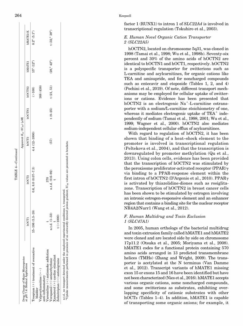
factor 1 (RUNX1) to intron 1 of SLC22A4 is involved intranscriptional regulation (Tokuhiro et al., 2003).
E. Human Novel Organic Cation Transporter2 (SLC22A5)
hOCTN2, located on chromosome 5q31, was cloned in1998 (Tamai et al., 1998; Wu et al., 1998b). Seventy-sixpercent and 30% of the amino acids of hOCTN2 areidentical to hOCTN1 and hOCT1, respectively. hOCTN2is a polyspecific transporter for zwitterions such asL-carnitine and acylcarnitines, for organic cations likeTEA and amisupride, and for noncharged compoundssuch as entecavir and etoposide (Tables 1, 2, and 4)(Pochini et al., 2019). Of note, different transport mech-anisms may be employed for cellular uptake of zwitter-ions or cations. Evidence has been presented thathOCTN2 is an electrogenic Na1-L-carnitine cotrans-porter with a sodium/L-carnitine stoichiometry of one,whereas it mediates electrogenic uptake of TEA1 inde-pendently of sodium (Tamai et al., 1998, 2001; Wu et al.,1999; Wagner et al., 2000). hOCTN2 also mediatessodium-independent cellular efflux of acylcarnitines.
With regard to regulation of hOCTN2, it has beenshown that binding of a heat–shock element to thepromoter is involved in transcriptional regulation(Peltekova et al., 2004), and that the transcription isdownregulated by promoter methylation (Qu et al.,2013). Using colon cells, evidence has been providedthat the transcription of hOCTN2 was stimulated bythe peroxisome proliferator-activated receptor (PPAR)gvia binding to a PPAR-response element within thefirst intron of hOCTN2 (D’Argenio et al., 2010). PPARgis activated by thiazolidine-diones such as rosiglita-zone. Transcription of hOCTN2 in breast cancer cellshas been shown to be stimulated by estrogen involvingan intronic estrogen-responsive element and an enhancerregion that contains a binding site for the nuclear receptorNR4A2/Nurr1 (Wang et al., 2012).
F. Human Multidrug and Toxin Exclusion1 (SLC47A1)
In 2005, human orthologs of the bacterial multidrugand toxin extrusion family called hMATE1 and hMATE2were cloned and are located side by side on chromosome17p11.2 (Otsuka et al., 2005; Moriyama et al., 2008).hMATE1 codes for a functional protein containing 570amino acids arranged in 13 predicted transmembranehelices (TMHs) (Zhang and Wright, 2009). The trans-porter is acetylated at the N terminus (Van Dammeet al., 2012). Transcript variants of hMATE1 missingexon 15 or exons 15 and 16 have been identified but havenot been characterized (Nies et al., 2016). hMATE1acceptsvarious organic cations, some noncharged compounds,and some zwitterions as substrates, exhibiting over-lapping specificity of cationic substrates with otherhOCTs (Tables 1–4). In addition, hMATE1 is capableof transporting some organic anions; for example, it
TABLE
4—Con
tinued
Dru
g(C
harge
ofMajor
Microsp
ecies
atpH
7.4)
Med
ical
App
lication
App
aren
tKm(IC50)[mM]
hOCT1
hOCT2
hOCT3
hOCTN1
hOCTN2
hMATE1
hMATE2-K
Trosp
ium
(1)trea
tmen
tof
overactive
blad
der
15–10
6(5.3–18
)0.6,
8.0(0.07–
7.3)
4.4(12–
1000
)(.
100)
15a(12a)
8.2a
(5.1
a)
Valpr
oylcarnitine(1
2)
anti-inflammatory
168(458
)
Varen
icline
(1)an
tism
okead
diction
t37
0Verap
amil(1
)an
tiarrh
ythm
icn.t.d.(1–33
)n.t.d.(0.6–92
)(57)
t(8–25
)t(21,
51)
(28,
a42
a)
t(32,
a38
a)
Xam
oterol
(1)card
iacstim
ulant
tt
Zolm
itriptan
(1)an
timigraine
t(.
1000
)
n.t.d.,notran
sportde
tected
und
ertheem
ploy
edex
perimen
talcondition
s;t,tran
sported.
aUptak
emea
suremen
tspe
rformed
inpr
esen
ceof
anou
twardlydirected
proton
grad
ient;IC
50va
lues
arepr
esen
tedin
bracke
ts.
264 Koepsell

has been shown to transport estrone sulfate with anapparent Km of 0.47 mM (Tanihara et al., 2007).Evidence has been provided that hMATE1 mediateselectroneutral proton–cation antiport in both directionsacross the plasma membrane, exhibiting a similaraffinity for protons from extracellular and intracellular(Otsuka et al., 2005; Tsuda et al., 2009b; Dangprapaiand Wright, 2011).Basal transcription of the SLC47A1 gene is regulated
by binding of the transcription factor Sp1 close to thetranscription start site (Kajiwara et al., 2007), bybinding of activation protein-1 (AP-1) and activationprotein-2 repressor (AP2-rep) to the promoter region(Ha Choi et al., 2009), and by the transcription factors,natural killer homeobox-2.5, sterol regulatory element-binding protein-1, and USF-1 (Kim et al., 2013).Concerning post-translational regulation of hMATE1,
it has been described that ischemia/reperfusion-inducibleprotein IRIP that interacts with regulatory protein RS1(RSC1A1) is involved in regulation of hMATE1 proteinabundance in the plasma membrane (Jiang et al., 2005;Li et al., 2013).
G. Human Multidrug and Toxin Exclusion 2, HumanMultidrug and Toxin Exclusion Kidney-Specific2 (SLC47A2)
In addition to the first cloned hMATE1 ortholog calledhMATE2, which comprises 602 amino acids (Otsukaet al., 2005), two shorter splice variants called humanMATE2-K (hMATE2-K) and hMATE2-B were clonedand characterized (Masuda et al., 2006). hMATE2-Kcontains 566 amino acids and is functionally active,whereas hMATE2-B contains 209 amino acids and doesnot mediate transport. hMATE2 and hMATE2-K have48% and 52% amino acid identity with hMATE1, re-spectively. Transport activity of hMATE2 was dem-onstrated after reconstitution of the transporter intoproteoliposomes (Komatsu et al., 2011). After expres-sion in HEK293 cells, hMATE2 has been localizedto intracellular vesicles and no transporter-mediatedcellular uptake could be detected (Komatsu et al., 2011).Future experiments are required to clarify whetherhMATE2 plays a role for cation uptake into intracellu-lar compartments and whether it can be targeted to theplasma membrane in vivo. Like hMATE1, hMATE2-Kis a polyspecific transporter of organic cations andzwitterions and noncharged compounds, showing broadlyoverlapping substrate selectivity with hMATE1 (Tables 1–4)(Tanihara et al., 2007; Astorga et al., 2012). Similar tohMATE1, hMATE-K2–mediated organic cation trans-port is stimulated by an opposed proton gradient (Masudaet al., 2006). It has been reported that hMATE2-K hasa higher affinity for protons than hMATE1 (Astorga et al.,2012).The transcription of hMATE2-K was shown to be
repressed by binding of the transcription factor MZ1 tothe promoter (Choi et al., 2011).
III. Structure–Function Relationships in OrganicCation Transporters
A. Primary Structures and Membrane Topology ofHuman Organic Cation Transporters
The OCTs, OCT1, OCT2, OCT3, OCTN1, and OCTN2,are members of the SLC22 family, which belongs to theMFS (Pao et al., 1998) (Fig. 1). The OCTs of the SLC22family have 12 predicted TMHs with intracellularNH2 and COOH termini (Fig. 2A). They contain a largeextracellular loop between TMH1 and TMH2 and a largeintracellular loop between TMH6 and TMH7. Thelarge extracellular loop comprises four or six cysteineresidues, which may form disulfide bridges (Keller et al.,2011; Brast et al., 2012) and at least one consensussequence for N-glycosylation. The large intracellular loopcontains consensus sequences for phosphorylation(Koepsell et al., 1999). The OCTs hMATE1 and hMATE2-K are members of the SLC47 family, which also belongsto the MATE family like transporter NorM from Vibriocholera (He et al., 2010) and transporter MATE fromPyrococcus furiosus (PfMATE) (Tanaka et al., 2013)(Fig. 1). Different from the OCTs of the SLC22 family,NorM and PfMATE, hMATE1, and hMATE2-K containan additional 13th C-terminal TMH, a large intracellu-lar loop between TMH12 and TMH13, and a shortextracellular C terminus (Fig. 2B) (Zhang and Wright,2009; Zhang et al., 2012).
B. Solved Crystal Structures of Transporters of theMajor Facilitator Superfamily and Multidrug andToxin Exclusion Family
Crystal structures of bacterial and fungal transport-ers of the MFS and of the MATE family have beensolved that are in four different conformations (Baiet al., 2017b). These include the L-fucose symporterFucP from Escherichia coli (Dang et al., 2010), theD-xylose–proton symporter XylE from E. coli (Sun et al.,2012), the MATE transporters NorM from V. cholerae(He et al., 2010), and transporter PfMATE (Tanakaet al., 2013) in outward-open conformations; the lactose–proton symporter LacY from E. coli in outward-facingoccluded state (Kumar et al., 2014); the multidrugtransporter EmrD from E. coli in an occluded state (Yinet al., 2006), the oligopeptide-H1 symporters PepTsofrom Shewanella oneidensis (Newstead et al., 2011),and the high-affinity phosphate transporter PiPTfrom Piriformospora indica (Pedersen et al., 2013) ininward-facing, occluded conformations; and LacY fromE. coli (Abramson et al., 2003), the glycerol-3-phosphate-phosphate antiporter GlpT from E. coli (Huang et al.,2003), and the peptide–proton symporter PepTst fromStreptococcus thermophiles (Solcan et al., 2012) ininward-open conformations. Examples for resolvedstructures in different conformations are depictedin previous reviews (Yan, 2013; Bai et al., 2017b).The resolved three-dimensional (3D) structures have
Organic Cation Transporters 265
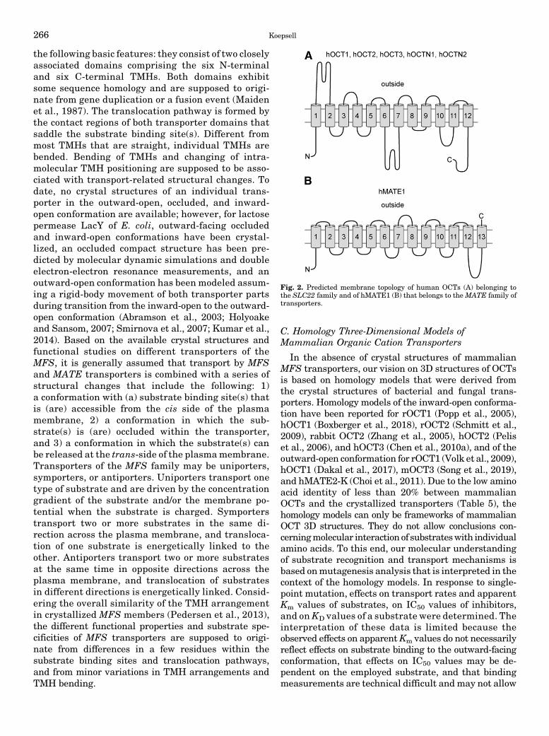
the following basic features: they consist of two closelyassociated domains comprising the six N-terminaland six C-terminal TMHs. Both domains exhibitsome sequence homology and are supposed to origi-nate from gene duplication or a fusion event (Maidenet al., 1987). The translocation pathway is formed bythe contact regions of both transporter domains thatsaddle the substrate binding site(s). Different frommost TMHs that are straight, individual TMHs arebended. Bending of TMHs and changing of intra-molecular TMH positioning are supposed to be asso-ciated with transport-related structural changes. Todate, no crystal structures of an individual trans-porter in the outward-open, occluded, and inward-open conformation are available; however, for lactosepermease LacY of E. coli, outward-facing occludedand inward-open conformations have been crystal-lized, an occluded compact structure has been pre-dicted by molecular dynamic simulations and doubleelectron-electron resonance measurements, and anoutward-open conformation has been modeled assum-ing a rigid-body movement of both transporter partsduring transition from the inward-open to the outward-open conformation (Abramson et al., 2003; Holyoakeand Sansom, 2007; Smirnova et al., 2007; Kumar et al.,2014). Based on the available crystal structures andfunctional studies on different transporters of theMFS, it is generally assumed that transport by MFSand MATE transporters is combined with a series ofstructural changes that include the following: 1)a conformation with (a) substrate binding site(s) thatis (are) accessible from the cis side of the plasmamembrane, 2) a conformation in which the sub-strate(s) is (are) occluded within the transporter,and 3) a conformation in which the substrate(s) canbe released at the trans-side of the plasmamembrane.Transporters of the MFS family may be uniporters,symporters, or antiporters. Uniporters transport onetype of substrate and are driven by the concentrationgradient of the substrate and/or the membrane po-tential when the substrate is charged. Symporterstransport two or more substrates in the same di-rection across the plasma membrane, and transloca-tion of one substrate is energetically linked to theother. Antiporters transport two or more substratesat the same time in opposite directions across theplasma membrane, and translocation of substratesin different directions is energetically linked. Consid-ering the overall similarity of the TMH arrangementin crystallized MFS members (Pedersen et al., 2013),the different functional properties and substrate spe-cificities of MFS transporters are supposed to origi-nate from differences in a few residues within thesubstrate binding sites and translocation pathways,and from minor variations in TMH arrangements andTMH bending.
C. Homology Three-Dimensional Models ofMammalian Organic Cation Transporters
In the absence of crystal structures of mammalianMFS transporters, our vision on 3D structures of OCTsis based on homology models that were derived fromthe crystal structures of bacterial and fungal trans-porters. Homology models of the inward-open conforma-tion have been reported for rOCT1 (Popp et al., 2005),hOCT1 (Boxberger et al., 2018), rOCT2 (Schmitt et al.,2009), rabbit OCT2 (Zhang et al., 2005), hOCT2 (Peliset al., 2006), and hOCT3 (Chen et al., 2010a), and of theoutward-open conformation for rOCT1 (Volk et al., 2009),hOCT1 (Dakal et al., 2017), mOCT3 (Song et al., 2019),and hMATE2-K (Choi et al., 2011). Due to the low aminoacid identity of less than 20% between mammalianOCTs and the crystallized transporters (Table 5), thehomology models can only be frameworks of mammalianOCT 3D structures. They do not allow conclusions con-cerningmolecular interaction of substrateswith individualamino acids. To this end, our molecular understandingof substrate recognition and transport mechanisms isbased onmutagenesis analysis that is interpreted in thecontext of the homology models. In response to single-point mutation, effects on transport rates and apparentKm values of substrates, on IC50 values of inhibitors,and onKD values of a substrate were determined. Theinterpretation of these data is limited because theobserved effects on apparentKm values do not necessarilyreflect effects on substrate binding to the outward-facingconformation, that effects on IC50 values may be de-pendent on the employed substrate, and that bindingmeasurements are technical difficult and may not allow
Fig. 2. Predicted membrane topology of human OCTs (A) belonging tothe SLC22 family and of hMATE1 (B) that belongs to the MATE family oftransporters.
266 Koepsell

to distinguish between binding to the outward-open orinward-open transporter conformation. An additionalimportant limitation is that, rather than altering themolecular interaction of a cation, the exchange of anamino acid may exhibit an indirect effect on cationbinding to a nearby or distant residue.
D. In-Depth Functional Characterization of OrganicCation Transporter 1 and Organic CationTransporter 2
A detailed functional characterization of rOCT1 andrOCT2 provided insight into the functional mechanismsof the OCT-subfamily OCT1–3. Using tracer uptakestudies and electrical measurements in oocytes ofX. laevis expressing rOCT1 or rOCT2 (Busch et al., 1996b;Nagel et al., 1997; Schmitt and Koepsell, 2005) andelectrical measurements in plasma membrane in giantpatches from oocytes expressing rOCT2 (Budiman et al.,2000), it was shown that rOCT1 and rOCT2 are sodium-independent electrogenic transporters that translocateorganic cations in both directions across plasma mem-branes, and also accept individual inorganic cationsas substrate. Experimental proof was provided thatrOCT1 and rOCT2 operate as transporters rather thanchannels by demonstrating trans-stimulation. Thus,rOCT1-mediated uptake of MPP into proteoliposomes,in which the membrane potential was clamped, wasstimulated by the alternative substrate choline on thetrans-side (Keller et al., 2005). Measuring uptake ofTEA or MPP and electrical charge simultaneously inoocytes expressing rOCT2 (Schmitt et al., 2009), chargeto cation stoichiometries of about one were obtained at
membrane potentials of 250 mV. This indicates ab-sence of anorganic ion leakage at250 mV, implying theexistence of transporter conformations with occludedsubstrates. At a membrane potential of 0 mV, charge tocation ratios between 3 and 4 were obtained for uptakeof TEA or MPP. This is interpreted to indicate cotrans-location of two or three inorganic cations under thiscondition.
Interaction of the nontransported inhibitors cortico-sterone and TBuA with rOCT2 was measured fromextracellular or intracellular sides, employing intactoocytes expressing rOCT2 or inside-out giant patchesfrom rOCT2-expressing oocytes, respectively (Volket al., 2003). Inward currents induced by superfusionof intact oocytes with choline and outward currentsinduced by superfusion of inside-out oriented giantpatches with choline were inhibited by addition ofcorticosterone or TBuA to the superfusion solutions.The data revealed that corticosterone and TBuA inhibitthe choline–induced currents from extracellular andintracellular with different efficacies showing differentapparent Ki values. Presuming that corticosterone andTBuA interact with the substrate-binding region ofrOCT1, the results indicate that the substrate-bindingregion may be exposed to extracellular or intracellularsurfaces, as predicted by 3D structures of crystallizedMFS transporters.
Using potential-induced fluorescence changes offluorescence-labeled rOCT1, high-affinity binding sitesof TEA, MPP, and TBuA were identified (Gorbunovet al., 2008). In a rOCT1 variant in which free cyste-ine residues had been removed, phenyalanine 483 in
TABLE 5Modeling of tertiary structures of OCTs and hMATE2-K
The analysis of homology was performed with program EMBOSS Needle (EMBL-EBI).
Transporter (ModeledConformation)
Crystallized Model Transporter(Conformational)
Homology a.a. Identity(a.a. Similarity) Procedure References
rOCT1 (i-open) Lactose permease LacY fromE. coli (i-open)
14.8% (23.0%) Crystal structure–basedhomology modeling
Abramson et al., 2003;Popp et al., 2005
rOCT1 (o-open) Based on prediction ofstructural changes ofmodeled i-open state
Volk et al., 2009
hOCT1 (i-open) Glycerol-3-phosphatetransporter GlpT from E. coli(i-open)
15.0% (25.5%) Crystal structure–basedhomology modeling
Huang et al., 2003;Boxberger et al., 2018
hOCT1 (o-open) Several members of the MFS Integrative combinationalmodeling
Roy et al., 2010; Dakalet al., 2017
rOCT2 (i-open) LacY from E. coli (i-open) 14.0% (24.0%) Crystal structure–basedhomology modeling
Abramson et al., 2003;Schmitt et al., 2009
rabbit OCT2 (i-open) Lactose permease LacY andglycerol-3-phosphate transporterGlpT from E. coli (i-open)
14.4% (22.3%)10.5% (20.0%)
Crystal structure–basedhomology modeling
Abramson et al., 2003;Huang et al., 2003;Zhang et al., 2005
hOCT2 (i-open) Lactose permease LacY andglycerol-3-phosphate transporterGlpT from E. coli (i-open)
13.4% (23.2%)14.6% (26.2%)
Crystal structure–basedhomology modeling
Abramson et al., 2003;Huang et al., 2003;Pelis et al., 2006
hOCT3 (i-open) Glycerol-3-phosphatetransporter GlpT from E. coli(i-open)
15.3% (26.4%) Crystal structure–basedhomology modeling
Huang et al., 2003;Chen et al., 2010a
mOCT3 (o-open) Human glucose transporterGLUT3 (o-occ)
19.5% (34.2%) Crystal structure–basedhomology modeling
Deng et al., 2015; Songet al., 2019
hMATE2-K (o-open) MATE transporter NorM fromvibrio cholerae (o-open)
17.6% (34.4%) Crystal structure–basedhomology modeling
He et al., 2010; Choiet al., 2011
a.a, amino acid; i-open, inward open conformation; o-occ, outward occluded conformation; o-open, outward open conformation.
Organic Cation Transporters 267

TMH11 located close to the extracellular surface wasreplaced by cysteine, which was subsequently labeledwith tetramethylrhodamine-6-maleinimide. Effects ofthe substrates choline, TEA, MPP, and the inhibitorTBuA on potential-induced fluorescence changes weremeasured, and EC50 were determined (Gorbunov et al.,2008). The data indicated that choline, TEA, and MPPnot only interacted at low-affinity binding sites withEC50 values similar to the apparent Km values of thesubstrates (EC50: 290 mM choline, 57 mMTEA, 0.86 mMMPP) but also at an additional high-affinity site (EC50:9 nM choline, 50 nMTEA, 24 pMMPP). For TBuA, threeinteraction sites with EC50 values of 1 pM, 32 nM, and277 nM were obtained.
E. Mutagenesis Studies in Rat Organic CationTransporter 1, Rat Organic Cation Transporter 2, andHuman Organic Cation Transporter 2
In rOCT1, detailed mutagenesis experiments wereperformed that induced effects on structure, transportactivity, and substrate selectivity. These studies in-creased the understanding of structure–function rela-tionship in OCT1–3. Some mutations were also studiedin rOCT2 and hOCT2.The impact of the large extracellular loop of rOCT1
on oligomerization and transport was investigated(Keller et al., 2011). The large extracellular loop locatedbetween TMH1 and TMH2 contains six cysteine resi-dues that are conserved in OCT1–3 and form disulfidebridges (Pelis et al., 2012). It was observed that cell-free–expressed rOCT1 solubilized with mild detergentsformed oligomers. Oligomerization of rOCT1 was alsodetectedwhen the transporterwas expressed inHEK293cells (Keller et al., 2011). In these studies, it could notbe distinguished whether rOCT1 forms homodimers orhomo-oligomers. Precipitation experiments were per-formed with conjointly expressed, differentially taggedrOCT1 wild-type (WT) and rOCT1 variants. In thevariants, either the large extracellular loop of rOCT1was replaced by the respective loop of rat organic aniontransporter (OAT)1, or cysteine residues in the largeextracellular loop of rOCT1 were exchanged for serines.The obtained data indicate that the structure of thelarge extracellular loop, and in particular the integrityof four cysteine residues in this loop, is required foroligomerization. Disulfide bridging between the cysteinesis required for oligomerization because no oligomerformation was observed in the presence of dithiothrei-tol. The data are consistent with a dimeric quarternarystructure of rOCT1 in the plasma membrane, as hasbeen shown for several bacterial transporters of theMFS (Veenhoff et al., 2001; Safferling et al., 2003). Aseries of experiments was performed trying to evaluatewhether the rOCT1 monomers transport indepen-dently (Keller et al., 2011). To this end, Vmax andapparent Km values measured in proteoliposomes con-taining rOCT1 WT versus nonoligomerizing rOCT1
loop variants were compared. In addition, Vmax andKm values of rOCT1 reconstituted into proteoliposomesor expressed in oocytes were measured in absenceand presence of dithiothreitol. Moreover, Vmax andKm values of a rOCT1-tandem protein mimicking therOCT1dimerweremeasuredwhenboth rOCT1moleculeswere functional, or when one molecule was blocked bymodification of a cysteine residue that had been intro-duced into the substrate-binding region. The data indicatethat rOCT1 monomers can operate independently, ashas been described for bacterial transporters of theMFS (Ambudkar et al., 1990; Auer et al., 2001). Theysuggest that the structure of the large loop has someminor impact on Km and turnover of the transporter.Oligomerization and the critical role of the sulfhydrylgroups in the large extracellular loop for oligomeriza-tion have also been described for hOCT2 (Brast et al.,2012).
In rOCT1, effects of point mutations on velocityand selectivity of transport, selectivity of inhibitors,and binding of MPP were investigated in detail. In anearly study, Asp475 in the middle of TMH 11 (Fig. 3)was replaced by glutamate, and effects on appar-ent Km values and Vmax values of choline, TEA, N1-methylnicotinamide, and MPP, and on IC50 values ofsome inhibitors were investigated (Gorboulev et al.,1999). Asp475 is conserved inOCT1–3, whereasOCTN1,OCTN2, and the organic anion transporters OAT1–3contain the positively charged arginine residue in thisposition. The Asp475Glu mutant of rOCT1 showed89% to 98% reduced Vmax values, although targetingof the transporter to the plasma membrane was notlargely impaired. The apparent Km values for choline,TEA, and N1-methylnicotinamide were decreased 4- to15-fold, whereas the apparent Km value for MPP wasnot altered. The IC50 values for inhibition of 10 mMTEAuptake by the nontransported inhibitors TBuA andtetrapentylammonium (TPeA) were decreased 5-fold.The data suggested a central role of Asp475 forsubstrate binding of structurally different substrates.Because it has been recently shown that Asp475 isdirectly involved in MPP binding to a transport-relatedlow-affinity MPP binding site (Keller et al., 2019),Asp475 probably interacts with all cationic substratesduring translocation.
In this binding study, MPP binding was measured tofusion proteins of green fluorescent protein (GFP) withrOCT1 WT (GFP-rOCT1) or rOCT1 mutants (Kelleret al., 2019). TheGFP fusion proteins were generated bycell-free expression and incorporated into nanodiscsduring translation. Measurements of MPP uptake intoproteoliposomes were performed in parallel. The nano-discs were formed by dialysis from solubilized mixturesof lipids and a nondimerizing variant of the membranescaffold protein 1. In nanodiscs formedwith appropriatelipids, per rOCT1 monomer about two low-affinity MPPbinding sites with a common apparent dissociation
268 Koepsell

constant (KD) of 36 mM and one high-affinity MPPbinding site with a KD of 0.23 mM were determined.The same data were obtained when oligomerizationof GFP-rOCT1 was prevented by replacement of thecysteines in the large extracellular loop of rOCT1. Theapparent KD of the low-affinity binding was similarto the apparent Km value (19 mM) obtained for MPPtransport in proteoliposomes. Of note, the apparentKm values for MPP uptake measured after reconstitu-tion of rOCT1 or GFP-rOCT into proteoliposomes weresimilar; however, they were about 10-fold higher thanthe apparent Km values for MPP uptake that weredetermined after expression of rOCT1 in oocytes orHEK293 cells.Asp475 belongs to a rOCT1 motif of five amino acids
in positions 474–478 that are located in the middle ofTMH11. Cys474, Asp475, Gly477, and Gly478 are con-served in OCT1–3. Cys474, Asp475, and Gly478 aresupposed to interact directly with substrates dur-ing transport somehow lining the transport path. Asobserved after replacement of Asp475 in rOCT1 byglutamate, after replacement of Cys474 in hOCT2 withalanine, the Vmax values for transport of TEA andMPP were decreased, the apparent Km for TEA wasdecreased, and the apparentKm forMPPwas not altered
(Pelis et al., 2012). In addition, some evidencewas providedthat Cys474 in hOCT2 contacts the aqueous phase becauseit could be modified by the hydrophilic sulfhydryl groupreagent (1)-biotinyl-3-maleimidopropionamidyl-3,6-dioxaoctanediamine (maleimide-PEO2-biotin) (Peliset al., 2006). Maleimide-PEO2-biotin inhibited transportirreversibly, and labeling of Cys474 with maleimide-PEO2-biotin was reduced in presence of TPeA. Locationof Gly478 in rOCT1 within the transport path wasdemonstrated in affinity-labeling experiments (Egenbergeret al., 2012). These experiments were performed with thevariant rOCT1(10DC) in which all cysteine residueswith exception of those in the large extracellular loopwere replaced by alanine or serine (Sturm et al., 2007).Affinity labeling of rOCT1(10DC) was performedwith the TEA-derived sulfhydryl group reagent 2-(trimethylammonium) ethylmethanethiosulfonate (MTSET),which is transported by rOCT1(10DC). When Gly478was exchanged against cysteine, MTSET blocked trans-port of organic cations irreversibly, and blockage wasblunted when the labeling was performed in presenceof substrates (Egenberger et al., 2012). The critical roleof Gly478 is supported by the observation that the IC50
values for inhibition of MPP uptake by TBuA weredecreased when Gly478 in rOCT1 or rOCT1(10DC) wasreplaced by cysteine.
Compared with channel activity, transporter func-tion is supposed to involve more substantial structuralchanges. In OCTs, transport-related structural changesinclude parts that are not directly involved in substratebinding and translocation such as TMHs 3, 6, 9, and 12(Fig. 3). In rOCT1 and rOCT2, transport-related struc-tural changes have been recorded by measuring cationeffects on membrane capacitance in oocytes expressingrOCT2 or rOCT1(10DC) (Schmitt and Koepsell, 2005;Gorbunov et al., 2008) and by measuring cation effectson potential-induced fluorescent changes in rOCT1(10DC)containing fluorescence-labeled cysteine residues close tothe extracellular surface of plasma membrane (Gorbunovet al., 2008; Egenberger et al., 2012). In the latter experi-ments, cysteine residues were introduced by replacingamino acids in peripheral parts of TMHs 4 (Cys260), 8(Cys380), and 11 (Cys483), and fluorescence labelingwas performedwith tetramethylrhodamine-6-maleimide(TMRM), which does not enter the transport path. Itwas observed that choline, MPP, and TBuA exhibiteddifferential effects on voltage-dependent fluorescentchanges when rOCT1(10DC) was labeled in differentpositions. When Gly478 within the transport pathwas replaced by cysteine, voltage-dependent fluores-cence changes of rOCT1(10DC) labeled in twopositionswithTMRM were altered. In addition, potential-dependent fluo-rescence changes observed in TMRM-labeled rOCT1(10DC)containing theGly478Cysmutationwere changedwhenCys478 was affinity-labeled with MTSET. The dataindicate extended potential- and cation-dependent struc-tural changes in rOCT1 when Gly478 is exchanged by
Fig. 3. Views into the modeled outward-open (upper panel) and inward-open (lower panel) binding cleft of rOCT1. Modeling was performed, asdescribed (Popp et al., 2005; Volk et al., 2009). Trp218, Arg440, andAsp475 located within the inner parts of the modeled outward-open andinward-open clefts are indicated.
Organic Cation Transporters 269

cysteine or when the introduced cysteine residue ismodified with MTSET. Glycine residues in TMHs pro-vide flexibility, allowing bending during transport thatmay be essential for formation of occluded conforma-tions (Pace and Scholtz, 1998). Hence, Gly477-Gly478 inOCT1–3 is supposed to represent a hinge domain in themiddle of TMH11 that allows bending during transport.Trying to determine the impact of amino acids in
TMH4 on substrate binding and/or translocation, theamino acids in TMH4 of rOCT1 were replaced individ-ually, and effects on apparent Km values of TEA andMPP transport were measured after expression of themutants in oocytes (Popp et al., 2005). The apparentKm values for TEA and MPP uptake were increased ordecreased when Val213, Trp218, or Phe222 was mu-tated. In mutant Thr226Ala, the apparent Km valuefor MPP, and in mutant Val229Ala, the apparent Km
for TEA, were increased, respectively. Because Val213,Trp218, Phe222, Tyr226, and Val229 are located on thesame side of TMH4, the data suggest that part of thesubstrate-binding region and transport path is formed byTMH4. Based on these data, it cannot be distinguishedwhether the individual mutations change a direct inter-action of the respective amino acid with a substrate orexhibit an allosteric effect on substrate interaction withnearby amino acids.For inhibition of TEA uptake or MPP uptake by
extracellular corticosterone, more than 30 times lowerIC50 values were obtained for rOCT2 compared withrOCT1. Investigating the molecular basis for this differ-ence, it was observed that after replacement of Ile443,Tyr447, and Glu448 in TMH10 of rOCT1 by alanine,leucine, and glutamine located in the correspondingpositions of rOCT2, the same IC50 values for corticoste-rone inhibition were observed as in rOCT2 (Gorboulevet al., 2005). In mutant rOCT1(Ile443Ala) the apparentKm value for TEA uptake was decreased, whereas indouble-mutant rOCT1(Tyr447Leu/Glu448Gln) the appar-ent Km value for MPP uptake was decreased, pointing toa role of these amino acids for binding and/or translocationof these substrates (Gorboulev et al., 2005).Effect of mutations in rOCT1 on inhibition of TEA-
induced inward currents by extracellular and intracel-lular corticosterone was characterized in cRNA-injectedoocytes or in stably transfectedHEK293 cells (Volk et al.,2009). For current measurements in oocytes, mutationswere performed in rOCT1(10DC), which showed higherTEA-induced inward currents and similar efficacy ofcorticosterone inhibition than rOCT1. Amino acids liningthe modeled outward-facing and/or inward-facing con-formation of rOCT1 were selected for mutagenesis. Forinterpretation of the data, corticosterone-docking simu-lations to the modeled outward- and inward-open cleft ofrOCT1 were performed. After mutation of Phe160 inTMH2, Trp218 in TMH4, Arg440 or Leu447 in TMH10,or Asp475 in THM11, the IC50 values for inhibition ofTEA-induced currents or of TEA uptake by corticosterone
added from extracellular or intracellular were changed.These amino acids are located within the inner partsof the modeled outward- and inward-facing cleft ofrOCT1 (Fig. 3) and were predicted to interact withcorticosterone by docking simulations. After replace-ment of Tyr222 by phenylalanine or Gln448 by gluta-mate, the IC50 value for inhibition of TEA-inducedcurrents by extracellular corticosterone was not al-tered. This is consistent with the inaccessibility ofthese amino acids in the modeled outward-facing cleft(Volk et al., 2009). The data suggest that amino acids inTMH4 (Trp218) and TMH11 (Asp475) located in theinward- and outward-facing conformations of rOCT1interact with TEA, MPP, and corticosterone.
To determine whether, in addition to amino acids inTMH4 and Asp475 in TMH11, also Phe160 in TMH2and Arg440 and Leu447 in TMH10 are critical fortranslocation of TEA and/or MPP, effects of muta-tions in these positions on apparent Km values foruptake of TEA and/or MPP into stably transfectedHEK293 cells were investigated (Gorboulev et al., 2018).The apparent Km value for uptake of TEA was alteredwhenArg440was replaced by lysine orwhenLeu447wasreplaced by tyrosine, whereas the apparent Km value forMPP was changed in the Phe160Tyr mutant. The datasuggest that Arg440 and Lys447 interact with TEA andthat Phe160 interacts with MPP during transport.
To further characterize the outward-facing bind-ing site, it was investigated which of the amino acidssupposed to interact with extracellular corticosteronealso interact with the nontransported inhibitors TBuAand TPeA in addition to Asp475 (Gorboulev et al., 2018).After mutation of Trp218, the IC50 values for inhibi-tion of MPP uptake by TBuA and TPeA were changed,whereas the exchange of Arg440 by lysine did not alterthe IC50 values of both inhibitors. After mutation ofPhe160 and Tyr222, only the IC50 for TBuA, and aftermutation of Leu447, only the IC50 for TPeAwas changed.Taken together the data suggest that TBuA interactswith Phe160, Trp218, Tyr222, and Asp475, whereasTPeA interacts with Trp218, Leu447, and Asp475.
Extending the binding studies to GFP-rOCT1 mutants,it was also investigated whether binding of MPP to thetwo low-affinity binding sites and the allosteric high-affinity MPP binding site in the rOCT1 monomer waschanged when, in addition of Asp475, Phe160, Trp218,Arg440, Leu447, or Gln448 was mutated (Keller et al.,2019). MPP-binding measurements were performedafter incorporation of GFP-rOCT1 WT or mutants intonanodiscs, whereas MPP uptake was measured in recon-stituted proteoliposomes. After replacement of Phe160by tyrosine, Leu447 by phenylalanine or tyrosine, andGln448 by glutamate, maximal binding to high- and low-affinity MPP binding sites was not changed. This meansthat binding to one high-affinity MPP binding site andtwo low-affinityMPP binding sites per rOCT1monomerwas not affected. However, as in Asp475Glu mutant,
270 Koepsell

after replacement of Trp218 by tyrosine, instead oftwo, only about one low-affinity MPP binding site permonomer was found, whereas high-affinity binding ofMPP was not altered (Fig. 4A). Because also about onehigh-affinity and one low-affinity MPP binding site perrOCT1 monomer were also observed in double-mutantAsp475Glu/Trp218Tyr, it was concluded that Asp475and Trp218 contribute to the same low-affinity MPPbinding site. Only about one low-affinity and one high-affinity MPP binding site were also observed whenArg440 was replaced by lysine (Fig. 4A). In double-mutant Arg440Lys/Asp475Glu, low-affinity binding ofMPPwas abolished, whereas high-affinityMPP bindingwas not changed. Therefore, it was concluded thatArg440 contributes to a different low-affinity MPPbinding site than Asp475 and Trp218. After reconstitu-tion into proteoliposomes, the Vmax of MPP uptake wasdecreased by 44% in mutant Trp218Tyr, by 79% inmutant Asp475Glu, by 29% in mutant Arg440Lys, andby 96% in double-mutant Asp475Glu/Arg440Lys. Thisindicates that the low-affinity MPP binding sites areinvolved in transport. The apparent dissociation con-stants of the low-affinity MPP binding sites (KD low-aff.)appear to be similar because they could not be distin-guished and only one common KD low-aff. value wasdetectable. Of note, the common KD low-aff. value ofMPP binding to GFP-rOCT1 (36 mM) was similar tothe apparentKmmeasured forMPPuptake byGFP-rOCT1in proteoliposomes (19 mM). This suggests that theKm value is mainly determined by MPP binding tothe transporter. In GFP-rOCT1 mutants Trp218Tyr,Asp475Glu, andTrp218Tyr/Asp475Glu, theKD low-aff. and
Km values were similar to GFP-rOCT1-WT, suggestingthat blockage of binding to and translocation via this low-affinity site do not alter binding to and translocation bytheother low-affinity site.At variance, theKD low-aff. and theKm value in mutant Arg440Lys were 2.2 or 3.6 timeshigher comparedwithGFP-rOCT1-WT, suggesting someeffect of this mutation on the other low-affinity MPPbinding site. In nanodiscs containing GFP-rOCT1, MPPmay bind to the outward- or inward-facing conformationof rOCT1. However, the neighboring location of Trp218and Asp475 and the more distant location of Arg440Lysin the modeled outward-facing conformation (Fig. 3upper panel; Fig. 4B) suggest that bindingwasmeasuredto the outward-facing conformation.
The data suggest that translocation of MPP orother organic cations may be initiated via substratebinding to two separate low-affinity binding sites.The two low-affinity substrate binding sites mayoperate independently or in a concerted mode, in-ducing translocation-related conformational changes.At substrate concentrations above the Km of the re-spective substrate, two substrate molecules may betransported together, as depicted for MPP in Fig. 4B.In this situation, the high-affinity binding site(s) of therespective substrate is (are) occupied, and binding ofinhibitors to high-affinity sites may not influence trans-port. At concentrations of MPP or other transportedcations far below the respective Km values, only onesubstrate molecule is transported per transport cycle,and binding of the transported substrate or othercations to high-affinity sites may exhibit allostericeffects on transport.
Fig. 4. Effects of mutations of amino acids lining the modeled outward-open cleft of rOct1 on MPP binding to low-affinity and high-affinity bindingsites. (A) Numbers of MPP molecules per transporter monomer that bind to GFP-fusion proteins with Oct1 WT or the indicated mutants. Bindingmeasurements were performed to fusion proteins that had been reconstituted into nanodiscs, and maximal low-affinity and high-affinity binding wasdetermined. *Significant difference compared with rOCT1. •Significant difference compared with all other rOct variants. (B) Hypothetic model basedon the observation that Trp218 (green) and Asp475 (red) are located in neighboring positions in the modeled outward-open cleft and more distant in themodeled inward-open cleft. Arg440 is indicated in blue.
Organic Cation Transporters 271

F. Mutagenesis Studies in Human Novel OrganicCation Transporter 1 and Human Novel OrganicCation Transporter 2
The amino acid sequences of hOCTN1 and hOCTN2are 88% identical, showing the smallest conservationsbetween the large intracellular loops and the intracellu-lar C termini. Both transporters accept organic cations,zwitterions, and noncharged compounds as substrates(Tables 2 and 4). They have partially overlapping sub-strate selectivities (Table 4) and employ partially differenttransport mechanisms. For example, hOCTN1 is a highlyefficient high-affinity transporter for ergothioneine, whichis not transported by hOCTN2. Human hOCTN2has a more than 100 times lower Km for transport ofL-carnitine than hOCTN1, and, different from hOCTN2,cellular uptake of TEA by hOCTN1 is stimulated byan outwardly directed proton gradient (Tamai et al.,1998). In hOCTN1, two binding sites are supposed to beinvolved in substrate recognition triggering transloca-tion, because hOCTN1-mediated transport of cytarabinecould not be inhibited by ergothioneine or TEA, but wasinhibited by nitrobenzylmercaptopurine ribonucleoside,whereas hOCTN1-mediated transport of ergothioneinewas not inhibited by nitrobenzylmercaptopurine ribonu-cleoside (Drenberg et al., 2017). In a mutant in whichLeu503 in hOCTN1 is replaced by phenylalanine, higherapparent Km values were obtained for L-carnitine andgabapentin, whereas lower apparent Km values weredetermined for ergothioneine and TEA (Table 8). Be-cause Leu503 is located in TMH12, which probably doesnot contribute to the substrate-binding region and trans-location path(s), the mutation is supposed to induce anallosteric alteration of (a) transport-relevant part(s) ofthe transporter. Trying to identify amino acids in hOCTN1that are responsible for the different efficacy of hOCTN1and hOCTN2 for transport of L-carnitine verus ergothio-neine, combinations of amino acids that differ betweenboth transporters were replaced mutually (Bacher et al.,2009). Transport of L-carnitine by hOCTN1 was in-creased when a combination of amino acids in TMHs 5,7, 8, 9, and 10 was exchanged, whereas transport ofergothioneine was not altered. The data support theexistence of different transport-relevant binding sites forthe two substrates.hOCTN2 mediates Na1-dependent transport of
L-carnitine as well as Na1-independent transport ofTEA. Because competitive interaction was observedfor inhibition of TEA uptake by L-carnitine and a mixedtype of interaction for TEA inhibition of L-carnitineuptake (Ohashi et al., 2002), overlapping or closely associ-ated binding sites for the two substrates were presumed.This notion is supported by differential effects ofmutationson efficacy of L-carnitine versus TEA transport. Thus, theexchange of Pro478 in TMH11 against leucine resulted ina strong reduction of L-carnitine transport and an increaseof TEA transport (Seth et al., 1999). Similarly, a reduction
of L-carnitine transport and no change in TEA transportwere observed when Tyr211 in TMH4 was replaced byphenylalanine (Seth et al., 1999). In both mutants, thesodium dependence of carnitine uptake was not changed.A selective effect on L-carnitine transport was alsoobserved when Ser467 in TMH 11 was replaced bycysteine (Ohashi et al., 2002). The efficacy of L-carnitinetransport was decreased because the apparent Km forcarnitine transport was increased 15-fold, whereastransport of several tested organic cations such as TEA,quinidine, and verapamil was not affected. As observedfor the mutants Pro478Leu and Tyr211Phe, mutantSer467Cys did not alter the activation of L-carnitinetransport by sodium. The data suggest that amino acidsin TMH11 are involved in transport-related binding ofL-carnitine. Because hOCTN2 contains an arginine inamino acid position 471, which corresponds to Asp475in rOCT1, similar to the organic anion transportersof the SLC22 family, the carboxyl group of L-carnitinemay interact with this Arg471. The EC50 for sodiumstimulation of L-carnitine transport was decreased todifferent degrees when Glu452 in the intracellular loopbetween TMH10 and TMH11 was replaced by differentamino acids (Wang et al., 2000b). Data allowing edu-cated speculations concerning the molecular effect ofthis mutation are not available.
G. Mutagenesis in Human Multidrug and ToxinExclusion 1 and Human Multidrug and ToxinExclusion Kidney-Specific 2
After replacement of His386 in TMH10 of hMATE1 andhistamine in the corresponding position of hMATE2-Kand of rat MATE (rMATE)1 by glutamine, TEA uptakewas largely decreased, although these mutations werenot supposed to decrease expression. Because pH depen-dence of TEA uptake-mediated rMATE1was not changeddramatically, this histidine residue is probably not di-rectly involved in proton binding (Asaka et al., 2007b).
Replacement of Cys59 or Cys123 of hMATE1 and incorresponding positions of hMATE-K2 and rMATE1 byglycine induced a large decrease of TEA uptake (Asakaet al., 2007b). The location of Cys59 at the transitionof TMH1 to the extracellular loop between TMH1 andTMH2 of hMATE1 and of Cys123 in TMH3 of hMATE1suggests that these mutations induced allosteric effectson TEA uptake.
IV. Expression and Locations of Human OrganicCation Transporters in Cells, Tissues, Organs,
and Tumors
A. Expression and Membrane Locations of HumanOrganic Cation Transporter 1, Human OrganicCation Transporter 2, and Human Organic CationTransporter 3
hOCT1 is one of the abundantly expressed drugtransporters in liver (Nishimura and Naito, 2005;
272 Koepsell

Hilgendorf et al., 2007; Drozdzik et al., 2019). Invarious other organs and tissues, including eye, brain,small intestine, kidney, lung, urinary bladder, heart,placenta, and skeletal muscle, hOCT1 is expressed atmuch lower levels (Table 6). In the eye, hOCT1 mRNAwas detected in the cornea and the choroidial–retinalbarrier (Zhang et al., 2008). In brain, hOCT1 mRNA isexpressed in microvessels (Geier et al., 2013). Notewor-thily, hOCT1 expression was also observed in adipo-cytes (Moreno-Navarrete et al., 2011) and in peripheralleukocytes, lymphocytes, and monocytes (Nishimuraand Naito, 2005; Minuesa et al., 2008). Immunohisto-chemical localization of hOCT1 has only been performedin a few tissues. In the liver, hOCT1 was localized to thesinusoidal membrane of hepatocytes, as has been de-scribed for rOCT1 (Meyer-Wentrup et al., 1998; Nieset al., 2009). In rodents, the highest expression of OCT1was observed in hepatocytes around the central veins,which represent the major site for glycolysis and lipidmetabolism (Meyer-Wentrup et al., 1998). In smallintestine, hOCT1 was localized to the BBM of enter-ocytes (Han et al., 2013). Luminal location of hOCT1was also detected in kidney where hOCT1 immunore-activity was observed at the BBM of proximal and distaltubules (Tzvetkov et al., 2009). In lung, immunoreac-tivity for hOCT1 was detected in ciliated cells of airwayepithelia, showing a predominantly intracellular loca-tion (Lips et al., 2005). In the bladder, hOCT1-relatedimmunoreactivity was observed in the plasma mem-brane of urothelial cells (Lips et al., 2007).The expression of hOCT2 is less ubiquitous than
hOCT1 and hOCT3 (Table 6). The highest expres-sion of hOCT2 mRNA was observed in kidney, whereasless mRNA expression was detected in several otherorgans, for example in lung, brain, spinal cord, placenta,and nasal mucosa. Expression of hOCT2 mRNA wasalso detected in dorsal root ganglia (Sprowl et al., 2013).No expression was detected in many organs, includ-ing small intestine, colon, liver, and heart (Table 6). Inkidney, hOCT2 has been localized by immunohisto-chemistry to the basolateral membrane of renal proxi-mal tubules, as has been described for rOCT2 (Karbachet al., 2000; Motohashi et al., 2002, 2013). In airwayepithelia of the lung, hOCT2-related immunoreactivitywas detected in apical membrane of ciliated epithelialcells and in plasma membranes of basal cells (Lipset al., 2005). In brain, hOCT2 was localized by in situhybridization and immunohistochemistry in neurons atdifferent brain areas (Busch et al., 1998). In addition,hOCT2 mRNA was observed in isolated microvessels(Geier et al., 2013). In rats and/or mice, OCT2 has alsobeen localized to neurons of different neuronal circuits(Bacq et al., 2012; Couroussé and Gautron, 2015), innerve cells of the spinal cord, and to the apicalmembraneof epithelial cells in the choroid plexus (Sweet et al.,2001). However, it has not been verified whether theselocations in rodents reflect the situation in humans.
hOCT3 is expressed in many organs and tissues.High mRNA abundance was observed in adrenalgland, prostate, salivary gland, and skeletal muscle(Table 6). Lower, but distinct mRNA expression wasdetected in adipose tissue, bone marrow, colon, liver,lung, stomach, small intestine, heart, testis, trachea,thyroid gland, uterus, and placenta, whereas lessabundant amounts of hOCT3 mRNA were observedin various other organs, including brain, kidney, andurinary bladder, and in monocytes (Table 6). Immuno-histochemistry in the submandibular gland revealeda predominant localization of hOCT3 in luminal mem-branes of acini and ducts (Lee et al., 2014a). In skeletalmuscle, hOCT3-related immunoreactivity was observedin the plasmamembrane of themuscle cells (Chen et al.,2010a), whereas in liver, hOCT3 was immunolocalizedto the sinusoidal membrane of hepatocytes (Nies et al.,2009). In the lung, hOCT3 protein was detected inbronchial epithelial cells and localized to the luminalmembrane of ciliated cells, the basolateral membraneof intercalated cells, and the entire plasma membraneof basal cells (Lips et al., 2005). At variance, in smallintestine hOCT3-related immunoreactivity was observedin the luminal membrane of the enterocytes (Mülleret al., 2005). Immunostaining in heart suggests thathOCT3 is located at the endothelium of capillaries andsmall vessels (Solbach et al., 2011). In the placenta,hOCT3-related immunostaining was observed in tro-phoblast cells (Solbach et al., 2011), and subcellularfractionation and Western blotting of placenatal cellsrevealed that hOCT3 is located in basolateral mem-branes facing the fetal side (Sata et al., 2005). hOCT3mRNAwas observed in various brain regions, includingsubstantia nigra, nucleus caudatus, pons, and cerebel-lum (Duan and Wang, 2010). Expression of mRNA andprotein was detected in isolated brain microvessels andin primary cultured astrocytes (Inazu et al., 2003; Geieret al., 2013). In kidney hOCT3 protein was located tothe basolateral membrane of renal proximal tubularcells (Chen et al., 2010a), whereas in urothelial cellshOCT3-related immunoreactivity was observed at theentire plasma membrane (Lips et al., 2007; Bschleipferet al., 2012).
B. Expression and Membrane Locations of HumanNovel Organic Cation Transporter 1 and HumanNovel Organic Cation Transporter 2
In nearly all investigated organs, tissues’ and cells’mRNA of hOCTN1 was detected; however, with theexceptions of bone marrow, colon, kidney, and skeletalmuscle, only low mRNA concentrations were observed(Table 6). The expression of hOCTN1 in bone marrow,spleen, lymphocytes, macrophages, monocytes, anderythrocyte progenitor cells suggests relevance of hOCTN1for blood formation and immune response. In smallintestine, hOCTN1 has been localized to the BBM ofenterocytes (Sugiura et al., 2010). In kidney, hOCTN1 is
Organic Cation Transporters 273

probably located in the BBM of renal proximal tubulesbecause a luminal location has been demonstrated forOCTN1 in mice, and it has been demonstrated thathOCTN1 interacts with the PDZ (PSD-95, Dlg, andZO-1) domain of proteins that are expressed in theBBM of renal proximal tubules (Gisler et al., 2001;Tamai et al., 2004; Kato et al., 2005). In lung, hOCTN1-related immunoreactivity was observed in the luminalmembrane of tracheal and bronchial epithelia, and in
alveolar epithelial cells (Berg et al., 2018). In skin,hOCTN1 is mainly expressed in the epidermis, where itwas localized to the basal membrane of proliferatingbasal cells and to terminally differentiated granularkeratinocytes (Markova et al., 2009).
hOCTN2 is also expressed ubiquitously. Relativelyhigh mRNA concentrations were observed in adrenalgland, brain vessels, colon, heart, kidney,mammary gland,salivary glands, and skeletal muscle. hOCTN2 mRNA
TABLE 6Abundance of mRNA and protein of human organic cation transporters in various organs, tissues, and cells
The degree of expression of mRNA (crosses) is estimated from observations reported in the literature. The related references are indicated in normal face. Due to partiallydiffering results, only an arbitrary classification could be performed. Detection of protein by Western blotting, immunohistochemistry, or proteomics is indicated by asterisks.The related references are indicated in italics when only protein expression was investigated and in underlined normal face when both mRNA and protein were notdetermined. The clasification has been performed on the basis of the following publications: hOCT1: Gorboulev et al., 1997; Pietig et al., 2001; Alcorn et al., 2002; Motohashiet al., 2002; Bottalico et al., 2004, 2007; Lips et al., 2005, 2007; Nishimura and Naito, 2005; Ballestero et al., 2006; Hilgendorf et al., 2007; Meier et al., 2007; Minuesa et al.,2008; Okabe et al., 2008; Zhang et al., 2008; Nies et al., 2009; Tzvetkov et al., 2009; Moreno-Navarrete et al., 2011; Gupta et al., 2012; Geier et al., 2013; Han et al., 2013;Drozdzik et al., 2014, 2019; Bexten et al., 2015; Cai et al., 2016; Arner et al., 2018; Breining et al., 2018, skin: K. Lips and H. Koepsell, unpublished data, hOCT2: Gorboulevet al., 1997; Busch et al., 1998; Pietig et al., 2001; Alcorn et al., 2002; Motohashi et al., 2002; Bottalico et al., 2004, 2007; Lips et al., 2005, 2007; Nishimura and Naito, 2005;Hilgendorf et al., 2007; Minuesa et al., 2008; Okabe et al., 2008; Filipski et al., 2009; Moreno-Navarrete et al., 2011; Bschleipfer et al., 2012; Geier et al., 2013; Sprowl et al.,2013; Bexten et al., 2015; Cai et al., 2016; Arner et al., 2018; Breining et al., 2018; Dolberg and Reichl, 2018, skin: K. Lips and H. Koepsell, unpublished data, hOCT3: Wuet al., 2000; Motohashi et al., 2002; Bottalico et al., 2004, 2007; Lips et al., 2005, 2007;Müller et al., 2005; Nishimura and Naito, 2005; Sata et al., 2005; Hilgendorf et al., 2007;Minuesa et al., 2008; Okabe et al., 2008; Nies et al., 2009; Duang and Wang, 2010; Solbach et al., 2011; Bschleipfer et al., 2012; Geier et al., 2013; Lee et al., 2014a; Bextenet al., 2015; Cai et al., 2016; Arner et al., 2018; Breining et al., 2018; Drozdzik et al., 2019; Song et al., 2019, skin: K. Lips and H. Koepsell, unpublished data, hOCTN1: Tamai et al.,1997; Pietig et al., 2001; Alcorn et al., 2002; Motohashi et al., 2002; Tokuhiro et al., 2003; Xuan et al., 2003; Kobayashi et al., 2004; Peltekova et al., 2004; Gründemann et al., 2005;Nishimura and Naito, 2005; Hiasa et al., 2006; Bottalico et al., 2007; Hilgendorf et al., 2007; Horvath et al., 2007; Meier et al., 2007; Minuesa et al., 2008; Okabe et al., 2008; Grigat et al.,2009; Markova et al., 2009; McBride et al., 2009; Sugiura et al., 2010; Bexten et al., 2015; Visentin et al., 2017b; Arner et al., 2018; Berg et al., 2018; Dolberg and Reichl, 2018, hOCTN2:Tamai et al., 1998; Wu et al., 1998b; Pietig et al., 2001; Motohashi et al., 2002; Tokuhiro et al., 2003; Xuan et al., 2003; Peltekova et al., 2004; Nishimura and Naito, 2005; Grube et al.,2006; Hilgendorf et al., 2007; Horvath et al., 2007; Meier et al., 2007; Minuesa et al., 2008; Okabe et al., 2008; Grigat et al., 2009; Geier et al., 2013; Bexten et al., 2015;Mooij et al., 2016;Berg et al., 2018; Dolberg and Reichl, 2018, hMATE1: Otsuka et al., 2005; Masuda et al., 2006; Tanihara et al., 2007; Ahmadimoghaddam et al., 2013; Geier et al., 2013; Motohashi et al.,2013; Bexten et al., 2015; Cai et al., 2016; Nies et al., 2016; Arner et al., 2018; Berg et al., 2018; Breining et al., 2018; Drozdzik et al., 2019, hMATE2-K: Masuda et al., 2006; Taniharaet al., 2007; Geier et al., 2013; Motohashi et al., 2013; Cai et al., 2016; Arner et al., 2018.
Organ, Issue, or Cells hOCT1 hOCT2 hOCT3 hOCTN1 hOCTN2 hMATE1 hMATE2-K
Adipose tissue 1 n.d. 11* 1 1 1,* n.d.Adrenal gland 1 n.d. 111 1 11 11,* (1)Bladder (urothelium) 1,* 1 1,* 1 1 1Bone marrow 1 n.d. 11 11 11 (1) (1)Brain 1 1 1 (1) 1 1 1Brain microvessels 11 11 11,* n.d. 11 11,* 11Colon 1,* n.d. 1,* 11 11 n.d. (1)Dorsal root ganglia 1Erythrocyte progenitor cells 1Eye, cornea 1 n.d. 1 1 1Eye, iris/ciliary body 1 1 1 1 1Eye, retina/choriodea 111 n.d. (1) 1 1Fibroblasts 1Granulocytes 1Heart 1 n.d. 11 1 11, * 1 (1).Kidney 11,* 111,* 1 11 11 11,* 111,*Liver 1111, * n.d. 11, * 1 1,* 11, * (1)Lung 1,* 1,* 11,* 1,* 1,* 1 (1)Lymphocytes 1,* 1,* 1,* 1,* *Macrophages 1,* 1,* *Mammary gland 1 1 11 1 11 11Monocytes 1 n.d. 1,* 1,* 1Nasal mucosa 1,* 1,* 1,*Ovary n.d. (1) 1 1 1 (1)Pancreas 1 n.d. 1 (1) 1Placenta 1 1 11,* 1 11 1 (1)Prostate 1 1 111 1 1 1,* (1)Salivary glands 1 n.d. 111,* 1 11 (1) (1)Skeletal muscle 1 n.d. 1111 11 11 11 1Skin 1,* 1,* 1,* 1,* 1Small intestine 1, * n.d. 1, * 11 11 n.d. (1)Sperm cells * *Spinal cord (1) 1 1 1 1 1Spleen 1 1 1 1 1 1 (1).Stomach 1 (1) 11 1 1 n.d. n.d.Testis 1 1 11 1 11 1 1Thymus (1) n.d. 1 1 1 — (1)Thyroid gland 1 n.d. 11 1 1 1Trachea 1,* 1,* 11,* 11,* 1,* 1,*Uterus 1 (1) 11,* 1 1 1 (1)
1111, very high expression;111, high expression;11, expression;1, low but significant expression; (1), low border-significant expression; n.d., no expression of mRNAdetected.
274 Koepsell

was also observed in many other organs and cells, in-cluding heart, small intestine, kidney, lung, brain, spleen,lymphocytes, macrophages, and monocytes (Table 6). Inaddition, hOCTN2has beendetected in exosomes (Consoleet al., 2018). In heart, hOCTN2 has been immunolocalizedto vascular endothelial cells (Grube et al., 2006). Immu-nolocalization of hOCTN2 in human small intestine andhuman kidney has not been reported. However, becauseOCTN2 was localized to the BBM of small intestine ofrenal proximal tubules in rodents (Tamai et al., 2001;Kato et al., 2006) and hOCTN2 binds to the BBM-associated PDZ containing proteins NHERF2 andPDZK1 (Kato et al., 2005), a luminal localization ofhOCTN2 in small intestine and kidney is also prob-able in humans. In the lung, hOCTN2-related immu-noreactivity was observed at the luminal membrane ofrespiratory epithelia and in alveolar epithelia (Horvathet al., 2007).
C. Expression and Membrane Locations of HumanMultidrug and Toxin Exclusion 1 and HumanMultidrug and Toxin Exclusion Kidney-Specific 2
mRNA of hMATE1 has been detected inmany organs,tissues, and cells (Table 6). The highest concentrationsof hMATE1mRNAwere found in adrenal gland, kidney,liver, and skeletal muscle (Otsuka et al., 2005; Masudaet al., 2006; Nies et al., 2016). Noteworthily, hMATE1mRNA was also detected in brain, showing an 11-foldhigher mRNA concentration in isolated brain micro-vessels than in brain homogenate (Geier et al., 2013). Inliver, hMATE1-related immunoreactivity was observedat the canalicular membrane of hepatocytes (Otsukaet al., 2005). In kidney, hMATE1 was immunolocalizedto the BBM of renal proximal tubules (Otsuka et al.,2005; Tanihara et al., 2007; Motohashi et al., 2013). Inindividual proximal tubular cells, coexpression of hMATE1with hOCT2 and the organic anion transporter hOAT1in the basolateral membrane has been demonstrated(Motohashi et al., 2013).The most abundant levels of hMATE2-K mRNA were
observed in kidney (Masuda et al., 2006). SignificanthMATE2-K–mRNA concentrations were also detectedin brain microvessels (Geier et al., 2013), and smallconcentrations of hMATE2-K mRNA were identified inseveral additional organs (Table 6). hMATE-K2 waslocalized to the BBM of renal proximal tubules (Masudaet al., 2006; Tanihara et al., 2007). In individual tubu-lar cells, coexpression of hMATE2-K in the BBM withhOCT2 in the basolateral membrane was demonstrated(Motohashi et al., 2013).
D. Transporters That Mediate Organic CationTransport in Small Intestine
Frequently, several OCTs are expressed in the samecell and are often located in the same plasma mem-brane of polarized cells. To anticipate cellular uptakeand transcellular movement of drugs transported by
OCTs, transport activities of coexpressedmore selectivetransporters also must be considered.
In small intestinal enterocytes, hOCT1, hOCT3,hOCTN1, and hOCTN2 are located in the BBM,where they are predominantly involved in the firststep of organic cation absorption (Fig. 5). In additionto absorption, the OCTs in the BBM may mediateefflux of organic cations performing electroneutralcation exchange (hOCT1, hOCT3, hOCTN1, hOCTN2)or proton–cation exchange (hOCTN1). To date, trans-porters in the basolateral membrane of enterocytes thatmediate organic cation uptake have not been identified.The small intestinal BBM also contains more selectivetransporters thatmay contribute to cation absorption orsecretion. For example, the choline transporter (CHT)1(SLC5A7), the norepinephrine (NE) transporter (NET)1(SLC6A2), the dopamine (DA) transporter (DAT)1(SLC6A3), the serotonin (SE) transporter (SERT)(SLC6A4), the thiamine transporter (THTR)2 (SLC19A3),the plasma membrane monoamine transporter (PMAT)(SLC29A4), and the choline-like transporters (CTL)1–5(SLC44A1–5) may participate in absorption of in-dividual cations (Said et al., 2004; Traiffort et al.,2005; Gröer et al., 2013; Han et al., 2015), whereas thehuman multidrug resistance protein 1 (hMDR1;ABCB1) mediates extrusion of some hydrophobic cati-ons across the BBM (Schinkel and Jonker, 2003).
E. Transporters That Mediate Organic CationTransport in Liver
In the liver, uptake of organic cations from the portalvein into hepatocytes is mainly mediated by hOCT1(Fig. 6). However, in individuals with relatively lowexpression of hOCT1 and high hepatic expression ofhOCT3 (Nies et al., 2009), or concerning substrateswith lower Km values for hOCT3 compared with hOCT1(Table 4), hOCT3 may contribute significantly (Fig. 6).hOCT1 and hOCT3 are mainly involved in electrogenic
Fig. 5. Location of OCTs in enterocytes of human small intestine.
Organic Cation Transporters 275

cation uptake, but may also mediate release of organiccations into the portal vein. The proton–cation exchangerhMATE1 in the biliary membrane is probably mostimportant for the second step of biliary secretion ofmany organic cations (Fig. 6). Of note, hMDR1 is alsolocated in the biliary membrane and may be pivotalfor cellular efflux of hydrophobic cationic substrates(Schinkel and Jonker, 2003). In human hepatocytes,expression of additional transporters that acceptsome cations as substrates, such as NET (SLC6A2),SERT (SLC6A4), PMAT(SL29A4), andTHTR1 (SLC19A2),has been reported (Nishimura and Naito, 2005; Roth et al.,2012); however, the biomedical relevance of these trans-porters has not been established.
F. Transporters that Mediate Organic CationTransport in Kidney
In renal proximal tubules, many transporters areinvolved in secretion and reabsorption of organic cati-ons. hOCT2, which is abundantly expressed in baso-lateralmembranes of the tubular cells (Motohashi et al.,2002), is critically involved in cellular uptake of manyorganic cations. hOCT2 operates in line with hMATE1and hMATE-K2 in the BBM (Otsuka et al., 2005;Masuda et al., 2006) that mediate cellular release ofvarious cations into the tubular lumen (Fig. 7). Hydro-phobic organic cations may be transported by hMDR1into the tubular lumen (Schinkel and Jonker, 2003).Uptake of organic cations across the basolateral mem-brane is complemented by hOCT3 (Chen et al., 2010a),whereas cation release across the BBM into the tubularlumen is supplemented by hOCTN1 and hOCTN2.hOCTN1 may operate as proton–cation exchanger likeMATE1 and MATE2-K. Reabsorption of organic cationsfrom the tubular lumen may be mediated by hOCT1(Tzvetkov et al., 2009), hOCTN1 (Tamai et al., 2004), orhOCTN2, which are located in the BBM (Kato et al.,
2006), in combination with cation release across thebasolateral membrane via cation exchange by hOCT2and/or hOCT3. In kidney, expression of PMAT, NET1,DAT1, SERT, CHT1, CTL2, and CTL4 also has beendemonstrated (Engel et al., 2004; Nishimura and Naito,2005; Traiffort et al., 2005). These transporters mayparticipate in renal secretion and/or reabsorption oforganic cations.
G. Transporters that Mediate Organic CationTransport in Lung
In lung, hOCT1–3, hOCTN1, and hOCTN2 have beenlocalized to the luminal membrane of ciliated bronchialepithelial cells (Lips et al., 2005; Horvath et al., 2007;Berg et al., 2018) (Fig. 8). Whereas the luminal OCTsmediate the first step in the absorption of inhaled cationicdrugs, the transporters in the basolateral membrane thatenable drug release into the interstitium have not beenidentified. Because hOCT2 and hOCTN1 can mediatecellular release of acetylcholine across the luminal mem-brane, these transporters are supposed to be involvedin non-neuronal cholinergic reactions (Lips et al.,2005; Pochini et al., 2012). Choline uptake into bron-chial epithelial cells by hOCT1, hOCT2, and hOCTN2(Table 2) may be important for the anabolic cell metab-olism. In addition, inhaled toxic cations that enteredthe epithelial cells may be excreted by exchange againstcholine (Table 3). Hydrophobic cations that entered theepithelial cells may also be expelled by hMDR1, which isalso located in the luminal membrane (Berg et al., 2018).
H. Transporters that Mediate Organic CationTransport in Brain
OCTs inbrainare supposed to play an important role forthe local adjustment of neurotransmitter concentrations
Fig. 6. Location of OCTs in hepatocytes of human liver.
Fig. 7. Location of OCTs in proximal epithelial cells of human kidney.
276 Koepsell

in brain interstitium and may modulate the tonus ofneuronal activities. Evidence has been provided onprotein and/or mRNA level that hOCT1–3, hOCTN2,hMATE1, and hMATE2-K are expressed in brainmicrovessels (Fig. 9; Table 6). The relatively highexpression levels of these transporters suggest thatthe OCTs are involved in uptake of endogeneouscompounds such as choline, L-carnitine, and thiamine(Table 2) and of cationic drugs (Table 4) across theblood brain barrier (BBB). In addition, the OCTs in theBBB may participate in the removal of organic cationsfrom brain, such as hMDR1 in the BBB (Schinkel andJonker, 2003). To date, the physiologic and biomedicalrole of the OCTs in the BBB is poorly understood. Onereason is that it has not been distinguished whetherthe OCTs are located in the luminal and/or basolateralmembrane of the endothelial cells. It has been ob-served that hOCT2 is expressed in neurons (Buschet al., 1998), and that hOCT3 is expressed in neuronsand glial cells (Inazu et al., 2003; Duan and Wang,2010). These transporters are involved in the reuptakeof secreted neurotransmitters (Table 2) and are sup-posed to modulate neuronal activities.
I. Organic Cation Transporters That Are Expressedin Tumors
OCTs are expressed in various tumors, such asbreast cancer, colon cancer, glioblastoma, hepatocel-lular carcinoma, lung non-small cell carcinoma, esoph-ageal cancer, renal cancer, and myeloic leukemia cells(Table 7). In some tumors, several OCTs are expressed.For example, hOCT1, hOCT2, hOCT3, hMATE1, and
hMATE-K2 were detected in breast cancer; hOCT1,hOCT3, and hOCTN1 in hepatocellular carcinoma;hOCT1, hOCT2, hOCT3, hOCTN1, and hOCTN2 in lungnon-small cell carcinoma; and hOCT1 and hOCTN1 inmyeloic leukemia cells (Wang et al., 2008; Drenberget al., 2017). In some cancers, OCTs exhibit differentexpression levels compared with the surrounding tis-sues. For example, in hepatocellular carcinoma, expres-sion of hOCT1 was decreased (Schaeffeler et al., 2011;Heise et al., 2012; Herraez et al., 2013; Al-Abdulla et al.,2019); in lung, non-small cell carcinoma; and in renalcarcinoma, expression of hOCT2 was decreased (Monkset al., 2007; Liu et al., 2016b), whereas in breast cancer,ovary cancer, prostate cancer, and uterine cancer, theexpression of hOCT3 was decreased (Rhodes et al., 2004;Tomlins et al., 2007; Chen et al., 2013; Cai et al., 2016).The decreased expression levels of hOCT1 in hepaticcancer and of hOCT3 in prostate carcinoma were corre-lated with promoter methylation (Schaeffeler et al.,2011; Chen et al., 2013). In hepatocellular and prostatecancer, the expression of hOCT1 and hOCT3 was nega-tively correlated with tumor malignancies (Tomlins et al.,2007; Heise et al., 2012). At variance, in glioblastoma theexpression of hOCTN2 was increased, and the expressionlevel was positively correlated with malignancy (Finket al., 2019). Positron emission tomography (PET) with14F-labeled model substrates (Wei et al., 2016; Visentinet al., 2017b, 2018), 11C-labeled choline (Glunde et al.,2015), 11C-labeled metformin (Sundelin et al., 2017), or
Fig. 8. Location of OCTs in ciliated epithelial cells of human trachea.
Fig. 9. Location of OCTs in human brain. It has not been resolvedwhether the depicted OCTs are located in the luminal and/or basalmembrane of the small brain vessels.
Organic Cation Transporters 277

11C-labeled sulpiride (Takano et al., 2017) may be usedto improve the localization of tumors in which expres-sion of OCTs is different compared with the surround-ing tissue.
V. Model Substrates and EndogenousCompounds That Are Transported by Human
Organic Cation Transporters
A. Model Substrates of Human OrganicCation Transporters
For analysis of transport by OCTs, model substrateshave been employed (Table 1). Transport activity wasevaluated by measuring fluorescence of fluorescentsubstrates (ASP, 49,6-diamidino-2-phenylindole, N,N,N-trimethyl-2-[methyl(7-nitrobenzo[c][1,2,5]oxadiazol-4-yl)amino]ethanaminium iodide, and rhodamine 123), bymeasuring radioactivity of 3H- or 14C-labeled substrates(MPP, TEA, or N-methylquinidine) or by performingPETwith 18F-labeled compounds (fluoromethyl choline,fluoro-L-a-methyltyrosine). The model substrates havepartially different but overlapping specificity for individualOCTs.
B. Neurotransmitters and Neuromodulators That AreTransported by Human Organic Cation Transporters
Various endogeneous cations and zwitterions are trans-ported by hOCTs implicating distinct physiologic rolesof OCTs in different organs (Table 2). For example,neurotransmitters are transported by hOCT1, hOCT2,and/or hOCT3, which are expressed in brain. Acetyl-choline is transported by hOCT2 and hOCTN1, whereasDA, SE, epinephrine, NE, and histamine are transportedby hOCT1, hOCT2, and hOCT3 (Table 2). Because thesetransporters operate most efficiently as electrogeniccation uptake systems, it is probable that they mediate
reuptake of released neurotransmitters and modulateneurotransmission. Because a detailed immunohisto-chemical localization of hOCT1–3 in human brain ismissing, our current understanding about impact ofOCT-mediated neurotransmitter uptake in brain isbased on experiments with rodents (see Cerebral Func-tions of Organic Cation Transporter 2 and Organic CationTransporter 3 in Rodents). These data suggest thathOCT2 and hOCT3 modulate behavior patterns, psy-chologic functions, and central metabolic regulations.
Exocytotic discharge of histamine from mast cells,basophilic granulocytes, or different resident cellsin skin, connective tissue, or bronchi mediates allergicreactions. The observations that hOCT3mediates highlyefficient cellular uptake of histamine and is expressed inskin, adipose tissue, and airway epithelial cells suggestthat it is involved in termination of allergic reactions.Because hOCT1 and hOCT2 are also expressed inbronchi, they may serve a similar function in the lung.hOCT1 and hOCT3 in the BBM of small intestine(Fig. 5) are supposed to be involved in the absorptionof histamine from food, which may be relevant forfood–histamine incompatibility.
The neuromodulator agmatine that is present inplasma and various tissues, including brain (Raaschet al., 1995), is transported by hOCT1–3 (Table 2).Agmatine is generated by decarboxylation of arginine.It binds to a2-adrenoreceptors at imidazoline bindingsites and may replace the antihypertensive drug cloni-dine (Li et al., 1994). Intracellular agmatine inducesexpression of antizyme that suppresses polyamine-induced proliferation of cells. Antizyme blocks ornithinedecarboxylase activity as well as cellular uptake of argi-nine, which leads to a decrease of intracellular polyamines(Satriano et al., 1998). Hence, agmatine exhibits anti-proliferative effects (Babál et al., 2001).
TABLE 7Expression of human organic cation transporters in tumors in different organs detected on mRNA and/or protein level
hOCT1: Kantarjian et al., 2004; Ballestero et al., 2006; Zhang et al., 2006; Monks et al., 2007; Wang et al., 2008; Schaeffeler et al., 2011; Heise et al., 2012; Herraez et al.,2013; Lin et al., 2013; Cai et al., 2016; Visentin et al., 2017b; Al-Abdulla et al., 2019, hOCT2: Rhodes et al., 2004; Zhang et al., 2006; Monks et al., 2007; Filipski et al., 2009;Burger et al., 2010; Lin et al., 2013; Cai et al., 2016; Liu et al., 2016b; Visentin et al., 2018, hOCT3: Rhodes et al., 2004; True et al., 2006; Monks et al., 2007; Tomlins et al.,2007; Wang et al., 2007; Eeles et al., 2008; Yokoo et al., 2008; Cui et al., 2011; Schaeffeler et al., 2011; Heise et al., 2012; Lin et al., 2013; Cai et al., 2016; Hu et al., 2018,hOCTN1: Rhodes et al., 2004; Monks et al., 2007; Wang et al., 2007; Drenberg et al., 2017; Visentin et al., 2017b; Hu et al., 2018, hOCTN2: Rhodes et al., 2004; Monks et al.,2007; Wang et al., 2007; Fink et al., 2019, MATE1: Cai et al., 2016, hMATE2-K: Cai et al., 2016; Liu et al., 2016b.
Cancerous Tissue, Localization hOCT1 hOCT2 hOCT3 hOCTN1 hOCTN2 MATE1 hMATE2-K
Breast cancer 1 1 11, d 11 1Buccal squamosal cell carcinoma 1, iColon cancer 1, s 1, i 1, iCholangiocarcinoma 1, dGlioblastoma 1, iHepatocellular carcinoma 1, d 1, s, d 1, iLung non-small cell carcinoma 1, s 1, d 1, s, d 1, s, d 1, sMyeloic leukemia 1 1Esophageal cancer 1, s 1, s 1, sOvary cancer (1) 1, dProstate cancer (1) 1, dRectal cancer 1, iRenal cell carcinoma 1, d 1, s 1, d 1 1, dStomach cancer 1, iUterin cancer 1, d
1, expression; (1), probable expression; d, decreased expression in tumor versus respective organ; i, increased expression in tumor versus respective organ; s, similarexpression in tumor and respective organ.
278 Koepsell

hOCT1–3 also transport salsolinol and histidyl-proline diketopiperazine [Cyclo (His-Pro)] (Table 2).Salsolinol is a neurotoxic metabolite of DA. Salsolinoland its derivative N-methyl-salsoninol are accumulatedin the substantia nigra of patients with Parkinson’sdisease (Naoi et al., 2002). Bothmetabolites are supposedto be involved in glutamate-induced, Ca21-mediatedexcitotoxic cell injury of dopaminergic neurons. In vitroexperiments with hOCT2-expressing cells indicate thatCyclo(His-Pro), a metabolite of thyrotropin-releasinghormone occurring in brain, exhibits a protective effecton salsolinol-induced cell injury (Taubert et al., 2007).
C. Metabolites and Food Components That AreTransported by Human Organic Cation Transporters
Choline is a crucial dietary component that is requiredfor synthesis of phospholipids, methyl metabolism, cho-linergic signal transduction, intracellular signaling,and lipid–cholesterol metabolism (Zeisel and Blusztajn,1994; Zeisel, 2000). Choline is absorbed in small in-testine and reabsorbed or secreted in renal proximaltubules dependent on the choline concentration in theblood (Dantzler et al., 1998). Choline is transportedinto various cells, including hepatocytes and choliner-gic neurons. In hepatocytes, choline is essential forthe formation of low-density lipoproteins (LDL), whichcontain phosphatidylcholine. In cholinergic neurons,choline is needed for synthesis of acetylcholine (Zeiseland Blusztajn, 1994). For the transfer of choline acrossplasma membranes, transporters are essential. Be-cause the choline concentration in the small intestinallumen, the portal vein, and at cholinergic synapses maychange dramatically, and choline in the systemic bloodmay increase transiently after a choline-rich meal, effi-cient choline uptake at different choline concentrationsmust be ensured at various locations. This is accom-plished by high-affinity and low-affinity choline trans-porters. High-affinity choline transporters belonging tothe SLC5 and SLC44 transporter families have beenidentified. Human CHT1 (SLC5A1) is mainly expressedin cholinergic neurons and mediates Na1- and Cl2-dependent choline uptake with a Km of ;2 mM(Apparsundaram et al., 2000; Okuda and Haga, 2000),whereas human CLT1 (SLC44A1) is expressed in brain,spinal cord, and lung and mediates Na1 independentuptake of choline (Traiffort et al., 2013). Choline trans-port has also been demonstrated for a splice variant ofhuman CLT2 (SLC44A2), which is expressed in varioustissues, including the inner ear, small intestine, andkidney (Kommareddi et al., 2010; Traiffort et al., 2013).hOCT1, hOCT2, hOCTN1, and hOCTN2 are low-affinity,high-capacity choline transporters (Table 2). Consideringthe locations of these transporters in enterocytes andproximal tubular cells (Figs. 5 and 7), hOCT1, hOCTN1,and hOCTN2 contribute to absorption and/or excretionof choline in small intestine, whereas hOCT1, hOCT2,hOCTN1, and hOCTN2 may be involved in renal
secretion and/or reabsorption of choline. hOCT1 inthe sinusoidal membrane of hepatocytes (Fig. 6) maymediate uptake of choline. In brain, hOCT2 in cholinergicneurons may be important for postexcitatory reuptakeof choline.
Thiamine, generally known as vitmin B1, plays animportant role in cell metabolism. After uptake intocells, thiamine is phosphorylated to thiaminepyro-phosphate, which serves as a cofactor of key metabolicenzymes such as pyruvate dehydrogenase,a-ketoglutaratedehydrogenase, and acetyl-CoA carboxylase. For smallintestinal thiamine absorption and thiamine uptakeinto cells, transporters are required that are efficientat a broad range of thiamine concentrations. Dependenton the food, thiamine concentration in the intestinallumen can vary from 0 to .100 mM. The thiamineconcentration in systemic blood plasma of healthyindividuals is between 40 and 120 nM (Thornalleyet al., 2007); however, the thiamine plasma concentra-tion in the portal veinmay be orders of magnitude higherdependent on food intake. Two high-affinity thiaminetransporters called human thiamine transporters(hTHTR)1 (SLC19A2) and hTHTR2 (SLC19A3) withrespective Km values of ;2.5 mM and 27 nM havebeen cloned (Dutta et al., 1999; Said et al., 2004).Expression of hTHTR1 and hTHTR2 was observed inskeletal muscle, heart, placenta, liver, kidney, smallintestine, and large intestine (Dutta et al., 1999; Eudyet al., 2000; Reidling et al., 2002; Said et al., 2004).Thiamine is also transported by hOCT1–3, hMATE1,and hMATE2-K (Table 2). For hOCT1, hOCT2, hMATE1,and hMATE2-K, apparent Km values between 3.5 and780 mM have been determined (Table 2). For thiaminetransport by hOCT2 and hMATE1, largely diverging Km
values have been reported from different laboratories.Considering the expression and location of hOCT1 andhOCT3 in the luminal membrane of enterocytes (Fig. 5)and the sinusoidal membrane of hepatocytes (Fig. 6)(Müller et al., 2005; Nies et al., 2009; Han et al., 2013),these transporters are supposed to be involved in smallintestinal thiamine absorption and thiamine uptakeinto hepatocytes. At very low thiamine concentrationsin the food, thiamine uptake across the BBM of enter-ocytes may be mediated by hTHTR2 with a Km valueof 27 nM, whereas thiamine uptake by hOCT1 andhOCT3 may be relevant at high thiamine content of thefood. The observation that treatment of patients withpyrimethamine, which inhibits transport by MATE1and MATE2-K with IC50 values ,0.01 mM, decreasedurinary thiamine excretion (Kato et al., 2014) suggeststhat MATE1 and/or MATE2-K are critically involved inrenal thiamine secretion.
Creatinine is a degradation product of creatinethat plays an important role in the energy supplyfor skeletal muscle. Creatine is supplied with the foodand is synthesized in the liver from arginine, glycine,and activated methionine. After intacellular synthesis,
Organic Cation Transporters 279

creatine is released into the blood. It enters skeletalmuscle cells via the Na1- and Cl2-dependent creatinetransporter SLC6A8 (Verhoeven et al., 2005). In musclecells, creatine is phosphorylated, providing an energystore for ATP synthesis. More than 90% of the total bodycreatine/phosphocreatine is present in skeletal muscle.Each day about 2% of the creatinine/phosphocreatinepool dehydrates to creatinine (Edison et al., 2007),which is released into the blood and excreted with theurine. Renal excretion occurs by glomerular filtra-tion in combination with active secretion in proximaltubules. In healthy individuals, the secretory compo-nent of creatinine excretion amounts to 10% to 40% ofurinary excretion (Levey et al., 1988). Active secretion ofcreatinine is mediated by organic anion transporterhOAT2 and by hOCT2 in the basolateral membrane incooperation with MATE2-K and MATE1 in the BBM(Opravil et al., 1993; Urakami et al., 2004; Endres et al.,2006; Imamura et al., 2011; Kusuhara et al., 2011;Ciarimboli et al., 2012; Lepist et al., 2014; Chu et al.,2016). In the presence of cimetidine, which inhibitsuptake by hOCT2, MATE1, and MATE2-K with similaraffinity (Table 4), the creatinine clearance is similar tothe clearance of inulin, which is exclusively excreted byglomerular ultrafiltration (Kabat-Koperska et al., 2004).The involvement of hMATE1 and/or hMATE2-K in creat-inine secretion is concluded from the clinical observationthat serum creatinine was increased when patients weretreated with pyrimethamine, which is a high-affinityinhibitor of MATE1 and hMATE2-K (Table 4) (Opravilet al., 1993; Kusuhara et al., 2011).L-carnitine is mainly supplied by the food, but can be
synthesized in kidney, liver, and brain (Vaz andWanders,2002). L-carnitine is essential for b-oxidation. Uptake ofL-carnitine and its derivative acetyl-L-carnitine acrossplasma membranes is mainly mediated by hOCTN2,which is ubiquitously expressed (Tables 2 and 6).For uptake of L-carnitine and acetyl-L-carnitine byhOCTN2, respective apparent Km values between 3.5and 8.5 mM have been reported (Table 2). The Km
value for L-carnitine is more than 5-fold lower thanthe mean free concentration of carnitine in the plasma(Schmidt-Sommerfeld et al., 1988). hOCTN2 in the BBMof enterocytes and renal proximal tubular cells (Figs. 5and 7) mediates the first steps in intestinal carnitineabsorption and renal carnitine reabsorption. Primarysystemic carnitine difficiency (SCD) is a genetic dis-ease that is due to loss-of-function polymorphisms inhOCTN2 (Nezu et al., 1999; Longo, 2016) (see GeneticVariants in SLC22A5 Cause Primary Systemic Carni-tine Deficiency). In patients with SCD, the carnitineconcentration in the blood is decreased due to impairedintestinal absorption and renal reabsorption of carnitine(Nezu et al., 1999; Longo, 2016). Human hOCTN1, whichis colocated with hOCTN2 in the BBM of small intestineand renal proximal tubules and also accepts L-carnitineas substrate (Table 2), does apparently not compensate
for loss of hOCTN2 function. SCD may cause cerebralsymptoms early in live and progressive skeletal weak-ness and cardiomyopathy later on (Karpati et al., 1975;Stanley et al., 1991). These symptoms suggest impor-tant roles of hOCTN2 for uptake of L-carnitine andacetyl-L-carnitine across the BBB and into muscle cells.Of note, neuroprotective effects of L-carnitine duringtraumatic and hypoxic brain injury have been reported(Ferreira and McKenna, 2017). Recent data suggestthat hOCT1 can mediate cellular efflux of acylcarni-tines (Kim et al., 2017).
L-carnitine, choline, betaine, and phosphatidylcho-line in lipid-rich food are metabolized by gut flora,whereby trimethylamine is generated. In the liver,trimethylamine is oxidized to trimethylamine N-oxide(TMAO), which is excreted in the kidney (Barrett andKwan, 1985; Bain et al., 2005). High plasma TMAOconcentrations have been associated with developmentof chronic kidney and cardiovascular diseases (Brownand Hazen, 2014; Tang et al., 2015; Kim et al., 2016).The observation that TMAO is transported by hOCT1and hOCT2 (Table 2) suggests that hOCT1maymediateefflux of TMAO from hepatocytes into the blood, whereashOCT2 may be involved in renal secretion of TMAO.
Ergothioneine is an antioxidant food component,which is present in plants and mushrooms. It isefficiently transported by hOCTN1 (Gründemann et al.,2005; Grigat et al., 2007). Because OCTN1 has beenlocated to the BBM of small intestinal enterocytes inhumans andmice (Sugiura et al., 2010) and ergothioneineabsorption was decreased in Octn12/2 mice, hOCTN1 isprobably involved in ergothioneine absorption. The ex-pression of hOCTN1 in lymphocytes, granulocytes,erythroid progenitor cells, monocytes, andmacrophages(Table 6) suggests that the integrity and/or functionalityof these cells is (are) influenced by hOCTN1-mediatedergothioneine uptake. Ergothioneine uptake by hOCTN1in erythroid progenitor cells may increase the concentra-tion of ergothioneine in erythrocytes and thereby protectoxyhemoglobolin against oxidation to methemoglobin(Arduini et al., 1992).
VI. Toxins That Are Transported by HumanOrganic Cation Transporters
Harmful effects of toxins, which are transported byOCTs, may be correlated with the activity and expres-sion levels of OCTs and may be attenuated by coappli-cation of alternative substrates and/or nontransportedinhibitors. Toxins transported by OCTs are the carci-nogenic mycotoxin aflatoxin that contaminates foodsupporting growth of Aspergillus fungi (Wogan et al.,2012), the mutagenic dye ethidium used in biochemicalresearch (Turner and Denny, 1996), the pyrrolizidinealkaloid monocrotaline found in Crotalaria plants (Tuet al., 2013), and the herbicide paraquat (1,19-dimethyl-4,49bipyridylium dichloride) (Vale et al., 1987) (Table 3).
280 Koepsell

The four toxins are hepatotoxic. Regarding paraquat, ithas been described that paraquat poisoning is charac-terized by high lung toxicity that may induce lungfibrosis (Dinis-Oliveira et al., 2008) and is nephrotoxic(Vale et al., 1987). In addition, paraquat may induceParkinson’s disease by destroying dopaminergic neu-rons in the substantia nigra (Ritz et al., 2009; Tanneret al., 2011). Because all above-named toxins are trans-ported by hOCT1, their small intestinal absorption maybe dependent on the expression and activity of hOCT1in small intestine and may be decreased after ingestionof substrates and/or inhibitors of hOCT1 (Fig. 5). Inaddition, the hepatotoxicity of these toxins may bedependent on expression and functional activity ofhOCT1 in the liver (Fig. 6). Transport of aflatoxin,ethidium, and paraquat by hOCT2 expressed in kidneysuggests that the renal excretion and a potential renaltoxicity of these compounds may depend on renalexpression and functionality of hOCT2 (Fig. 7). In caseof paraquat, which is also transported by hMATE1,renal excretion and toxicity may also be modulated byrenal expression and functionality of hMATE1. Trans-port of paraquat across the luminal membrane ofbronchial epithelial cells via hOCT1 or hOCT2 (Fig. 8)may be relevant for paraquat-induced lung toxicity (Lipset al., 2005). Moreover, hOCT1, hOCT2, and hMATE1in the BBB (Fig. 9) may be involved in paraquat uptakeinto brain, and hOCT2-mediated uptake of paraquatinto neurons of the substantia nigra may be critical forsporadic occurrence of Parkinson’s disease.
VII. Drugs That Are Substrates and/or Inhibitorsof Human Organic Cation Transporters
A. General Considerations
About 40% of all prescribed drugs are organic cations(Neuhoff et al., 2003). Many of these are substratesand/or inhibitors of hOCTs. To anticipate the roles ofOCTs on pharmacokinetics, treatment efficacies, andpotential drug toxicities, the interaction of drugs withthe individual hOCTs must be known in addition to thelocation of theOCTs.Moreover, the capability of passivedrug permeation across plasma membranes and colo-cation of transporters from other families with over-lapping substrate selectivity needs to be considered.It is also mandatory to know which drugs are trans-ported by individual hOCTs or whether they are non-transported inhibitors. The transport efficiacy and theinhibitory potential of individual transporters can beanticipated from in vitro determinations of apparentKm
and IC50 values. To anticipate the clinical relevance ofindividual OCTs, clinically relevant drug concentra-tions must be known. For this purpose, total plasmaconcentrations must be determined and drug binding toplasma proteins needs to be considered.Recently, evidence has been provided that the efficacy
of drugs to inhibit organic cation uptake by hOCT1,
hOCT2, and hMATE1 is dependent on the molecularstructure of the transported substrate (Belzer et al.,2013; Martínez-Guerrero and Wright, 2013; Thévenodet al., 2013; Wittwer et al., 2013). The use of differentmodel cations (Table 1) for uptake measurements canprovide one explanation why for many compoundsinteracting with individual OCTs largely divergingIC50 have been reported (Tables 1, 2, 4, and 9). Of note,largely diverging IC50 were also obtained for the in-hibition of rOCT1-mediatedMPPuptakewhen differentMPP concentrations far below the apparent Km valuefor MPP were employed for the uptake measurements(Gorboulev et al., 2018). In addition to diverging IC50
values, largely diverging apparent Km values for trans-port of drugs by individual transporter have beenreported from different laboratories (Table 4). Possiblecauses for differing Km values are different employedexpression systems and different incubation times usedfor uptake measurements (Gorboulev et al., 2018). An-other cause could be a recording of high- and low-affinityuptake sites when different ranges of substrate con-centrations are used. Recently, apparent high- andlow-affinityKmvalues have been distinguished for hOCT2-mediated uptake of amphetamine (Wagner et al., 2017).
In Table 4, apparent Km values and IC50 values ofdrugs are compiled that have been shown to be trans-ported by at least one hOCT. It is also indicated whentransport has not been detected. In Table 4, the reportedIC50 values are also indicated. In Table 9, IC50 values ofdrugs are shown, which are supposed to be high-affinityinhibitors because an IC50 value less than 20 mM hasbeen reported for at least one OCT. Most of these drugshave not been tested for transport. The apparent Km
values reported in Table 4 range from submicromolar tomillimolar concentrations. Please note that a relativelyhigh Km does not exclude that a drug concentration farbelow the apparentKmmay inhibit transport of anotherdrug because the respective IC50 value may be ordersof magnitudes lower than the respective Km value. Forexample, for hOCTN1-mediated uptake of saracatinib,an apparent Km value of 42 mM was determined,whereas for inhibition of hOCTN1-mediated uptakeof 1 mM ASP by saracatinib, an IC50 value of 72 nMwas obtained (Harrach et al., 2017). Dramatic differ-ences between apparent Km and IC50 values may beobtained when the concentration of the transporteddrug used for inhibition experiments is very low becauseunder this condition binding to high-affinity sites maybe inhibitory. For example, Minuesa et al. (2009) deter-mined a Km value of 1.9 mM for hOCT2-mediated lam-ivudine uptake, and an IC50 value of 8 pM for lamivudineinhibition of hOCT2-mediated uptake of 1.3 nM MPP(Table 4).
To date, transport by OCTs has been demonstratedfor smaller number of drugs compared with inhibition.The reasons are that only a minor fraction of drugs hasbeen tested for transport because demonstration of
Organic Cation Transporters 281

transport is technically more demanding. Of note, it isdifficult to exclude that a drug is transported. For thispurpose, a methodological approach is required thatdetects transport at low substrate concentrations wheretransporter-mediated translocation of lipophilic drugsexceeds their passive permeation across the plasmamembrane. With the exceptions in which uptake meas-urements were performed with radioactively labeleddrugs, transport has been tested by electrical measure-ments performed with transfected oocytes or by uptakemeasurements into epithelial cells measuring intracel-lular drug concentrations by high-pressure liquid chro-matography. The two latter methods require relativelyhigh substrate concentrations. The electrical measure-ments only detect relatively high charge translocationrates, whereas detection of intracellular compoundsby high-pressure liquid chromatography is insensitive.In Tables 1–4, it is indicated that no transport wasdetected for individual inhibitory compounds; however,in many cases, such as for transport of cisplatin byhOCTN1 or hOCTN2 (Yonezawa et al., 2006), transportat low clinically relevant concentrations has not beenexcluded.
B. Antidiabetic Drugs
Oral application of the biguanide metformin is regu-larly employed for first-line treatment of type 2 diabetes(T2D) (see first paragraph of Treatment of Type 2Diabetes with Metformin). Metformin is transportedby most hOCTs (Table 4). Interaction of three addi-tional antidiabetic drugs with OCTs has been observed(Table 9). Rapaglinide, which increases the release ofinsulin from pancreatic b cells, and the insulin sensi-tizer rosiglitazone have been identified as high-affinityinhibitors of hOCT1. The dipeptidyl-4-peptidase inhib-itor sitagliptin, which increases the plasma concen-tration of the glucagon-like peptide-1, is a high-affinityinhibitor of hOCT2.
C. Antineoplastic Drugs
Many antineoplastic drugs interact with hOCTs.They include platinum-based drugs, nucleoside ana-logs, kinase inhibitors, and topoisomerase inhibitors.Cisplatin is transported by hOCT1, hOCT2, hMATE1,and hMATE2-K; oxaliplatin by all seven hOCTs; andpicoplatin by hOCT1–3 (Table 4). Transport of cisplatinby hOCT2 is probably most relevant because highlyefficient uptakewith an apparentKmvalue of 11mMhasbeen observed (Yonezawa et al., 2006; Sprowl et al., 2013).The antineoplastic tyrosine kinase inhibitor, imatinib, istransported by hOCTN1 and hMATE1 and inhibits thehOCT1–3, hOCTN1, and hMATE2-K with high efficacy(see Table 4 and Treatment of Myeloid Leukemia withImatinib). At variance, the antineoplastic tyrosine kinaseinhibitors erlotinib, dasatinib, gefitinib, nilotinib, andsunitinib have only been identified as high-affinityinhibitors of hOCTs (Table 9). The antineoplastic tyrosine
kinase inhibitor sorafenib inhibits most hOCTs with lowaffinity. Sorafenib is probably not transported by hOCT1(Chenet al., 2019), as has been suggested (MinematsuandGiacomini, 2011; Johnston et al., 2014; Al-Abdulla et al.,2019). The cyclin-dependent kinase inhibitor abemaciclibhas been identified as highly efficient inhibitor of hOCT2(Chappell et al., 2019). The antineoplastic topoiso-merase inhibitor etoposide is transported by hOCT2,whereas the topoisomerase inhibitor topotecan is trans-ported by hMATE1 and hMATE2-K and inhibits hOCT2(Table 4). At variance, the topoisomerase inhibitorsirinotecan and mitoxantrone have only been identi-fied as inhibitors of hOCTs (Table 9). Moreover, it hasbeen shown that the alkylating cytostatic ifosfamidis transported by hOCT2 (Table 4). Based on studieswith tumor cells and clinical studies, metformin is alsosupposed to have antineoplastic effects that are dependenton OCT-mediated metformin uptake (Heckman-Stoddardet al., 2017; Cai et al., 2019).
D. Antiallergic Drugs
Various histamine receptor antagonists, which areemployed for treatment of allergy and/or gastric ulcera,interact with hOCTs. Thus, the antihistaminic anti-allergic drugs fexofenandine, pyrilamine, desloratidine,epinastine, ketotifen, and tripelenamine have been iden-tified as substrates and/or inhibitors. Fexofenandine andpyrilamine are transported by hMATE1 and hOCTN1,respectively, whereas epinastine, tripelenamine, andketotifen inhibit individual hOCTs (Tables 4 and 9).
The antihistaminic antacids cimetidine, famotidine,and ranitidine have also been identified as substratesand/or inhibitors (Tables 4 and 9). Cimetidine is trans-ported by hOCT1, hOCT2, hMATE1, and hMATE2-K,and it inhibits hOCT3, hOCTN1, and hOCTN2. Atvariance, famotidine and ranitidine are hOCT2 sub-strates and inhibitors of other hOCTs.
E. Antiarrhythmic, Antihypertensive, andHypertensive Drugs
The antiarrhythmic drugs dispyramide, procainamide,quinidine, flecainide, propaferone, and mexiletine,which are sodium channel blockers, have been identi-fied as substrates and/or inhibitors of hOCTs. Dispyr-amide is transported by hOCT1 and hOCT3 and inhibitshOCT2, hMATE1, and hMATE-K2. Quinidine is trans-ported by hOCT3, hOCTN1, and hOCTN2 and inhibitsthe other hOCTs, whereas procainamide is trans-ported by hOCT1–3, hMATE1, and hMATE2-K (Table 4).Flecainide, propaferone, and mexiletine have been iden-tified as OCT inhibitors (Table 9).
The antiarrhythmic calcium channel blocker ve-rapamil is transported by hOCTN1, hOCTN2, andhMATE2-K and inhibits the other OCTs, whereas thecalcium channel blocker diltiazem has been identi-fied as substrate of hOCT1 and as inhibitor of mostother hOCTs (Table 4). In addition, the antiarrhythmic
282 Koepsell

b-receptor antagonist propranolol is transported by hOCT2and inhibits several hOCTs (Table 4). Finally, the antiar-rhythmic drugs nifekalant and propaferone have beenshown to inhibit various hOCTs (Table 9).Several antihypertensive drugs interact with hOCTs.
Naldolol is transported by hOCT1, hOCT2, hMATE1, andhMATE2-K, whereas diltiazem, prazosin, and captoprilare transported by hOCT1, hOCT2, and hMATE2-K,respectively (Fig. 4). Spironolactone, carvediol, guana-benz, guanfacine, phentolamine, and bisprolol havebeen identified as OCT inhibitors (Table 9).The hypertensive drug etilefrine is transported by
hOCT3and inhibits hOCT1andhOCT2 (Table 4), whereasdihydroergotamine has been identified as inhibitor ofhOCT2, hMATE1, and hMATE2-K (Table 9).
F. Antiemetic and Antimigraine Drugs
Several antiemetic drugs interact with hOCTs. Diphe-nylhydramine and tropisetron have been identified assubstrates of hOCT1 (Table 4), and diphenylhydramine,ondansetron, domperidone, and granisetron as OCTinhibitors (Table 9).Also, antimigraine 5-hydroxytryptamine 1B/D/F re-
ceptor antagonists interact with hOCTs. Thus, suma-triptan, naratriptan, rizatriptan, and zolmitriptan havebeen identified as substrates of hOCT1, sumatriptan assubstrate of hOCT2 (Table 4), and eletriptan as high-affinity inhibitor of hOCT1 (Table 9).
G. Psychotropic and Analgetic Drugs
Several antidepressant drugs have been shown to betransported and/or to inhibit hOCTs. The DA receptorantagonist sulpiride is transported by all OCTs. The SEreuptake inhibitor fluoxetine is transported by hOCT1and inhibits hOCT2, whereas the SE reuptake inhibi-tor citalopram inhibits hOCT1–3. Bupropion has beenidentified as substrate of hOCT2 (Table 4) and de-sipramine, imipramine, amitriptyline, clomipramine,and trimipramine as inhibitors of hOCTs (Table 9).The weak antidepressant berberine is transported byhOCT1–3 (Table 4). The antiepileptic drugs gabapen-tin and sparteine have been identified as substrate ofhOCTN1 and inhibitor of hOCT2, respectively. More-over, the antipsychotic drugs chlorpromazine, olanzapine,and prochlorperazine; the anxiolytic drugs buspirone andflurazepam; and the sedative drugs clomacran and zolpi-dem have been identified as inhibitors of hOCTs (Fig. 9).The analgetic drug metabolite O-desmethyltramadol issubstrate of hOCT1 and the analgetic ketamine sub-strate of hOCT1–3.
H. Antibiotic Drugs and Antiseptic Substances
Some antibiotics have been tested for transport and/orinhibition of hOCTs. It was observed that trimetho-prim is transported by hOCT1, hOCT2, hMATE1, andhMATE2-K, that ethambutol is transported by hOCT1,hOCT2, hOCTN1, and hOCTN2, and that tetracyclin is
transported by MATE-K2. Cephaloridrine and levofloxacinhave been identified as substrates of hOCTN2 andhMATE1, respectively. Des-fluoro (6)-quinolone has beenidentified as high-affinity inhibitor of hOCT2, hMATE1,and hMATE2-K and leucomycin as inhibitor of hOCT2,hMATE1, and hMATE2-K. The antiseptic compoundschlorhexidine, ethylacridinium, and tetrachlorosalicy-lanilide are high-affinity inhibitors of hOCTs. Chlo-rhexidine inhibits hOCT2, hMATE1, and hMATE2-K,whereas ethylacridinium and tetrachlorosalicylanilideinhibit hOCT2.
I. Antifungal, Antiparasitic, and Antiviral Drugs
The antifungal drugs clotrimazole, dichlorophene,and griseofulvin have been identified as inhibitorsof hOCT1, and Gentian violet and sulconazole asinhibitors of hOCT2. Ketoconazole inhibits severalhOCTs (Table 9).
Interaction with hOCTs has been also demonstratedfor several antiparasitic drugs. Pentamidine is trans-ported by hOCT1, hOCT2, and MATE2-K and inhib-its hOCT3 and hMATE1. Quinine is transported byhMATE1andhMATE2-Kand inhibits hOCT1–3.Bithionoland pyrimethamine inhibit hOCT1, hOCT2, hMATE1, andhMATE2-K, and closantel inhibits hOCT1. The antímalar-ial prodrug proguanil and its active metabolite cycloguanilhave been identified as substrates of hOCT1, hOCT2,hOCT3, and hMATE1.
The antiviral drugs acyclovir and ganciclovir aretransported by hOCT1, MATE1, and MATE2-K; lam-ivudine is transported by hOCT1–3; amantadine andcobistat by hOCT2; and ribavirin by hOCTN1. More-over, the antiviral drugs indinavir, ritonavir, abacavir,emtricitabine, tenofir disoproxil fumate, nelfinavir, saqui-navir, dolutegravir, rimantadine, anddarunavirhavebeenidentified as inhibitors of hOCTs (Table 9).
VIII. Genetic Variants in Human OrganicCation Transporters
A. Genetic Variants in Human Organic CationTransporter 1
SLC22A1 coding for hOCT1 is highly polymorphic.Twenty-seven nonsynonymous SNVs have been analyzedfor impact of transporter function and/or expression(Table 8). The SNVs have different effects on transportof different cations. Various nonsynonymous SNVsresult in almost complete elimination of transportactivity for all tested substrates (Ser29Leu, Cys88Arg,Gly220Val, Pro283Leu, Glu284Lys, Arg287Gly, Gly465Arg).This may be due to blockage of plasma membranetargeting, as demonstrated for Ser29Leu and Glu284Lys(Seitz et al., 2015), or to substrate-independent disruptionof transport activity. Variants Gln97Lys, Pro117Leu, andArg206Cys appear to decrease transport activity ina substrate-independent way. In case of Arg206Cys,this appears to be due to impaired plasma membrane
Organic Cation Transporters 283

TABLE 8Nonsynonymous genetic variants of human organic cation transporters with analyzed impact on function and/or expression
MAFs reported from different groups are presented in the order of their appearance in the literature. Superscript letters indicate the numbers of employed individuals inthe studies: a, 200; b, 60; c, 20; d, 455; e, 570; f, 220; g, 116; h, 240; i, 3023; k, 125; l, 150; m, 96; n, 7968; o, 160; p, 120; q, 100; r, 623; s, 89; t, 136; u, 68. References in which theindicated MAFs are reported are printed in italics. SLC22A1: Ser14Phe: Shu et al., 2003; More et al., 2010; Seitz et al., 2015, Ser29Leu: Seitz et al., 2015, Phe41Leu: Itodaet al., 2004, Arg61Cys: Kerb et al., 2002; Shu et al., 2003; Chen et al., 2010b, 2017b; More et al., 2010; Saadatmand et al., 2012; Tzvetkov et al., 2012, 2013, 2018; dos SantosPereira et al., 2014; Dujic et al., 2015; Seitz et al., 2015; Matthaei et al., 2016, 2019, Leu85Phe: Shu et al., 2003, Leu85Phe: Shu et al., 2003, Cys88Arg: Kerb et al., 2002;Saadatmand et al., 2012; Dujic et al., 2015; Seitz et al., 2015,Gln97Lys: Chen et al., 2010b, Pro117Leu: Itoda et al., 2004; Seitz et al., 2015, Phe160Leu: Kerb et al., 2002; Shuet al., 2003; Itoda et al., 2004; Sakata et al., 2004; Kang et al., 2007; Chen et al., 2010b, Ser189Leu: Shu et al., 2003; More et al., 2010; Seitz et al., 2015, Pro197Ser: Herraezet al., 2013; Seitz et al., 2015, Arg206Cys: Yoon et al., 2013; Seitz et al., 2015, Gly220Val: Shu et al., 2003; Chen et al., 2010b; More et al., 2010; Seitz et al., 2015, Thr245Met:Seitz et al., 2015, Pro283Leu: Sakata et al., 2004; Kang et al., 2007, Glu284Lys: Seitz et al., 2015, Arg287Gly: Sakata et al., 2004, Pro341Leu: Shu et al., 2003; Itoda et al.,2004; Kang et al., 2007; Chen et al., 2010b; More et al., 2010; Yoon et al., 2013,Arg342His: Shu et al., 2003;Gly401Ser: Kerb et al., 2002; Shu et al., 2003; Chen et al., 2010b, 2017b;More et al., 2010; Saadatmand et al., 2012; Tzvetkov et al., 2012, 2018; dos Santos Pereira et al., 2014; Dujic et al., 2015; Seitz et al., 2015; Matthaei et al., 2019,Met408Val: Kerbet al., 2002; Shu et al., 2003; Itoda et al., 2004; Kang et al., 2007; Chen et al., 2010b; More et al., 2010, Gly414Ala: Kerb et al., 2002, Met420del: Kerb et al., 2002; Shu et al., 2003,2007; More et al., 2010; Saadatmand et al., 2012; Tzvetkov et al., 2012, 2013, 2018; dos Santos Pereira et al., 2014; Dujic et al., 2015; Seitz et al., 2015; Chen et al., 2017b; Lianget al., 2018; Matthaei et al., 2019,Met440Ile: Shu et al., 2003, Ile449Thr: Seitz et al., 2015, Val461Ile: Shu et al., 2003,Gly465Arg: Kerb et al., 2002; Shu et al., 2003; Chen et al.,2010b; More et al., 2010; Saadatmand et al., 2012; Dujic et al., 2015; Arg488Met: Shu et al., 2003. SLC22A2: Pro54Ser: Leabman et al., 2002, Pro160Ser: Leabman et al., 2002,Phe161Leu: Leabman et al., 2002, Met165Val: Leabman et al., 2002, Met165Ile: Leabman et al., 2002, Thr199Ile: Kang et al., 2007; Song et al., 2008a, Yoon et al., 2013,Thre201Met: Kang et al., 2007; Song et al., 2008a; Yoon et al., 2013,Ala270Ser: Leabman et al., 2002; Kang et al., 2007; Song et al., 2008a,b; Chen et al., 2009a, 2017b; Zolk et al.,2009a; Yoon et al., 2013; Teft et al., 2017, Ala297Gly: Leabman et al., 2002, Arg400Cys: Leabman et al., 2002, Lys432Gln: Leabman et al., 2002. SLC22A3: Thr44Met: Chenet al., 2010a, Ala116Ser: Chen et al., 2010a, Leu186Phe: Chen et al., 2010a, Met370Ile: Lazar et al., 2008, Val388Met: Chen et al., 2010a, Thr400Ile: Chen et al., 2010a; Yoonet al., 2013, Val423Phe: Chen et al., 2010a; Yoon et al., 2013, Ala439Val: Nies et al., 2011b,Gly475Ser: Nies et al., 2011b. SLC22A4: Val159Met: Urban et al., 2007, Asp165Gly:Urban et al., 2007, Met205Ile: Urban et al., 2007; Dujic et al., 2015, Arg282Stop: Urban et al., 2007, Ile306Thr: Urban et al., 2007; Toh et al., 2009; Yoon et al., 2013; Futatsugiet al., 2016, Gly462Glu: Kawasaki et al., 2004, Leu503Phe: Peltekova et al., 2004; Newman et al., 2005b; Taubert et al., 2005; Urban et al., 2007, 2008; Toh et al., 2009; Pochiniet al., 2012; Yoon et al., 2013; Dujic et al., 2015; Futatsugi et al., 2016. SLC22A5: Phe17Leu: Urban et al., 2006, Trp132Stop: Koizumi et al., 1999; Tang et al., 1999, Leu144Phe:Urban et al., 2006,Met179Leu: Koizumi et al., 1999, Tyr211Cys: Vaz et al., 1999, Arg282Stop: Vaz et al., 1999; Wang et al., 1999, Trp283Cys: Koizumi et al., 1999,Met352Arg:Seth et al., 1999, Tyr401Stop: Wang et al., 1999, Tyr449Asp: Urban et al., 2006; Amat di San Filippo et al., 2008, Ser467Cys: Koizumi et al., 1999; Ohashi et al., 2002,Glu475Arg: Wang et al., 2000a, Pro478Leu: Seth et al., 1999; Tang et al., 1999; Urban et al., 2006, Val481Phe: Urban et al., 2006, Val481Ile: Urban et al., 2006, Phe508Leu:Urban et al., 2006,Met530Val:Urban et al., 2006,Pro549Ser:Urban et al., 2006. SLC47A1:Val10Leu: Kajiwara et al., 2009,Gly64Asp: Chen et al., 2009b; Kajiwara et al., 2009;Yoon et al., 2013, Leu125Phe: Chen et al., 2009b, Kim et al., 2013; Yoon et al., 2013, Thr159Met: Meyer zu Schwabedissen et al., 2010, Ala310Val: Kajiwara et al., 2009,Asp328Ala: Kajiwara et al., 2009; Meyer zu Schwabedissen et al., 2010, Val338Ile: Chen et al., 2009b; Meyer zu Schwabedissen et al., 2010, Asn474Ser: Kajiwara et al., 2009,Val480Met: Chen et al., 2009b,Cys497Ser: Chen et al., 2009b,Cys497Phe: Meyer zu Schwabedissen et al., 2010,Gln519His: Chen et al., 2009b. SLC47A2: Lys64Asn: Kajiwaraet al., 2009; Yoon et al., 2013, Pro162Leu: Choi et al., 2011, Gly211Val: Kajiwara et al., 2009; Yoon et al., 2013, Gly393Arg: Choi et al., 2011, Thr505Ile: Choi et al., 2011,Ala525Thr: Choi et al., 2011.
Gene Amino Acid Exchange (MAF in Different Populations)In Vitro Uptake Compared with WT [% of WT]
Substrate: Effect on Uptake Measured Below Km, Effect on Vmax, (Effect on Km)
SLC22A1 Ser14Phe (Af: 0.031a, 0.012d; Eu: 0a, 0e; As: 0b, 0.002f,Me: 0c)
TEA: ;190, ;100; MPP: ;190; metformin: ;60; picoplatin: ;70
Ser29Leu (Af: 0d; Eu: 0e; As: 0f) TEA: 0; MPP: 0; metformin: 0; tropisetron: 0Phe41Leu (Ja: 0.004g)Arg61Cys (Af: 0a, 0.010d; Eu: 0.156h, 0.072a, 0.009e;
As: 0b, 0.0166f; Me: 0.056c)TEA: ;30; MPP: ;30, ;50, (;100, 640); metformin: ;5; 22, (103); picoplatin:
;20; tropisetron:;10; debrisoquine:;37, (;100); proguanil:;50; cycloguanil;80; fenoterol: ;30; sumatriptan: 54; ipratropium: 43; amilsulpride: ;30;sulpride: ,5
Leu85Phe (Af: 0.01a; Eu: 0a, 0.004h; As: 0b; Me: 0c) MPP: ;100Cys88Arg (Af: 0.002d; Eu: 0.012h, 0.0005e, 0.004i; As:
0.0009f)TEA: ;6; MPP: ;2, serotonin: ;12; debrisoquine: 0
Gln97Lys (EA: 0.017k) TEA: ;60; MPP: ;60; metformin: ;60, 88, (280)Phe160Leu (Af: 0.005a; Eu: 0.340h, 0.065a, 4.6; As:
0.017b; EA: 0.12k; Me: 0.05c; Ko: 13l; Ja: 0.086g)TEA: ;100, ;100, (;100); MPP: ;100, ;100, (;60)
Pro117Leu (Af: 0d; Eu: 0e; As: 0.002f; Ja: 0.004g) TEA: ;45; MPP: ;50; metformin: ;55, 51, (128)Ser189Leu (Af: 0a, 0d; Eu: 0.005a, 0e; As: 0b, 0f; Me: 0c) TEA: ;100, ;20; MPP: ;100; metformin: ;20; picoplatin: ;60Pro197Ser TEA: ;100Arg206Cys (Af: 0d; Eu: 0e; As: 0.001f; Ko: 0m) TEA: ;20; MPP: ;25; metformin: ;20, 20, (111)Gly220Val (Af: 0.005a, 0d; Eu: 0a, 0e; As: 0b, 0f; Me: 0c) TEA: 0; MPP: ;7; metformin: 0, picoplatin: ;10Thr245Met (Af: 0d; Eu: 0e; As: 0.002f)Pro283Leu (Ko: 0.02l) TEA: 0; MPP: 0;Glu284Lys (Af: 0d; Eu: 0e; As: 0f, 0.14) TEA: 0; MPP: 0; metformin: 0; tropisetron: 0Arg287Gly TEA: 0; MPP: 0;Pro341Leu (Af: 0.082a; Eu: 0a; As: 0.117b; EA: 0.19k;
Me: 0c; Ja: 0.168g; Ko: 0.16l, 0.135m)TEA: ;65, 56, (63); MPP: 60–100, 56, (180); metformin: ;100; picoplatin: ;100
Arg342His (Af: 0.031a; Eu: 0a; As: 0b; Me: 0c) MPP: ;100; 1–8; picoplatin: ;100Gly401Ser (Af: 0.007a, 0.0009d; Eu: 0.065h, 0.011a,
0.0047e, 0.0024i; As: 0b, 0f; Me: 0c)TEA: 1–8; MPP: ,2; serotonin: ;10; metformin: ;10; picoplatin: ;30;
tropisetron 0; debrisoquine: ;9, (;100); proguanil: ;30; cycloguanil ;60;fenoterol:;30; sumatriptan: 13; ipratropium: 10; amilsulpride: ;20; sulpride:;30
Met408Val (Af: 0.74a; Eu: 0.60a; As: 0.76b; EA: 0.81k;Me: 0.79c; Ko: 74l; Ja: 0.81g)
MPP: ;100; picoplatin: ;140
Gly414Ala (Eu: 0.004h)Met420del (Af: 0.029a, 0.040d; Eu: 0.263h, 0.185a,
0.027e; 0.191i; As: 0b, 0.065f; Me: 0.21c)TEA: ;100; MPP: ;100, ;100, (;100); metformin: ;30; picoplatin: ;61;
morphine: 25, (147); tropisetron: ;5; debrisoquine: ;50, (;100); proguanil:;70; cycloguanil ;100; fenoterol: ;70; sumatriptan: ;100, 66; ipratropium:55; thiamine: ;60, 30, (;150); amilsulpride: ;30; sulpride: ;30
Met440Ile (Af: 0.005a; Eu: 0a; As: 0b; Me: 0c) MPP: ;100Ile449Thr (Af: 0d; Eu: 0e; As: 0f)Val461Ile (Af: 0.001a; Eu: 0a; As: 0b; Me: 0c) MPP: ;100Gly465Arg (Af: 0a, 0.002d; Eu: 0.003h, 0.04a, 0.003e,
0.0032i; As: 0b, 0.004f; Me:0c)TEA: 0; MPP: ;7; metformin: 0; picoplatin: ;8; debrisoquine: 0
Arg488Met (Af: 0.005a; Eu: 0a; As: 0b; Me: 0c) MPP: ;100; picoplatin: ;120
(continued )
284 Koepsell

TABLE 8—Continued
Gene Amino Acid Exchange (MAF in Different Populations)In Vitro Uptake Compared with WT [% of WT]
Substrate: Effect on Uptake Measured Below Km, Effect on Vmax, (Effect on Km)
SLC22A2 Pro54Ser (Af: 0.005a; Eu: 0a; As: 0b; Me: 0c) MPP: ;150Pro160Ser (Af: 0.005a; Eu: 0a; As: 0b; Me: 0c)Phe161Leu (Af: 0a; Eu: 0.005a; As: 0b; Me: 0c) MPP: ;100Met165Val (Af: 0.005a; Eu: 0a; As: 0b; Me: 0c) MPP: ;100Met165Ile (Af: 0.01a; Eu: 0a; As: 0b; Me: 0c) TEA: decrease; MPP: ;50, (;100)Thr199Ile (Ko: 0.01l, 0m) TEA: ;30; MPP: ;3, ;30, 4, (372), metformin: 3, 14, (50)Thr201Met (Ko: 0.02l, 0.005m) TEA: ;50, ;30; MPP: ;2, ;40, 5, (509); metformin: 1, 15, (40)Ala270Ser (Af: 0.11a; Eu 0.157a, 0.11n; As: 0.086a; Me:
0.15a; Ko: 0.14l, 0.078m)TEA: ;40; MPP: ;6, ;40, 9, 70, (;100), (200); metformin: 150, 9, 53, 135,
(;100); propranolol: 44, (;50); ipratropium: 121, (;200); TMAO: ;100Ala297Gly (Af: 0a; Eu: 0.005a; As: 0b; Me: 0c) MPP: ;80Arg400Cys (Af: 0.015a; Eu: 0a; As: 0b; Me: 0c) TEA: decrease; MPP: ;30, (;100)Lys432Gln (Af: 0.01a; Eu: 0a; As: 0b; Me: 0.05c) MPP: (;56)
SLC22A3 Thr44Met (Af: 0.006a; Eu: 0.006a; As: 0b; Me: 0c) Metformin: 136, (105)Ala116Ser (Af: 0.02a; Eu: 0a; As: 0b; Me: 0c) Metformin: 92, (94)Leu186Phe Metformin: 97, (95)Met370Ile Norepinephrine: 60, 280, (210)Val388Met Metformin: 105, (104)Thr400Ile (Af: 0a; Eu: 0.005a; As: 0b; Me: 0c; Ko: 0m) Metformin: 71, (350)Val423Phe (Ko: 0m) Metformin: 75, (271)Ala439ValGly475Ser
SLC22A4 Val159Met (Af: 0.006°; Eu: 0°; As: 0p; Me: 0q) TEA: ;100; betaine: ;80Asp165Gly (Af: 0°; Eu: 0°; As: 0.008p; Me: 0q) TEA: ,10; betaine: 0Met205Ile (Af: 0.006°; Eu: 0°; As: 0p; Me: 0q) TEA: ;60; betaine: ;60Arg285Stop (Af: 006°; Eu: 0°; As: 0p; Me: 0q) TEA: 0; betaine: 0Thr306Ile (Af: 0.27°; Eu: 0.34°, 0.41n; As: 0.64p; Me:
0.24q; CS: 0.40m; IS: 0.14m; Ko: 0.63m)TEA: ;70; betaine: ;110; ergothioneine: 95, (106); gabapentin: 66, (129)
Gly462Glu TEA: 0Leu503Phe (Af: 0.09°; Eu: 0.41°, 0.39n; As: 0p; Me:
0.23p; Ca: 0.43r; CS: 0m; IS: 0.07m; Ko: 0m)Ergothioneine: 52, 63, (34), (47); L-carnitine: 46, (238); TEA: ;240, 133, 316,
(17), (39); gabapentin: ;50, 273, (453); acetylcholine: ;100, ;100; betaine:;30; metformin: ;30; buformin: ;30; phenformin: ;30
SLC22A5 Phe17Leu (Af: 0°; Eu: 0°; As: 0.0.7p; Me: 0q) L-carnitine: ;60, 52, (93); TEA: ;10, 65, (206)Trp132Stop L-carnitine: 0Leu144Phe (Af: 0.075°; Eu: 0°; As: 0p; Me: 0.01q) L-carnitine: ;100, ;100, (;100); TEA: ;100, ;100, (;100)Met179Leu L-carnitine: ;70Tyr211Cys L-carnitine: 0Arg282Stop L-carnitine: 0Trp283Cys L-carnitine: ;2Met352Arg L-carnitine: 0; TEA: ;10Tyr401Stop L-carnitine: 0Tyr449Asp (Af: 0.006°; Eu: 0°; As: 0p; Me: 0q) L-carnitine: ;80; TEA: ;170Glu452Arg L-carnitine: ;4, ;6, (;100)Ser467Cys L-carnitine: 9.9, 150, (1660); acetyl-L-carnitine: 13; TEA: 105, 180, (122);
pyrilamine: 74; quinidine: 88; verapamil: 10Pro478Leu L-carnitine: ;4; TEA: ;370Val481Phe (Af: 0°; Eu: 0.006°; As: 0p; Me: 0q) L-carnitine: ;80; TEA: ;30Val481Ile (Af: 0.006°; Eu: 0°; As: 0p; Me: 0q) L-carnitine: ;100; TEA: ;100Phe508Leu (Af: 0°; Eu: 0°; As: 0p; Me: 0.01q) L-carnitine: ;10; TEA: ;100Met530Val (Af: 0°; Eu: 0°; As: 0.008p; Me: 0q) L-carnitine: ;80; TEA: ;150Pro549Ser (Af: 0.10°; Eu: 0°; As: 0p; Me: 0q) L-carnitine: ;100, ;100, (;100); TEA: ;100, ;100, (;100)
SLC47A1 Val10Leu (Ja: 0.022s) TEA: ;100; metformin: ;100Gly64Asp (Af: 0t; Eu: 0t; As: 0.007t; Me: 0t; Ja: 0.006s;
Ko: 0.005m)TEA: 0, ;10; metformin: ;2, ;7; paraquat: 0; oxaliplatin: 0
Leu125Phe (Af: 0t; Eu: 0t; As: 0.007t; Me: 0.05t; Ko:0.005m)
TEA: ;50; metformin: ;60, 78, (126); paraquat: ;70, 114, (116); oxaliplatin:;90
Thr159Met TEA: ;45; metformin: ;45Ala310Val (Ja: 0.0022s) TEA: ;20, 38, (;376); metformin: ;40Asp328Ala (Ja: 0.006s) TEA: ;20, ;100, 27, (;129); metformin: ;27, ;100Val338Ile (Af: 0.05t; Eu: 0t; As: 0t; Me: 0t) TEA: ;40, ;50; metformin: ;50, ;60, 89, (116); paraquat: ;100; oxaliplatin:
;80Asn474Ser (Ja: 0.006s) TEA: ;64, 70, (;143); metformin: ;98Gly480Ala (Af: 0t; Eu: 0t; As: 0.008t; Me: 0t) TEA: 0; metformin: ;5; paraquat: ;5; oxaliplatin: 0Cys497Phe TEA: ;100; metformin: ;80, ;120Cys497Ser (Af: 0.02t; Eu: 0t; As: 0t; Me: 0t) TEA: ;80; metformin: ;80; paraquat: ;120; oxaliplatin: ;170Gln519His (Af: 0.08t; Eu: 0t; As: 0t; Me: 0t) TEA: ;90; metformin: ;80; paraquat: ;100; oxaliplatin: ;90
SLC47A2 Lys64Asn (Ja: 0.006s; Ko: 0m) TEA: ;50, 37, (;57); metformin: ;70Pro162Leu (Af: 0.06u; Eu: 0u; As: 0.09u; Me: 0u) TEA: ;20; metformin: ;25; amiloride: ;50; trospium: ;15Gly211Val (Ja: 0.017s; Ko: 0m) TEA: ;2; metformin: 0Gly393Arg (Af: 0u; Eu: 0.009u; As: 0u; Me: 0.008u) TEA: ;25, metformin: ;40, amiloride: ;50, trospium: ;35Thr505Ile (Af: 0.07u; Eu: 0u; As: 0u; Me: 0u) TEA: ;80, metformin: ;60, amiloride: ;105, trospium: ;115Ala525Thr (Af: 0.07u; Eu: 0u; As: 0u; Me: 0u) TEA: ;80, metformin: ;75, amiloride: ;130, trospium: ;90
Af, Africans i.e., American Africans (research group of K. Giacomini) or Africans from North Africa, Sub-Sahara and Middle East (Seitz et al., 2015); As, American Asians(research group of K. Giacomini) or Asians from Central Asia (Seitz et al., 2015); Ca, Canadians; C/J, mixed Chinese and Japanese population; CS, Chinese from Singapore;EA, Asians from East Asia; Eu, Europeans or American Europeans; IS, Indians from Singapore; Ja, Japanese; Ko, Koreans; Me, American Mexicans.
Organic Cation Transporters 285

targeting (Seitz et al., 2015). Arg61Cys, Ser189Leu, andMet420del are examples for genetic variants that causesubstrate-dependent effects. The functional effects ofthese variants may involve changes in Km and/or Vmax
values with corresponding effects on uptake rates mea-sured at different substrate concentrations.One SNV in the promoter region of hOCT12510 T.C
(rs1867351) was associatedwith a decreased transporterexpression (Tzvetkov et al., 2009). In tumor cell lines,splice variants of hOCT1 have been identified, some ofwhich are not targeted to the plasma membrane (Hayeret al., 1999; Herraez et al., 2013).Tzvetkov and coworkers investigated the abundance
of 19 nonsynonymous SNVs in populations fromAmerica, Europe, Central Asia, North Africa plus MiddleEast, and Sub-Saharan Africa, and determined theireffects on transport of various substrates (Seitz et al.,2015). They defined 16 different main groups of haplo-type allele combinations. In addition to one group calledOCT1*1 withWT activity comprising seven allele combi-nations, they defined 15 groups (OCT1*2–16) containingone to three different allele combinations, which exhibitmore than 50% decreased or increased transport activ-ity of at least one substrate. The authors demonstrateda strong interethnic variability of some genetic variations(Seitz et al., 2015). Interethnic variability of reducedfunction variants Pro341Leu and Met420del becomesapparent from the major allele frequencies (MAFs)indicated in Table 8.In the upstream 5-kb DNA sequence of hOCT1, 11
SNVs with allele frequencies .1% were observed; how-ever, no effect on hOCT1 transcription was identified(Bokelmann et al., 2018). At variance it was observedthat a very rare SNV in the promoter (2201C.G)decreased hOCT1 transcription in the luciferace assay(Bokelmann et al., 2018).
B. Genetic Variants in Human Organic CationTransporter 2
In the coding region of SLC22A2, 10 nonsynonymousSNVs that are rare in many investigated populationsand one variant (Val270Ser) with relatively highabundance in all investigated populations were ob-served (Table 8). For Ala270Ser, minor allele fre-quencies between 0.09 and 0.16 were determined.Two nonsynonymous genetic variants with relativelyhigh MAFs in Koreans (Thr199Ile MAF 0.01, Thr201MetMAF 0.02) and the variant Ala270Ser led to stronglydecreased transport of individual substrates. In var-iants Thr199Ile and Thr201Met, the Vmax values forMPP transport were decreased by 95% or 96%, and theapparent Km values were increased 3.7- or 7.5-fold,whereas for metformin transport both Vmax and appar-ent Km values were decreased. Although partially di-vergent data were reported from different laboratories,also the Ala270Ser variant is supposed to inducesubstrate-specific effects. Thus, differential effects on
Vmax and/or apparentKm values were reported forMPP,metformin, and propranolol (Table 8).
C. Genetic Variants in Human Organic CationTransporter 3
In SLC22A3, nine nonsynonymus SNVs were identi-fied that are rare in most investigated populations(Table 8). One exception is variant Thre44Met withaMAFof 0.017 inAfricanAmericans (Chen et al., 2010a).For variants Met370Ile, Thr400Ile, and Val423Phe,functional changes have been described.
Eight SNVs with minor MAFs between 0.01 and 0.29have been detected in the promoter of hOCT3 (Lazaret al., 2003; Chen et al., 2013; Kwon et al., 2018). OneSNV in the proximal promoter (22G.A), which wasobserved with high frequencies in African American,European American, Asian American, and MexicanAmerican populations (MAFs between 0.32 and 0.55),was not detected in individuals from Korea (Chen et al.,2013; Kwon et al., 2018). For the seven SNVs observedin the Korean population, MAFs between 0.01 and 0.29were determined (Kwon et al., 2018). The three abun-dant SNVs in the Korean population with MAFs of0.26 (21603G.A), 0.26 (21547T.G), and 0.29 (229G.A)are mostly linked, forming a promoter haplotype (H2)with a frequency of 0.24 (Kwon et al., 2018). For themost abundant promoter haplotype without any of theobserved SNVs (H1), a frequency of 0.70 was deter-mined. Functional effects on hOCT3 transcription wereobserved for the22G.ASNV observed in the Americanpopulation and the H2 promoter haplotype detected inthe Korean population. In the luciferase promoter assay,higher transcription was observed in the 22A.22Gvariant and in the H2 versus H1 haplotype. VariousSNVswere also detected in the 39 untranslated sequenceof the hOCT3 gene. One of these SNVs (rs2076828, C.G)caused a decreased transcription of hOCT3 in theluciferase assay (Chen et al., 2015).
D. Genetic Variants in Human Novel Organic CationTransporter 1
In the exons of SLC22A4, four rare nonsynonymousgenetic variants (Val159Met, Asp165Gly, Met205Ile,Gly462Glu), two frequent nonsynonymous genetic var-iants (Thr306Ile, Leu503Phe), and one rare stop codonafter the triplet coding Arg285 were observed (Table 8).The MAF of variant Leu503Phe is largely different indiverse populations. In the rare variants Asp165Glyand Gly462Glu, transport of TEA and betaine wasabsent or largely decreased, respectively. For the mostfrequent mutant Thr306Ile, only minor effects ontransport of TEA and betaine were observed. MutantLeu503Phe, which is very frequently observed inEuropeans and Canadians, showed complex functionalchanges.Whereas theVmax values for ergothioneine andcarnitine were decreased by about 50%, the Vmax valuesfor TEA and acetylcholine were increased or unchanged,
286 Koepsell

respectively. In contrast, the apparent Km values forergothioneine and TEA were decreased, for L-carnitineincreased, and for acetylcholine unchanged. Hence,this variant causes differential effects on transport ofdifferent cations, which are dependent on substrateconcentrations.In the promoter of hOCTN1, six SNVs with high
MAFs in different populations were observed thatdid not show effects on transcription in luciferasereporter assays (Tahara et al., 2009). One SNV in intron1 located in a RUNX1 binding site has been shown tochange transcription (Tokuhiro et al., 2003).
E. Genetic Variants in Human Novel Organic CationTransporter 2
The frequency of nonsynonymous SNVs in theSLC22A5 gene is very low (Table 8). Many identifiednonsynonymous SNVs represent mutants of hOCTN2with abolished or largely decreased L-carnitine trans-port, which was identified in patients suffering fromSCD (see Genetic Variants in SLC22A5 Cause PrimarySystemic Carnitine Deficiency). In screening for geneticvariants in a group of 540 individuals from differentethnic groups, variants showing higher MAFs in indi-vidual populations were observed (Table 8). For somenonsynonymous SNVs, differential effects on transportactivity, Vmax, and apparent Km of different substrateswere observed.In position 2207 of the hOCTN2 promoter, cytidine
was found to be monomorphic in the Asian Americanpopulation, whereas hOCTN2 2207G haplotypes werefrequently observed in African American and EuropeanAmerican populations (Tahara et al., 2009). The2207Gvariant was associated with increased transcriptionin luciferase reporter assays (Peltekova et al., 2004).In individuals suspicious for SCD in which no nonsynon-ymous SNV causing SCD was detected, a 2149G.Aexchange was identified that generated a novel func-tional out-of-frame AUG initiation codon (Ferdinandusseet al., 2019). It was demonstrated that this initiationcodon suppressed protein translation from WT ATG,resulting in a reduced expression of L-carnitine transport.
F. Genetic Variants in Human Multidrug and ToxinExclusion 1
In SLC47A1, 12 nonsynonymous SNVs have beenidentified (Table 8). The genetic variants were rare(MAF ,0.008) with the exceptions of Val10Leu inJapanese, Leu125Phe in Koreans, and Val338Ile andCys497Ser in Africans. Whereas no effect on transportwas observed for Val10Leu, in variant Gly64Asp trans-port of all tested substrates was reduced by at least 90%.In variants Ala310Val and Asp328Ala, the Vmax valuesfor TEA transport were decreased.In the promoter of hMATE1, two SNVs have been
identified (232G.A, 266T.C) that decrease promoteractivity (Kajiwara et al., 2007; Ha Choi et al., 2009). The
MAF of 232G,A was 0.037 in a Japanese population(Kajiwara et al., 2007), whereas for 266T.C, MAFsbetween0.23and0.45were obtained indifferentAmericanpopulations (HaChoi et al., 2009). In the232G.Avariant,binding of the transcription factor Sp1 was decreased.
G. Genetic Variants in Human Multidrug and ToxinExclusion 2 Kidney-Specific
Six nonsynonymous SNVs have been identified inSLC47A2 (Table 8). Pro162Leu, Gly211Val, Thr505Ile,and Ala525Thr are expressed relatively abundantly(MAF . 0.015) in Africans, Asians, and/or Japanese.In variant Gly211Val, uptake of TEA and metforminwas almost abolished, whereas in variant Pro162Leuuptake of TEA, metformin, and trospiumwas decreasedbetween 75% and 85%.
In the basal promoter of hMATE2-K, four SNVs wereidentified. The most abundant SNV (2130G.A) withMAFs between 0.26 in European Americans and 0.49in Asian Americans showed higher promoter activityin the presence of the transcription factor MZ1 (Choiet al., 2011).
IX. Effects of Genetic Variants in Organic CationTransporters on Drug Treatment
A. Treatment of Type 2 Diabetes with Metformin
Metformin is the first-line drug for the treatment ofT2D. It reduces blood glucose by inhibiting gluconeo-genesis and increasing glycogensynthesis in the liver,by increasing insulin senitivity and peripheral glucoseuptake, and probably also by altering lipid metabolism(Kirpichnikov et al., 2002; Rena et al., 2013; Kashi et al.,2016). At the molecular level, inhibition of mitochondrialglycerol-3-phosphate dehydrogenase, interference withglucagon signaling, and activation of AMP-stimulatedprotein kinase (AMPK) are involved (Zhou et al., 2001;Kirpichnikov et al., 2002; Musi et al., 2002; Miller et al.,2013; Madiraju et al., 2014). Metformin is a hydrophilicbase that is positively charged at physiologic pH andis not metabolized (Sirtori et al., 1978). After oralapplication, about 50% of metformin is absorbed insmall intestine (Pentikäinen et al., 1979; Tuckeret al., 1981). In the blood, metformin does not bindto plasma proteins (Sirtori et al., 1978). The bulk ofmetformin is eliminated in the kidney by glomerularfiltration and by tubular secretion (Pentikäinen et al.,1979; Scheen, 1996; Graham et al., 2011). Genetic vari-ability of pharmacokinetics and pharmacodynamicsof metformin has been described (Cook et al., 2007;Graham et al., 2011).
hOCT1–3, hOCTN1, hMATE1, and hMATE2-K thataccept metformin as substrate (Table 4) may be in-volved in absorption, cellular uptake, and excretion ofmetformin. In the BBM of small intestinal, hOCT1,hOCT3, and hOCTN1 may be engaged in the first stepof metformin absorption in parallel with the monamine
Organic Cation Transporters 287
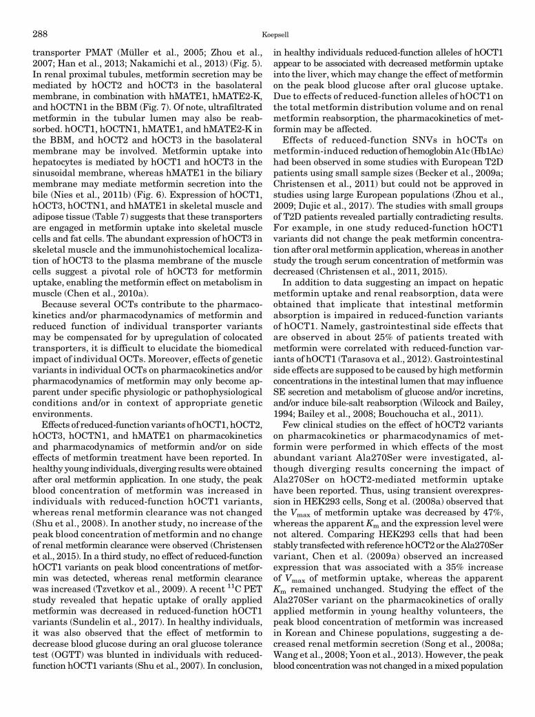
transporter PMAT (Müller et al., 2005; Zhou et al.,2007; Han et al., 2013; Nakamichi et al., 2013) (Fig. 5).In renal proximal tubules, metformin secretion may bemediated by hOCT2 and hOCT3 in the basolateralmembrane, in combination with hMATE1, hMATE2-K,and hOCTN1 in the BBM (Fig. 7). Of note, ultrafiltratedmetformin in the tubular lumen may also be reab-sorbed. hOCT1, hOCTN1, hMATE1, and hMATE2-K inthe BBM, and hOCT2 and hOCT3 in the basolateralmembrane may be involved. Metformin uptake intohepatocytes is mediated by hOCT1 and hOCT3 in thesinusoidal membrane, whereas hMATE1 in the biliarymembrane may mediate metformin secretion into thebile (Nies et al., 2011b) (Fig. 6). Expression of hOCT1,hOCT3, hOCTN1, and hMATE1 in skeletal muscle andadipose tissue (Table 7) suggests that these transportersare engaged in metformin uptake into skeletal musclecells and fat cells. The abundant expression of hOCT3 inskeletal muscle and the immunohistochemical localiza-tion of hOCT3 to the plasma membrane of the musclecells suggest a pivotal role of hOCT3 for metforminuptake, enabling the metformin effect on metabolism inmuscle (Chen et al., 2010a).Because several OCTs contribute to the pharmaco-
kinetics and/or pharmacodynamics of metformin andreduced function of individual transporter variantsmay be compensated for by upregulation of colocatedtransporters, it is difficult to elucidate the biomedicalimpact of individual OCTs. Moreover, effects of geneticvariants in individual OCTs on pharmacokinetics and/orpharmacodynamics of metformin may only become ap-parent under specific physiologic or pathophysiologicalconditions and/or in context of appropriate geneticenvironments.Effects of reduced-function variants of hOCT1, hOCT2,
hOCT3, hOCTN1, and hMATE1 on pharmacokineticsand pharmacodynamics of metformin and/or on sideeffects of metformin treatment have been reported. Inhealthy young individuals, diverging resultswere obtainedafter oral metformin application. In one study, the peakblood concentration of metformin was increased inindividuals with reduced-function hOCT1 variants,whereas renal metformin clearance was not changed(Shu et al., 2008). In another study, no increase of thepeak blood concentration of metformin and no changeof renal metformin clearance were observed (Christensenet al., 2015). In a third study, no effect of reduced-functionhOCT1 variants on peak blood concentrations of metfor-min was detected, whereas renal metformin clearancewas increased (Tzvetkov et al., 2009). A recent 11C PETstudy revealed that hepatic uptake of orally appliedmetformin was decreased in reduced-function hOCT1variants (Sundelin et al., 2017). In healthy individuals,it was also observed that the effect of metformin todecrease blood glucose during an oral glucose tolerancetest (OGTT) was blunted in individuals with reduced-function hOCT1 variants (Shu et al., 2007). In conclusion,
in healthy individuals reduced-function alleles of hOCT1appear to be associated with decreased metformin uptakeinto the liver, which may change the effect of metforminon the peak blood glucose after oral glucose uptake.Due to effects of reduced-function alleles of hOCT1 onthe total metformin distribution volume and on renalmetformin reabsorption, the pharmacokinetics of met-formin may be affected.
Effects of reduced-function SNVs in hOCTs onmetformin-induced reduction of hemoglobinA1c (Hb1Ac)had been observed in some studies with European T2Dpatients using small sample sizes (Becker et al., 2009a;Christensen et al., 2011) but could not be approved instudies using large European populations (Zhou et al.,2009; Dujic et al., 2017). The studies with small groupsof T2D patients revealed partially contradicting results.For example, in one study reduced-function hOCT1variants did not change the peak metformin concentra-tion after oralmetformin application, whereas in anotherstudy the trough serum concentration of metformin wasdecreased (Christensen et al., 2011, 2015).
In addition to data suggesting an impact on hepaticmetformin uptake and renal reabsorption, data wereobtained that implicate that intestinal metforminabsorption is impaired in reduced-function variantsof hOCT1. Namely, gastrointestinal side effects thatare observed in about 25% of patients treated withmetformin were correlated with reduced-function var-iants of hOCT1 (Tarasova et al., 2012). Gastrointestinalside effects are supposed to be caused by highmetforminconcentrations in the intestinal lumen that may influenceSE secretion and metabolism of glucose and/or incretins,and/or induce bile-salt reabsorption (Wilcock and Bailey,1994; Bailey et al., 2008; Bouchoucha et al., 2011).
Few clinical studies on the effect of hOCT2 variantson pharmacokinetics or pharmacodynamics of met-formin were performed in which effects of the mostabundant variant Ala270Ser were investigated, al-though diverging results concerning the impact ofAla270Ser on hOCT2-mediated metformin uptakehave been reported. Thus, using transient overexpres-sion in HEK293 cells, Song et al. (2008a) observed thatthe Vmax of metformin uptake was decreased by 47%,whereas the apparent Km and the expression level werenot altered. Comparing HEK293 cells that had beenstably transfectedwith reference hOCT2or theAla270Servariant, Chen et al. (2009a) observed an increasedexpression that was associated with a 35% increaseof Vmax of metformin uptake, whereas the apparentKm remained unchanged. Studying the effect of theAla270Ser variant on the pharmacokinetics of orallyapplied metformin in young healthy volunteers, thepeak blood concentration of metformin was increasedin Korean and Chinese populations, suggesting a de-creased renal metformin secretion (Song et al., 2008a;Wang et al., 2008; Yoon et al., 2013). However, the peakblood concentrationwasnot changed in amixed population
288 Koepsell

of African and European Americans (Chen et al., 2009a)and in a Caucasian population (Christensen et al., 2013).Consistently, the renal clearance was decreased in theAsian populations (Song et al., 2008a; Wang et al., 2008),only slightly increased in the American populations(Chen et al., 2009a), and not changed in Caucasianpopulations (Tzvetkov et al., 2009; Christensen et al.,2013). In Caucasians, a SNV in the promoter ofhMATE1 (266T.C) that decreased hMATE1 expres-sion influenced the impact of the Ala270Ser variant inhOCT1 on renal metformin clearance (Christensenet al., 2013). In a study including 7968 European T2Dpatients, no effect of Ala270Ser on metformin-induceddecrease of blood Hb1Ac was observed (Dujic et al.,2017). In summary, the most abundant genetic variantin hOCT2 that probably decreases metformin uptakemay influence the pharmacokinetics and pharmacody-namics of metformin, but does not exhibit a relevanteffect on glycemic response to metformin in EuropeanT2D patients.In the amino acid sequence of hOCT3, only very rare
variants were observed. To date, it has not been investi-gated whether the two rare hOCT3 variants (Thr400Ile,Ala423Phe) that reduce the Vmax for metformin uptake(Table 8) (Chen et al., 2010a) and the two SNVs in thehOCT3 promoter that increase transcription (Chen et al.,2013; Kwon et al., 2018) have impact on the pharmacoki-netics and/or pharmacodynamics of metformin. However,it was observed in healthy individuals that a SNV inthe 39-untranslated region that decreased transcription(rs2076828) was associated with a blunted metformin-mediated downregulation of the increase of blood glucoseduring the OGTT (Chen et al., 2015).Although hOCTN1 is located in the BBM of small
intestine and renal proximal tubules (Figs. 5 and 7) andtransports metformin (Nakamichi et al., 2013), theidentified variants of hOCTN1 have not been testedfor metformin uptake (Table 8). In a small Koreanpopulation, in which the Thr306Leu variant of hOCTN1is highly abundant, higher blood peak concentrationsof metformin were observed after oral metformin appli-cation in individuals carrying the Thr306Leu variantcompared with individuals without this variant (Yoonet al., 2013). However, in a large population of EuropeanT2D patients, no impact on metformin-induced reduc-tion of blood Hb1Ac was correlated with the Thr306Leuvariant (Dujic et al., 2017).In hMATE1, six amino acid variants were identified
that lead to reduced metformin transport after ex-pression in cells (Table 8). The highest MAFs of thesevariants were observed for Leu125Phe in Mexicans (0.05)and for Val338Ile in Africans (0.05). Due to low MAFs,only heterozygous reduced-function alleles have beenemployed for clinical testing in a preliminary study onJapanese T2D patients (Toyama et al., 2010). In thisstudy, no effect of reduced function alleles on the oralclearance of metformin was observed.
Impact of a highly frequent SNV in the hMATE1 pro-moter (266T.C) that decreases expression onmetforminpharmacokinetics and pharmacodynamics was tested(Ha Choi et al., 2009; Stocker et al., 2013). In healthyindividuals from a mixed Asian, African, and Europeanpopulation, no effect on metformin pharmacokineticswas detected. However, in healthy individuals and T2Dpatients, a reinforced metformin-mediated reduction ofHbA1 in the blood was observed (Stocker et al., 2013).Studying a large population of metformin-treatedEuropean T2D patients, no correlation between the266T.C variant and blood Hb1Ac was detected (Dujicet al., 2017).
A frequent intronic SNV of hMATE1 (rs2289669) witha MAF of 0.43 in Caucasians has been described thatincreased the therapeutic effect of metformin (Beckeret al., 2009b). In aged T2D patients, presence of this SNVwas correlated with a reinforced metformin-induced de-crease of the Hb1Ac concentration in the blood. This SNVprobably decreases the expression of hMATE1 in hepato-cytes, resulting in an increase of intracellular metformin.Of note, the effect of SNVrs2289669342was only observedin individuals carrying alleles with SNV rs622342, whichdecreases expression of hOCT1 (Becker et al., 2010). Thus,downregulation of hMATE1 in hepatocytes may beonly critical for the therapeutic relevant concentrationof intracellular metformin if hOCT1-mediated metfor-min uptake is moderate. At variance, no correlationbetween the rs2289669 variant and blood Hb1Ac wasobserved in a large population of metformin-treatedEuropean T2D patients (Dujic et al., 2017).
To evaluate the impact of hMATE2-K on the efficacyof metformin treatment, the correlation between alleleswith SNV 2130G.A in the hMATE2-K promoter thatincreases hMATE2-K expression and blood Hb1Ac wasinvestigated in T2D patients treated with metformin(Choi et al., 2011). The frequent promoter polymorphism(MAF of 0.26 in a mixed group of African Americans andCaucasians) that increases hMATE2-K expression wascorrelated with an attenuation of metformin-induceddecrease of Hb1Ac. However, this correlation also wasnot confirmed in an analysis using a large group ofEuropean T2D patients (Dujic et al., 2017).
B. Antineoplastic Treatment with Cisplatin
Since approval by the FDA in 1978, cisplatin (cis-diamminedichloroplatinum) is in clinical use for treat-ment of cancer. Because serious side effects includingnephrotoxicity, neurotoxicity, and ototoxicity (Wangand Lippard, 2005; Pabla and Dong, 2008) limit therapywith cisplatin, additional platinum-based cytostaticssuch as carboplatin, oxaliplatin, picoplatin, and cis-diammine(pyridine) platinum (II) were developed. Car-boplatin and oxaliplatin are in clinical use. They showless nephrotoxicity than cisplatin; however, they havedifferent antitumor specificities (Ardizzoni et al., 2007;Schuler et al., 2010; Ho et al., 2016; Liu et al., 2016a).
Organic Cation Transporters 289

Currently, cisplatin is employed for treatment ofbladder cancer, cervical cancer, ovarian cancer, non-smallcell lung cancer, malignant mesothelioma, squamouscell carcinoma of head and neck, and cancer of testis(Kelland, 2007; Cutler and Choo, 2011; Ishikawa et al.,2014). After treatment of testicular cancer with cisplatin,90% to 95%of patients survive (Raghavan, 2003). Cisplatinis predominantly eliminated by excretion in renal proximaltubules. Nephrotoxicity of cisplatin due to cisplatin enrich-ment in the tubular cells is the main limiter in therapy(dos Santos et al., 2012).In addition to OCTs, the copper transporter CTR1
(SLC31A1) and the P-ATPases ATP7A and ATP7Baccept cisplatin as substrate (Samimi et al., 2004; Larsonet al., 2009). Cisplatin is transported by hOCT1, hOCT2,hMATE1, and hMATE2-K, among which hOCT2 is mosteffective (Table 4) (Ciarimboli et al., 2005b; Yonezawaet al., 2006; Yokoo et al., 2007; Filipski et al., 2008;Sprowl et al., 2013). Whereas the impact of geneticvariants in hOCT1, hMATE1, and hMATE2-K oncisplatin treatment has not been explored, it hasbeen investigated whether the Ala270Ser variantin hOCT2 affects clearance and/or nephrotoxicity ofcisplatin (Filipski et al., 2008, 2009; Iwata et al., 2012).After expression of the hOCT2 variant Ala270Ser incells, transport of TEA, MPP, and propranolol wasdecreased compared with hOCT2 WT type (Table 8).However, a decrease of cisplatin uptake has not beenverified. Due to the relatively low allele abundance ofthe Ala270Ser variant, clinical studies were employedwith heterocygote patients. No significant effect of theAla270Ser variant on total or renal cisplatin clearancewas observed (Filipski et al., 2008); however, data wereobtained, suggesting that the nephrotoxicity of cisplatinis decreased in patients carrying the Ala270Ser variant.Consistently, different from control patients, no decreaseof renal creatinine clearancewas observedwhenpatientswith the Ala270Ser variant were treated with cisplatin(Filipski et al., 2009; Iwata et al., 2012). The datasuggest that hOCT2 is critically involved in cisplatinuptake across the basolateral membrane of renal prox-imal epithelial cells, althoughCTR1 is also located in thismembrane (Pabla et al., 2009).
C. Treatment of Myeloid Leukemia with Imatinib
The tyrosine kinase inhibitor imatinib is used for thefirst-line treatment of CML that is due to the abnormalpresence of a unique fusion gene, termed BCR-ABL1(Goldman and Melo, 2003). In about 80% of patients,imatinib induces complete cytogenic remission (O’Brienet al., 2003; Kantarjian et al., 2004). However, primaryand secondary resistance attenuates or prevents thetherapeutic effect in some of the patients (Marin et al.,2008). In addition to two genetic variants in the kinasedomain of BCR-ABL1 preventing imatinib-mediatedtyrosine kinase inhibition (Kantarjian et al., 2003),imatinib resistance may result from changed functions
of transporters that adjust the imatinib concentrationwithin the myeloma cells. In human acute myelomacell lines, several transporters are expressed that aresupposed to accept imatinib as substrate (Hu et al., 2008;Harrach et al., 2016). Expressing these transporters inoocytes and HEK293 cells, data were obtained thatsuggest that cellular influx of imatinib is mediated bythe human OAT peptides 1A2 and 1B3, by hOCTN1,and by hMATE1, whereas imatinib efflux is mediatedby p-glycoprotein ABCB1 and breast cancer resistanceprotein ABCG2 (Table 4) (Hu et al., 2008; Harrach et al.,2016). Because it was observed that imatinib uptakeinto lymphoblastoid cells and mononuclear cells ofperipheral blood derived from CML patients was re-duced by the hOCT1 inhibitors prazosine and amanta-dine, it was concluded that hOCT1 transports imatinib(Thomas et al., 2004; White et al., 2007). This interpre-tation is probably false because the employed inhibitorswere not specific for hOCT1 (Table 4) and no significanthOCT1-mediated uptake of imatinib was detected whenhOCT1 was expressed in oocytes, epithelial and leuke-mic mammalian cell lines, and/or primary CD341 cellsfrom CML patients (Hu et al., 2008; Nies et al., 2014;Blanc Mettral et al., 2019). In one divergent study,Harrach et al. (2016) observed 2-fold higher cellularimatinib uptake in aHEK293 cell line, which was stablytransfected with hOCT1 than in mock-transfectedHEK293 cells. However, they did not control whetherthe expression of other imatinib transporters was un-affected in this cell line. Various clinical studies revealedthat the abundance of hOCT1-mRNAwas correlated withthe therapeutic effect of imatinib (Crossman et al., 2005;Marin et al., 2010; Nardinelli et al., 2012; Zhong et al.,2012; Gromicho et al., 2013). Nevertheless, becausecorrelationsbetweengeneexpressionofABCB1,SLC22A1,and/orABCG2 have been observed (Gromicho et al., 2013),it is possible that the observed changes in hOCT1 mRNAabundance were associated with changed expression ofanother transporter involved in imatinib uptake or efflux.Because hOCT1 transports endogenous compounds suchas choline, thiamine, and histamine (Table 2), which mayhave an impact on metabolism and/or functional state ofthe myeloma cells, changes in hOCT1 expression mayindirectly influence the therapeutic effect of imatinib.
About 20 clinical studies were perfomed in which theeffects of various polymorphisms in hOCT1, includingvariants Leu160Phe, Met408Val, and Met420del, onclinical outcome after treatment of CML patients withimatinib were investigated. These studies revealedinconsistent, mostly nonreproducible data that do notallow an unambiguous interpretation (Gromicho et al.,2013; Watkins et al., 2015).
D. Analgetic Treatment with Tramadol
The analgetic opoid tramadol has a high passivemembrane permeability and is not transported by hOCT1.In the liver, the active metabolite O-desmethyltramadol
290 Koepsell

is formed by the P450 cytochrome CYP2D6 (Paar et al.,1992). The metabolite has a 10-fold lower passivemembrane permeability than the parental drug andis transported by hOCT1 (Tzvetkov et al., 2011). Aftera single oral dose of tramadol, the increase of the plasmaconcentrations of O-desmethyltramadol was significantlyhigher in individuals containing one or two inactivehOCT1 alleles (Tzvetkov et al., 2011). In parallel,tramadol-induced miosis, a surrogate marker of thecentral analgetic tramadol effect, persisted longer inindividuals with inactive hOCT1 variants (Tzvetkovet al., 2011). It has been hypothesized that tramadolis absorbed in small intestine via passive diffusion andpassively enters the hepatocytes where it ismetabolizedtoO-desmethyltramadol. The bulk ofO-desmethyltramadolis supposed to be released into the blood by a nonidentifiedtransporter but partially re-enters the hepatocytes viahOCT1. In hepatocytes, O-desmethyltramadol may alsobe glucuronidated and excreted into the bile. In brain,hOCT1-mediated transport may be involved in passageof O-desmethyltramadol across the BBB (Fig. 9). Theobserved increase of O-desmethyltramadol in the bloodof individuals with inactive hOCT1 alleles suggests thatthe reuptake of O-desmethyltramadol into hepato-cytes is critical in pharmacokinetics. In anotherstudy, it has been demonstrated that, in addition togenetic variants in CYP2D6, which affect the forma-tion of O-desmethyltramadol (Stamer et al., 2003;Pedersen et al., 2006), reduced-function variants ofhOCT1 are also clinically relevant (Stamer et al.,2016). Employing patient-controlled application oftramadol after surgery, the consumption of tramadolwas measured in patients in relation to CYP2D6 andSLC22A1 genotypes. The tramadol consumption wasnegatively correlated with metabolic active CYP2D6alleles, indicating that poor metabolizers requiredmore tramadol. In addition, tramadol consumptionwas negatively correlated with nonfunctional SLC22A1alleles, suggesting that blunting of uptake of the ac-tive metabolite O-desmethyltramadol into hepatocytesincreases the analgetic effect.
E. Pain-Relieving Treatment with Morphine andAntitussive Treatment with Codeine
In response to treatment of pain with morphine,interindividual and ethnic differences have been de-scribed (Cepeda et al., 2001; Edwards et al., 2005).These differences have been shown to be partially due togenetic variants in hOCT1 that transports morphinewith high affinity and efficacy (Tzvetkov et al., 2013)(Table 4). About 90% of morphine is glucuronidatedby uridine 59-diphosphate glucuronosyltransferase 2B7(Coffman et al., 1997) and excreted in the liver, whereasabout 10% is excreted unchanged in the urine (Osborneet al., 1990; Hasselström and Sawe, 1993). Underclinical conditions, more than 60% of morphine uptakeinto hepatocytes is supposed to occur via hOCT1
(Tzvetkov et al., 2013). In children treated withmorphine for pain relief after tonsillectomy, the abun-dance of nonfunctional hOCT1 alleles was correlatedwith low morphine clearance and high abundance ofmorphine-related adverse effects such as nausea, vom-iting, and respiratory depression (Fukuda et al., 2013;Venkatasubramanian et al., 2014; Balyan et al., 2017).The higher abundance of nonfunctional hOCT1 allelesin the Caucasian versus African American popula-tion provides an explanation why more adverse effectsduring morphine treatment were observed in Caucasianversus African American children (Sadhasivam et al.,2012a,b).
The antitussive drug codeine enters hepatocytesby passive diffusion. In the hepatocytes codeine isdemethylated to morphine by cytochrome P4502D6(CYP2D6) (Dayer et al., 1988). Morphine may be glucur-onidated in the hepatocytes and excreted with the bile ormay leave the hepatocyte by passive diffusion or via a notyet identified transporter across the sinusoidal mem-brane. Morphine in the blood is distributed throughoutthe body and may re-enter the hepatocytes via hOCT1(Tzvetkov et al., 2013). At low morphine plasma con-centrations, hOCT1-mediated morphine uptake largelyexceeds passive diffusion and is critical for the concen-tration of morphine in the blood. In individuals con-taining one or two inactive hOCT1 alleles, the plasmaconcentration of morphine during treatment with codeinewas more than 2-fold than in individuals with two activehOCT1 alleles (Tzvetkov et al., 2013).
F. Treatment of Side Effects of Chemotherapywith Tropisetron
The SE receptor 5-hydroxytryptamine 3 antagonisttropisetron is employed for treatment of nausea andvomiting during chemotherapy of cancer. In the liver,tropisetron is metabolized by CYP2D6, and CYP2D6polymorphisms have been linked to interindividualvariations during antiemetic treatment (Kaiser et al.,2002). Tropisetron is a substrate of hOCT1, and cancerpatients receiving chemotherapy in which vomiting wastreated with tropisetron showed 3-fold less vomitingevents when they contained one or two nonfunctionalSLC22A1 alleles compared with patients in which bothSLC22A1 alleles were functional (Tzvetkov et al., 2012).Consistently, the higher efficacy of the antiemetic ther-apy with tropisetron was correlated with higher concen-trations of tropisetron in the blood.
G. Treatment of Migraine with Sumatriptan
Like other 5-hydroxy tryptamine 1B/DF receptor ago-nists, sumatriptan is employed for treatment of migraine.Sumatriptan has a low oral bioavailability due to a limitedsmall intestinal absorption and a distinctive first-passmetabolism involving monoaminooxidase A in liver,intestine, and some other organs (Grimsby et al., 1990;Dixon et al., 1994; Saura et al., 1996). After intravenous
Organic Cation Transporters 291

application of sumatriptan to healthy individuals withloss of function alleles of hOCT1, a higher blood concen-tration and reduced apparent distribution volume of thedrug were observed compared with individuals withfunctional hOCT1 alleles (Matthaei et al., 2016). Theseeffects are supposed to be due to a reduced hepaticuptake leading to slowed-down metabolism. The datasuggest an increased risk for adverse side effects duringtreatment with sumatriptan in patients with loss-of-function polymorphisms of hOCT1.
H. Treatment of Asthma with Fenoterol
The b2-adrenergic agonist fenoterol is employed invarious countries for treatment of asthma and fortocolysis in obstetrics, although it may cause cardiovas-cular and metabolic side effects. The side effects arecorrelated with high blood concentrations of the drugand include hypokalemia, arrhythmia, hypotension,and hyperglycemia, and may be associated with fataloutcome (Spitzer et al., 1992; Bremner et al., 1993).Because more than 99% of fenoterol in the blood ispositively charged, transporters are required to allowpassage of fenoterol across plasmamembranes (Hochhausand Möllmann, 1992). Fenoterol is metabolized by sulfa-tion and glucuronidation in the liver and mostly elimi-nated as metabolite (Hochhaus and Möllmann, 1992).Fenoterol is transported by hOCT1 and hOCT3 withapparent Km values of 1.8 and 20 mM, respectively(Tzvetkov et al., 2018). The lowerKm value and higherabundance of hOCT1 in the sinusoidal membrane ofhepatocytes strongly suggest that fenoterol uptakeinto hepatocytes is mainly mediated by hOCT1. Inaccordance with this notion, after intravenous appli-cation of fenoterol in healthy individuals containingnonfunctional hOCT1 alleles, higher blood concentra-tions were observed compared with individuals withtwo functional hOCT1 alleles (Tzvetkov et al., 2018). Inaddition, side effects of fenoterol treatment such ashigher heart rate, drop in diastolic blood pressure, andincrease of blood glucose correlated with the abun-dance of nonfunctional hOCT1 alleles. The data sug-gest that patients with nonfunctional hOCT1 variantsare a risk group for adverse effects during treatmentwith fenoterol.
I. Treatment of Malaria with Proguanil
Proguanil is a prodrug that is employed for prophylaxisand treatment of malaria (Wattanagoon et al., 1987).In the blood, more than 99% of proguanil is positivelycharged. Proguanil is transported into hepatocytes viahOCT1 and to a small extent by hOCT3 (Matthaei et al.,2019). In hepatocytes, the active metabolite cycloguanilis formed by cytochrome P4502C19 (Coller et al., 1997;Hoskins et al., 1998). Cycloguanil attacks pre-erythrocyticmalaria parasite forms in the liver and malaria para-sites within erythrocytes (Delves et al., 2012). Thetransporter(s) mediating efflux of positively charged
cycloguanil from hepatocytes has (have) not beenidentified. Cycloguanil–proguanil exchange via hOCT1may be involved. In healthy individuals treated withproguanil, the concentration of cycloguanil and the cyclo-guanil/proguanil ratio in the blood was correlated withfunctionality of hOCT1 alleles, whereas no correlationwas observed for the blood concentration of proguanil(Matthaei et al., 2019). These data suggest that theformation of cycloguanil is dependent on hOCT1-mediated influx, that hepatic efflux of cycloguanil mayoccur by hOCT1-mediated cycloguanil–proguanil antiport,and that polymorphisms in hOCT1may be one reason forthe failure of proguanil treatment in patients (Kanekoet al., 1999).
X. Drug–Drug Interactions of Substrates andInhibitors at Organic Cation Transporters
A. General Considerations
Clinical tests in healthy and diseased individualsrepresent the only reliable method to evaluate interac-tion of drugs that are substrates and/or inhibitors ofOCTs in humans. The reasons are that cellular testsystems do not represent the complexity of in vivointeractions and that animal models do not mimichuman features sufficiently. This includes differencesin properties, expression and regulation of OCTs, andtransporters from other families that transport organiccations. The interpretation of clinically observed drug–drug interactions requires a detailed pharmacokineticcharacterization of the involved drugs, an establishedunderstanding of transporter expression and functionincluding disease-related changes, and a well-foundedknowledge concerning interactions of the respectivedrugs with individual transporters. To select drugs forclinical testing of drug–drug interactions, knowledgeconcerning functions of OCTs in different organs, abouttransported substrates, and aboutKm and IC50 values isrequired.
Reflecting on in vivo drug–drug interactions at thelevel of OCTs, interaction of drugs at one OCT type inone location, interaction of drugs at one OCT type indifferent locations, and interaction of drugs at differentOCT types operating in concert, must be considered.Although transport of many drugs by hOCTs (Table 4)and inhibition of hOCTs by many drugs have beeninvestigated (Tables 4 and 9), the anticipation of anin vivo interaction of two drugs at one OCT at a specificlocation remains uncertain, rendering the anticipationof effects of in vivo interactions of drugs with OCTs indifferent locations highly speculative. One reason is thatthe IC50 values that have been determined for inhibitionof OCTs by drugs only reflect individual experimentalconditions. Thus, the IC50 values of OCTs are not onlydependent on the structure of the employed substratebut also on the substrate concentration (Gorboulev et al.,2018), and most available IC50 values have not been
292 Koepsell

TABLE
9Dru
gsthat
hav
ebe
ensh
ownto
inhibithu
man
orga
niccation
tran
sporters
andha
veno
tbe
entested
fortran
sportor
couldno
tbe
iden
tified
assu
bstrate(onlydr
ugsforwhich
anIC
50va
lue,20
mM
was
determ
ined
forat
leas
ton
eOCT
areindicated)
Charge
ofmajor
microsp
eciesat
pH7.4was
calculatedwithpr
ogram
MarvinSke
tchof
Chem
Axo
n.A
bac
avir:Minuesaet
al.,20
09,a
bem
aciclib:Cha
ppellet
al.,20
19,a
dap
alen
e:Kidoet
al.,20
11,a
lfuzo
sin:Che
net
al.,
2017
a,am
itriptyline:Sataet
al.,20
05;A
hlinet
al.,20
08;Z
olket
al.,20
09b;
Belzeret
al.,20
13;T
zvetko
vet
al.,20
13;J
ouan
etal.,20
14;H
acke
ret
al.,20
15;M
atthae
ietal.,20
16;S
ando
vale
tal.,20
18;Z
huet
al.,20
18,a
mad
iaquin
e:va
nde
rVelde
net
al.,20
17,D
-amphetam
ine:
Amph
ouxet
al.,20
06,a
msa
crin
e:Ahlinet
al.,20
08,a
rtem
isin
:Hub
enyet
al.,20
16,a
tomox
etin
e:San
dova
letal.,20
18,a
zidot
hym
idin
e:Minuesaet
al.,20
09,b
eclomethas
one:
Lipset
al.,20
05,b
enze
thon
ium:K
idoet
al.,20
11,b
enztro
pin
e:San
dova
let
al.,20
18,b
ithionol:K
idoet
al.,20
11;W
ittw
eret
al.,20
13;C
hen
etal.,20
17a,
bosu
tinib:J
ohnston
etal.,20
14,b
ucindolol:A
hlinet
al.,20
08,2
011,
budes
onid
e:Lipset
al.,20
05,b
uflom
edil:K
idoet
al.,20
11;H
endr
ickx
etal.,20
13,b
usp
iron
e:Wittw
eret
al.,20
13,S
ando
vale
tal.,20
18,b
isop
rolol:Bachmak
ovet
al.,20
09,c
amylofin
e:Chen
etal.,20
17a,
carb
etap
enta
ne:
Chen
etal.,20
17a,
carv
edilol:B
achmak
ovet
al.,20
09;Z
olket
al.,20
09b;
Gru
beet
al.,20
11;B
elzeret
al.,20
13;Y
inet
al.,20
16;C
hen
etal.,20
17a,
clem
astine:
Ahlin
etal.,20
08,c
lomac
ran:K
idoet
al.,20
11,c
hlorh
exid
ine:
Wittw
eret
al.,20
13,c
ediran
ib:J
ohns
tonet
al.,20
14,c
hlorp
hen
esin
:Kidoet
al.,20
11,c
hlorp
romaz
ine:
Bed
narczyk
etal.,20
03;A
hlin
etal.,20
08,2
011;
Zolket
al.,20
09a;
Belzeret
al.,20
13,c
italop
ram:A
hlinet
al.,20
08;
Koe
pselle
tal.,unpu
blishe
dda
ta,c
lomip
ramin
e:Ahlinet
al.,20
08;H
endr
ickx
etal.,20
13,c
lonid
ine:
Zhan
get
al.,19
98;W
uet
al.,20
00;M
üller
etal.,20
05;S
uhre
etal.,20
05;A
hlinet
al.,20
08;Z
olket
al.,20
09a;
Astorga
etal.,
2012
;Belzeret
al.,20
13;H
endr
ickx
etal.,20
13;T
zvetko
vet
al.,20
13;C
hen
etal.,20
17a,
clos
antel:Chen
etal.,20
17a,
clot
rimaz
ole:
Chen
etal.,20
17a,
cloz
apin
e:Hen
drickx
etal.,20
13;W
anget
al.,20
19,c
odeine:
Tzv
etko
vet
al.,20
13,d
arunav
ir:M
osset
al.,20
15,d
asat
inib
:Minem
atsu
andGiacomini,20
11,d
es-flu
oro(6)-quin
olon
e:Oku
daet
al.,20
06;Imam
ura
etal.,20
11;d
extrom
ethor
phan
:Che
net
al.,20
17a,
dichloro
phen
e:Chen
etal.,
2017
a,des
ipra
min
e:Gorbo
ulev
etal.,19
97;Z
han
get
al.,19
98;W
uet
al.,20
00;A
hlinet
al.,20
08,2
011;
Tsu
daet
al.,20
09b;
Zolket
al.,20
09b;
Chen
etal.,20
17a,
des
lora
tadin
e:Zolket
al.,20
09b,
dicyc
lomin
e:Kidoet
al.,20
11,
dip
yram
idole:
Kidoet
al.,20
11,dob
uta
min
e:Chen
etal.,20
17a,
dolutegr
avir:Ree
seet
al.,20
13;Lep
istet
al.,20
14,dom
per
idon
e:Wittw
eret
al.,20
13,don
epez
il:Martínez-G
uerrero
etal.,20
16;Matthae
iet
al.,20
16,
des
lora
tadin
e:Zolket
al.,20
09b,
dih
ydro
ergo
tamin
e:Wittw
eret
al.,20
13,d
oxaz
osin
:Ahlinet
al.,20
11,d
oxep
in:Z
olket
al.,20
09b;
Belzeret
al.,20
13;H
acke
ret
al.,20
15;C
hen
etal.,20
17a,
eletriptan
:Matthae
ietal.,20
16,
epin
astine:
Wittw
eret
al.,20
13,er
lotinib
:Minem
atsu
and
Giacomini,
2011
;Chen
etal.,20
17a,
esmolol:Martínez-G
uerreroet
al.,20
16,ethin
yles
trad
iol:
Wittw
eret
al.,20
13,ethylac
ridin
ium:Belzeret
al.,20
13,
exem
esta
ne:
Kidoet
al.,20
11,fen
flura
min
e:Zolket
al.,20
09a,
flec
ainid
e:Umeh
araet
al.,20
08;Z
olket
al.,20
09b;
Gru
beet
al.,20
11;B
elzeret
al.,20
13,flu
raze
pam
:Zolket
al.,20
09a,b,
gabex
ate:
Kidoet
al.,20
11;W
ittw
eret
al.,20
13,g
efitin
ib:G
alettiet
al.,20
10;M
inem
atsu
andGiacomini,20
11,g
entian
violet:K
idoet
al.,20
11,g
ranisetro
n:W
ittw
eret
al.,20
13,g
rise
ofulvin
:Chen
etal.,20
17a,
guan
aben
z:Kidoet
al.,20
11;C
hen
etal.,20
17a,
guan
facine:
Astorga
etal.,20
12,imip
ramin
e:Wuet
al.,20
00;A
hlinet
al.,20
08;T
suda
etal.,20
09b;
Zolket
al.,20
09b;
Kidoet
al.,20
11;B
elzeret
al.,20
13;H
endr
ickx
etal.,20
13;T
zvetko
vet
al.,20
13;S
ando
vale
tal.,20
18;Z
huet
al.,20
18,imiq
uim
od:A
storga
etal.,20
12,indin
avir:Z
han
get
al.,20
00;J
unget
al.,20
08;W
ittw
eret
al.,20
13,irinotecan
:Tzv
etko
vet
al.,20
13;W
ittw
eret
al.,20
13;S
ando
vale
tal.,20
18;Z
huet
al.,20
18,jat
rorr
hizin
e:Sun
etal.,20
19,ketoc
onaz
ole:
Ahlin
etal.,20
11;Astorga
etal.,20
12;Chen
etal.,20
17a,
ketot
ifen
:Martínez-G
uerrero
etal.,20
16,lanso
pra
zole:Nieset
al.,20
11a;
Hacke
ret
al.,20
15,leukom
ycin
:Wittw
eret
al.,20
13,
meb
ever
ine:
Kidoet
al.,20
11,3,4-methylen
edioxy
metham
phetam
ine:
Amph
ouxet
al.,20
06,mex
iletin
e:Zo
lket
al.,20
09b;
San
dova
let
al.,20
18,mefloquin
e:Hube
nyet
al.,20
16,mid
azolam
:Zha
nget
al.,19
98,
mitox
antron
e:Mey
erzu
Sch
wab
edissenet
al.,20
10;W
ittw
eret
al.,20
13,n
elfinav
ir:Z
han
get
al.,20
00;J
unget
al.,20
08,n
ifek
alan
t:Wittw
eret
al.,20
13,n
ilot
inib
:Minem
atsu
andGiacomini,20
11,o
ndan
setron
:Kidoet
al.,
2011
;Tzv
etko
vet
al.,20
12,2
013;
Wittw
eret
al.,20
13;L
echn
eret
al.,20
16;Z
huet
al.,20
18,o
xaliplatin:Y
onezaw
aet
al.,20
06;Y
okoo
etal.,20
07,2
008;
Ahlinet
al.,20
08,2
011;
Jonget
al.,20
11;S
prow
letal.,20
13,o
lanza
pin
e:Kido
etal.,
2011
,om
epra
zole:Nieset
al.,
2011
a;Martínez-G
uerrero
etal.,
2016
,or
phen
adrine:
Ahlin
etal.,
2008
;Kido
etal.,
2011
,pan
topra
zole:Nieset
al.,
2011
a;Wittw
eret
al.,
2013
;Hacke
ret
al.,
2015
,phen
oxyb
enza
min
e:Hay
er-Zillgen
etal.,20
02;Ahlinet
al.,20
08,phen
toloam
ine:
Astorga
etal.,20
12,phen
ylto
loxa
min
e:Kidoet
al.,20
11,phen
ytoin:Has
annejad
etal.,20
04,pro
chlorp
eraz
ine:
Ahlinet
al.,20
08;
Hen
drickx
etal.,20
13;M
artínez-G
uerrero
etal.,20
16,p
rocy
clid
ine:
Kidoet
al.,20
11,p
rogu
anil:A
storga
etal.,20
12;v
ande
rVelde
net
al.,20
17,p
romaz
ine:
Ahlinet
al.,20
08,p
ropafen
one:
Ahlin
etal.,20
08,2
011;
Zolket
al.,
2009
b;Gru
beet
al.,20
11;B
elzeret
al.,20
13;H
endr
ickx
etal.,20
13;C
hen
etal.,20
17a,
pro
pan
thelin
ebro
mid
e:Kidoet
al.,20
11,p
yrim
etham
ine:
Matsu
shim
aet
al.,20
09;T
suda
etal.,20
09b;
Astorga
etal.,20
12;H
ube
nyet
al.,
2016
;Lechner
etal.,20
16;Y
inet
al.,20
16;C
hen
etal.,20
17a;
vande
rVelde
net
al.,20
17,q
uetiapin
e:Wan
get
al.,20
19,r
abep
razo
le:K
idoet
al.,20
11;N
ieset
al.,20
11a,
repag
linid
e:Ahlinet
al.,20
08;B
achmak
ovet
al.,20
08,
rapam
ycin
:Mey
erzu
Sch
wab
edissenet
al.,20
10,r
iman
tadin
e:Wittw
eret
al.,20
13,r
iton
avir:Z
han
get
al.,20
00;J
unget
al.,20
08;M
eyer
zuSch
wab
edissenet
al.,20
10;W
ittw
eret
al.,20
13;L
epistet
al.,20
14;K
ikuch
ietal.,
2019
,ro
siglitaz
one:
Bachm
akov
etal.,20
08,sa
quin
avir:Zhan
get
al.,20
00;Ju
nget
al.,20
08,sc
opolam
ine:
Hen
drickx
etal.,20
13;Chen
etal.,20
17b,
sita
glip
tin:Hacke
ret
al.,20
15,sp
artein
e:Kidoet
al.,20
11,
spiron
olac
tone:
Gru
beet
al.,20
06,2
011;
Ahlinet
al.,20
11;H
acke
ret
al.,20
15,s
ulcon
azole:
Kidoet
al.,20
11,s
unitin
ib:M
inem
atsu
andGiacomini,20
11;C
henet
al.,20
17a,
tacrine:
Kidoet
al.,20
11;A
storga
etal.,20
12;C
hen
etal.,20
17a;
San
dova
let
al.,20
18;tenat
opra
zole:Nieset
al.,20
11a,
tenofov
irdisop
roxil:Minuesaet
al.,20
09;Shen
etal.,20
17,irtetra
chloro
salicy
lanilid
e:Kidoet
al.,20
11,t
oltero
din
e:Kidoet
al.,20
11,tram
adol:
Astorga
etal.,20
12,trim
ipra
min
e:Ahlin
etal.,20
08;Hacke
ret
al.,20
15,trip
ilen
amin
e:San
dova
let
al.,20
18,tu
boc
ura
rine:
Wittw
eret
al.,20
13,va
lspod
ar:Lechner
etal.,20
16,ve
curo
niu
m:Wittw
eret
al.,20
13,
zafirlukas
t:Wittw
eret
al.,20
13,z
ipra
sidon
e:Wan
get
al.,20
19,z
olpid
em:Hacke
ret
al.,20
15.
Dru
g(C
harge
ofMajor
Microsp
ecies
atpH
7.4)
Med
ical
App
lication
IC50[mM]
hOCT1
hOCT2
hOCT3
hOCTN1
hOCTN2
hMATE1
hMATE2-K
Aba
cavir(0)an
tiviral
7�
1025
4�
1025
5�
1025
Abe
maciclib(1
)an
tineoplas
tic
1.5
0.52
0.75
Ada
palene(2
)trea
tmen
tof
acne
,1
Alfuzosin(1
)an
tipr
ostate
hyp
erplas
ia15
Amod
iaqu
ine(1
)an
timalarial
11Amsa
crine(1
)an
tine
oplastic
5Amitriptyline(1
)an
tide
pressa
nt1.9–
580.4–
14.10
0Artem
isin
(0)an
timalarial
4.2
Atomox
etine(1
)trea
tmen
tof
ADHS
3.5–
20Atrop
ine(1
)myd
riatic
1.2,
121.3–
2944
65.9a
53a
Azido
thym
idine(1
/2)an
tiviral
0.00
020.00
030.00
04Beclometha
sone(0)an
tias
thmatic
4.4
Ben
zethon
ium
(1)an
tine
oplastic
,1
Ben
ztropine(1
)an
ti-Parkinson
0.3–
13Bithionol
(2)an
thelmintic
2.2
1.9,
,1
5.2a
6.6a
Buc
indo
lol(1
)va
sodilatory
15,2
7Bud
eson
ide(0)an
tias
thmatic
7.3
Buflom
edil(1
)va
sodilatory
n.t.d.
,1,
n.t.d.
Busp
iron
e(1
)an
xiolytic
8.6–
691.7a
46a
Bisop
rolol(1
)an
tihy
perten
sive
2.4
Bosutinib(0)an
tich
ronicmye
loic
leuk
emia
2.1
Cam
ylofine(1
)mus
clerelaxa
nt
9.1
Carbe
tape
ntane
(1)an
titussive
1.6
(con
tinued
)
Organic Cation Transporters 293

TABLE
9—Con
tinued
Dru
g(C
harge
ofMajor
Microsp
ecies
atpH
7.4)
Med
ical
App
lication
IC50[mM]
hOCT1
hOCT2
hOCT3
hOCTN1
hOCTN2
hMATE1
hMATE2-K
Carve
dilol(1
)an
tihyp
ertens
ive
1.4,
3.4
2.3–
6374
7312
4,a14
6a79
,a13
1a
Ced
iran
ib(1
)an
tine
oplastic
2.5
Chlorh
exidine(1
1)an
tiseptic
0.4
0.7a
0.5a
Chlorph
enesin
(0)mus
clerelaxa
nt
,1
Chlorpr
omaz
ine(1
)an
tips
ycho
tic
2.6–
522.6,
14Cloza
pine(1
)an
tide
pressa
nt1.8
Citalop
ram
(1)an
tide
pressa
nt
3,19
1214
5Clemas
tine(1
)an
tiallergic
4.9
Clomacran(1
)seda
tive
andhyp
notic
,1
Clomipramine(1
)an
tide
pressa
nt
5,19
,n.t.d.
n.t.d.
Clonidine(1
)seda
tive
0.6–
23,n.t.d.
2–63
,n.t.d.
110,
373
8.1a
54a
Clope
rastine(1
)an
titussive
15Closa
ntel(2
)an
tipa
rasitc
3Clotrim
azole(0)an
tifung
al12
Cloza
pine(1
)an
tips
ychotic
1.8,
n.t.d.
Cod
eine(1
)an
titussive,
analge
sic
11Daruna
vir(0)an
tiviral
16Das
atinib
(1)an
tineoplas
tic
1.1
2.1
4.5
0.8a
0.8a
Des-fluoro(6)-qu
inolon
e(c.n.d.)an
tibiotic
0.9,
1.3
0.8a
0.1a
Dex
trom
ethorph
an(1
)an
titussive
10Dichloroph
ene(2
)an
tifunga
l8.4
Dicyclomine(1
)an
tisp
asmod
ic,1
Dipyram
idole(0)an
tithrombo
tic
812.6
2674
Don
epezil(1
)an
ti-A
lzhe
imer
0.8a–1.5a
Desipramine(1
)an
tide
pressa
nt5–
5716
1456
a28
3a
Dihyd
roergo
tamine(1
)an
timigraine
502.8a
13a
Dob
utamine(1
)trea
tmen
tof
hea
rtfailur
e4.2
Doluteg
ravir(0)an
tiviral
0.07
,1.9
.10
04.7a
.10
0a
Dom
perido
ne(0)an
temetic
337.9
2.3a
15a
Dox
azosin
(1)an
tihy
perten
sive
15Dox
epin
(1)an
tide
pressa
nt
1.5,
120.25
–13
Eletriptan(0)an
timigraine
6.8
Epina
stine(1
)an
tiallergic
,1,
4.3
1.1a
30a
Erlotinib
(0)an
tineoplas
tic
0.34
,16
5.2
4.2
3.8,
a7.9a
3.5,
a6.2a
Esm
olol
(1)trea
tmen
tof
tach
ycardia
11a–25
a
Ethinylestrad
iol(0)contracep
tive
2.2
21a
20a
Ethylacridinium
(1)an
tiseptic
0.08
Exe
mestane
(0)trea
tmen
tof
brea
stcanc
er,1
Fen
fluramine(1
)ap
petite
supp
ressan
t10
Flecainide(1
)an
tiarrh
ythm
ic2.5,
4219
1,23
460
176
Flurazepa
m(1
)an
ticonvu
lsan
t53
,60
Gab
exate(1
)an
ticoag
ulan
t0.9
0.5a–0.70
a11
a
Gefitinib
(0)an
tine
oplastic
1.1
245.5
1.8a
0.19
a
Gen
tian
violet
(1)fung
icidal
,1
Granisetron
(1)an
tiem
etic
4.3
5.0a
311a
Griseofulvin(0)an
timycotic
7.3
Guan
aben
z(1
)an
tihy
perten
sive
4.9
,1
Guan
facine(1
)an
tihyp
ertensive
3.5a
218a
Imipramine(1
)an
tide
pressa
nt6–
37,n
.t.d.
0.4–
6,n.t.d.
4242
,a10
183a
Imiquim
od(0)im
mune
resp
onse
mod
ifier
14a
19a
Indinav
ir(0)an
tiviral
37–20
814
2,27
5.50
07.8a
.50
0a
(con
tinued
)
294 Koepsell

TABLE
9—Con
tinued
Dru
g(C
harge
ofMajor
Microsp
ecies
atpH
7.4)
Med
ical
App
lication
IC50[mM]
hOCT1
hOCT2
hOCT3
hOCTN1
hOCTN2
hMATE1
hMATE2-K
Irinotecan
(1)an
tine
oplastic
1.4–
210.31
–16
757.9a
79a
Jatrorrh
izine(1
)pr
omotingGImotility
0.12
,0.82
0.18
,0.28
Ketocon
azole(0)an
tifunga
l2.6,
7.4
1.3a
9.3a
Ketotifen
(1)an
tiallergic
13a–27
a
Lan
sopr
azole(0)an
tacid
363.3–
9.5
3.1
Leu
komycin
(12)an
tibiotic
182
11a
125a
Meb
everine(1
)an
tisp
asmod
ic,1
Mefloqu
ine(1
)an
timalarial
6.6
153,4-Methy
lened
ioxy
metha
mph
etam
ine(1
)stim
ulant
241.6
74Mex
iletine(1
)an
tiarrh
ythmic
19–50
Midaz
olam
(0)hy
pnotic,an
esthetic
3.7
Mitox
antron
e(1
1)an
tine
oplastic
4473
610.5a;4.4;
5.2
0.83
a
Nelfinav
ir(1
)an
tiviral
7,22
13Nifek
alan
t(1
12)an
tiarrh
ythmic
.30
010
.314
66.5a
2.7a
Nilotinib
(0)an
tineoplas
tic
2.9
.30
0.35
3.4a
1.8a
Onda
nsetron
(1)an
tiem
etic
0.3–
640.2–
161.7,
170.04
a–4.0a,0.15
0.03
a–1.7,
a6.9
Olanz
apine(1
)an
tips
ychotic
,1
Omep
razole
(0)an
tacid
166.7
2216
–24
a
Orp
hen
adrine(1
)muscle
relaxa
nt
13,.
100
2.5
65.10
0Pan
topr
azole(0)trea
tmen
tof
gastriculcera
31,.50
00.56
–2.8
23,1
3743
a.50
0a
Phen
oxyb
enza
mine(1
)an
tihyp
ertensive
2.7,
155
6Phen
tolamine(1
)va
sodilatory
4.6a
5.3a
Phen
yltoloxa
mine(1
)en
hancerof
analge
tics
,1
Phen
ytoin(0)an
ticonvu
lsan
t0.75
Proch
lorp
eraz
ine(1
)an
tips
ycho
tic
50,n
.t.d.
n.t.d.
7.9a–15
a
Procyclidine(1
)an
ti-Parkins
on,0.1
Progu
anil(1
)an
timalarial
134.4a
1.4a
Promaz
ine(1
)an
tips
ychotic
17Propa
fenon
e(1
)an
tiarrh
ythmic
1–16
,n.t.d.
3.6,
25,n.t.d.
68Propa
nthe
line(1
)trea
tmen
tof
hyp
erhidrosis
,1
Pyrim
etham
ine(1
)an
timalarial
8.5,
141.6–
230.04
a–1.2a,0.08
0.01
a–0.83
,a0.06
Que
tiap
ine(0)an
tide
pressa
nt13
Rab
eprazole
(0)an
tacid
3.0
,1,
5.7
3.0
Rep
aglinide(0)an
tidiab
etic
1.6,
9.2
Rap
amycin
(0)im
munesu
ppressive
3.3,
3.5
Rim
antadine(1
)an
tiviral
4.4
7.3a
288a
Riton
avir
(0)an
tiviral
5–34
20,25
.10
01.3a–4.4a
14,1
524
,a90
a
Rosiglitazone(2
)an
tidiab
etic
6.9
Saq
uinav
ir(1
)an
tiviral
8,37
205
Scopo
lamine(0)an
tiem
etic
6.7,
n.t.d.
541,
n.t.d.
218
119a
700a
Sitag
liptin
(1)an
tidiab
etic
7.4
Spa
rteine(1
1)an
ticonvu
lsan
t,an
tiarrh
ythm
ic,1
Spirono
lacton
e(0)an
tihy
perten
sive
1.2,
31.3
7312
526
,36
Sulconaz
ole(0)an
tifunga
l,1
Sun
itinib
(1)an
tine
oplastic
0.33
,6.1
1.7
5.2
0.28
a0.86
Tacrine(1
)an
ti-A
lzhe
imer
22,8
30.4–
2.3
0.5a,1
.11.1,
a.10
0Ten
atop
razole
(0)an
tacid
2320
15Ten
ofov
irdisopo
xil(0)an
tiviral
9�
1025,.10
6�
1025
5�
1027
Tetrach
lorosa
licylanilide(2
)an
tiseptic
,1
Tolterodine(1
)sp
asmolyticum
,1
Tramad
ol(1
)18
a75
a
(con
tinued
)
Organic Cation Transporters 295

determined using appropriate drugs at clinically rele-vant concentrations as substrates. In addition, the IC50
values for OCTs mediating cellular export such ashMATE1, hMATE2-K, and hOCTN1have been determinedby measuring uptake rather than efflux. Another reasonfor the insecurity of in vivo predictions is that, at varianceto drug concentrations in systemic and portal blood,intracellular drug concentrations and drug concentra-tions in the lumen of renal proximal tubules are notknown. Intracellular drugs may induce trans-stimulationif they arewell-transported substrates, or trans-inhibitionif they are nontransported inhibitors that have enteredthe cells by an alternative transporter. Ultrafiltratedcationic drugs within the lumen of renal proximaltubules that may have been concentrated during waterreabsorption may inhibit transporters in the BBM thatmediate cellular efflux of secreted drugs such as hMATE1,hMATE2-K, or hOCTN1. Drugs in the tubular lumenmaybe reabsorbed and increase the concentration of therespective drug within tubular cells.
Anticipating potential drug–drug interactions at OCTs,transport inhibition of drugs that are present at concen-trations close to their respective Km values and inhibitionof drugs with concentrations far below their respective Km
values must be distinguished. In the first case, inhibitionis mediated by drug interaction at low-affinity sites thatare probably directly involved in transport, whereas inthe second case allosteric inhibition by interaction ofinhibitors with high-affinity cation binding sites maybe observed. The clinically approved drug–drug inter-actions at the level of OCTs reflect interactions at thelow-affinity sites.
B. Interactions of Drugs during Treatment of Type 2Diabetes with Metformin
hOCT1–3, hOCTN1, hMATE1, and hMATE2-K ac-cept metformin as substrate and are supposed to beinvolved in intestinal metformin absorption, metforminuptake into hepatocytes, muscle cells and fat cells, and/or in renal metformin secretion (Figs. 5–7; Tables 4 and6). The critical role(s) of hOCT1, hOCT2, hMATE1, and/or hMATE2-K for pharmacokinetics and/or pharmaco-dynamics of metformin has (have) been supported byclinical studies in healthy individuals and/or T2Dpatientswith reduced-function genetic variants (see Treatmentof Type 2 Diabetes with Metformin). In this context, itis not surprising that coadministration of metforminwith drugs that are transported by OCTs and/or inhibitOCTs changed the metformin concentration in theblood, renal metformin excretion, and/or the therapeu-tic effect of metformin on diabetes (Wang et al., 2008;Kusuhara et al., 2011; Cho et al., 2014; Dujic et al.,2015). Thus, coadministration of metformin with cit-alopram, verapamil, or codeine representing high-affinity inhibitors of individual OCTs (Tables 4 and 9)to T2D patients was correlated with metformin intoler-ance (Dujic et al., 2015). In case of verapamil, metformin
TABLE
9—Con
tinued
Dru
g(C
harge
ofMajor
Microsp
ecies
atpH
7.4)
Med
ical
App
lication
IC50[mM]
hOCT1
hOCT2
hOCT3
hOCTN1
hOCTN2
hMATE1
hMATE2-K
Trimipramine(1
)an
tide
pressa
nt28
0.44
Tripe
lenam
ine(1
)an
tiallergic
2.1–
12Tubo
curarine(1
1)mus
clerelaxa
nt
799.4a
56a
Valsp
odar
(0),an
tidr
ugresistan
t2.0a–.15
a0.11
a–0.24
a
Vecuronium
(11)muscle
relaxa
nt
3.5
1.9a
25a
Zafirluk
ast(2
)an
tias
thmatic
9.7
4.3a
7.6a
Ziprasido
ne(0)an
tide
pressa
nt
3.6
Zolpide
m(0)seda
tive
,an
ticonv
ulsan
t0.15
c.n.d.,ch
arge
not
determ
ined
;GI,
gastrointestinal;n
.t.d.,notran
sportde
tected
unde
rtheem
ploy
edex
perimen
talcondition
s.aUptak
emea
suremen
tspe
rformed
inpr
esen
ceof
anou
twardlydirected
proton
grad
ient.
296 Koepsell

intolerance is supposed to be induced by inhibition ofhOCT1-mediated metformin uptake into hepatocytesbecause coadministration of verapamil with metforminblunted the glucose-lowering effect of metformin withoutchanging either themetformin concentration in the bloodor renal metformin clearance (Cho et al., 2014).Coadministration of cimetidine with metformin de-
creased renal metformin clearance, suggesting clini-cally relevant interactions of cimetidine with OCTs inrenal proximal tubules that mediate metformin secre-tion (Somogyi et al., 1987; Wang et al., 2008). Cimet-idine has been identified as substrate of hOCT1,hOCT2, hMATE1, and hMATE2-K and as inhibitorof hOCT3, hOCTN1, and hOCTN2 (Table 4). Becauseno cimetidine effect on renal metformin clearance wasobserved in patients containing the Ala270Ser poly-morphism in hOCT2 that decreases translocation ofsome substrates (Table 8), it has been suggested thata cimetidine–metformin interaction at hOCT2 maybe responsible for the observed cimetidine-induceddecrease of metformin clearance (Wang et al., 2008).However, this interpretation may be false because itwas observed later on that metformin transport mediatedby hMATE1 and hMATE2-K rather than by hOCT2 canbe inhibited at clinical relevant cimetidine concentrationsin the blood (Tsuda et al., 2009b; Ito et al., 2012b).A decrease of renal metformin secretion was also
observed after coadministration of pyrimethamine,which is used to treat protozoal infections (Kusuharaet al., 2011). In this case, the metformin concentra-tion in the blood was slightly increased. It has beenreported that pyrimethamine inhibits transportmediatedby hOCT1, hOCT2, hMATE1, and hMATE2-K (Table 9).Because the IC50 values determined for inhibition ofhMATE1 or hMATE2-K by pyrimethamine were morethan 100-fold lower compared with the IC50 valuesobtained for inhibition of hOCT2, and the mean bloodconcentration of unbound pyrimethamine duringtreatment is more than 10-fold lower than the IC50
value for inhibition of hOCT2, pyrimethamine probablydecreases metformin secretion by inhibition of hMATE1and hMATE2-K similar to cimetidine.
C. Interactions of Drugs during Treatment of Cancerwith Cisplatin
The clinical use of cisplatin during treatment ofmalignancies is limited by nephrotoxicity (dos Santoset al., 2012). It has been shown that cisplatin is trans-ported by hOCT1, hOCT2, hMATE1, and hMATE2-K(Table 4). Because hOCT2 transports cisplatin with highefficacy and patients carrying hOCT2 alleles coding foran amino acid exchange affecting translocation of sev-eral cations exhibited a blunted cisplatin-induced neph-rotoxicity, hOCT2 is supposed to be critically involvedin renal cisplatin secretion (Filipski et al., 2009; Iwataet al., 2012). Hence, coadministration of drugs thatinhibit hOCT2-mediated uptake of cisplatin across the
basolateral membrane of renal tubular cells representsan attractive strategy to prevent cisplatin-induced neph-rotoxicity. However, because hMATE1 and/or hMATE2-K may be critical for cellular efflux of cisplatin acrossthe BBM, drugs inhibiting hOCT2 that also inhibithMATE1 and/or hMATE2-K may increase the intracellu-lar cisplatin concentration and thereby promote cisplatin-induced nephrotoxicity. In a clinical trial including18 patients, protective effects of cimetidine and verap-amil on nephrotoxicity during cisplatin treatment ofcancer patients were observed (Sleijfer et al., 1987).This suggests more effective inhibition of cisplatintransport by hOCT2 versus hMATE1 and hMATE2-Kunder clinical conditions or a minor role of the MATEtransporters for cisplatin efflux. Of note, in vitro meas-urements revealed that cimetidine inhibits metformintransport by hMATE1 and hMATE2-K with higheraffinity compared with hOCT2 (Tsuda et al., 2009b;Ito et al., 2012b). Thus, in vitro determination of IC50
values for inhibition of cisplatin uptake by the threetransporters using clinically relevant concentrations isrecommended before further clinical evaluation of pro-tective effects of cimetidine, verapamil, or other drugsshould be performed.
D. Decrease of Renal Drug Clearance by Cimetidine
Upon coadministration of cimetidine, the renal clear-ance of various drugs transported by OCTs is decreasedin addition to metformin clearance (Ito et al., 2012b).Cimetidine, which is used for treatment of heartburnand stomach ulcers, can be obtained without prescrip-tion. Coadministration of cimetidine reduced the renalexcretion of the diuretic amiloride (Somogyi et al., 1989),the antibiotic cephalexin (van Crugten et al., 1986), theantiallergic fexofenandine (Yasui-Furukori et al., 2005),the antiarrhythmic procainamide (Somogyi and Heinzow,1982; Rodvold et al., 1987), the antacid ranitidine (vanCrugten et al., 1986), the antiepileptic gabapentin (Urbanet al., 2008), and varenicline, which is used to supportsmoking cessation (Feng et al., 2008). The data suggestthat cimetidine inhibits secretion of these drugs in renalproximal tubules by inhibiting hOCT2-mediated uptakeacross the basolateral membrane or inhibiting effluxacross the BBM, which may be mediated by hMATE1,hMATE2-K, hOCTN1, and/or hOCTN2 (Fig. 7). Trans-port of these drugs by the individual OCTs has not beeninvestigated in sufficient details. The available data in-dicate that amiloride is transported by hOCT1, hOCT2,and hMATE2-K; cephalexin and fexofenandine are trans-ported by hMATE1; procainamide by hOCT1–3, hMATE1,and hMATE2-K; ranitine and varenicline by hOCT1 andhOCT2; and gabapentin by hOCTN1 (Table 4). To identifythe individual OCTs in which the pharmacokineticallyrelevant interactions with cimetidine occur, the IC50
values for cimetidine inhibition of transport of the indi-vidual drugs must be determined and effects of muta-tions with abolished cimetidine transport in individual
Organic Cation Transporters 297

OCTs on renal cimetidine evaluated. In any case, theincreased blood concentrations of procainamide, gaba-pentin, and varenicline during coadministration ofcimetidine must be considered to minimize side effectssuch as disturbances of cardiorhythm after treatmentwith procainamide, ataxia, dizziness, and drowsinessafter treatment with gabapentin, and insomnia andnausea after uptake of varenicline.
XI. Diseases That Are Associated with GeneticVariants in Organic Cation Transporters
A. General Considerations
Because OCTs are involved in uptake, excretion,and tissue distribution of endogeneous compounds likeneurotransmitters, essential nutrient components suchas creatinine and ergothioneine, and toxic compoundslike aflatoxin, polymorphisms of OCTs may changephysiologic functions and influence emergence of dis-eases and their course of healing as well as the over-coming of intoxications. Due to overlapping selectivitiesbetween OCTs and polyspecific transporters of otherfamilies, effects of genetic variants with changedfunction in individual OCTs are often compensatedfor. Thus, there is only one example that geneticvariants in an individual OCT cause a specific disease,namely primary systemic carnitine deficiency that iscaused by defect mutations in hOCTN2 (Longo et al.,2016). More frequently, change of function variants inOCTs has been associated with higher prevalence ofdiseases, for example polymorphisms in hOCTN1 andhOCTN2withCrohn’sdisease (CD) (Newmanet al., 2005a;Palmieri et al., 2006). In such cases, an unequivocalidentification of the causal relationships has not beenachieved. In addition to effects of function and/orexpression on pathogenesis, the expression of OCTsmay be changed throughout the course of diseases andmay influence the outcome. Examples are a reducedhepatic expression of hOCT1 during cholestasis andcarcinoma (Nies et al., 2009; Schaeffeler et al., 2011), anincreased expression of hOCT1 in fat tissue of morbidobese individuals (Moreno-Navarrete et al., 2011), anda decreased cardiac expression of hOCTN2 in patientswith dilated cardiomyopathy (Grube et al., 2011). Disease-induced changes in expression of OCTs may also impactthe pharmacodynamics of drugs if they are transportedby the respective transporters.
B. Association of Human Organic Cation Transporter 1Variants with Blood Lipids, Blood Isobutylcarnitine,and Body Weight
Comprehensive worldwide genome analyses revealedassociations of genetic variants of hOCT1 with bloodlevels of total cholesterol and LDL (Liang et al., 2018).For example, variant Met420del, in which the apparentKm value for thiamine uptake was increased by about50% and theVmax value was decreased by 60% (Table 8),
was significantly associated with the increase of totalcholesterol and LDL in the blood. These associationswere also observed for the hOCT1 variants Pro341Leuand Val408Met, which showed decreased expression insome tissues (Innocenti et al., 2011; Battle et al., 2017).Combining reduced-function and protein truncationvariants, a strong association of the variants with in-creased body weight was observed. Because the hOCT1variants were not linked to other risk loci for high serumlipids or obesity, reduced functions of hOCT1 repre-sent risk factors for metabolic syndromes. SNVs in thehOCT1 gene, which are linked with variants Arg61CysorMet420del,were shown tobe associatedwitha reducedserum concentration of isobutylcarnitine (Suhre et al.,2011; Kim et al., 2017). Because the hOCT1 variantsexhibit decreased cellular efflux of acylcarnitines andhOCT1 is highly expressed in liver, the data suggest thatreduced hepatic efflux of acylcarnitines causes decreasedblood concentrations of acylcarnitines, which is supposedto influence lipid metabolism.
C. Association of a Single-Nucleotide Variant inSLC22A3 with Distal Colon Cancer
Genome-wide association studies (GWAS) have linkedSNVs in hOCT3 to prostate cancer and colorectal cancer(True et al., 2006; Tomlins et al., 2007; Eeles et al., 2008;Cui et al., 2011). In an Asian population, it has beenobserved that a SNV within intron 5 of SLC22A3 wasassociated with prevalence of distal colon cancer (Cuiet al., 2011). It has been shown that the expression ofOCTs is altered in various tumors (Table 7). Expressionof OCTs may be changed during malignant transforma-tion. It may influence the course of tumor developmentbecause OCT-mediated uptake of endogeneous OCTsubstrates may influence tumor progression. In addi-tion, changed expression of OCTs in tumors or geneticvariants in OCTs may influence treatment with cyto-static drugs, as has been shown for the expression ofhOCT2 during treatment of colorectal cancer patientswith oxaliplatin (Tatsumi et al., 2013).
D. Potential Association of Single-Nucleotide Variantsin SLC22A3 with Coronary Artery Disease andObsessive-Compulsive Disorder
In a GWAS, the SLC22A3-LPAL2-LPA cluster on chro-mosome 6q26-q27 was identified as risk locus for coronaryartery disease (Trégouët et al., 2009). LPA encodes apoli-poprotein, and LPA2 is a pseudogene of apoliporotein 2.LPA has been associated with coronary artery disease(Brazier et al., 1999). Because hOCT3 is highly expressedin heart and an effective transporter of epinephrine,loss-of-function polymorphisms in hOCT3 may increasethe extracellular epinephrine concentration in heart,promote vascular constriction, and increase heart rate.
Obsessive-compulsive disorder (OCD) is character-ized by recurrent intrusive thoughts that are usuallyaccompanied by distinct behavioral patterns. Potential
298 Koepsell

genetic inheritance of OCD has been established (Paulset al., 1995). Disturbances of neurotransmitter systemsincluding SE-related systems are involved (Albert et al.,2002). Sequencing the exons and the promoter region ofSLC22A3 in 84 Caucasian children and adolescentswith OCD, deletion of AG upstream of the start codon(2106/2107) and the Ile370Met exchange was observedin three patients and one patient, respectively, but notdetected in 204 healthy individuals (Lazar et al., 2008).Because the AG deletion induced a decrease of promoteractivity and the affinity of NE uptake mediated byhOCT3 was decreased in the Ile370Met mutant, it wassuggested that reduced-function variants in SLC22A3may contribute to the pathogenesis of OCD.
E. Association of Single-Nucleotide Variants inSLC22A4 with Rheumatoid Arthritis in Japanese
In a Japanese population, it was observed that a SNVin intron 1 of SCL22A4 that is located within a RUNX1binding site, as well as a polymorphism in the RUNX1gene, were associated with rheumatoid arthritis (RA)(Tokuhiro et al., 2003). The effects were independentfrom each other and were observed in homozygous andheterozygous alleles, respectively. In another studyperformed in Japan, no correlation between heterozy-gous carriers of the intronic SNV in SLC22A4 and RAwas observed (Kuwahara et al., 2005). RA is character-ized by inflammation of joint synovium with invasion ofinflammatory cells and destruction of joints involvingautoimmune processes (Goldbach-Mansky et al., 2000).Proliferation of inflammatory cells and autoimmunereactions may be influenced by RUNX1 that has beenidentified as hematopoietic transcription factor and hasbeen shown to alter transcription via interaction withcofactors (Lutterbach and Hiebert, 2000; Helms et al.,2003). Both the SNV in the RUNX1 binding site ofSLC222A4 and the polymorphism in RUNX1 proba-bly alter expression of hOCTN1 that is expressed insynoviumand leukocytes (Tokuhiro et al., 2003;Kobayashiet al., 2004). The SNV in theRUNX1 binding site observedin Japanese is less frequent in Canadian, Britain, andSpanish people. In these populations, no significantassociation between this SNV and RA could be estab-lished (Barton et al., 2005; Newman et al., 2005b;Martínez et al., 2006b). Also, in Dutch people, noassociation between the SNV in RUNX1 and RA wasdetected (Wesoly et al., 2005). The data suggest thattheassociationbetween theSNV in the intron ofSLC222A4and the SNV in RUNX with RA observed in Japaneseis not relevant or very seldom in various other pop-ulations and may be dependent on a specific geneticconstellation (Lutterbach and Hiebert, 2000).
F. Association of Single-Nucleotide Variants inSLC22A4 with Diabetes Type 1
Type 1 diabetes (T1D) is most frequently caused by anautoimmune dysregulation. Because T1D is associated
withadecreasedblood concentration ofL-carnitine (Winteret al., 1989; Mamoulakis et al., 2004) and L-carnitineincreases the sensitivity of cells to insulin (Proulx et al.,1997), an impact of carnitine-transportingOCTs is conceiv-able. Hence, it was investigated in a Spanish populationwhether genetic variants in theSLC22A4 and/orSLC22A5genes are associated with T1D. Four SNVs in SLC22A4and one SNV in SLC22A5 were analyzed for associationwith T1D (Santiago et al., 2006). For one variant inSLC22A4, a significant associationwith TIDwas observed.In addition, a different haplotype distribution betweenTID patients and healthy individuals was observed. Thedata suggest an impact of hOCTN1 on T1D.
G. Genetic Variants in SLC22A5 Cause PrimarySystemic Carnitine Deficiency
Loss-of-function mutations in hOCTN2 or SNVs inthe promotor region cause the rare, recessively inheriteddisease SCD (Nezu et al., 1999; Tang et al., 1999; Wanget al., 1999). The incidence of SCD in newborns variesfrom 1:142,000 in USA to 1:300 in the Faroe Islandsin the North Atlantic (Frigeni et al., 2017). Due to anabolished or impaired function of hOCTN2 in renalproximal tubules (Fig. 7), the renal reabsorption ofL-carnitine is impaired in SCD. This leads to a decreaseof the L-carnitine concentration in the serum and to lowintracellular L-carnitine concentrations (Longo, 2016).L-carnitine has important metabolic functions that areparticularly relevant during fasting and stress and fororgans that rely on fatty acid oxidation such as heartand skeletal muscle. L-carnitine is essential for thetransfer of long-chain fatty acids from the cytosol intothe mitochondrial matrix, where b-oxidation takesplace (Vaz and Wanders, 2002; Longo et al., 2016).L-carnitine is also required for the import of peroxi-somal b-oxidationmetabolites intomitochondria, allow-ing their complete oxidation (Vaz and Wanders, 2002).In addition, L-carnitine is a scavenger of reactive oxygenspecies and reduces the risk of oxidant injury (Ribas et al.,2010). During infancy SCD may cause hypoketotic hypo-glycemia; Reye Syndrome, which is characterized byenzephalopathy with hyperammonia; and sudden in-fant death. Manifestations of SCD in adults includeprogessive skeletal muscle weakness and cardiomyop-athy (Karpati et al., 1975; Stanley et al., 1991). SCDshould be diagnosed in newborns by measuring theserum concentration of L-carnitine and L-carnitineuptake into cultivated skin fibroblasts and can be treatedby lifelong supplementation with L-carnitine (Karpatiet al., 1975; Treem et al., 1988; Lamhonwah et al., 2002;Frigeni et al., 2017).
H. Association of Single-Nucleotide Variants inSLC22A4 and/or SLC22A5 with InflammatoryBowel Diseases
CD and ulcerative colitis (UC) are inflammatorybowel diseases (IBD) that have similar multifactorial
Organic Cation Transporters 299

etiologies, including genetic and environmental fac-tors and disturbed interplay between intestinalmicroflora and T cell responses (Fiocchi, 1998; Elson, 2002).IBD susceptibility loci have been identified on chromo-somes 16q, 12q, 6p, 14q, 5q, and 19p (Newman et al.,2005a). One IBD relevant locus (IBD1) is locatedon chromosome 16 and contains the CARD15 gene(Cuthbert et al., 2002; Lesage et al., 2002; Newmanet al., 2004). Another IBD relevant locus (IBD5) wasdetected on chromosome 5q31 in the cluster codingfor SCL22A4 and SLC22A5. It was observed thatindividuals containing a combined two-allele haplotypein the coding region of hOCTN1 and in the promoter ofhOCTN2 called SLC22A-TC showed an increased riskfor CD and UC (Rioux et al., 2001; Peltekova et al.,2004; Newman et al., 2005a; Babusukumar et al., 2006;Palmieri et al., 2006; Russell et al., 2006). In hOCTN1,Leu503 was exchanged by phenylalanine, leading todecreased Km and Vmax values of ergothioneine trans-port, a decreased Km value and an increased Vmax valueof L-carnitine transport, and decreased transport ofbetaine (see Leu503Phe in Table 8), whereas the pro-moter of hOCTN2 contained a2207G.C exchange thatcauses decreased transcription (Peltekova et al., 2004).Further investigations indicated that the associationbetween the SLC22A-TC haplotype and CD was notobserved in all populations and appears to depend onadditional genetic factors (Leung et al., 2006; Martínezet al., 2006a; Silverberg et al., 2007). It has also beenreported that the SLC22A-TC haplotype may be associ-atedwithUC (Waller et al., 2006) and that theLeu503Phemutation in hOCTN1 alone can be a risk factor forCD (Silverberg et al., 2007). The Leu503Phe variant issupposed to cause an increased ergothioneine uptakeinto immune cells at physiologic low ergothioneine con-centrations that may promote inflammations (Taubertet al., 2005). In summary, the expression of hOCTN1 inCD1 monocytes/macrophages probably plays a key rolein the pathogenesis of CD (Mahida, 2000).
I. Association of Single-Nucleotide Variants inSLC47A1 with Chronic Kidney Disease
Chronic kidney disease (CKD) is a common illnesswith increasing prevalence that leads to kidney failureand death (Eckardt et al., 2013). The causes of CKDare diverse and multifactorial, including diseases likehypertension and diabetes, side effects of drugs, andgenetic factors. CKD is accompanied by a decrease ofglomerular filtration rate (GFR), which can be accu-rately determined measuring excretion of endogenousor exogeneous filtration markers and can be estimatedby measuring serum creatinine (Scr) concentrations.Besides GFR, Scr is influenced by metabolic generationof creatinine in skeletal muscle and by renal creatininesecretion. Trying to explore genetic causes for CKD,GWAS were performed in which Scr levels were corre-lated with 67,093 Caucasian individuals from different
populations and with 2230 Icelanders (Köttgen et al.,2010; Sveinbjornsson et al., 2014). In addition to variousother genes that were correlated with altered Scr levels,it was observed that polymorphisms in SLC22A2 andSLC47A1 were associated with increased Scr (Köttgenet al., 2010; Sveinbjornsson et al., 2014). Using cystatinC as marker for GFR, a correlation of SLC47A1 withreduced GFP was confirmed at variance to SCL22A2.The data suggest an impact of hMATE1 on CKD anda critical involvement of hOCT2 in renal creatininesecretion.
XII. Insights in Potential Physiologic andBiomedical Functions of Human Organic Cation
Transporters from Animal Experiments
A. General Considerations
In vivo studies with rodents in particular, with micein which individual transporters had been removedgenetically [knockout (KO) mice], provided valuableinsights in potential physiologic and biomedical func-tions of OCTs. However, species differences betweenOCTs of humans and rodents impose severe limitationson the possibility to transfer conclusions obtained fromthe animal studies to humans. Concerning Km, Vmax,and IC50 values, differences between humans and rodentsmay be determined using transfected cell lines; however,it is almost impossible to fully resolve the differences inphysiologic and biomedical in vivo properties. First, thetissue distributions, expression levels, and/or subcellularlocations of OCTs may be different between rodents andhumans. Second, the regulation of OCT expression levelsand/or functional states of OCTs in response to differentphysiologic and pathophysiological conditions and differ-ent medial treatments may differ. Third, after removal ofan individual OCT transporter in mice, transporters withoverlapping substrate selectivity may be upregulated ordownregulated. Fourth, due to different levels in braindevelopment imposing limitations of behavioral testsperformed in rodents, experiments on brain functions inrodents provide very limited information about humans.In contrast, animal experiments are necessary becausemany functional investigations cannot be performedin humans for ethical reasons, and studies in animalsdepict directions for clinical investigations.
B. Species Differences of Organic Cation Transportersbetween Rodents and Humans
For OCT orthologs of rodents and humans, partiallydifferent Km, Vmax, and/or IC50 values have beenreported. For example, for uptake of ergothioneineby mouse OCTN1 (mOCTN)1, an apparent Km valueof 4.7 mMwas determined (Kato et al., 2010), whereasapparent Km values of 21 und 85 mM were reported forhOCTN1-mediated ergothioneine uptake (Gründemannet al., 2005; Futatsugi et al., 2016). For L-carnitineuptake by mOCTN2 and hOCTN2, respective apparent
300 Koepsell

Km values of 22 and 3.5–5.0 mM were obtained (Tamaiet al., 1998, 2000; Seth et al., 1999; Ohashi et al., 2002).Considering that different Km values for individualOCTs have been reported from different laboratories,the above-mentioned difference between orthologs couldbe partially due to different experimental conditions(Gorboulev et al., 2018). Employing side-by-sidemeasure-ments under identical experimental conditions, Tzvetkovand coworkers (M. J. Meyer and M. V. Tzvetkov, unpub-lished data) determined Km values for uptake of severalorganic cations by hOCT1, mOCT1, and rOCT1. Forseveral cations, they measured largely divergent ap-parent Km values between humans and rodents. Insome cases, they also obtained smaller but significantdifferences between mouse and rat. For example, theapparent Km values for trospium transport by mOCT1and rOCT1 were 9-fold and 4.5-fold lower comparedwith hOCT1, respectively. For ASP transport by mOCT1and rOCT1, they obtained 5-fold and 3-fold lower Km
than hOCT1, respectively, whereas the Km values forfenoterol transport by mOCT1 and rOCT1 were 2.5-foldand 2.3-fold higher compared with hOCT1.Afterexpressionof rOCT1andhOCT1inoocytes,maximal
MPP uptake rates of 180 and 3.7 pmol� occyte21� hour21
were obtained, respectively (Zhang et al., 1997; Arndtet al., 2001). For inhibition of mOCT1-mediated MPPuptake by cimetidine, an IC50 value of 0.59 mM wasobtained, whereas for inhibition of hOCT1-mediateduptake of different cations IC50 values between 95 and2830 mM were determined (Zhang et al., 1998; Kakehiet al., 2002; Ciarimboli et al., 2004; Lee et al., 2009). Theobservation that TBuA is transported by hOCT1, butnot by rOCT1 or mOCT1 (Dresser et al., 2000), is anexample that the selectivity for transported cationsmayalso be different.Differences in expression and subcellular location
between OCT orthologous may be exemplified as fol-lows. Whereas the mRNA abundance of hOCT1 inhuman liver was more than 100 times higher comparedwith hOCT3 (Nies et al., 2009), a similar mRNAabundance of rOCT1 and rOCT3 mRNA was observedin rat liver (Slitt et al., 2002; Choudhuri et al., 2003). Inboth human and rat liver, OCT1 and OCT3 are locatedin the sinusoidalmembraneofhepatocytes (Meyer-Wentrupet al., 1998; Nies et al., 2009). In human kidney, highexpression of hOCT2 and low expression of hOCT1 with20-fold lower mRNA abundance have been described(Motohashi et al., 2002; Nishimura and Naito, 2005). Atvariance, in rat kidney a similar, high expression ofrOCT1 and rOCT2 has been observed (Slitt et al., 2002;Choudhuri et al., 2003). Of note, in renal proximaltubules, rOCT1, rOCT2, and hOCT2 are located in thebasolateral membrane (Karbach et al., 2000; Motohashiet al., 2002, 2013), whereas hOCT1 has been localizedto the BBM (Tzvetkov et al., 2009).Species-dependent gender effects on expression of
various OCTs have been described (Yonezawa et al.,
2005; Saboli�c et al., 2016). For example, in kidneysof male rats or mice, higher expression levels ofOCT2 were observed, whereas the expression ofhOCT2 in human kidney appeared to be independentof gender.
Differences in mRNA abundance of OCT orthologousbetween rodents and humans, as exemplified for OCT1in kidney, implicate differences in tissue-specific tran-scriptional regulation. Considering species differencesbetween promoter sequences and proteins that areinvolved in transcription and regulation of OCTs,considerable differences in regulation of expressionof OCTs are expected. As presumed, large differencesbetween rodents and humans were observed in short-term post-translational regulations of OCT orthologs(Schlatter, 2016). For example, rOCT-mediated transportof ASP is upregulated by PKA and PKC and down-regulated by PKG, whereas hOCT1-mediated ASPtransport is downregulated by PKA and not alteredby PKC and PKG (Mehrens et al., 2000; Ciarimboliet al., 2004; Schlatter, 2016).
C. Studies in Knockout Mice on the Impact of OrganicCation Transporter 1 and Organic Cation Transporter3 on Metabolism
Experiments with KO mice provided evidence forimportant roles of OCT1 and OCT3 in lipid and glucosemetabolism, which are probably relevant for humans.The impact of these transporters is supposed to belinked to transport of thiamine by OCT1, and to trans-port of NE by OCT3 (Table 2).
Removal of OCT1 inmice resulted in decreased concen-trations of thiamine and thiamine pyrophosphate inhepatocytes (Liang et al., 2018). The deficiency ofthiamine in hepatocytes in OCT1-KO mice was associ-ated with reduced activity of thiamine-dependentenzymes, including pyruvate dehydrogenase. Thisleads to disturbance of the glucose–fatty acid cycleand is combined with phosphorylation of regulatoryproteins, including phosphorylation of AMPK. Thereby,glucose utilization was impaired, whereas fatty acidoxidation and gluconeogenesis were enhanced. Thesechanges resulted in increased hepatic concentrationsof glucose and glycogen, and to a decreased hepaticcontent of triglycerides, explaining the observation ofa protective effect of OCT1 removal on diet-inducedhepatic steatosis (Chen et al., 2014; Liang et al., 2018).The OCT1-KO mice had a higher body weight withincreased fat content and decreased lean mass. Inaddition, blood concentrations of cholesterol and LDLwere increased. Importantly, the metabolic changesinduced by OCT1 removal were similar to changesduring a thiamine-deficient diet. Although, in mouseliver, OCT3 is expressed to a similar level as OCT1(Slitt et al., 2002; Choudhuri et al., 2003), OCT3 canapparently not compensate for OCT1 removal in mice.Therefore, and because similar apparent Km values
Organic Cation Transporters 301
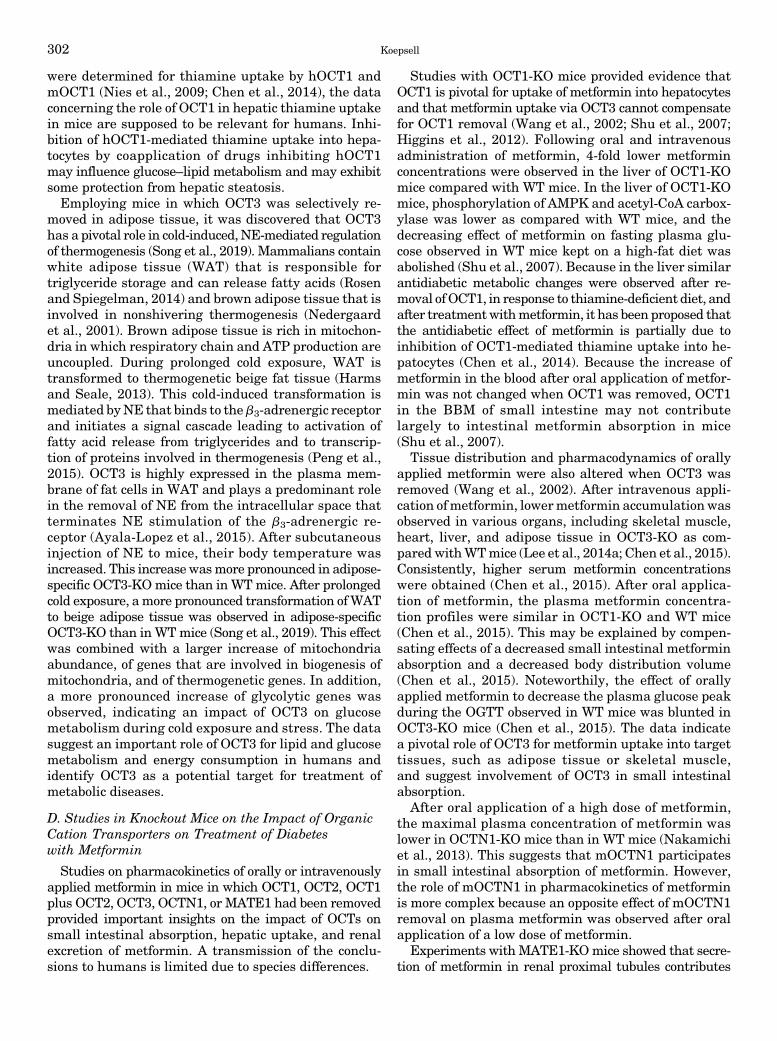
were determined for thiamine uptake by hOCT1 andmOCT1 (Nies et al., 2009; Chen et al., 2014), the dataconcerning the role of OCT1 in hepatic thiamine uptakein mice are supposed to be relevant for humans. Inhi-bition of hOCT1-mediated thiamine uptake into hepa-tocytes by coapplication of drugs inhibiting hOCT1may influence glucose–lipid metabolism and may exhibitsome protection from hepatic steatosis.Employing mice in which OCT3 was selectively re-
moved in adipose tissue, it was discovered that OCT3has a pivotal role in cold-induced,NE-mediated regulationof thermogenesis (Song et al., 2019). Mammalians containwhite adipose tissue (WAT) that is responsible fortriglyceride storage and can release fatty acids (Rosenand Spiegelman, 2014) and brown adipose tissue that isinvolved in nonshivering thermogenesis (Nedergaardet al., 2001). Brown adipose tissue is rich in mitochon-dria in which respiratory chain and ATP production areuncoupled. During prolonged cold exposure, WAT istransformed to thermogenetic beige fat tissue (Harmsand Seale, 2013). This cold-induced transformation ismediated byNE that binds to theb3-adrenergic receptorand initiates a signal cascade leading to activation offatty acid release from triglycerides and to transcrip-tion of proteins involved in thermogenesis (Peng et al.,2015). OCT3 is highly expressed in the plasma mem-brane of fat cells in WAT and plays a predominant rolein the removal of NE from the intracellular space thatterminates NE stimulation of the b3-adrenergic re-ceptor (Ayala-Lopez et al., 2015). After subcutaneousinjection of NE to mice, their body temperature wasincreased. This increasewasmore pronounced in adipose-specific OCT3-KOmice than in WTmice. After prolongedcold exposure, amore pronounced transformation ofWATto beige adipose tissue was observed in adipose-specificOCT3-KO than inWTmice (Song et al., 2019). This effectwas combined with a larger increase of mitochondriaabundance, of genes that are involved in biogenesis ofmitochondria, and of thermogenetic genes. In addition,a more pronounced increase of glycolytic genes wasobserved, indicating an impact of OCT3 on glucosemetabolism during cold exposure and stress. The datasuggest an important role of OCT3 for lipid and glucosemetabolism and energy consumption in humans andidentify OCT3 as a potential target for treatment ofmetabolic diseases.
D. Studies in Knockout Mice on the Impact of OrganicCation Transporters on Treatment of Diabeteswith Metformin
Studies on pharmacokinetics of orally or intravenouslyapplied metformin in mice in which OCT1, OCT2, OCT1plus OCT2, OCT3, OCTN1, or MATE1 had been removedprovided important insights on the impact of OCTs onsmall intestinal absorption, hepatic uptake, and renalexcretion of metformin. A transmission of the conclu-sions to humans is limited due to species differences.
Studies with OCT1-KO mice provided evidence thatOCT1 is pivotal for uptake of metformin into hepatocytesand that metformin uptake via OCT3 cannot compensatefor OCT1 removal (Wang et al., 2002; Shu et al., 2007;Higgins et al., 2012). Following oral and intravenousadministration of metformin, 4-fold lower metforminconcentrations were observed in the liver of OCT1-KOmice compared with WT mice. In the liver of OCT1-KOmice, phosphorylation of AMPK and acetyl-CoA carbox-ylase was lower as compared with WT mice, and thedecreasing effect of metformin on fasting plasma glu-cose observed in WT mice kept on a high-fat diet wasabolished (Shu et al., 2007). Because in the liver similarantidiabetic metabolic changes were observed after re-moval ofOCT1, in response to thiamine-deficient diet, andafter treatmentwithmetformin, it has been proposed thatthe antidiabetic effect of metformin is partially due toinhibition of OCT1-mediated thiamine uptake into he-patocytes (Chen et al., 2014). Because the increase ofmetformin in the blood after oral application of metfor-min was not changed when OCT1 was removed, OCT1in the BBM of small intestine may not contributelargely to intestinal metformin absorption in mice(Shu et al., 2007).
Tissue distribution and pharmacodynamics of orallyapplied metformin were also altered when OCT3 wasremoved (Wang et al., 2002). After intravenous appli-cation ofmetformin, lowermetformin accumulationwasobserved in various organs, including skeletal muscle,heart, liver, and adipose tissue in OCT3-KO as com-pared withWTmice (Lee et al., 2014a; Chen et al., 2015).Consistently, higher serum metformin concentrationswere obtained (Chen et al., 2015). After oral applica-tion of metformin, the plasma metformin concentra-tion profiles were similar in OCT1-KO and WT mice(Chen et al., 2015). This may be explained by compen-sating effects of a decreased small intestinal metforminabsorption and a decreased body distribution volume(Chen et al., 2015). Noteworthily, the effect of orallyapplied metformin to decrease the plasma glucose peakduring the OGTT observed in WT mice was blunted inOCT3-KO mice (Chen et al., 2015). The data indicatea pivotal role of OCT3 for metformin uptake into targettissues, such as adipose tissue or skeletal muscle,and suggest involvement of OCT3 in small intestinalabsorption.
After oral application of a high dose of metformin,the maximal plasma concentration of metformin waslower in OCTN1-KO mice than in WT mice (Nakamichiet al., 2013). This suggests that mOCTN1 participatesin small intestinal absorption of metformin. However,the role of mOCTN1 in pharmacokinetics of metforminis more complex because an opposite effect of mOCTN1removal on plasma metformin was observed after oralapplication of a low dose of metformin.
Experiments with MATE1-KOmice showed that secre-tion of metformin in renal proximal tubules contributes
302 Koepsell
significantly to urinarymetformin excretion. Theyprovideevidence that MATE1 is pivotal for metformin transportacross the BBM of renal proximal tubules and plays animportant role for transport of metformin across thebiliarymembrane of hepatocytes.After an oral applicationof metformin, the area below the blood concentration–time curve in MATE1-KO mice was larger than in WTmice, whereas the renal excretion of metformin wasdecreased (Tsuda et al., 2009a; Toyama et al., 2010). Inaddition, the concentration of metformin in the liverwas markedly higher in the MATE1-KO than in WTmice (Toyama et al., 2012). During treatment withmetformin, an impaired renal excretion and increasedhepatic concentration of metformin may promote lacticacidosis. After applying metformin for 7 days with thedrinking water, the plasma concentration of lactatewas higher and the pH in the plasma was lower inMATE1-KO than in WT mice (Toyama et al., 2012).
E. Studies in Knockout Mice on the Impact of OrganicCation Transporters on Nephrotoxicity of Cisplatin,Ethidium, and Paraquat
Nephrotoxicity limits the use of cisplatin in cancertherapy (Pabla and Dong, 2008). Studies with KO miceindicated that mOCT1 and mOCT2 in the basolateralmembrane and mMATE1 in the BBM of renal proximaltubules are critically involved in cisplatin secretion andhave impact on severity of cisplatin-induced nephrotox-icity. The amount of cisplatin in urine collected during24 hours after intraperitoneal injection of cisplatin was50% lower in OCT1/OCT2 double-KO mice than in WTmice (Filipski et al., 2009). Renal dysfunction, as indi-cated by increased blood urea nitrogen and increasedurine glucose that was observed in WT mice, did notoccur in the OCT1/OCT2-KO mice (Filipski et al., 2009;Ciarimboli et al., 2010). In MATE1-KO mice, cisplatininduced a more severe renal failure combined witha shortened time of animal survival than in WT mice(Nakamura et al., 2010b). One hour after intraperi-toneal injection of cisplatin, higher cisplatin concen-trations were measured in blood and kidney and morepronounced increases of blood urea nitrogen and Scrwere observed 3 days after cisplatin administration(Nakamura et al., 2010b).OCT1/OCT2-KO mice were also used to evaluate the
impact of OCT1 and/or OCT2 on the nephrotoxicity ofethidium. After arterial infusion with ethidium bromide,in cortical renal tubules of WT mice, distinctly higherconcentrations of ethidium were observed than inOCT1/OCT2-KO mice (Lee et al., 2009). The data sug-gest that mOCT1 and/or mOCT2 are critically involvedin uptake of ethidium into renal proximal tubules andmodulate nephrotoxicity of ethidium.Paraquat-induced nephrotoxicity inWTandMATE1-KO
mice was compared (Li et al., 2011). After intravenousinjection of paraquat, a 57% higher maximal plasmaconcentration of paraquat was observed in MATE1-KO
mice than in WT mice. Ninety minutes after paraquatapplication, a 2-fold higher paraquat concentration wasmeasured in kidneys of MATE-KO than in WT mice.Three days after the application of paraquat, inMATE1-KO mice a higher degree of nephrotoxicitywas diagnosed, as evaluated by histology and expres-sion of indicator proteins for acute renal injury. Thedata indicate that MATE1 is involved in renal paraquatexcretion and that decreased expression, dysfunction,or inhibition of MATE1 may exacerbate paraquat-induced nephrotoxicity.
F. Cerebral Functions of Organic Cation Transporter2 and Organic Cation Transporter 3 in Rodents
Our knowledge concerning functions of OCT2 andOCT3 in brain is almost entirely based on studies withrodents. To date, detailed information about cerebraldistributions of OCT2 and OCT3 is only available forrodents. The impact of removal of OCT2 and OCT3 onbehavioral and psychologic traits has been investigatedin rats and mice. DA, SE, NE, epinephrine, andhistidine are transported by rOCT2 and hOCT2; how-ever, different Km values have been reported for SE andepinephrine (Amphoux et al., 2006). NE, epinephrine,and histamine are transported by hOCT3 and rOCT3.In this case, different apparent Km values were de-termined for epinephrine and histamine. Of note,DA and SE are transported by rOCT3, but not byhOCT3. Immunostaining and in situ hybridization inmice and/or rats indicated overlapping, but partiallydifferent cerebral distributions of OCT2 and OCT3.Whereas OCT2 seems to be exclusively expressed inneurons (Bacq et al., 2012; Couroussé et al., 2015),OCT3 is expressed in neurons, glial cells, and ependy-mal cells (Wu et al., 1998a; Haag et al., 2004; Gasseret al., 2006, 2017; Nakayama et al., 2007; Vialou et al.,2008; Cui et al., 2009; Mayer et al., 2018). Different fromsodium-dependent, high-affinity monoamine reuptaketransporters that are enriched in nerve terminals,OCT2 and OCT3 are mainly located in neuronal cellmembranes and dendrites, indicating an extrasynapticlocation (Bacq et al., 2012; Gasser et al., 2017). BothOCT2 and OCT3 are expressed in cerebral cortex,cerebellum, hippocampus, thalamus, hypothalamus,amygdala, and hindbrain (Vialou et al., 2004, 2008;Bacq et al., 2012; Couroussé et al., 2015). However,different from OCT2, OCT3 is also expressed in circum-ventricular organs, in the substantia nigra, and in thechoroid plexus (Haag et al., 2004; Vialou et al., 2004;Nakayama et al., 2007; Gasser et al., 2009). Detectionof amine-neurotransmitter transport by OCT2 andOCT3 and cerebral expression suggested modulatoryroles during aminergic neurotransmission (Busch et al.,1998; Schmitt et al., 2003). This idea was supported bymeasurements in which effects of OCT inhibitors oncerebral clearance of SE or histamine were investigated(Daws et al., 2005; Gasser et al., 2006; Baganz et al.,
Organic Cation Transporters 303

2008). For example, it was observed that decynium22decelerated the clearance of SE in the hippocampus ofmice in which the sodium-dependent SERT had beenremoved (Baganz et al., 2008). Integrated behaviors andpersonality traits, including motor activity, mood, motiva-tion, anxiety, depression, and aggression, are modulatedby aminergic neurotransmitters and can be influenced byinhibitors of high-affinity reuptake systems (Charney,1998; Torres et al., 2003). For these reasons, effects ofOCT2 or OCT3 removal on anxiety-related behavior,behavior-despair paradigms of depression-like behaviorpatterns, and stress response were investigated. Be-cause OCT3 is expressed in the area postrema (AP) andthe subfornical organ (SFO) that are involved in thecontrol of osmotic homeostasis, the effect of OCT3removal on intake of salt and water also was tested.After removal of OCT2 in mice, sodium-independent,
decynium22-blockable uptake of NE and SE into sus-pended brain cells was decreased (Bacq et al., 2012).Accordingly, the in vivo clearance of iontophoreticallyapplied NE or SE measured in presence of venlafaxine,a selective blocker of NET and SET, was slowed down(David et al., 2003; Bacq et al., 2012). Several behav-ioral, mood-related tests were performed trying toevaluate the impact of OCT2 on anxiety and depression.After removal of OCT2, several indicators for anxietywere decreased. For example, in the open-field test,OCT2-KO mice remained for longer time periods in thecenter of the open field and showed more rearingactivity than WT mice. Similarly, in the elevatedO-maze test, OCT2-KO mice spent longer time periodsin the elevated open parts of the experimental device(Bacq et al., 2012). Evaluating more complex behav-ioral traits related to depression in humans, removalof OCT2 revealed different effects than antidepres-sive inhibitors of SERT and NET. For evaluation ofdepressive-like behavior, a tail-suspension test anda forced-swim test (FST) were applied. In both tests,time periods of immobility after exposition of the animalsto water (exposure to inescapable stress) that are sup-posed to correlate with depression-like behavior (Porsoltet al., 2001) were measured. Whereas the times ofimmobility were decreased by the SERT-inhibitor ven-laflexin, indicating a diminished level of resignation,they were increased after removal of OCT2 (Bacq et al.,2012). Applying a chronic corticosterone-induced de-pression model in WT and OCT2-KO mice, long-termtreatment with venlaflexin decreased the corticosterone-induced depressive-like traits evaluated in various testsin WT mice, but not in the OCT2-KO mice (Bacq et al.,2012). The data indicate that OCT2 modulates short-term and long-term effects of antidepressant inhibitorsof amine neurotransmitter uptake by SERT.Detailed immunohistochemical investigations revealed
that OCT2 is expressed in nuclei of stess-related neuronalcircuits that trigger the activation of the hypothalamic-pituitary-adrenocortical axis, culminating in corticosterone
secretion (Herman et al., 2003; Kvetnansky et al., 2009).In OCT2-KO mice, basal corticosterone secretion andenhanced corticosteone secretion in response to acutestress during FST were more distinctly increased thanin WT mice (Couroussé et al., 2015). Upon exposure tounpredictable chronic mild stress, OCT2-KO miceshowed an increased vulnerability to changes that re-flect depression-related symptoms such as dimishedself-grooming and accelerated development of specialmemory deficits. Because removal of OCT2 did notchange the affinity of adrenocorticotrophic hormonefor stimulation of adrenal corticosterone secretion, theobserved OCT2-related effects are supposed to be due toeffects on central neuronal circuits. In hippocampus,Ser9 phosphorylation of glycogen synthase kinase-3bunder basal conditions and after acute stress was differentbetweenOCT2-KOversusWTmice (Couroussé etal., 2015).Ser9 phosphorylation of glycogen synthase kinase-3b hasbeen shown to be involved in mood disorders (Beaulieuet al., 2008) and to be decreased by SE-dependentactivation (Li et al., 2004). Hence, the data suggest thatOCT2 influences SE concentrations within stress-relatedneuronal circuits, leading to an increased vulnerabilityduring chronic stress.
The effects of OCT3 removal on brain depression-related functions were investigated by two groupsemploying different lines of OCT3-KO mice (Vialouet al., 2008; Wultsch et al., 2009). Vialou et al. (2008)used a mutant mouse line that has been functionallycharacterized (Zwart et al., 2001b). After intravenousinjection ofMPP, largely decreasedMPP concentrationswere measured in heart and salivary glands, that is, intissues with high expression of OCT3 (Zwart et al.,2001b; Lee et al., 2014a). Vialou et al. (2008) observedthat the DA concentration in several brain regions, forexample, in the substantia nigra and the tegmentalarea, was decreased upon OCT3 removal (Vialou et al.,2008). Performing behavioral tests, they observed sim-ilar basal locomotor activity in the OCT3-KO and WTmice. In OCT3-KO mice they observed higher para-digms for anxiety in the open-field and Y-maze teststhan in WT mice. After application of relatively lowdoses of D-amphetamine or cocaine, the locomotoractivity was increased about 3-fold in both WT andOCT3-KOmice. However, high doses of both stimulantsincreased locomotor activity in OCT3-KO mice, but notin WTmice. Using a homemade, sparsely characterizedOCT3-KO mouse line, Wultsch et al. (2009) evaluatedthe effects of OCT3 removal on locomotor activity,spatial orientation, and anxiety. They detected noeffects on locomotor activity and spatial orientationsimilar to Vialou et al. (2008) but observed increasedparadigms for anxiety in the open-field and elevatedplus-maze test. Taken together the data suggest thatOCT3 has an impact on depression-related behavior inmice and on addiction to amphetamine that may beinfluenced by additional genetic factors.
304 Koepsell

Decreasing the cerebral expression of OCT3 in miceby infusion of antisense oligonucleotide into the thirdbrain ventricle, the time of immobility in the FST thatrepresents a paradigm for depression-related behaviorwas largely decreased (Kitaichi et al., 2005). Thissuggests an antidepressive effect of OCT3 removal. Animpact of OCT3 on the stimulatory effect of metham-phetamine (METH) was indicated by the observationthat the stimulation of locomotor activity byMETHwasconsiderably higher when the expression of OCT3 wasreduced (Kitaichi et al., 2005). This effect of ventricle-injected OCT3-antisense oligonucleotides on METH-stimulated locomotor activity was confirmed in rats(Nakayama et al., 2007). In rats, the authors addition-ally determined the effect of s.c. injected METH onextracellular cerebral DA concentrations. After METHapplication, extracelluar DA in the prefrontal cortexand the nucleus accumbens was increased. This increasewas significantly enhancedwhen the expression of OCT3was decreased by intraventricular injection of antisenseoligonucleotides. Because mOCT3 does not transportamphetamine like hOCT3 (Wagner et al., 2017, 2018),the increased cerebral concentration of METH inOCT3-KO mice is probably due to an effect of OCT3removal on function or expression of an unknowntransporter that accepts METH as substrate.Recently, data have been reported that suggest that
OCT3 may be involved in cellular DA release fromstimulated dopaminergic neurons (Mayer et al., 2018).Comparing effects of amphetamine on neuronal activityin WT and OCT3-KO mice, the authors observed thatamphetamine-induced hyperactivity was dependent onthe presence of OCT3. They showed that inWTmice theextracellular DA concentration close to dopaminergicneurons increased after stimulation with amphetamine.In WT mice the amphetamine-induced increase of DAwas partially blocked by inhibitors of DAT and by theOCT-inhibitor decynium22; however, the decynium22-induced inhibition was absent in OCT3-KOmice. Tryingto test the hypothesis that OCT3 mediates DA releasefrom amphetamine-stimulated neurons, they performedefflux studies withMPP in rat superior cervical ganglioncells that express OCT3, but neither OCT1 nor OCT2(Kristufek et al., 2002; Mayer et al., 2018). Amphetamine-stimulated MPP efflux from MPP-preloaded superiorcervical ganglion cells was completely blocked by decy-nium22. The authors raised the hypothesis that amphet-amine enters dopaminergic neurons via a nonidentifiedtransporter or by passive diffusion and mediates therelease of DA from intracellular vesicles by disruptingthe vesicular monoamine transporter 2 (Sulzer et al.,2005), and that the increased intracellular DA concen-tration provides the driving force for OCT3-mediated DAefflux. An alternative explanation for the impact ofOCT3on amphetamine-stimulated DA-mediated neurotrans-mission is that OCT3 is critically involved in reuptake ofextracellular DA into neurons and/or glia cells.
Because it was observed that OCT3 is expressed inneurons of the SFO and the AP that are involved insensing of osmolarity changes and regulation of salt andwater intake, the effect of OCT3 removal on salt intakewas investigated (Vialou et al., 2004). After dehydrationthat was induced by withdrawal of water and injectionof the sodium-wasting diuretic furosemide, the sponta-neous ingestion of hypertonic saline was 40% higherin OCT3-KO mice than in WT mice. To elucidate theimpact of OCT3 removal on neuronal activity, theeffect on salt and water deprivation on the expressionof c-Fos was investigated. Neuronal expression ofc-Fos has been shown to indicate central responses toexcitatory stimuli, including hyperosmolarity and hypo-volemia (Lane et al., 1997; Thunhorst et al., 1998). Underbasal conditions, a similar expression of OCT3 wasobserved in SFO neurons of WT and OCT3-KO mice.Of note, after dehydration, the neuronal cFos expres-sion in the SFO was inceased more than 10-fold in WTmice and less than 5-fold in OCT3-KO mice (Vialouet al., 2004). The data suggest that OCT3-mediatedneurotransmitter transport influences the regulationof salt intake. The impact of OCT3 on salt intakeregulation is probably not exclusively due to effects onneuronal activity in the SFO and PA because OCT3 isalso expressed in additional structures such as hypo-thalamic paraventricular nuclei, supraoptical nuclei,and the solitary tract nucleus, which are supposed to beinvolved in sensing of blood osmolarity changes and inregulation of salt intake (Ferguson and Bains, 1996;Bourque and Oliet, 1997).
XIII. In Vitro Drug Testing
Preclinical testing of new molecular entities (NMEs)for interaction with hOCTs is required to anticipatepotential adverse drug effects (ADEs) in patients and todesign suitable clinical studies. Adverse effects of drugsinteracting with hOCTs may be caused by toxicity incells or organs in which drugs are enriched in responseto OCT-mediated cellular uptake or to inhibition ofOCT-mediated cellular export. OCT-related ADEs mayalso be caused by drug interaction with pivotal endoge-neous functions of OCTs, such as hOCT1-mediatedthiamine uptake into hepatocytes or fat cells, cellularuptake and/or efflux of neurotransmitters by hOCT2and hOCT3, and OCTN2-mediated cellular uptake ofcarnitine. In addition, NMEs interacting with OCTsmay alter the pharmacokinetics of coapplied drugs andchange their pharmacodynamics and/or toxicity.
The following procedures are suggested as paradig-matic examples for identification of NMEs that interactwith any hOCT under biomedically and/or physiologi-cally relevant conditions. These suggestions are basedon current knowledge on transporters and do not considercompromises due to commercial needs and/or politicalframework. Test conditions for interaction of NMEs
Organic Cation Transporters 305

with all hOCTs, hMATE1 and hMATE-2K, ratherthan exclusively with hOCT1 and hOCT2 as recom-mended by the FDA and EMA, are described in thiswork. In silico prediction for interaction of NMEs withhOCT1, hOCT2, and hMATE1 by pharmacophore mod-eling (Ahlin et al., 2008; Wittwer et al., 2013; Xu et al.,2013; Chen et al., 2017a; Sandoval et al., 2018) cannot beused to exclude NMEs from in vitro testing becauseinteraction with OCTs was predicted for less than 85% ofthe drugs that were identified by in vitro assays(Koepsell, 2019). In vitro testing is recommended for allpositively charged and uncharged NMEs regardless oftheir molecular weight.It is recommended to employ inhibition assays using
very low substrate concentrations for the identificationof NMEs that interact with OCTs because this allowsdetection of inhibitory interactions at high- and low-affinity binding sites (see In-Depth Functional Charac-terization of Organic Cation Transporter 1 and OrganicCation Transporter 2, last paragraph). Allosteric, high-affinity inhibition of transport can only be observedat low substrate concentrations when only one low-affinity binding site is involved in translocation (seeFig. 4B). The inhibition assays should be performed atnanomolar test substrate concentrations. This allowsthe unequivocal detection of transporter-mediateduptake even in the presence of high passive membranepermeation. Hence, the uptake measurements shouldbe performed with radioactively labeled substrates.Because the apparent Km values determined for MPPuptake by hOCT1, hOCT2, hOCT3, hMATE1, andhMATE-K2 are relatively low (between 3 and 111 mM;see Table 1), it is recommended to use radioactivelylabeled MPP at two concentrations (1 and 10 nM) forinitial screening of hOCT1–3, hMATE1, and hMATE-K2.To elucidate whether NMEs can cause high-affinityinhibition by interaction with allosteric binding sites, itis proposed to start with NME concentrations of 1 pM,1 mM, and 5 mM. Initial screening of NMEs for in-teraction with hOCTN1 and hOCTN2 should be per-formed with radioactively labeled ergothioneine andL-carnitine, respectively, using concentrations of 1and 10 nM, and NME concentrations of 1 pM, 1 mM,and 5 mM. Because IC50 values of OCTs are dependenton the molecular structure of the test substrate (Belzeret al., 2013; Thévenod et al., 2013; Yin et al., 2016), itis recommended to repeat in vitro inhibition testingfor NMEs, which do not show up as inhibitors in theprimary assay. To evaluate the interaction of NMEswith hOCT1–3, hMATE1, and hMATE2-K in the follow-up assay, it is proposed to use radioactively labeledtrospium as test substrate (apparent Km values be-tween 4.4 and 106 mM; see Table 4) (Bexten et al., 2015).Like withMPP, trospium concentrations of 1 and 10 nMand NME concentrations of 1 pM, 1 mM, and 5 mMshould be used. In a follow-up assay, the interaction ofNMEs with hOCTN1 and hOCTN2 may be evaluated
by measuring the inhibition of uptake of 1 and 10 nMradioactively labeled sulpiride (apparent Km valuesof 235 and 250 mM; Table 4) (dos Santos Pereira et al.,2014).
It is recommended to test those NMEs that inhibitOCTs also for transport because OCT-mediated trans-port may impact pharmacokinetics, pharmacodynam-ics, and/or toxicity of the NMEs. Designing transportmeasurements, it must be considered that the ratiobetween transporter-mediated uptake and passivemem-brane permeation generally decreases with increasingdrug concentrations. Hence, transport may be over-looked if too high concentrations of NMEs are used.Because it is difficult to anticipate the concentration ofthe respective NME in different body compartments,transport should be tested with NME concentrationsin the submicromolar range. If transported NMEs willbe pursued for drug development, the apparent Km
value(s) for the involved OCT(s) must be determinend.It is also recommended to characterize the effects offrequent polymorphisms in the involved OCTs if theyhave been shown to exhibit substrate-specific effectson transport (Table 8). For example, effects ofmutationsSer14Phe, Arg61Cys, Gly401Ser, and Met420del inhOCT1 and of mutation Ala270Ser in hOCT2 on Km
and Vmax values should be determined.During the first phase of clinical studies of novel
drugs interacting with OCTs, in vitro testing is recom-mended to anticipate potential effects on endogenousfunctions of the involved OCTs and effects on pharma-cokinetics and/or toxicity of coadministered drugs.For in vitro testing at this stage of drug development,unbound blood concentrations of the novel drugs needto be determined in clinical studies so that expectedminimal, average, and peak free plasma drug concen-trations can be estimated. Using these drug concentra-tions, the effects of novel drugs on transport of physiologicimportant endogeneous substrates by the respectiveOCTs should be tested. Effects on estimated minimal,average, and maximal blood and/or tissue concentra-tions of the endogenous substrates should be analyzed.For example, if a novel drug interacts with hOCT1, itshould also be tested to determine whether it inhibitshOCT1-mediated uptake of thiamine or, if a novel druginteracts with hOCT3, it is recommended to determinethe drug effect on hOCT3-mediated uptake of DA, SE,epinepherine, NE, and histamine. In vitro tests are alsoimportant to anticipate potential effects of novel drugson pharmacokinetics, pharmacodynamics, and/or toxic-ity on coadministered drugs. Hence, novel drugs inter-acting with OCTs should be tested for inhibition withcoadministered drugs that are transported by the re-spective OCTs. Testing for interactions with uptake ofmetformin and cisplatin should be obligatory. If a noveldrug interacts with hOCT1, hOCT2, hOCT3, MATE1,and/or MATE2-K, it is recommended to test whetherit inhibits uptake of 1, 5, and 15 mM metformin.
306 Koepsell

The metformin concentrations represent the troughplasma membrane concentration, the intermediate con-centration, and maximal plasma metformin concentra-tion, repectively (Shu et al., 2008; Tzvetkov et al., 2009;Christensen et al., 2011). If a novel drug interacts withhOCT2, hMATE1, and/or MATE-K2, inhibition ofcisplatin transport mediated by the respective OCT(s)at clinically relevant concentrations should be tested.Ínhibition of hOCT2-mediated cisplatin uptake suggestsretarded renal cisplatin excretion associated with reducednephrotoxicity, whereas inhibition of cisplatin uptake byMATE1 and/or MATE2-K indicates a risk for aggrava-tion of cisplatin-induced nephrotoxicity. The informa-tion obtained from in vitro testing concerning theinhibitory potential of novel drugs on OCT-mediateduptake of endogenous substrates and coadministereddrugs provides a basis to decide which additionalclinical studies are required to exclude ADEs relatedto drug interactions at organic cation transporters.
XIV. Concluding Remarks
Twenty-five years of intensive investigation on OCTsafter cloning of the first member of the SLC22 familyhave established multiple pivotal roles of OCTs in phys-iology and pharmacology. The large variety of functions ofOCTs is due to their capability for polyspecific recognitionand translocation of cationic and/or zwitterionic sub-strates. The elucidation of functional traits after over-expression, purification, and reconstitution of OCTs aswell as the development of experimentally based hy-potheses on the molecular basics for substrate recogni-tion and translocation provided a rough idea about thecomplexity of ligand interactions at OCTs. Realizing thebroad impact of OCTs on pharmacokinetics, pharmaco-dynamics, and toxicity of drugs, the interaction of manydrugs with individual OCTs has been investigated, andin vitro testing of NMEs for interaction with OCTs hasbecome standard during drug development. Unfortu-nately, the routinely employed procedures, includingthose that have been proposed by the InternationalTransporter Consortium, FDA, and EMA, only providean incomplete picture because they do not detect anypossible interactions of drugs withOCTs. Extension andrefinement of in vitro testing procedures as proposedin this review can avoid the shortcomings. The single-nucleotide variations in hOCTs have been identified,and their frequencies in various ethnic groups weredetermined. In addition, effects of mutations on trans-port of some endogenous substrates and some drugsand effects on treatments with a few drugs have beeninvestigated. The current knowledge of the impact ofmutations on drug treatment and on physiologic func-tions is very fragmentary. Studies that were performedin KO mice revealed important hints on endogeneousphysiologic functions of OCTs in humans such as onthe impact of OCT2 and OCT3 for behavioral traits.
To enlighten the endogeneous physiologic functions ofOCTs in humans, intensive future investigations arerequired in which, for example, physiologic effects ofloss-of-function mutants on distributions and metaboliceffects of endogeneous substrates are investigated orthe distribution of 11C- or 18F-labeled drugs is determinedby PET. In summary, previous research indicates theenormous potential of OCT-related pharmacological char-acterizations to improve drug treatments of patients,including the development of personalized therapies. In-tensive future research in the field is required to use thishigh potential for improvement of medical treatments.
Authorship Contributions
Participated in research design: Koepsell.Conducted experiments: Koepsell.Contributed new reagents or analytic tools: Koepsell.Performed data analysis: Koepsell.Wrote or contributed to the writing of the manuscript: Koepsell.
ReferencesAbramson J, Smirnova I, Kasho V, Verner G, Kaback HR, and Iwata S (2003)Structure and mechanism of the lactose permease of Escherichia coli. Science 301:610–615.
Ahlin G, Chen L, Lazorova L, Chen Y, Ianculescu AG, Davis RL, Giacomini KM,and Artursson P (2011) Genotype-dependent effects of inhibitors of the organiccation transporter, OCT1: predictions of metformin interactions. Pharmacoge-nomics J 11:400–411.
Ahlin G, Karlsson J, Pedersen JM, Gustavsson L, Larsson R, Matsson P, Norinder U,Bergström CAS, and Artursson P (2008) Structural requirements for drug in-hibition of the liver specific human organic cation transport protein 1. J Med Chem51:5932–5942.
Ahmadimoghaddam D, Zemankova L, Nachtigal P, Dolezelova E, Neumanova Z,Cerveny L, Ceckova M, Kacerovský M, Micuda S, and Staud F (2013) Organiccation transporter 3 (OCT3/SLC22A3) and multidrug and toxin extrusion 1(MATE1/SLC47A1) transporter in the placenta and fetal tissues: expressionprofile and fetus protective role at different stages of gestation. Biol Reprod 88:55.
Al-Abdulla R, Lozano E, Macias RIR, Monte MJ, Briz O, O’Rourke CJ, Serrano MA,Banales JM, Avila MA, Martinez-Chantar ML, et al. (2019) Epigenetic events in-volved in organic cation transporter 1-dependent impaired response of hepatocel-lular carcinoma to sorafenib. Br J Pharmacol 176:787–800.
Albert U, Bergesio C, Pessina E, Maina G, and Bogetto F (2002) Management oftreatment resistant obsessive-compulsive disorder: algorithms for pharmacother-apy. Panminerva Med 44:83–91.
Alcorn J, Lu X, Moscow JA, and McNamara PJ (2002) Transporter gene expression inlactating and nonlactating human mammary epithelial cells using real-time reversetranscription-polymerase chain reaction. J Pharmacol Exp Ther 303:487–496.
Amat di San Filippo C, Taylor MRG, Mestroni L, Botto LD, and Longo N (2008)Cardiomyopathy and carnitine deficiency. Mol Genet Metab 94:162–166.
Ambudkar SV, Anantharam V, and Maloney PC (1990) UhpT, the sugar phosphateantiporter of Escherichia coli, functions as a monomer. J Biol Chem 265:12287–12292.
Amphoux A, Vialou V, Drescher E, Brüss M, Mannoury La Cour C, Rochat C, MillanMJ, Giros B, Bönisch H, and Gautron S (2006) Differential pharmacological in vitroproperties of organic cation transporters and regional distribution in rat brain.Neuropharmacology 50:941–952.
Aoki M, Terada T, Kajiwara M, Ogasawara K, Ikai I, Ogawa O, Katsura T, and Inui K(2008) Kidney-specific expression of human organic cation transporter 2(OCT2/SLC22A2) is regulated by DNA methylation. Am J Physiol Renal Physiol295:F165–F170.
Apparsundaram S, Ferguson SM, George AL Jr, and Blakely RD (2000) Molecularcloning of a human, hemicholinium-3-sensitive choline transporter. Biochem Bio-phys Res Commun 276:862–867.
Ardizzoni A, Boni L, Tiseo M, Fossella FV, Schiller JH, Paesmans M, Radosavljevic D,Paccagnella A, Zatloukal P, Mazzanti P, et al.; CISCA (CISplatin versus CArboplatin)Meta-Analysis Group (2007) Cisplatin- versus carboplatin-based chemotherapy infirst-line treatment of advanced non-small-cell lung cancer: an individual patientdata meta-analysis. J Natl Cancer Inst 99:847–857.
Arduini A, Mancinelli G, Radatti GL, Hochstein P, and Cadenas E (1992) Possiblemechanism of inhibition of nitrite-induced oxidation of oxyhemoglobin by ergo-thioneine and uric acid. Arch Biochem Biophys 294:398–402.
Arndt P, Volk C, Gorboulev V, Budiman T, Popp C, Ulzheimer-Teuber I, Akhoundova A,Koppatz S, Bamberg E, Nagel G, et al. (2001) Interaction of cations, anions, and weakbase quinine with rat renal cation transporter rOCT2 compared with rOCT1.Am J Physiol Renal Physiol 281:F454–F468.
Arner P, Kulyté A, Batchelor K, Laurencikiene J, Livingston J, and Rydén M (2018)Mapping of biguanide transporters in human fat cells and their impact on lipolysis.Diabetes Obes Metab 20:2416–2425.
Organic Cation Transporters 307

Asaka J, Terada T, Ogasawara K, Katsura T, and Inui K (2007a) Characterizationof the Basal promoter element of human organic cation transporter 2 gene.J Pharmacol Exp Ther 321:684–689.
Asaka J, Terada T, Tsuda M, Katsura T, and Inui K (2007b) Identification ofessential histidine and cysteine residues of the H1 /organic cation antiportermultidrug and toxin extrusion (MATE). Mol Pharmacol 71:1487–1493.
Astorga B, Ekins S, Morales M, and Wright SH (2012) Molecular determinants ofligand selectivity for the human multidrug and toxin extruder proteins MATE1and MATE2-K. J Pharmacol Exp Ther 341:743–755.
Auer M, Kim MJ, Lemieux MJ, Villa A, Song J, Li XD, and Wang DN (2001) High-yield expression and functional analysis of Escherichia coli glycerol-3-phosphatetransporter. Biochemistry 40:6628–6635.
Ayala-Lopez N, Jackson WF, Burnett R, Wilson JN, Thompson JM, and Watts SW(2015) Organic cation transporter 3 contributes to norepinephrine uptake intoperivascular adipose tissue. Am J Physiol Heart Circ Physiol 309:H1904–H1914.
Babál P, Ruchko M, Campbell CC, Gilmour SP, Mitchell JL, Olson JW, and GillespieMN (2001) Regulation of ornithine decarboxylase activity and polyamine transportby agmatine in rat pulmonary artery endothelial cells. J Pharmacol Exp Ther 296:372–377.
Babusukumar U, Wang T, McGuire E, Broeckel U, and Kugathasan S (2006) Con-tribution of OCTN variants within the IBD5 locus to pediatric onset Crohn’s dis-ease. Am J Gastroenterol 101:1354–1361.
Bacher P, Giersiefer S, Bach M, Fork C, Schömig E, and Gründemann D (2009)Substrate discrimination by ergothioneine transporter SLC22A4 and carnitinetransporter SLC22A5: gain-of-function by interchange of selected amino acids.Biochim Biophys Acta 1788:2594–2602.
Bachmakov I, Glaeser H, Endress B, Mörl F, König J, and Fromm MF (2009) In-teraction of beta-blockers with the renal uptake transporter OCT2. Diabetes ObesMetab 11:1080–1083.
Bachmakov I, Glaeser H, Fromm MF, and König J (2008) Interaction of oral anti-diabetic drugs with hepatic uptake transporters: focus on organic anion trans-porting polypeptides and organic cation transporter 1. Diabetes 57:1463–1469.
Bacq A, Balasse L, Biala G, Guiard B, Gardier AM, Schinkel A, Louis F, Vialou V,Martres MP, Chevarin C, et al. (2012) Organic cation transporter 2 controls brainnorepinephrine and serotonin clearance and antidepressant response. Mol Psychiatry17:926–939.
Baganz NL, Horton RE, Calderon AS, Owens WA, Munn JL, Watts LT,Koldzic-Zivanovic N, Jeske NA, Koek W, Toney GM, et al. (2008) Organiccation transporter 3: keeping the brake on extracellular serotonin in serotonin-transporter-deficient mice. Proc Natl Acad Sci USA 105:18976–18981.
Bai M, Ma Z, Sun D, Zheng C, Weng Y, Yang X, Jiang T, and Jiang H (2017a)Multiple drug transporters mediate the placental transport of sulpiride. ArchToxicol 91:3873–3884.
Bai X, Moraes TF, and Reithmeier RAF (2017b) Structural biology of solute carrier(SLC) membrane transport proteins. Mol Membr Biol 34:1–32.
Bailey CJ, Wilcock C, and Scarpello JH (2008) Metformin and the intestine.Diabetologia 51:1552–1553.
Bain MA, Fornasini G, and Evans AM (2005) Trimethylamine: metabolic, pharma-cokinetic and safety aspects. Curr Drug Metab 6:227–240.
Ballestero MR, Monte MJ, Briz O, Jimenez F, Gonzalez-San Martin F, and Marin JJ(2006) Expression of transporters potentially involved in the targeting of cy-tostatic bile acid derivatives to colon cancer and polyps. Biochem Pharmacol 72:729–738.
Balyan R, Zhang X, Chidambaran V, Martin LJ, Mizuno T, Fukuda T, Vinks AA,and Sadhasivam S (2017) OCT1 genetic variants are associated with postoperativemorphine-related adverse effects in children. Pharmacogenomics 18:621–629.
Barrett EL and Kwan HS (1985) Bacterial reduction of trimethylamine oxide. AnnuRev Microbiol 39:131–149.
Barton A, Eyre S, Bowes J, Ho P, John S, and Worthington J (2005) Investigation ofthe SLC22A4 gene (associated with rheumatoid arthritis in a Japanese population)in a United Kingdom population of rheumatoid arthritis patients. Arthritis Rheum52:752–758.
Battle A, Brown CD, Engelhardt BE, and Montgomery SB; GTEx Consortium;Laboratory, Data Analysis &Coordinating Center (LDACC)—Analysis WorkingGroup; Statistical Methods Groups—Analysis Working Group; Enhancing GTEx(eGTEx) Groups; NIH Common Fund; NIH/NCI; NIH/NHGRI; NIH/NIMH; NIH/NIDA;Biospecimen Collection Source Site—NDRI; Biospecimen Collection Source Site—RPCI;Biospecimen Core Resource—VARI; Brain Bank Repository—University of MiamiBrain Endowment Bank; Leidos Biomedical—Project Management; ELSI Study;Genome Browser Data Integration & Visualization—EBI; Genome Browser DataIntegration & Visualization—UCSC Genomics Institute, University of CaliforniaSanta Cruz; Lead Analysts; Laboratory, Data Analysis & Coordinating Center(LDACC); NIH Program Management; Biospecimen Collection; Pathology; eQTLManuscript Working Group (2017) Genetic effects on gene expression across hu-man tissues. Nature 550:204–213.
Beaulieu JM, Zhang X, Rodriguiz RM, Sotnikova TD, Cools MJ, Wetsel WC, Gain-etdinov RR, and Caron MG (2008) Role of GSK3 beta in behavioral abnormalitiesinduced by serotonin deficiency. Proc Natl Acad Sci USA 105:1333–1338.
Becker ML, Visser LE, van Schaik RH, Hofman A, Uitterlinden AG, and Stricker BH(2009a) Genetic variation in the organic cation transporter 1 is associated with met-formin response in patients with diabetes mellitus. Pharmacogenomics J 9:242–247.
Becker ML, Visser LE, van Schaik RHN, Hofman A, Uitterlinden AG, and StrickerBH (2009b) Genetic variation in the multidrug and toxin extrusion 1 transporterprotein influences the glucose-lowering effect of metformin in patients with di-abetes: a preliminary study. Diabetes 58:745–749.
Becker ML, Visser LE, van Schaik RHN, Hofman A, Uitterlinden AG, and StrickerBH (2010) Interaction between polymorphisms in the OCT1 and MATE1 trans-porter and metformin response. Pharmacogenet Genomics 20:38–44.
Bednarczyk D, Ekins S, Wikel JH, and Wright SH (2003) Influence of molecularstructure on substrate binding to the human organic cation transporter, hOCT1.Mol Pharmacol 63:489–498.
Belzer M, Morales M, Jagadish B, Mash EA, and Wright SH (2013) Substrate-dependent ligand inhibition of the human organic cation transporter OCT2.J Pharmacol Exp Ther 346:300–310.
Berg T, Hegelund-Myrbäck T, Öckinger J, Zhou XH, Brännström M, Hagemann-Jensen M, Werkström V, Seidegård J, Grunewald J, Nord M, et al. (2018) Ex-pression of MATE1, P-gp, OCTN1 and OCTN2, in epithelial and immune cells inthe lung of COPD and healthy individuals. Respir Res 19:68.
Bexten M, Oswald S, Grube M, Jia J, Graf T, Zimmermann U, Rodewald K, Zolk O,Schwantes U, Siegmund W, et al. (2015) Expression of drug transporters and drugmetabolizing enzymes in the bladder urothelium in man and affinity of the bladderspasmolytic trospium chloride to transporters likely involved in its pharmacoki-netics. Mol Pharm 12:171–178.
Biermann J, Lang D, Gorboulev V, Koepsell H, Sindic A, Schröter R, Zvirbliene A,Pavenstädt H, Schlatter E, and Ciarimboli G (2006) Characterization of regulatorymechanisms and states of human organic cation transporter 2. Am J Physiol CellPhysiol 290:C1521–C1531.
Blanc Mettral J, Faller N, Cruchon S, Sottas L, Buclin T, Schild L, Choong E,Nahimana A, and Decosterd LA (2019) Imatinib uptake into cells is not mediatedby organic cation transporters OCT1, OCT2, or OCT3, but is influenced byextracellular pH. Drug Metab Lett DOI: 10.2174/1872312813666190207150207[published ahead of print].
Bokelmann K, Brockmöller J, and Tzvetkov MV (2018) Impact of promoter poly-morphisms on the transcriptional regulation of the organic cation transporterOCT1 (SLC22A1). J Pers Med 8:42, DOI: 10.3390/jpm.8040042.
Bottalico B, Larsson I, Brodszki J, Hernandez-Andrade E, Casslén B, Marsál K,and Hansson SR (2004) Norepinephrine transporter (NET), serotonin transporter(SERT), vesicular monoamine transporter (VMAT2) and organic cation trans-porters (OCT1, 2 and EMT) in human placenta from pre-eclamptic and normo-tensive pregnancies. Placenta 25:518–529.
Bottalico B, Noskova V, Pilka R, Larsson I, Domanski H, Casslén B, and Hansson SR(2007) The organic cation transporters (OCT1, OCT2, EMT) and the plasmamembrane monoamine transporter (PMAT) show differential distribution and cy-clic expression pattern in human endometrium and early pregnancy decidua. MolReprod Dev 74:1303–1311.
Bouchoucha M, Uzzan B, and Cohen R (2011) Metformin and digestive disorders.Diabetes Metab 37:90–96.
Bourdet DL, Pritchard JB, and Thakker DR (2005) Differential substrate and in-hibitory activities of ranitidine and famotidine toward human organic cationtransporter 1 (hOCT1; SLC22A1), hOCT2 (SLC22A2), and hOCT3 (SLC22A3).J Pharmacol Exp Ther 315:1288–1297.
Bourque CW and Oliet SH (1997) Osmoreceptors in the central nervous system. AnnuRev Physiol 59:601–619.
Boxberger KH, Hagenbuch B, and Lampe JN (2014) Common drugs inhibit humanorganic cation transporter 1 (OCT1)-mediated neurotransmitter uptake. DrugMetab Dispos 42:990–995.
Boxberger KH, Hagenbuch B, and Lampe JN (2018) Ligand-dependent modulation ofhOCT1 transport reveals discrete ligand binding sites within the substratetranslocation channel. Biochem Pharmacol 156:371–384.
Brast S, Grabner A, Sucic S, Sitte HH, Hermann E, Pavenstädt H, Schlatter E,and Ciarimboli G (2012) The cysteines of the extracellular loop are crucial fortrafficking of human organic cation transporter 2 to the plasma membrane and areinvolved in oligomerization. FASEB J 26:976–986.
Brazier L, Tiret L, Luc G, Arveiler D, Ruidavets JB, Evans A, Chapman J, CambienF, and Thillet J (1999) Sequence polymorphisms in the apolipoprotein(a) gene andtheir association with lipoprotein(a) levels and myocardial infarction: The ECTIMStudy. Atherosclerosis 144:323–333.
Breining P, Jensen JB, Sundelin EI, Gormsen LC, Jakobsen S, Busk M, Rolighed L,Bross P, Fernandez-Guerra P, Markussen LK, et al. (2018) Metformin targetsbrown adipose tissue in vivo and reduces oxygen consumption in vitro. DiabetesObes Metab 20:2264–2273.
Bremner P, Woodman K, Burgess C, Crane J, Purdie G, Pearce N, and Beasley R(1993) A comparison of the cardiovascular and metabolic effects of formoterol,salbutamol and fenoterol. Eur Respir J 6:204–210.
Brown JM and Hazen SL (2014) Metaorganismal nutrient metabolism as a basis ofcardiovascular disease. Curr Opin Lipidol 25:48–53.
Bschleipfer T, Weidner W, Kummer W, and Lips KS (2012) Does bladder outletobstruction alter the non-neuronal cholinergic system of the human urothelium?Life Sci 91:1082–1086.
Budiman T, Bamberg E, Koepsell H, and Nagel G (2000) Mechanism of electrogeniccation transport by the cloned organic cation transporter 2 from rat. J Biol Chem275:29413–29420.
Burger H, Zoumaro-Djayoon A, Boersma AWM, Helleman J, Berns EMJJ, Mathijs-sen RHJ, Loos WJ, and Wiemer EAC (2010) Differential transport of platinumcompounds by the human organic cation transporter hOCT2 (hSLC22A2). BrJ Pharmacol 159:898–908.
Busch AE, Karbach U, Miska D, Gorboulev V, Akhoundova A, Volk C, Arndt P,Ulzheimer JC, Sonders MS, Baumann C, et al. (1998) Human neurons express thepolyspecific cation transporter hOCT2, which translocates monoamine neuro-transmitters, amantadine, and memantine. Mol Pharmacol 54:342–352.
Busch AE, Quester S, Ulzheimer JC, Gorboulev V, Akhoundova A, Waldegger S,Lang F, and Koepsell H (1996a) Monoamine neurotransmitter transport mediatedby the polyspecific cation transporter rOCT1. FEBS Lett 395:153–156.
Busch AE, Quester S, Ulzheimer JC, Waldegger S, Gorboulev V, Arndt P, Lang F,and Koepsell H (1996b) Electrogenic properties and substrate specificity of thepolyspecific rat cation transporter rOCT1. J Biol Chem 271:32599–32604.
Cai H, Everett RS, and Thakker DR (2019) Efficacious dose of metformin for breastcancer therapy is determined by cation transporter expression in tumours.Br J Pharmacol 176:2724–2735.
Cai H, Zhang Y, Han TK, Everett RS, and Thakker DR (2016) Cation-selectivetransporters are critical to the AMPK-mediated antiproliferative effects of met-formin in human breast cancer cells. Int J Cancer 138:2281–2292.
308 Koepsell

Cepeda MS, Farrar JT, Roa JH, Boston R, Meng QC, Ruiz F, Carr DB, and Strom BL(2001) Ethnicity influences morphine pharmacokinetics and pharmacodynamics.Clin Pharmacol Ther 70:351–361.
Çetinkaya I, Ciarimboli G, Yalçinkaya G, Mehrens T, Velic A, Hirsch JR, Gorboulev V,Koepsell H, and Schlatter E (2003) Regulation of human organic cation transporterhOCT2 by PKA, PI3K, and calmodulin-dependent kinases. Am J Physiol RenalPhysiol 284:F293–F302.
Chan G, Houle R, Lin M, Yabut J, Cox K, Wu J, and Chu X (2019) Role of trans-porters in the disposition of a novel b-lactamase inhibitor: relebactam (MK-7655).J Antimicrob Chemother 74:1894–1903.
Chang Q, Zhang Y, Beezhold KJ, Bhatia D, Zhao H, Chen J, Castranova V, Shi X,and Chen F (2009) Sustained JNK1 activation is associated with altered histoneH3 methylations in human liver cancer. J Hepatol 50:323–333.
Chappell JC, Turner PK, Pak YA, Bacon J, Chiang AY, Royalty J, Hall SD, Kulan-thaivel P, and Bonventre JV (2019) Abemaciclib inhibits renal tubular secretionwithout changing glomerular filtration rate. Clin Pharmacol Ther 105:1187–1195.
Charney DS (1998) Monoamine dysfunction and the pathophysiology and treatmentof depression. J Clin Psychiatry 59 (Suppl 14):11–14.
Chen EC, Khuri N, Liang X, Stecula A, Chien HC, Yee SW, Huang Y, Sali A,and Giacomini KM (2017a) Discovery of competitive and noncompetitive ligands ofthe organic cation transporter 1 (OCT1; SLC22A1). J Med Chem 60:2685–2696.
Chen EC, Liang X, Yee SW, Geier EG, Stocker SL, Chen L, and Giacomini KM (2015)Targeted disruption of organic cation transporter 3 attenuates the pharmacologicresponse to metformin. Mol Pharmacol 88:75–83.
Chen J, Brockmöller J, Seitz T, König J, Chen X, and Tzvetkov MV (2017b) Tropanealkaloids as substrates and inhibitors of human organic cation transporters of theSLC22 (OCT) and the SLC47 (MATE) families. Biol Chem 398:237–249.
Chen L, Hong C, Chen EC, Yee SW, Xu L, Almof EU, Wen C, Fujii K, Johns SJ,Stryke D, et al. (2013) Genetic and epigenetic regulation of the organic cationtransporter 3, SLC22A3. Pharmacogenomics J 13:110–120.
Chen L, Pawlikowski B, Schlessinger A, More SS, Stryke D, Johns SJ, Portman MA,Chen E, Ferrin TE, Sali A, et al. (2010a) Role of organic cation transporter3 (SLC22A3) and its missense variants in the pharmacologic action of metformin.Pharmacogenet Genomics 20:687–699.
Chen L, Shu Y, Liang X, Chen EC, Yee SW, Zur AA, Li S, Xu L, Keshari KR, Lin MJ,et al. (2014) OCT1 is a high-capacity thiamine transporter that regulates hepaticsteatosis and is a target of metformin. Proc Natl Acad Sci USA 111:9983–9988.
Chen L, Takizawa M, Chen E, Schlessinger A, Segenthelar J, Choi JH, Sali A, KuboM, Nakamura S, Iwamoto Y, et al. (2010b) Genetic polymorphisms in organiccation transporter 1 (OCT1) in Chinese and Japanese populations exhibit alteredfunction. J Pharmacol Exp Ther 335:42–50.
Chen M, Neul C, Schaeffeler E, Frisch F, Winter S, Schwab M, Koepsell H, Hu S,Laufer S, Baker SD, et al. (2019) Sorafenib activity and disposition in liver cancerdoes not depend on organic cation transporter 1 (OCT1). Clin Pharmacol Ther DOI:10.1002/cpt.1588 [published ahead of print].
Chen Y, Li S, Brown C, Cheatham S, Castro RA, Leabman MK, Urban TJ, Chen L,Yee SW, Choi JH, et al. (2009a) Effect of genetic variation in the organic cationtransporter 2 on the renal elimination of metformin. Pharmacogenet Genomics 19:497–504.
Chen Y, Teranishi K, Li S, Yee SW, Hesselson S, Stryke D, Johns SJ, Ferrin TE,Kwok P, and Giacomini KM (2009b) Genetic variants in multidrug and toxiccompound extrusion-1, hMATE1, alter transport function. Pharmacogenomics J 9:127–136.
Chen Y, Zhang S, Sorani M, and Giacomini KM (2007) Transport of paraquat byhuman organic cation transporters and multidrug and toxic compound extrusionfamily. J Pharmacol Exp Ther 322:695–700.
Cheng Y, Wright SH, Hooth MJ, and Sipes IG (2009) Characterization of the dis-position and toxicokinetics of N-butylpyridinium chloride in male F-344 rats andfemale B6C3F1 mice and its transport by organic cation transporter 2. Drug MetabDispos 37:909–916.
Cho SK, Kim CO, Park ES, and Chung JY (2014) Verapamil decreases the glucose-lowering effect of metformin in healthy volunteers. Br J Clin Pharmacol 78:1426–1432.
Choi JH, Yee SW, Ramirez AH, Morrissey KM, Jang GH, Joski PJ, Mefford JA,Hesselson SE, Schlessinger A, Jenkins G, et al. (2011) A common 59-UTR variant inMATE2-K is associated with poor response to metformin. Clin Pharmacol Ther 90:674–684.
Choudhuri S, Cherrington NJ, Li N, and Klaassen CD (2003) Constitutive expressionof various xenobiotic and endobiotic transporter mRNAs in the choroid plexus ofrats. Drug Metab Dispos 31:1337–1345.
Christensen MM, Brasch-Andersen C, Green H, Nielsen F, Damkier P, Beck-NielsenH, and Brosen K (2011) The pharmacogenetics of metformin and its impact onplasma metformin steady-state levels and glycosylated hemoglobin A1c. Pharma-cogenet Genomics 21:837–850.
Christensen MM, Pedersen RS, Stage TB, Brasch-Andersen C, Nielsen F, Damkier P,Beck-Nielsen H, and Brøsen K (2013) A gene-gene interaction between poly-morphisms in the OCT2 and MATE1 genes influences the renal clearance ofmetformin. Pharmacogenet Genomics 23:526–534.
Christensen MMH, Højlund K, Hother-Nielsen O, Stage TB, Damkier P, Beck-Nielsen H,and Brøsen K (2015) Steady-state pharmacokinetics of metformin is independent of theOCT1 genotype in healthy volunteers. Eur J Clin Pharmacol 71:691–697.
Chu X, Bleasby K, Chan GH, Nunes I, and Evers R (2016) Transporters affecting bio-chemical test results: creatinine-drug interactions. Clin Pharmacol Ther 100:437–440.
Ciarimboli G, Deuster D, Knief A, Sperling M, HoltkampM, Edemir B, Pavenstädt H,Lanvers-Kaminsky C, am Zehnhoff-Dinnesen A, Schinkel AH, et al. (2010) Organiccation transporter 2 mediates cisplatin-induced oto- and nephrotoxicity and isa target for protective interventions. Am J Pathol 176:1169–1180.
Ciarimboli G, Holle SK, Vollenbröcker B, Hagos Y, Reuter S, Burckhardt G, Bierer S,Herrmann E, Pavenstädt H, Rossi R, et al. (2011) New clues for nephrotoxicityinduced by ifosfamide: preferential renal uptake via the human organic cationtransporter 2. Mol Pharm 8:270–279.
Ciarimboli G, Koepsell H, Iordanova M, Gorboulev V, Dürner B, Lang D, Edemir B,Schröter R, Van Le T, and Schlatter E (2005a) Individual PKC-phosphorylationsites in organic cation transporter 1 determine substrate selectivity and transportregulation. J Am Soc Nephrol 16:1562–1570.
Ciarimboli G, Lancaster CS, Schlatter E, Franke RM, Sprowl JA, Pavenstädt H,Massmann V, Guckel D, Mathijssen RH, Yang W, et al. (2012) Proximal tubularsecretion of creatinine by organic cation transporter OCT2 in cancer patients. ClinCancer Res 18:1101–1108.
Ciarimboli G, Ludwig T, Lang D, Pavenstädt H, Koepsell H, Piechota H-J, Haier J,Jaehde U, Zisowsky J, and Schlatter E (2005b) Cisplatin nephrotoxicity is criticallymediated via the human organic cation transporter 2. Am J Pathol 167:1477–1484.
Ciarimboli G, Struwe K, Arndt P, Gorboulev V, Koepsell H, Schlatter E, and HirschJR (2004) Regulation of the human organic cation transporter hOCT1. J CellPhysiol 201:420–428.
Coffman BL, Rios GR, King CD, and Tephly TR (1997) Human UGT2B7 catalyzesmorphine glucuronidation. Drug Metab Dispos 25:1–4.
Coller JK, Somogyi AA, and Bochner F (1997) Association between CYP2C19 geno-type and proguanil oxidative polymorphism. Br J Clin Pharmacol 43:659–660.
Console L, Scalise M, Tonazzi A, Giangregorio N, and Indiveri C (2018) Character-ization of exosomal SLC22A5 (OCTN2) carnitine transporter. Sci Rep 8:3758.
Cook MN, Girman CJ, Stein PP, and Alexander CM (2007) Initial monotherapy witheither metformin or sulphonylureas often fails to achieve or maintain currentglycaemic goals in patients with type 2 diabetes in UK primary care. Diabet Med24:350–358.
Couroussé T, Bacq A, Belzung C, Guiard B, Balasse L, Louis F, Le Guisquet AM,Gardier AM, Schinkel AH, Giros B, et al. (2015) Brain organic cation transporter 2controls response and vulnerability to stress and GSK3b signaling. Mol Psychiatry20:889–900.
Couroussé T and Gautron S (2015) Role of organic cation transporters (OCTs) in thebrain. Pharmacol Ther 146:94–103.
Crossman LC, Druker BJ, Deininger MW, Pirmohamed M, Wang L, and Clark RE(2005) hOCT 1 and resistance to imatinib. Blood 106:1133–1134, author reply1134.
Cui M, Aras R, Christian WV, Rappold PM, Hatwar M, Panza J, Jackson-Lewis V,Javitch JA, Ballatori N, Przedborski S, et al. (2009) The organic cation transporter-3 is a pivotal modulator of neurodegeneration in the nigrostriatal dopaminergicpathway. Proc Natl Acad Sci USA 106:8043–8048.
Cui R, Okada Y, Jang SG, Ku JL, Park JG, Kamatani Y, Hosono N, Tsunoda T,Kumar V, Tanikawa C, et al. (2011) Common variant in 6q26-q27 is associatedwith distal colon cancer in an Asian population. Gut 60:799–805.
Cuthbert AP, Fisher SA, Mirza MM, King K, Hampe J, Croucher PJ, Mascheretti S,Sanderson J, Forbes A, Mansfield J, et al. (2002) The contribution of NOD2 genemutations to the risk and site of disease in inflammatory bowel disease. Gastro-enterology 122:867–874.
Cutler MJ and Choo EF (2011) Overview of SLC22A and SLCO families of druguptake transporters in the context of cancer treatments. Curr Drug Metab 12:793–807.
Dakal TC, Kumar R, and Ramotar D (2017) Structural modeling of human organiccation transporters. Comput Biol Chem 68:153–163.
Dang S, Sun L, Huang Y, Lu F, Liu Y, Gong H, Wang J, and Yan N (2010) Structureof a fucose transporter in an outward-open conformation. Nature 467:734–738.
Dangprapai Y and Wright SH (2011) Interaction of H1 with the extracellular andintracellular aspects of hMATE1. Am J Physiol Renal Physiol 301:F520–F528.
Dantzler WH, Evans KK, and Wright SH (1998) Basolateral choline transport inisolated rabbit renal proximal tubules. Pflugers Arch 436:899–905.
D’Argenio G, Petillo O, Margarucci S, Torpedine A, Calarco A, Koverech A, Boccia A,Paolella G, and Peluso G (2010) Colon OCTN2 gene expression is up-regulatedby peroxisome proliferator-activated receptor gamma in humans and mice andcontributes to local and systemic carnitine homeostasis. J Biol Chem 285:27078–27087.
David C, Rumrich G, and Ullrich KJ (1995) Luminal transport system for H1/organiccations in the rat proximal tubule: kinetics, dependence on pH; specificity ascompared with the contraluminal organic cation-transport system. Pflugers Arch430:477–492.
David DJ, Bourin M, Jego G, Przybylski C, Jolliet P, and Gardier AM (2003) Effects ofacute treatment with paroxetine, citalopram and venlafaxine in vivo on nor-adrenaline and serotonin outflow: a microdialysis study in Swiss mice. Br J Phar-macol 140:1128–1136.
Daws LC, Montañez S, Owens WA, Gould GG, Frazer A, Toney GM, and GerhardtGA (2005) Transport mechanisms governing serotonin clearance in vivo revealedby high-speed chronoamperometry. J Neurosci Methods 143:49–62.
Dayer P, Desmeules J, Leemann T, and Striberni R (1988) Bioactivation of thenarcotic drug codeine in human liver is mediated by the polymorphic mono-oxygenase catalyzing debrisoquine 4-hydroxylation (cytochrome P-450 dbl/bufI).Biochem Biophys Res Commun 152:411–416.
Delves M, Plouffe D, Scheurer C, Meister S, Wittlin S, Winzeler EA, Sinden RE,and Leroy D (2012) The activities of current antimalarial drugs on the life cyclestages of Plasmodium: a comparative study with human and rodent parasites.PLoS Med 9:e1001169.
Deng D, Sun P, Yan C, Ke M, Jiang X, Xiong L, Ren W, Hirata K, Yamamoto M, FanS, et al. (2015) Molecular basis of ligand recognition and transport by glucosetransporters. Nature 526:391–396.
Dinis-Oliveira RJ, Duarte JA, Sánchez-Navarro A, Remião F, Bastos ML,and Carvalho F (2008) Paraquat poisonings: mechanisms of lung toxicity, clinicalfeatures, and treatment. Crit Rev Toxicol 38:13–71.
Dixon CM, Park GR, and Tarbit MH (1994) Characterization of the enzyme re-sponsible for the metabolism of sumatriptan in human liver. Biochem Pharmacol47:1253–1257.
Dolberg AM and Reichl S (2018) Expression analysis of human solute carrier (SLC)family transporters in nasal mucosa and RPMI 2650 cells. Eur J Pharm Sci 123:277–294.
Organic Cation Transporters 309

dos Santos NA, Carvalho Rodrigues MA, Martins NM, and dos Santos AC (2012)Cisplatin-induced nephrotoxicity and targets of nephroprotection: an update. ArchToxicol 86:1233–1250.
Dos Santos Pereira JN, Tadjerpisheh S, Abu Abed M, Saadatmand AR, Weksler B,Romero IA, Couraud PO, Brockmöller J, and Tzvetkov MV (2014) The poorlymembrane permeable antipsychotic drugs amisulpride and sulpiride are sub-strates of the organic cation transporters from the SLC22 family. AAPS J 16:1247–1258.
Drenberg CD, Gibson AA, Pounds SB, Shi L, Rhinehart DP, Li L, Hu S, Du G, NiesAT, Schwab M, et al. (2017) OCTN1 is a high-affinity carrier of nucleoside ana-logues. Cancer Res 77:2102–2111.
Dresser MJ, Gray AT, and Giacomini KM (2000) Kinetic and selectivity differencesbetween rodent, rabbit, and human organic cation transporters (OCT1).J Pharmacol Exp Ther 292:1146–1152.
Dresser MJ, Xiao G, Leabman MK, Gray AT, and Giacomini KM (2002) Interactionsof n-tetraalkylammonium compounds and biguanides with a human renal organiccation transporter (hOCT2). Pharm Res 19:1244–1247.
Drozdzik M, Busch D, Lapczuk J, Müller J, Ostrowski M, Kurzawski M, and OswaldS (2019) Protein abundance of clinically relevant drug transporters in the humanliver and intestine: a comparative analysis in paired tissue specimens. ClinPharmacol Ther 105:1204–1212.
Drozdzik M, Gröer C, Penski J, Lapczuk J, Ostrowski M, Lai Y, Prasad B, UnadkatJD, Siegmund W, and Oswald S (2014) Protein abundance of clinically relevantmultidrug transporters along the entire length of the human intestine. Mol Pharm11:3547–3555.
Duan H and Wang J (2010) Selective transport of monoamine neurotransmitters byhuman plasma membrane monoamine transporter and organic cation transporter3. J Pharmacol Exp Ther 335:743–753.
Dudley AJ, Bleasby K, and Brown CD (2000) The organic cation transporter OCT2mediates the uptake of beta-adrenoceptor antagonists across the apical membraneof renal LLC-PK(1) cell monolayers. Br J Pharmacol 131:71–79.
Dujic T, Zhou K, Donnelly LA, Tavendale R, Palmer CN, and Pearson ER (2015)Association of organic cation transporter 1 with intolerance to metformin in type 2diabetes: a GoDARTS study. Diabetes 64:1786–1793.
Dujic T, Zhou K, Yee SW, van Leeuwen N, de Keyser CE, Javorský M, Goswami S,Zaharenko L, Hougaard Christensen MM, Out M, et al. (2017) Variants in phar-macokinetic transporters and glycemic response to metformin: a metgen meta-analysis. Clin Pharmacol Ther 101:763–772.
Dutta B, Huang W, Molero M, Kekuda R, Leibach FH, Devoe LD, Ganapathy V,and Prasad PD (1999) Cloning of the human thiamine transporter, a member of thefolate transporter family. J Biol Chem 274:31925–31929.
Eckardt KU, Coresh J, Devuyst O, Johnson RJ, Köttgen A, Levey AS, and Levin A(2013) Evolving importance of kidney disease: from subspecialty to global healthburden. Lancet 382:158–169.
Edison EE, Brosnan ME, Meyer C, and Brosnan JT (2007) Creatine synthesis: pro-duction of guanidinoacetate by the rat and human kidney in vivo. Am J PhysiolRenal Physiol 293:F1799–F1804.
Edwards RR, Moric M, Husfeldt B, Buvanendran A, and Ivankovich O (2005) Ethnicsimilarities and differences in the chronic pain experience: a comparison of AfricanAmerican, Hispanic, and White patients. Pain Med 6:88–98.
Eeles RA, Kote-Jarai Z, Giles GG, Olama AA, Guy M, Jugurnauth SK, Mulholland S,Leongamornlert DA, Edwards SM, Morrison J, et al.; UK Genetic Prostate CancerStudy Collaborators; British Association of Urological Surgeons’ Section of Oncol-ogy; UK ProtecT Study Collaborators (2008) Multiple newly identified loci associ-ated with prostate cancer susceptibility. Nat Genet 40:316–321.
Egenberger B, Gorboulev V, Keller T, Gorbunov D, Gottlieb N, Geiger D, Mueller TD,and Koepsell H (2012) A substrate binding hinge domain is critical for transport-related structural changes of organic cation transporter 1. J Biol Chem 287:31561–31573.
Elson CO (2002) Genes, microbes, and T cells--new therapeutic targets in Crohn’sdisease. N Engl J Med 346:614–616.
Endres CJ, Hsiao P, Chung FS, and Unadkat JD (2006) The role of transporters indrug interactions. Eur J Pharm Sci 27:501–517.
Engel K, Zhou M, and Wang J (2004) Identification and characterization of a novelmonoamine transporter in the human brain. J Biol Chem 279:50042–50049.
Eraly SA, Hamilton BA, and Nigam SK (2003) Organic anion and cation transportersoccur in pairs of similar and similarly expressed genes. Biochem Biophys ResCommun 300:333–342.
Eudy JD, Spiegelstein O, Barber RC, Wlodarczyk BJ, Talbot J, and Finnell RH (2000)Identification and characterization of the human and mouse SLC19A3 gene:a novel member of the reduced folate family of micronutrient transporter genes.Mol Genet Metab 71:581–590.
Feng B, Obach RS, Burstein AH, Clark DJ, de Morais SM, and Faessel HM (2008)Effect of human renal cationic transporter inhibition on the pharmacokinetics ofvarenicline, a new therapy for smoking cessation: an in vitro-in vivo study. ClinPharmacol Ther 83:567–576.
Ferdinandusse S, Te Brinke H, Ruiter JPN, Haasjes J, Oostheim W, van Lenthe H,IJlst L, Ebberink MS, Wanders RJA, Vaz FM, et al. (2019) A mutation creating anupstream translation initiation codon in SLC22A5 5’UTR is a frequent cause of primarycarnitine deficiency. Hum Mutat DOI: 10.1002/humu.23839 [published ahead of print].
Ferguson AV and Bains JS (1996) Electrophysiology of the circumventricular organs.Front Neuroendocrinol 17:440–475.
Ferreira GC and McKenna MC (2017) L-Carnitine and acetyl-L-carnitine roles andneuroprotection in developing brain. Neurochem Res 42:1661–1675.
Filipski KK, Loos WJ, Verweij J, and Sparreboom A (2008) Interaction of Cisplatinwith the human organic cation transporter 2. Clin Cancer Res 14:3875–3880.
Filipski KK, Mathijssen RH, Mikkelsen TS, Schinkel AH, and Sparreboom A (2009)Contribution of organic cation transporter 2 (OCT2) to cisplatin-induced nephro-toxicity. Clin Pharmacol Ther 86:396–402.
Fink MA, Paland H, Herzog S, Grube M, Vogelgesang S, Weitmann K, Bialke A,Hoffmann W, Rauch BH, Schroeder HWS, et al. (2019) L-carnitine-mediated tumor
cell protection and poor patient survival associated with OCTN2 overexpression inglioblastoma multiforme. Clin Cancer Res 25:2874–2886.
Fiocchi C (1998) Inflammatory bowel disease: etiology and pathogenesis. Gastroen-terology 115:182–205.
Frigeni M, Balakrishnan B, Yin X, Calderon FRO, Mao R, Pasquali M, and Longo N(2017) Functional and molecular studies in primary carnitine deficiency. HumMutat 38:1684–1699.
Fukuda T, Chidambaran V, Mizuno T, Venkatasubramanian R, Ngamprasertwong P,Olbrecht V, Esslinger HR, Vinks AA, and Sadhasivam S (2013) OCT1 geneticvariants influence the pharmacokinetics of morphine in children. Pharmacoge-nomics 14:1141–1151.
Futatsugi A, Masuo Y, Kawabata S, Nakamichi N, and Kato Y (2016) L503F variantof carnitine/organic cation transporter 1 efficiently transports metformin and otherbiguanides. J Pharm Pharmacol 68:1160–1169.
Gai Z, Visentin M, Hiller C, Krajnc E, Li T, Zhen J, and Kullak-Ublick GA (2016)Organic cation transporter 2 overexpressionmay confer an increased risk of gentamicin-induced nephrotoxicity. Antimicrob Agents Chemother 60:5573–5580.
Galetti M, Alfieri RR, Cavazzoni A, La Monica S, Bonelli M, Fumarola C, Mozzoni P,De Palma G, Andreoli R, Mutti A, et al. (2010) Functional characterization ofgefitinib uptake in non-small cell lung cancer cell lines. Biochem Pharmacol 80:179–187.
Gasser PJ, Hurley MM, Chan J, and Pickel VM (2017) Organic cation transporter 3(OCT3) is localized to intracellular and surface membranes in select glial andneuronal cells within the basolateral amygdaloid complex of both rats and mice.Brain Struct Funct 222:1913–1928.
Gasser PJ, Lowry CA, and Orchinik M (2006) Corticosterone-sensitive monoaminetransport in the rat dorsomedial hypothalamus: potential role for organic cationtransporter 3 in stress-induced modulation of monoaminergic neurotransmission.J Neurosci 26:8758–8766.
Gasser PJ, Orchinik M, Raju I, and Lowry CA (2009) Distribution of organic cationtransporter 3, a corticosterone-sensitive monoamine transporter, in the rat brain.J Comp Neurol 512:529–555.
Geier A, Wagner M, Dietrich CG, and Trauner M (2007) Principles of hepatic organicanion transporter regulation during cholestasis, inflammation and liver regeneration.Biochim Biophys Acta 1773:283–308.
Geier EG, Chen EC, Webb A, Papp AC, Yee SW, Sadee W, and Giacomini KM (2013)Profiling solute carrier transporters in the human blood-brain barrier. ClinPharmacol Ther 94:636–639.
Giacomini KM, Huang S-M, Tweedie DJ, Benet LZ, Brouwer KL, Chu X, Dahlin A,Evers R, Fischer V, Hillgren KM, et al.; International Transporter Consortium(2010) Membrane transporters in drug development. Nat Rev Drug Discov 9:215–236.
Gisler SM, Stagljar I, Traebert M, Bacic D, Biber J, and Murer H (2001) Interactionof the type IIa Na/Pi cotransporter with PDZ proteins. J Biol Chem 276:9206–9213.
Glunde K, Penet MF, Jiang L, Jacobs MA, and Bhujwalla ZM (2015) Cholinemetabolism-based molecular diagnosis of cancer: an update. Expert Rev Mol Diagn15:735–747.
Goldbach-Mansky R, Lee J, McCoy A, Hoxworth J, Yarboro C, Smolen JS, Steiner G,Rosen A, Zhang C, Ménard HA, et al. (2000) Rheumatoid arthritis associatedautoantibodies in patients with synovitis of recent onset. Arthritis Res 2:236–243.
Goldman JM and Melo JV (2003) Chronic myeloid leukemia--advances in biology andnew approaches to treatment. N Engl J Med 349:1451–1464.
Gorboulev V, Rehman S, Albert CM, Roth U, Meyer MJ, Tzvetkov MV, Mueller TD,and Koepsell H (2018) Assay conditions influence affinities of rat organic cationtransporter 1: analysis of mutagenesis in the modeled outward-facing cleft bymeasuring effects of substrates and inhibitors on initial uptake.Mol Pharmacol 93:402–415.
Gorboulev V, Shatskaya N, Volk C, and Koepsell H (2005) Subtype-specific affinity forcorticosterone of rat organic cation transporters rOCT1 and rOCT2 depends on threeamino acids within the substrate binding region. Mol Pharmacol 67:1612–1619.
Gorboulev V, Ulzheimer JC, Akhoundova A, Ulzheimer-Teuber I, Karbach U,Quester S, Baumann C, Lang F, Busch AE, and Koepsell H (1997) Cloning andcharacterization of two human polyspecific organic cation transporters. DNA CellBiol 16:871–881.
Gorboulev V, Volk C, Arndt P, Akhoundova A, and Koepsell H (1999) Selectivity ofthe polyspecific cation transporter rOCT1 is changed by mutation of aspartate 475to glutamate. Mol Pharmacol 56:1254–1261.
Gorbunov D, Gorboulev V, Shatskaya N, Mueller T, Bamberg E, Friedrich T,and Koepsell H (2008) High-affinity cation binding to organic cation transporter 1induces movement of helix 11 and blocks transport after mutations in a modeledinteraction domain between two helices. Mol Pharmacol 73:50–61.
Grabner A, Brast S, Sucic S, Bierer S, Hirsch B, Pavenstädt H, Sitte HH, Schlatter E,and Ciarimboli G (2011) LAPTM4A interacts with hOCT2 and regulates its en-docytotic recruitment. Cell Mol Life Sci 68:4079–4090.
Graham GG, Punt J, Arora M, Day RO, Doogue MP, Duong JK, Furlong TJ,Greenfield JR, Greenup LC, Kirkpatrick CM, et al. (2011) Clinical pharmacoki-netics of metformin. Clin Pharmacokinet 50:81–98.
Green RM, Lo K, Sterritt C, and Beier DR (1999) Cloning and functional expressionof a mouse liver organic cation transporter. Hepatology 29:1556–1562.
Grigat S, Fork C, Bach M, Golz S, Geerts A, Schömig E, and Gründemann D (2009)The carnitine transporter SLC22A5 is not a general drug transporter, but it effi-ciently translocates mildronate. Drug Metab Dispos 37:330–337.
Grigat S, Harlfinger S, Pal S, Striebinger R, Golz S, Geerts A, Lazar A, Schömig E,and Gründemann D (2007) Probing the substrate specificity of the ergothioneinetransporter with methimazole, hercynine, and organic cations. Biochem Pharmacol74:309–316.
Grimsby J, Lan NC, Neve R, Chen K, and Shih JC (1990) Tissue distribution ofhuman monoamine oxidase A and B mRNA. J Neurochem 55:1166–1169.
Gröer C, Brück S, Lai Y, Paulick A, Busemann A, Heidecke CD, Siegmund W,and Oswald S (2013) LC-MS/MS-based quantification of clinically relevant in-testinal uptake and efflux transporter proteins. J Pharm Biomed Anal 85:253–261.
310 Koepsell

Gromicho M, Magalhães M, Torres F, Dinis J, Fernandes AR, Rendeiro P, Tavares P,Laires A, Rueff J, and Sebastião Rodrigues A (2013) Instability of mRNA expres-sion signatures of drug transporters in chronic myeloid leukemia patients resistantto imatinib. Oncol Rep 29:741–750.
Grube M, Ameling S, Noutsias M, Köck K, Triebel I, Bonitz K, Meissner K, Jedlitschky G,Herda LR, Reinthaler M, et al. (2011) Selective regulation of cardiac organic cationtransporter novel type 2 (OCTN2) in dilated cardiomyopathy. Am J Pathol 178:2547–2559.
Grube M, Meyer zu Schwabedissen HEU, Präger D, Haney J, Möritz K-U, MeissnerK, Rosskopf D, Eckel L, Böhm M, Jedlitschky G, et al. (2006) Uptake of cardio-vascular drugs into the human heart: expression, regulation, and function of thecarnitine transporter OCTN2 (SLC22A5). Circulation 113:1114–1122.
Gründemann D, Gorboulev V, Gambaryan S, Veyhl M, and Koepsell H (1994) Drugexcretion mediated by a new prototype of polyspecific transporter. Nature 372:549–552.
Gründemann D, Hahne C, Berkels R, and Schömig E (2003) Agmatine is efficientlytransported by non-neuronal monoamine transporters extraneuronal monoaminetransporter (EMT) and organic cation transporter 2 (OCT2). J Pharmacol Exp Ther304:810–817.
Gründemann D, Harlfinger S, Golz S, Geerts A, Lazar A, Berkels R, Jung N, RubbertA, and Schömig E (2005) Discovery of the ergothioneine transporter. Proc NatlAcad Sci USA 102:5256–5261.
Gründemann D, Schechinger B, Rappold GA, and Schömig E (1998) Molecularidentification of the corticosterone-sensitive extraneuronal catecholamine trans-porter. Nat Neurosci 1:349–351.
Gupta S, Wulf G, Henjakovic M, Koepsell H, Burckhardt G, and Hagos Y (2012)Human organic cation transporter 1 is expressed in lymphoma cells and increasessusceptibility to irinotecan and paclitaxel. J Pharmacol Exp Ther 341:16–23.
Haag C, Berkels R, Gründemann D, Lazar A, Taubert D, and Schömig E (2004) Thelocalisation of the extraneuronal monoamine transporter (EMT) in rat brain.J Neurochem 88:291–297.
Ha Choi J, Wah Yee S, Kim MJ, Nguyen L, Ho Lee J, Kang J-O, Hesselson S, CastroRA, Stryke D, Johns SJ, et al. (2009) Identification and characterization of novelpolymorphisms in the basal promoter of the human transporter, MATE1. Phar-macogenet Genomics 19:770–780.
Hacker K, Maas R, Kornhuber J, FrommMF, and Zolk O (2015) Substrate-dependentinhibition of the human organic cation transporter OCT2: a comparison of met-formin with experimental substrates. PLoS One 10:e0136451.
Han TK, Everett RS, Proctor WR, Ng CM, Costales CL, Brouwer KL, and ThakkerDR (2013) Organic cation transporter 1 (OCT1/mOct1) is localized in the apicalmembrane of Caco-2 cell monolayers and enterocytes. Mol Pharmacol 84:182–189.
Han TK, Proctor WR, Costales CL, Cai H, Everett RS, and Thakker DR (2015) Fourcation-selective transporters contribute to apical uptake and accumulation ofmetformin in Caco-2 cell monolayers. J Pharmacol Exp Ther 352:519–528.
Harms M and Seale P (2013) Brown and beige fat: development, function and ther-apeutic potential. Nat Med 19:1252–1263.
Harrach S, Edemir B, Schmidt-Lauber C, Pap T, Bertrand J, and Ciarimboli G (2017)Importance of the novel organic cation transporter 1 for tyrosine kinase inhibitionby saracatinib in rheumatoid arthritis synovial fibroblasts. Sci Rep 7:1258.
Harrach S, Schmidt-Lauber C, Pap T, Pavenstädt H, Schlatter E, Schmidt E, BerdelWE, Schulze U, Edemir B, Jeromin S, et al. (2016) MATE1 regulates cellularuptake and sensitivity to imatinib in CML patients. Blood Cancer J 6:e470.
Hasannejad H, Takeda M, Narikawa S, Huang X-L, Enomoto A, Taki K, Niwa T,Jung SH, Onozato ML, Tojo A, et al. (2004) Human organic cation transporter 3mediates the transport of antiarrhythmic drugs. Eur J Pharmacol 499:45–51.
Hasselström J and Säwe J (1993) Morphine pharmacokinetics and metabolism inhumans: enterohepatic cycling and relative contribution of metabolites to activeopioid concentrations. Clin Pharmacokinet 24:344–354.
Hayer M, Bönisch H, and Brüss M (1999) Molecular cloning, functional character-ization and genomic organization of four alternatively spliced isoforms of the hu-man organic cation transporter 1 (hOCT1/SLC22A1). Ann Hum Genet 63:473–482.
Hayer-Zillgen M, Brüss M, and Bönisch H (2002) Expression and pharmacologicalprofile of the human organic cation transporters hOCT1, hOCT2 and hOCT3. BrJ Pharmacol 136:829–836.
He X, Szewczyk P, Karyakin A, Evin M, Hong W-X, Zhang Q, and Chang G (2010)Structure of a cation-bound multidrug and toxic compound extrusion transporter.Nature 467:991–994.
Heckman-Stoddard BM, DeCensi A, Sahasrabuddhe VV, and Ford LG (2017)Repurposing metformin for the prevention of cancer and cancer recurrence. Dia-betologia 60:1639–1647.
Heise M, Lautem A, Knapstein J, Schattenberg JM, Hoppe-Lotichius M, Foltys D,Weiler N, Zimmermann A, Schad A, Gründemann D, et al. (2012) Downregulationof organic cation transporters OCT1 (SLC22A1) and OCT3 (SLC22A3) in humanhepatocellular carcinoma and their prognostic significance. BMC Cancer 12:109.
Helms C, Cao L, Krueger JG, Wijsman EM, Chamian F, Gordon D, Heffernan M,Daw JA, Robarge J, Ott J, et al. (2003) A putative RUNX1 binding site variantbetween SLC9A3R1 and NAT9 is associated with susceptibility to psoriasis. NatGenet 35:349–356.
Hendrickx R, Johansson JG, Lohmann C, Jenvert RM, Blomgren A, Börjesson L,and Gustavsson L (2013) Identification of novel substrates and structure-activityrelationship of cellular uptake mediated by human organic cation transporters 1and 2. J Med Chem 56:7232–7242.
Herman JP, Figueiredo H, Mueller NK, Ulrich-Lai Y, Ostrander MM, Choi DC,and Cullinan WE (2003) Central mechanisms of stress integration: hierarchicalcircuitry controlling hypothalamo-pituitary-adrenocortical responsiveness. FrontNeuroendocrinol 24:151–180.
Herraez E, Lozano E, Macias RI, Vaquero J, Bujanda L, Banales JM, Marin JJ,and Briz O (2013) Expression of SLC22A1 variants may affect the response ofhepatocellular carcinoma and cholangiocarcinoma to sorafenib. Hepatology 58:1065–1073.
Hiasa M, Matsumoto T, Komatsu T, and Moriyama Y (2006) Wide variety of locationsfor rodent MATE1, a transporter protein that mediates the final excretion step fortoxic organic cations. Am J Physiol Cell Physiol 291:C678–C686.
Higgins JW, Bedwell DW, and Zamek-Gliszczynski MJ (2012) Ablation of both or-ganic cation transporter (OCT)1 and OCT2 alters metformin pharmacokinetics buthas no effect on tissue drug exposure and pharmacodynamics. Drug Metab Dispos40:1170–1177.
Hilgendorf C, Ahlin G, Seithel A, Artursson P, Ungell A-L, and Karlsson J (2007)Expression of thirty-six drug transporter genes in human intestine, liver, kidney,and organotypic cell lines. Drug Metab Dispos 35:1333–1340.
Ho GY, Woodward N, and Coward JI (2016) Cisplatin versus carboplatin: compara-tive review of therapeutic management in solid malignancies. Crit Rev OncolHematol 102:37–46.
Hochhaus G and Möllmann H (1992) Pharmacokinetic/Pharmacodynamic charac-teristics of the beta-2-agonists terbutaline, salbutamol and fenoterol. Int J ClinPharmacol Ther Toxicol 30:342–362.
Holohan PD and Ross CR (1980) Mechanisms of organic cation transport in kidneyplasma membrane vesicles: 1. Countertransport studies. J Pharmacol Exp Ther215:191–197.
Holyoake J and Sansom MSP (2007) Conformational change in an MFS protein: MDsimulations of LacY. Structure 15:873–884.
Horvath G, Schmid N, Fragoso MA, Schmid A, Conner GE, Salathe M, and Wanner A(2007) Epithelial organic cation transporters ensure pH-dependent drug absorp-tion in the airway. Am J Respir Cell Mol Biol 36:53–60.
Hoskins JM, Shenfield GM, and Gross AS (1998) Relationship between proguanilmetabolic ratio and CYP2C19 genotype in a Caucasian population. Br J ClinPharmacol 46:499–504.
Hu C, Lancaster CS, Zuo Z, Hu S, Chen Z, Rubnitz JE, Baker SD, and Sparreboom A(2012) Inhibition of OCTN2-mediated transport of carnitine by etoposide. MolCancer Ther 11:921–929.
Hu S, Franke RM, Filipski KK, Hu C, Orwick SJ, de Bruijn EA, Burger H, Baker SD,and Sparreboom A (2008) Interaction of imatinib with human organic ion carriers.Clin Cancer Res 14:3141–3148.
Hu Y, Qian Y, Lin L, Chen W, Yang L, Hu X, Tian K, Xia K, and Su T (2018)Differential expression of organic cation transporter 3 in oral submucous fibrosis-associated buccal squamous cell carcinoma. Oral Surg Oral Med Oral Pathol OralRadiol 126:48–53.
Huang Y, Lemieux MJ, Song J, Auer M, and Wang D-N (2003) Structure andmechanism of the glycerol-3-phosphate transporter from Escherichia coli. Science301:616–620.
Hubeny A, Keiser M, Oswald S, Jedlitschky G, Kroemer HK, SiegmundW, and GrubeM (2016) Expression of organic anion transporting polypeptide 1A2 in red bloodcells and its potential impact on antimalarial therapy. Drug Metab Dispos 44:1562–1568.
Imamura Y, Murayama N, Okudaira N, Kurihara A, Inoue K, Yuasa H, Izumi T,Kusuhara H, and Sugiyama Y (2013) Effect of the fluoroquinolone antibacterialagent DX-619 on the apparent formation and renal clearances of 6b-hydroxycortisol, anendogenous probe for CYP3A4 inhibition, in healthy subjects. Pharm Res 30:447–457.
Imamura Y, Murayama N, Okudaira N, Kurihara A, Okazaki O, Izumi T, Inoue K,Yuasa H, Kusuhara H, and Sugiyama Y (2011) Prediction of fluoroquinolone-induced elevation in serum creatinine levels: a case of drug-endogenous substanceinteraction involving the inhibition of renal secretion. Clin Pharmacol Ther 89:81–88.
Inazu M, Takeda H, and Matsumiya T (2003) Expression and functional character-ization of the extraneuronal monoamine transporter in normal human astrocytes.J Neurochem 84:43–52.
Innocenti F, Cooper GM, Stanaway IB, Gamazon ER, Smith JD, Mirkov S, Ramirez J,Liu W, Lin YS, Moloney C, et al. (2011) Identification, replication, and functionalfine-mapping of expression quantitative trait loci in primary human liver tissue.PLoS Genet 7:e1002078.
Ishikawa T, Kubota T, Abe S, Watanabe Y, Sugano T, Inoue R, Iwanaga A, Seki K,Honma T, and Yoshida T (2014) Hepatic arterial infusion chemotherapy with cis-platin before radical local treatment of early hepatocellular carcinoma (JIS score 0/1) improves survival. Ann Oncol 25:1379–1384.
Ito S, Kusuhara H, Kumagai Y, Moriyama Y, Inoue K, Kondo T, Nakayama H, HoritaS, Tanabe K, Yuasa H, et al. (2012a) N-methylnicotinamide is an endogenous probefor evaluation of drug-drug interactions involving multidrug and toxin extrusions(MATE1 and MATE2-K). Clin Pharmacol Ther 92:635–641.
Ito S, Kusuhara H, Yokochi M, Toyoshima J, Inoue K, Yuasa H, and Sugiyama Y(2012b) Competitive inhibition of the luminal efflux by multidrug and toxinextrusions, but not basolateral uptake by organic cation transporter 2, is the likelymechanism underlying the pharmacokinetic drug-drug interactions caused by ci-metidine in the kidney. J Pharmacol Exp Ther 340:393–403.
Itoda M, Saito Y, Maekawa K, Hichiya H, Komamura K, Kamakura S, Kitakaze M,Tomoike H, Ueno K, Ozawa S, et al. (2004) Seven novel single nucleotide poly-morphisms in the human SLC22A1 gene encoding organic cation transporter 1(OCT1). Drug Metab Pharmacokinet 19:308–312.
Iwata K, Aizawa K, Kamitsu S, Jingami S, Fukunaga E, Yoshida M, Yoshimura M,Hamada A, and Saito H (2012) Effects of genetic variants in SLC22A2 organiccation transporter 2 and SLC47A1 multidrug and toxin extrusion 1 transporter oncisplatin-induced adverse events. Clin Exp Nephrol 16:843–851.
Jiang W, Prokopenko O, Wong L, Inouye M, and Mirochnitchenko O (2005) IRIP,a new ischemia/reperfusion-inducible protein that participates in the regulation oftransporter activity. Mol Cell Biol 25:6496–6508.
Johnston RA, Rawling T, Chan T, Zhou F, andMurrayM (2014) Selective inhibition of humansolute carrier transporters by multikinase inhibitors. Drug Metab Dispos 42:1851–1857.
Jong NN, Nakanishi T, Liu JJ, Tamai I, and McKeage MJ (2011) Oxaliplatintransport mediated by organic cation/carnitine transporters OCTN1 and OCTN2 inoverexpressing human embryonic kidney 293 cells and rat dorsal root ganglionneurons. J Pharmacol Exp Ther 338:537–547.
Organic Cation Transporters 311

Jouan E, Le Vee M, Denizot C, Da Violante G, and Fardel O (2014) The mitochondrialfluorescent dye rhodamine 123 is a high-affinity substrate for organic cationtransporters (OCTs) 1 and 2. Fundam Clin Pharmacol 28:65–77.
Jung N, Lehmann C, Rubbert A, Knispel M, Hartmann P, van Lunzen J, StellbrinkH-J, Faetkenheuer G, and Taubert D (2008) Relevance of the organic cationtransporters 1 and 2 for antiretroviral drug therapy in human immunodeficiencyvirus infection. Drug Metab Dispos 36:1616–1623.
Kabat-Koperska J, Motyl W, Doma�nski L, Safranow K, Gołembiewska E, Kedzierska K,and Ciechanowski K (2004) Methods of GFR determination--creatinine clearanceafter cimetidine administration in clinical practice. Acta Med Austriaca 31:51–55.
Kaiser R, Sezer O, Papies A, Bauer S, Schelenz C, Tremblay PB, Possinger K, Roots I,and Brockmöller J (2002) Patient-tailored antiemetic treatment with 5-hydroxytryptaminetype 3 receptor antagonists according to cytochrome P-450 2D6 genotypes. J Clin Oncol 20:2805–2811.
Kajiwara M, Terada T, Asaka J, Aoki M, Katsura T, Ikai I, and Inui K (2008) Reg-ulation of basal core promoter activity of human organic cation transporter 1(OCT1/SLC22A1). Am J Physiol Gastrointest Liver Physiol 295:G1211–G1216.
Kajiwara M, Terada T, Asaka J, Ogasawara K, Katsura T, Ogawa O, Fukatsu A, DoiT, and Inui K (2007) Critical roles of Sp1 in gene expression of human and ratH1/organic cation antiporter MATE1. Am J Physiol Renal Physiol 293:F1564–F1570.
Kajiwara M, Terada T, Ogasawara K, Iwano J, Katsura T, Fukatsu A, Doi T, and InuiK (2009) Identification of multidrug and toxin extrusion (MATE1 and MATE2-K)variants with complete loss of transport activity. J Hum Genet 54:40–46.
Kakehi M, Koyabu N, Nakamura T, Uchiumi T, Kuwano M, Ohtani H, and Sawada Y(2002) Functional characterization of mouse cation transporter mOCT2 comparedwith mOCT1. Biochem Biophys Res Commun 296:644–650.
Kamiyama Y, Matsubara T, Yoshinari K, Nagata K, Kamimura H, and Yamazoe Y(2007) Role of human hepatocyte nuclear factor 4alpha in the expression of drug-metabolizing enzymes and transporters in human hepatocytes assessed by use ofsmall interfering RNA. Drug Metab Pharmacokinet 22:287–298.
Kaneko A, Bergqvist Y, Taleo G, Kobayakawa T, Ishizaki T, and Björkman A (1999)Proguanil disposition and toxicity in malaria patients from Vanuatu with highfrequencies of CYP2C19 mutations. Pharmacogenetics 9:317–326.
Kang HJ, Song IS, Shin HJ, Kim WY, Lee CH, Shim JC, Zhou HH, Lee SS,and Shin JG (2007) Identification and functional characterization of geneticvariants of human organic cation transporters in a Korean population. DrugMetab Dispos 35:667–675.
Kantarjian HM, Cortes JE, O’Brien S, Luthra R, Giles F, Verstovsek S, Faderl S,Thomas D, Garcia-Manero G, Rios MB, et al. (2004) Long-term survival benefit andimproved complete cytogenetic and molecular response rates with imatinibmesylate in Philadelphia chromosome-positive chronic-phase chronic myeloidleukemia after failure of interferon-alpha. Blood 104:1979–1988.
Kantarjian HM, Talpaz M, O’Brien S, Giles F, Garcia-Manero G, Faderl S, Thomas D,Shan J, Rios MB, and Cortes J (2003) Dose escalation of imatinib mesylate canovercome resistance to standard-dose therapy in patients with chronic myeloge-nous leukemia. Blood 101:473–475.
Karbach U, Kricke J, Meyer-Wentrup F, Gorboulev V, Volk C, Loffing-Cueni D,Kaissling B, Bachmann S, and Koepsell H (2000) Localization of organiccation transporters OCT1 and OCT2 in rat kidney. Am J Physiol RenalPhysiol 279:F679–F687.
Karpati G, Carpenter S, Engel AG, Watters G, Allen J, Rothman S, Klassen G,and Mamer OA (1975) The syndrome of systemic carnitine deficiency: clinical,morphologic, biochemical, and pathophysiologic features. Neurology 25:16–24.
Kashi Z, Mahrooz A, Kianmehr A, and Alizadeh A (2016) The role of metforminresponse in lipid metabolism in patients with recent-onset type 2 diabetes: HbA1clevel as a criterion for designating patients as responders or nonresponders tometformin. PLoS One 11:e0151543.
Kato K, Mori H, Kito T, Yokochi M, Ito S, Inoue K, Yonezawa A, Katsura T, KumagaiY, Yuasa H, et al. (2014) Investigation of endogenous compounds for assessing thedrug interactions in the urinary excretion involving multidrug and toxin extrusionproteins. Pharm Res 31:136–147.
Kato Y, Kubo Y, Iwata D, Kato S, Sudo T, Sugiura T, Kagaya T, Wakayama T,Hirayama A, Sugimoto M, et al. (2010) Gene knockout and metabolome analysis ofcarnitine/organic cation transporter OCTN1. Pharm Res 27:832–840.
Kato Y, Sai Y, Yoshida K, Watanabe C, Hirata T, and Tsuji A (2005) PDZK1 directlyregulates the function of organic cation/carnitine transporter OCTN2. Mol Phar-macol 67:734–743.
Kato Y, Sugiura M, Sugiura T, Wakayama T, Kubo Y, Kobayashi D, Sai Y, Tamai I,Iseki S, and Tsuji A (2006) Organic cation/carnitine transporter OCTN2 (Slc22a5)is responsible for carnitine transport across apical membranes of small intestinalepithelial cells in mouse. Mol Pharmacol 70:829–837.
Kawasaki Y, Kato Y, Sai Y, and Tsuji A (2004) Functional characterization of humanorganic cation transporter OCTN1 single nucleotide polymorphisms in the Japa-nese population. J Pharm Sci 93:2920–2926.
Keiser M, Hasan M, and Oswald S (2018) Affinity of ketamine to clinically relevanttransporters. Mol Pharm 15:326–331.
Kekuda R, Prasad PD, Wu X, Wang H, Fei Y-J, Leibach FH, and Ganapathy V (1998)Cloning and functional characterization of a potential-sensitive, polyspecific or-ganic cation transporter (OCT3) most abundantly expressed in placenta. J BiolChem 273:15971–15979.
Kelland L (2007) The resurgence of platinum-based cancer chemotherapy. Nat RevCancer 7:573–584.
Keller T, Egenberger B, Gorboulev V, Bernhard F, Uzelac Z, Gorbunov D, Wirth C,Koppatz S, Dötsch V, Hunte C, et al. (2011) The large extracellular loop of organiccation transporter 1 influences substrate affinity and is pivotal for oligomerization.J Biol Chem 286:37874–37886.
Keller T, Elfeber M, Gorboulev V, Reiländer H, and Koepsell H (2005) Purificationand functional reconstitution of the rat organic cation transporter OCT1. Bio-chemistry 44:12253–12263.
Keller T, Gorboulev V, Mueller TD, Dötsch V, Bernhard F, and Koepsell H (2019) Ratorganic cation transporter 1 contains three binding sites for substrate 1-methyl-4-phenylpyridinium per monomer. Mol Pharmacol 95:169–182.
Kerb R, Brinkmann U, Chatskaia N, Gorbunov D, Gorboulev V, Mornhinweg E, KeilA, Eichelbaum M, and Koepsell H (2002) Identification of genetic variations of thehuman organic cation transporter hOCT1 and their functional consequences.Pharmacogenetics 12:591–595.
Kido Y, Matsson P, and Giacomini KM (2011) Profiling of a prescription drug libraryfor potential renal drug-drug interactions mediated by the organic cation trans-porter 2. J Med Chem 54:4548–4558.
Kikuchi R, Chiou WJ, Kasai MA, de Morais SM, and Bow DAJ (2019) No inhibition ofMATE1/2K-mediated renal creatinine secretion predicted with ritonavir or cobi-cistat. J Pharm Sci 108:3118–3123.
Kim HI, Raffler J, Lu W, Lee JJ, Abbey D, Saleheen D, Rabinowitz JD, Bennett MJ,Hand NJ, Brown C, et al. (2017) Fine mapping and functional analysis reveal a roleof SLC22A1 in acylcarnitine transport. Am J Hum Genet 101:489–502.
Kim RB, Morse BL, Djurdjev O, Tang M, Muirhead N, Barrett B, Holmes DT, MadoreF, Clase CM, Rigatto C, et al.; CanPREDDICT Investigators (2016) Advancedchronic kidney disease populations have elevated trimethylamine N-oxide levelsassociated with increased cardiovascular events. Kidney Int 89:1144–1152.
Kim TH, Kim KH, Park HJ, Kim S, and Choi JH (2013) Identification and functionalcharacterization of novel MATE1 genetic variations in Koreans. Biochem BiophysRes Commun 434:334–340.
Kimura N, Masuda S, Katsura T, and Inui K (2009) Transport of guanidine com-pounds by human organic cation transporters, hOCT1 and hOCT2. BiochemPharmacol 77:1429–1436.
Kimura N, Masuda S, Tanihara Y, Ueo H, Okuda M, Katsura T, and Inui K (2005)Metformin is a superior substrate for renal organic cation transporter OCT2 ratherthan hepatic OCT1. Drug Metab Pharmacokinet 20:379–386.
Kirpichnikov D, McFarlane SI, and Sowers JR (2002) Metformin: an update. AnnIntern Med 137:25–33.
Kitaichi K, Fukuda M, Nakayama H, Aoyama N, Ito Y, Fujimoto Y, Takagi K, TakagiK, and Hasegawa T (2005) Behavioral changes following antisense oligonucleotide-induced reduction of organic cation transporter-3 in mice. Neurosci Lett 382:195–200.
Kito T, Ito S, Mizuno T, Maeda K, and Kusuhara H (2019) Investigation of non-linearMate1-mediated efflux of trimethoprim in the mouse kidney as the mechanismunderlying drug-drug interactions between trimethoprim and organic cations inthe kidney. Drug Metab Pharmacokinet 34:87–94.
Knop J, Hoier E, Ebner T, FrommMF, and Müller F (2015) Renal tubular secretion ofpramipexole. Eur J Pharm Sci 79:73–78.
Kobayashi D, Aizawa S, Maeda T, Tsuboi I, Yabuuchi H, Nezu J, Tsuji A, and Tamai I(2004) Expression of organic cation transporter OCTN1 in hematopoietic cellsduring erythroid differentiation. Exp Hematol 32:1156–1162.
Koehler MR, Wissinger B, Gorboulev V, Koepsell H, and Schmid M (1997) The twohuman organic cation transporter genes SLC22A1 and SLC22A2 are located onchromosome 6q26. Cytogenet Cell Genet 79:198–200.
Koepsell H (1998) Organic cation transporters in intestine, kidney, liver, and brain.Annu Rev Physiol 60:243–266.
Koepsell H (2019) Multiple binding sites in organic cation transporters require so-phisticated procedures to identify interactions of novel drugs. Biol Chem 400:195–207.
Koepsell H, Gorboulev V, and Arndt P (1999) Molecular pharmacology of organiccation transporters in kidney. J Membr Biol 167:103–117.
Koizumi A, Nozaki J, Ohura T, Kayo T, Wada Y, Nezu J, Ohashi R, Tamai I, Shoji Y,Takada G, et al. (1999) Genetic epidemiology of the carnitine transporter OCTN2gene in a Japanese population and phenotypic characterization in Japanese ped-igrees with primary systemic carnitine deficiency. Hum Mol Genet 8:2247–2254.
Komatsu T, Hiasa M, Miyaji T, Kanamoto T, Matsumoto T, Otsuka M, Moriyama Y,and Omote H (2011) Characterization of the human MATE2 proton-coupled poly-specific organic cation exporter. Int J Biochem Cell Biol 43:913–918.
Kommareddi PK, Nair TS, Thang LV, Galano MM, Babu E, Ganapathy V, KanazawaT, McHugh JB, and Carey TE (2010) Isoforms, expression, glycosylation, and tissuedistribution of CTL2/SLC44A2. Protein J 29:417–426.
Köttgen A, Pattaro C, Böger CA, Fuchsberger C, Olden M, Glazer NL, Parsa A, Gao X,Yang Q, Smith AV, et al. (2010) New loci associated with kidney function and chronickidney disease. Nat Genet 42:376–384.
Kristufek D, Rudorfer W, Pifl C, and Huck S (2002) Organic cation transportermRNA and function in the rat superior cervical ganglion. J Physiol 543:117–134.
Kumar H, Kasho V, Smirnova I, Finer-Moore JS, Kaback HR, and Stroud RM (2014)Structure of sugar-bound LacY. Proc Natl Acad Sci USA 111:1784–1788.
Kusuhara H, Ito S, Kumagai Y, Jiang M, Shiroshita T, Moriyama Y, Inoue K, Yuasa H,and Sugiyama Y (2011) Effects of a MATE protein inhibitor, pyrimethamine, on therenal elimination of metformin at oral microdose and at therapeutic dose in healthysubjects. Clin Pharmacol Ther 89:837–844.
Kuwahara M, Ikari K, Momohara S, Nakamura T, Hara M, Yamanaka H, Tomatsu T,and Kamatani N (2005) Failure to confirm association between SLC22A4 poly-morphism and rheumatoid arthritis in a Japanese population. Arthritis Rheum 52:2947–2948.
Kvetnansky R, Sabban EL, and Palkovits M (2009) Catecholaminergic systems instress: structural and molecular genetic approaches. Physiol Rev 89:535–606.
Kwon EY, Chung JY, Park HJ, Kim BM, Kim M, and Choi JH (2018) OCT3 promoterhaplotype is associated with metformin pharmacokinetics in Koreans. Sci Rep 8:16965.
Lamhonwah A-M, Olpin SE, Pollitt RJ, Vianey-Saban C, Divry P, Guffon N, BesleyGTN, Onizuka R, De Meirleir LJ, Cvitanovic-Sojat L, et al. (2002) Novel OCTN2mutations: no genotype-phenotype correlations: early carnitine therapy preventscardiomyopathy. Am J Med Genet 111:271–284.
Lane JM, Herbert J, and Fitzsimons JT (1997) Increased sodium appetite stimulatesc-fos expression in the organum vasculosum of the lamina terminalis. Neuroscience78:1167–1176.
312 Koepsell

Larson CA, Blair BG, Safaei R, and Howell SB (2009) The role of the mammaliancopper transporter 1 in the cellular accumulation of platinum-based drugs. MolPharmacol 75:324–330.
Lazar A, Gründemann D, Berkels R, Taubert D, Zimmermann T, and Schömig E(2003) Genetic variability of the extraneuronal monoamine transporter EMT(SLC22A3). J Hum Genet 48:226–230.
Lazar A,Walitza S, Jetter A, GerlachM,Warnke A, Herpertz-DahlmannB,GründemannD,Grimberg G, Schulz E, Remschmidt H, et al. (2008) Novel mutations of theextraneuronal monoamine transporter gene in children and adolescents withobsessive-compulsive disorder. Int J Neuropsychopharmacol 11:35–48.
Leabman MK, Huang CC, Kawamoto M, Johns SJ, Stryke D, Ferrin TE, DeYoung J,Taylor T, Clark AG, Herskowitz I, et al., et al. (2002) Polymorphisms in a humankidney xenobiotic transporter, OCT2, exhibit altered function. Pharmacogenetics12:395–405.
Lechner C, Ishiguro N, Fukuhara A, Shimizu H, Ohtsu N, Takatani M, Nishiyama K,Washio I, Yamamura N, and Kusuhara H (2016) Impact of experimental conditionson the evaluation of interactions between multidrug and toxin extrusion proteinsand candidate drugs. Drug Metab Dispos 44:1381–1389.
Lee JH, Lee JE, Kim Y, Lee H, Jun H-J, and Lee S-J (2014b) Multidrug and toxiccompound extrusion protein-1 (MATE1/SLC47A1) is a novel flavonoid transporter.J Agric Food Chem 62:9690–9698.
Lee N, Duan H, Hebert MF, Liang CJ, Rice KM, and Wang J (2014a) Taste of a pill:organic cation transporter-3 (OCT3) mediates metformin accumulation and se-cretion in salivary glands. J Biol Chem 289:27055–27064.
Lee W-K, Reichold M, Edemir B, Ciarimboli G, Warth R, Koepsell H, and Thévenod F(2009) Organic cation transporters OCT1, 2, and 3 mediate high-affinity transportof the mutagenic vital dye ethidium in the kidney proximal tubule. Am J PhysiolRenal Physiol 296:F1504–F1513.
Lepist EI, Zhang X, Hao J, Huang J, Kosaka A, Birkus G, Murray BP, Bannister R,Cihlar T, Huang Y, et al. (2014) Contribution of the organic anion transporterOAT2 to the renal active tubular secretion of creatinine and mechanism for serumcreatinine elevations caused by cobicistat. Kidney Int 86:350–357.
Lesage S, Zouali H, Cézard JP, Colombel JF, Belaiche J, Almer S, Tysk C, O’MorainC, Gassull M, Binder V, et al.; EPWG-IBD Group; EPIMAD Group; GETAID Group(2002) CARD15/NOD2 mutational analysis and genotype-phenotype correlation in612 patients with inflammatory bowel disease. Am J Hum Genet 70:845–857.
Leung E, Hong J, Fraser AG, Merriman TR, Vishnu P, and Krissansen GW (2006)Polymorphisms in the organic cation transporter genes SLC22A4 and SLC22A5 andCrohn’s disease in a New Zealand Caucasian cohort. Immunol Cell Biol 84:233–236.
Le Vee M, Lecureur V, Moreau A, Stieger B, and Fardel O (2009) Differential reg-ulation of drug transporter expression by hepatocyte growth factor in primaryhuman hepatocytes. Drug Metab Dispos 37:2228–2235.
Levey AS, Perrone RD, and Madias NE (1988) Serum creatinine and renal function.Annu Rev Med 39:465–490.
Li G, Regunathan S, Barrow CJ, Eshraghi J, Cooper R, and Reis DJ (1994) Agmatine:an endogenous clonidine-displacing substance in the brain. Science 263:966–969.
Li L, Tu M, Yang X, Sun S, Wu X, Zhou H, Zeng S, and Jiang H (2014) The contri-bution of human OCT1, OCT3, and CYP3A4 to nitidine chloride-induced hepato-cellular toxicity. Drug Metab Dispos 42:1227–1234.
Li L, Weng Y, Wang W, Bai M, Lei H, Zhou H, and Jiang H (2017) Multiple organiccation transporters contribute to the renal transport of sulpiride. Biopharm DrugDispos 38:526–534.
Li Q, Peng X, Yang H, Wang H, and Shu Y (2011) Deficiency of multidrug and toxinextrusion 1 enhances renal accumulation of paraquat and deteriorates kidney in-jury in mice. Mol Pharm 8:2476–2483.
Li Q, Yang H, Peng X, Guo D, Dong Z, Polli JE, and Shu Y (2013) Ischemia/Reper-fusion-inducible protein modulates the function of organic cation transporter 1 andmultidrug and toxin extrusion 1. Mol Pharm 10:2578–2587.
Li X, Zhu W, Roh MS, Friedman AB, Rosborough K, and Jope RS (2004) In vivoregulation of glycogen synthase kinase-3beta (GSK3beta) by serotonergic activityin mouse brain. Neuropsychopharmacology 29:1426–1431.
Liang X, Yee SW, Chien HC, Chen EC, Luo Q, Zou L, Piao M, Mifune A, Chen L,Calvert ME, et al. (2018) Organic cation transporter 1 (OCT1) modulates multiplecardiometabolic traits through effects on hepatic thiamine content. PLoS Biol 16:e2002907.
Lin R, Li X, Li J, Zhang L, Xu F, Chu Y, and Li J (2013) Long-term cisplatin exposurepromotes methylation of the OCT1 gene in human esophageal cancer cells. Dig DisSci 58:694–698.
Lips KS, Volk C, Schmitt BM, Pfeil U, Arndt P, Miska D, Ermert L, Kummer W,and Koepsell H (2005) Polyspecific cation transporters mediate luminal release ofacetylcholine from bronchial epithelium. Am J Respir Cell Mol Biol 33:79–88.
Lips KS, Wunsch J, Zarghooni S, Bschleipfer T, Schukowski K, Weidner W, Wessler I,Schwantes U, Koepsell H, and Kummer W (2007) Acetylcholine and molecular compo-nents of its synthesis and release machinery in the urothelium. Eur Urol 51:1042–1053.
Liu L, Zheng YH, Han L, and Qin SK (2016a) Efficacy and safety of the oxaliplatin-based chemotherapy in the treatment of advanced primary hepatocellular carci-noma: a meta-analysis of prospective studies. Medicine (Baltimore) 95:e4993.
Liu Y, Zheng X, Yu Q, Wang H, Tan F, Zhu Q, Yuan L, Jiang H, Yu L, and Zeng S(2016b) Epigenetic activation of the drug transporter OCT2 sensitizes renal cellcarcinoma to oxaliplatin. Sci Transl Med 8:348ra97.
Longo N (2016) Primary carnitine deficiency and newborn screening for disorders ofthe carnitine cycle. Ann Nutr Metab 68 (Suppl 3):5–9.
Longo N, Frigeni M, and Pasquali M (2016) Carnitine transport and fatty acid oxi-dation. Biochim Biophys Acta 1863:2422–2435.
Lovejoy KS, Todd RC, Zhang S, McCormick MS, D’Aquino JA, Reardon JT, Sancar A,Giacomini KM, and Lippard SJ (2008) cis-Diammine(pyridine)chloroplatinum(II),a monofunctional platinum(II) antitumor agent: uptake, structure, function, andprospects. Proc Natl Acad Sci USA 105:8902–8907.
Lutterbach B and Hiebert SW (2000) Role of the transcription factor AML-1 in acuteleukemia and hematopoietic differentiation. Gene 245:223–235.
Ma Z, Yang X, Jiang T, Bai M, Zheng C, Zeng S, Sun D, and Jiang H (2017) MultipleSLC and ABC transporters contribute to the placental transfer of entecavir. DrugMetab Dispos 45:269–278.
Madiraju AK, Erion DM, Rahimi Y, Zhang XM, Braddock DT, Albright RA,Prigaro BJ, Wood JL, Bhanot S, MacDonald MJ, et al. (2014) Metforminsuppresses gluconeogenesis by inhibiting mitochondrial glycerophosphate de-hydrogenase. Nature 510:542–546.
Mahida YR (2000) The key role of macrophages in the immunopathogenesis of in-flammatory bowel disease. Inflamm Bowel Dis 6:21–33.
Maiden MC, Davis EO, Baldwin SA, Moore DC, and Henderson PJ (1987) Mamma-lian and bacterial sugar transport proteins are homologous. Nature 325:641–643.
Mamoulakis D, Galanakis E, Dionyssopoulou E, Evangeliou A, and Sbyrakis S (2004)Carnitine deficiency in children and adolescents with type 1 diabetes. J DiabetesComplications 18:271–274.
Marin D, Bazeos A, Mahon FX, Eliasson L, Milojkovic D, Bua M, Apperley JF, SzydloR, Desai R, Kozlowski K, et al. (2010) Adherence is the critical factor for achievingmolecular responses in patients with chronic myeloid leukemia who achieve com-plete cytogenetic responses on imatinib. J Clin Oncol 28:2381–2388.
Marin D, Milojkovic D, Olavarria E, Khorashad JS, de Lavallade H, Reid AG,Foroni L, Rezvani K, Bua M, Dazzi F, et al. (2008) European LeukemiaNetcriteria for failure or suboptimal response reliably identify patients with CML inearly chronic phase treated with imatinib whose eventual outcome is poor. Blood112:4437–4444.
Markova NG, Karaman-Jurukovska N, Dong KK, Damaghi N, Smiles KA,and Yarosh DB (2009) Skin cells and tissue are capable of using L-ergothioneine asan integral component of their antioxidant defense system. Free Radic Biol Med 46:1168–1176.
Martel F, Keating E, Calhau C, Gründemann D, Schömig E, and Azevedo I (2001)Regulation of human extraneuronal monoamine transporter (hEMT) expressed inHEK293 cells by intracellular second messenger systems. Naunyn SchmiedebergsArch Pharmacol 364:487–495.
Martínez A, Martín MC, Mendoza JL, Taxonera C, Díaz-Rubio M, de la Concha EG,and Urcelay E (2006a) Association of the organic cation transporter OCTN geneswith Crohn’s disease in the Spanish population. Eur J Hum Genet 14:222–226.
Martínez A, Valdivia A, Pascual-Salcedo D, Balsa A, Fernández-Gutiérrez B, De laConcha E, and Urcelay E (2006b) Role of SLC22A4, SLC22A5, and RUNX1 genesin rheumatoid arthritis. J Rheumatol 33:842–846.
Martínez-Guerrero LJ, Morales M, Ekins S, and Wright SH (2016) Lack of influenceof substrate on ligand interaction with the human multidrug and toxin extruder,MATE1. Mol Pharmacol 90:254–264.
Martínez-Guerrero LJ and Wright SH (2013) Substrate-dependent inhibition of hu-man MATE1 by cationic ionic liquids. J Pharmacol Exp Ther 346:495–503.
Massmann V, Edemir B, Schlatter E, Al-Monajjed R, Harrach S, Klassen P, Holle SK,Sindic A, Dobrivojevic M, Pavenstädt H, et al. (2014) The organic cation trans-porter 3 (OCT3) as molecular target of psychotropic drugs: transport character-istics and acute regulation of cloned murine OCT3. Pflugers Arch 466:517–527.
Masuda S, Terada T, Yonezawa A, Tanihara Y, Kishimoto K, Katsura T, Ogawa O,and Inui K (2006) Identification and functional characterization of a new humankidney-specific H1/organic cation antiporter, kidney-specific multidrug and toxinextrusion 2. J Am Soc Nephrol 17:2127–2135.
Matsson EM, Eriksson UG, Palm JE, Artursson P, Karlgren M, Lazorova L,Brännström M, Ekdahl A, Dunér K, Knutson L, et al. (2013) Combined in vitro-invivo approach to assess the hepatobiliary disposition of a novel oral thrombininhibitor. Mol Pharm 10:4252–4262.
Matsushima S, Maeda K, Inoue K, Ohta KY, Yuasa H, Kondo T, Nakayama H, Horita S,Kusuhara H, and Sugiyama Y (2009) The inhibition of human multidrug and toxinextrusion 1 is involved in the drug-drug interaction caused by cimetidine. Drug MetabDispos 37:555–559.
Matthaei J, Kuron D, Faltraco F, Knoch T, Dos Santos Pereira JN, Abu Abed M,Prukop T, Brockmöller J, and Tzvetkov MV (2016) OCT1 mediates hepatic uptakeof sumatriptan and loss-of-function OCT1 polymorphisms affect sumatriptanpharmacokinetics. Clin Pharmacol Ther 99:633–641.
Matthaei J, Seitz T, Jensen O, Tann A, Prukop T, Tadjerpisheh S, Brockmöller J,and Tzvetkov MV (2019) OCT1 deficiency affects hepatocellular concentrations andpharmacokinetics of cycloguanil, the active metabolite of the antimalarial drugproguanil. Clin Pharmacol Ther 105:190–200.
Mayer FP, Schmid D, Owens WA, Gould GG, Apuschkin M, Kudlacek O, Salzer I,Boehm S, Chiba P, Williams PH, et al. (2018) An unsuspected role for organiccation transporter 3 in the actions of amphetamine. Neuropsychopharmacology 43:2408–2417.
McBride BF, Yang T, Liu K, Urban TJ, Giacomini KM, Kim RB, and Roden DM(2009) The organic cation transporter, OCTN1, expressed in the human heart,potentiates antagonism of the HERG potassium channel. J Cardiovasc Pharmacol54:63–71.
McKinney TD and Kunnemann ME (1985) Procainamide transport in rabbit renalcortical brush border membrane vesicles. Am J Physiol 249:F532–F541.
Mehrens T, Lelleck S, Çetinkaya I, Knollmann M, Hohage H, Gorboulev V, Bokník P,Koepsell H, and Schlatter E (2000) The affinity of the organic cation transporterrOCT1 is increased by protein kinase C-dependent phosphorylation. J Am SocNephrol 11:1216–1224.
Meier Y, Eloranta JJ, Darimont J, Ismair MG, Hiller C, Fried M, Kullak-Ublick GA,and Vavricka SR (2007) Regional distribution of solute carrier mRNA expressionalong the human intestinal tract. Drug Metab Dispos 35:590–594.
Meyer-Wentrup F, Karbach U, Gorboulev V, Arndt P, and Koepsell H (1998) Mem-brane localization of the electrogenic cation transporter rOCT1 in rat liver. Bio-chem Biophys Res Commun 248:673–678.
Meyer zu Schwabedissen HE, Verstuyft C, Kroemer HK, Becquemont L, and Kim RB(2010) Human multidrug and toxin extrusion 1 (MATE1/SLC47A1) transporter:functional characterization, interaction with OCT2 (SLC22A2), and single nucle-otide polymorphisms. Am J Physiol Renal Physiol 298:F997–F1005.
Organic Cation Transporters 313

Miller RA, Chu Q, Xie J, Foretz M, Viollet B, and Birnbaum MJ (2013) Biguanidessuppress hepatic glucagon signalling by decreasing production of cyclic AMP.Nature 494:256–260.
Minematsu T and Giacomini KM (2011) Interactions of tyrosine kinase inhibitorswith organic cation transporters and multidrug and toxic compound extrusionproteins. Mol Cancer Ther 10:531–539.
Minematsu T, Iwai M, Umehara K, Usui T, and Kamimura H (2010) Characterizationof human organic cation transporter 1 (OCT1/SLC22A1)- and OCT2 (SLC22A2)-mediated transport of 1-(2-methoxyethyl)-2-methyl-4,9-dioxo-3-(pyrazin-2-ylmethyl)-4,9-dihydro-1H-naphtho[2,3-d]imidazolium bromide (YM155 monobromide), a novelsmall molecule survivin suppressant. Drug Metab Dispos 38:1–4.
Ming X, Ju W, Wu H, Tidwell RR, Hall JE, and Thakker DR (2009) Transport ofdicationic drugs pentamidine and furamidine by human organic cation trans-porters. Drug Metab Dispos 37:424–430.
Minuesa G, Purcet S, Erkizia I, Molina-Arcas M, Bofill M, Izquierdo-Useros N,Casado FJ, Clotet B, Pastor-Anglada M, and Martinez-Picado J (2008) Expressionand functionality of anti-human immunodeficiency virus and anticancer drug up-take transporters in immune cells. J Pharmacol Exp Ther 324:558–567.
Minuesa G, Volk C, Molina-Arcas M, Gorboulev V, Erkizia I, Arndt P, Clotet B,Pastor-Anglada M, Koepsell H, and Martinez-Picado J (2009) Transport of lam-ivudine [(-)-b-L-29,39-dideoxy-39-thiacytidine] and high-affinity interaction of nu-cleoside reverse transcriptase inhibitors with human organic cation transporters 1,2, and 3. J Pharmacol Exp Ther 329:252–261.
Misaka S, Knop J, Singer K, Hoier E, Keiser M, Müller F, Glaeser H, König J,and Fromm MF (2016) The nonmetabolized b-blocker nadolol is a substrate ofOCT1, OCT2, MATE1, MATE2-K, and P-glycoprotein, but not of OATP1B1 andOATP1B3. Mol Pharm 13:512–519.
Miyake T, Mizuno T, Mochizuki T, Kimura M, Matsuki S, Irie S, Ieiri I, Maeda K,and Kusuhara H (2017) Involvement of organic cation transporters in the kineticsof trimethylamine N-oxide. J Pharm Sci 106:2542–2550.
Monk D, Arnaud P, Apostolidou S, Hills FA, Kelsey G, Stanier P, Feil R, and MooreGE (2006) Limited evolutionary conservation of imprinting in the human placenta.Proc Natl Acad Sci USA 103:6623–6628.
Monks NR, Liu S, Xu Y, Yu H, Bendelow AS, and Moscow JA (2007) Potent cyto-toxicity of the phosphatase inhibitor microcystin LR and microcystin analogues inOATP1B1- and OATP1B3-expressing HeLa cells. Mol Cancer Ther 6:587–598.
Mooij MG, van de Steeg E, van Rosmalen J, Windster JD, de Koning BA, Vaes WH,van Groen BD, Tibboel D, Wortelboer HM, and de Wildt SN (2016) Proteomicanalysis of the developmental trajectory of human hepatic membrane transporterproteins in the first three months of life. Drug Metab Dispos 44:1005–1013.
More SS, Li S, Yee SW, Chen L, Xu Z, Jablons DM, and Giacomini KM (2010) Organiccation transporters modulate the uptake and cytotoxicity of picoplatin, a third-generation platinum analogue. Mol Cancer Ther 9:1058–1069.
Moreno-Navarrete JM, Ortega FJ, Rodríguez-Hermosa JI, Sabater M, Pardo G,Ricart W, and Fernández-Real JM (2011) OCT1 expression in adipocytes couldcontribute to increased metformin action in obese subjects. Diabetes 60:168–176.
Moriyama Y, Hiasa M, Matsumoto T, and Omote H (2008) Multidrug and toxiccompound extrusion (MATE)-type proteins as anchor transporters for the excretionof metabolic waste products and xenobiotics. Xenobiotica 38:1107–1118.
Moss DM, Liptrott NJ, Siccardi M, and Owen A (2015) Interactions of antiretroviraldrugs with the SLC22A1 (OCT1) drug transporter. Front Pharmacol 6:78.
Motohashi H, Nakao Y, Masuda S, Katsura T, Kamba T, Ogawa O, and Inui K (2013)Precise comparison of protein localization among OCT, OAT, and MATE in humankidney. J Pharm Sci 102:3302–3308.
Motohashi H, Sakurai Y, Saito H, Masuda S, Urakami Y, Goto M, Fukatsu A, Ogawa O,and Inui K (2002) Gene expression levels and immunolocalization of organic iontransporters in the human kidney. J Am Soc Nephrol 13:866–874.
Müller F, König J, Glaeser H, Schmidt I, Zolk O, Fromm MF, and Maas R (2011)Molecular mechanism of renal tubular secretion of the antimalarial drug chloro-quine. Antimicrob Agents Chemother 55:3091–3098.
Müller F, Weitz D, Mertsch K, König J, and Fromm MF (2018) Importance of OCT2and MATE1 for the cimetidine-metformin interaction: insights from investigationsof polarized transport in single- and double-transfected MDCK cells with a focus onperpetrator disposition. Mol Pharm 15:3425–3433.
Müller J, Lips KS, Metzner L, Neubert RHH, Koepsell H, and Brandsch M (2005)Drug specificity and intestinal membrane localization of human organic cationtransporters (OCT). Biochem Pharmacol 70:1851–1860.
Musi N, Hirshman MF, Nygren J, Svanfeldt M, Bavenholm P, Rooyackers O, Zhou G,Williamson JM, Ljunqvist O, Efendic S, et al. (2002) Metformin increases AMP-activated protein kinase activity in skeletal muscle of subjects with type 2 diabetes.Diabetes 51:2074–2081.
Nagel G, Volk C, Friedrich T, Ulzheimer JC, Bamberg E, and Koepsell H (1997) Areevaluation of substrate specificity of the rat cation transporter rOCT1. J BiolChem 272:31953–31956.
Nakamichi N, Shima H, Asano S, Ishimoto T, Sugiura T, Matsubara K, Kusuhara H,Sugiyama Y, Sai Y, Miyamoto K, et al. (2013) Involvement of carnitine/organiccation transporter OCTN1/SLC22A4 in gastrointestinal absorption of metformin.J Pharm Sci 102:3407–3417.
Nakamura T, Nakanishi T, Haruta T, Shirasaka Y, Keogh JP, and Tamai I (2010a)Transport of ipratropium, an anti-chronic obstructive pulmonary disease drug, ismediated by organic cation/carnitine transporters in human bronchial epithelialcells: implications for carrier-mediated pulmonary absorption. Mol Pharm 7:187–195.
Nakamura T, Yonezawa A, Hashimoto S, Katsura T, and Inui K (2010b) Disruption ofmultidrug and toxin extrusion MATE1 potentiates cisplatin-induced nephrotoxi-city. Biochem Pharmacol 80:1762–1767.
Nakayama H, Kitaichi K, Ito Y, Hashimoto K, Takagi K, Yokoi T, Takagi K, Ozaki N,Yamamoto T, and Hasegawa T (2007) The role of organic cation transporter-3 inmethamphetamine disposition and its behavioral response in rats. Brain Res 1184:260–269.
Naoi M, Maruyama W, Akao Y, and Yi H (2002) Dopamine-derived endogenousN-methyl-(R)-salsolinol: its role in Parkinson’s disease. Neurotoxicol Teratol 24:579–591.
Nardinelli L, Sanabani SS, Didone A, Ferreira PdeB, Serpa M, Novaes MM,Marchiani M, Ruiz AL, Lima IS, Chamone DdeA, et al. (2012) Pretherapeuticexpression of the hOCT1 gene predicts a complete molecular response to imatinibmesylate in chronic-phase chronic myeloid leukemia. Acta Haematol 127:228–234.
Nedergaard J, Golozoubova V, Matthias A, Asadi A, Jacobsson A, and Cannon B(2001) UCP1: the only protein able to mediate adaptive non-shivering thermo-genesis and metabolic inefficiency. Biochim Biophys Acta 1504:82–106.
Neuhoff S, Ungell AL, Zamora I, and Artursson P (2003) pH-dependent bidirectionaltransport of weakly basic drugs across Caco-2 monolayers: implications for drug-drug interactions. Pharm Res 20:1141–1148.
Newman B, Gu X, Wintle R, Cescon D, Yazdanpanah M, Liu X, Peltekova V, VanOene M, Amos CI, and Siminovitch KA (2005a) A risk haplotype in the SoluteCarrier Family 22A4/22A5 gene cluster influences phenotypic expression ofCrohn’s disease. Gastroenterology 128:260–269.
Newman B, Silverberg MS, Gu X, Zhang Q, Lazaro A, Steinhart AH, Greenberg GR,Griffiths AM, McLeod RS, Cohen Z, et al. (2004) CARD15 and HLA DRB1 allelesinfluence susceptibility and disease localization in Crohn’s disease. Am J Gastroenterol99:306–315.
Newman B, Wintle RF, van Oene M, Yazdanpanah M, Owen J, Johnson B, Gu X,Amos CI, Keystone E, Rubin LA, et al. (2005b) SLC22A4 polymorphisms implicatedin rheumatoid arthritis and Crohn’s disease are not associated with rheumatoidarthritis in a Canadian Caucasian population. Arthritis Rheum 52:425–429.
Newstead S, Drew D, Cameron AD, Postis VL, Xia X, Fowler PW, Ingram JC, Car-penter EP, Sansom MS, McPherson MJ, et al. (2011) Crystal structure of a pro-karyotic homologue of the mammalian oligopeptide-proton symporters, PepT1 andPepT2. EMBO J 30:417–426.
Nezu J, Tamai I, Oku A, Ohashi R, Yabuuchi H, Hashimoto N, Nikaido H, Sai Y,Koizumi A, Shoji Y, et al. (1999) Primary systemic carnitine deficiency is caused bymutations in a gene encoding sodium ion-dependent carnitine transporter. NatGenet 21:91–94.
Nies AT, Damme K, Kruck S, Schaeffeler E, and Schwab M (2016) Structure andfunction of multidrug and toxin extrusion proteins (MATEs) and their relevance todrug therapy and personalized medicine. Arch Toxicol 90:1555–1584.
Nies AT, Herrmann E, BromM, and Keppler D (2008) Vectorial transport of the plantalkaloid berberine by double-transfected cells expressing the human organic cationtransporter 1 (OCT1, SLC22A1) and the efflux pump MDR1 P-glycoprotein(ABCB1). Naunyn Schmiedebergs Arch Pharmacol 376:449–461.
Nies AT, Hofmann U, Resch C, Schaeffeler E, Rius M, and Schwab M (2011a) Protonpump inhibitors inhibit metformin uptake by organic cation transporters (OCTs).PLoS One 6:e22163.
Nies AT, Koepsell H, Damme K, and Schwab M (2011b) Organic cation transporters(OCTs, MATEs), in vitro and in vivo evidence for the importance in drug therapy.Handb Exp Pharmacol 201:105–167.
Nies AT, Koepsell H, Winter S, Burk O, Klein K, Kerb R, Zanger UM, Keppler D,Schwab M, and Schaeffeler E (2009) Expression of organic cation transportersOCT1 (SLC22A1) and OCT3 (SLC22A3) is affected by genetic factors and chole-stasis in human liver. Hepatology 50:1227–1240.
Nies AT, Schaeffeler E, van der Kuip H, Cascorbi I, Bruhn O, Kneba M, Pott C,Hofmann U, Volk C, Hu S, et al. (2014) Cellular uptake of imatinib into leukemiccells is independent of human organic cation transporter 1 (OCT1). Clin Cancer Res20:985–994.
Nishimura M and Naito S (2005) Tissue-specific mRNA expression profiles of humanATP-binding cassette and solute carrier transporter superfamilies. Drug MetabPharmacokinet 20:452–477.
O’Brien SG, Guilhot F, Larson RA, Gathmann I, Baccarani M, Cervantes F,Cornelissen JJ, Fischer T, Hochhaus A, Hughes T, et al.; IRIS Investigators(2003) Imatinib compared with interferon and low-dose cytarabine for newly di-agnosed chronic-phase chronic myeloid leukemia. N Engl J Med 348:994–1004.
O’Brien VP, Bokelmann K, Ramírez J, Jobst K, Ratain MJ, Brockmöller J,and Tzvetkov MV (2013) Hepatocyte nuclear factor 1 regulates the expression ofthe organic cation transporter 1 via binding to an evolutionary conserved region inintron 1 of the OCT1 gene. J Pharmacol Exp Ther 347:181–192.
Ohashi R, Tamai I, Inano A, Katsura M, Sai Y, Nezu J, and Tsuji A (2002) Studies onfunctional sites of organic cation/carnitine transporter OCTN2 (SLC22A5) usinga Ser467Cys mutant protein. J Pharmacol Exp Ther 302:1286–1294.
Ohashi R, Tamai I, Nezu Ji J, Nikaido H, Hashimoto N, Oku A, Sai Y, Shimane M,and Tsuji A (2001) Molecular and physiological evidence for multifunctionality ofcarnitine/organic cation transporter OCTN2. Mol Pharmacol 59:358–366.
Ohashi R, Tamai I, Yabuuchi H, Nezu JI, Oku A, Sai Y, Shimane M, and Tsuji A(1999) Na(1)-dependent carnitine transport by organic cation transporter(OCTN2): its pharmacological and toxicological relevance. J Pharmacol ExpTher 291:778–784.
Ohnishi S, Okamura N, Sakamoto S, Hasegawa H, Norikura R, Kanaoka E, Taka-hashi K, Horie K, Sakamoto K, and Baba T (2008) Role of Na1/L-carnitine trans-porter (OCTN2) in renal handling of pivaloylcarnitine and valproylcarnitineformed during pivalic acid-containing prodrugs and valproic acid treatment. DrugMetab Pharmacokinet 23:293–303.
Ohta KY, Inoue K, Yasujima T, Ishimaru M, and Yuasa H (2009) Functional char-acteristics of two human MATE transporters: kinetics of cimetidine transport andprofiles of inhibition by various compounds. J Pharm Pharm Sci 12:388–396.
Okabe H, Satoh S, Kato T, Kitahara O, Yanagawa R, Yamaoka Y, Tsunoda T, Fur-ukawa Y, and Nakamura Y (2001) Genome-wide analysis of gene expression inhuman hepatocellular carcinomas using cDNA microarray: identification of genesinvolved in viral carcinogenesis and tumor progression. Cancer Res 61:2129–2137.
Okabe M, Szakács G, Reimers MA, Suzuki T, Hall MD, Abe T, Weinstein JN,and Gottesman MM (2008) Profiling SLCO and SLC22 genes in the NCI-60 cancercell lines to identify drug uptake transporters. Mol Cancer Ther 7:3081–3091.
314 Koepsell

Okuda M, Kimura N, and Inui K (2006) Interactions of fluoroquinolone anti-bacterials, DX-619 and levofloxacin, with creatinine transport by renal organiccation transporter hOCT2. Drug Metab Pharmacokinet 21:432–436.
Okuda M, Saito H, Urakami Y, Takano M, and Inui K (1996) cDNA cloning andfunctional expression of a novel rat kidney organic cation transporter, OCT2.Biochem Biophys Res Commun 224:500–507.
Okuda M, Urakami Y, Saito H, and Inui K (1999) Molecular mechanisms of organiccation transport in OCT2-expressing Xenopus oocytes. Biochim Biophys Acta 1417:224–231.
Okuda T and Haga T (2000) Functional characterization of the human high-affinitycholine transporter. FEBS Lett 484:92–97.
Opravil M, Keusch G, and Lüthy R (1993) Pyrimethamine inhibits renal secretion ofcreatinine. Antimicrob Agents Chemother 37:1056–1060.
Osborne R, Joel S, Trew D, and Slevin M (1990) Morphine and metabolite behaviorafter different routes of morphine administration: demonstration of the importanceof the active metabolite morphine-6-glucuronide. Clin Pharmacol Ther 47:12–19.
Otsuka M, Matsumoto T, Morimoto R, Arioka S, Omote H, and Moriyama Y (2005) Ahuman transporter protein that mediates the final excretion step for toxic organiccations. Proc Natl Acad Sci USA 102:17923–17928.
Paar WD, Frankus P, and Dengler HJ (1992) The metabolism of tramadol by humanliver microsomes. Clin Investig 70:708–710.
Pabla N and Dong Z (2008) Cisplatin nephrotoxicity: mechanisms and renoprotectivestrategies. Kidney Int 73:994–1007.
Pabla N, Murphy RF, Liu K, and Dong Z (2009) The copper transporter Ctr1 con-tributes to cisplatin uptake by renal tubular cells during cisplatin nephrotoxicity.Am J Physiol Renal Physiol 296:F505–F511.
Pace CN and Scholtz JM (1998) A helix propensity scale based on experimentalstudies of peptides and proteins. Biophys J 75:422–427.
Palmieri O, Latiano A, Valvano R, D’Incà R, Vecchi M, Sturniolo GC, Saibeni S,Peyvandi F, Bossa F, Zagaria C, et al. (2006) Variants of OCTN1-2 cation trans-porter genes are associated with both Crohn’s disease and ulcerative colitis. Ali-ment Pharmacol Ther 23:497–506.
Pao SS, Paulsen IT, and Saier MH Jr (1998) Major facilitator superfamily. MicrobiolMol Biol Rev 62:1–34.
Parvez MM, Kaisar N, Shin HJ, Lee YJ, and Shin JG (2018) Comprehensive sub-strate characterization of 22 antituberculosis drugs for multiple solute carrier(SLC) uptake transporters, in vitro. Antimicrob Agents Chemother 27:62.
Pauls DL, Alsobrook JP II, Goodman W, Rasmussen S, and Leckman JF (1995) Afamily study of obsessive-compulsive disorder. Am J Psychiatry 152:76–84.
Pedersen BP, Kumar H, Waight AB, Risenmay AJ, Roe-Zurz Z, Chau BH, Schles-singer A, Bonomi M, Harries W, Sali A, et al. (2013) Crystal structure ofa eukaryotic phosphate transporter. Nature 496:533–536.
Pedersen RS, Damkier P, and Brøsen K (2006) Enantioselective pharmacokinetics oftramadol in CYP2D6 extensive and poor metabolizers. Eur J Clin Pharmacol 62:513–521.
Pelis RM, Dangprapai Y, Cheng Y, Zhang X, Terpstra J, and Wright SH (2012)Functional significance of conserved cysteines in the human organic cation trans-porter 2. Am J Physiol Renal Physiol 303:F313–F320.
Pelis RM, Zhang X, Dangprapai Y, and Wright SH (2006) Cysteine accessibility in thehydrophilic cleft of human organic cation transporter 2. J Biol Chem 281:35272–35280.
Peltekova VD, Wintle RF, Rubin LA, Amos CI, Huang Q, Gu X, Newman B, Van OeneM, Cescon D, Greenberg G, et al. (2004) Functional variants of OCTN cationtransporter genes are associated with Crohn disease. Nat Genet 36:471–475.
Peng XR, Gennemark P, O’Mahony G, and Bartesaghi S (2015) Unlock the thermo-genic potential of adipose tissue: pharmacological modulation and implications fortreatment of diabetes and obesity. Front Endocrinol (Lausanne) 6:174.
Pentikäinen PJ, Neuvonen PJ, and Penttilä A (1979) Pharmacokinetics of metforminafter intravenous and oral administration to man. Eur J Clin Pharmacol 16:195–202.
Pietig G, Mehrens T, Hirsch JR, Çetinkaya I, Piechota H, and Schlatter E (2001)Properties and regulation of organic cation transport in freshly isolated humanproximal tubules. J Biol Chem 276:33741–33746.
Pochini L, Galluccio M, Scalise M, Console L, and Indiveri C (2019) OCTN: a smalltransporter subfamily with great relevance to human pathophysiology, drug dis-covery, and diagnostics. SLAS Discov 24:89–110.
Pochini L, Scalise M, Galluccio M, Pani G, Siminovitch KA, and Indiveri C (2012) Thehuman OCTN1 (SLC22A4) reconstituted in liposomes catalyzes acetylcholinetransport which is defective in the mutant L503F associated to the Crohn’s disease.Biochim Biophys Acta 1818:559–565.
Pochini L, Scalise M, and Indiveri C (2015) Immuno-detection of OCTN1 (SLC22A4) inHeLa cells and characterization of transport function. Int Immunopharmacol 29:21–26.
Popp C, Gorboulev V, Müller TD, Gorbunov D, Shatskaya N, and Koepsell H (2005)Amino acids critical for substrate affinity of rat organic cation transporter 1 linethe substrate binding region in a model derived from the tertiary structure oflactose permease. Mol Pharmacol 67:1600–1611.
Porsolt RD, Brossard G, Hautbois C, and Roux S (2001) Rodent models of depression:forced swimming and tail suspension behavioral despair tests in rats and mice.Curr Protoc Neurosci Chapter 8:Unit 8.10A.
Proulx F, Lacroix J, Qureshi IA, Nadeau D, Gauthier M, and Lambert M (1997) Acquiredcarnitine abnormalities in critically ill children. Eur J Pediatr 156:864–869.
Qu Q, Qu J, Zhan M, Wu LX, Zhang YW, Lou XY, Fu LJ, and Zhou HH (2013)Different involvement of promoter methylation in the expression of organic cation/carnitine transporter 2 (OCTN2) in cancer cell lines. PLoS One 8:e76474.
Raasch W, Regunathan S, Li G, and Reis DJ (1995) Agmatine, the bacterial amine, iswidely distributed in mammalian tissues. Life Sci 56:2319–2330.
Raghavan D (2003) Testicular cancer: maintaining the high cure rate. Oncology(Williston Park) 17:218–228, discussion 228–229, 234–235 passim.
Reese MJ, Savina PM, Generaux GT, Tracey H, Humphreys JE, Kanaoka E, WebsterLO, Harmon KA, Clarke JD, and Polli JW (2013) In vitro investigations into the roles
of drug transporters and metabolizing enzymes in the disposition and drug inter-actions of dolutegravir, a HIV integrase inhibitor. Drug Metab Dispos 41:353–361.
Reidling JC, Subramanian VS, Dudeja PK, and Said HM (2002) Expression andpromoter analysis of SLC19A2 in the human intestine. Biochim Biophys Acta1561:180–187.
Rena G, Pearson ER, and Sakamoto K (2013) Molecular mechanism of action ofmetformin: old or new insights? Diabetologia 56:1898–1906.
Reznicek J, Ceckova M, Cerveny L, Müller F, and Staud F (2017) Emtricitabine isa substrate of MATE1 but not of OCT1, OCT2, P-gp, BCRP or MRP2 transporters.Xenobiotica 47:77–85.
Rhodes DR, Yu J, Shanker K, Deshpande N, Varambally R, Ghosh D, Barrette T,Pandey A, and Chinnaiyan AM (2004) ONCOMINE: a cancer microarray databaseand integrated data-mining platform. Neoplasia 6:1–6.
Ribas GS, Manfredini V, de Marco MG, Vieira RB, Wayhs CY, Vanzin CS, BianciniGB, Wajner M, and Vargas CR (2010) Prevention by L-carnitine of DNA damageinduced by propionic and L-methylmalonic acids in human peripheral leukocytesin vitro. Mutat Res 702:123–128.
Rioux JD, Daly MJ, Silverberg MS, Lindblad K, Steinhart H, Cohen Z, Delmonte T,Kocher K, Miller K, Guschwan S, et al. (2001) Genetic variation in the 5q31 cy-tokine gene cluster confers susceptibility to Crohn disease. Nat Genet 29:223–228.
Ritz BR, Manthripragada AD, Costello S, Lincoln SJ, Farrer MJ, Cockburn M,and Bronstein J (2009) Dopamine transporter genetic variants and pesticides inParkinson’s disease. Environ Health Perspect 117:964–969.
Rodvold KA, Paloucek FP, Jung D, and Gallastegui J (1987) Interaction of steady-state procainamide with H2-receptor antagonists cimetidine and ranitidine. TherDrug Monit 9:378–383.
Rosen ED and Spiegelman BM (2014) What we talk about when we talk about fat.Cell 156:20–44.
Roth M, Obaidat A, and Hagenbuch B (2012) OATPs, OATs and OCTs: the organicanion and cation transporters of the SLCO and SLC22A gene superfamilies. BrJ Pharmacol 165:1260–1287.
Roy A, Kucukural A, and Zhang Y (2010) I-TASSER: a unified platform for auto-mated protein structure and function prediction. Nat Protoc 5:725–738.
Rulcova A, Krausova L, Smutny T, Vrzal R, Dvorak Z, Jover R, and Pavek P (2013)Glucocorticoid receptor regulates organic cation transporter 1 (OCT1, SLC22A1)expression via HNF4a upregulation in primary human hepatocytes. PharmacolRep 65:1322–1335.
Russell RK, Drummond HE, Nimmo ER, Anderson NH, Noble CL, Wilson DC, GillettPM, McGrogan P, Hassan K, Weaver LT, et al. (2006) Analysis of the influence ofOCTN1/2 variants within the IBD5 locus on disease susceptibility and growthindices in early onset inflammatory bowel disease. Gut 55:1114–1123.
Saadatmand AR, Tadjerpisheh S, Brockmöller J, and Tzvetkov MV (2012) The pro-totypic pharmacogenetic drug debrisoquine is a substrate of the genetically poly-morphic organic cation transporter OCT1. Biochem Pharmacol 83:1427–1434.
Saboli�c I, Breljak D, and Smital T (2016) Translational relevance of animal modelsfor the study of organic cation transporter function, in Organic Cation Trans-porters: Integration of Physiology, Pathology and Pharmacology (Ciarmboli G,Gautron S, and Schlatter E eds) pp 217–242, Springer/International PublishingAG, Cham, Switzerland.
Saborowski M, Kullak-Ublick GA, and Eloranta JJ (2006) The human organic cationtransporter-1 gene is transactivated by hepatocyte nuclear factor-4a. J PharmacolExp Ther 317:778–785.
Sadhasivam S, Chidambaran V, Ngamprasertwong P, Esslinger HR, Prows C, ZhangX, Martin LJ, and McAuliffe J (2012a) Race and unequal burden of perioperativepain and opioid related adverse effects in children. Pediatrics 129:832–838.
Sadhasivam S, Krekels EH, Chidambaran V, Esslinger HR, Ngamprasertwong P,Zhang K, Fukuda T, and Vinks AA (2012b) Morphine clearance in children: doesrace or genetics matter? J Opioid Manag 8:217–226.
Safferling M, Griffith H, Jin J, Sharp J, De Jesus M, Ng C, Krulwich TA, and WangDN (2003) TetL tetracycline efflux protein from Bacillus subtilis is a dimer in themembrane and in detergent solution. Biochemistry 42:13969–13976.
Said HM, Balamurugan K, Subramanian VS, and Marchant JS (2004) Expressionand functional contribution of hTHTR-2 in thiamin absorption in human intestine.Am J Physiol Gastrointest Liver Physiol 286:G491–G498.
Saito J, Hirota T, Kikunaga N, Otsubo K, and Ieiri I (2011) Interindividual differencesin placental expression of the SLC22A2 (OCT2) gene: relationship to epigeneticvariations in the 59-upstream regulatory region. J Pharm Sci 100:3875–3883.
Sakata T, Anzai N, Kimura T, Miura D, Fukutomi T, Takeda M, Sakurai H,and Endou H (2010) Functional analysis of human organic cation transporterOCT3 (SLC22A3) polymorphisms. J Pharmacol Sci 113:263–266.
Sakata T, Anzai N, Shin HJ, Noshiro R, Hirata T, Yokoyama H, Kanai Y, and Endou H(2004) Novel single nucleotide polymorphisms of organic cation transporter 1 (SLC22A1)affecting transport functions. Biochem Biophys Res Commun 313:789–793.
Salomon JJ, Hagos Y, Petzke S, Kühne A, Gausterer JC, Hosoya K, and Ehrhardt C(2015) Beta-2 adrenergic agonists are substrates and inhibitors of human organiccation transporter 1. Mol Pharm 12:2633–2641.
Samimi G, Katano K, Holzer AK, Safaei R, and Howell SB (2004) Modulation of thecellular pharmacology of cisplatin and its analogs by the copper exporters ATP7Aand ATP7B. Mol Pharmacol 66:25–32.
Sandoval PJ, Zorn KM, Clark AM, Ekins S, and Wright SH (2018) Assessment ofsubstrate-dependent ligand interactions at the organic cation transporter OCT2using six model substrates. Mol Pharmacol 94:1057–1068.
Santiago JL, Martínez A, de la Calle H, Fernández-Arquero M, Figueredo MA, de laConcha EG, and Urcelay E (2006) Evidence for the association of the SLC22A4and SLC22A5 genes with type 1 diabetes: a case control study. BMC Med Genet7:54.
Santini D, Perrone G, Vincenzi B, Lai R, Cass C, Alloni R, Rabitti C, Antinori A,Vecchio F, Morini S, et al. (2008) Human equilibrative nucleoside transporter 1(hENT1) protein is associated with short survival in resected ampullary cancer.Ann Oncol 19:724–728.
Organic Cation Transporters 315

Sata R, Ohtani H, Tsujimoto M, Murakami H, Koyabu N, Nakamura T, Uchiumi T,Kuwano M, Nagata H, Tsukimori K, et al. (2005) Functional analysis of organic cationtransporter 3 expressed in human placenta. J Pharmacol Exp Ther 315:888–895.
Satriano J, Matsufuji S, Murakami Y, Lortie MJ, Schwartz D, Kelly CJ, Hayashi S,and Blantz RC (1998) Agmatine suppresses proliferation by frameshift induction ofantizyme and attenuation of cellular polyamine levels. J Biol Chem 273:15313–15316.
Saura J, Nadal E, van den Berg B, Vila M, Bombi JA, and Mahy N (1996)Localization of monoamine oxidases in human peripheral tissues. Life Sci 59:1341–1349.
Schaeffeler E, Hellerbrand C, Nies AT, Winter S, Kruck S, Hofmann U, van der KuipH, Zanger UM, Koepsell H, and Schwab M (2011) DNA methylation is associatedwith downregulation of the organic cation transporter OCT1 (SLC22A1) in humanhepatocellular carcinoma. Genome Med 3:82.
Scheen AJ (1996) Clinical pharmacokinetics of metformin. Clin Pharmacokinet 30:359–371.
Schinkel AH and Jonker JW (2003) Mammalian drug efflux transporters of the ATPbinding cassette (ABC) family: an overview. Adv Drug Deliv Rev 55:3–29.
Schlatter E (2016) Physiological and pathophysiological regulation of transporters fororganic cations, in Organic Cation Transporters: Integration of Physiology, Pa-thology and Pharmacology (Ciarmboli G, Gautron S, and Schlatter E eds) pp139–169, Springer/International Publishing AG, Cham, Switzerland.
Schmidt-Lauber C, Harrach S, Pap T, Fischer M, Victor M, Heitzmann M, Hansen U,Fobker M, Brand SM, Sindic A, et al. (2012) Transport mechanisms and theirpathology-induced regulation govern tyrosine kinase inhibitor delivery in rheu-matoid arthritis. PLoS One 7:e52247.
Schmidt-Sommerfeld E, Werner D, and Penn D (1988) Carnitine plasma concen-trations in 353 metabolically healthy children. Eur J Pediatr 147:356–360.
Schmitt A, Mössner R, Gossmann A, Fischer IG, Gorboulev V, Murphy DL, Koepsell H,and Lesch KP (2003) Organic cation transporter capable of transporting serotonin isup-regulated in serotonin transporter-deficient mice. J Neurosci Res 71:701–709.
Schmitt BM, Gorbunov D, Schlachtbauer P, Egenberger B, Gorboulev V, Wischmeyer E,Müller T, and Koepsell H (2009) Charge-to-substrate ratio during organic cationuptake by rat OCT2 is voltage dependent and altered by exchange of glutamate 448with glutamine. Am J Physiol Renal Physiol 296:F709–F722.
Schmitt BM and Koepsell H (2005) Alkali cation binding and permeation in the ratorganic cation transporter rOCT2. J Biol Chem 280:24481–24490.
Schuler PJ, Trellakis S, Greve J, BasM, Bergmann C, Bölke E, Lehnerdt G, Mattheis S,Albers AE, Brandau S, et al. (2010) In vitro chemosensitivity of head and neck cancercell lines. Eur J Med Res 15:337–344.
Seitz T, Stalmann R, Dalila N, Chen J, Pojar S, Dos Santos Pereira JN, Krätzner R,Brockmöller J, and Tzvetkov MV (2015) Global genetic analyses reveal stronginter-ethnic variability in the loss of activity of the organic cation transporterOCT1. Genome Med 7:56.
Sekine T, Kusuhara H, Utsunomiya-Tate N, Tsuda M, Sugiyama Y, Kanai Y,and Endou H (1998) Molecular cloning and characterization of high-affinity car-nitine transporter from rat intestine. Biochem Biophys Res Commun 251:586–591.
Seth P, Wu X, Huang W, Leibach FH, and Ganapathy V (1999) Mutations in novelorganic cation transporter (OCTN2), an organic cation/carnitine transporter, withdifferential effects on the organic cation transport function and the carnitinetransport function. J Biol Chem 274:33388–33392.
Severance AC, Sandoval PJ, and Wright SH (2017) Correlation between apparentsubstrate affinity and OCT2 transport turnover. J Pharmacol Exp Ther 362:405–412.
Shen H, Li W, Humphreys WG, and Lai Y (2017) Tenofovir disoproxil fumarate is notan inhibitor of human organic cation transporter 1. J Pharmacol Exp Ther 360:341–342.
Shu Y, Brown C, Castro RA, Shi RJ, Lin ET, Owen RP, Sheardown SA, Yue L,Burchard EG, Brett CM, et al. (2008) Effect of genetic variation in the organiccation transporter 1, OCT1, on metformin pharmacokinetics. Clin Pharmacol Ther83:273–280.
Shu Y, Leabman MK, Feng B, Mangravite LM, Huang CC, Stryke D, Kawamoto M,Johns SJ, DeYoung J, Carlson E, et al.; Pharmacogenetics Of Membrane Trans-porters Investigators (2003) Evolutionary conservation predicts function of var-iants of the human organic cation transporter, OCT1. Proc Natl Acad Sci USA 100:5902–5907.
Shu Y, Sheardown SA, Brown C, Owen RP, Zhang S, Castro RA, Ianculescu AG, YueL, Lo JC, Burchard EG, et al. (2007) Effect of genetic variation in the organic cationtransporter 1 (OCT1) on metformin action. J Clin Invest 117:1422–1431.
Silverberg MS, Duerr RH, Brant SR, Bromfield G, Datta LW, Jani N, Kane SV,Rotter JI, Philip Schumm L, Hillary Steinhart A, et al.; NIDDK IBD GeneticsConsortium (2007) Refined genomic localization and ethnic differences observed forthe IBD5 association with Crohn’s disease. Eur J Hum Genet 15:328–335.
Sirtori CR, Franceschini G, Galli-Kienle M, Cighetti G, Galli G, Bondioli A, and ContiF (1978) Disposition of metformin (N,N-dimethylbiguanide) in man. Clin Phar-macol Ther 24:683–693.
Sleijfer DT, Offerman JJG, Mulder NH, Verweij M, van der HemGK, Schraffordt KoopsHS, and Meijer S (1987) The protective potential of the combination of verapamil andcimetidine on cisplatin-induced nephrotoxicity in man. Cancer 60:2823–2828.
Slitt AL, Cherrington NJ, Hartley DP, Leazer TM, and Klaassen CD (2002) Tissuedistribution and renal developmental changes in rat organic cation transportermRNA levels. Drug Metab Dispos 30:212–219.
Smirnova I, Kasho V, Choe J-Y, Altenbach C, Hubbell WL, and Kaback HR (2007)Sugar binding induces an outward facing conformation of LacY. Proc Natl Acad SciUSA 104:16504–16509.
Solbach TF, Grube M, Fromm MF, and Zolk O (2011) Organic cation transporter 3:expression in failing and nonfailing human heart and functional characterization.J Cardiovasc Pharmacol 58:409–417.
Solcan N, Kwok J, Fowler PW, Cameron AD, Drew D, Iwata S, and Newstead S(2012) Alternating access mechanism in the POT family of oligopeptide trans-porters. EMBO J 31:3411–3421.
Somogyi A and Heinzow B (1982) Cimetidine reduces procainamide elimination.N Engl J Med 307:1080.
Somogyi A, Stockley C, Keal J, Rolan P, and Bochner F (1987) Reduction of metforminrenal tubular secretion by cimetidine in man. Br J Clin Pharmacol 23:545–551.
Somogyi AA, Hovens CM, Muirhead MR, and Bochner F (1989) Renal tubular se-cretion of amiloride and its inhibition by cimetidine in humans and in an animalmodel. Drug Metab Dispos 17:190–196.
Song IS, Shin HJ, Shim EJ, Jung IS, KimWY, Shon JH, and Shin JG (2008a) Geneticvariants of the organic cation transporter 2 influence the disposition of metformin.Clin Pharmacol Ther 84:559–562.
Song IS, Shin HJ, and Shin JG (2008b) Genetic variants of organic cation transporter 2(OCT2) significantly reduce metformin uptake in oocytes. Xenobiotica 38:1252–1262.
Song W, Luo Q, Zhang Y, Zhou L, Liu Y, Ma Z, Guo J, Huang Y, Cheng L, Meng Z,et al. (2019) Organic cation transporter 3 (Oct3) is a distinct catecholaminesclearance route in adipocytes mediating the beiging of white adipose tissue. PLoSBiol 17:e2006571.
Spitzer WO, Suissa S, Ernst P, Horwitz RI, Habbick B, Cockcroft D, Boivin JF,McNutt M, Buist AS, and Rebuck AS (1992) The use of beta-agonists and the risk ofdeath and near death from asthma. N Engl J Med 326:501–506.
Sprowl JA, Ciarimboli G, Lancaster CS, Giovinazzo H, Gibson AA, Du G, Janke LJ,Cavaletti G, Shields AF, and Sparreboom A (2013) Oxaliplatin-induced neurotox-icity is dependent on the organic cation transporter OCT2. Proc Natl Acad Sci USA110:11199–11204.
Sprowl JA, Ong SS, Gibson AA, Hu S, Du G, LinW, Li L, Bharill S, Ness RA, Stecula A,et al. (2016) A phosphotyrosine switch regulates organic cation transporters. NatCommun 7:10880.
Sreenivasan Tantuan S and Viljoen CD (2018) Imatinib affects the expression ofSLC22A1 in a non-linear concentration-dependent manner within 24 hours. MedSci Monit Basic Res 24:59–62.
Stamer UM, Lehnen K, Höthker F, Bayerer B, Wolf S, Hoeft A, and Stuber F (2003)Impact of CYP2D6 genotype on postoperative tramadol analgesia. Pain 105:231–238.
Stamer UM, Musshoff F, Stüber F, Brockmöller J, Steffens M, and Tzvetkov MV(2016) Loss-of-function polymorphisms in the organic cation transporter OCT1 areassociated with reduced postoperative tramadol consumption. Pain 157:2467–2475.
Stanley CA, DeLeeuw S, Coates PM, Vianey-Liaud C, Divry P, Bonnefont JP, Sau-dubray JM, Haymond M, Trefz FK, Breningstall GN, et al. (1991) Chronic car-diomyopathy and weakness or acute coma in children with a defect in carnitineuptake. Ann Neurol 30:709–716.
Stocker SL, Morrissey KM, Yee SW, Castro RA, Xu L, Dahlin A, Ramirez AH, RodenDM, Wilke RA, McCarty CA, et al. (2013) The effect of novel promoter variants inMATE1 and MATE2 on the pharmacokinetics and pharmacodynamics of metfor-min. Clin Pharmacol Ther 93:186–194.
Sturm A, Gorboulev V, Gorbunov D, Keller T, Volk C, Schmitt BM, Schlachtbauer P,Ciarimboli G, and Koepsell H (2007) Identification of cysteines in rat organic cationtransporters rOCT1 (C322, C451) and rOCT2 (C451) critical for transport activityand substrate affinity. Am J Physiol Renal Physiol 293:F767–F779.
Sugiura T, Kato S, Shimizu T, Wakayama T, Nakamichi N, Kubo Y, Iwata D, SuzukiK, Soga T, Asano M, et al. (2010) Functional expression of carnitine/organic cationtransporter OCTN1/SLC22A4 in mouse small intestine and liver. Drug MetabDispos 38:1665–1672.
Suhre K, Shin SY, Petersen AK, Mohney RP, Meredith D, Wägele B, Altmaier E,Deloukas P, Erdmann J, Grundberg E, et al.; CARDIoGRAM (2011) Human met-abolic individuality in biomedical and pharmaceutical research. Nature 477:54–60.
Suhre WM, Ekins S, Chang C, Swaan PW, and Wright SH (2005) Molecular deter-minants of substrate/inhibitor binding to the human and rabbit renal organiccation transporters hOCT2 and rbOCT2. Mol Pharmacol 67:1067–1077.
Sulzer D, Sonders MS, Poulsen NW, and Galli A (2005) Mechanisms of neurotrans-mitter release by amphetamines: a review. Prog Neurobiol 75:406–433.
Sun L, Zeng X, Yan C, Sun X, Gong X, Rao Y, and Yan N (2012) Crystal structure ofa bacterial homologue of glucose transporters GLUT1-4. Nature 490:361–366.
Sun S, Wang K, Lei H, Li L, Tu M, Zeng S, Zhou H, and Jiang H (2014) Inhibition oforganic cation transporter 2 and 3 may be involved in the mechanism of theantidepressant-like action of berberine. Prog Neuropsychopharmacol Biol Psychi-atry 49:1–6.
Sun S, Zhou S, Lei S, Zhu S, Wang K, Jiang H, and Zhou H (2019) Jatrorrhizinereduces 5-HT and NE uptake via inhibition of uptake-2 transporters and producesantidepressant-like action in mice. Xenobiotica 49:1237–1243.
Sundelin E, Gormsen LC, Jensen JB, Vendelbo MH, Jakobsen S, Munk OL, Chris-tensen M, Brøsen K, Frøkiaer J, and Jessen N (2017) Genetic polymorphisms inorganic cation transporter 1 attenuates hepatic metformin exposure in humans.Clin Pharmacol Ther 102:841–848.
Sveinbjornsson G, Mikaelsdottir E, Palsson R, Indridason OS, Holm H, JonasdottirA, Helgason A, Sigurdsson S, Jonasdottir A, Sigurdsson A, et al. (2014) Raremutations associating with serum creatinine and chronic kidney disease. Hum MolGenet 23:6935–6943.
Sweet DH, Miller DS, and Pritchard JB (2001) Ventricular choline transport: a rolefor organic cation transporter 2 expressed in choroid plexus. J Biol Chem 276:41611–41619.
Tachampa K, Takeda M, Khamdang S, Noshiro-Kofuji R, Tsuda M, Jariyawat S,Fukutomi T, Sophasan S, Anzai N, and Endou H (2008) Interactions of organicanion transporters and organic cation transporters with mycotoxins. J PharmacolSci 106:435–443.
Tahara H, Kusuhara H, Endou H, Koepsell H, Imaoka T, Fuse E, and Sugiyama Y(2005) A species difference in the transport activities of H2 receptor antagonists byrat and human renal organic anion and cation transporters. J Pharmacol Exp Ther315:337–345.
Tahara H, Yee SW, Urban TJ, Hesselson S, Castro RA, Kawamoto M, Stryke D,Johns SJ, Ferrin TE, Kwok P-Y, et al. (2009) Functional genetic variation in thebasal promoter of the organic cation/carnitine transporters OCTN1 (SLC22A4) andOCTN2 (SLC22A5). J Pharmacol Exp Ther 329:262–271.
316 Koepsell

Takano H, Ito S, Zhang X, Ito H, Zhang M-R, Suzuki H, Maeda K, Kusuhara H,Suhara T, and Sugiyama Y (2017) Possible role of organic cation transporters inthe distribution of [11C]sulpiride, a dopamine D2 receptor antagonist. J Pharm Sci106:2558–2565.
Takano M, Inui K, Okano T, Saito H, and Hori R (1984) Carrier-mediated transportsystems of tetraethylammonium in rat renal brush-border and basolateral mem-brane vesicles. Biochim Biophys Acta 773:113–124.
Takeda M, Khamdang S, Narikawa S, Kimura H, Kobayashi Y, Yamamoto T, ChaSH, Sekine T, and Endou H (2002) Human organic anion transporters and humanorganic cation transporters mediate renal antiviral transport. J Pharmacol ExpTher 300:918–924.
Tamai I, China K, Sai Y, Kobayashi D, Nezu J, Kawahara E, and Tsuji A (2001) Na(1)-coupled transport of L-carnitine via high-affinity carnitine transporter OCTN2 andits subcellular localization in kidney. Biochim Biophys Acta 1512:273–284.
Tamai I, Nakanishi T, Kobayashi D, China K, Kosugi Y, Nezu J, Sai Y, and Tsuji A(2004) Involvement of OCTN1 (SLC22A4) in pH-dependent transport of organiccations. Mol Pharm 1:57–66.
Tamai I, Ohashi R, Nezu J, Yabuuchi H, Oku A, Shimane M, Sai Y, and Tsuji A(1998) Molecular and functional identification of sodium ion-dependent, high af-finity human carnitine transporter OCTN2. J Biol Chem 273:20378–20382.
Tamai I, Ohashi R, Nezu JI, Sai Y, Kobayashi D, Oku A, Shimane M, and Tsuji A(2000) Molecular and functional characterization of organic cation/carnitinetransporter family in mice. J Biol Chem 275:40064–40072.
Tamai I, Yabuuchi H, Nezu J, Sai Y, Oku A, Shimane M, and Tsuji A (1997) Cloningand characterization of a novel human pH-dependent organic cation transporter,OCTN1. FEBS Lett 419:107–111.
Tanaka Y, Hipolito CJ, Maturana AD, Ito K, Kuroda T, Higuchi T, Katoh T, Kato HE,Hattori M, Kumazaki K, et al. (2013) Structural basis for the drug extrusionmechanism by a MATE multidrug transporter. Nature 496:247–251.
Tang NLS, Ganapathy V, Wu X, Hui J, Seth P, Yuen PMP, Wanders RJ, Fok TF,and Hjelm NM (1999) Mutations of OCTN2, an organic cation/carnitine trans-porter, lead to deficient cellular carnitine uptake in primary carnitine deficiency.Hum Mol Genet 8:655–660.
Tang WH, Wang Z, Kennedy DJ, Wu Y, Buffa JA, Agatisa-Boyle B, Li XS, LevisonBS, and Hazen SL (2015) Gut microbiota-dependent trimethylamine N-oxide(TMAO) pathway contributes to both development of renal insufficiency andmortality risk in chronic kidney disease. Circ Res 116:448–455.
Tanihara Y, Masuda S, Katsura T, and Inui K (2009) Protective effect of concomitantadministration of imatinib on cisplatin-induced nephrotoxicity focusing on renalorganic cation transporter OCT2. Biochem Pharmacol 78:1263–1271.
Tanihara Y, Masuda S, Sato T, Katsura T, Ogawa O, and Inui K (2007) Substratespecificity of MATE1 and MATE2-K, human multidrug and toxin extrusions/H(1)-organic cation antiporters. Biochem Pharmacol 74:359–371.
Tanner CM, Kamel F, Ross GW, Hoppin JA, Goldman SM, Korell M, Marras C,Bhudhikanok GS, Kasten M, Chade AR, et al. (2011) Rotenone, paraquat, andParkinson’s disease. Environ Health Perspect 119:866–872.
Tarasova L, Kalnina I, Geldnere K, Bumbure A, Ritenberga R, Nikitina-Zake L,Fridmanis D, Vaivade I, Pirags V, and Klovins J (2012) Association of geneticvariation in the organic cation transporters OCT1, OCT2 and multidrug and toxinextrusion 1 transporter protein genes with the gastrointestinal side effects andlower BMI in metformin-treated type 2 diabetes patients. PharmacogenetGenomics 22:659–666.
Tatsumi S, Matsuoka H, Hashimoto Y, Hatta K, Maeda K, and Kamoshida S (2013)Organic cation transporter 2 and tumor budding as independent prognostic factorsin metastatic colorectal cancer patients treated with oxaliplatin-based chemo-therapy. Int J Clin Exp Pathol 7:204–212.
Taubert D, Grimberg G, Jung N, Rubbert A, and Schömig E (2005) Functional role ofthe 503F variant of the organic cation transporter OCTN1 in Crohn’s disease. Gut54:1505–1506.
Taubert D, Grimberg G, Stenzel W, and Schömig E (2007) Identification of the en-dogenous key substrates of the human organic cation transporter OCT2 and theirimplication in function of dopaminergic neurons. PLoS One 2:e385.
Teft WA, Morse BL, Leake BF, Wilson A, Mansell SE, Hegele RA, Ho RH, and KimRB (2017) Identification and characterization of trimethylamine-N-oxide uptakeand efflux transporters. Mol Pharm 14:310–318.
Thévenod F, Ciarimboli G, Leistner M, Wolff NA, Lee WK, Schatz I, Keller T, Al-Monajjed R, Gorboulev V, and Koepsell H (2013) Substrate- and cell contact-dependent inhibitor affinity of human organic cation transporter 2: studies withtwo classical organic cation substrates and the novel substrate cd21. Mol Pharm10:3045–3056.
Thomas J, Wang L, Clark RE, and PirmohamedM (2004) Active transport of imatinibinto and out of cells: implications for drug resistance. Blood 104:3739–3745.
Thornalley PJ, Babaei-Jadidi R, Al Ali H, Rabbani N, Antonysunil A, Larkin J,Ahmed A, Rayman G, and Bodmer CW (2007) High prevalence of low plasmathiamine concentration in diabetes linked to a marker of vascular disease. Dia-betologia 50:2164–2170.
Thunhorst RL, Xu Z, Cicha MZ, Zardetto-Smith AM, and Johnson AK (1998) Fosexpression in rat brain during depletion-induced thirst and salt appetite. AmJ Physiol 274:R1807–R1814.
Toh DSL, Koo SH, Limenta LMG, Yee JY, Murray M, and Lee EJD (2009) Geneticvariations of the SLC22A4 gene in Chinese and Indian populations of Singapore.Drug Metab Pharmacokinet 24:475–481.
Tokuhiro S, Yamada R, Chang X, Suzuki A, Kochi Y, Sawada T, Suzuki M, NagasakiM, Ohtsuki M, Ono M, et al. (2003) An intronic SNP in a RUNX1 binding site ofSLC22A4, encoding an organic cation transporter, is associated with rheumatoidarthritis. Nat Genet 35:341–348.
Tomlins SA, Mehra R, Rhodes DR, Cao X, Wang L, Dhanasekaran SM, Kalyana-Sundaram S, Wei JT, Rubin MA, Pienta KJ, et al. (2007) Integrative molecularconcept modeling of prostate cancer progression. Nat Genet 39:41–51.
Torres GE, Gainetdinov RR, and Caron MG (2003) Plasma membrane monoaminetransporters: structure, regulation and function. Nat Rev Neurosci 4:13–25.
Toyama K, Yonezawa A, Masuda S, Osawa R, Hosokawa M, Fujimoto S, Inagaki N,Inui K, and Katsura T (2012) Loss of multidrug and toxin extrusion 1 (MATE1) isassociated with metformin-induced lactic acidosis. Br J Pharmacol 166:1183–1191.
Toyama K, Yonezawa A, Tsuda M, Masuda S, Yano I, Terada T, Osawa R, Katsura T,Hosokawa M, Fujimoto S, et al. (2010) Heterozygous variants of multidrug andtoxin extrusions (MATE1 and MATE2-K) have little influence on the disposition ofmetformin in diabetic patients. Pharmacogenet Genomics 20:135–138.
Traiffort E, O’Regan S, and Ruat M (2013) The choline transporter-like familySLC44: properties and roles in human diseases. Mol Aspects Med 34:646–654.
Traiffort E, Ruat M, O’Regan S, and Meunier FM (2005) Molecular characterizationof the family of choline transporter-like proteins and their splice variants.J Neurochem 92:1116–1125.
Treem WR, Stanley CA, Finegold DN, Hale DE, and Coates PM (1988) Primarycarnitine deficiency due to a failure of carnitine transport in kidney, muscle, andfibroblasts. N Engl J Med 319:1331–1336.
Trégouët DA, König IR, Erdmann J, Munteanu A, Braund PS, Hall AS, GrosshennigA, Linsel-Nitschke P, Perret C, DeSuremain M, et al.; Wellcome Trust Case Con-trol Consortium; Cardiogenics Consortium (2009) Genome-wide haplotype associ-ation study identifies the SLC22A3-LPAL2-LPA gene cluster as a risk locus forcoronary artery disease. Nat Genet 41:283–285.
True L, Coleman I, Hawley S, Huang CY, Gifford D, Coleman R, Beer TM, Gelmann E,Datta M, Mostaghel E, et al. (2006) A molecular correlate to the Gleason gradingsystem for prostate adenocarcinoma. Proc Natl Acad Sci USA 103:10991–10996.
Tsuda M, Terada T, Mizuno T, Katsura T, Shimakura J, and Inui K (2009a) Targeteddisruption of the multidrug and toxin extrusion 1 (mate1) gene in mice reducesrenal secretion of metformin. Mol Pharmacol 75:1280–1286.
Tsuda M, Terada T, Ueba M, Sato T, Masuda S, Katsura T, and Inui K (2009b)Involvement of human multidrug and toxin extrusion 1 in the drug interactionbetween cimetidine and metformin in renal epithelial cells. J Pharmacol Exp Ther329:185–191.
Tu M, Sun S, Wang K, Peng X, Wang R, Li L, Zeng S, Zhou H, and Jiang H (2013)Organic cation transporter 1 mediates the uptake of monocrotaline and plays animportant role in its hepatotoxicity. Toxicology 311:225–230.
Tucker GT, Casey C, Phillips PJ, Connor H, Ward JD, and Woods HF (1981) Met-formin kinetics in healthy subjects and in patients with diabetes mellitus. BrJ Clin Pharmacol 12:235–246.
Turner PR and Denny WA (1996) The mutagenic properties of DNA minor-groovebinding ligands. Mutat Res 355:141–169.
Tzvetkov MV, dos Santos Pereira JN, Meineke I, Saadatmand AR, Stingl JC,and Brockmöller J (2013) Morphine is a substrate of the organic cation transporterOCT1 and polymorphisms in OCT1 gene affect morphine pharmacokinetics aftercodeine administration. Biochem Pharmacol 86:666–678.
Tzvetkov MV, Matthaei J, Pojar S, Faltraco F, Vogler S, Prukop T, Seitz T,and Brockmöller J (2018) Increased systemic exposure and stronger cardiovascularand metabolic adverse reactions to fenoterol in individuals with heritable OCT1deficiency. Clin Pharmacol Ther 103:868–878.
Tzvetkov MV, Saadatmand AR, Bokelmann K, Meineke I, Kaiser R, and BrockmöllerJ (2012) Effects of OCT1 polymorphisms on the cellular uptake, plasma concen-trations and efficacy of the 5-HT(3) antagonists tropisetron and ondansetron.Pharmacogenomics J 12:22–29.
Tzvetkov MV, Saadatmand AR, Lötsch J, Tegeder I, Stingl JC, and Brockmöller J(2011) Genetically polymorphic OCT1: another piece in the puzzle of the variablepharmacokinetics and pharmacodynamics of the opioidergic drug tramadol. ClinPharmacol Ther 90:143–150.
Tzvetkov MV, Vormfelde SV, Balen D, Meineke I, Schmidt T, Sehrt D, Saboli�c I,Koepsell H, and Brockmöller J (2009) The effects of genetic polymorphisms in theorganic cation transporters OCT1, OCT2, and OCT3 on the renal clearance ofmetformin. Clin Pharmacol Ther 86:299–306.
Ullrich KJ, Rumrich G, Neiteler K, and Fritzsch G (1992) Contraluminal transport oforganic cations in the proximal tubule of the rat kidney. II. Specificity: anilines,phenylalkylamines (catecholamines), heterocyclic compounds (pyridines, quino-lines, acridines). Pflugers Arch 420:29–38.
Umehara K-I, Iwatsubo T, Noguchi K, and Kamimura H (2007) Functional in-volvement of organic cation transporter1 (OCT1/Oct1) in the hepatic uptake oforganic cations in humans and rats. Xenobiotica 37:818–831.
Umehara K-I, Iwatsubo T, Noguchi K, Usui T, and Kamimura H (2008) Effect ofcationic drugs on the transporting activity of human and rat OCT/Oct 1-3 in vitroand implications for drug-drug interactions. Xenobiotica 38:1203–1218.
Urakami Y, Kimura N, Okuda M, and Inui K (2004) Creatinine transport by baso-lateral organic cation transporter hOCT2 in the human kidney. Pharm Res 21:976–981.
Urban TJ, Brown C, Castro RA, Shah N, Mercer R, Huang Y, Brett CM, BurchardEG, and Giacomini KM (2008) Effects of genetic variation in the novel organiccation transporter, OCTN1, on the renal clearance of gabapentin. Clin PharmacolTher 83:416–421.
Urban TJ, Gallagher RC, Brown C, Castro RA, Lagpacan LL, Brett CM, Taylor TR,Carlson EJ, Ferrin TE, Burchard EG, et al. (2006) Functional genetic diversity inthe high-affinity carnitine transporter OCTN2 (SLC22A5). Mol Pharmacol 70:1602–1611.
Urban TJ, Yang C, Lagpacan LL, Brown C, Castro RA, Taylor TR, Huang CC, StrykeD, Johns SJ, Kawamoto M, et al. (2007) Functional effects of protein sequencepolymorphisms in the organic cation/ergothioneine transporter OCTN1(SLC22A4). Pharmacogenet Genomics 17:773–782.
Vale JA, Meredith TJ, and Buckley BM (1987) Paraquat poisoning: clinical featuresand immediate general management. Hum Toxicol 6:41–47.
van Crugten J, Bochner F, Keal J, and Somogyi A (1986) Selectivity of the cimetidine-induced alterations in the renal handling of organic substrates in humans: studieswith anionic, cationic and zwitterionic drugs. J Pharmacol Exp Ther 236:481–487.
Van Damme P, Lasa M, Polevoda B, Gazquez C, Elosegui-Artola A, Kim DS, DeJuan-Pardo E, Demeyer K, Hole K, Larrea E, et al. (2012) N-terminal acetylome
Organic Cation Transporters 317

analyses and functional insights of the N-terminal acetyltransferase NatB. ProcNatl Acad Sci USA 109:12449–12454.
van der Velden M, Bilos A, van den Heuvel JJMW, Rijpma SR, Hurkmans EGE,Sauerwein RW, Russel FGM, and Koenderink JB (2017) Proguanil and cycloguanilare organic cation transporter and multidrug and toxin extrusion substrates.Malar J 16:422.
van Montfoort JE, Müller M, Groothuis GMM, Meijer DKF, Koepsell H, and Meier PJ(2001) Comparison of “type I” and “type II” organic cation transport by organiccation transporters and organic anion-transporting polypeptides. J Pharmacol ExpTher 298:110–115.
Vaz FM, Scholte HR, Ruiter J, Hussaarts-Odijk LM, Pereira RR, Schweitzer S, deKlerk JB, Waterham HR, and Wanders RJ (1999) Identification of two novelmutations in OCTN2 of three patients with systemic carnitine deficiency. HumGenet 105:157–161.
Vaz FM and Wanders RJ (2002) Carnitine biosynthesis in mammals. Biochem J 361:417–429.
Veenhoff LM, Heuberger EH, and Poolman B (2001) The lactose transport protein isa cooperative dimer with two sugar translocation pathways. EMBO J 20:3056–3062.
Venkatasubramanian R, Fukuda T, Niu J, Mizuno T, Chidambaran V, Vinks AA,and Sadhasivam S (2014) ABCC3 and OCT1 genotypes influence pharmacokineticsof morphine in children. Pharmacogenomics 15:1297–1309.
Verhaagh S, Schweifer N, Barlow DP, and Zwart R (1999) Cloning of the mouse andhuman solute carrier 22a3 (Slc22a3/SLC22A3) identifies a conserved cluster ofthree organic cation transporters on mouse chromosome 17 and human 6q26-q27.Genomics 55:209–218.
Verhoeven NM, Salomons GS, and Jakobs C (2005) Laboratory diagnosis of defects ofcreatine biosynthesis and transport. Clin Chim Acta 361:1–9.
Veyhl M, Keller T, Gorboulev V, Vernaleken A, and Koepsell H (2006) RS1 (RSC1A1)regulates the exocytotic pathway of Na1-D-glucose cotransporter SGLT1. AmJ Physiol Renal Physiol 291:F1213–F1223.
Veyhl M, Wagner CA, Gorboulev V, Schmitt BM, Lang F, and Koepsell H (2003)Downregulation of the Na(1)- D-glucose cotransporter SGLT1 by protein RS1(RSC1A1) is dependent on dynamin and protein kinase C. J Membr Biol 196:71–81.
Veyhl-Wichmann M, Friedrich A, Vernaleken A, Singh S, Kipp H, Gorboulev V,Keller T, Chintalapati C, Pipkorn R, Pastor-Anglada M, et al. (2016) Phosphory-lation of RS1 (RSC1A1) steers inhibition of different exocytotic pathways for glu-cose transporter SGLT1 and nucleoside transporter CNT1, and an RS1-derivedpeptide inhibits glucose absorption. Mol Pharmacol 89:118–132.
Vialou V, Amphoux A, Zwart R, Giros B, and Gautron S (2004) Organic cationtransporter 3 (Slc22a3) is implicated in salt-intake regulation. J Neurosci 24:2846–2851.
Vialou V, Balasse L, Callebert J, Launay J-M, Giros B, and Gautron S (2008) Alteredaminergic neurotransmission in the brain of organic cation transporter 3-deficientmice. J Neurochem 106:1471–1482.
Visentin M, Gai Z, Torozi A, Hiller C, and Kullak-Ublick GA (2017a) Colistin issubstrate of the carnitine/organic cation transporter 2 (OCTN2, SLC22A5). DrugMetab Dispos 45:1240–1244.
Visentin M, Torozi A, Gai Z, Häusler S, Li C, Hiller C, Schraml PH, Moch H,and Kullak-Ublick GA (2018) Fluorocholine transport mediated by the organiccation transporter 2 (OCT2, SLC22A2): implication for imaging of kidney tumors.Drug Metab Dispos 46:1129–1136.
Visentin M, van Rosmalen BV, Hiller C, Bieze M, Hofstetter L, Verheij J, Kullak-Ublick GA, Koepsell H, Phoa SSKS, Tamai I, et al. (2017b) Impact of organic cationtransporters (OCT-SLC22A) on differential diagnosis of intrahepatic lesions. DrugMetab Dispos 45:166–173.
Volk C, Gorboulev V, Budiman T, Nagel G, and Koepsell H (2003) Different affinitiesof inhibitors to the outwardly and inwardly directed substrate binding site of or-ganic cation transporter 2. Mol Pharmacol 64:1037–1047.
Volk C, Gorboulev V, Kotzsch A, Müller TD, and Koepsell H (2009) Five amino acidsin the innermost cavity of the substrate binding cleft of organic cation transporter 1interact with extracellular and intracellular corticosterone. Mol Pharmacol 76:275–289.
Wagner CA, Lükewille U, Kaltenbach S, Moschen I, Bröer A, Risler T, Bröer S,and Lang F (2000) Functional and pharmacological characterization of humanNa(1)-carnitine cotransporter hOCTN2. Am J Physiol Renal Physiol 279:F584–F591.
Wagner DJ, Sager JE, Duan H, Isoherranen N, and Wang J (2017) Interaction andtransport of methamphetamine and its primary metabolites by organic cation andmultidrug and toxin extrusion transporters. Drug Metab Dispos 45:770–778.
Wagner DJ, Shireman LM, Ahn S, Shen DD, and Wang J (2018) Disposition ofmethamphetamine and major metabolites in mice: role of organic cation trans-porter 3 in tissue-selective accumulation of para-hydroxymethamphetamine. DrugMetab Dispos 46:1277–1284.
Waller S, Tremelling M, Bredin F, Godfrey L, Howson J, and Parkes M (2006) Evi-dence for association of OCTN genes and IBD5 with ulcerative colitis. Gut 55:809–814.
Wang C, Uray IP, Mazumdar A, Mayer JA, and Brown PH (2012) SLC22A5/OCTN2expression in breast cancer is induced by estrogen via a novel intronic estrogen-response element (ERE). Breast Cancer Res Treat 134:101–115.
Wang D and Lippard SJ (2005) Cellular processing of platinum anticancer drugs.NatRev Drug Discov 4:307–320.
Wang DS, Jonker JW, Kato Y, Kusuhara H, Schinkel AH, and Sugiyama Y (2002)Involvement of organic cation transporter 1 in hepatic and intestinal distribution ofmetformin. J Pharmacol Exp Ther 302:510–515.
Wang L, Giannoudis A, Lane S, Williamson P, Pirmohamed M, and Clark RE (2008)Expression of the uptake drug transporter hOCT1 is an important clinical de-terminant of the response to imatinib in chronic myeloid leukemia. Clin PharmacolTher 83:258–264.
Wang T, Li J, Chen F, Zhao Y, He X, Wan D, and Gu J (2007) Choline transporters inhuman lung adenocarcinoma: expression and functional implications. Acta Bio-chim Biophys Sin (Shanghai) 39:668–674.
Wang W, Bai M, Jiang T, Li C, Li P, Zhou H, Wang Z, Li L, and Jiang H (2019)Clozapine-induced reduction of l-carnitine reabsorption via inhibition/down-regu-lation of renal carnitine/organic cation transporter 2 contributes to liver lipidmetabolic disorder in mice. Toxicol Appl Pharmacol 363:47–56.
Wang Y, Kelly MA, Cowan TM, and Longo N (2000a) A missense mutation in theOCTN2 gene associated with residual carnitine transport activity. Hum Mutat 15:238–245.
Wang Y, Meadows TA, and Longo N (2000b) Abnormal sodium stimulation of car-nitine transport in primary carnitine deficiency. J Biol Chem 275:20782–20786.
Wang Y, Ye J, Ganapathy V, and Longo N (1999) Mutations in the organic cation/carnitine transporter OCTN2 in primary carnitine deficiency. Proc Natl Acad SciUSA 96:2356–2360.
Wang ZJ, Yin OQ, Tomlinson B, and Chow MS (2008) OCT2 polymorphisms and in-vivo renal functional consequence: studies with metformin and cimetidine. Phar-macogenet Genomics 18:637–645.
Watkins DB, Hughes TP, and White DL (2015) OCT1 and imatinib transport in CML:is it clinically relevant? Leukemia 29:1960–1969.
Wattanagoon Y, Taylor RB, Moody RR, Ochekpe NA, Looareesuwan S, and White NJ(1987) Single dose pharmacokinetics of proguanil and its metabolites in healthysubjects. Br J Clin Pharmacol 24:775–780.
Wei L, Tominaga H, Ohgaki R, Wiriyasermkul P, Hagiwara K, Okuda S, Kaira K,Kato Y, Oriuchi N, Nagamori S, et al. (2016) Transport of 3-fluoro-L-a-methyl-tyrosine (FAMT) by organic ion transporters explains renal background in [(18)F]FAMT positron emission tomography. J Pharmacol Sci 130:101–109.
Wenge B, Geyer J, and Bönisch H (2011) Oxybutynin and trospium are substrates ofthe human organic cation transporters. Naunyn Schmiedebergs Arch Pharmacol383:203–208.
Wesoly J, Toes RE, Slagboom PE, and Huizinga TW (2005) RUNX1 intronic SNP isnot associated with rheumatoid arthritis susceptibility in Dutch Caucasians.Rheumatology (Oxford) 44:1196.
White DL, Saunders VA, Dang P, Engler J, Venables A, Zrim S, Zannettino A, Lynch K,Manley PW, and Hughes T (2007) Most CML patients who have a suboptimal re-sponse to imatinib have low OCT-1 activity: higher doses of imatinib may overcomethe negative impact of low OCT-1 activity. Blood 110:4064–4072.
Wilcock C and Bailey CJ (1994) Accumulation of metformin by tissues of the normaland diabetic mouse. Xenobiotica 24:49–57.
Winter SC, Simon M, Zorn EM, Szabo-Aczel S, Vance WH, O’Hara T, and Higashi L(1989) Relative carnitine insufficiency in children with type I diabetes mellitus. AmJ Dis Child 143:1337–1339.
Wittwer MB, Zur AA, Khuri N, Kido Y, Kosaka A, Zhang X, Morrissey KM, Sali A,Huang Y, and Giacomini KM (2013) Discovery of potent, selective multidrug andtoxin extrusion transporter 1 (MATE1, SLC47A1) inhibitors through prescriptiondrug profiling and computational modeling. J Med Chem 56:781–795.
Wogan GN, Kensler TW, and Groopman JD (2012) Present and future directions oftranslational research on aflatoxin and hepatocellular carcinoma: a review. FoodAddit Contam Part A Chem Anal Control Expo Risk Assess 29:249–257.
Wu X, Huang W, Ganapathy ME, Wang H, Kekuda R, Conway SJ, Leibach FH,and Ganapathy V (2000) Structure, function, and regional distribution of the or-ganic cation transporter OCT3 in the kidney. Am J Physiol Renal Physiol 279:F449–F458.
Wu X, Huang W, Prasad PD, Seth P, Rajan DP, Leibach FH, Chen J, Conway SJ,and Ganapathy V (1999) Functional characteristics and tissue distribution patternof organic cation transporter 2 (OCTN2), an organic cation/carnitine transporter.J Pharmacol Exp Ther 290:1482–1492.
Wu X, Kekuda R, Huang W, Fei YJ, Leibach FH, Chen J, Conway SJ, and GanapathyV (1998a) Identity of the organic cation transporter OCT3 as the extraneuronalmonoamine transporter (uptake2) and evidence for the expression of the trans-porter in the brain. J Biol Chem 273:32776–32786.
Wu X, Prasad PD, Leibach FH, and Ganapathy V (1998b) cDNA sequence, transportfunction, and genomic organization of human OCTN2, a new member of the or-ganic cation transporter family. Biochem Biophys Res Commun 246:589–595.
Wultsch T, Grimberg G, Schmitt A, Painsipp E, Wetzstein H, Breitenkamp AFS,Gründemann D, Schömig E, Lesch K-P, Gerlach M, et al. (2009) Decreased anxietyin mice lacking the organic cation transporter 3. J Neural Transm (Vienna) 116:689–697.
Xiao G, Rowbottom C, Boiselle C, and Gan LS (2018) Fampridine is a substrate andinhibitor of human OCT2, but not of human MATE1, or MATE2K. Pharm Res 35:159.
Xu Y, Liu X, Li S, Zhou N, Gong L, Luo C, Luo X, Zheng M, Jiang H, and Chen K(2013) Combinatorial pharmacophore modeling of organic cation transporter 2(OCT2) inhibitors: insights into multiple inhibitory mechanisms. Mol Pharm 10:4611–4619.
Xuan W, Lamhonwah A-M, Librach C, Jarvi K, and Tein I (2003) Characterization oforganic cation/carnitine transporter family in human sperm. Biochem Biophys ResCommun 306:121–128.
Yabuuchi H, Tamai I, Nezu J, Sakamoto K, Oku A, Shimane M, Sai Y, and Tsuji A(1999) Novel membrane transporter OCTN1 mediates multispecific, bidirectional,and pH-dependent transport of organic cations. J Pharmacol Exp Ther 289:768–773.
Yan N (2013) Structural advances for the major facilitator superfamily (MFS)transporters. Trends Biochem Sci 38:151–159.
Yang X, Ma Z, Zhou S, Weng Y, Lei H, Zeng S, Li L, and Jiang H (2016) Multiple drugtransporters are involved in renal secretion of entecavir. Antimicrob Agents Che-mother 60:6260–6270.
Yasui-Furukori N, Uno T, Sugawara K, and Tateishi T (2005) Different effects ofthree transporting inhibitors, verapamil, cimetidine, and probenecid, on fex-ofenadine pharmacokinetics. Clin Pharmacol Ther 77:17–23.
318 Koepsell

Yasujima T, Ohta K, Inoue K, and Yuasa H (2011) Characterization of human OCT1-mediated transport of DAPI as a fluorescent probe substrate. J Pharm Sci 100:4006–4012.
Yasujima T, Ohta K-Y, Inoue K, Ishimaru M, and Yuasa H (2010) Evaluation of 4’,6-diamidino-2-phenylindole as a fluorescent probe substrate for rapid assays of thefunctionality of human multidrug and toxin extrusion proteins. Drug Metab Dispos38:715–721.
Yee SW, Brackman DJ, Ennis EA, Sugiyama Y, Kamdem LK, Blanchard R, Galetin A,Zhang L, and Giacomini KM (2018) Influence of transporter polymorphisms on drugdisposition and response: a perspective from the international transporter consor-tium. Clin Pharmacol Ther 104:803–817.
Yin J, Duan H, and Wang J (2016) Impact of substrate-dependent inhibition on renalorganic cation transporters hOCT2 and hMATE1/2-K-mediated drug transport andintracellular accumulation. J Pharmacol Exp Ther 359:401–410.
Yin Y, He X, Szewczyk P, Nguyen T, and Chang G (2006) Structure of the multidrugtransporter EmrD from Escherichia coli. Science 312:741–744.
Yokoo S, Masuda S, Yonezawa A, Terada T, Katsura T, and Inui K-I (2008) Signifi-cance of OCT3/SLC22A3, organic cation transporter 3, expression for the cytotoxiceffect of oxaliplatin in colorectal cancer. Drug Metab Dispos 36:2299–2306.
Yokoo S, Yonezawa A, Masuda S, Fukatsu A, Katsura T, and Inui K (2007) Differ-ential contribution of organic cation transporters, OCT2 and MATE1, in platinumagent-induced nephrotoxicity. Biochem Pharmacol 74:477–487.
Yonezawa A, Masuda S, Nishihara K, Yano I, Katsura T, and Inui K (2005) Associ-ation between tubular toxicity of cisplatin and expression of organic cation trans-porter rOCT2 (Slc22a2) in the rat. Biochem Pharmacol 70:1823–1831.
Yonezawa A, Masuda S, Yokoo S, Katsura T, and Inui K (2006) Cisplatin and oxaliplatin,but not carboplatin and nedaplatin, are substrates for human organic cation trans-porters (SLC22A1-3 and multidrug and toxin extrusion family). J Pharmacol ExpTher 319:879–886.
Yoon H, Cho HY, Yoo HD, Kim SM, and Lee YB (2013) Influences of organic cationtransporter polymorphisms on the population pharmacokinetics of metformin inhealthy subjects. AAPS J 15:571–580.
Zamek-GliszczynskiMJ, Giacomini KM, and Zhang L (2018) Emerging clinical importanceof hepatic organic cation transporter 1 (OCT1) in drug pharmacokinetics, dynamics,pharmacogenetic variability, and drug Interactions. Clin Pharmacol Ther 103:758–760.
Zeisel SH (2000) Choline: an essential nutrient for humans. Nutrition 16:669–671.Zeisel SH and Blusztajn JK (1994) Choline and human nutrition. Annu Rev Nutr 14:269–296.
Zeng Q, Bai M, Li C, Lu S, Ma Z, Zhao Y, Zhou H, Jiang H, Sun D, and Zheng C (2019)Multiple Drug Transporters Contribute to the Placental Transfer of Emtricitabine.Antimicrob Agents Chemother 63(8), DOI: 10.1128/AAC.00199-1931160284.
Zhang L, Dresser MJ, Gray AT, Yost SC, Terashita S, and Giacomini KM (1997)Cloning and functional expression of a human liver organic cation transporter.MolPharmacol 51:913–921.
Zhang L, Gorset W, Washington CB, Blaschke TF, Kroetz DL, and Giacomini KM(2000) Interactions of HIV protease inhibitors with a human organic cationtransporter in a mammalian expression system. Drug Metab Dispos 28:329–334.
Zhang L, Schaner ME, and Giacomini KM (1998) Functional characterization of anorganic cation transporter (hOCT1) in a transiently transfected human cell line(HeLa). J Pharmacol Exp Ther 286:354–361.
Zhang S, Lovejoy KS, Shima JE, Lagpacan LL, Shu Y, Lapuk A, Chen Y, Komori T,Gray JW, Chen X, et al. (2006) Organic cation transporters are determinants ofoxaliplatin cytotoxicity. Cancer Res 66:8847–8857.
Zhang T, Xiang CD, Gale D, Carreiro S, Wu EY, and Zhang EY (2008) Drug trans-porter and cytochrome P450 mRNA expression in human ocular barriers: impli-cations for ocular drug disposition. Drug Metab Dispos 36:1300–1307.
Zhang X, He X, Baker J, Tama F, Chang G, and Wright SH (2012) Twelve trans-membrane helices form the functional core of mammalian MATE1 (multidrug andtoxin extruder 1) protein. J Biol Chem 287:27971–27982.
Zhang X, Shirahatti NV, Mahadevan D, and Wright SH (2005) A conserved gluta-mate residue in transmembrane helix 10 influences substrate specificity of rabbitOCT2 (SLC22A2). J Biol Chem 280:34813–34822.
Zhang X and Wright SH (2009) MATE1 has an external COOH terminus, consistentwith a 13-helix topology. Am J Physiol Renal Physiol 297:F263–F271.
Zhong JS, Meng FY, Xu D, Zhou HS, and Dai M (2012) Correlation between imatinibtrough concentration and efficacy in Chinese chronic myelocytic leukemia patients.Acta Haematol 127:221–227.
Zhou G, Myers R, Li Y, Chen Y, Shen X, Fenyk-Melody J, Wu M, Ventre J, Doebber T,Fujii N, et al. (2001) Role of AMP-activated protein kinase in mechanism of met-formin action. J Clin Invest 108:1167–1174.
Zhou K, Donnelly LA, Kimber CH, Donnan PT, Doney ASF, Leese G, Hattersley AT,McCarthy MI, Morris AD, Palmer CN, et al. (2009) Reduced-function SLC22A1polymorphisms encoding organic cation transporter 1 and glycemic response tometformin: a GoDARTS study. Diabetes 58:1434–1439.
Zhou M, Xia L, and Wang J (2007) Metformin transport by a newly cloned proton-stimulated organic cation transporter (plasma membrane monoamine transporter)expressed in human intestine. Drug Metab Dispos 35:1956–1962.
Zhu H-J, Appel DI, Gründemann D, and Markowitz JS (2010) Interaction of organiccation transporter 3 (SLC22A3) and amphetamine. J Neurochem 114:142–149.
Zhu P, Ye Z, Guo D, Xiong Z, Huang S, Guo J, Zhang W, Polli JE, Zhou H, Li Q, et al.(2018) Irinotecan alters the disposition of morphine via inhibition of organic cationtransporter 1 (OCT1) and 2 (OCT2). Pharm Res 35:243.
Zolk O, Solbach TF, König J, and Fromm MF (2009a) Functional characterization ofthe human organic cation transporter 2 variant p.270Ala.Ser. Drug Metab Dispos37:1312–1318.
Zolk O, Solbach TF, König J, and Fromm MF (2009b) Structural determinants ofinhibitor interaction with the human organic cation transporter OCT2 (SLC22A2).Naunyn Schmiedebergs Arch Pharmacol 379:337–348.
Zwart R, Sleutels F, Wutz A, Schinkel AH, and Barlow DP (2001a) Bidirectionalaction of the Igf2r imprint control element on upstream and downstream imprintedgenes. Genes Dev 15:2361–2366.
Zwart R, Verhaagh S, Buitelaar M, Popp-Snijders C, and Barlow DP (2001b) Im-paired activity of the extraneuronal monoamine transporter system known asuptake-2 in Orct3/Slc22a3-deficient mice. Mol Cell Biol 21:4188–4196.
Organic Cation Transporters 319