organ donation in ontario: a guide for critical care ... · ian m ball, janice beitel, kim bowman,...
TRANSCRIPT

Organ Donation in Ontario: A Guide for Critical Care Residents1st Edition
NDD V025

ISBN: 978-0-9951644-0-6
Organ Donation in Ontario: A Guide for Critical Care Residents 1st. Edition
Edited by Pierre Cardinal MD FRCPC MScEpi
Contributors:Ian M Ball, Janice Beitel, Kim Bowman, Pierre Cardinal, Sonny Dhanani, Ronish Gupta, Mike Hartwick, Andrew Healey, Eli Malus,
Edwin Poon, Louise Pope-Rhoden, Sam Shemie, Karim Soliman, Sabira Valiani, Alissa Visram, Lindsay Wilson
Produced by the Trillium Gift of Life Network and the Practice Performance and Innovation Unit ofthe Royal College of Physicians and Surgeons of Canada
Copyright © (2016) Trillium Gift of Life Network
All rights reserved. This material may be reproduced in whole or in part for educational, personal or public non-commercial purposes only.
Written permission from the Trillium Gift of Life Network is required for all other uses.
DisclaimerAll material found in this guide, including text, images, audio and other formats, was created for informational purposes only. The content is not intended to be a substitute for professional medical consultation, diagnosis, treatment or care of actual patients. Always seek the advice of other qualified health providers with any questions you may have regarding a medical situation,
medication or procedure described in this guide. Never disregard professional medical consultation or delay seeking it because of something you have read in this guide.The Trillium Gift of Life Network and the Royal College of Physicians and Surgeons of Canada do not recommend or endorse any specific tests, products, procedures, opinions or other informa-tion that may be mentioned within this guide or the content to which it links. The Trillium Gift of Life Network, the Royal College of Physicians and Surgeons of Canada, and the authors assume no responsibility for any circumstances arising out of the use, misuse, interpretation or application of any information within this material. Reliance on any information within the guide or
associated web content is solely at your own risk.

Contributors
This eBook was made possible through the generous contribution of time and effort of our esteemed colleagues.
Funding for this ebook was provided by the Trillium Gift of Life Network, working in cooperation with the Royal College of Physicians and Surgeons of Canada.
The Trillium Gift of Life Network would also like to acknowledge the leadership of Canadian Blood Services (Canadian Council for Donation and Transplantation) as well as members of the national forums that contributed to the development of the leading practice guidelines on which this material is based.
Peer review of this eBook was provided by: Claudio Martin MD, FRCPC, Giuseppe Pagliarello MD, FRCSC,Michael Sharpe MD, FRCPC,Jeffery Singh MD, FRCPC
Janice Beitel RN, MScN, CNCC(c)
Ian M Ball MD, FRCPC
Louise Pope-Rhoden RN
Edwin Poon BSc, RRT/RRCP, MBA
Sonny Dhanani MD FRCPC
Alissa Visram MD
Kim Bowman RN, BScN, Med
Karim Soliman MD, FRCPC
Andrew Healey MD, RDCS, RDMS, FRCPC
Lindsay Wilson MHA(c), BA
Pierre Cardinal MD, FRCPC, MScEpi
Sabira Valiani MD, FRCPC
Sam Shemie MD, FRCPC
Mike Hartwick MD, MEd, FRCPC
Eli Malus MD, FRCPC
Ronish Gupta MD, FRCPC

Content
Design NoteUnlike a traditional printed publication, this eBook was designed to be experienced in a non-linear manner with the thought that clinicians can use the material as reference or at the bedside.
Within the material you will see several conceptual maps that represent expert consensus on how to approach a particular treatment goal. These maps, allow you to jump around the book content quickly.
The book also includes a set of icons that provide access to layers of information that we hope you will find useful.
Tap this shape to jump to an overview map for the current chapter
Tap this shape to jump to an overview map for a different chapter
Tap this symbol to jump to a sub-map within the chapter
Tap this symbol to play multimedia content
Tap this symbol to access material that supports the map
Underlined text indicates that a short definition is available on tap.
Ellipses like these... indicate that tapping the item reveals additional text
Organ Donation in Ontario: A Guide for Critical Care Residents
Contributors 3
Neurological Declaration of Death 5
Donor Management 38

5
Neurological Declaration of Death
Introduction Brain death is defined as the irreversible loss of consciousness com-bined with the irreversible loss of all brainstem functions, including the capacity to breathe. For patients who die as a result of severe brain injury, standard end-of-life care should include offering the option of organ and tissue donation. All patients who are suspected of being brain dead should have an assessment to document death by neurologic
Organ Donation in Ontario: A Guide for Critical Care Residents
Authors: Sonny Dhanani MD FRCPC, Michael Hartwick MD MEd FRCPC, Sabira Valiani MD FRCPC, Alissa Visram MD, Pierre Cardinal MD MScEpi FRCPC, Janice Beitel RN MScN CNCC(c), Ian M Ball MD MSc FRCPC, Karim Soliman MD FRCPC, Eli Malus MD FRCPC, Lindsay Wilson MHA(c) BA, Sam D. Shemie MD FRCPC, Andrew Healey MD FRCPC Collaborators: Louise Pope-Rhoden RN, Kim Bowman RN BScN MEd, Edwin Poon B.Sc. RRT
criteria and to establish donor eligibility. This chapter reviews the concept of brain death, pathophysiology, and diagnostic criteria for declaration including minimum clinical criteria and ancillary testing. In addition, it presents a clinical approach to the declaration of neuro- logical death which is supported by evidence for each task performed during the process (refer to the interactive map under Clinical Approach).

6Organ Donation in Ontario: A Guide for Critical Care Residents - Neurological Declaration of Death
Key Concepts
Brain Death ConceptIn Canada, brain death is defined as the irreversible loss of the capacity for conscious-ness combined with the irreversible loss of all brainstem functions, including the capacity to breathe.The concept of brain death served to address 2 major issues that arose as a result of advances in health care in the 1960’s:1 practi-cal issues that arose with the advent of inten-sive care units with artificial airways and mechanical ventilators regarding the treatment of apneic patients and ethical concerns assoc- iated with organ donation arising from the then-new discipline of transplant surgery 2. The clini-cal appearance of brain death was first described in seminal work by the French in 1959 and termed “coma dépassé” meaning “a state beyond coma”. In 1968, the Ad Hoc Committee of the Harvard Medical School defined irreversible coma and brain death and the definition of irreversible loss of brainstem function in brain
1 Mollaret P, Goulon M. Le coma dépassé. Rev Neurol (Paris). 1959;101:3-15
2 A definition of irreversible coma: Report of the Ad Hoc Committee of the Harvard Medical School to Examine the Definition of Brain death. JAMA. 1968;205:337-40
death was formally adopted in the UK in 19953 In the United States, the Uniform Deter- mination of Death Act states than “an indi-vidual who has sustained irreversible cessation of all functions of the entire brain, includ-ing the brainstem, is dead.” It is important to understand that the clinical bedside evaluation assesses the complete loss of brainstem function, but it does not distinguish between brainstem death, as may be seen in isolated brainstem infarction, or whole brain death that involves the cerebrum and the brainstem4.
Brain death is a term and a concept that remains a source of misunderstanding for many practitioners, casual observers, and the public. From a physiological perspective, it is better understood as irreversible brain arrest or com-plete and irreversible cessation of clinical func-tions of the brain. Neurological determination
3 Diagnosis of brain death: Statement issued by the honor-ary secretary of the Conference of Medical Royal Colleges and Their Faculties in the United Kingdom on 11 October 1976. BMJ. 1976;2:1187-8
4 The President’s Council on Bioethics: Controversies in the determination of death: a white paper by the President’s Council on Bioethics 2008. http://bioethicsprint.bioethics.gov/reports/death/Controversies%20in%20the%20Deter-mination%20of%20Death%20for%20the%20Web%20(2)
of death is the process and procedure to deter-mine death in the context of a ventilated patient with irreversible brain failure. It should never be confused with other forms of severe brain injury, such as unconsciousness, reversible coma, persistent vegetative state, cortical death or anencephaly5,6.
PathophysiologyRegardless of the primary etiology of brain injury, tissue edema or mass effect leads to the final common pathway characterized by increasing intracranial pressure, which progres-sively impairs cerebral blood flow. As pressure rises inside the rigid intracranial vault, it may do so heterogeneously throughout the brain or selectively within compartments. Pressure- related ischemia ensues leading to further neuronal/glial injury, abnormal vascular auto-regulation, and edema7. This contributes to a continued rise in ICP until intracranial pressure
5 Wijdicks EF, "Brain death worldwide: Accepted fact but no global consensus in diagnostic criteria", Neurology, 2002;58;20-25
6 Bernat JL.Contemporary controversies in the definition of death. Prog Brain Res. 2009;177:21-31.
7 Shemie SD, Doig C, Belitsky P. Advancing towards a modern death: The path from severe brain injury to neurological determination of death. CMAJ. 2003 Apr 15;168(8):993-5.

7Organ Donation in Ontario: A Guide for Critical Care Residents - Neurological Declaration of Death
exceeds arterial inflow pressure and irreversible brain arrest/failure occurs. In response to rising intracranial pressure, the brain herniates through paths of least resistance, most commonly seen as downward descent of the brainstem and cere-bellum through the foramen magnum. Cellular disruption and herniation triggers an inflamma-tory cascade that affects cardiorespiratory func-tion and hormonal regulation. This cascade affects pituitary and hypothalamic function, and the brainstem resulting in catecholamine, thyroid, and vasopressin abnormalities. The duration of time from injury to brain death may vary, depending on mechanism and severity of initial injury and the response to neuro- protective therapies8.
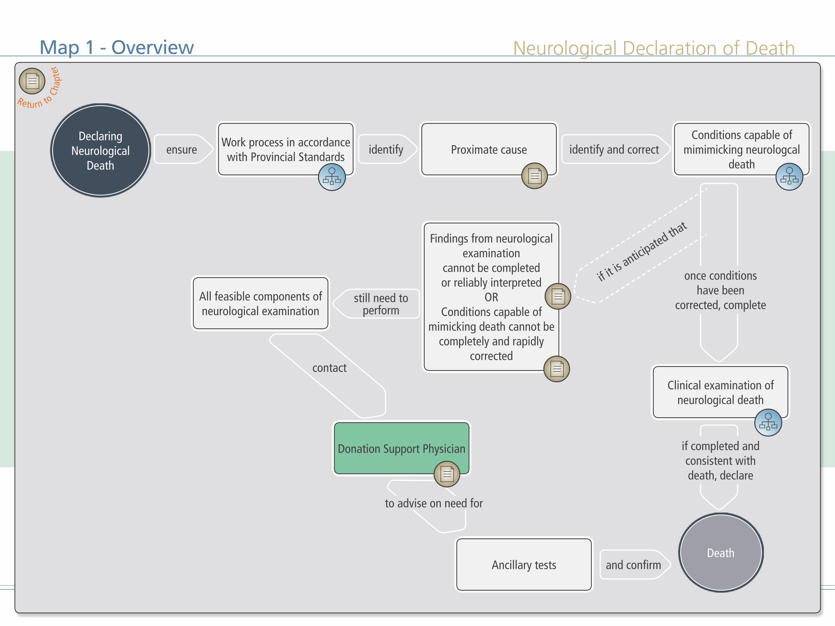
Clinical Approach (Map 1 ) Neurologic Determination of Death is a detailed clinical examination that documents the complete and irreversible loss of conscious-ness and absence of brainstem function includ-ing the capacity to breathe. In accordance with the Trillium Gift of Life Network Act, Tril-lium Gift of Life Network (TGLN) endorses recommendations from the Canadian Coun-
8 Dhanani S and Shemie SD. “Brain Death”. Pediatric Crit-ical Care Medicine: Basic Science and Clinical Evidence, 2nd Edition. Eds Derek S. Wheeler, Hector R. Wong, Thomas P. Shanley, Springer, 2014
cil for Donation and Transplantation (CCDT) 2003 Forum on Severe Brain Injury to Neu-rological Determination of Death, and 2007 Forum on Brain Blood Flow in the Neuro- logical Determination of Death. Adaptations have been made based on other newer recom-mendations9,10,11.
The physician must work in accordance with provincial standards (Map2 ) to iden-tify irreversible proximate cause for death and account for conditions capable of mim-icking neurologic death (Map 3 ). Once potential confounding conditions are identified and corrected, the physician with appropriate skills and knowledge must perform a clinical neurologic exam confirming neurologic death (Map 4 ). Once the exam is completed and is consistent with neurologic death, the pronouncement of death with specific
9 Shemie SD, Doig C, Dickens B, Byrne P et al. Severe brain injury to neurological determination of death: Canadian Council for Donation and Transplantation Forum recom-mendations. CMAJ. 2006 Mar 14;174(6):S1-13.
10 Shemie SD, Lee D, Sharpe M, Tampieri D, Young B; Ca-nadian Critical Care Society. Brain blood flow in the neu-rological determination of death: Canadian expert report. Can J Neurol Sci. 2008 May;35(2):140-5.
11 Wijdicks EF, Varelas PN, Gronseth GS, Greer DM; Amer-ican Academy of Neurology.Evidence-based guideline up-date: determining brain death in adults: report of the Qual-ity Standards Subcommittee of the American Academy of Neurology. Neurology. 2010 Jun 8;74(23):1911-8.
documentation is completed. A second exam is necessary for the purposes of organ donation, consistent with existing provincial laws.
If conditions capable of mimicking neuro- logical death cannot be corrected or if any part of the neurological exam cannot be reliably interpreted or fully performed, then the appropriate use of ancillary testing, where specifically indicated, is required. Even with the addition of ancillary testing, all feasible comp-onents of the clinical neurological exam should be completed by two physicians. Once ancillary testing confirms the findings of the clini-cal exam, then the pronouncement of neuro- logic death is complete. Contacting the TGLN Donation Support Physician may be appro- priate to discuss best practices for management of neurological determination of death and organ donation.
8Organ Donation in Ontario: A Guide for Critical Care Residents - Neurological Declaration of Death
ConclusionThe neurologic determination of death is currently the standard of practice as one of the prereq-uisites to organ donation in ventilated patients with irreversible cessation of all clinical functions of the brain. It is being used to initiate withdrawal of mechanical support discussions and is a prerequisite to organ donation. Its concept has been internationally accepted as a medical and legal definition of death in many countries with advanced health care systems. Brain death is defined as the irreversible loss of the capacity for consciousness combined with the irreversible loss of all brainstem functions, including the capacity to breathe. NDD remains a fundamentally clinical examination establishing a clear mechanism for catastrophic brain injury in the absence of confounding or reversible factors. Ancillary radiologic testing is only recommended when a full clinical exam cannot be reliably completed or when mimickers of neurological death cannot be rapidly and completely reversed. Stringent legal and clinical criteria exist to ensure transpar-ency, safety and trust in the process.
Neurological Declaration of DeathMap 1 - Overview
Work process in accordance with Provincial Standards
All feasible components of neurological examination
Ancillary tests
Clinical examination of neurological death
Donation Support Physician
Findings from neurological examination
cannot be completed or reliably interpreted
ORConditions capable of
mimicking death cannot be completely and rapidly
corrected
Proximate causeConditions capable of
mimimicking neurologcal death
Declaring Neurological
Death
Death
ensure identify
and confirm
identify and correct
once conditions have been
corrected, complete
if completed and consistent with death, declare
contact
if it is a
nticipated that
still need to perform
to advise on need for
Return to
Cha
pter

Neurological Declaration of DeathMap 2 - Provincial Standards
Neonates(less than 30 days)
Children (1 year or older), adolescents, and adults
Infants (30 days to less than 1 year)
At different times with a minimum of 24 hours
between determinations
At different times with no specified time interval between determinations
Simultaneously or at different times with no specified time interval
between determinations
Minimum of two determinations
No association to the proposed recipient
ANDFull and current licensure for independent
medical practice in the relevant Canadian jurisdictionAND
Skill and knowledge in the management of patients with severe brain injury and in NDD in the patient's
age group
Physicians
Declaring Neurological
Death
Working in Accordance with
Provincial Standards
with
done done either done
which specify
if performed in
Return to
Cha
pter

Neurological Declaration of DeathMap 3 - Mimickers of Neurological Death
Declaring Neurological
Death
Identifying Conditions
Capable of Mimicking Neurological
Death
Hypothermia
Peripheral nerve or muscle dysfunction
Neuromuscular blockade
Clinically significant drug intoxications, liver and
renal dysfucntion - hypothermia
Electrolyte abnormalities
Inborn errors of metabolism, severe liver and/or renal
dysfunction
Wait until medication has beenmetabolized/excreted, or give
antidotes
Unresuscitated shock
Hypoxic brain damage secondary to cardiac arrest
24 hours following return of spontaneous circulation
Clinical Examinaition of
Neurological Death
consider
wait for
if deemed
uncorrectable
correct, then proceed to
either
then proceed to
Consult the Donation Support Physician
if unsure
Defined as: Core temperature < 340 C in infants, children and adults: core temperature < 360 C in newborn
Return to
Cha
pter

Neurological Declaration of Death
Neonates (less than 30 days and minimum time from birth of 48h, and correct
gestational age of 36 weeks)add both oculocephalic and suck reflexassessment. Minimum core temperature
36 degrees C
recognize
Apnea test
Infants (30 days to less than 1 year)
add oculocephalic reflexassessment
Adults, adolescents,and children (1 year or older)
Deep unresponsive coma
Chronic respiratory insufficiency and responsiveness
to only supranormal levels of carbon dioxide
ORPatients dependent on
hypoxic drive
Cough and Gag ReflexesCorneal ReflexesVestibulo Ocular Response
Map 4 - Clinical Examination of Neurological Death
Declaring Neurological
Death
Clinical Examination of Neurological
Death
Assessment
Brainstem reflexes
Donation Support Physician
perform
perform
confirm
assess
consult
if performed in
Return to
Cha
pter
Select image to see a demonstration of oculocephalic relex. Requires internet connection.

13Organ Donation in Ontario: A Guide for Critical Care Residents - Neurological Declaration of Death
DescriptionIdentification of a proximate cause of death is a prerequisite to NDD. There must be definite clinical or neuroimaging evidence of an acute central nervous system (CNS) event consis-tent with the irreversible loss of neurological function1.
1 Shemie SD, Doig C, Dickens B, et al. Severe brain injury to neurological determination of death: Canadian forum recommendations. CMAJ : Canadian Medical Association Journal. 2006;174(6):S1-S12. doi:10.1503/cmaj.045142.
RationaleIn order for NDD to be declared, a definitive cause of death must be identified and condi-tions mimicking neurological death must be ruled out. The identified proximate cause of death must be consistent with irreversible loss of neurological function1.Examples of conditions leading to neurological death may include (but are not limited to) the following:
• Acute brain injury - e.g. cerebrovascular accidents, head trauma, intracranial tumors, intracranial hemorrhage or acute hydrocephalus
• Hypoxic-ischemic injury - e.g. resuscitated cardiac arrest, near drowning asphyxia, hypovolemic shock
• CNS infection - e.g. meningitis, encephali-tis
• Miscellaneous - e.g. Metabolic encepha-lopathy from liver disease, diabetic ketoac-idosis, metabolic disorders, acute hypona-tremia or vasculitis2.
2 "Determination of Death". Donation Resource Manual: A Tool To Assist Hospitals With The Process Of Organ And Tissue Donation. 6th ed. Toronto: Queen's Printer, 2014. Print.
Identifying Proximate CauseKim Bowman
Return to overview map
However these conditions must also be associ-ated evidence of increased intracranial pressure or cerebral edema in order to lead to irreversible neuronal injury. In the clinical judgement of the treating physician, the proximate cause of death must be confirmed by a clinical presenta-tion (e.g. prolonged circulatory arrest) or neuro-imaging (e.g. cerebral edema on CT head) that fully explain neurological death1. NDD for the purposes of donation cannot take place unless a proximate cause has been established.
14Organ Donation in Ontario: A Guide for Critical Care Residents - Neurological Declaration of Death
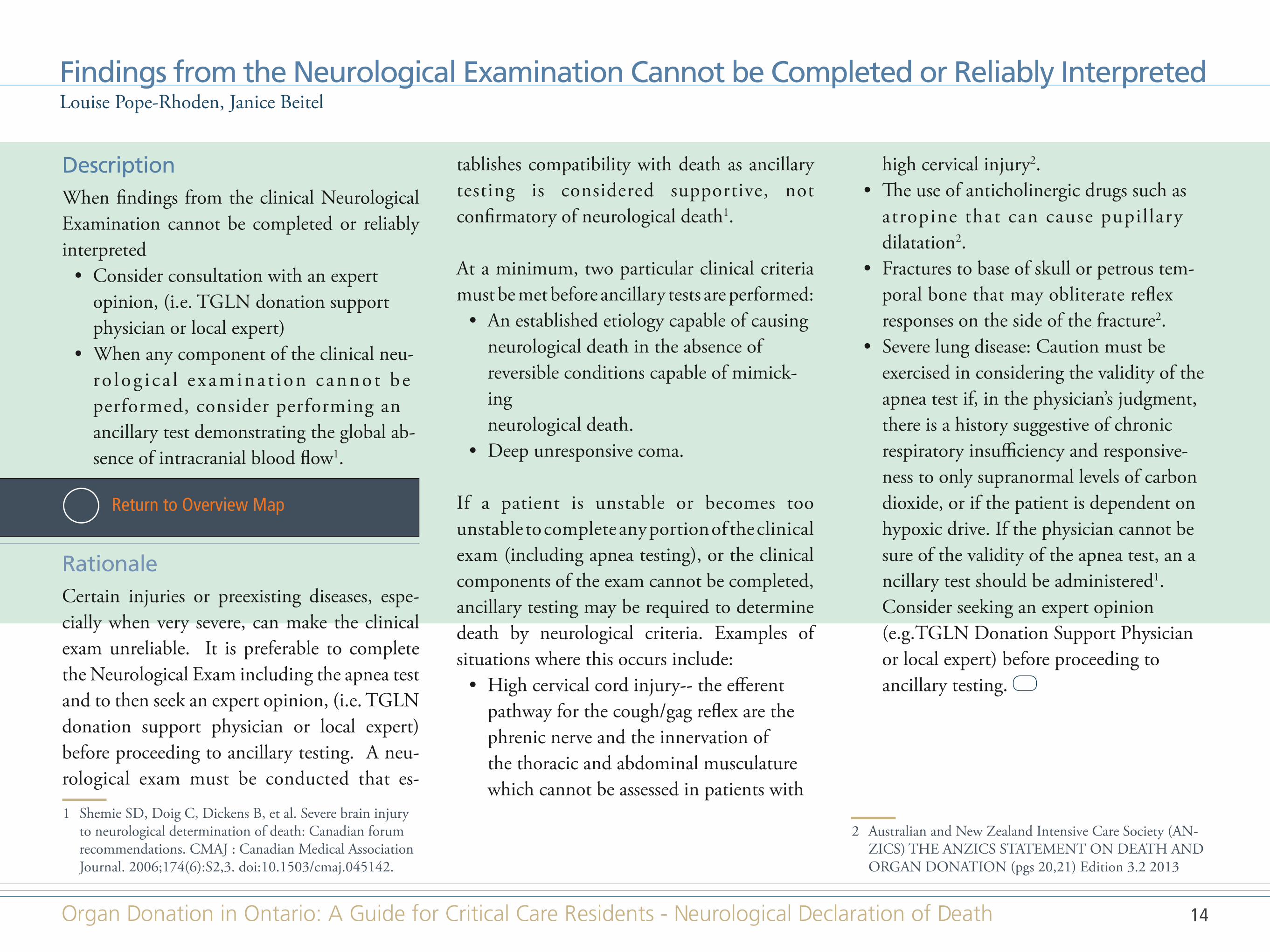
DescriptionWhen findings from the clinical Neurological Examination cannot be completed or reliably interpreted
• Consider consultation with an expert opinion, (i.e. TGLN donation support physician or local expert)
• When any component of the clinical neu-ro log i c a l e x amina t ion c anno t be performed, consider performing an ancillary test demonstrating the global ab-sence of intracranial blood flow1.
RationaleCertain injuries or preexisting diseases, espe-cially when very severe, can make the clinical exam unreliable. It is preferable to complete the Neurological Exam including the apnea test and to then seek an expert opinion, (i.e. TGLN donation support physician or local expert) before proceeding to ancillary testing. A neu-rological exam must be conducted that es-
1 Shemie SD, Doig C, Dickens B, et al. Severe brain injury to neurological determination of death: Canadian forum recommendations. CMAJ : Canadian Medical Association Journal. 2006;174(6):S2,3. doi:10.1503/cmaj.045142.
Return to Overview Map
tablishes compatibility with death as ancillary testing is considered supportive, not confirmatory of neurological death1.
At a minimum, two particular clinical criteria must be met before ancillary tests are performed:
• An established etiology capable of causing neurological death in the absence of reversible conditions capable of mimick-ing neurological death.
• Deep unresponsive coma.
If a patient is unstable or becomes too unstable to complete any portion of the clinical exam (including apnea testing), or the clinical components of the exam cannot be completed, ancillary testing may be required to determine death by neurological criteria. Examples of situations where this occurs include:
• High cervical cord injury-- the efferent pathway for the cough/gag reflex are the phrenic nerve and the innervation of the thoracic and abdominal musculature which cannot be assessed in patients with
high cervical injury2.• The use of anticholinergic drugs such as
atropine that can cause pupillary dilatation2.
• Fractures to base of skull or petrous tem-poral bone that may obliterate reflex responses on the side of the fracture2.
• Severe lung disease: Caution must be exercised in considering the validity of the apnea test if, in the physician’s judgment, there is a history suggestive of chronic respiratory insufficiency and responsive-ness to only supranormal levels of carbon dioxide, or if the patient is dependent on hypoxic drive. If the physician cannot be sure of the validity of the apnea test, an a ncillary test should be administered1. Consider seeking an expert opinion (e.g.TGLN Donation Support Physician or local expert) before proceeding to ancillary testing.
2 Australian and New Zealand Intensive Care Society (AN-ZICS) THE ANZICS STATEMENT ON DEATH AND ORGAN DONATION (pgs 20,21) Edition 3.2 2013
Findings from the Neurological Examination Cannot be Completed or Reliably InterpretedLouise Pope-Rhoden, Janice Beitel

15Organ Donation in Ontario: A Guide for Critical Care Residents - Neurological Declaration of Death
DescriptionMay include but are not limited to factors such as hypothermia, electrolyte abnormali-ties, neuromuscular blockage etc.
• If such conditions cannot be complete-ly or rapidly corrected, a clinical exam should still be conducted to the extent possible.
• An ancillary test should be considered1.• Consult with the donation support physi-
cian (DSP) or local expert should be considered before ordering any ancillary tests to ensure that all efforts have been made to perform neurological declara-tion of death (NDD) using clinical criteria including the apnea test.
1 Shemie SD, Doig C, Dickens B, et al. Severe brain injury to neurological determination of death: Canadian forum recommendations. CMAJ : Canadian Medical Association Journal. 2006;174(6):S1-S12. doi:10.1503/cmaj.045142.
RationaleConditions capable of mimicking neurologi-cal death have been well cited in numerous do-nation related documents 1,2. In order to de-termine neurological declaration of death for the purposes of donation, conditions capa-ble of mimicking neurological death3 should be identified and corrected. Ancillary testing should be considered if in the physician’s judge-ment some of these conditions cannot be fully corrected or if correction of a mimicker of neu-rological death (e.g. a serum sodium of >160), would unduly prolong the time period before donation as perceived by the donor’s family. Because ancillary testing is considered support-ive, not confirmatory of neurological death, a neurological exam must be conducted that establishes (to the extent possible) compatibility with death2. Consider seeking an expert opin-ion (e.g.TGLN Donation Support Physician or local expert) before proceeding to ancillary testing.
2 "Determination Of Death". Donation Resource Manu-al: A Tool To Assist Hospitals With The Process Of Organ And Tissue Donation. 6th ed. Toronto: Queen's Printer, 2014. Print.
3 Shemie, Sam D. et al. "International Guideline Develop-ment For The Determination Of Death". Intensive Care Med 40.6 (2014): 788-797. Web.
Conditions Capable of Mimicking Death Cannot be Completely or Rapidly CorrectedKim Bowman
Return to overview map

16Organ Donation in Ontario: A Guide for Critical Care Residents - Neurological Declaration of Death
DescriptionContact Donation Support Physician (DSP) or local expert: TGLN 1-877-363-8456
RationaleThe Donation Support Physician (DSP) is an intensivist on-call and available by phone 24/7 within Ontario. Their main role is to support and advise the provincial resource centre, the organ and tissue donation coordinator, and the
most responsible physician. Discussions might include donor identification, declaration of death, organ suitability, and donation logistics.
Consultation with a physician expert from TGLN prior to declaration of death may appear to represent a conflict of interest. However, it is well recognized that appropriate expertise and experience for NDD is greatest at the OPO with the organ and tissue donation coordinator and the donation support physician. This is an im-portant mechanism to support this process and physicians invested in the donation system will
be committed to the integrity of this system. A consultation to the DSP is always done with acknowledgements of ethical practice standards and with the utmost professionalism
When findings from neurological examination cannot performed or reliably interpreted OR conditions mimicking neurological death can-not be corrected, it is important to consider contacting either the DSP or a local expert to discuss concerns or performing ancillary tests. Even though TGLN documents suggest that ancillary tests may be performed in these situa-tions, these tests should only be ordered during appropriate situations while considering risks and benefits of the chosen ancillary test The DSP or local expert can help determine their appropriateness given the clinical context. If examination according to the minimum clini-cal criteria (to the extent possible) is compati-ble with death, an ancillary test demonstrating the global absence of intracranial blood flow supports the declaration of death1. Any ancillary test is considered supportive, not con-
1 Shemie SD, Doig C, Dickens B, et al. Severe brain injury to neurological determination of death: Canadian forum recommendations. CMAJ : Canadian Medical Association Journal. 2006;174(6):S1-S12. doi:10.1503/cmaj.045142.
firmatory, of neurologic death.
Contacting the DSP or local expert should be considered before ordering any ancillary tests to ensure that all efforts have been made to perform NDD using clinical criteria includ-ing the apnea test. It is our experience that on occasion, with advice from a DSP or local expert, it still may be possible to perform NDD using clinical criteria obviating the need for an-cillary tests. If after consultation with the DSP or local expert it is deemed that an ancillary test is required, the DSP/local expert can also help guide the selection of the most appropriate ancillary test given the clinical situation and local expertise.
Contact Donation Support PhysicianEli Malus, Sonny Dhanani
Return to overview map

17Organ Donation in Ontario: A Guide for Critical Care Residents - Neurological Declaration of Death
DescriptionThe brainstem examination is different in neonates (36 weeks, gestation to 29 days old (corrected for gestational age)1:
• Minimum clinical criteria include absence of oculocephalic reflex and suck reflex.
• Minimum temperature must be a core temperature 36°C1.
• Minimum time from birth to first deter-mination is 48 h1.
• Two examinations are required, by two
different physicians, at a minimum time interval of 24 hours between each exam-ination is required1.
• Ancillary testing, as defined by demonstra-tion of the absence of intracranial blood flow, should be performed when any of the minimum clinical criteria cannot be established or confounding factors remain
1 Shemie SD, Doig C, Dickens B, et al. Severe brain injury to neurological determination of death: Canadian forum recommendations. CMAJ : Canadian Medical Association Journal. 2006;174(6):S2,3. doi:10.1503/cmaj.045142.
unresolved2.• The physicians performing NDD should
have special expertise in the patient’s age group. The minimum level of physician qualifications should be understood as specialists with skill and knowledge in the management of newborns/infants/chil-dren and/or adolescents, respectively with brain injury and the determination of death based on neurological criteria1.
RationaleIt is prudent to have an independent examina-tion because of the lack of collective experience and research on brain death in this age group a repeat examination no less than 24 hours apart is recommended to ensure independent confirmation by another qualified physician, regardless of the primary mechanism of the brain injury. Accuracy of gestational age should be supported by clinical history (e.g., dates and prenatal ultrasound) and physical examination. Inability to confirm a gestational age > 36 weeks should preclude NDD.
2 CCDT 2003 Severe brain injury to neurological determi-nation of death Canadian Forum Report and Recommen-dations
Should uncertainty or confounding issues arise that cannot be resolved, the time interval may be extended according to physician judgement, or an ancillary test demonstrating absence of intracranial blood flow may be used2. The 48 hour recommendation from injury to first determination reflects a reduced certainty of neurological prognostication in the newborn1.
Determining Neurological Death in Neonates According to Provincial StandardsLouise Pope-Rhoden
Return to sub map
Return to overview map
18Organ Donation in Ontario: A Guide for Critical Care Residents - Neurological Declaration of Death
Return to sub map
Return to overview map
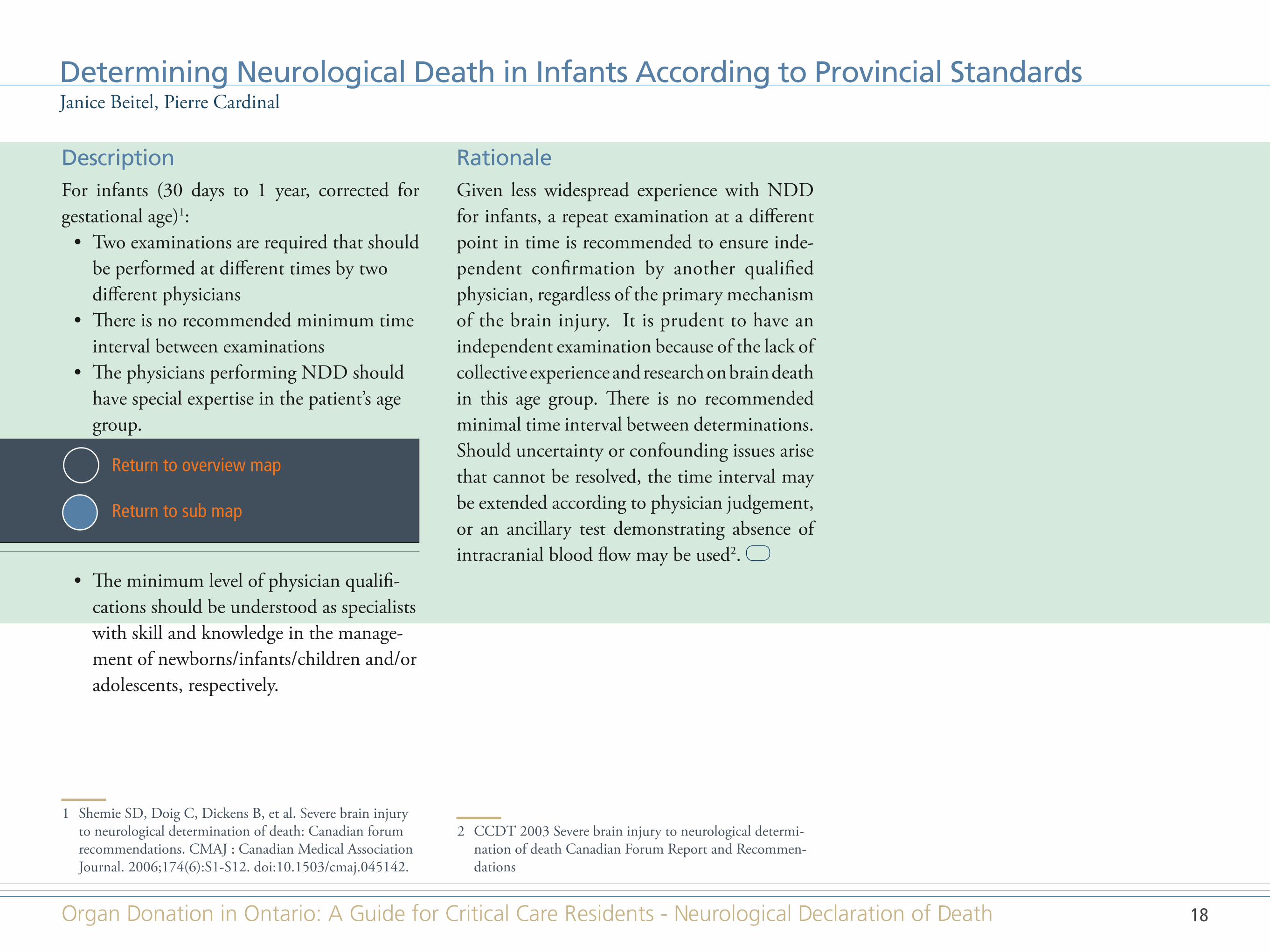
DescriptionFor infants (30 days to 1 year, corrected for gestational age)1:
• Two examinations are required that should be performed at different times by two different physicians
• There is no recommended minimum time interval between examinations
• The physicians performing NDD should have special expertise in the patient’s age group.
• The minimum level of physician qualifi-cations should be understood as specialists with skill and knowledge in the manage-ment of newborns/infants/children and/or adolescents, respectively.
1 Shemie SD, Doig C, Dickens B, et al. Severe brain injury to neurological determination of death: Canadian forum recommendations. CMAJ : Canadian Medical Association Journal. 2006;174(6):S1-S12. doi:10.1503/cmaj.045142.
RationaleGiven less widespread experience with NDD for infants, a repeat examination at a different point in time is recommended to ensure inde-pendent confirmation by another qualified physician, regardless of the primary mechanism of the brain injury. It is prudent to have an independent examination because of the lack of collective experience and research on brain death in this age group. There is no recommended minimal time interval between determinations. Should uncertainty or confounding issues arise that cannot be resolved, the time interval may be extended according to physician judgement, or an ancillary test demonstrating absence of intracranial blood flow may be used2.
2 CCDT 2003 Severe brain injury to neurological determi-nation of death Canadian Forum Report and Recommen-dations
Determining Neurological Death in Infants According to Provincial StandardsJanice Beitel, Pierre Cardinal

19Organ Donation in Ontario: A Guide for Critical Care Residents - Neurological Declaration of Death
Return to sub map
Return to overview map
Description• Core temperature should be obtained
through central blood, rectal or esopha-geal–gastric measurement1.
• The core body temperature required to apply the minimum clinical criteria should be 34°C in infants, children, and adults2.
• The brainstem examination is different in neonates, for newborns aged 36 weeks gestation to 29 days old (corrected for
gestational age) minimal core body temperature should be 36°C1.
1 Shemie SD, Doig C, Dickens B, et al. Severe brain injury to neurological determination of death: Canadian forum recommendations. CMAJ : Canadian Medical Association Journal. 2006;174(6):S2-4. doi:10.1503/cmaj.045142.
2 Trillium Gift of Life Network (TGLN), Guidelines for the neurological determination of death (NDD) for the pur-poses of organ donation in ontario: adult and paediatric patients 1 year and older
RationaleWhen body temperature is below normal, hypo-thermia should be considered as a confounding factor which might prevent the observation of neurologic responses and/or mimic neurologi-cal death. Confounding conditions that may invalidate testing for cessation of brain function include naturally occurring and/or therapeutic hypothermia3. There is little evidence to inform the threshold temperature below which NDD becomes unreliable. Given the lack of evidence, a consensus decision was made to adopt 36°C and 34°C as rational, safe and attainable standards in neonates and in any patients older than 1 month, respectively. Ideally, temperature should be as close to normal as possible as these threshold tempera-tures are the minimal temperature at which the test is considered valid. Raising a patient’s tem-perature to 34°C (or to 36°C in neonates) is also easy to achieve1.
In neonates, it is also important to ensure the accuracy of gestational age based on clinical
3 S. D. Shemie, L. Hornby, A. Baker, et al. Internation-al guideline development for the determination of brain death Intensive Care Med (2014) 40:791 DOI 10.1007/s00134-014-3242-7
history (e.g. dates and prenatal ultrasound) and physical examination. Inability to confirm a gestational age >36 weeks should preclude NDD1. The higher recommended temperature threshold in neonates reflects uncertainty about hypothermic effects on neurological function in the newborn and the fact that normothermia is an easily attainable standard1.
Hypothermia as a Mimicker of Neurological DeathLouise Pope-Rhoden, Janice Beitel
20Organ Donation in Ontario: A Guide for Critical Care Residents - Neurological Declaration of Death
Return to sub map
Return to overview map
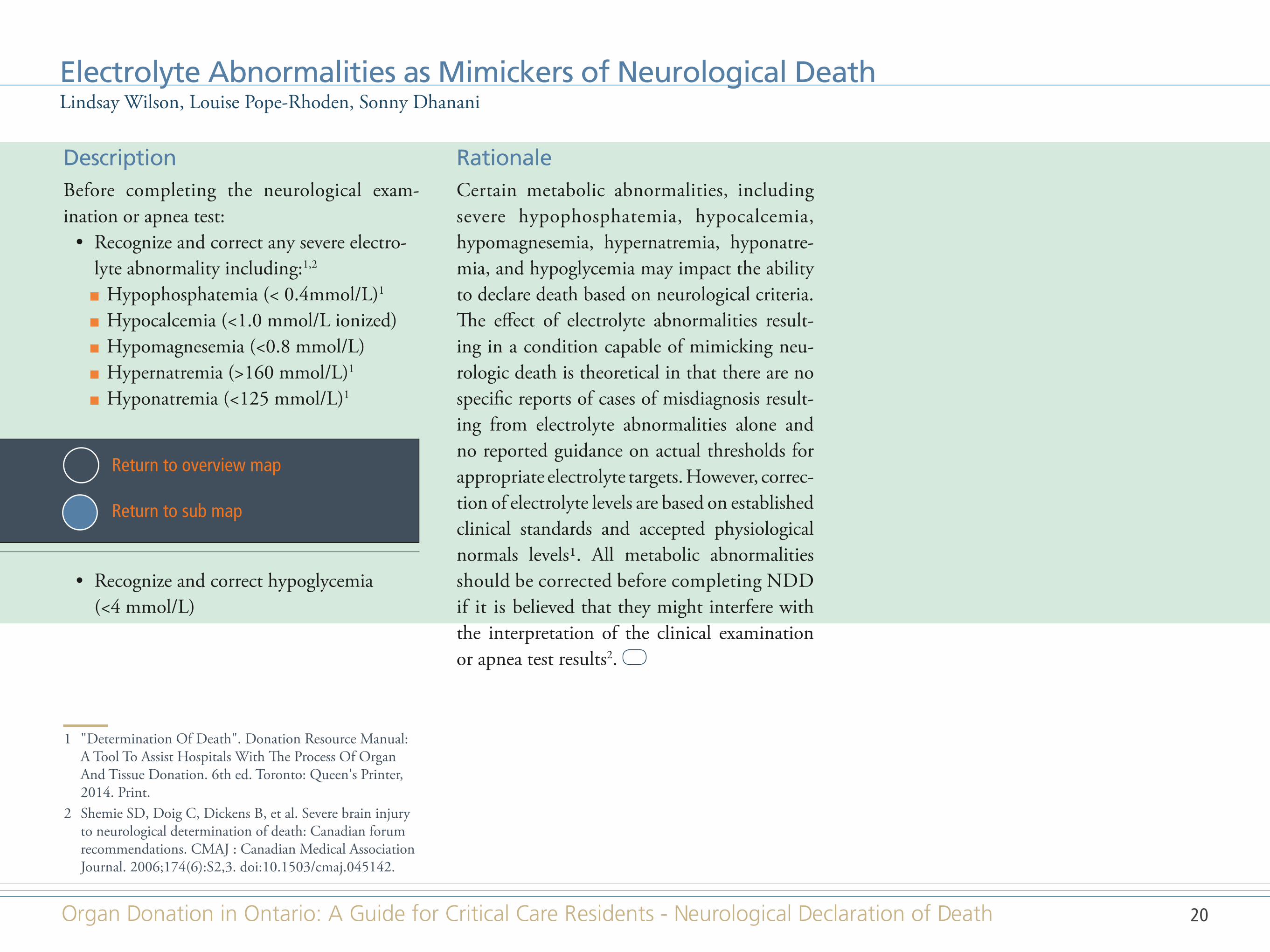
DescriptionBefore completing the neurological exam- ination or apnea test:
• Recognize and correct any severe electro-lyte abnormality including:1,2
◼ Hypophosphatemia (< 0.4mmol/L)1
◼ Hypocalcemia (<1.0 mmol/L ionized) ◼ Hypomagnesemia (<0.8 mmol/L) ◼ Hypernatremia (>160 mmol/L)1
◼ Hyponatremia (<125 mmol/L)1
• Recognize and correct hypoglycemia (<4 mmol/L)
1 "Determination Of Death". Donation Resource Manual: A Tool To Assist Hospitals With The Process Of Organ And Tissue Donation. 6th ed. Toronto: Queen's Printer, 2014. Print.
2 Shemie SD, Doig C, Dickens B, et al. Severe brain injury to neurological determination of death: Canadian forum recommendations. CMAJ : Canadian Medical Association Journal. 2006;174(6):S2,3. doi:10.1503/cmaj.045142.
RationaleCertain metabolic abnormalities, including severe hypophosphatemia, hypocalcemia, hypomagnesemia, hypernatremia, hyponatre-mia, and hypoglycemia may impact the ability to declare death based on neurological criteria. The effect of electrolyte abnormalities result-ing in a condition capable of mimicking neu-rologic death is theoretical in that there are no specific reports of cases of misdiagnosis result-ing from electrolyte abnormalities alone and no reported guidance on actual thresholds for appropriate electrolyte targets. However, correc-tion of electrolyte levels are based on established clinical standards and accepted physiological normals levels1. All metabolic abnormalities should be corrected before completing NDD if it is believed that they might interfere with the interpretation of the clinical examination or apnea test results2.
Electrolyte Abnormalities as Mimickers of Neurological DeathLindsay Wilson, Louise Pope-Rhoden, Sonny Dhanani

21Organ Donation in Ontario: A Guide for Critical Care Residents - Neurological Declaration of Death
Return to sub map
Return to overview map
DescriptionStop any neuromuscular blockade agent before performing the clinical examination or apnea test for NDD1.
1 Shemie SD, Doig C, Dickens B, et al. Severe brain injury to neurological determination of death: Canadian forum recommendations. CMAJ : Canadian Medical Association Journal. 2006;174(6):S1-S12. doi:10.1503/cmaj.045142.
2. International guideline development for the determination of death Intensive Care Med (2014) 40:788–797 DOI 10.1007/s00134-014-3242-7
RationaleNeuromuscular blocking agents inhibit the action of acetylcholine by blocking neuromus-cular transmission and thus causing paralysis of all skeletal muscles. Common agents include vecuronium, pancuronium, cisatracurium and rocuronium. Depolarizing agents such as suc-cinylcholine are short acting and often not a consideration in the ICU patient but should also be recognized. Neuromuscular blockade will affect all striated muscles including the diaphragm and therefore will mimic unrespon-siveness and lead to absence of breathing during apnea tests1,2. Cranial nerve functions are also affected with the exception of the pupillary light reflex which is preserved in normal patients on neuromuscular blocking agents. Thus, any neu-romuscular blockade agent should be stopped before performing the clinical examination or apnea test for NDD2. If any concerns about persisting pharmacologic neuromuscular block-ade, train of four testing to confirm reversal of paralysis should be conducted.
Neuromuscular Blockade as a Mimicker of Neurological DeathKim Bowman, Janice Beitel

22Organ Donation in Ontario: A Guide for Critical Care Residents - Neurological Declaration of Death
DescriptionRecognize and treat unresuscitated shock:
• Persistent hypotension despite fluid resuscitation and vasopressors
AND/OR• Persistent hypoperfusion despite fluid
resuscitation or inotropes1.• In the presence of shock refractory to
resuscitative measures, contact the dona-tion support physician (DSP) or local expert before proceeding with NDD or ordering ancillary tests.
1 "Determination Of Death". Donation Resource Manual: A Tool To Assist Hospitals With The Process Of Organ And Tissue Donation. 6th ed. Toronto: Queen's Printer, 2014. Print.
Return to sub map
Return to overview map
RationaleShock is associated with inadequate oxygenated circulation to the brain2. In the presence of unresuscitated shock (i.e. persistent hypoten-sion despite fluid resuscitation and vasopres-sors or persistent hypoperfusion despite fluid resuscitation or inotropes), the motor response, cranial nerve examination and apnea test may be falsely positive mimicking neurological death. It is important to assess for both hypo-tension and hypoperfusion and to correct the shock state prior to proceeding with a diagnosis of brain death for the purposes of donation. In the presence of shock refractory to all resus-citative measures, a consultation with the DSP or local expert should be considered before ordering ancillary tests2.
2 International guideline development for the determination of death Intensive Care Med (2014) 40:788–797 DOI 10.1007/s00134-014-3242-7
Unresuscitated Shock as a Mimicker of Neurological DeathKim Bowman, Janice Beitel

23Organ Donation in Ontario: A Guide for Critical Care Residents - Neurological Declaration of Death
Return to sub map
Return to overview map
DescriptionConsider peripheral nerve or muscle dys-function as mimickers of neurological death especially in the presence of diseases that may lead to severe neuromuscular dysfunction such as:
• Myasthenia gravis• Guillain-Barre Syndrome• Botulism
RationaleCertain peripheral nerve and muscle diseases, especially when very severe, can make it diffi-cult to interpret the result of the neurological exam especially the apnea test. In the presence of severe neuromuscular diseases, the motor response, cranial nerve examination and apnea test may be falsely positive mimicking neuro-logical death. In such cases, it is preferable to complete the clinical neurological examination, including the apnea test, and to then seek a second opinion or consultation with neurology. Ancillary testing may be warranted to confirm absence of brain blood flow. An expert opinion (e.g. TGLN donation support physician or a local expert) before proceeding to ancillary test-ing should also be considered1.
1 Shemie SD, Doig C, Dickens B, et al. Severe brain injury to neurological determination of death: Canadian forum recommendations. CMAJ : Canadian Medical Association Journal. 2006;174(6):S1-S12. doi:10.1503/cmaj.045142.
Peripheral Nerve or Muscle Dysfunction as a Mimicker of Neurological DeathKarim Soliman

24Organ Donation in Ontario: A Guide for Critical Care Residents - Neurological Declaration of Death
Return to sub map
Return to overview map
DescriptionAlways perform a detailed review of the past medical history to exclude inborn errors of metabolism (IEM).
RationaleInborn errors of metabolism (IEM) may result in unresponsiveness and preclude the diagnosis of neurological determination of death. IEM typically present in the neonatal pe-riod or infancy but can occur at any time, even in adulthood1. Potential symptoms include: hy-poglycemia, metabolic acidosis, and electrolyte imbalances. Diagnoses include fructose intol-erance, galactosemia, phenylketonuria (PKU)2. As a minimum consideration, a thorough review of the patient’s medical history should be conducted to exclude IEM.
Severe metabolic disorders may appear as part of the primary presentation. Symptoms and signs can complicate management for the physician and thus may impact the ability to declare death based on neurological criteria3. Attempts should
1 Emedicine.medscape.com,. "Inborn Errors Of Metabo-lism: Background, Pathophysiology, Epidemiology". N.p., 2016. Web. 2 Mar. 2016.
2 Updated by: Chad Haldeman-Englert, and the A.D.A.M. Editorial team. "Inborn Errors Of Metabolism: Medlin-eplus Medical Encyclopedia". Nlm.nih.gov. N.p., 2016. Web. 3 Mar. 2016.
3 Shemie SD, Doig C, Dickens B, et al. Severe brain injury to neurological determination of death: Canadian forum recommendations. CMAJ : Canadian Medical Association Journal. 2006;174(6):S1-S12. doi:10.1503/cmaj.045142.
be made to correct any metabolic abnormality that might play interfere with the neu-rological declaration of death. If the metabolic abnormalities cannot be corrected, it is prefer-able to complete the neurological examination, including the apnea test, and then consider consultation with an expert (e.g.TGLN Donation Support Physician or a local expert) before proceeding to ancillary testing.
Inborn Errors of Metabolism as Mimickers of Neurological DeathKim Bowman, Janice Beitel

25Organ Donation in Ontario: A Guide for Critical Care Residents - Neurological Declaration of Death
Return to sub map
Return to overview map
DescriptionMany drugs can affect neurological function and may even mimic neurological death. Given the disastrous consequences of a false positive NDD, it is essential to always consider drugs as potential confounders especially in the ab-sence of a clear proximate cause of death. This can be very challenging given that a complete drug history is often missing and that current screening tests are notoriously inaccurate. In
addition, patients considered for NDD may have hepatic and renal dysfunction or be hypo- thermic which may influence the metabolism and action of various drugs in unpredictable ways. It is important to distinguish drugs that may have played a role in the primary presen-tation of coma (i.e. overdose) versus drugs that are used in routine ICU care. For lingering effects of therapeutic narcotics or sedatives that may impair appropriate neurologic examina-tion, delaying the testing for neurologic death
or considering ancillary test is warranted. A consultation with a DSP or local expert should be considered.
RationaleA negative urine “drug screen” is completely inadequate for ruling out the presence of con-founding agents because most hospital urine 'drug screens' only test for five or six classes of drugs. Even when the drug belongs to one of these classes, a negative result does not neces-sarily exclude an intoxication given the poor performance of these tests (low sensitivity and specificity)1,2,3. Positive test results do not necessarily confound the interpretation of the clinical examination for NDD. For example, patients who overdose on sympathomimetic agents (e.g. cocaine) may sustain intracranial hemorrhages and irreversible neurological in-jury as a result of the overdose, yet these agents
1 Hammett-Stabler CA, Pesce AJ, Cannon DJ: Urine drug screening in the medical setting. Clin Chim Acta 2002;315:125-135.
2 Hepler BR, Sutheimer CA, SUnshine I: The role of the toxicology laboratory in emergency medicine.II. Study of an integrated approach. J Toxicol Clin Toxicol 1984-85;22:503-528.
3 Kellerman AL, Fihn SD, Logerfro JP, et al: Impact of drug screening in suspected overdose. Ann Emerg Med 1987;16:1206-1216.
could never confound the clinical NDD giv-en their short half-lives. Opioids and sedative hypnotics are the agents most likely to con-found the NDD examination, particularly when administered at high doses, for long durations, or in patients with compromised hepatic and/or renal function. Clinicians should be proac-tive at stopping these agents as early as possi-ble when they are not required clinically (i.e. for ICP control), in anticipation of an NDD assessment. When agents are required therap- eutically, clinicians are well advised to use short acting agents that are less reliant on hepatic metabolism, such as propofol. In addition, antidotes (particularly naloxone and flumaze-nil) are not recommended as they may precipitate acute withdrawal, leading to catecholamine surges that may be injurious to vulnerable organs.
Clinicians should consider consultation with local poison centers when drug intoxica-tion may be affecting the NDD assessment. Consider seeking an expert opinion (e.g.TGLN Donation Support Physician or local expert).
Clinically Significant Drug Intoxications as Mimickers of Neurological DeathIan Ball, Pierre Cardinal

26Organ Donation in Ontario: A Guide for Critical Care Residents - Neurological Declaration of Death
Return to sub map
Return to overview map
DescriptionConsider hypoxic-ischemic brain damage secondary to cardiac arrest as mimicker of neurological death. In particular the first 24 hours post arrest where the clinical exam may be confounded.
RationaleHypoxic-ischemic injury post cardiac arrest can mimic neurological death especially in the first 24 hours post arrest. Canadian guidelines require a minimum of 24 hours between cardiac arrest and clinical declarations1. Sever-al other national neurological death declaration guidelines carry an observation period between anoxic-ischemic brain injury and neuro- logical declarations2. Current recommendation in Ontario is to delay declaration testing until 24 hours after the time of the hypoxic-ischemic event. Clinical context is important and consultation with the DSP or local expert should be considered. When hypothermia is used post arrest, the prolonged effect of sedatives due to altered drug metabolism may warrant further consideration to ensure that they too are not potential
1 Shemie SD, Doig C, Dickens B, et al. Severe brain injury to neurological determination of death: Canadian forum recommendations. CMAJ : Canadian Medical Association Journal. 2006;174(6):S1-S12. doi:10.1503/cmaj.045142.
2 Wijdicks EF. Brain death worldwide: Accepted fact but no global consensus in diagnostic criteria. Neurology 2002;58;20-25
mimickers of neurological death3,4. Testing should be delayed until the patient has been rewarmed.
3 Wijdicks EF, Determining Brain Death. Continuum: Life-long Learning in Neurology 2015;21:1419
4 Greer DM, Busl KM. Pitfalls in the Diagnosis of Brain Death. Neurocritical Care 2009; 11:276–287
Hypoxic Brain Damage Secondary to Cardiac Arrest as a Mimicker of Neurological DeathKarim Soliman

27Organ Donation in Ontario: A Guide for Critical Care Residents - Neurological Declaration of Death
DescriptionAn apnea test is typically the last of the clinical tests performed for purposes of declaring NDD and involves the following steps1:
• Attempt to achieve normal baseline arte-rial gases: pH 7.35-7.45, PaCO₂ 35-45 mmHg, PaO₂ > 100 mmHg2.
• Preoxygenate with 100% O2 for 10 min-utes2.
• Apply one of either open-circuit technique or closed-circuit technique as described
below:Open-circuit technique (“Flow” apnea testing): ◼ Disconnect ETT from ventilator and in-sert catheter into ETT to deliver O₂ at
1 Shemie SD, Ross H, Pagliarello J, Baker AJ, Greig PD, Brand T, et al. Organ donor management in Canada: rec-ommendations of the forum on Medical Management to Optimize Donor Organ Potential. CMAJ : Canadian Med-ical Association Journal 2006;174(6):S13-32.
2 "Determination Of Death". Donation Resource Manu-al: A Tool To Assist Hospitals With The Process Of Organ And Tissue Donation. 6th ed. Toronto: Queen's Printer, 2014. Print.
Return to sub map
Return to overview map
1-4 L/minute (alternatively set ventilator to achieve this effect)2.
◼ For patients with high FiO2 or PEEP requirements, the following may be prepared for use to maintain optimal oxygenation:
• Connect a BVM system with a com-plete seal (reusable bags are suggested for their air tight qualities) to the in-line O2 flow meter.
• Attach appropriate PEEP valve and swivel ETT adapter for patient sup-port.
• Inflate the BVM system with inline O2
to the minimal setting required for ad-equate inflation of the BVM system; suggest using O2 flow greater than or equal to 10 l/minute3.
Closed-circuit technique (“CPAP” ap-nea testing): ◼ The patient remains on mechanical ventilation for the duration of the apnea test
◼ Ensure the following conditions on the ventilator:
• Ventilator circuit is free of water or
3 Trillium Gift of Life Network (TGLN), Neurological De-termination of Death Policy and Procedure. 2014.
condensation• Ventilator circuit is not resting on
patient’s body• Ventilator Trigger Sensitivity setting
within reasonable range• Ventilator Apnea Alarm is off or at
most lenient allowable setting• Ventilator Apnea Backup Ventilation
setting is off or at most lenient allow-able setting
• FiO2 = 1• Change ventilator mode to CPAP /
PEEP-only• Set CPAP level to PEEP level of
ventilation settings prior to apnea test• For monitoring during the apnea test:
a) If using O2 tubing:• Draw patient’s gown to umbilicus level
to facilitate the observation of the thorax and abdomen and confirm the lack of respiratory effort
b) If using BVM with PEEP:• Observe as above• Provide one breath after bag-valve-
mask set-up has been attached• At completion of spontaneous exhala-
tion and bag-valve-mask refill, the apnea test begins3.
Apnea Testing During Clinical Examination of Neurological DeathLindsay Wilson, Edwin Poon
28
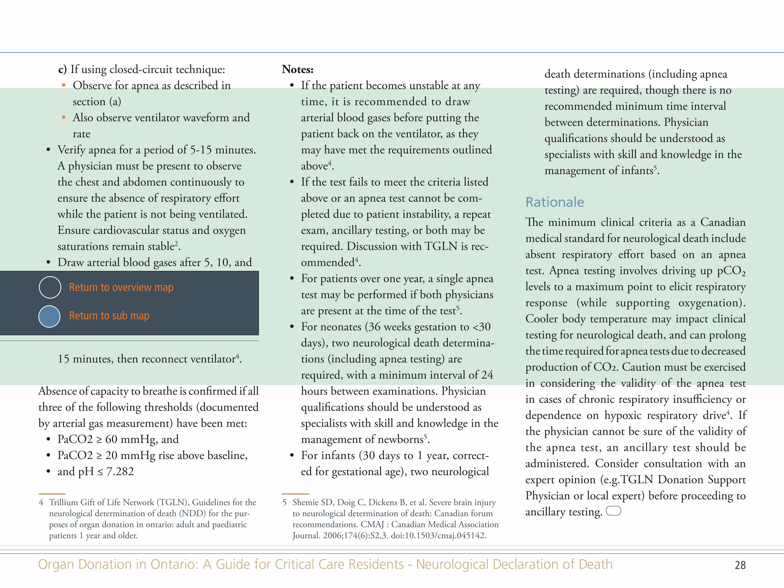
c) If using closed-circuit technique:• Observe for apnea as described in
section (a)• Also observe ventilator waveform and
rate• Verify apnea for a period of 5-15 minutes.
A physician must be present to observe the chest and abdomen continuously to ensure the absence of respiratory effort while the patient is not being ventilated. Ensure cardiovascular status and oxygen saturations remain stable2.
• Draw arterial blood gases after 5, 10, and
15 minutes, then reconnect ventilator4. Absence of capacity to breathe is confirmed if all three of the following thresholds (documented by arterial gas measurement) have been met:
• PaCO2 ≥ 60 mmHg, and • PaCO2 ≥ 20 mmHg rise above baseline,• and pH ≤ 7.282
4 Trillium Gift of Life Network (TGLN), Guidelines for the neurological determination of death (NDD) for the pur-poses of organ donation in ontario: adult and paediatric patients 1 year and older.
Notes:• If the patient becomes unstable at any
time, it is recommended to draw arterial blood gases before putting the patient back on the ventilator, as they may have met the requirements outlined above4.
• If the test fails to meet the criteria listed above or an apnea test cannot be com-pleted due to patient instability, a repeat exam, ancillary testing, or both may be required. Discussion with TGLN is rec-ommended4.
• For patients over one year, a single apnea test may be performed if both physicians are present at the time of the test5.
• For neonates (36 weeks gestation to <30 days), two neurological death determina-tions (including apnea testing) are required, with a minimum interval of 24 hours between examinations. Physician qualifications should be understood as specialists with skill and knowledge in the management of newborns5.
• For infants (30 days to 1 year, correct-ed for gestational age), two neurological
5 Shemie SD, Doig C, Dickens B, et al. Severe brain injury to neurological determination of death: Canadian forum recommendations. CMAJ : Canadian Medical Association Journal. 2006;174(6):S2,3. doi:10.1503/cmaj.045142.
death determinations (including apnea testing) are required, though there is no recommended minimum time interval between determinations. Physician qualifications should be understood as specialists with skill and knowledge in the management of infants5.
RationaleThe minimum clinical criteria as a Canadian medical standard for neurological death include absent respiratory effort based on an apnea test. Apnea testing involves driving up pCO₂ levels to a maximum point to elicit respiratory response (while supporting oxygenation). Cooler body temperature may impact clinical testing for neurological death, and can prolong the time required for apnea tests due to decreased production of CO2. Caution must be exercised in considering the validity of the apnea test in cases of chronic respiratory insufficiency or dependence on hypoxic respiratory drive4. If the physician cannot be sure of the validity of the apnea test, an ancillary test should be administered. Consider consultation with an expert opinion (e.g.TGLN Donation Support Physician or local expert) before proceeding to ancillary testing.
Organ Donation in Ontario: A Guide for Critical Care Residents - Neurological Declaration of Death
Return to sub map
Return to overview map

29Organ Donation in Ontario: A Guide for Critical Care Residents - Neurological Declaration of Death
Return to sub map
Return to overview map
DescriptionRecognize chronic respiratory insufficiency and response to only supranormal levels of carbon dioxide as special consideration when undergo-ing apnea testing in setting of neurological test.
RationaleThe main objective of apnea testing is to test respiratory drive via a carbon dioxide challenge. In patients with chronic carbon dioxide reten-tion due to COPD or other diseases, central and peripheral chemoreceptors have been reset to the new baseline carbon dioxide levels and the kidneys compensate for the chronic respira-tory acidosis1,2. Special consideration should be given to these patients so that their pre-apnea exam arterial blood gas is normalized to their baseline. This will prevent the apnea testing from being unnecessarily prolonged and hence is safer for the patient.
The guidelines also suggest that in patients with chronic respiratory insufficiency who only respond to supranormal levels of carbon di-oxide, the validity of the apnea exam must
1 Acid-Base Response to Chronic Hypercapnia in Man. Newton C. Brackett, Jr., M.D., Charles F. Wingo, M.D., Orhan Muren, M.D., and Jose T. Solano, M.D. N Engl J Med 1969; 280:124-130
2 Lumb, BL. Nunn’s Applied Respiratory Physiology. 7th edition. Churchill Livingston, c2016. Chapter 5: Control of Breathing.
Recognition of Chronic Respiratory Insufficiency and Response to Supranormal Levels of Carbon Dioxide During Apnea TestingKarim Soliman
be considered with caution3. In these cases, consultation with expert opinion should be considered (e.g. TGLN donation support phy-sician or a local expert) to discuss the use of ancillary testing based on this clinical context.
3 Shemie SD, Doig C, Dickens B, et al. Severe brain injury to neurological determination of death: Canadian forum recommendations. CMAJ : Canadian Medical Association Journal. 2006;174(6):S1-S12. doi:10.1503/cmaj.045142.

30Organ Donation in Ontario: A Guide for Critical Care Residents - Neurological Declaration of Death
Return to sub map
Return to overview map
DescriptionRecognize patients with central hypoventilation as a contraindication to interpreting the apnea test for neurological death declarations1.
1 Shemie SD, Doig C, Dickens B, et al. Severe brain injury to neurological determination of death: Canadian forum recommendations. CMAJ : Canadian Medical Association Journal. 2006;174(6):S1-S12. doi:10.1503/cmaj.045142.
RationaleThe Canadian guideline recommends the iden-tification of patients who are dependent on their hypoxic drive to breathe1. Such patients usually suffer from either congenital or acquired central hypoventilation. Recently, congenital central hypoventilation syndrome has been to be linked to mutations of the PHOX2B gene2. Acquired hypoventilation is associated with traumatic, ischemic, and inflammatory processes affecting the brain-stem3. Congenital disorders such as myelo-meningocele associated with Arnold Chiari Type II malformation may also lead to central hypoventilation. The pathogenesis of hypoven-tilation in these patients is usually multi- factorial denoting conditions resulting from underlying neurologic disorders affecting the sensors ( i.e. peripheral and central chemore-ceptors, and mechanoreceptors) and/or the central controller that receive input and
2 Amiel J, Laudier B, Attie-Bitach T, et al. Polyalanine ex-pansion and frameshift mutations of the paired like ho-meobox gene PHOX2B in congenital central hypoventila-tion syndrome. Nat Genet 2003;33(4): 459–61.
integrate the response from sensors3,4. Most patients not only have an impaired response to hypercarbia but also to hypoxemia. Such patients would not be adequately challenged with rising carbon dioxide levels thus any concerns with the performance or interpreta-tion of apnea testing warrants ancillary testing. In these cases, it is important that consultation with expert opinion should be considered (e.g. TGLN donation support physician or a local expert) to discuss the use of ancillary testing.
3 Muzumdar H, Arens R. Central Alveolar Hypoventilation Syndromes. Sleep Medicine Clinics. 2008;3:601-615.
4 Ramanantsoa N, Gallego J. Congenital central hypoventi-lation syndrome. Respiratory Physiology & Neurobiology. 2013;189:272-279.
Recognition of Patients Dependant on Hypoxic Drive During Apnea TestingKarim Soliman, Pierre Cardinal

31Organ Donation in Ontario: A Guide for Critical Care Residents - Neurological Declaration of Death
Description• Assess level of consciousness (Glasgow
Coma Scale = 3)1.• By definition, “deep unresponsive coma
includes the absence of any centrally mediated motor responses to pain or cen-trally mediated spontaneous movements. Any CNS-mediated motor response to pain in any distribution, seizures, decor-ticate and decerebrate responses are not
consistent with NDD. Spinal reflexive movements confined to spinal distribution may persist.” 1
• Test CNS-mediated motor response to pain on all extremities and above the clavicles. Any centrally mediated response to painful stimulation excludes neuro- logical death. Movements should be
1 Shemie SD, Doig C, Dickens B, et al. Severe brain injury to neurological determination of death: Canadian forum recommendations. CMAJ : Canadian Medical Association Journal. 2006;174(6):S1-S12. doi:10.1503/cmaj.045142.
Return to sub map
Return to overview map
examined closely to be distinguished from intact spinal reflexive movements. If there is difficulty distinguishing between spinal reflexive movements and centrally mediated motor responses, consider consulttion with TGLN or a local expert to determine whether an ancillary test should be performed.
RationaleThe minimum clinical criteria as a Canadian medical standard for neurological death in-cludes deep unresponsive coma with absence of bilateral motor responses, excluding spinal reflexive movements. Spinal reflexive move-ments can be either spontaneous or elicited by stimulation, including painful stimuli ap-plied to limbs or sternum, tactile stimulation applied to palmar or plantar areas, neck flex-ion, limb elevation or hypoxia (as may occur during ventilation disconnection). Spinal reflexes are not to be confused with a patho-logical flexion or extension response. Spinal reflexive movements are only observed in a spinal (i.e. not cranial nerve) distribution. They may be triggered by stimulating spinal nerves (e.g. sternal or nail bed pressure) but not by stimu-
lating cranial nerves (e.g. pressure on eyebrow). Spinal reflexive movements may include: exten-sion-pronation movements of the upper limbs or non-specific flexion of the lower limbs; undulating toe reflex (plantar flexion of great toe, followed by brief plantar flexion sequen- tially of second to fifth toes); Lazarus sign (bilateral arm flexion, shoulder adduction, hand raising to above the chest, and may in-clude flexion of trunk, hips and knees); deep tendon reflexes; plantar responses, either flexor or extensor; respiratory-like movements (shoul-der elevation and adduction, back arching or intercostal expansion) without significant tidal volume; and head turning.”2 If there are any questions about spinal reflexes, it is preferable to complete the neurological examination, including the apnea test, and consider con-sultation with an expert opinion (e.g.TGLN Donation Support Physician) before proceed-ing to ancillary testing.
2 Australian and New Zealand Intensive Care Society. The ANZICS Statement on Death and Organ Donation (Edi-tion 3.2). Melbourne: ANZICS, 2013.
Confirming Deep Unresponsive Coma During Clinical Examination of Neurological DeathLindsay Wilson

32Organ Donation in Ontario: A Guide for Critical Care Residents - Neurological Declaration of Death
Description1,2
Assess corneal reflex: • Stimulate the cornea above and below the
pupil with a tissue and observe both eyelids for any response.
• Any response, such as blinking or tearing excludes neurological determination of death.
Assess cough reflex: • Insert a suction catheter into the endotra-
cheal tube and stimulate the trachea. • Any effort to cough excludes neuro-
1 Step-by step overview of the clinical practice guidelines for declaring neurological death in adults and children. Video available at http://www.organsandtissues.ca/s/english-ex-pert/leading-practices-public-awareness-and-education-2 Canadian Council for Donation and Transplantation (CCDT); 2007.
2 "Determination Of Death". Donation Resource Manual: A Tool To Assist Hospitals With The Process Of Organ And Tissue Donation. 6th ed. Toronto: Queen's Printer, 2014. Print.
Return to sub map
Return to overview map
logical death.Assess gag reflex:
• Insert a yankauer or tongue depressor to stimulate the back of the pharynx.
• Any effort to gag excludes neurological death1.
Assess pupillary response: • In a darkened room, shine a light into
each eye and observe change in pupil size. • Absent reflex involves fixed and dilated
pupils that are unreactive to light. Pupils smaller than 3 mm in diameter in adults or any direct or consensual reaction exclude neurological death1.
Assess caloric/vestibulo-ocular response: • Position head at 30° from horizontal• Using an otoscope, confirm that the exter-
nal auditory canals are not obstructed• Irrigate the auditory canal with at least 50
ml of ice water, and observe both eyes. • Any eye movement excludes neurological
death. • Five minutes should be observed before
the other auditory canal is irrigated. • Halt the cold caloric testing if the
tympanic membrane is perforated.Consider contacting the TGLN donation sup-port physician or a local expert if the neuro- logical exam cannot be completed or there is doubt regarding the interpretation of some neurological findings.
RationaleThe minimum clinical criteria as a Canadian medical standard for neurological death in-clude absent brainstem reflexes as defined by absent gag and cough reflexes and the bilateral absence of corneal responses, pupillary respons-es to light, with pupils at mid-size or greater, and vestibulo-ocular responses. Although the mechanism that moderates the vestibular ocu-lar response is independent of the presence of an intact tympanic membrane, many guide-lines including the Trillium Gift of Life Guide-lines currently recommends to halt the cold cal- oric testing and perform ancillary testing if the tympanic membrane is perforated2. In other jurisdictions practice differs. For example, the Australian guidelines state that the presence of a ruptured tympanic membrane does not inval-
Assessing Brainstem Reflexes in Adults, Adolescents and Children when Examining Neurological DeathLindsay Wilson

33Organ Donation in Ontario: A Guide for Critical Care Residents - Neurological Declaration of Death
idate the test2,3. In such cases that the brainstem assessment cannot be completed (i.e. a glass eye), ancillary testing may be recommended4. Con-sider seeking an expert opinion (e.g.TGLN Donation Support Physician or local expert) before proceeding to ancillary testing5.
3 Australian and New Zealand Intensive Care Society. The ANZICS Statement on Death and Organ Donation (Edi-tion 3.2). Melbourne: ANZICS, 2013.
4 Trillium Gift of Life Network (TGLN), Guidelines for the neurological determination of death (NDD) for the pur-poses of organ donation in ontario: adult and paediatric patients 1 year and older
5 Shemie SD, Doig C, Dickens B, et al. Severe brain injury to neurological determination of death: Canadian forum recommendations. CMAJ : Canadian Medical Association Journal. 2006;174(6):S1-S12. doi:10.1503/cmaj.045142.
Return to sub map
Return to overview map

34Organ Donation in Ontario: A Guide for Critical Care Residents - Neurological Declaration of Death
Description1,2
Assess corneal reflex: • Stimulate the cornea with a tissue and
observe both eyelids for any response.• Any response, such as blinking, excludes
neurological determination of death.
Assess cough reflex: • Insert a suction catheter into the endotra-
cheal tube and stimulate the trachea. • Any effort to cough excludes neurological
death.
1 Step-by step overview of the clinical practice guidelines for declaring neurological death in adults and children. Video available at http://www.organsandtissues.ca/s/english-ex-pert/leading-practices-public-awareness-and-education-2 Canadian Council for Donation and Transplantation (CCDT); 2007.
2 Shemie SD, Doig C, Dickens B, et al. Severe brain injury to neurological determination of death: Canadian forum recommendations. CMAJ : Canadian Medical Association Journal. 2006;174(6):S2-4. doi:10.1503/cmaj.045142.
Return to sub map
Return to overview map
Assess gag reflex: • Insert a yankauer or tongue depressor to
stimulate the back of the pharynx. • Any effort to gag excludes neurological
death.
Assess pupillary response: • In a darkened room, shine a light into
each eye and observe change in pupil size. • Absent reflex involves fixed and dilated
pupils that are unreactive to light. Pupils smaller than 3 mm in diameter or any direct or consensual reaction exclude neurological death.
Assess oculocephalic reflex:• Should only be performed if there is no
suspicion of cervical spinal cord injury• Turn head both left and right looking at
the position of the eyes• With brain death, the eyes remain
mid-positioned as the head is turned • Any eye movement excludes neurological
death
Assess caloric/vestibulo-ocular response: • Position head at 30° horizontally
• Using an otoscope, confirm that the exter-nal auditory canals are not obstructed
• Irrigate the auditory canal with at least 50 ml of ice water, and observe both eyes.
• Any eye movement excludes neurological death
• Five minutes should be observed before the other auditory canal is irrigated.
• Halt the cold caloric testing if the tympanic membrane is perforated.
Consider contacting the TGLN donation support physician or a local expert if the neu-rological exam cannot be completed or there is doubt regarding the interpretation of some neurological findings.
RationaleIn infants (aged 30 days to 1 year (corrected for gestational age), clinical examination of neuro-logical death is the same as in adults, except that oculocephalic reflex should also be absent. This test may be more reliable than the vestibu-lo ocular reflex in infants due to the unique anatomy of the external auditory canal2. Al-though the mechanism that moderates the ves-tibular ocular response is independent of the
Assessing Brainstem Reflexes in Infants When Examining Neurological DeathLouise Pope-Rhoden, Janice Beitel, Sonny Dhanani

35Organ Donation in Ontario: A Guide for Critical Care Residents - Neurological Declaration of Death
presence of an intact tympanic membrane, many guidelines including the Trillium Gift of Life Guidelines currently recommends to halt the cold caloric testing and perform ancillary testing if the tympanic membrane is perforated3. In other jurisdictions practice differs. For example, the Australian guidelines state that the presence of a ruptured tympanic membrane does not invalidate the test3,4.
3 "Determination Of Death". Donation Resource Manual: A Tool To Assist Hospitals With The Process Of Organ And Tissue Donation. 6th ed. Toronto: Queen's Printer, 2014. Print.
4 Australian and New Zealand Intensive Care Society. The AN-ZICS Statement on Death and Organ Donation (Edition 3.2). Melbourne: ANZICS, 2013.
Return to sub map
Return to overview map

36Organ Donation in Ontario: A Guide for Critical Care Residents - Neurological Declaration of Death
Description1
Assess corneal reflex: • Stimulate the cornea with a tissue and ob-
serve both eyelids for any response.• Any response, such as blinking, excludes
neurological determination of death.
Assess cough reflex: • Insert a suction catheter into the endotra-
cheal tube and stimulate the trachea. • Any effort to cough excludes neurological
death.
Assess gag reflex: • Insert a yankauer or tongue depressor to
stimulate the back of the pharynx. • Any effort to gag excludes neurological
death. Assess pupillary response:
1 Step-by step overview of the clinical practice guidelines for declaring neurological death in adults and children. Video available at http://www.organsandtissues.ca/s/english-ex-pert/leading-practices-public-awareness-and-education-2 Canadian Council for Donation and Transplantation (CCDT); 2007.
Return to sub map
Return to overview map
• In a darkened room, shine a light into each eye and observe change in pupil size.
• Absent reflex involves fixed and dilated pupils that are unreactive to light. Pupils smaller than 3 mm in diameter or any direct or consensual reaction exclude neurological death
Assess suck reflex: • Place and lightly press the pad of the small
finger against the roof of the mouth to determine if a response (movement of the tongue, mouth or pharynx) is elicited
• Any sucking action excludes the diagnosis of neurological death
Assess oculocephalic reflex:• Should only be performed if there is no
suspicion of cervical spinal cord injury• Turn head both left and right looking at
the position of the eyes• With brain death, the eyes remain
mid-positioned as the head is turned • Any eye movement excludes neurological
death
Assess caloric/vestibulo-ocular response: • Position head at 30° horizontally
• Using an otoscope, confirm that the external auditory canals are not obstructed
• Irrigate the auditory canal with at least 50 ml of ice water, and observe both eyes.
• Any eye movement excludes neurological death
• Five minutes should be observed before the other auditory canal is irrigated.
• Halt the cold caloric testing if the tym-panic membrane is perforated.
Consider contacting the TGLN donation support physician or a local expert if the neurological exam cannot be completed or there is doubt regarding the interpretation of some neurological findings.
RationaleIn neonates 36 weeks gestation to 29 days old (corrected for gestational age), the clinical examination of Neurological Death is the same as in adults, except that both oculocephalic reflex and suck reflex should also be absent, and the minimum temperature must be a core tem-perature of 36°C. The accuracy of gestational age should be supported by clinical history (e.g. dates and prenatal ultrasound) and physical examination. Inability to confirm a gestational
Assessing Brainstem Reflexes in Neonates when Examining Neurological DeathLouise Pope-Rhoden, Janice Beitel, Sonny Dhanani

37Organ Donation in Ontario: A Guide for Critical Care Residents - Neurological Declaration of Death
age >36 weeks should preclude NDD2.
Although the mechanism that moderates the vestibular ocular response is independent of the presence of an intact tympanic membrane, many guidelines including the Trillium Gift of Life Guidelines currently recommends to halt the cold caloric testing and perform ancillary testing if the tympanic membrane is per-forated3. In other jurisdictions practice differs. For example, the Australian guidelines state that the presence of a ruptured tympan-ic membrane does not invalidate the test3,4.
A call to the TGLN donation support physician or local expert is warranted to discuss individual cases.
2 Shemie SD, Doig C, Dickens B, et al. Severe brain injury to neurological determination of death: Canadian forum recommendations. CMAJ : Canadian Medical Association Journal. 2006;174(6):S2-4. doi:10.1503/cmaj.045142.
3 "Determination Of Death". Donation Resource Manu-al: A Tool To Assist Hospitals With The Process Of Organ And Tissue Donation. 6th ed. Toronto: Queen's Printer, 2014. Print.
4 Australian and New Zealand Intensive Care Society. The ANZICS Statement on Death and Organ Donation (Edi-tion 3.2). Melbourne: ANZICS, 2013.
Return to sub map
Return to overview map

38
Authors: Ian M Ball MD MSc FRCPC, Andrew Healey MD FRCPC, Karim Soliman MD FRCPC, Alissa Visram MD, Sabira Valiani MD FRCPC, Michael Hartwick MD MEd FRCPC, Eli Malus MD FRCPC, Ronish Gupta MD FRCP, Pierre Cardinal MD MScEpi FRCPC, Janice Beitel RN MScN CNCC(c), Sam D. Shemie MD FRCPC, Sonny Dhanani MD FRCPCCollaborators: Louise Pope-Rhoden RN, Kim Bowman RN BScN MEd, Lindsay Wilson MHA(c) BA
Introduction For the purposes of this chapter, donor management begins at the time of the declaration of neurological death and ends at the time of organ retrieval. This chapter will not address the management of Donation After Cardiac Death donors. Medical management is critical to actualizing the individual or family’s intent to donate and maximizing the benefit of that intent. Attentive patient care during this period is of paramount impor-
tance, as it ensures consented donors are actually able to donate and affects both the number and quality of organs available for transplantation. The brain dead donor may experience autonomic dysfunction that results in precipitous changes in vital signs, potentially compromising transplant-able organs, with consequences on graft function, graft survival, and overall transplantability.
Donor Management
39
Until recently, the donor management litera-ture was underdeveloped, and physicians were compelled to rely on physiology, and extrapo-lations from related research. Fortunately, there is an increasing body of literature available to guide physicians through donor management decisions, and this trend is expected to increase in the coming years.
In the past, organ donors were sometimes relegated to a less important status than other critically ill patients. We encourage clinicians to view donors not just as patients deserving of the highest quality end of life care, but also for their ability to save and enhance the lives of recipients. This requires attention and vigilance to principles of multi-organ support and resuscitation.
Key ConceptsThe process of organ transplantation is often lengthy, and the physiologic changes prior to and after neurologic death can lead to irrevers-ible end-organ dysfunction preventing organ donation. Missed opportunities to donate have obvious ramifications on the number of transplants, but more importantly, may have
detrimental effects on families of consented donors. A review of 1,554 organ donors revealed that the average time from neurolog-ic declaration of death to organ procurement was 34 hours, and that in 15% of cases, pro-curement took place greater than 48 hours af-ter declaration1. During this time period, the potential donor must be aggressively managed in order to minimize end-organ dysfunction, increase the number of organs available for surgical recovery, and optimize the post-trans-plant graft function. This is important as it has been shown that almost one-quarter of potential donors were unable to donate due to pre-pro-curement instability2. In addition, meeting or-gan donor management goals (normal cardiac, pulmonary, renal and endocrine parameters) increases rates of organ procurement (≥
1 Inaba K, Branco BC, Lam L, et al. Organ donation and time to procurement: Late is not too late. J Trauma - Inj Infect Crit Care. 2010;68(6):1362-1366. doi:10.1097/TA.0b013e3181db30d3\r00005373-201006000-00014 [pii].
2 Malinoski DJ, Patel MS, Daly MC, Oley-Graybill C, Salim A. The impact of meeting donor management goals on the number of organs transplanted per donor. Crit Care Med. 2012;40(10):2773-2780. doi:10.1097/CCM.0b013e-31825b252a.
4 organs)3. The delay between declaration of neurologic death and organ retrieval can be advantageous. With appropriate management of fluids and vasoactive agents, it is not un-common for cardiac function to improve over the first 24-36 hours, particularly after a cardiac arrest.
Canadian guidelines have been created to standardize the multisystem management4.These have been used to create Trillium Gift of Life Network and hospital specific order sets for the management of the potential organ donor after neurological determination of death. A close collaborative relationship and excellent communication between the ICU team and the organ and tissue donation coordinators is para-mount for optimal organ donor management.
3 Kotloff RM, Blosser S, Fulda GJ, et al. Manage-ment of the Potential Organ Donor in the ICU. Crit Care Med. 2015;43(6):1291-1325. doi:10.1097/CCM.0000000000000958.
4 Shemie SD, Ross H, Pagliarello J, et al. Organ donor man-agement in Canada: Recommendations of the forum on Medical Management to Optimize Donor Organ Potential. CMAJ. 2006;174:S13-S30. doi:10.1503/cmaj.045131.
Organ Donation in Ontario: A Guide for Critical Care Residents - Donor Management
Donor Management

40
Clinical Approach (Map 1 )The multisystem management of the potential donor starts with the neurologic determination of death, consent for donation, and the in-stitution of standardized order sets outlining the necessary monitoring and investigations required. In essence, the intensive care is optimized as per any other patient, and should not be considered as different or unique. The optimization of oxygenation and hemody-namic parameters are considered. In addition, the unique physiology including autonomic in-stability and hormonal dysregulation should be attended to.
Standard monitoring and investigation (Map 2 ) to ensure appropriate oxy-gen delivery to end organs should continue. Organ specific investigations (Map 3 ) may also be needed to ensure suitability for eventual transplantation. This may include additional and repeated laboratory samples as well as specific testing for function such as bron-choscopy, echocardiography, and ultrasound. Routine prophylaxis measures such as for DVT, ulcers, VAP should be continued.
Normalization of hemodynamics with appro-priate fluid, vasopressors, and inotropes may be necessary based on the clinical situation (Map 9 ,10 ). Aggressive treatment of arrhythmias i s warranted. Neurogenic pulmonary edema may occur requiring increased support. More aggressive standard strategies should be instituted if ARDS is pres-ent. A balanced approach is needed for fluid resuscitation to optimize renal perfusion but minimize pulmonary edema. Infections, both isolated and systemic, do not preclude donation and should be treated. One of the most common physiologic complications of neuro-logic death includes abnormalities in hormonal regulation. Diabetes insipidus is common and should be identified early to prevent hypo-volemia and hypernatremia. Triple hormonal therapy with methylprednisolone, thyroxine, and insulin has shown to improve potential for donation and improve organ utilization (Maps 4-8 ). Summary/ConclusionSuccessful medical management of the organ donor is critical to actualizing the individual or family’s intent to donate and maximizing the
benefit of that intent. After brain death, the time needed to obtain consent, assess suit-ability, and organize transplant recovery teams may be prolonged. During this phase, risks for hemodynamic instability and compromise of end organ function are high necessitating aggressive, and standardized donor man-agement strategies to improve the opportunity to donate and increase the number and quality of transplants.
Organ Donation in Ontario: A Guide for Critical Care Residents - Donor Management
Donor Management

Donor ManagementMap 1 - Overview
Donor Management with Support From TGLN
Endocrine systemHemodynamics/
Circulation
GI system
Management
Prophylaxis
Standard investigations and monitoring
Organ specific investigations
Respiratory systemInfectionRenal system
Central line associatedblood stream infection
Decubitus ulcer
VAP
Stress ulcer
DVT prophylaxis
perform
of
of
initiate
Return to
Cha
pter
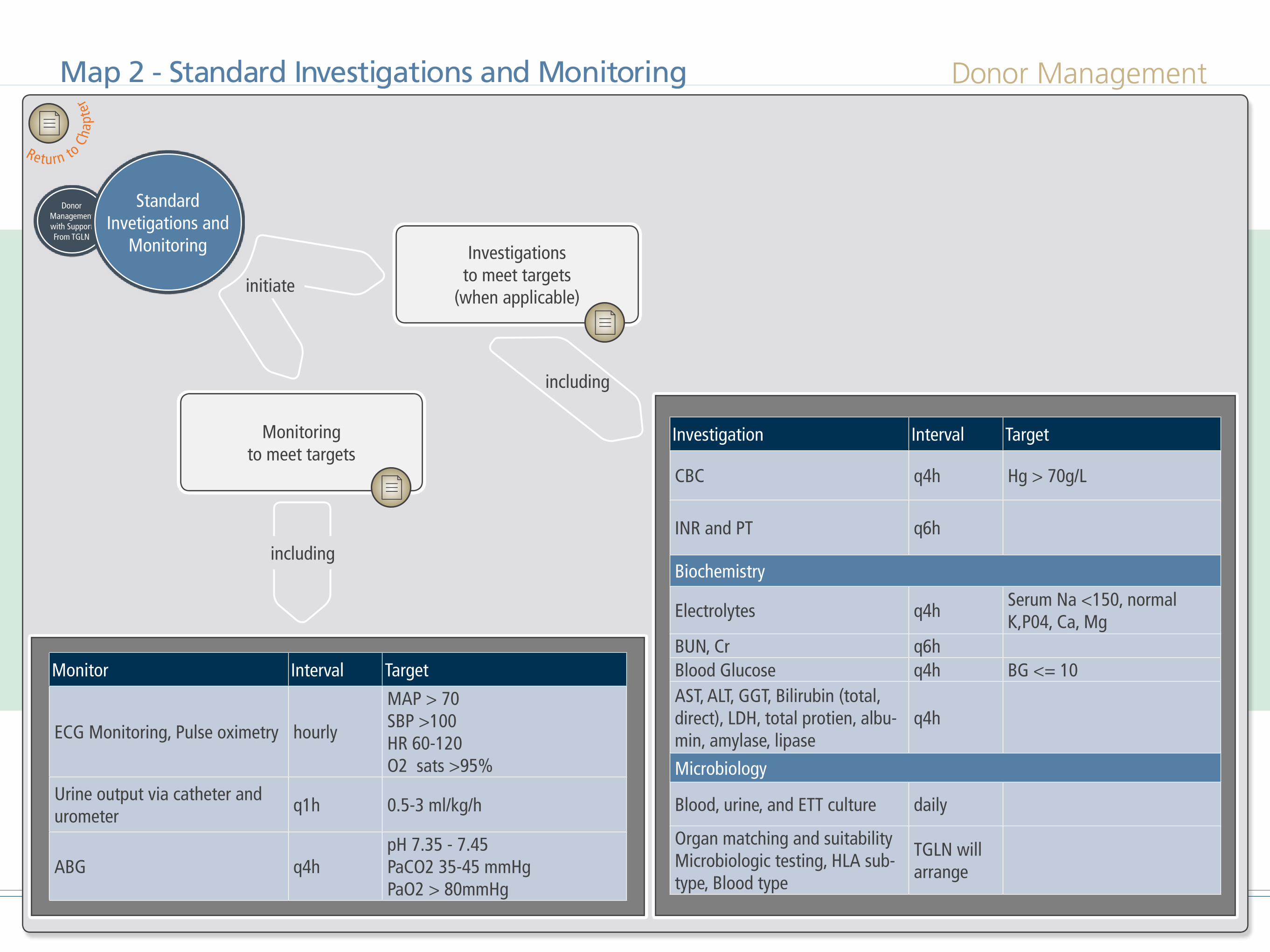
Donor Management
Donor Management with Support From TGLN
Investigationsto meet targets
(when applicable) including
Standard monitoring
to meet targets including...
Investigationsto meet targets
(when applicable)
Monitoringto meet targets
Standard Invetigations and
Monitoring
initiate
Investigation Interval Target
CBC q4h Hg > 70g/L
INR and PT q6h
Biochemistry
Electrolytes q4hSerum Na <150, normal K,P04, Ca, Mg
BUN, Cr q6hBlood Glucose q4h BG <= 10AST, ALT, GGT, Bilirubin (total, direct), LDH, total protien, albu-min, amylase, lipase
q4h
Microbiology
Blood, urine, and ETT culture daily
Organ matching and suitability Microbiologic testing, HLA sub-type, Blood type
TGLN will arrange
Monitor Interval Target
ECG Monitoring, Pulse oximetry hourly
MAP > 70SBP >100HR 60-120O2 sats >95%
Urine output via catheter and urometer
q1h 0.5-3 ml/kg/h
ABG q4hpH 7.35 - 7.45PaCO2 35-45 mmHgPaO2 > 80mmHg
including
including
Map 2 - Standard Investigations and Monitoring
Return to
Cha
pter

Donor Management
Donor Management with Support From TGLN
Return to
Cha
pter
Map 3 - Organ Specific Investigations
Organ Specific Investigations
Heart Donor
Hemodynamically unstable
Serial echo
Coronary angiogram
Baseline ECG, 2D echo, TnI or TnT
One of:1) male > 55, female > 60
2) Two risk factors AND male >40, female >453) >Three risk factors
4) History of cocaine use5) Upon transplant program request
Lung Donor
Bronchoscopy withbronchial washing sent for
culture, gram stain
CXR
Liver Donor
Weight > 100kg orBMI > 30 or
HCVAb positiveor request by the
transplant program
UltrasoundTransplant program
Kidney Donor
UrinalysisRequested by
transplant program
Ultrasound or other investigations
if possibleif possible
if possibleif possible
perform
if
request
if consider
perform
consult
if
ifperform
performperform initial
perform daily
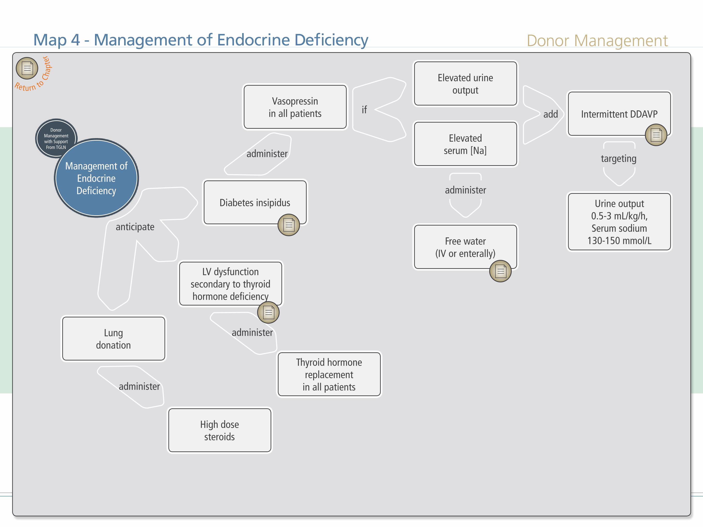
Donor Management
Donor Management with Support From TGLN
Map 4 - Management of Endocrine Deficiency
Management of Endocrine Deficiency
anticipate
Diabetes insipidus
LV dysfunction secondary to thyroid hormone deficiency
Lungdonation
High dosesteroids
Thyroid hormone replacementin all patients
Vasopressinin all patients
Elevated urine output
Intermittent DDAVP
Urine output 0.5-3 mL/kg/h, Serum sodium
130-150 mmol/L
Elevatedserum [Na]
Free water(IV or enterally)
administer
if
administer
administer
administer
add
targeting
Return to
Cha
pter

Donor Management
Donor Management with Support From TGLN
initiate
continue
Map 5 - Managing Infection
Management of Infection
Proven or presumedinfection
Antibiotics
Transplant program maydelay donation to
provide time for treatmentto take effect
Measures to reducehospital-acquired infection
if
be aware
Return to
Cha
pter

Donor Management
Donor Management with Support From TGLN
consider
continue
Map 6 - Management of the Gastrointestinal System
Management of the GI System
Donation Support Physician
Enternal nutrition only
Glycemic control
Possible liverdonation
Hyponatremia/Hypernatremia
Insulin infusion BG <=10with
avoid
target
if small bowel donation considered, consult
Return to
Cha
pter

Donor Management
Donor Management with Support From TGLN
Return to
Cha
pter
Map 7 - Management of the Renal System
Management of the Renal System
Nephrotoxic agent
Mild hypothermia(temp 34-35C)
Pre-defined BP and perfusion targets
Fluid bolus prior tocontrast administration
Administering isotonic fluid solution prior to
contrast administration
consider
consideradminister
avoid
ensure
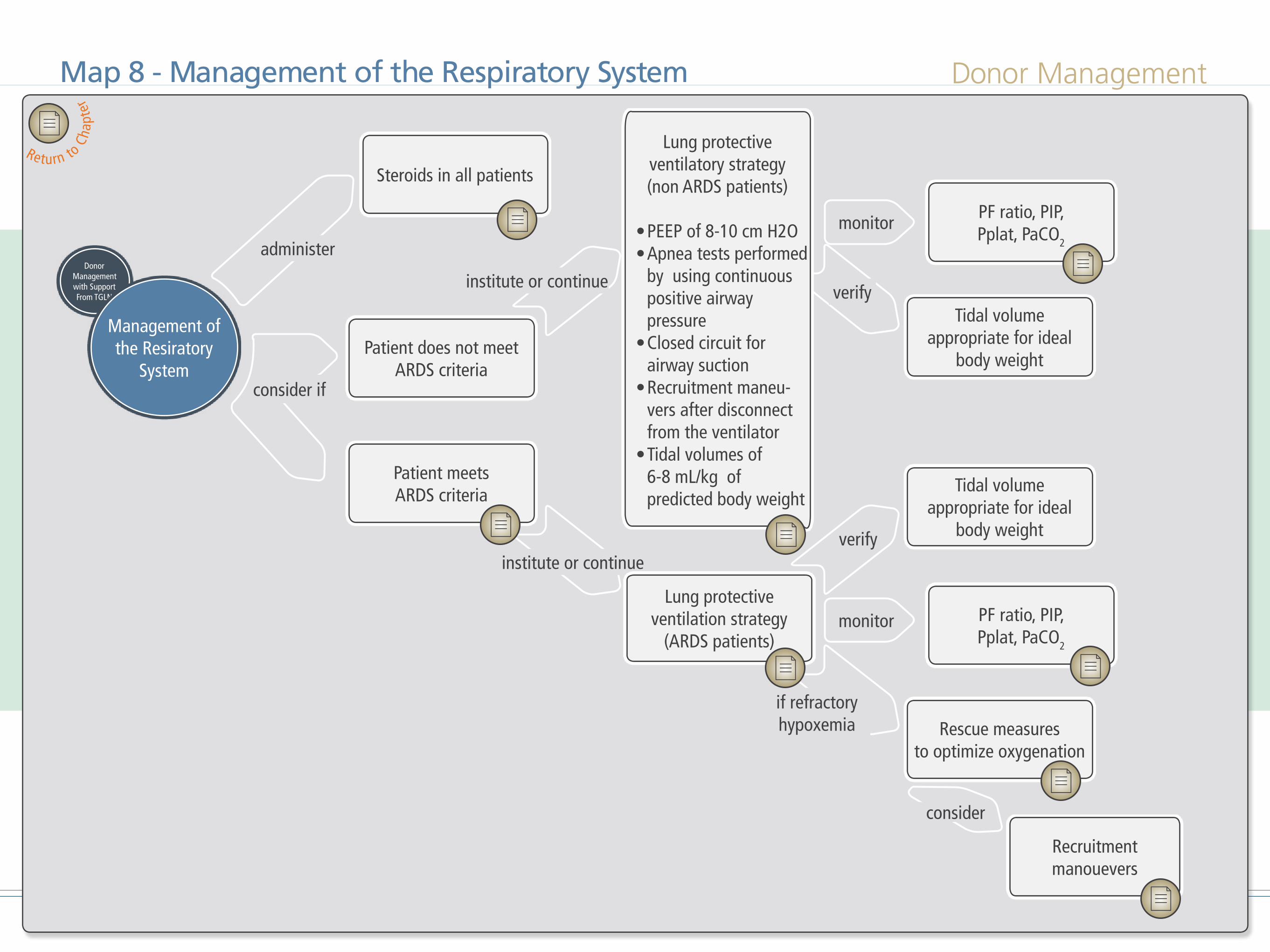
Donor Management
Donor Management with Support From TGLN
institute or continue
institute or continue
verify
consider
verify
Map 8 - Management of the Respiratory System
Management of the Resiratory
System
Steroids in all patients
Patient does not meet ARDS criteria
Patient meetsARDS criteria
Lung protectiveventilation strategy
(ARDS patients)
Rescue measuresto optimize oxygenation
PF ratio, PIP, Pplat, PaCO2
PF ratio, PIP, Pplat, PaCO2
Tidal volume appropriate for ideal
body weight
Tidal volume appropriate for ideal
body weight
Recruitment manouevers
monitor
monitor
administer
consider if
if refractoryhypoxemia
Return to
Cha
pter
Lung protective ventilatory strategy (non ARDS patients)
• PEEP of 8-10 cm H2O• Apnea tests performed
by using continuous positive airway pressure
• Closed circuit for airway suction
• Recruitment maneu-vers after disconnect from the ventilator
• Tidal volumes of 6-8 mL/kg of predicted body weight
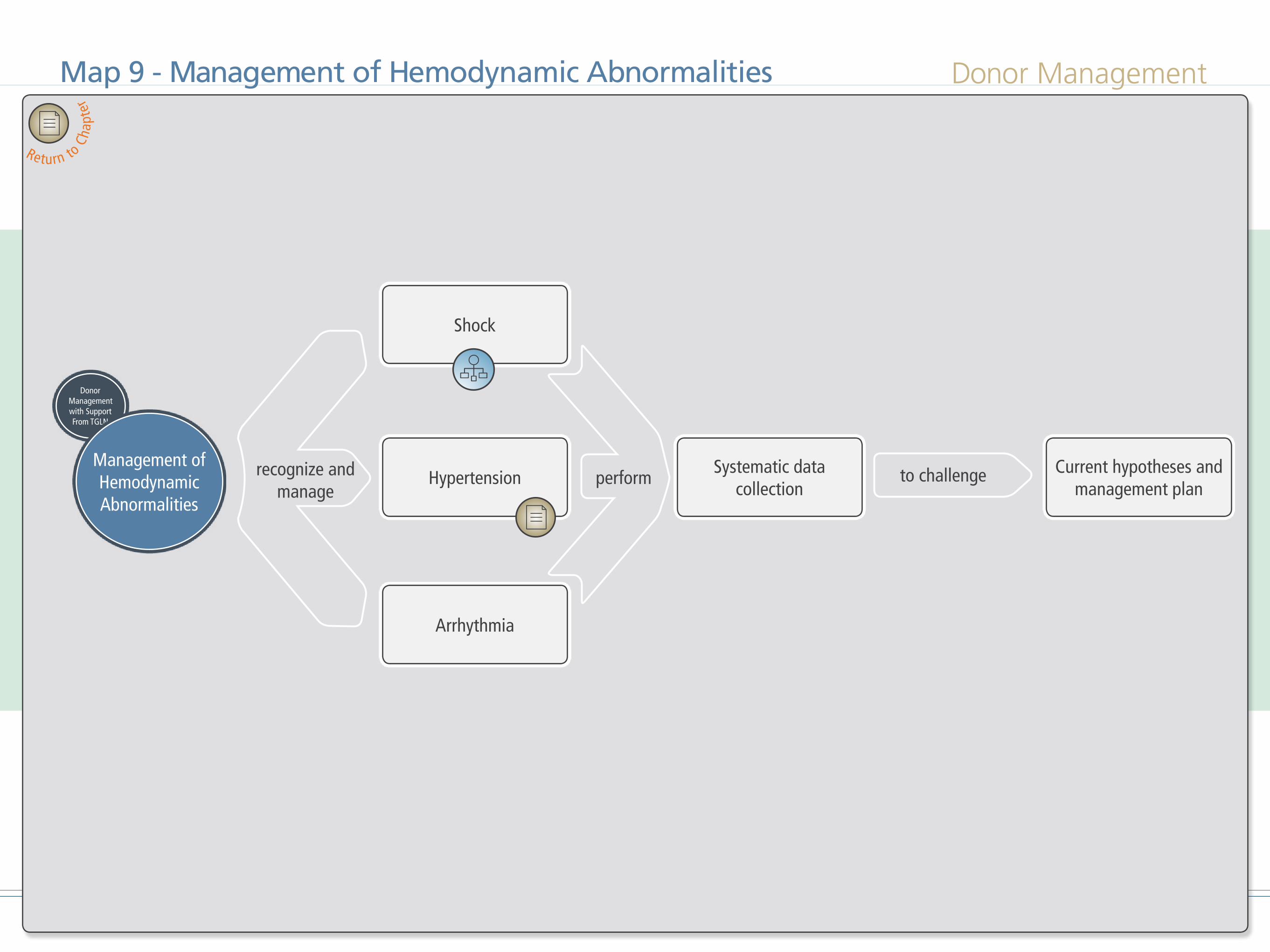
Donor Management
Donor Management with Support From TGLN
performrecognize and manage
Map 9 - Management of Hemodynamic Abnormalities
Management of Hemodynamic Abnormalities
Shock
HypertensionSystematic data
collectionCurrent hypotheses and
management plan
Arrhythmia
to challenge
Return to
Cha
pter
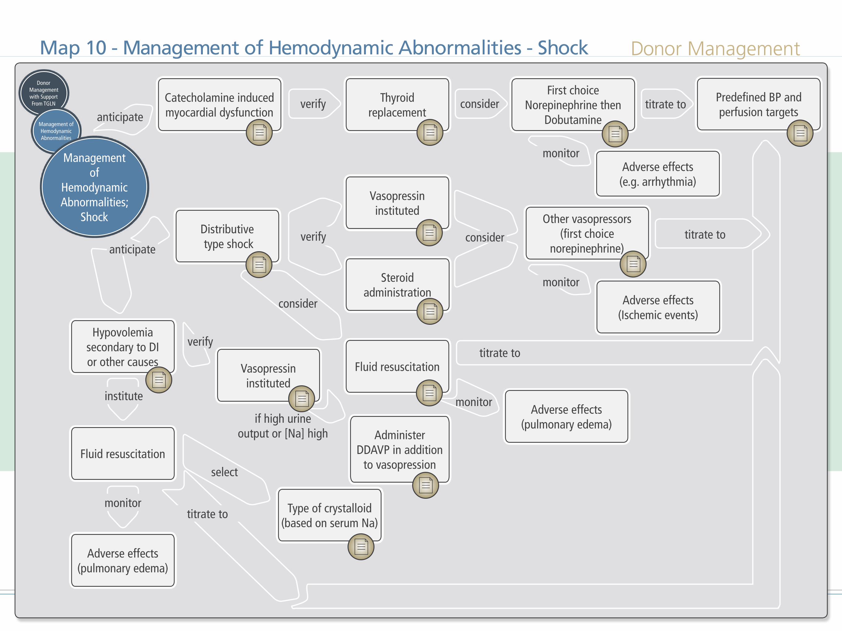
Donor Management
if high urine output or [Na] high
monitor
considerverify
Map 10 - Management of Hemodynamic Abnormalities - Shock
Donor Management with Support From TGLN
Management of Hemodynamic Abnormalities
Catecholamine inducedmyocardial dysfunction
Distributive type shock
Other vasopressors(first choice
norepinephrine)
Adverse effects(Ischemic events)
Vasopressininstituted
Steroid administration
Fluid resuscitation
Adverse effects(pulmonary edema)
Hypovolemiasecondary to DI or other causes
Fluid resuscitation
Type of crystalloid(based on serum Na)
Adverse effects(pulmonary edema)
Vasopressininstituted
AdministerDDAVP in addition
to vasopression
Thyroid replacement
First choice Norepinephrine then
Dobutamine
Adverse effects(e.g. arrhythmia)
titrate to
titrate to
titrate to
monitor
monitor
verify
select
consider
institute
monitor
consider
titrate to
verify
Management of
Hemodynamic Abnormalities;
Shock
anticipate
anticipate
Predefined BP and perfusion targets

51Organ Donation in Ontario: A Guide for Critical Care Residents - Donor Management
DescriptionStart or continue measures to prevent:
• Deep vein thrombosis• Stress ulcers• Decubitus ulcers• Ventilator-associated pneumonia• Central line-associated
bloodstream infection
RationaleAs intensivists become increasingly involved in donor management, it is imperative that thesame rigor that is applied to the care of living patients be employed in the care of organDonors1. Through provision of meticulous and aggressive care, and in collaboration with theTGLN and transplant teams, the intensivist has the opportunity to both preserve the option oforgan donation for patients and their families and provide the gift of life to others.
1 Kotloff RM, Blosser S, Fulda GJ, et al. Manage-ment of the Potential Organ Donor in the ICU. Crit Care Med. 2015;43(6):1291-1325. doi:10.1097/CCM.0000000000000958.
Performing Prophylaxis for Organ DonorsLouise Pope-Rhoden
Return to Overview map
52Organ Donation in Ontario: A Guide for Critical Care Residents - Donor Management
DescriptionStandard monitoring of potential donors includes1,2:
• Vital signs q1h• Core temperature q4h • Continuous electrocardiogram (EKG)
monitoring• Continuous pulse oximetry • Arterial blood pressure q1h• Continuous central venous pressure
(CVP) monitoring
• Urine output q1h• Infectious disease assessment (via q24h
blood, urine & sputum cultures)1
1 "Donor Screening and Testing". Donation Resource Manual: A Tool To Assist Hospitals With The Process Of Organ And Tissue Donation. 6th ed. Toronto: Queen's Printer, 2014. Print.
2 Shemie SD, Ross H, Pagliarello J, Baker AJ, Greig PD, Brand T, et al. Organ donor management in Canada: recommendations of the forum on Medical Management to Optimize Donor Organ Potential. CMAJ : Canadian Medical Association Journal 2006;174(6):S13-32.
• Optional pulmonary artery catheter in situ or central venous line (central venous oximetry)1,2,3
RationaleClose monitoring of hemodynamic and ventilatory function is required to optimize resuscitation and maintain optimal organ function at the time of surgical recovery 1, 2,3,4. Monitoring for possible sources of infections is also recommended.
3 Menza R. Evaluation and Assessment of Organ Donors. In: Dianne LaPointe R, Linda O, Teresa S, ed. by. A Clinician's Guide to Donation and Transplantation. 1st ed. Lenexa: NATCO, The Organization for Transplant Professionals; 2016.
4 Kotloff RM, Blosser S, Fulda GJ, et al. Manage-ment of the Potential Organ Donor in the ICU. Crit Care Med. 2015;43(6):1291-1325. doi:10.1097/CCM.0000000000000958.
Standard Monitoring to Meet TargetsJanice Beitel
Return to Overview map
Return to Standard Investigations and Monitoring map

53Organ Donation in Ontario: A Guide for Critical Care Residents - Donor Management
DescriptionStandard investigations for all potential donors include1,2:
• ABO - Blood Group (Cross and Type)• Arterial Blood Gases (ABGs)on positive
end expiratory pressure (PEEP) 8-10 cm H2O q2-4h. Contact the transplant team for patients on higher PEEP who may become hypoxemic with lower PEEP levels.
• CBC q4h
• Serum Lactate q 2-4h• Mixed venous blood gases or central
venous oxygen saturation q6h through
1 "Donor Screening and Testing". Donation Resource Man-ual: A Tool To Assist Hospitals With The Process Of Or-gan And Tissue Donation. 6th ed. Toronto: Queen's Print-er, 2014. Print.
2 Shemie SD, Ross H, Pagliarello J, Baker AJ, Greig PD, Brand T, et al. Organ donor management in Canada: rec-ommendations of the forum on Medical Management to Optimize Donor Organ Potential. CMAJ : Canadian Medi-cal Association Journal 2006;174(6):S13-32.
either pulmonary artery catheter in situ or central venous line, respectively
• Liver profile – Bilirubin (total and direct), AST, ALT, ALP, LDH, Total Protein, Albumin, Amylase, Lipase, GGT q4h
• PT/INR, PTT q6h• Electrolytes, Creatinine, Urea, Glucose,
Ca, PO4, Mg, Lactate q4h• CK, CK-MB q4-8h• Troponin (I or T) q8h• Urinalysis q24h• C & S – sputum, urine and blood
(include gram stain) q24h• 12 lead EKG• CXR q4h
Note: Please see organ specific investigations for lung and heart serological tests for infectious diseases are requested by TGLN and sent to be processed in central laboratories in Ontario
RationaleStandard investigations provide baseline in-formation on organ function and physiologi-cal homeostasis3. Potential organ donors may be hemodynamically unstable with electrolyte disturbances related to the underlying cause of death, disease process or treatments2, 3. Standard investigations are well accepted in practice and serial testing provides trending information on organ function for the early identification and correction of electrolyte imbalances and/or the implementation of other interventions to meet targets2.
3 Menza R. Evaluation and Assessment of Organ Donors. In: Dianne LaPointe R, Linda O, Teresa S, ed. by. A Cli-nician's Guide to Donation and Transplantation. 1st ed. Lenexa: NATCO, The Organization for Transplant Pro-fessionals; 2016.
Standard Investigations to Meet TargetsJanice Beitel
Return to Overview map
Return to Standard Investigations and Monitoring map

54Organ Donation in Ontario: A Guide for Critical Care Residents - Donor Management
Description• EKG• Troponin I or T q12h• Baseline echocardiography• If hemodynamically unstable,or reduced
ejection fraction on baseline echo, perform serial echocardiogram
• In patients with risk factors, request coronary angiography
RationaleIn evaluating the heart donor, three critical key concepts should be considered:
1. With the advent of expertly managed ven-tricular assist devices, the quality of the donor heart must be sufficient that trans-plantation of the donor heart results in mortality reduction compared to ongoing circulatory support.
2. (Serial examinations of the heart by EKG,
biochemistry, and echocardiography sig-nificantly increases the number of hearts suitable for transplantation as the often severe myocardial dysfunction resolve with time and hormonal replacement therapy (T4) 1,2,3,4,5. The heart is rarely ruled out for transplantation based on the initial echocardiogram 5. Therefore, medical support and re-evaluation by serial echo-cardiogram should be performed prior to decisions about transplantability.
3. Knowledge of the data supporting improvement will assist intensivists in discussing the need for serial evaluation with colleagues and the healthcare team.
1 Casartelli M, Bombardini T, Simion D, Gaspari MG, Pro-caccio F. Wait, treat and see: echocardiographic monitoring of brain-dead potential donors with stunned heart. Car-diovasc Ultrasound. 2012;10:25.
2 Zaroff JG, Babcock WD, Shiboski SC. The impact of left ventricular dysfunction on cardiac donor transplant rates. J Heart Lung Transplant. 2003;22(3):334-7.
3 Mascia L, Mastromauro I, Viberti S, Vincenzi M, Zanello M. Management to optimize organ procurement in brain dead donors. Minerva Anestesiol. 2009;75(3):125-33.
4 Borbely XI, Krishnamoorthy V, Modi S, et al. Temporal Changes in Left Ventricular Systolic Function and Use of Echocardiography in Adult Heart Donors. Neurocrit Care. 2015;23(1):66-71.
5 Sopko N, Shea KJ, Ludrosky K, et al. Survival is not com-promised in donor hearts with echocardiographic abnor-malities. J Surg Res. 2007;143(1):141-4.
The EKG and Troponin are useful to assess for conduction abnormalities and ongoing isch-emia6. In Canada, Health Canada requires these investigations of all donors where possi-ble. When initially abnormal, serial ECGs and Troponins assist transplanters assess suitability as commonly the abnormalities are transient.
The hemodynamic assessment of all donors begins with a surface echocardiogram7. This begins the assessment of the suitability of the heart for transplant but also critically informs the hemodynamic management of patients who are not heart donors. The time following herniation is associated with an initial excess of sympathetic outflow, followed by loss of sym-pathetic tone and systemic vascular resistance. These result in often dramatic changes in the ECG, Troponin and the echocardiogram. Both left ventricular dysfunction and vasodilation often lead to hypotension. In the most hemody-namically unstable potential donors, echocar-
6 Shemie SD, Ross H, Pagliarello J, et al. Organ donor man-agement in Canada: recommendations of the forum on Medical Management to Optimize Donor Organ Potential. CMAJ. 2006;174(6):S13-32.
7 Venkateswaran RV, Townend JN, Wilson IC, Mascaro JG, Bonser RS, Steeds RP. Echocardiography in the potential heart donor. Transplantation. 2010;89(7):894-901.
Investigations for Heart DonorsAndrew Healey
Return to Overview map
Return to Organ Specific Investigations map
55
diography can provide information on causes of instability (e.g. stress cardiomyopathy) and guide a goal-directed resuscitation and choice of initial vasopressors 7. While early echocar-diography may assist the physician with management of the hemodynamics, an abnor-mal echocardiogram should not be seen as an exclusion to potential heart donation 5. There is a significant body of literature that cites the marked effects of time and hormonal treatment, resulting in the resolution of both regional and
global left ventricular dysfunction to complete recovery 1,2,3,4,5 In special circumstances, trans-esophageal echocardiography may be required at the request of the transplant teams or in the event transthoracic echocardiography does not produce adequate imaging windows.
Coronary angiography is an essential and often final step in the assessment of the donor heart suitability. In perfect circumstances, angiogra-phy is not required but this is rare. It behoves
intensivists to remember that coronary artery disease in an allograft progresses rapidly and is one of the main causes of graft loss after transplantation. The CCDT guidelines6 sug-gest if one of the following are present, coronary angiography should be performed:
1. male > 55 or female > 602. two cardiac risk factors* AND male > 40
or female >453. > three risk factors4. history of cocaine use5. transplant program request
*Risk Factors include: smoking, hypertension, diabetes, hyperlipidemia, body mass index > 32, family history of the disease, prior histo-ry of coronary artery disease, ischemia on electrocardiogram, anterolateral regional wall motion abnormalities on echocardiogram, 2-dimensional echocardiographic assessment of ejection fraction of ≤ 40%.
The authors recommend contact with the organ donor organization with all other data prior to proceeding to coronary angiography to ensure potential suitability as there is risk associated
with both the contrast load and the transfer of donors.
The use of the pulmonary artery catheter is of-ten found within algorithms for donor manage-ment6. Experience over time with CVP moni-toring, targeted therapy to address deficits in organ perfusion and serial echocardiography has diminished the need for PA catheters. In special circumstances in the donor or recipient, a pulmonary artery catheter might be request-ed to evaluate a specific parameter of donor heart function. This is almost exclusively per-formed at the request of the accepting transplant program and after all other testing suggests suitability of the organ.
Return to Overview map
Return to Organ Specific Investigations map
Organ Donation in Ontario: A Guide for Critical Care Residents - Donor Management

56Organ Donation in Ontario: A Guide for Critical Care Residents - Donor Management
DescriptionFor any potential donors when lungs are being considered for transplantation, perform:
• Arterial Blood Gases q 4h on 100% O2 (challenge gases)
• Chest radiography q41,2
• Bronchoscopy 1,2 a second bronchoscopy is performed in recovery surgery1
• Bronchoalveolar lavage (BAL) 2
• BAL gram stain, culture and sensitivity
RationaleOngoing assessment for oxygenation oc-curs throughout management and organ specific investigation of the potential donor.
1 "Donor Screening and Testing". Donation Resource Man-ual: A Tool To Assist Hospitals With The Process Of Or-gan And Tissue Donation. 6th ed. Toronto: Queen's Print-er, 2014. Print.
2 Shemie SD, Ross H, Pagliarello J, Baker AJ, Greig PD, Brand T, et al. Organ donor management in Canada: rec-ommendations of the forum on Medical Management to Optimize Donor Organ Potential. CMAJ : Canadian Medi-cal Association Journal 2006;174(6):S13-32.
ABG’s (challenge gases) are routinely drawn after 20 min on a FiO2 of 1 to assess the PaO2:-FiO2 ratio. Chest radiography is used to assess atelectasis, infiltrates, or infection in combina-tion with other clinical findings3. Although not supported by strong evidence, investigations (CXR/ABGs) are requested every four hours to monitor lung function. The role of bedside ultrasound to evaluate lung volume status and cardiac function is evolving, but is not con-sidered standard at this time. Bronchoscopy is required to investigate any CXR findings, identify aspiration, infection and to clear air-way secretions as needed 3. Bronchoscopy is also performed to exclude any endobronchial lesions and to define anatomy. A bronchial lavage for gram stain and sensitivity may help confirm infection and refine treatment that will often need to continue in the immunocompromised recipient 2,3. A second bronchoscopy is almost universally repeated in the OR by the transplant team prior to lung recovery 1,3.
3 Menza R. Evaluation and Assessment of Organ Donors. In: Dianne LaPointe R, Linda O, Teresa S, ed. by. A Cli-nician's Guide to Donation and Transplantation. 1st ed. Lenexa: NATCO, The Organization for Transplant Pro-fessionals; 2016.
Investigations for Lung DonorsJanice Beitel, Pierre Cardinal, Andrew Healey, Kim Bowman
Return to Overview map
Return to Organ Specific Investigations map

57Organ Donation in Ontario: A Guide for Critical Care Residents - Donor Management
DescriptionFor any potential donors when liver is being considered for potential transplantation, biochemical tests should include: 1,2,3
• Bilirubin (total and direct), AST, ALT, ALP, LDH, Total Protein, Albumin, Amy-lase, Lipase, GGT q 4 h
• INR, PTT q6• Serum sodium levels should be
maintained within normal ranges of 135 - 145 mmol/L
• Liver ultrasound may be requested by
1 "Donor Screening and Testing". Donation Resource Man-ual: A Tool To Assist Hospitals With The Process Of Or-gan And Tissue Donation. 6th ed. Toronto: Queen's Print-er, 2014. Print.
2 Shemie SD, Ross H, Pagliarello J, Baker AJ, Greig PD, Brand T, et al. Organ donor management in Canada: rec-ommendations of the forum on Medical Management to Optimize Donor Organ Potential. CMAJ : Canadian Medi-cal Association Journal 2006;174(6):S13-32.
3 Menza R. Evaluation and Assessment of Organ Donors. In: Dianne LaPointe R, Linda O, Teresa S, ed. by. A Clinician's Guide to Donation and Transplantation. 1st ed. Lenexa: NATCO, The Organization for Transplant Professionals; 2016.
transplant team, especially if the donor weighs over 100 kg or has a body mass index greater than 30.
• Uncommonly, intraoperative liver biopsy
Note:Serologic testing is requested by TGLN and sent to be processed in in central laboratories in Ontario
RationaleAssessment of liver enzymes through biochem-ical testing provides information to assess for acute ischemia related to events either pre-ceding or surrounding neurological death 2,3,4. Repeated testing provides the opportunity to see a trend in liver enzymes as an indication that the insult is over and the liver is show-ing signs of recovery. Since the bile ducts have only one blood supply (hepatic artery), they are most susceptible to ischemic injury placing the potential recipient at risk for bile duct complications.
4 Kotloff RM, Blosser S, Fulda GJ, et al. Manage-ment of the Potential Organ Donor in the ICU. Crit Care Med. 2015;43(6):1291-1325. doi:10.1097/CCM.0000000000000958.
Donor serum sodium greater than 155 has been weakly associated with primary liver dysfunc-tion in transplant recipients but reversible if hypernatremia is corrected4.
While the use of ultrasound to assess for fat-ty liver remains controversial, the test may be requested by the transplant program when donor characteristics are associated with an increased risk of graft failure in the recipient. Ultrasounds are also requested to assist with assessment especially when the donor is located in a geographically remote area3. The transplant team will very rarely perform liver biopsies intraoperatively at the time of procurement in donors who weigh over 100 kg or have a body mass index greater than 30 3.
Investigations for Liver DonorsJanice Beitel, Pierre Cardinal, Andrew Healey, Kim Bowman
Return to Overview map
Return to Organ Specific Investigations map
58Organ Donation in Ontario: A Guide for Critical Care Residents - Donor Management
DescriptionIn addition to the standard investigations needed for all NDD donors, investigations for renal donors should include:
• Urinalysis daily• Serum creatinine and urea every 6 hours• Uncommonly, intraoperative kidney
biopsy (if age >65, or age <65 with a history of hypertension, diabetes, abnormal urinalysis, or serum creatinine >133µmol/L) - this decision is made in
concert with the transplant team and the procedure usually carried out intraopera-tively at the time of procurement.
• Consider radiographic imaging of kidneys if requested by the transplant teams. Indications for radiographic imaging include donors with a family history of polycystic kidney disease, renal stones or other urologic abnormalities.
RationaleA urinalysis is a useful screening tool to assess for microscopic hematuria and proteinuria, both of which can be markers of glomerular pathology and may affect the decision to trans-plant the kidney1. Persistent microscopic hema-turia should prompt a workup to exclude uro-logic causes such as urinary tract infections or nephrolithiasis. If the microscopic hematu-ria persists, consultation with the transplant physician is warranted in order to determine if a kidney biopsy is required. Urinalysis and urine culture are Health Canada requirements of all organ donors.
Routine evaluation of the donor’s creatinine and urea is used to monitor for objective signs of end organ dysfunction and acute kidney injury. Ser ia l measurements can document improvement (or deterioration) with time and treatment.
The Canadian guidelines recommend kidney biopsies for patients older than 65, or those
1 Andrews P, Burnapp L, Manas D, Bradley J, Dudley C. Summary of the British Transplantation Society/Renal As-sociation UK Guidelines for Living Donor Kidney Trans-plantation. Transplantation. 2012;93:666-673.
younger than 65 with a creatinine of 133µmol/L or greater, an abnormal urinalysis, or a history of hypertension or diabetes2. If a kidney biopsy is warranted, it is usually performed intraoper-atively at the time of procurement, and not in the intensive care unit2. A kidney biopsy in a potential extended criteria donor can identify conditions such as glomerulosclerosis, vascular abnormalities, or interstitial fibrosis that may affect the decision of the team to transplant the kidney3. Expanded criteria donors are those who are at increased risk of graft failure, and include donors older than 60 years, or those 50-59 years with at least two comorbidities (cere-brovascular cause of death, renal insufficiency, or hypertension)4.
The Society of Critical Care Medicine
2 Shemie SD, Ross H, Pagliarello J, et al. Organ donor man-agement in Canada: Recommendations of the forum on Medical Management to Optimize Donor Organ Potential. CMAJ. 2006;174:S13-S30. doi:10.1503/cmaj.045131.
3 Kotloff RM, Blosser S, Fulda GJ, et al. Manage-ment of the Potential Organ Donor in the ICU. Crit Care Med. 2015;43(6):1291-1325. doi:10.1097/CCM.0000000000000958.
4 Port F, Bragg-Gresham J, Metzger R, Dykstra D, Gilles-pie B, Young E, Delmonico F, Wynn J, Merion R, Wolfe R, Held P. Donor characteristics associated with reduced graft survival: an approach to expanding the pool of kid-ney donors. Transplantation. 2002;74:1281-1286.
Investigations for Kidney DonorsAlissa Visram, Sabira Valiani, Pierre Cardinal
Return to Overview map
Return to Organ Specific Investigations map

59Organ Donation in Ontario: A Guide for Critical Care Residents - Donor Management
suggests that radiographic imaging of the kidneys may be warranted in donors who have a family history of polycystic kidney disease, or a history of renal stones or urologic abnormal-ities3. Renal imaging in Canada is currently per formed only on the request of the transplant teams.
Return to Overview map
Return to Organ Specific Investigations map

60Organ Donation in Ontario: A Guide for Critical Care Residents - Donor Management
Description• Anticipate that diabetes insipidus (DI) is
common in patients who are dead by neurological criteria.
• Insert a foley catheter and monitor fluid inputs and outputs.
• Recognize dilute urine (< 200 mOsm/kg•H2O), polyuria (> 4mL/kg/h), as well as hypernatremia (> 145mmol/L) as signs of diabetes insipidus in the potential NDD donor.
RationaleArginine vasopressin (AVP) is a peptide that is formed in the hypothalamus and then stored in as well as secreted from the posterior pitu-itary. Its anti-diuretic effect on the V2 receptors within the renal collecting system promotes the reabsorption of free water. AVP regulates extracellular fluid volume by the reabsorption
of water from the kidneys. After brain death, a lack of AVP leads to unregulated renal losses of free water resulting in a state of diabetes insip-idus1. Diabetes insipidus is confirmed if urine output remains excessive (> 4mL/kg/h) as well as dilute (< 200 mOsm/kg•H2O) and associ-ated with hypernatremia (> 145mmol/L) and an increased serum osmolality (> 300mOsm/kg•H2O)2. In the context of brain death, diabetes insipidus should be presumed even if all criteria are not met, especially in the context of clinical hypovolemia.
1 Loh JA, Verbalis JG. Disorders of water and salt metab-olism associated with pituitary disease. Endocrinol Metab Clin North Am 2008; 37:213–234
2 Trillium Gift of Life Network. Donation Resource Manu-al: A tool to assist hospitals with the process of organ and tissue Donation. Queen's Printer for Ontario: 2010.
Anticipating Diabetes Insipidus in the Organ DonorMichael Hartwick, Sonny Dhanani
Return to Overview map
Return to Endocrine Deficiency map

61Organ Donation in Ontario: A Guide for Critical Care Residents - Donor Management
DescriptionTo anticipate the transient left ventricular car-diac dysfunction after brain death that may be responsive to empiric thyroid hormone replace-ment.
RationaleThyroid hormone plays a part in cardiovascular regulation by improving both contractility and chronotropy, as well as by decreasing systemic vascular resistance. The damage to the hypotha-
lamic-pituitary axis that ensues with herniation triggers hormonal dysregulation and may lead to clinical hypothyroidism1. However, in some patients, the clinical picture is more consistent with sick euthyroid syndrome rather than true hypothyroidism. Animal models demonstrate significant decline in triiodothyronine (T3) and free thyroxine (T4) levels after brain death. This results in a depletion of myocardial energy
1 1. Chen EP, Bittner HB, Kendall SW, et al: Hormonal and hemodynamic changes in a validated animal model of brain death. Crit Care Med 1996; 24:1352–1359
stores, a shift from aerobic to anaerobic metab-olism, and a reduction in cardiac function. In addition, thyroid replacement has been shown to exert a positive effect on myocardial gene ex-pression2. In animal models, the transition to anaerobic metabolism is reversed with T3 re-placement and is accompanied by a return of myocardial function3,4.
The evidence supports starting thyroxine in the presence of LV dysfunction. However, in light of minimal harm, the recommendation is to start thyroid replacement even without con-firmed LV dysfunction as part of empiric triple hormonal therapy regimens (vasopressin, and corticosteroids)5.
2 James SR, Ranasinghe AM, Venkateswaran R, McCabe CJ, Franklyn JA, Bonser RS.The effects of acute triiodo-thyronine therapy on myocardial gene expression in brain stem dead cardiac donors. J Clin Endocrinol Metab. 2010 Mar;95(3):1338-43. 159.
3 Novitzky D, Cooper DK, Rosendale JD, et al: Hormonal therapy of the brain-dead organ donor: Experimental and clinical studies.Transplantation 2006; 82:1396–1401
4 Cooper DK, Novitzky D, Wicomb WN: The pathophysi-ological effects of brain death on potential donor organs, with particular reference to the heart. Ann R Coll Surg Engl 1989; 71:261–266
5. Kutsogiannis DJ, Pagliarello G, Doig C, Ross H, She-mie SD. Medical management to optimize donor organ potential: review of the literature. Can J Anaesth. 2006 Aug;53(8):820-30.
Anticipating LV Dysfunction Secondary to Thyroid Hormone DeficiencySonny Dhanani
Return to Overview map
Return to Endocrine Deficiency map

62Organ Donation in Ontario: A Guide for Critical Care Residents - Donor Management
DescriptionTo administer vasopressin and or intermittent DDAVP in most potential donors after brain death.
RationaleAlthough the mechanism has not been estab-lished, diabetes insipidus has the potential to impair organ function beyond that sim-ply resulting from diuresis, hypovolemia, and hemodynamic instability. Potential organ do-
nors with diabetes insipidus may be managed with an intravenous AVP infusion or inter-mittent desmopressin. An AVP infusion is the preferred agent for patients with concom-itant hypotension. Given that it provides the dual benefits of anti-diuresis as well as hemo-dynamic support, it is administered in most NDD patients who are not hypertensive. In hypertensive patients, AVP is best avoided giv-en concerns around possible interference with end-organ perfusion based on animal models of
splanchnic blood flow1.
Diabetes insipidus without hypotension may also be managed with desmopressin (1-desami-no-8-d-arginine vasopressin, or DDAVP). Des-mopressin is an AVP analogue whose mechanism of action is highly specific for the V2 vasopres-sin receptors in the renal collecting system. This property allows desmopressin to inhibit diuresis with virtually no vasopressor side effect. Multi-ple routes of administration are available, but the intravenous route is preferred in the organ donor2. Although concerns for potential thrombogenic complications have been raised with desmopressin use, it does not seem to impair graft survival. One trial of 97 adult or-gan donors randomized to receive desmopressin or nothing for diabetes insipidus demonstrat-ed no impairment in pre-procurement renal function or post-transplant need for dialysis in those who received desmopressin3. A more
1 Chen JM, Cullinane S, Spanier TB, Artrip JH, John R, Ed-wards NM, Oz MC, Landry DW. Vasopressin deficiency and pressor hypersensitivity in hemodynamically unstable organ donors. Circulation 1999; 100 (suppl II):II-246.
2 Pennefather SH, Bullock RE, Mantle D, Dark JH. Use of low dose arginine vasopressin to support brain-dead organ donors. Transplantation 1995; 59(1):58-62.
3 Nakagawa K, Tang JF. Physiologic response of human brain death and the use of vasopressin for successful or-gan transplantation. J Clin Anesth. 2011 Mar;23(2):145-8.
recent retrospective analysis of 458 neurolog-ically deceased adult kidney donors suggest-ed that those treated with desmopressin actu-ally had improved pre-recovery renal function (creatinine 97 ± 44 vs 124 ± 106 µmol/L; p < 0.001)4.In either case, AVP infusions or desmopressin boluses should be titrated to target serum sodi-um levels 130 – 155 mmol/L and urine output 0.5 – 3 mL/kg/h. If needed, AVP and desmo-pressin may also be used simultaneously for pa-tients who continue to have a high urine output despite treatment with AVP alone. Intermittent intravenous DDAVP 4 micrograms every 6 hours is a common regimen. Intravenous AVP can be titrated between 0.5 units/h and maxi-mum of 2.4 units/h. Intravenous maintenance fluids to maintain blood pressure and perfusion and the administration of free water (either through the enteral route (free water flushes) or intravenously (D5W)) should be considered to aggressively correct hypernatremia5. 4 Dunser MW, Mayr AJ, Ulmer H, Knotzer H, Sumann
G, Pajk W, Friesenecker B, Hasibeder WR. Arginine va-sopressin in advanced vasodilatory shock: a prospec-tive, randomized,controlled study. Circulation. 2003 May 13;107(18):2313-9.
5 Kutsogiannis DJ, Pagliarello G, Doig C, Ross H, She-mie SD. Medical management to optimize donor organ potential: review of the literature. Can J Anaesth. 2006 Aug;53(8):820-30.
Administering Intermittent DDAVP when Managing DonorsSonny Dhanani, Ronish Gupta, Pierre Cardinal, Andrew Healey
Return to Overview map
Return to Endocrine Deficiency map

63Organ Donation in Ontario: A Guide for Critical Care Residents - Donor Management
DescriptionIf euvolemic with serum Na between 135-145 mmol/L:
• Initial maintenance intravenous fluids should be administered to achieve euvole-mia with 0.9%NS or Ringer’s Lactate
If euvolemic with serum Na>145mmol/L:• Administer a IV bolus of 250 mL of D5W
or free water enterally• and immediately assess for diabetes
insipidus
• Change maintenance fluids to D5W. If clinically hypovolemic and hypernatremic:
• Administer Ringer’s Lactate bolus of 500 mL IV
• Once fluid resuscitated administer free water (intravenously or enterally)
RationaleHypernatremia (serum sodium > 155 mmol/L) secondary to excessive sodium administration during resuscitation or diabetes insipidus may
Administering Free Water in the Organ DonorSonny Dhanani, Pierre Cardinal, Andrew Healey
lead to the accumulation of compensatory idiogenic osmoles in donor organs. Once trans-planted, significant intracellular fluid shifts may develop as these organs are exposed to the recipient normal serum; the different osmo-lalities resulting in a water influx into the transplanted organs.
Traditionally, hypernatremia was considered to be independently associated with hepatic and renal dysfunction or graft loss after transplan-tation. However, more recent evidence suggests that this effect may be less significant than once believed and that detrimental effects of hyper-natremia are reversible if serum sodium levels are corrected 1,2,3.
Given that avoiding or treating hypernatremia is easily achieved using inexpensive management
1 Bloom MB, Raza S, Bhakta A, et al. Impact of Deceased Organ Donor Demographics and Critical Care End Points on Liver Transplantation and Graft Survival Rates. J Am Coll Surg. 2015;220(1):38-47. doi:10.1016/j.jamcoll-surg.2014.09.020.
2 Kazemeyni SM, Esfahani F. Influence of hypernatremia and polyuria of brain-dead donors before organ pro-curement on kidney allograft function. Urol J. 2008 Sum-mer;5(3):173-7.
3 Mangus RS, Fridell JA, Vianna RM, Milgrom ML, Chestovich P, Vandenboom C,Tector AJ. Severe hy-pernatremia in deceased liver donors does not impact early transplant outcome. Transplantation. 2010 Aug 27;90(4):438-43.
strategies, it is recommended to target normal serum sodium levels by selecting appropriate maintenance intravenous fluids, the ear-ly recognition of diabetes insipidus and hypovolemia, and the aggressive correction of hypernatremia.
Return to Overview map
Return to Endocrine Deficiency map

64Organ Donation in Ontario: A Guide for Critical Care Residents - Donor Management
Description• Invasive bacterial infection may not be a
contraindication to organ donation. • In the presence of proven or suspected
bacterial infection, organ procurement may be delayed to ensure that donors are treated with appropriate targeted antibiot-ics and to reduce the risk of transmission to the recipient.
• Consult with transplant team and infec-tious diseases specialists to determine the appropriate duration of therapy.
RationaleIt is important to appreciate that bacterial in-fections are not necessarily contraindications to organ donation1,2. Donors in the intensive care unit are at increased risk of bacterial infections from sources such as indwelling catheters/indwelling monitoring devices3, bronchopneu-monia or other sites as observed in all critically ill patients. On occasion, the event leading to NDD may be infectious in origin (e.g. bacterial meningitis). However, bacteremic donors who receive pathogen-directed antibiotic therapy for 24 to 48 hours prior to organ procurement transmit few, if any, infections to the recipi-ents4. Therefore, bacteremic donors should be
1 Shemie SD, Ross H, Pagliarello J, et al. Organ donor man-agement in Canada: Recommendations of the forum on Medical Management to Optimize Donor Organ Potential. CMAJ. 2006;174:S13-S30. doi:10.1503/cmaj.045131.
2 Kotloff RM, Blosser S, Fulda GJ, et al. Manage-ment of the Potential Organ Donor in the ICU. Crit Care Med. 2015;43(6):1291-1325. doi:10.1097/CCM.0000000000000958.
3 Cerutti E, Stratta C, Romagnoli R, Serra R, Lepore M, Fop F, Mascia L, Lupo F, Franchello A, Panio A, Salizzoni M. Bacterial- and fungal-positive cultures in organ donors: Clinical impact in liver transplantation. Liver Transpl. 2006;12:1253-1259.
4 Cohen J, Michowiz R, Ashkenazi T, Pitlik S, Singer P. Successful Organ Transplantation from Donors with Acinetobacter Baumannii Septic Shock. Transplantation. 2006;81:853-855.
promptly treated with pathogen-directed anti-biotic therapy. Empiric antibiotic therapy may be considered in those NDD donors at risk of bronchopneumonia. Similarly, NDD donors with bacterial meningitis may also be suitable organ donors assuming that they receive antibi-otic therapy directed at the presumed pathogen. Of note, a similar antibiotic regimen is often continued for a 5-10 day duration in recipients whose organs are retrieved from donors with proven or suspected infections2. The risks asso-ciated with organ procurement from fungemic donors has not been clearly established but this too does not represent a contraindication to transplantation. Expert consultation is advised in the event of fungemia.
Consultation with infectious disease specialists may be beneficial in order to provide recom-mendations regarding the choice of antibiotic. In addition, the transplant team should be informed of any proven or suspected infections (whether bacterial, fungal, or viral) as this will influence the decision to transplant or its timing1.
Donation Delays Due to Infection TreatmentAlissa Visram, Sabira Valiani, Pierre Cardinal, Andrew Healey
Return to Overview map
Return to Management of Infection map

65Organ Donation in Ontario: A Guide for Critical Care Residents - Donor Management
Description• Medical management of the donor is
similar to the management of critically ill patients
• Routine enteral feedings should be initiated and continued in donors1
• If initiated, parenteral nutrition should be continued1
1 Shemie SD, Ross H, Pagliarello J, et al. Organ donor man-agement in Canada: Recommendations of the forum on Medical Management to Optimize Donor Organ Potential. CMAJ. 2006;174:S13-S30. doi:10.1503/cmaj.045131.
RationaleThe influence of donor nutrition on graft sur-vival has been previously studied in several smallanimal studies but not formally in humans2. Tube feedings may be beneficial for small bow-el donors due to a perceived protective effect on mucosal structure3. It is recommended that nutrition in donors be continued. Feedings should be discontinued on call to the operating room prior to organ recovery. Earlier dis-continuation may be requested if lung retrieval is being considered1.
2 Kutsogiannis, D.J., Shemie, S.D., Doig, C. Donor Organ Management: Literature Review (short version). Edmon-ton: The Canadian Council for Donation and Transplanta-tion, 2004.
3 Kotloff RM, Blosser S, Fulda GJ, et al. Manage-ment of the Potential Organ Donor in the ICU. Crit Care Med. 2015;43(6):1291-1325. doi:10.1097/CCM.0000000000000958.
Enternal Nutrition in the Organ DonorKim Bowman
Return to Overview map
Return to Management of Gastrointestinal System map

66Organ Donation in Ontario: A Guide for Critical Care Residents - Donor Management
Description• Loss of glycemic control part of hormonal
changes accompanying brain death1
• Hyperglycemia is not uncommon in brain dead donors
• Glucose level should be assessed q4h2 and normal parameters maintained
• Standard management includes maintain-ing glucose level of 6-10 mmol/L3
1 Kutsogiannis, D.J., Shemie, Sam D., Doig, Christopher. Donor Organ Management: Literature Review (short ver-sion). Edmonton: The Canadian Council for Donation and Transplantation, 2004.
2 "Donor Screening and Testing". Donation Resource Man-ual: A Tool to Assist Hospitals with The Process Of Or-gan And Tissue Donation. 6th ed. Toronto: Queen's Print-er, 2014. Print.
RationaleThe goals of donor management are similar to those used to treat other patients in the ICU. Hemodynamic stability, normovolemia and normothermia should be preserved. Endocrine disorders (including changes in glycemic control) are commonly observed in brain dead donors1.
Several factors may impact glucose levels, includ-ing underlying diabetes mellitus, pancreatic insufficiency, and hypotonic dextrose solutions used for the treatment of free water replacement in patients with hypernatremia. Traditionally, protocols have recommended to initiate and titrate insulin infusion to maintain serum glucose level at 6-10 mmol/L3. Without specific evidence in NDD patients, more recent guidelines recommend that glucose should be maintained within normal parameters, as per current practice in the ICU3.
3 Kotloff RM, Blosser S, Fulda GJ, et al. Manage-ment of the Potential Organ Donor in the ICU. Crit Care Med. 2015;43(6):1291-1325. doi:10.1097/CCM.0000000000000958.
Glycemic Control in the Organ DonorKim Bowman, Sonny Dhanani, Andrew Healey
Return to Overview map
Return to Management of Gastrointestinal System map
67Organ Donation in Ontario: A Guide for Critical Care Residents - Donor Management
DescriptionSerum sodium levels should be maintained within normal ranges of 135 - 145 mmol/L to enable pronouncement of neurological death1, promote optimal donor organ function2, and improve liver outcomes for transplant recipi-ents 2,3,4.
Interventions associated with donor care to avoid hyper/hyponatremia include1,3,4:
1 Shemie SD, Doig C, Dickens B, et al. Severe brain injury to neurological determination of death: Canadian forum recommendations. CMAJ : Canadian Medical Association Journal. 2006;174(6):S2,3. doi:10.1503/cmaj.045142.
2 Shemie SD, Ross H, Pagliarello J, et al. Organ donor man-agement in Canada: Recommendations of the forum on Medical Management to Optimize Donor Organ Potential. CMAJ. 2006;174:S13-S30. doi:10.1503/cmaj.045131.
3 Kotloff RM, Blosser S, Fulda GJ, et al. Manage-ment of the Potential Organ Donor in the ICU. Crit Care Med. 2015;43(6):1291-1325. doi:10.1097/CCM.0000000000000958.
4 Totsuka E, Dodson F, Urakami A, Moras N, Ishii T, Lee MC, et al. Influence of high donor serum sodium levels on early postoperative graft function in human liver transplan-tation: Effect of correction of donor hypernatremia. Liver Transplantation and Surgery 1999;5:421-8
• Standard investigation to manage targets (frequent electrolyte monitoring)
• Assessment of underlying causes (free water diuresis, diabetes insipidus, osmotic diuresis (e.g. glucosuria))
• Treating diabetes insipidus with v asopressin infusions or desmopressin
• Administering vasopressin infusions in patients who are at high risk of death by neurological criteria and hypotensive
• Appropriate fluid resuscitation and sodium based IV fluids (Ringer Lactate, Normal Saline) for hyponatremia
• Use of hypotonic IV fluids (e.g. D5W) and enteral free water replacement for hypernatremia
RationaleThe most common electrolyte disturbances in patients who have been pronounced dead by neurological criteria or who are at risk for progressing to death are sodium disturbances, usually hypernatremia.
Severe electrolyte abnormalities are considered a confounding factor which might rarely pre-vent the observation of neurological responses
and/or mimic neurological death1,2, specifically hypernatremia (> 160 mmol/L) is of concern. Further, donor serum sodium greater than 155 has been weakly associated with primary liver dysfunction in transplant recipients but re-versible if hypernatremia is corrected2,3. Giv-en the potential impact of serum sodium abnormalities on pronouncement of death by neurological criteria and possibly liver graft outcomes, it is necessary to proactively manage levels and address imbalances.
Avoid Hyponatremia or HypernatremiaJanice Beitel, Andrew Healey
Return to Overview map
Return to Management of Gastrointestinal System map

68Organ Donation in Ontario: A Guide for Critical Care Residents - Donor Management
DescriptionSmall bowel transplant is the rarest form of or-gan transplantation. If small bowel donation is being considered, consultation with TGLN is recommended. Should it be determined there is potential for small bowel donation:
• Hemodynamics should be optimized (blood pressure, pressor requirements, and lactate clearance) to ensure normal perfusion of the bowel1
• Administration of oral and/or intravenous antibiotics to reduce infection and bacterial translocation following transplant may be requested1
• Bowel preparation to remove stool from the colon may be requested
1 Kotloff RM, Blosser S, Fulda GJ, et al. Manage-ment of the Potential Organ Donor in the ICU. Crit Care Med. 2015;43(6):1291-1325. doi:10.1097/CCM.0000000000000958.
RationaleThe selection of small bowel donors varies somewhat from donor selection for other organ allografts. The organs procured for in-testine transplantation include the small bowel alone or with the some or all of the stomach, duodenum, liver, large bowel, and kidneys. While awaiting the organ recovery surgery, it is important that the donor is well oxygenated and well perfused with normal electrolyte lev-els. High dose vasopressors should be avoided or minimized because they can cause splanch-nic vasoconstriction which leads to intestinal ischemia. Excessive intravenous fluids and/or a low sodium should be avoided because they can cause bowel edema. Enteral feeds should be stopped for at least six hours prior to organ retrieval.
The unique consequences of intestinal ischemic injury could result in bacterial translocation or intestinal perforation, so donors with excessive vasopressor requirements or prolonged arrest are generally avoided. Intestinal decontamina-tion with enteral antibiotic mixtures is aimed at decreasing the bacterial content of the intestinal allograft and may be requested by the transplant
team. In some circumstances, intravenous anti-biotics will also be requested. Some centers may ask for a CT with intravenous and oral contrast to define the intra-abdominal anatomy. If the latter request is made, consider the potential for acute kidney injury and discuss with the donation physician if you are concerned.
Consulting the Donation Support Physician when Small Bowel Donation is ConsideredLouise Pope-Rhoden, Andrew Healey
Return to Overview map
Return to Management of Gastrointestinal System map

69Organ Donation in Ontario: A Guide for Critical Care Residents - Donor Management
Description• Target a default MAP of ≥ 70 mmHg
in adult donors1, which can be adjusted based on the patient’s baseline blood pressure
• Monitor perfusion and cardiac output using the following targets:• Clinical surrogates - no mottling,
capillary refill less than 4 seconds, and warm extremities
• Laboratory surrogates - a normal or decreasing lactate, and a central venous oxygen saturation ≥ 60%
• Noninvasive means - normal stroke olume by echocardiography, or non-invasive cardiac output monitors
• Monitor fluid balance as fluid deficit and fluid overload may both adversely affect the function of kidneys
1 Shemie S. Organ donor management in Canada: recom-mendations of the forum on Medical Management to Op-timize Donor Organ Potential. Canadian Medical Associa-tion Journal. 2006;174:S13-S30.
RationaleIt is essential to closely monitor both blood pressure and perfusion in NDD patients to ensure the early identification of patients in need of further resuscitation. Achieving the targeted pressures and perfusion parameters increases the likelihood of successful multi-organ donation. Frequent reassessments are necessary to ensure that therapeutic interventions meet their goals and do not result in unintended adverse effects.
Both the 2006 Canadian1 and the 2015 SCCM2 guidelines recommend that fluid replacement should be administered to maintain euvolemia as defined by central venous pressure (CVP) and pulmonary artery occlusion pressure (PAOP) measurements. However, using CVP to assess volume is no longer supported by recent work. A systematic review has demonstrated that the CVP is not a reliable measure of blood volume and cannot be used to predict the hemody-
2 Kotloff RM, Blosser S, Fulda GJ, et al. Manage-ment of the Potential Organ Donor in the ICU. Crit Care Med. 2015;43(6):1291-1325. doi:10.1097/CCM.0000000000000958.
namic response to a fluid challenge3. Given that assessment of volume status is difficult, we suggest focusing on blood pressure and perfu-sion monitoring. In the presence of shock (poor pressure and/or perfusion), bedside echocardi-ography4 or other tests to assess fluid respon-siveness (e.g. respiratory variation of systolic pressure, pulse pressure, stroke index, passive straight leg raising) may help identify patients who require fluid administration.
In patients with normal pressure and perfusion, fluid should not be administered to ‘protect’ the kidneys. In the absence of hypotension or hypoperfusion excessive fluid administration may also impair renal function as it may lead to renal venous congestion5. While there are no
3 Marik P, Baram M, Vahid B. Does Central Venous Pres-sure Predict Fluid Responsiveness?*: A Systematic Review of the Literature and the Tale of Seven Mares. Chest. 2008;134:172-178.
4 Kumar A, Anel R, Bunnell E, Habet K, Zanotti S, Mar-shall S, Neumann A, Ali A, Cheang M, Kavinsky C, Par-rillo J. Pulmonary artery occlusion pressure and central venous pressure fail to predict ventricular filling volume, cardiac performance, or the response to volume infusion in normal subjects. Critical Care Medicine. 2004;32:691-699.
5 Prowle J, Echeverri J, Ligabo E, Ronco C, Bellomo R. Fluid balance and acute kidney injury. Nat Rev Nephrol. 2009;6:107-115.
BP and Perfusion Targets when Managing the Renal System for NDD DonorsAlissa Visram, Sabira Valiani, Pierre Cardinal, Andrew Healey
Return to Overview map
Return to Management of the Renal System map

70Organ Donation in Ontario: A Guide for Critical Care Residents - Donor Management
randomized control trials comparing a liberal fluid strategy to a restricted one, two retro-spective studies in NDD donors suggest that a liberal fluid strategy does not improve renal function6,7 and possibly decreases heart, lung, and kidney procurement for transplantation6.
6 Abdelnour T, Rieke S: Relationship of hormonal resus-citation therapy and central venous pressure on increas-ing organs for transplant. J Heart Lung Transplant 2009; 28:480–485.
7 Miñambres E, Rodrigo E, Ballesteros MA, et al: Impact of restrictive fluid balance focused to increase lung pro-curement on renal function after kidney transplantation. Nephrol Dial Transplant 2010; 25:2352–2356.
Return to Overview map
Return to Management of the Renal System map
71Organ Donation in Ontario: A Guide for Critical Care Residents - Donor Management
DescriptionAvoid the use of nephrotoxic agents when caring for NDD donors, in order to prevent renal injury.
RationaleMuch like the care of standard ICU patients, it is recommended that nephrotoxic agents (ie: contrast agents, nephrotoxic antibiotics) be avoided if possible when caring for NDD donors, in order to prevent renal injury1.
If aminoglycoside antibiotics are required, once daily dosing is preferred to maximize con-centration dependent bactericidal activity and limit aminoglycoside induced acute kidney injury. Additionally, monitoring of aminogly-coside levels is recommended when adminis-tration is required beyond single doses2. When vancomycin is used, careful monitoring of levels is important to avoid nephrotoxicity. Appro-priate fluid replacement should be considered in order to optimize renal clearance.
1 Shemie SD, Ross H, Pagliarello J, et al. Organ donor man-agement in Canada: Recommendations of the forum on Medical Management to Optimize Donor Organ Potential. CMAJ. 2006;174:S13-S30. doi:10.1503/cmaj.045131.
2 Khwaja A. KDIGO Clinical Practice Guidelines for Acute Kidney Injury. Nephron Clinical Practice. 2012;120:179-184.
Avoiding Nephrotoxic Agents when Managing the Renal SystemAlissa Visram, Sabira Valiani, Pierre Cardinal
Return to Overview map
Return to Management of the Renal System map

72Organ Donation in Ontario: A Guide for Critical Care Residents - Donor Management
DescriptionConsider targeting a body temperature between 34-35°C in NDD renal donors in order to reduce the rate of delayed renal graft function
RationaleCurrently there are no national or provincial guidelines that recommend altering tem-perature targets based on the organ to be trans-planted. The Trillium Gift of Life Network
guidelines recommend NDD donor tempera-ture should be maintained between 35.5 and 37°C in order to avoid deleterious effects of hypothermia, such as myocardial suppression, hypoxemia, and acidosis1 (see 'Standard In-vestigations and Monitoring'). However, hypo-thermia also has potential beneficial effects as it may reduce ischemia-reperfusion
1 Trillium Gift of Life Network. Donation Resource Manu-al: A tool to assist hospitals with the process of organ and tissue Donation. Queen's Printer for Ontario: 2010.
injury thereby improving allograft function post-transplantation.
A study published by Malinoski et al. in 2015 showed that, in NDD renal donors, targeting a body temperature of 34-35°C (hypothermia) compared to 36.5-37.5°C (normothermia) significantly reduced the rate of delayed graft function (the need for kidney recipients to undergo dialysis within 7 days of transplant). The beneficial effects of hypothermia on reducing the rate of delayed graft function was more pronounced when kidneys were trans-planted from expanded criteria donors (i.e. old-er than 60 years, or those 50-59 years with at least two comorbidities - cerebrovascular cause of death, renal insufficiency, or hypertension) when compared to standard donors2,3. There was also no significant difference in the num-ber of organs transplanted from donors in the
2 Niemann CU, Feiner J, Swain S, Bunting S, Friedman M, Crutchfield M, Broglio K, Hirose R, Roberts JP, Malinoski D. Therapeutic hypothermia in deceased organ donors and kidney-graft function. New England Journal of Medicine. 2015 Jul 30;373(5):405-14.
3 Port F, Bragg-Gresham J, Metzger R, Dykstra D, Gilles-pie B, Young E, Delmonico F, Wynn J, Merion R, Wolfe R, Held P. Donor characteristics associated with reduced graft survival: an approach to expanding the pool of kid-ney donors. Transplantation. 2002;74:1281-1286.
hypothermia group as compared to the normo-thermia group which suggests that hypother-mia does not significantly alter the transplant potential of other organs (specifically heart, lung, liver, or pancreas donation)2. In addition, the rate of adverse events was similar in the experimental and control groups. The results of this study cannot be applied to coagulopathic NDD donors (INR >2.5, PTT > 3 times normal, platelets <50,000/mcl) or hemodynam-ically unstable donors (>1 vasopressor or one agent at high dose, dopamine >10mcg/kg/min, norepinephrine >10mcg/min, or phenyleph-rine >60mcg/min) as they were excluded from the study. Though this study did not address the effects of donor hypothermia on long term renal outcomes, the findings present a com-pelling argument that hypothermia should be considered in hemodynamically stable renal NDD donors.
Consider Mild Hypothermia when Managing the Renal SystemAlissa Visram, Pierre Cardinal
Return to Overview map
Return to Management of the Renal System map

73Organ Donation in Ontario: A Guide for Critical Care Residents - Donor Management
DescriptionFor prophylaxis against contrast induced ne-phropathy in patients with an abnormal creat-inine or acute kidney injury, consider admin-istering isotonic crystalloid solutions to ensure normovolemia.
RationaleIn NDD donors with acute kidney injury or an abnormal creatinine who are scheduled to receive intravenous contrast, the donor should be adequately hydrated with intravenous isotonic crystalloid solutions1,2,3. Fluids should be started three hours prior to contrast admin-istration where possible, and should be contin-ued three to six hours after contrast exposure. Fluids should be titrated to achieve a urine out-put of >150 mL/hour for the first six hours after contrast exposure4, acknowledging that the
1 Shemie SD, Ross H, Pagliarello J, Baker AJ, Greig PD, Brand T, et al. Organ donor management in Canada: rec-ommendations of the forum on Medical Management to Optimize Donor Organ Potential. CMAJ : Canadian Medical Association journal = journal de l'Association medicale canadienne. 2006;174(6):S13-32. doi: 10.1503/cmaj.045131. PubMed PMID: 16534070; PubMed Central PMCID: PMCPMC1402396.
2 Kotloff RM, Blosser S, Fulda GJ, Malinoski D, Ahya VN, Angel L, et al. Management of the Potential Organ Do-nor in the ICU: Society of Critical Care Medicine/Amer-ican College of Chest Physicians/Association of Organ Procurement Organizations Consensus Statement. Crit-ical care medicine. 2015;43(6):1291-325. doi: 10.1097/CCM.0000000000000958. PubMed PMID: 25978154.
3 Khwaja A. KDIGO Clinical Practice Guidelines for Acute Kidney Injury. Nephron Clinical Practice. 2012;120:179-184.
4 Trillium Gift of Life Network. Donation Resource Manu-al: A tool to assist hospitals with the process of organ and tissue Donation. Queen's Printer for Ontario: 2010.
presence of diabetes insipidus may confound the urine output assessment. The fluid regimen suggested above may need to be increased if volume contraction is suspected.
The use of N-acetyl-cysteine (NAC) has also been previously recommended to prevent con-trast induced nephropathy (CIN)1,3. The mechanism by which NAC prevents renal inju-ry has not been clearly established, however its vasodilatory and antioxidant effects are thought to play a role in renal protection4.
In a recent systematic review of various strat-egies5, none was shown to be clearly effective for contrast induced nephropathy prevention. Crystalloid therapy should be instituted first, with NAC administration left to the discretion of the care team.
5 Subramaniam RM, Suarez-Cuervo C, Wilson RF,Turban S, Zhang A, Sherrod C, Aboagye J, Eng J, Choi MJ, Hutf-less S, Bass EB, Effectiveness of Prevention Strategies for Contrast-Induced Nephropathy: A Systematic Review and Meta-analysis Ann Intern Med. 2016;164:406-416
Consider Saline to Prevent Contrast Induced Nephropathy Alissa Visram, Sabira Valiani, Pierre Cardinal, Sonny Dhanani, Andrew Healey
Return to Overview map
Return to Management of the Renal System map

74Organ Donation in Ontario: A Guide for Critical Care Residents - Donor Management
Description• High-dose methylprednisolone should be
administered to all NDD donors1. • The recommended dose of
methylprednisolone is 15mg/kg IV daily (to a maximum of 1000mg IV daily)2.
1 Shemie SD, Ross H, Pagliarello J, et al. Organ donor man-agement in Canada: Recommendations of the forum on Medical Management to Optimize Donor Organ Potential. CMAJ. 2006;174:S13-S30. doi:10.1503/cmaj.045131.
2 Trillium Gift of Life Network. Donation Resource Manu-al: A tool to assist hospitals with the process of organ and tissue Donation. Queen's Printer for Ontario: 2010.
RationaleHigh-dose methylprednisolone is recommend-ed in all patients for respiratory optimization and protection following the neurologic decla-ration of death (NDD)1. The administration of high-dose methylprednisolone is believed to mitigate the effects of the inflammatory cascade on organ donor function3. The recommendation for high dose steroid administration is based on a systematic review of nonrandomized studies demonstrating that steroid administration may improve donor lung PaO2/FiO2 ratios and lung retrieval rates4. Although the evidence is limit-ed, methylprednisolone administration is rec-ommended for steroid replacement therapy in all NDD patients unless their lungs are not con-sidered suitable for donation. In these patients, stress dose steroids may be still be required to manage shock (see ‘Steroid Administration in Patients with Distributive Shock’).
3 Kotloff RM, Blosser S, Fulda GJ, et al. Manage-ment of the Potential Organ Donor in the ICU. Crit Care Med. 2015;43(6):1291-1325. doi:10.1097/CCM.0000000000000958.
4 Dupuis, S., Amiel, J., Desgroseilliers, M., Williamson, D., Thiboutot, Z., & Serri, K. et al. (2014). Corticosteroids in the management of brain-dead potential organ donors: a systematic review. British Journal Of Anaesthesia, 113(3), 346-359. http://dx.doi.org/10.1093/bja/aeu154
Administering Steroids when Managing the Respiratory SystemSabira Valiani, Alissa Visram, Pierre Cardinal
Return to Overview map
Return to Management of the Respiratory System map

75Organ Donation in Ontario: A Guide for Critical Care Residents - Donor Management
Description• Institute a lung protective ventilation
strategy for NDD donors not meeting ARDS criteria with the following initial settings1:• Tidal volume 6-8mL/kg of ideal body
weight for both adult and pediatric donors
• PEEP of 8-10 cm H20
• Avoid derecruitment in NDD patients by1:• Performing closed-circuit suctioning• Providing recruitment maneuvers after
any disconnection from the ventilator• Performing the apnea test using
continuous positive airway pressure• Institute chest physiotherapy, therapeutic
bronchoscopy, routine suctioning and
1 Mascia L, Pasero D, Slutsky AS, et al. Effect of a lung pro-tective strategy for organ donors on eligibility and avail-ability of lungs for transplantation: a randomized con-trolled trial. JAMA. 2010;304(23):2620-7. doi: 10.1001/jama.2010.1796.
repositioning as required• Utilize a judicious fluid management
strategy
RationaleIn NDD donors not meeting ARDS criteria, lung protective ventilation strategies aim to minimize ventilator induced lung injury which is postulated to be mediated, in part, by damage caused by overdistention of alveo-li (volutrauma), and ventilation at low lung volumes (atelectrauma)2. Such strategies gener-ally consist of some combination of low tidal volume ventilation, the use of higher PEEP, recruitment manoeuvres, and a limitation of plateau airway pressures2,3.
The Ranieri protocol, which includes low tidal volume ventilation, is the lung protective ven-tilation strategy of choice in NDD donors who
2 Slutsky AS, Ranieri VM. Ventilator-induced lung inju-ry. NEJM. 2013;369(22):2126-36. doi: 10.1056/NEJM-ra1208707.
3 Kotloff RM, Blosser S, Fulda GJ, Malinoski D, Ahya VN, Angel L, et al. Management of the Potential Organ Do-nor in the ICU: Society of Critical Care Medicine/Amer-ican College of Chest Physicians/Association of Organ Procurement Organizations Consensus Statement. Crit-ical care medicine. 2015;43(6):1291-325. doi: 10.1097/CCM.0000000000000958.
do not meet ARDS criteria (see “Patient meets ARDS criteria” for details). This protocol was tested in a randomized controlled trial of 118 NDD donors, and may increase the number of lungs available for transplant1. Potential donors with a history of smoking, asthma, COPD, purulent secretions, aspiration, or infiltrates on chest x-ray were excluded from the study. NDD donors in the lung protective ventilation (inter-vention) arm were ventilated with tidal volumes of 6-8mL/kg of ideal body weight and a PEEP of 8-10 cm H2O. Additionally, the intervention arm included closed circuit suctioning, recruit-ment after any disconnect from the ventilator, and apnea testing performed with continuous positive airway pressure. This was compared to a conventional ventilation strategy, consisting of tidal volumes of 10-12mL/kg of ideal body weight and PEEP of 3-5 cm H2O. The con-trol group had open circuit suctioning, no recruitment maneuvers, and apnea testing per-formed with high flow oxygen alone. The study demonstrated that a lung protective strategy in potential NDD donors increased the number of lungs eligible for transplant (from 54% in the conventional strategy to 95% in the pro-tective strategy, p<0.01) and harvested lungs
Lung Protective Ventilation Strategy in NDD Donors - Non ARDS Sabira Valiani, Ian Ball, Alissa Visram, Pierre Cardinal
Return to Overview map
Return to Management of the Respiratory System map

76Organ Donation in Ontario: A Guide for Critical Care Residents - Donor Management
(from 27% in the conventional strategy to 54% in the protective strategy, p=0.04). Six-month survival rates did not differ between recipients who received lungs from donors ventilated with the conventional strategy compared with the protective strategy (69% vs 75%, respectively).
The SCCM and Canadian guidelines also recommend further adjuncts to the above lung protective ventilation strategy3,4 These adjuncts include chest physiotherapy, therapeutic bron-choscopy, routine suctioning and reposition-ing. Finally, judicious fluid management is
recommended to prevent volume overload which may lead to pulmonary edema, while ensuring adequate volume resuscitation3,4.
4 Shemie SD, Ross H, Pagliarello J, et al. Organ donor man-agement in Canada: Recommendations of the forum on Medical Management to Optimize Donor Organ Potential. CMAJ. 2006;174:S13-S30. doi:10.1503/cmaj.045131.
Return to Overview map
Return to Management of the Respiratory System map
77Organ Donation in Ontario: A Guide for Critical Care Residents - Donor Management
Description• In NDD patients who meet ARDS
criteria, monitor ventilatory management and adjust initial settings to meet the following targets:• PaO2 55-80 mmHg or oxygen
saturation 88-95%• Peak inspiratory pressure (PIP) < 35
cm H2O • Plateau pressure (Pplat) < 30 cm H20• pH 7.30-7.45, with permissive
hypercapnea
RationaleARDS is defined clinically by an acute onset (less than 7 days) of bilateral infiltrates on chest x-ray or computed tomography (CT) scan, with a PaO2/FiO2 ratio of < 300, which cannot be ful-
ly explained by cardiac failure or fluid overload1. Pathophysiologically, ARDS is characterized by increased shunt and dead space fraction, as well as poor lung compliance2. In NDD patients with ARDS, implementation of a lung protec-tive ventilation strategy based on the ARDSnet protocol is recommended (see 'Lung Protective Ventilation Strategy for ARDS')3. Initial tidal volumes should be set at 6mL/kg of ideal body weight. Plateau pressure (Pplat) should then be measured, and tidal volumes can be decreased to a minimum of 4mL/kg until Pplat is < 30 cm H2O.
The oxygenation target for NDD patients with ARDS is not well defined. Given the lack of ran-domized controlled trials in the NDD donors
1 Force ADT, Ranieri VM, Rubenfeld GD, Thompson BT, Ferguson ND, Caldwell E, et al. Acute respiratory distress syndrome: the Berlin Definition. JAMA : the journal of the American Medical Association. 2012;307(23):2526-33. doi: 10.1001/jama.2012.5669.
2 Ware LB, Matthay MA. The Acute Respirato-ry Distress Syndrome. New England Journal of Medicine. 2000;342(18):1334-49. doi:10.1056/NEJM200005043421806.
3 ARDS Network. Ventilation with Lower Tidal Volumes as Compared with Traditional Tidal Volumes for Acute Lung Injury and the Acute Respiratory Distress Syndrome. New England Journal of Medicine. 2000;342(18):1301-8. doi:10.1056/NEJM200005043421801.
specifically meeting ARDS criteria, the 2006 Canadian guidelines suggest an oxygen satura-tion >95% in all NDD donors4. However, con-sideration should be made to adhering to the ARDSnet goal PaO2 is 55-80 mmHg or oxygen saturation 88-95%, with PEEP titration based on FiO2. Discussion with TGLN and the dona-tion support physician to best optimize respira-tory management for these cases may be war-ranted. In cases of refractory hypoxemia, rescue measures can be used (see 'Instituting Rescue Measures for Refractory Hypoxemia').
Due to the increased dead space in ARDS, hypercapnia is common, and the pH goal has been liberalized to 7.30-7.45. In the ARDSnet protocol, patients who were difficult to venti-late due to increased dead space were managed with bicarbonate infusions, or had their Pplat goals liberalized if their pH was less than 7.153. Local protocols should be followed in these extreme cases.
4 Shemie SD, Ross H, Pagliarello J, et al. Organ donor man-agement in Canada: Recommendations of the forum on Medical Management to Optimize Donor Organ Potential. CMAJ. 2006;174:S13-S30. doi:10.1503/cmaj.045131.
Monitoring Oxygenation when Managing the Respiratory System - ARDSSabira Valiani, Alissa Visram, Pierre Cardinal
Return to Overview map
Return to Management of the Respiratory System map

78Organ Donation in Ontario: A Guide for Critical Care Residents - Donor Management
DescriptionRecognize ARDS in NDD donors, as defined by an acute onset (less than 7 days) of bilat-eral infiltrates on chest x-ray or computed tomography (CT) scan, with a PaO2/FiO2 ratio of < 300, which cannot be fully explained by cardiac failure or fluid overload1.
1 Force ADT, Ranieri VM, Rubenfeld GD, Thompson BT, Ferguson ND, Caldwell E, et al. Acute respirato-ry distress syndrome: the Berlin Definition. JAMA. 2012;307(23):2526-33. doi: 10.1001/jama.2012.5669.
RationalePotential donors that have been declared dead by neurological criteria remain at risk for the Acute Respiratory Distress Syndrome (ARDS), as any other patient who has sustained a similar physiologic insult. The incidence of ARDS in severe traumatic brain injury is approximately 20-30%2,3. ARDS is defined by the Berlin Criteria as an acute (<7 days) impairment in oxygenation that is not fully explained by car-diac failure or volume overload, with bilateral infiltrates on chest x-ray or CT. The impair-ment in oxygenation is quantified by the ratio of PaO2 to FiO2 (PF ratio), measured at a PEEP of > 5 cm H2O. ARDS is subdivided into mild (PF ratio < 300), moderate (PF ratio < 200), and severe (PF ratio < 100)1. One should dis-tinguish ARDS from neurogenic pulmonary edema that may accompany brain herniation. Neurogenic pulmonary edema often presents
2 Hendrickson CM, Howard BM, Kornblith LZ, Conroy AS, Nelson MF, Zhuo H, et al. The acute respiratory distress syndrome following isolated severe traumatic brain injury. The journal of trauma and acute care surgery. 2016. Epub 2016/02/18. doi: 10.1097/ta.0000000000000982.
3 Aisiku IP, Yamal JM, Doshi P, Rubin ML, Benoit JS, Han-nay J, et al. The incidence of ARDS and associated mor-tality in severe TBI using the Berlin definition. The journal of trauma and acute care surgery. 2016;80(2):308-12. doi: 10.1097/TA.0000000000000903.
with rapid onset infiltrates, which responds well to high levels of PEEP and is generally reversible over hours.
Considering if Patients Meet ARDS CriteriaSabira Valiani, Ian Ball, Alissa Visram, Pierre Cardinal
Return to Overview map
Return to Management of the Respiratory System map
79Organ Donation in Ontario: A Guide for Critical Care Residents - Donor Management
DescriptionIn patients who do not meet ARDS criteria, monitor ventilatory management, and adjust initial settings to ensure that the following are targets are met1,2:
• PaCO2 of 35-45 mmHg and pH 7.35-7.45
• PaO2 > 90 mmHg or oxygen saturation > 95%
• Peak inspiratory pressure (PIP) < 35 cm H2O
• Plateau pressure (Pplat) < 30 cm H2O
1 Mascia L, Pasero D, Slutsky A, et al., Effect of a Lung Protective Strategy for Organ Donors on Eligibility and Availability of Lungs for Transplantation. JAMA. 2010;304:2620.
2 Shemie SD, Ross H, Pagliarello J, et al. Organ donor man-agement in Canada: Recommendations of the forum on Medical Management to Optimize Donor Organ Potential. CMAJ. 2006;174:S13-S30. doi:10.1503/cmaj.045131.
RationaleIn patients who do not meet ARDS criteria, ventilation strategies must be monitored to ensure that adequate ventilation and oxygen-ation are being provided, while limiting the effects of ventilator induced lung injury. As the patient’s clinical status changes, ventilator settings should be adjusted to address chang-ing lung compliance, resistance, dead space, shunt, and acid-base status. The 2006 Canadian guidelines suggest a target pH of 7.35-7.45 and PaCO2 of 35-45 mmHg2. The recommended lung protective ventilation strategy is based on the Ranieri protocol , which uses a target PaO2 of >90 mmHg (see 'Lung Protective Ventilation Strategy')1. Finally, recommended peak inspi-ratory pressure (PIP) <35 cm H2O and plateau pressure < 30 cm H2O were targets used in the ARDSnet protocol3.
3 ARDS Network. Ventilation with Lower Tidal Volumes as Compared with Traditional Tidal Volumes for Acute Lung Injury and the Acute Respiratory Distress Syndrome. New England Journal of Medicine. 2000;342(18):1301-8. Doi: 0.1056/NEJM200005043421801.
Monitoring PF Ratios, Peak and Plateau Pressures, and PaCO2 – Non ARDSSabira Valiani, Alissa Visram, Pierre Cardinal
Return to Overview map
Return to Management of the Respiratory System map
80Organ Donation in Ontario: A Guide for Critical Care Residents - Donor Management
DescriptionIf the NDD donor meets ARDS criteria, initiate lung protective ventilation strategies according to the ARDSnet protocol and local practice protocols, including:
• Low tidal volume ventilation of 4-6mL/kg of ideal body weight
• Goal plateau pressures (Pplat) < 30 cm H2O
• PEEP titration based on FiO2
RationaleNDD donors meeting ARDS criteria should be ventilated according to local practice protocols that implement low tidal volume ventilation based on the ARDSnet protocol1. Low tidal volume ventilation has been shown to reduce absolute mortality and increase ventilator free days in living patients with ARDS1. In NDD patients, this ventilation strategy is suggested in order to optimize oxygenation and prevent further pulmonary deterioration, thereby pre-serving the transplant potential of other organs.
In the ARDSnet protocol, patients in the low tidal volume strategy were ventilated with target tidal volumes of 4-6mL/kg of ideal body weight, and maximal plateau pressures of 30 cm H2O, compared to con-ventional ventilation with tidal volumes of 12mL/kg of ideal body weight and maximal plateau pressures of 50 cm H2O. PEEP was titrated based on FiO2 requirements, and this or similar titration protocols are generally
1 ARDS Network. Ventilation with Lower Tidal Volumes as Compared with Traditional Tidal Volumes for Acute Lung Injury and the Acute Respiratory Distress Syndrome. New England Journal of Medicine. 2000;342(18):1301-8. doi:10.1056/NEJM200005043421801.
also included in local practice protocols.
Lung Protective Ventilation Strategy in NDD Donors - ARDSSabira Valiani, Ian Ball, Alissa Visram, Pierre Cardinal
Return to Overview map
Return to Management of the Respiratory System map

81Organ Donation in Ontario: A Guide for Critical Care Residents - Donor Management
DescriptionIn the donor patient with refractory hypox-emia, several evidence based strategies from the non-organ donation literature should be imple-mented:
• Neuromuscular blockade1
• Prone positioning2
• Recruitment maneuvers with increase in peak airway pressure to 30 cm H2O for 30-60 seconds
RationalePotential donors that have been declared dead by neurologic criteria remain at risk for the Acute Respiratory Distress Syndrome (ARDS), in the same way that any other non-NDD patient who had sustained the same physiologic insult would be. Current evidence for ARDS
1 Neuromuscular Blockers in Early Acute Respiratory Dis-tress Syndrome Papazian L, Forel J-M, Gacouin A, et al. N Engl J Med 2010; 363:1107-1116
2 Prone Positioning in Severe Acute Respiratory Distress Syndrome. Guérin, C, Reignier J, Richard J-C, et al. N Engl J Med 2013; 368:2159-2168
management, not specifically for NDD, support the use of neuromuscular blockade to improve compliance, reduce respiratory work, and lessen metabolic demand1. Individual agents are not recommended over others, and local preference should be instituted. Prone positioning has also shown success in ARDS2, but also has not been specifically studied in the NDD patient. Local ARDS protocols for prone positioning should be instituted2.
There is no high level evidence for nitric oxide, epoprostenol, or oscillation in potential lung donors, and recent evidence has failed to show a survival benefit in the general literature3,4. However, both nitric oxide and epoprostenol may transiently improve systemic oxygenation, thereby improving oxygen delivery prior to organ donation. They should be considered second line therapies for cases refractory to more evidence based treatments.
3 Adhikari NK et al. Inhaled nitric oxide does not reduce mortality in patients with acute respiratory distress syn-drome regardless of severity: systematic review and me-ta-analysis. Crit Care Med. 2014 Feb;42(2):404-12.
4 High-Frequency Oscillation in Early Acute Respiratory Distress Syndrome Ferguson ND, Cook DJ, Guyat GH, et al. N Engl J Med 2013; 368:795-805
Routine recruitment maneuvers have also not shown survival benefit in patients with refrac-tory hypoxemia; however, a systematic review published in 20085 and a subsequent Cochrane Review in 20096 demonstrated that the use of recruitment maneuvers results in transient increases in oxygenation by PaO2 and P:F mea-surements. This may be requested by lung recovery teams. The target pressure and time for recruitment maneuvers varies by centre; however, the Canadian recommendations are for an increase in pressure to 30 cm H2O for 30-60 seconds7,8.
5 Fan E, Wilcox M, Brower R, Stewart T, Mehta S, Lap-insky S, Meade M, Ferguson N. Recruitment Maneu-vers for Acute Lung Injury. Am J Respir Crit Care Med. 2008;178:1156-1163.
6 Hodgson C, Keating JL, Holland AE, Davies AR, Smir-neos L, Bradley SJ, Tuxen D. Recruitment manoeuvres for adults with acute lung injury receiving mechanical ventila-tion. Cochrane Database Syst Rev. 2009 Jan 1;2(2).
7 Shemie SD, Ross H, Pagliarello J, et al. Organ donor man-agement in Canada: Recommendations of the forum on Medical Management to Optimize Donor Organ Potential. CMAJ. 2006;174:S13-S30. doi:10.1503/cmaj.045131.,
8 Trillium Gift of Life Network. Donation Resource Manu-al: A tool to assist hospitals with the process of organ and tissue Donation. Queen's Printer for Ontario: 2010.
Instituting Rescue Measures to Optimize Oxygenation in NDD Donors with ARDS and Refractory HypoxemiaIan Ball, Sonny Dhanani
Return to Overview map
Return to Management of the Respiratory System map

82Organ Donation in Ontario: A Guide for Critical Care Residents - Donor Management
Description• Perform routine recruitment maneuvers in
all NDD patients, regardless of PaO2/FiO2 ratio1
• In NDD patients with refractory hypox-emia, perform recruitment maneuvers as a rescue intervention to improve oxygen-ation1,2
1 Shemie SD, Ross H, Pagliarello J, et al. Organ donor man-agement in Canada: Recommendations of the forum on Medical Management to Optimize Donor Organ Poten-tial. CMAJ. 2006;174:S13-S30. doi:10.1503/cmaj.045131. Shemie SD, Ross H, Pagliarello J, et al. Organ donor man-agement in Canada: Recommendations of the forum on Medical Management to Optimize Donor Organ Potential. CMAJ. 2006;174:S13-S30. doi:10.1503/cmaj.045131.
2 Kotloff RM, Blosser S, Fulda GJ, et al. Manage-ment of the Potential Organ Donor in the ICU. Crit Care Med. 2015;43(6):1291-1325. doi:10.1097/CCM.0000000000000958.
• Recruitment maneuvers done on an FiO2
of 1 should be performed with a sustained inflation to a pressure of 30 cm H2O for 30-60 seconds1,3.
RationaleRecruitment maneuvers improve oxygenation by re-inflating collapsed alveoli4. These recruit-ment maneuvers consist of a sustained inflation to a target pressure, which may result in un-intended deleterious effects of hypotension or pneumothorax4. The target pressure and time for recruitment maneuvers varies by centre; however, the Canadian recommendations are for an increase in pressure to 30 cm H2O for 30-60 seconds1,3. In the Ranieri protocol, which implemented a lung protective ventilation strategy for potential lung donors, recruitments were performed by doubling of target tidal volumes (target tidal volumes in this study were
3 Trillium Gift of Life Network. Donation Resource Manu-al: A tool to assist hospitals with the process of organ and tissue Donation. Queen's Printer for Ontario: 2010.
4 Slutsky AS, Ranieri VM. Ventilator-induced lung in-jury. The New England journal of medicine. 2013;369(22):2126-36. doi: 10.1056/NEJMra1208707. PubMed PMID: 24283226
6-8mL/kg of ideal body weight) for 10 respiratory cycles5.
Routine recruitment maneuvers are recom-mended by the 2006 Canadian guidelines as part of the management of all NDD donors and continue to be used despite weak evidence1. The recommended frequency of these recruit-ment maneuvers has not been specified.
Rescue recruitment maneuvers are recom-mended by the SCCM and Canadian guide-lines to potentially improve lung procurement in NDD donors whose PaO2 to FiO2 ratio is < 3001,2. These recruitment maneuvers are part of an aggressive lung donor management protocol that includes chest physiotherapy, therapeutic bronchoscopy, routine suctioning and reposi-tioning, and maintenance of euvolemia1,2,3.
5 Mascia L, Pasero D, Slutsky AS, Arguis MJ, Berardi-no M, Grasso S, et al. Effect of a lung protective strat-egy for organ donors on eligibility and availability of lungs for transplantation: a randomized controlled trial. JAMA : the journal of the American Medical Association. 2010;304(23):2620-7. Epub 2010/12/16. doi: 10.1001/jama.2010.1796. PubMed PMID: 21156950.
Consider Recruitment ManoeuvresSabira Valiani, Alissa Visram, Pierre Cardinal
Return to Overview map
Return to Management of the Respiratory System map
83Organ Donation in Ontario: A Guide for Critical Care Residents - Donor Management
DescriptionAppropriate agents to manage acute hyperten-sion secondary to neurologic herniation includeHydralazine, phentolamine, and nitroprusside.
RationalePotential donors that have been declared dead by neurologic criteria are at risk of malig-nant hypertension secondary to a catecholamine release resulting from medullary ischemia1,2.
This is the brain’s effort to maintain an adequate cerebral perfusion pressure. Human autopsy evidence and baboon models of brain death demonstrate an association between brainstem
1 Wilhelm MJ, Pratschke J, Laskowski IA, Paz DM, Til-ney NL. Brain death and its impact on the donor heart — lessons from animal models. J Heart Lung Transplant 2000;19:414-8.
2 Baroldi G, Di Pasquale G, Silver MD, Pinelli G, Lusa AM, Fineschi V. Type and extent of myocardial injury related to brain damage and its significance in heart trans- plan-tation: a morphometric study. J Heart Lung Transplant 1997;16:994-1000.
ischemia and left ventricular subendocardial ischemia3,4. Despite the lack of supporting high level evidence, medical management of brain death induced hypertension is physiologically justified, to optimize transplanted organ outcomes. The ideal antihypertensive agent needs to have predominantly peripheral effects, with minimal inotropy or chronotropy. It needs to be titratable and shorter acting. Hypotension subsequent to spinal cord ischemia with resultant peripheral vasodilation, decrease in serum catecholamines, and decreased beta receptor activity5,6 is likely to follow the hypertensive emergency and will be worsened by antihypertensive therapy.
3 Novitzky D, Horak A, Cooper DK, Rose AG. Electro-cardiographic and histopatho- logic changes developing during experi- mental brain death in the baboon. Trans- plant Proc 1989;21:2567-9.
4 Kolin A, Norris JW. Myocardial damage from acute cere-bral lesions. Stroke 1984;15: 990-3.
5 Shivalkar B, Van Loon J, Wieland W, et al. Variable effects of explosive or gradual increase of intracranial pressure on myocardial structure and function. Circulation 1993; 87:230-9.
6 Wood KE, Becker BN, McCartney JG, et al., Care of the Potential Organ Donor. N Engl J Med 2004;351:2730-9.
Ideal agents include:
1. Hydralazine2. Phentolamine3. *Nitroprusside
All should be administered as intravenous infu-sions in a monitored setting. It is not unusual for some patients to require multiple antihyper-tensive agents. Clinicians should be prepared to administer vasopressors at a moment’s notice in case of hypotension from evolving brainstem ischemia. *Caution with infusions administered for greater than 48 hours due to the accumulation of toxic metabolites
Recognize and Manage Hypertension in an Organ Donor Ian Ball, Sonny Dhanani, Andrew Healey
Return to Overview map
Return to Management of the Hemodynamics map

84Organ Donation in Ontario: A Guide for Critical Care Residents - Donor Management
DescriptionAnticipate catecholamine induced myocar-dial dysfunction after typical sympathetic storm following brain death.
RationaleMyocardial dysfunction may be related to many different mechanisms including contusion, anoxic-ischemic injuries, or underlying comor-bidities. However, myocardial injury following
brain death is often related to catecholamine release. Neurological insults that cause progres-sive and sustained rise in intracranial pressure produce ischemia of the brainstem. When extension into the medulla oblongata occurs, the vagal cardiomotor nucleus becomes isch-emic, preventing tonic vagal stimuli resulting in unopposed sympathetic stimulation. The re-sult is a massive release of catecholamines which may last for minutes to hours and clinically presents as arterial hypertension with the po-
tential for tachydysrhythmias1,2,3. This often re-ferred the “autonomic” or “sympathetic storm” during which time significant cardiac dam-age can occur secondary to changes in ox-ygen consumption and delivery3,4. The release of endogenous catecholamines results in increased peripheral resistance causing a sud-den increase in myocardial work and oxygen consumption possibly leading to myocardial ischemia or infarction. In animals, the rise of epinephrine after brain death and the extent of myocardial damage have been shown to depend
1 Novitzky D, Wicomb WN, Cooper DKC, Rose AG, Fraser RC, Barnard CW. Electrocardiographic, hemodynamic and endocrine changes occurring during experimental brain death in the chacma baboon. Heart Transplantation 1984; 4:63-9.
2 Ferrera R, Hadour G, Tamion F, Henry JP, Mulder P, Rich-ard V, Thuillez C,Ovize M, Derumeaux G. Brain death provokes very acute alteration in myocardial morphology detected by echocardiography: preventive effect of be-ta-blockers. Transpl Int. 2011 Mar;24(3):300-6.
3 Li J, Konstantinov IE, Cai S, Shimizu M, Redington AN. Systemic and myocardial oxygen transport respons-es to brain death in pigs. Transplant Proc. 2007 Jan-Feb;39(1):21-6
4 Pérez López S, Otero Hernández J, Vázquez Moreno N, et al: Brain death effects on catecholamine levels and sub-sequent cardiac damage assessed in organ donors. J Heart Lung Transplant 2009; 28(8):815-820.
on the rate of rise in ICP5,6. This culminates in the commonly seen, and often reversible, stress cardiomyopathy following herniation.
Interestingly, when studied in patients with sub-arachnoid hemorrhages, autopsies have shown scattered foci of transmural myocardial inju-ry that are not seen in patients dying of non- neurogenic causes7,8. Brain dead patients with elevations in cardiac troponin I and T have been shown to have diffuse subendocardial myocy-tolysis and coagulative necrosis. Echocardio-graphic systolic myocardial dysfunction is present in 42% of adult brain death and asso-ciated with ventricular dysrhythmias. More of-
5 Kono T, Morita H, Kuroiwa T, Onaka H, Takatsuka H, Fujiwara A. Left ventricular wall motion abnormalities in patients with subarachnoid hemorrhage: neurogenic stunned myocardium. J Am Coll Cardiol 1994; 24:636-40.
6 Temes RE, Tessitore E, Schmidt JM, Naidech AM, Fer-nandez A, Ostapkovich ND,Frontera JA, Wartenberg KE, Di Tullio MR, Badjatia N, Connolly ES, Mayer SA,Parra A. Left ventricular dysfunction and cerebral infarction from vasospasm after subarachnoid hemorrhage. Neurocrit Care. 2010 Dec;13(3):359-65.
7 Macmillan CSA, Grant IS, Andrews PJD. Pulmonary and cardiac sequelae of subarachnoid haemorrhage: time for active management? Intensive Care Med 2002; 28:1012-23.
8 Tung P, Kopelnik A, Banki N, Ong K, Ko N, Lawton MT, Gress D, Drew B, Foster E, Parmley W, Zaroff J. Predic-tors of neurocardiogenic injury after subarachnoid hemor-rhage. Stroke. 2004 Feb;35(2):548-51. Epub 2004 Jan 22.
Anticipate Catecholamine Induced Myocardial InjurySonny Dhanani
Return to Management of Shock map
Return to Management of Hemodynamics map

85Organ Donation in Ontario: A Guide for Critical Care Residents - Donor Management
ten, the myocardial injury resulting from the sympathetic storm is transient and potentially reversible and has been defined as the “neu-rogenically stunned myocardium”. With tem-porary support, this often resolves even to the point where the heart may be suitable for transplantation despite a brief period of severe systolic dysfunction9.
9 Dujardin KS, McCully RB, Wijdicks EF, Tazelaar HD, Seward JB, McGregor CG, Olson LJ. Myocardial dysfunc-tion associated with brain death: clinical, echocardiograph-ic, and pathologic features. J Heart Lung Transplant 2001; 20:350-7.
Return to Management of Shock map
Return to Management of Hemodynamics map

86Organ Donation in Ontario: A Guide for Critical Care Residents - Donor Management
DescriptionThyroid replacement should be considered in all NDD patients as part of triple-hormonal replacement therapy.
• Administer T4/ L-thyroxine 100 micro-grams IV bolus, then 50 micrograms IV every 12 hours.
OR• Administer T4/ L-thyroxine 20 micro-
grams IV bolus followed by 10µg/hr IV infusion
RationaleThe benefit of using thyroid hormone replace-ment therapy in brain dead donors is based on conflicting weak evidence, though evidence in animal models is supportive. Alone, T3 replace-ment has been shown to reduce vasopressor use and improve cardiac function on echocard- iography. Studies that also include the co- administration of steroids, and vasopressin have been more impressive. These have shown that routine use may improve the chance of heart
transplantation. However, studies on thyroid replacement in the brain dead donor are of low quality with poor study design, limiting objective analysis1,2.
Given the limited evidence, some have sug-gested restricting the use of thyroid replacement to patients with documented hypothyroidism, identified cardiac dysfunction, or hemody-namic instability. Because of minimal side effects, many donor management protocols advocate for its empiric use in all NDD patients. Empiric thyroid hormone administration with vasopressin and methylprednisolone (so called triple hormone therapy) has been shown to increase the number of transplanted organs, especially the heart3,4.
1 Klein I, Ojamaak K. Thyroid hormone and the cardiovas-cular system. NEJM 2001; 344(7): 501.
2 Sazontseva IE, Kozlov IA, Moisuc YG, Ermolenko AE, Afonin VV, Illnitskiy VV. Hormonal response to brain death. Transplantation Proceedings 1991; 23(5):2467.
3 James SR, Ranasinghe AM, Venkateswaran R, McCabe CJ, Franklyn JA, Bonser RS.The effects of acute triiodo-thyronine therapy on myocardial gene expression in brain stem dead cardiac donors. J Clin Endocrinol Metab. 2010 Mar;95(3):1338-43.
4 Roels L, Pirenne J, Delooz H, Lauwers P, Vandermeersch E. Effect of triiodothyronine replacement therapy on maintenance characteristics and organ availability in hemo-dynamically unstable donors. Transplantation Proceedings 2000; 32:1564-6.
Two forms of thyroid replacement are available. There are advantages of parenteral T3 over T4 (stability for intravenous infusion, rapid onset as T3 does not require peripheral tissue conversion), but T3 is extremely expensive in comparison to intravenous T4 and may not be commercially available in many countries5. There has not been evidence of benefit of one form over the other. Thus, most sites con-tinue to use T4 more commonly6. While there is some evidence for an oral route7, we con-tinue to advocate for intravenous use until lar- ger studies of equivalence have been completed.
5 Zuppa AF, Nadkarni V, Davis L, Adamson PC, Helfaer MA, Elliott MR, Abrams J,Durbin D. The effect of a thy-roid hormone infusion on vasopressor support in critical-ly ill children with cessation of neurologic function. Crit Care Med.2004 Nov;32(11):2318-22.
6 Kutsogiannis DJ, Pagliarello G, Doig C, Ross H, She-mie SD. Medical management to optimize donor organ potential: review of the literature. Can J Anaesth. 2006 Aug;53(8):820-30.
7 Sharpe MD, van Rassel B, Haddara W. Oral and intra-venous thyroxine (T4) achieve comparable serum levels for hormonal resuscitation protocol in organ donors: a randomized double-blinded study.Can J Anaesth. 2013 Oct;60(10):998-1002.
Verifying Thyroid Replacement Sonny Dhanani, Ronish Gupta, Pierre Cardinal, Andrew Healey
Return to Management of Shock map
Return to Management of Hemodynamics map

87Organ Donation in Ontario: A Guide for Critical Care Residents - Donor Management
Description• Institute norepinephrine as the vasopres-
sor of choice in patients who are suspected to have shock secondary to catecholamine induced myocardial dysfunction• The maximal dose recommended is
20mcg/minute or 0.2mcg/kg/minute1, 2
• If the targets are not met, the addition of dobutamine may be considered• Recognize and monitor for tachyar-
rhythmias and hypotension as the most common adverse effects of dobutamine administration3
• Milrinone and epinephrine may also be considered as inotropic support.
• The above vasopressors and inotropes
1 Shemie SD, Ross H, Pagliarello J, et al. Organ donor man-agement in Canada: Recommendations of the forum on Medical Management to Optimize Donor Organ Potential. CMAJ. 2006;174:S13-S30. doi:10.1503/cmaj.045131.
2 Trillium Gift of Life Network. Donation Resource Manu-al: A tool to assist hospitals with the process of organ and tissue Donation. Queen's Printer for Ontario: 2010.
should be instituted along thyroid hor-mone replacement (see 'Verifying Thyroid Replacement').
RationaleIn NDD patients presenting with cardiogenic shock, the initial step is to verify that thyroid hormone therapy has been instituted. Thyroid hormone therapy with intravenous T3 may reduce cardiac dysfunction in NDD patients3. However, patients may remain in shock despite thyroid hormone replacement. In these cas-es, vasopressors and inotropes should be add-ed based on physiologic rationale1. Norepin- ephrine is often the first vasopressor of choice because it provides both vasoconstrictor activity via α1-adrenergic stimulation, and ino-tropy via ß1-adrenergic stimulation. This results in an improvement in MAP and a small increase in cardiac output and stroke volume3. If the administration of norepinephrine fails to improve perfusion and surrogate markers for cardiac output, an inotrope, such as dobuta-mine can be added. Dobutamine has primarily ß1-adrenergic activity, which increases myocar-
3 Hollenberg SM. Vasoactive drugs in circulatory shock. American journal of respiratory and critical care medicine. 2011;183(7):847-55. doi: 10.1164/rccm.201006-0972CI.
dial contractility and heart rate. Its effects are limited by the development of tachyarrythmias and vasodilation resulting in hypotension3. Other vasoactive agents, such as dopamine, mil-rinone or epinephrine could also be considered in these situations4. Although dopamine is traditionally considered first line for the man-agement of shock in NDD donors, there is lim-ited evidence to recommend its use over other vasopressor agents, and there are concerns that it may lead to increased arrhythmia5.
In NDD patients who are potential heart donors, catecholamines should be used with caution. The Canadian guidelines state that downregulation of ß-receptors and depletion of ATP occurs with the use of catecholamines1. The SCCM guidelines also caution about the use of α-agonists, as stimulation of this recep-tor predisposes to increased pulmonary
4 Kotloff RM, Blosser S, Fulda GJ, et al. Manage-ment of the Potential Organ Donor in the ICU. Crit Care Med. 2015;43(6):1291-1325. doi:10.1097/CCM.0000000000000958.
5 De Backer D, Biston P, Devriendt J, Madl C, Chochrad D, Aldecoa C, et al. Comparison of dopamine and norepinephrine in the treatment of shock. NEJM. 2010;362(9):779-89. doi: 10.1056/NEJMoa0907118.
Consider First Choice Norepinephrine then Dobutamine Sabira Valiani, Alissa Visram, Pierre Cardinal
Return to Management of Shock map
Return to Management of Hemodynamics map
88Organ Donation in Ontario: A Guide for Critical Care Residents - Donor Management
capillary permeability, and may result in coro-nary vasoconstriction4.
Due to the potential harms of catecholamine administration to NDD patients who are potential heart donors, optimization with thyroid hormone administration should be performed first. If a shock state is persistent, the minimal doses of vasopressors and inotropes required to meet blood pressure and perfusion targets should be instituted.
Return to Management of Shock map
Return to Management of Hemodynamics map
89Organ Donation in Ontario: A Guide for Critical Care Residents - Donor Management
DescriptionAnticipate distributive shock given its high prevalence in NDD patients.
RationaleThe etiology of hypotension in brain dead patients is characterized by low preload due to vascular volume depletion, neurogenically- mediated contractile myocardial dysfunction, and a fall in the systemic vascular resistance
resulting from a catecholamine and sympathet-ic hormone depletion.
During the process of brainstem ischemia and herniation, the regulation of sympathetic and parasympathetic activity becomes unbalanced. Per iods of unopposed sympathet ic activity known as “sympathetic storm” occur within minutes of marked increases in intra- cranial pressure, and coincide with elevated levels of circulating catecholamines. With-in hours of this initial phase, a state of cate-
cholamine depletion ensues leading to de-creased systemic vascular resistance and profound vasodilation1.
From a hormonal perspective, the combination of increased intracranial pressure, ischemia and inflammatory mediated damage in neurologic death interfere with hypothalamus and pitu-itary functioning. As a result, several hormonal axes begin to fail. Secretion of arginine vasopres-sin from the posterior pituitary is impaired in over three-quarters of potential organ donors2,3. A reduction in vasopressin (a direct vasocon-strictor), as well as ACTH (cortisol stimulation) result in decreased vascular tone and hypoten-sion. The decreased vascular tone of the venous capacitance vessels often lowers the mean sys-temic venous pressure reducing venous return,
1 Salim A., Martin M., Brown C., et al: Complications of brain death: frequency and impact on organ retrieval. Am Surg 2006; 72(5):377-381.
2 Huang J, Trinkaus K, Huddleston CB, Mendeloff EN, Spray TL, Canter CE. Risk factors for primary graft failure after pediatric cardiac transplantation: importance of re-cipient and donor characteristics. J Heart Lung Transplant. 2004 Jun;23(6):716-22.
3 Nakagawa K, Tang JF. Physiologic response of human brain death and the use of vasopressin for successful or-gan transplantation. J Clin Anesth. 2011 Mar;23(2):145-8.
hence cardiac output4,5.
Overall, sympathetic arrest due to catechol-amine and sympathetic hormone depletion and consequent reduced systemic vascular resistance underly hypotension and distributive shock in the potential donor. Other etiologies of distributive shock (e.g. sepsis) should always be considered but are far less common. Aggressive management of hypotension during the inter-val from admission to procurement is essential to prevent the development of multiple organ failure and possibly cardiac arrest5.
4 Shah VR. Aggressive management of multiorgan donor. Transplant Proc. 2008 May;40(4):1087-90.
5 Zaroff JG, Rosengard BR, Armstrong WF, et al., Consen-sus conference report Maximizing use of organs recovered from the cadaver donor: cardiac recommendations. Circu-lation 2002; 106:836-41.
Anticipate Distributive Shock Sonny Dhanani, Pierre Cardinal, Andrew Healey
Return to Management of Shock map
Return to Management of Hemodynamics map

90Organ Donation in Ontario: A Guide for Critical Care Residents - Donor Management
DescriptionAdults
• Administer vasopressin at a rate of 2.4 units/hour (0.04 units/min) in all adult patients unless hypertensive
RationaleBrain death and hypotension are often asso-ciated with vasopressin deficiency1. Arginine Vasopressin (AVP) is a peptide that is formed in
the hypothalamus and then stored in as well as secreted from the posterior pituitary. Its activity upon the V1 receptors of vascular smooth mus-cle leads to vasoconstriction (i.e. its “vasopres-sor” effect); and its activity upon the V2 recep-tors within the renal collecting system promote the reabsorption of free water (i.e. its “anti- diuretic” effect).
1 Chen JM, Cullinane S, Spanier TB, Artrip JH, John R, Ed-wards NM, Oz MC, Landry DW. Vasopressin deficiency and pressor hypersensitivity in hemodynamically unstable organ donors. Circulation 1999; 100 (suppl II):II-246.
Given that the sequence of brainstem ischemia and herniation typically results in diminished sympathetic activity as well as AVP deficiency, optimizing vascular tone becomes a priority. Secretion of AVP from the posterior pituitary is impaired in over three-quarters of potential organ donors. Low-dose AVP infusions have been shown to improve hemodynamic stabil-ity and spare catecholamine use2,3. Prolonged hemodynamic stability can be maintained after brain death with low-dose AVP (1–2 units/hour), permitting a significant decrease in epinephrine and extended preservation of renal function4,5. This finding is considered advantageous as it may limit excessive catechol-amine driven demands on an already comp-
2 Pennefather SH, Bullock RE, Mantle D, Dark JH. Use of low dose arginine vasopressin to support brain-dead organ donors. Transplantation 1995; 59(1):58-62.
3 Holmes CL, Patel BM, Russell JA, Walley KR. Physiology of vasopressin relevant to management of septic shock. Chest 2001; 120:989-1002.
4 Nakagawa K, Tang JF. Physiologic response of human brain death and the use of vasopressin for successful or-gan transplantation. J Clin Anesth. 2011 Mar;23(2):145-8.
5 Dunser MW, Mayr AJ, Ulmer H, Knotzer H, Sumann G, Pajk W, Friesenecker B, Hasibeder WR. Arginine va-sopressin in advanced vasodilatory shock: a prospec-tive, randomized,controlled study. Circulation. 2003 May 13;107(18):2313-9.
romised heart. Supporting this idea, an analysis of 12,322 donors demonstrated that AVP use was associated with higher numbers of organs retrieved per donor (3.75 vs 3.33; p < 0.001)6.
Caution should still be maintained when admin-istering AVP as reductions in splanchnic blood flow have been demonstrated when compared to dopamine2. Additional benefit may be con-ferred to hemodynamically unstable patients when AVP is used in combination with other hormone therapies. Intravenous AVP/vaso-pressin infusion can be titrated from minimum of 0.5 units/h and maximum of 2.4 units/h to obtain a systolic blood pressure of 100 mmHg or mean arterial pressure of 70 mmHg.
6 Plurad DS, Bricker S, Neville A, Bongard F, Putnam B. Ar-ginine vasopressin significantly increases the rate of suc-cessful organ procurement in potential donors. Am J Surg. 2012;204(6):856-861. doi:10.1016/j.amjsurg.2012.05.011.
Verifing Vasopressin Administration for Distributive ShockSonny Dhanani, Ronish Gupta, Pierre Cardinal
Return to Management of Shock map
Return to Management of Hemodynamics map

91Organ Donation in Ontario: A Guide for Critical Care Residents - Donor Management
Description• Verify if donor is already receiving high-
dose steroid (e.g. methylprednisolone) as an immune modulator in preparation for potential lung donation
• In presence of shock for patients who are not on high-dose steroid, administer intra-venous steroids (e.g. hydrocortisone) for maintenance of hemodynamic targets
RationaleThe anterior pituitary secretes adrenocorti-cotropic hormone (ACTH) which stimulates cortisol release from the adrenal glands. Cortisol has numerous functions in healthy individuals, but its most relevant roles in the organ donor are maintaining hemodynamic stability and modulating the body’s immune system.
It is unclear whether potential organ donors
develop a state of adrenal insufficiency and/or suppression that is unique to the brain death process or similar to critically ill patients in general1. A study of 37 adults with severe trau-matic brain injury revealed that compared to matched non-neurologically deceased patients, the 17 neurologically deceased individuals had significantly lower circulating cortisol levels (234 ± 171 vs 469 ± 182 nmol/L; p < 0.001) and significantly reduced peak cortisol response to ACTH administration (466 ± 174 vs 659 ± 157 nmol/L; p = 0.001)1.
Steroid replacement for the maintenance of hemodynamic targets in the management of distributive shock has been recommended. This is in addition to the recommendation of high-dose steroid administration for the anti- inflammatory benefits shown for lung transplan-tation. Note that patient receiving high-dose steroids in preparation for lung donation do not need to receive stress-dose steroids. A prospec-tive multi-centre cluster study of 208 neurolog-ically deceased adults has studied the effect of
1 Callahan DS, Kim D, Bricker S, et al. Trends in Organ Donor Management: 2002 to 2012. J Am Coll Surg. 2014;219(4):752-756. doi:10.1016/j.jamcoll-surg.2014.04.017.
steroid replacement therapy (50 milligram bolus of hydrocortisone, followed by 10 milligram per hour continuous infusion). In this study, the group receiving steroid replacement had re-duced need for norepinephrine (mean 1.18 ± 0.92 vs 1.49 ± 1.29 milligrams per hour; p = 0.03), shorter time on norepinephrine (median 874 vs 1,160 minutes; p < 0.0001) and a greater likelihood of being weaned off norepinephrine support (33.8% vs 9.5%; p < 0.0001). Steroid use however, was not associated with higher or-gan procurement or improved graft function2.
Therefore, in patients who are not on high-dose steroids (administered in preparation for lung transplantation) and present with cardiovascu-lar instability requiring inotrope/pressor, rou-tine administration of low dose steroids should be considered to address the possibility of adre-nal insufficiency.
2 Pinsard M, Ragot S, Mertes PM, et al. Interest of l ow-dose hydrocortisone therapy during brain-dead organ donor resuscitation : the CORTICOME study. Crit Care. 2014;18(R158):1-8.
Steroid Administration in Patients with Distributive ShockSonny Dhanani, Ronish Gupta, Pierre Cardinal
Return to Management of Shock map
Return to Management of Hemodynamics map

92Organ Donation in Ontario: A Guide for Critical Care Residents - Donor Management
Description• Initiate Norepinephrine (0-1.3 mcg/kg/
min) and titrate to effect• Select default MAP target of 65 mmHg.
Target MAP may be readjusted based on the patient’s baseline BP
• Monitor for hypoperfusion using clinical examination (prolonged capillary refill, cold extremities, presence of mottling); rising serum lactate; decrease in central venous oxygen saturation; or invasive or
noninvasive means to identify any de-crease in cardiac output
• Monitor for ischemic events (extremities, bowel, kidneys, and heart)
RationaleIn NDD patients presenting with distributive shock, vasopressin is often the first vasopressor of choice because it not only increases vasomo-tor tone but may also treat or prevent the de-velopment of diabetes insipidus (see 'Verifying
Vasopressin Instituted'). Given the abnormal pituitary-adrenal axis (see 'Verifying Steroid Ad-ministration') of NDD patients, it is important to verify that all patients in distributive shock also be receiving corticosteroids to maintain a normal vasopressor response to catecholamine. However, many NDD patients remain hypo-tensive despite adequate volume resuscitation and the administration of vasopressin and cor-ticosteroids and thus may require the addition of other vasopressors.
The 2006 Canadian guidelines recommend the use of norepinephrine, epinephrine and/or phenylephrine for hemodynamic support with dosing titrated to achieve clinical effect, but with no predetermined upper limit1. The guidelines also distinguish between pure vasopressors (va-sopressin, phenylephrine) and vasopressors with beta-agonist inotropic action (norepinephrine, epinephrine). They recommend cautious use of vasopressors with beta-agonist action in poten-tial heart donors, given concerns about myo-cardial adenosine triphosphate (ATP) deple-
1 Shemie SD, Ross H, Pagliarello J, et al. Organ donor man-agement in Canada: Recommendations of the forum on Medical Management to Optimize Donor Organ Potential. CMAJ. 2006;174:S13-S30. doi:10.1503/cmaj.045131.
tion and downregulation of beta-receptors. In potential cardiac donors, the guidelines recom-mend limiting the maximal infusion rate (e.g. for dopamine, limiting to a rate no higher than 10 µg/kg per minute). The recent American guidelines note that dopamine has traditional-ly been the first-line vasoactive agent for man-agement of cardiovascular collapse following brainstem death, but there remain insufficient data to preferentially recommend this drug over other vasopressor agents2. The guidelines also note that norepinephrine or phenylephrine are recommended in the predominantly vasodila-tory component of shock (low systemic vascular resistance). In other causes of shock states with low SVR (e.g. septic patients), norepinephrine as compared to dopamine, was demonstrated to increase mean perfusion pressures without adverse effects to renal and splanchnic blood flow3. In addition, the rate of complications, in particular arrhythmia, was lower in patients
2 Kotloff RM, Blosser S, Fulda GJ, et al. Manage-ment of the Potential Organ Donor in the ICU. Crit Care Med. 2015;43(6):1291-1325. doi:10.1097/CCM.0000000000000958.88.
3 De Backer D, Creteur J, Silva E, Vincent JL. Effects of do-pamine,norepinephrine, and epinephrine on the splanchnic circulation in septic shock:which is best? Crit Care Med. 2003 Jun;31(6):1659-67.
Considering Other Vasopressor Use in Donors with Distributive ShockSonny Dhanani, Pierre Cardinal, Andrew Healey
Return to Management of Shock map
Return to Management of Hemodynamics map
93Organ Donation in Ontario: A Guide for Critical Care Residents - Donor Management
treated with norepinephrine compared to those treated with dopamine4.
It is therefore recommended that norepineph-rine be administered in NDD patients with distributive shock and persistent hypotension despite adequate fluid resuscitation and admin-istration of vasopressin and corticosteroids.
4 De Backer, D, Biston P, et al. Comparison of Dopamine and Norepinephrine in the Treatment of ShockN Engl J Med 2010; 362:779-789
Return to Management of Shock map
Return to Management of Hemodynamics map

94Organ Donation in Ontario: A Guide for Critical Care Residents - Donor Management
Description• Recognize the need for fluid bolus admin-
istration in distributive and hypovolemic shock.
• Administer fluid boluses to improve hemodynamic stability and reach pre-defined blood pressure and perfusion targets.
• Choose fluid resuscitation based on the composition and physiologic properties of the fluid, as well as the patient’s electrolyte and acid-base status.
• Recognize that there is no strong evidence to suggest the preferential use of colloids versus crystalloids when resuscitating NDD donors in shock.
• Avoid hydroxyethyl starch during resusci-tative efforts given the risk of renal injury (both AKI and delayed renal graft function)
RationaleThe overall goal of donor management is to ensure adequate end organ perfusion in order to preserve organ function for donation. The principles of fluid resuscitation in an NDD donor are the same as living patients.
Fluid resuscitation is indicated in NDD donors who have hypovolemic or distributive type shock. The purpose of fluid resuscitation is to increase preload and thereby cardiac output1. The decision to administer a fluid bolus should be based on a clinical assessment suggestive of hypovolemia. Hypovolemia can be assessed by using static and dynamic indices, and a full dis-cussion of the predictive value and limitations of these indices is beyond the scope of this chapter. However, common indicators of hypovolemia include significant stroke volume variation or significant pulse pressure variation1.
The TGLN guidelines suggest fluid bolus-es to be administered in 250-500mL aliquots over 10 minutes, with frequent reassessment of
1 Bendjelid K, Romand JA. Fluid responsiveness in mechan-ically ventilated patients: a review of indices used in inten-sive care. Intensive care medicine. 2003;29(3):352-60. doi: 10.1007/s00134-002-1615-9.
response to fluid boluses2. A positive response to a fluid challenge could consist of an increase in blood pressure (or a decreased need for vasopressor support), increased urine output, a decrease in heart rate, or an improvement in peripheral perfusion (e.g. mottling, cool ex-tremities) or other markers of hypoperfusion (serum lactate, pulse pressure variation, etc). Fluid should not be administered when there is a lack of a response to the previous fluid bolus, or if pulmonary edema develops2.
Isotonic crystalloids or colloids may be chosen for initial fluid resuscitation in NDD donors3. There is limited evidence to guide the choice of crystalloid or colloid fluid for initial resus-citation in NDD patients3. In living patients with shock, the administration of 4% albumin, as compared to normal saline, for fluid resus-citation did not show any mortality benefit, and there were no differences in rates of or-gan failure or days of renal replacement thera-
2 Trillium Gift of Life Network. Donation Resource Manu-al: A tool to assist hospitals with the process of organ and tissue Donation. Queen's Printer for Ontario: 2010.
3 Kotloff RM, Blosser S, Fulda GJ, et al. Manage-ment of the Potential Organ Donor in the ICU. Crit Care Med. 2015;43(6):1291-1325. doi:10.1097/CCM.0000000000000958.
Fluid Resuscitation in Distributive Shock Alissa Visram, Sabira Valiani, Pierre Cardinal, Andrew Healey
Return to Management of Shock map
Return to Management of Hemodynamics map
95Organ Donation in Ontario: A Guide for Critical Care Residents - Donor Management
py4. The lack of benefit for colloid resuscitation in critically ill patients was also confirmed in a Cochrane review in 20135. It should be noted that colloids are more expensive than crystalloids6.
Ringer’s lactate and 0.9% saline are the recom-mended isotonic crystalloid solutions used in the resuscitation of critically ill patients3. Their use should be guided by the patient’s electrolyte and acid-base status. Excessive use of 0.9%
saline can lead to hypernatremia or a hyperchlo-remic metabolic acidosis. In NDD donors with hypernatremia and hypovolemia secondary to
4 Finfer S, Bellomo R, Boyce N, French J, Myburgh J, Nor-ton R. A comparison of albumin and saline for fluid re-suscitation in the intensive care unit. The New England journal of medicine. 2004;350(22):2247-56. doi: 10.1056/NEJMoa040232.
5 Perel P, Roberts I, Ker K. Colloids versus crystalloids for fluid resuscitation in critically ill patients. The Cochrane database of systematic reviews. 2013;2:Cd000567. Epub 2013/03/02. doi: 10.1002/14651858.CD000567.pub6.
6 Vassalos A, Rooney K. Surviving Sepsis Guidelines 2012. Critical Care Medicine. 2013;41:e485-e486.
diabetes insipidus, first correct the patient’s hypovolemia with an isotonic crystalloid (pref-erentially ringer’s lactate given its lower sodium content) and then correct the hypernatremia with D5W intravenously or free water enteral-ly. If the patient is euvolemic, correct the pa-tient’s hypernatremia with D5W or free water, as outlined in the “Endocrine - diabetes insip-idus management” sections (see 'Anticipating Diabetes Insipidus')When using colloids for fluid resuscitation, the primary options are starches or blood products (ie: albumin or packed red blood cells). The Society of Critical Care Medicine and TGLN guidelines recommend that hydroxyethyl starch (HES) should NOT be used given its association with delayed graft function and renal failure 3,7.
Fluid resuscitation alone may be unsuccessful in optimizing blood pressure and perfusion in the NDD donor. Given the abnormal pituitary- adrenal axis (see 'Verifying Steroid Administra-tion') of NDD patients, it is important to veri-
7 Shemie SD, Ross H, Pagliarello J, et al. Organ donor man-agement in Canada: Recommendations of the forum on Medical Management to Optimize Donor Organ Potential. CMAJ. 2006;174:S13-S30. doi:10.1503/cmaj.045131.
fy that all patients in distributive shock also be receiving corticosteroids to maintain a normal vasopressor response to catecholamine. In pa-tients suspected to be hypovolemic from diabe-tes insipidus, administration of vasopressin is important to limit further fluid loss, and must accompany fluid resuscitation. Many NDD patients remain hypotensive despite fluid re-suscitation and commonly require the addition of other vasopressors or inotropes.
Return to Management of Shock map
Return to Management of Hemodynamics map

96Organ Donation in Ontario: A Guide for Critical Care Residents - Donor Management
Description• To anticipate hypovolemia in brain dead
patient• To identify that diabetes insipidus (DI) is
a common cause of hypovolemia in brain dead patient
• To recognize other causes of hypovolemia
RationaleHypovolemia is frequently observed in brain dead patient. Fluid restriction and the ad-ministration of diuretics and mannitol to treat increased intracranial pressure, hidden blood loss in trauma patients, are but some of the many causes of hypovolemia in potential NDD donors. However, diabetes insipidus is so frequent that it should be expected in all NDD donors. Arginine vasopressin (AVP) is a peptide that is formed in the hypothalamus and then stored in as well as secreted from the posterior pituitary. Its anti-diuretic effect on the V2 receptors within the renal collecting system promotes the reabsorption of free water. AVP regulates extracellular fluid volume by the reabsorption of water from the kidneys. After brain death, a lack of AVP leads to unregulated renal losses of free water resulting in a state of diabetes insipidus. This almost universally leads to free water deficit and hypovolemia1. Diabetes insipidus is confirmed if urine output remains excessive (> 4mL/kg/h) as well as dilute (< 200 mOsm/kg•H2O) and associated with hyperna-
1 Loh JA, Verbalis JG: Disorders of water and salt metab-olism associated with pituitary disease. Endocrinol Metab Clin North Am 2008; 37:213–234
tremia (> 145mmol/L) and an increased serum osmolality (> 300mOsm/kg•H2O). In the con-text of brain death, diabetes insipidus should be presumed even if all criteria not met, especially in the context of clinical hypovolemia2,3.
2 Ball SG: Vasopressin and disorders of water balance: The physiology and pathophysiology of vasopressin. Ann Clin Biochem 2007;44:417–431
3 Kutsogiannis DJ, Pagliarello G, Doig C, Ross H, She-mie SD. Medical management to optimize donor organ potential: review of the literature. Can J Anaesth. 2006 Aug;53(8):820-30.
Anticipate Hypovolemia Secondary to DI or Other CausesSonny Dhanani, Pierre Cardinal, Andrew Healey
Return to Management of Shock map
Return to Management of Hemodynamics map

97Organ Donation in Ontario: A Guide for Critical Care Residents - Donor Management
DescriptionTitrate vasopressin intravenous infusion (0 to a maximal dose of 2.4 units/hr) targeting a systolic blood pressure above 100 mmHg, se-rum sodium levels between 130 – 155 mmol/L, and urine output 0.5 – 3 mL/kg/h
RationaleAlthough the mechanism has not been estab-lished, diabetes insipidus has the potential to impair organ function beyond that simply resulting from diuresis, hypovolemia, and hemodynamic instability. Potential organ do-nors with diabetes insipidus often present with hypotension from excessive fluid losses. This may be managed with fluid replacement but more importantly needs to be managed for ongoing losses. A vasopressin (AVP) infusion is the preferred agent for patients with concom-itant hypotension. It provides the dual bene-fits of anti-diuresis as well as hemodynamic support1. However, concerns around possible interference with end-organ perfusion exist based on animal models of splanchnic blood flow. For this reason, in hypertensive patients, high-doses of AVP might be avoided2. Vaso-pressin intravenous infusion should be start-ed at 0-2.4 units/hr for systolic blood pressure
1 Chen JM, Cullinane S, Spanier TB, Artrip JH, John R, Ed-wards NM, Oz MC, Landry DW. Vasopressin deficiency and pressor hypersensitivity in hemodynamically unstable organ donors. Circulation 1999; 100 (suppl II):II-246.
2 Holmes CL, Patel BM, Russell JA, Walley KR. Physiology of vasopressin relevant to management of septic shock. Chest 2001; 120:989-1002.
< 100 mmHg or mean arterial pressure < 70 mmHg3,4.
In situations, where signs of diabetes insipidus persist despite vasopressin infusion, intermit-tent desmopressin may be added to vasopressin. Desmopressin inhibits diuresis but has virtu-ally no vasopressor side effect, so is not helpful in the context of hypotension.
In either case, AVP infusions should be titrated to target systolic blood pressure above 100 mmHg, serum sodium levels 130 – 155 mmol/L and urine output 0.5 – 3 mL/kg/h.
3 Nakagawa K, Tang JF. Physiologic response of human brain death and the use of vasopressin for successful or-gan transplantation. J Clin Anesth. 2011 Mar;23(2):145-8.
4 Dunser MW, Mayr AJ, Ulmer H, Knotzer H, Sumann G, Pajk W, Friesenecker B, Hasibeder WR. Arginine va-sopressin in advanced vasodilatory shock: a prospec-tive, randomized,controlled study. Circulation. 2003 May 13;107(18):2313-9.
Verifying Vasopressin Administration in the Context of DISonny Dhanani, Ronish Gupta, Pierre Cardinal
Return to Management of Shock map
Return to Management of Hemodynamics map

98Organ Donation in Ontario: A Guide for Critical Care Residents - Donor Management
DescriptionIn patients in shock and diabetes insipidus (DI) treated with vasopressin but with persistently high urine output
• Start desmopressin (DDAVP) 4 micrograms IV every 6 hours and titrate to urine output 0.5 - 3 mL/kg/hr with close monitoring of serum sodium levels (target 130-150 mmol)
RationaleAn AVP infusion is the preferred agent for patients with both shock and diabetes insipi-dus (DI) because vasopressin provides the dual benefits of anti-diuresis as well as weak vaso-pressor support. However, some patients may continue to have high water losses resulting in hypernatremia despite a vasopressin infu-sion. In such patients, desmopressin (1-desa-mino-8-d-arginine vasopressin, or DDAVP) is often added to vasopressin because of its highly
specific affinity for the V2 vasopressin receptors in the renal collecting system1. This property allows desmopressin to inhibit diuresis but without any vasopressor effect. Multiple routes of administration are available, but the intravenous route is preferred in the organ donor. Although concerns for potential throm-bogenic complications have been raised with desmopressin use, it does not seem to impair graft survival2. One trial of 97 adult organ donors randomized to receive desmopres-sin or nothing for diabetes insipidus demon- strated no impairment in pre-procurement renal function or post-transplant need for dialysis in those who received desmopressin. A more recent retrospective analysis of 458 neuro-logically deceased adult kidney donors suggest-ed that those treated with desmopressin actu-ally had improved renal function (creatinine 97
1 Chen JM, Cullinane S, Spanier TB, Artrip JH, John R, Ed-wards NM, Oz MC, Landry DW. Vasopressin deficiency and pressor hypersensitivity in hemodynamically unstable organ donors. Circulation 1999; 100 (suppl II):II-246.
2 Pennefather SH, Bullock RE, Mantle D, Dark JH. Use of low dose arginine vasopressin to support brain-dead organ donors. Transplantation 1995; 59(1):58-62.
± 44 vs 124 ± 106 µmol/L; p < 0.001)3,4.
Desmopressin boluses should be titrated to target urine output 0.5 – 3 mL/kg/h with close monitoring of the serum sodium levels (target 130 – 155 mmol/L). Intermittent intravenous DDAVP 4 micrograms every 6 hours is a com-mon regiment. Intravenous maintenance fluids to maintain blood pressure and perfusion and the administration of free water (either through the enteral route or intravenously) should be considered to correct hypernatremia5.
3 Nakagawa K, Tang JF. Physiologic response of human brain death and the use of vasopressin for successful or-gan transplantation. J Clin Anesth. 2011 Mar;23(2):145-8.
4 Dunser MW, Mayr AJ, Ulmer H, Knotzer H, Sumann G, Pajk W, Friesenecker B, Hasibeder WR. Arginine va-sopressin in advanced vasodilatory shock: a prospec-tive, randomized,controlled study. Circulation. 2003 May 13;107(18):2313-9. Epub 2003 May 5.
5 Kutsogiannis DJ, Pagliarello G, Doig C, Ross H, She-mie SD. Medical management to optimize donor organ potential: review of the literature. Can J Anaesth. 2006 Aug;53(8):820-30.
Administer Infusion of DDAVP in Addition to VasopressinSonny Dhanani, Ronish Gupta, Pierre Cardinal, Andrew Healey
Return to Management of Shock map
Return to Management of Hemodynamics map

99Organ Donation in Ontario: A Guide for Critical Care Residents - Donor Management
Description• Recognize the need for fluid bolus admin-
istration in hypovolemic shock.• Administer fluid boluses to improve he-
modynamic stability and reach pre- defined blood pressure and perfusion targets.
• If clinically hypovolemic and hyponatremic (serum Na < 135 mmol/L)• Administer 0.9%NS 500 mL IV and
repeat until fluid resuscitated.
• If clinically hypovolemic and serum Na between 135-145 mmol/L• Administer 0.9%NS or Ringer’s
Lactate bolus of 500 mL IV repeat un-til fluid resuscitated.
• If clinically hypovolemic and hypernatremic (serum Na>145mmol/L)• Administer Ringer’s Lactate bolus of
500 mL IV• Once fluid resuscitated administer free
water (intravenously or enterally)
Select Type of Crystalloid Based on Serum Na in a DI ContextMichael Hartwick, Alissa Visram, Sabira Valiani, Sonny Dhanani, Andrew Healey
◾ Administer a IV bolus of 250 mL of D5W or free water enterally.
◾ Change maintenance fluids to D5W.
RationaleThe principles of fluid resuscitation in an NDD donor are the same as in living patients. Fluid resuscitation is indicated in NDD donors who have hypovolemic shock. The purpose of fluid resuscitation is to increase preload and thereby cardiac output1. The decision to administer a fluid bolus should be based on a clinical assess-ment suggestive of hypovolemia. Hypovolemia can be assessed by using static and dynamic indices, and a full discussion of the predictive value and limitations of these indices is beyond the scope of this chapter. However, common indicators of hypovolemia include significant stroke volume variation or significant pulse pressure variation1.
The fluid selection is guided by the serum sodium level. In the presence of a normal serum sodium, either Ringer’s lactate or 0.9% saline
1 Bendjelid K, Romand JA. Fluid responsiveness in mechan-ically ventilated patients: a review of indices used in inten-sive care. Intensive care medicine. 2003;29(3):352-60. doi: 10.1007/s00134-002-1615-9.
are recommended. For patients with hypona-tremia, 0.9%NS solution is preferred, whereas RL is preferred in the patients with hyperna-tremia given its lower sodium content2. Once the patient becomes euvolemic, Ringer’s lactate should be discontinued and the hypernatremia corrected with either D5W or free water2.
Hypernatremia (serum sodium > 155 mmol/L) secondary to excessive sodium administration during resuscitation or diabetes insipidus may lead to the accumulation of compensatory idiogenic osmoles in donor organs. Once trans-planted, significant intracellular fluid shifts may develop as these organs are exposed to the recipient normal serum; the different osmolalities resulting in a water influx into the transplanted organs3,4.
2 Trillium Gift of Life Network. Donation Resource Manu-al: A tool to assist hospitals with the process of organ and tissue Donation. Queen's Printer for Ontario: 2010.
3 Bloom MB, Raza S, Bhakta A, et al. Impact of Deceased Organ Donor Demographics and Critical Care End Points on Liver Transplantation and Graft Survival Rates. J Am Coll Surg. 2015;220(1):38-47. doi:10.1016/j.jamcoll-surg.2014.09.020.
4 Kazemeyni SM, Esfahani F. Influence of hypernatremia and polyuria of brain-dead donors before organ pro-curement on kidney allograft function. Urol J. 2008 Sum-mer;5(3):173-7.
Return to Management of Shock map
Return to Management of Hemodynamics map

100Organ Donation in Ontario: A Guide for Critical Care Residents - Donor Management
Traditionally, hypernatremia was thought to be independently associated with hepatic and renal dysfunction or graft loss after transplanta-tion4. However, more recent evidence suggests that this effect may be less significant than once believed4,5. Given that avoiding or treating hy-pernatremia is easily achieved using inexpensive management strategies, it is recommended to target normal serum sodium levels by se-lecting with appropriate maintenance intra-venous fluids, and the aggressive correction of hypernatremia.
5 Mangus RS, Fridell JA, Vianna RM, Milgrom ML, Chestovich P, Vandenboom C,Tector AJ. Severe hy-pernatremia in deceased liver donors does not impact early transplant outcome. Transplantation. 2010 Aug 27;90(4):438-43.
Return to Management of Shock map
Return to Management of Hemodynamics map

101Organ Donation in Ontario: A Guide for Critical Care Residents - Donor Management
Description• Titrate first line agent to pre-defined
blood pressure and perfusion targets1,2
• A default target MAP should be ≥ 70mmHg1, which can be adjusted based on the patient’s baseline blood pressure
• Monitor perfusion and cardiac output using the following targets: ◾ Clinical surrogates: No mottling, capillary refill less than 4 seconds,
and warm extremities ◾ Laboratory surrogates: a normal or decreasing lactate, and a central venous oxygen saturation ≥ 60%
◾ Noninvasive means if skills and
1 Shemie SD, Ross H, Pagliarello J, et al. Organ donor man-agement in Canada: Recommendations of the forum on Medical Management to Optimize Donor Organ Poten-tial. CMAJ. 2006;174(6 SUPPL.):S13-S30. doi:10.1503/cmaj.045131.
2 Trillium Gift of Life Network. Donation Resource Manu-al: A tool to assist hospitals with the process of organ and tissue Donation. Queen's Printer for Ontario: 2010.
Titrate to Pre-Defined TargetsSabira Valiani, Alissa Visram, Sonny Dhanani, Pierre Cardinal
expertise: normal stroke volume by echocardiography, or non-invasive cardiac output monitors
• If the above targets are not met, the addition of second line agents should be considered• Recognize and monitor for common
adverse effect of high dose vasopressor and inotrope administration
RationaleIt is essential to closely monitor both blood pressure and perfusion in NDD patients to ensure the early identification of patients in need of further resuscitation. Achieving the targeted pressures and perfusion parameters increases the likelihood of successful multi-organ donation and the likelihood of maintaining a donor to successful procurement3. The response to ther-apeutic interventions should also be close-ly monitored and should encompass not only the intended effect (e.g. a rising blood pressure with vasopressors) but also unintended effects
3 Malinoski DJ, Patel MS, Daly MC, Oley-Graybill C, Salim A. The impact of meeting donor management goals on the number of organs transplanted per donor. Crit Care Med. 2012;40(10):2773-2780. doi:10.1097/CCM.0b013e-31825b252a.
(e.g. a reduction in perfusion due to arteriolar constriction) as well as adverse effects. Note that there is little clinical evidence to support the proposed targets for both blood pressure and perfusion.
Return to Management of Shock map
Return to Management of Hemodynamics map