orbital involvement by nut midline carcinoma: new
TRANSCRIPT

CASE REPORT Open Access
Orbital involvement by NUT midlinecarcinoma: new presentation andencouraging outcome managed byradiotherapy combined with tyrosinekinase inhibitor: a case reportPeiwei Chai1,2†, Chuandi Zhou1,2†, Renbing Jia1,2* and Yefei Wang1,2*
Abstract
Background: NUT midline carcinoma (NMC) is a poorly differentiated squamous cancer with a median survival atless than 7 months. NMC is resistant to conventional chemotherapies and characterized by rearrangement of theNUT gene.
Case presentation: Here, we described a patient who initially presented with epiphora, and an orbit involvedNMC. In addition, we for the first time demonstrated that local radiotherapy combined with tyrosine kinaseinhibitor (TKI) could significantly inhibit tumor progression in orbital involvement by NMC.
Conclusions: Our study for the first time described an orbit involved NMC patient initially presented with epiphora.In addition, we provided an alternative to the management of orbit involved NMC.
Keywords: NUT midline carcinoma, Epiphora, Anlotinib, Tyrosine kinase inhibitor
BackgroundNUT midline carcinoma (NMC) is a rare tumor arisingprimarily in the midline with a median survival at lessthan 7 months [1,2]. NMC is resistant to conventionalchemotherapies and characterized by rearrangement ofthe NUT gene [3]. To our best knowledge, no report hasdocumented an NMC with primary presentation ofepiphora. Due to the rapid progression of NMC, thepatient missed the best chance of surgical resection.Notably, the recent availability of targeted therapy withBromo- and Extra-Terminal domain inhibitors (BETi)may hold promise in treating NMC. However, BETi isnot currently available in China. In this report, we per-formed local radiotherapy with Anlotinib, which signifi-cantly inhibited NMC progression and could provide anexample of an alternative therapeutic option for NMC.
Case presentationA 59-year-old female presented with 14 days of epiphora.She denied vision loss, pain, epistaxis, or fevers. Hercomplete blood count examination showed Hb 112 g/L[ref. 113~151 g/L], WBCs 4.2 × 10⁹/L [ref. 4.0~10.0 ×10⁹/L], and Platelets 213 × 109/L [ref. 100~300 × 109/L].Her renal function test (RFL), liver function test (LFT)and lipid profile were within normal limits (DBIL 2.0 μmol/L [ref. 0.51~3.42umol/L], I-BIL 2.4 μmol/L [ref.1.71~13.8umol/L], Scr 66 μmol/L [ref. 30~110umol/L],BUN 3mmol/L [ref. 2.9~7.5mmol/L] and LDL 2.78mmol/L [ref. 1.9~3.6mmol/L]). Irrigation of the lacrimal passagesuggested no blockage, no purulent or hemorrhagicdischarge. Three months later, the symptom of epiphoraaggravated. Orbital computed tomography (CT) and mag-netic resonance imaging (MRI) scans showed a right orbitalmass extending to the adjacent paranasal sinuses (Fig. 1).The results of a gastrointestinal tract endoscopy, colonos-copy, nasal endoscope and 18F-2-Fluoro-2-Deoxy-D-Gluco-pyranose positron emission tomography (18F-FDG PET/
© The Author(s). 2020 Open Access This article is distributed under the terms of the Creative Commons Attribution 4.0International License (http://creativecommons.org/licenses/by/4.0/), which permits unrestricted use, distribution, andreproduction in any medium, provided you give appropriate credit to the original author(s) and the source, provide a link tothe Creative Commons license, and indicate if changes were made. The Creative Commons Public Domain Dedication waiver(http://creativecommons.org/publicdomain/zero/1.0/) applies to the data made available in this article, unless otherwise stated.
* Correspondence: [email protected]; [email protected]†Peiwei Chai and Chuandi Zhou contributed equally to this work.1Department of Ophthalmology, Ninth People’s Hospital, Shanghai JiaoTongUniversity School of Medicine, Shanghai 200025, People’s Republic of ChinaFull list of author information is available at the end of the article
Chai et al. Diagnostic Pathology (2020) 15:2 https://doi.org/10.1186/s13000-019-0922-1

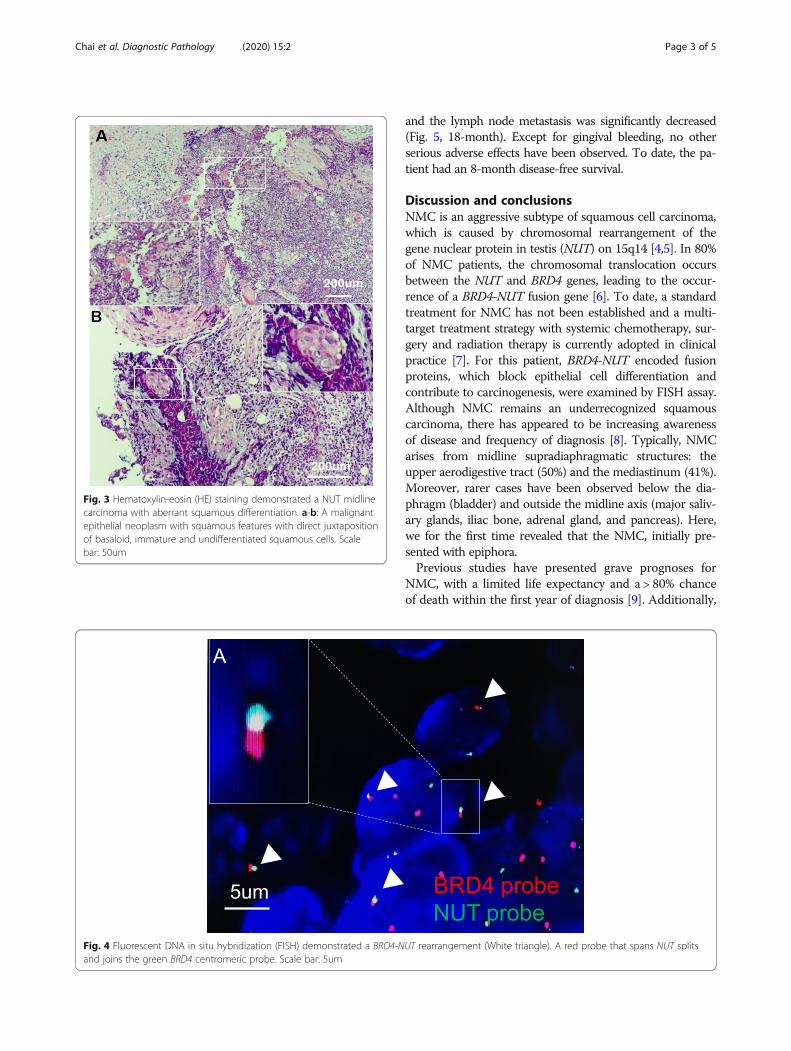
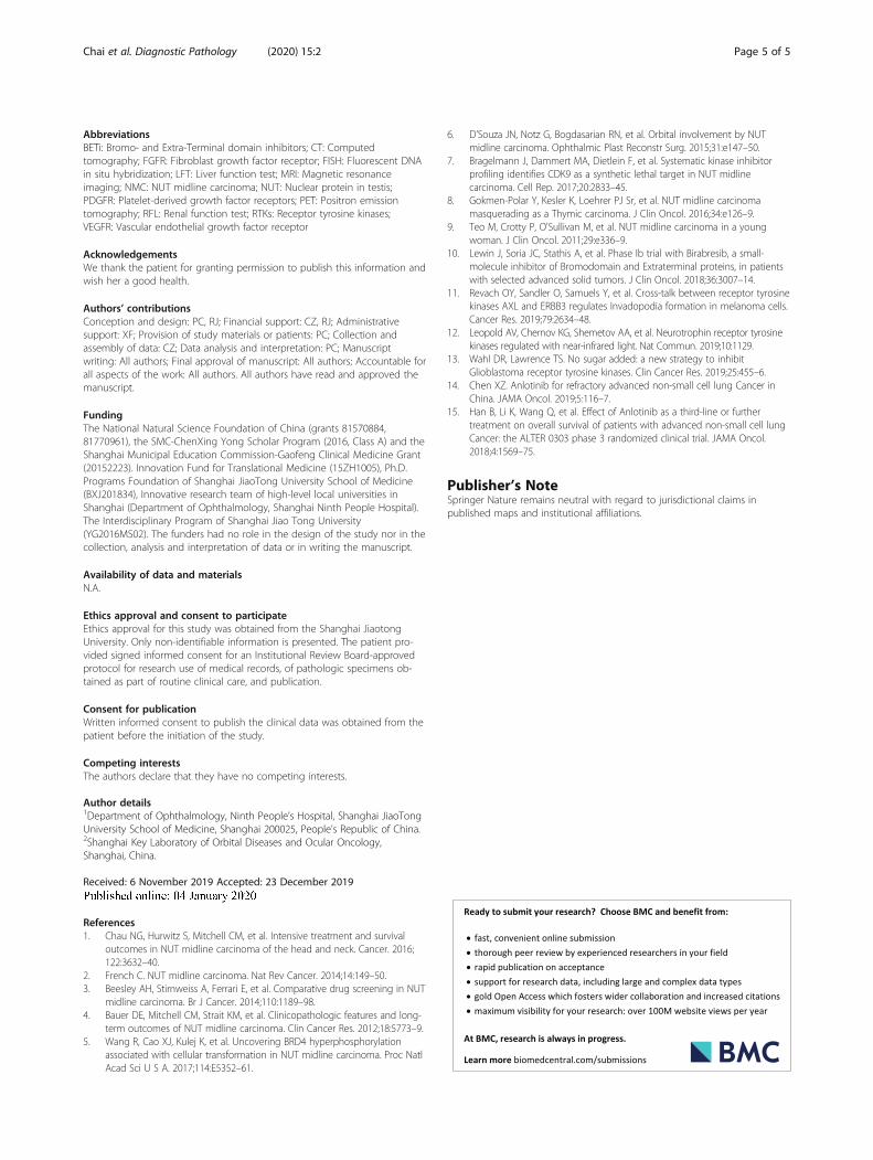
CT) revealed no other malignancies (Fig. 2). The mass wassurgically removed. Pathologic analysis suggested a malig-nant epithelial neoplasm with squamous features with directjuxtaposition of basaloid, immature and undifferentiatedsquamous cells (Fig. 3). A panel of immunohistochemistrystains showed positive staining for markers of squamous dif-ferentiation, for p40(+), p63(+), CK5/6(+), NUT (+) andKi67(50%+). Fluorescent DNA in situ hybridization (FISH)demonstrated the presence of NUT-BRD4 rearrangement(Fig. 4). In this condition, orbital exenteration was indicated,however, the patient refused. The mass grew rapidly afterprimary resection, which metastasized to cervical lymphnode 2months later (pathologically proved with biopsy).The patient developed severe dyspnea and could hardly per-form prostrations (Fig. 5, 8-month). 4months later, the pa-tient was treated with first round of local radiotherapy (50Gy/25 Fx), tumors shrunk, and the symptom of dyspneaeased. No remarkable adverse effects were observed. How-ever, 2months later, more metastasis was observed in theforehead and neck (Fig. 5, red arrow). The patient was thentreated with multi-targeting tyrosine kinase inhibitor (Anlo-tinib, 12mg, qd) and second round of local radiotherapy(50Gy/25 Fx) thereafter. The masses continued to shrink,
Fig. 1 An orbit involved NUT midline carcinoma (NMC). a Theorbital computed tomography (CT) and (b) Magnetic resonanceimaging (MRI) showed an orbital mass extending to the adjacentparanasal sinuses
Fig. 2 A systemic 18F-2-Fluoro-2-Deoxy-D-Glucopyranose positron emission tomography (18F-FDG PET/CT) demonstrated an orbital mass withincreased FDG uptake but no other remarkable malignancy in the trunk
Chai et al. Diagnostic Pathology (2020) 15:2 Page 2 of 5
and the lymph node metastasis was significantly decreased(Fig. 5, 18-month). Except for gingival bleeding, no otherserious adverse effects have been observed. To date, the pa-tient had an 8-month disease-free survival.
Discussion and conclusionsNMC is an aggressive subtype of squamous cell carcinoma,which is caused by chromosomal rearrangement of thegene nuclear protein in testis (NUT) on 15q14 [4,5]. In 80%of NMC patients, the chromosomal translocation occursbetween the NUT and BRD4 genes, leading to the occur-rence of a BRD4-NUT fusion gene [6]. To date, a standardtreatment for NMC has not been established and a multi-target treatment strategy with systemic chemotherapy, sur-gery and radiation therapy is currently adopted in clinicalpractice [7]. For this patient, BRD4-NUT encoded fusionproteins, which block epithelial cell differentiation andcontribute to carcinogenesis, were examined by FISH assay.Although NMC remains an underrecognized squamouscarcinoma, there has appeared to be increasing awarenessof disease and frequency of diagnosis [8]. Typically, NMCarises from midline supradiaphragmatic structures: theupper aerodigestive tract (50%) and the mediastinum (41%).Moreover, rarer cases have been observed below the dia-phragm (bladder) and outside the midline axis (major saliv-ary glands, iliac bone, adrenal gland, and pancreas). Here,we for the first time revealed that the NMC, initially pre-sented with epiphora.Previous studies have presented grave prognoses for
NMC, with a limited life expectancy and a > 80% chanceof death within the first year of diagnosis [9]. Additionally,
Fig. 3 Hematoxylin-eosin (HE) staining demonstrated a NUT midlinecarcinoma with aberrant squamous differentiation. a-b: A malignantepithelial neoplasm with squamous features with direct juxtapositionof basaloid, immature and undifferentiated squamous cells. Scalebar: 50um
Fig. 4 Fluorescent DNA in situ hybridization (FISH) demonstrated a BRD4-NUT rearrangement (White triangle). A red probe that spans NUT splitsand joins the green BRD4 centromeric probe. Scale bar: 5um
Chai et al. Diagnostic Pathology (2020) 15:2 Page 3 of 5

aggressive surgical resection and early radiotherapy mayprolong life expectancy for the non-metastatic NMC pa-tient [4]. In this case, we assumed that the NMC grew rap-idly after primary resection was due to the incompleteresection. And the patient refused orbital exenteration fordisfigurement of facial appearance. The recent availabilityof targeted therapy with acetyl-histone mimics (BETi) mayhold promise in treating NMC [2]. Jeremy Lewin et al.found that NMC had a partial response with 1.4 to 8.4months of BETi treatment [10]. However, BETi is notcurrently available in China. In this report, we per-formed local radiotherapy combined with multi-targeting tyrosine kinase inhibitor (Anlotinib) could sig-nificantly suppress NMC progression, which might pro-vide an alternative to the treatment of NMC.Receptor tyrosine kinases (RTKs) are transmembrane
glycoproteins that communicate with cellular growthfactors and extracellular ligands [11]. They play vitalroles in intracellular tyrosine phosphorylation and intra-cellular signaling [12]. RTK activation mediates manyvital physiological processes, including proliferation, mi-gration, differentiation and apoptosis [13]. In addition,RTKs have been implicated in a variety of cancers, suchas non-small-cell lung cancer (NSCLC), renal cell carcin-oma (RCC) and advanced medullary thyroid cancer [13].Anlotinib is a novel, orally administered RTKs inhibitor
that multi-targets fibroblast growth factor receptor(FGFR), vascular endothelial growth factor receptor(VEGFR) and platelet-derived growth factor receptors(PDGFR) [14]. Compared to placebo, it largely improvedboth progression-free survival (PFS) and overall survival(OS) in a phase III trial in patients with advanced non-small-cell lung cancer (NSCLC) [15]. Here, we for thefirst time demonstrated that the clinical application ofAnlotinib in treating with orbital NMC.In summary, we initially reported a case of NMC with
the primary complaint of epiphora. In addition to routinetests of lacrimal duct irrigation, a CT or MRI should becarried out if the symptoms persist. Heightened awarenessand early recognition of NMC are critical, and the clin-ician should possess adequate suspicion for NMC, espe-cially when encountering patients with refractoryepiphora. NMC is histopathologically characterized byabrupt squamous differentiation, a NUT immunostainingpositive result and BRD4-NUT rearrangement by FISHassay. Once diagnosed with NMC, complete surgical re-section should be performed. Adequate radiotherapy isrecommended postoperatively. Additionally, other adju-vant therapies, such as BETi, local radiotherapy and tyro-sine kinase inhibitor, could also be considered for NMCtreatment. Larger studies and longer follow up time stillneeded to validate our finding.
Fig. 5 The appearance of the NUT midline carcinoma (NMC) patient at the time of onset, 4, 8, 16 and 18months after diagnosis. The patient’sconsent for using their photos for publication has been obtained
Chai et al. Diagnostic Pathology (2020) 15:2 Page 4 of 5
AbbreviationsBETi: Bromo- and Extra-Terminal domain inhibitors; CT: Computedtomography; FGFR: Fibroblast growth factor receptor; FISH: Fluorescent DNAin situ hybridization; LFT: Liver function test; MRI: Magnetic resonanceimaging; NMC: NUT midline carcinoma; NUT: Nuclear protein in testis;PDGFR: Platelet-derived growth factor receptors; PET: Positron emissiontomography; RFL: Renal function test; RTKs: Receptor tyrosine kinases;VEGFR: Vascular endothelial growth factor receptor
AcknowledgementsWe thank the patient for granting permission to publish this information andwish her a good health.
Authors’ contributionsConception and design: PC, RJ; Financial support: CZ, RJ; Administrativesupport: XF; Provision of study materials or patients: PC; Collection andassembly of data: CZ; Data analysis and interpretation: PC; Manuscriptwriting: All authors; Final approval of manuscript: All authors; Accountable forall aspects of the work: All authors. All authors have read and approved themanuscript.
FundingThe National Natural Science Foundation of China (grants 81570884,81770961), the SMC-ChenXing Yong Scholar Program (2016, Class A) and theShanghai Municipal Education Commission-Gaofeng Clinical Medicine Grant(20152223). Innovation Fund for Translational Medicine (15ZH1005), Ph.D.Programs Foundation of Shanghai JiaoTong University School of Medicine(BXJ201834), Innovative research team of high-level local universities inShanghai (Department of Ophthalmology, Shanghai Ninth People Hospital).The Interdisciplinary Program of Shanghai Jiao Tong University(YG2016MS02). The funders had no role in the design of the study nor in thecollection, analysis and interpretation of data or in writing the manuscript.
Availability of data and materialsN.A.
Ethics approval and consent to participateEthics approval for this study was obtained from the Shanghai JiaotongUniversity. Only non-identifiable information is presented. The patient pro-vided signed informed consent for an Institutional Review Board-approvedprotocol for research use of medical records, of pathologic specimens ob-tained as part of routine clinical care, and publication.
Consent for publicationWritten informed consent to publish the clinical data was obtained from thepatient before the initiation of the study.
Competing interestsThe authors declare that they have no competing interests.
Author details1Department of Ophthalmology, Ninth People’s Hospital, Shanghai JiaoTongUniversity School of Medicine, Shanghai 200025, People’s Republic of China.2Shanghai Key Laboratory of Orbital Diseases and Ocular Oncology,Shanghai, China.
Received: 6 November 2019 Accepted: 23 December 2019
References1. Chau NG, Hurwitz S, Mitchell CM, et al. Intensive treatment and survival
outcomes in NUT midline carcinoma of the head and neck. Cancer. 2016;122:3632–40.
2. French C. NUT midline carcinoma. Nat Rev Cancer. 2014;14:149–50.3. Beesley AH, Stirnweiss A, Ferrari E, et al. Comparative drug screening in NUT
midline carcinoma. Br J Cancer. 2014;110:1189–98.4. Bauer DE, Mitchell CM, Strait KM, et al. Clinicopathologic features and long-
term outcomes of NUT midline carcinoma. Clin Cancer Res. 2012;18:5773–9.5. Wang R, Cao XJ, Kulej K, et al. Uncovering BRD4 hyperphosphorylation
associated with cellular transformation in NUT midline carcinoma. Proc NatlAcad Sci U S A. 2017;114:E5352–61.
6. D’Souza JN, Notz G, Bogdasarian RN, et al. Orbital involvement by NUTmidline carcinoma. Ophthalmic Plast Reconstr Surg. 2015;31:e147–50.
7. Bragelmann J, Dammert MA, Dietlein F, et al. Systematic kinase inhibitorprofiling identifies CDK9 as a synthetic lethal target in NUT midlinecarcinoma. Cell Rep. 2017;20:2833–45.
8. Gokmen-Polar Y, Kesler K, Loehrer PJ Sr, et al. NUT midline carcinomamasquerading as a Thymic carcinoma. J Clin Oncol. 2016;34:e126–9.
9. Teo M, Crotty P, O'Sullivan M, et al. NUT midline carcinoma in a youngwoman. J Clin Oncol. 2011;29:e336–9.
10. Lewin J, Soria JC, Stathis A, et al. Phase Ib trial with Birabresib, a small-molecule inhibitor of Bromodomain and Extraterminal proteins, in patientswith selected advanced solid tumors. J Clin Oncol. 2018;36:3007–14.
11. Revach OY, Sandler O, Samuels Y, et al. Cross-talk between receptor tyrosinekinases AXL and ERBB3 regulates Invadopodia formation in melanoma cells.Cancer Res. 2019;79:2634–48.
12. Leopold AV, Chernov KG, Shemetov AA, et al. Neurotrophin receptor tyrosinekinases regulated with near-infrared light. Nat Commun. 2019;10:1129.
13. Wahl DR, Lawrence TS. No sugar added: a new strategy to inhibitGlioblastoma receptor tyrosine kinases. Clin Cancer Res. 2019;25:455–6.
14. Chen XZ. Anlotinib for refractory advanced non-small cell lung Cancer inChina. JAMA Oncol. 2019;5:116–7.
15. Han B, Li K, Wang Q, et al. Effect of Anlotinib as a third-line or furthertreatment on overall survival of patients with advanced non-small cell lungCancer: the ALTER 0303 phase 3 randomized clinical trial. JAMA Oncol.2018;4:1569–75.
Publisher’s NoteSpringer Nature remains neutral with regard to jurisdictional claims inpublished maps and institutional affiliations.
Chai et al. Diagnostic Pathology (2020) 15:2 Page 5 of 5