oral sc','uiernallkes,,{ - dental news · oraleuropean sc','uiernallkes,,{...
TRANSCRIPT
European ','urnal ,,{ ~'
Oral Scielkes ' ,~:Elir J Oral Sci 2009; /17: 447-.453Printed in Singapore. All rigilts reserved
© 2009 The Authors.Jo"rnal compilation <f) 2009 E"r J Oral Sci
European JOllrtlal ifOral Sdetlces
Annalisa Mazzoni', Giulio Marchesi2,
Milena Cadenaro2, GiovanniMazzotti', Roberto Oi Lenarda2,Marco Ferrari3, Lorenzo Breschi2,4
'Department of SAU & FAL,UniversityofBologna, Bologna, Italy;2Divisionof DentalSciences and Biomaterials, Department ofBiomedicine,Universityof Trieste, Trieste,Italy;3Department of Fixed Prosthodontics andDental Materials, Universityof Siena, Siena,Italy;4IGM-CNR,Unitof Bolognarc/o lOR,Bologna, Italy
Push-out stress for fibre posts lutedusing different adhesive strategiesJo"fazzoniA, Marchesi G, Cadenaro 1\1, l\1azzotti G, Di Lenarda R, Ferrari M, Breschi L.Push-out stress for fibre posts luted using different adhesive strategies. Eur J OralSci 2009; 117; 447-·453. © 20 9 The Authors. Journal compilalion © 2009 Eur J OralSci
The influence of thermocycli 19 on the bond strength of fibre posts cemented withdifferent luting approaches as investigated. A total of 84 human incisors wereselected for the study. Sixty teeth were assigned to one of the following adhesive/cement combinations for push-out bond-strength evaluation: group 1, XP Bond/CoreXFlow + DT Light-Post; group 2, Panavia F 2.0 + Tech 21; or group 3, RelyXUnicem + RelyX. Bonded specimens were cut into I-mm-thick slabs and eitherthermocycled (40,000 cycles) or stored in artificial saliva (control specimens) beforepush-out bond-strength testing. Additional specimens were processed for quantitativeinterfacial nanoleakage analysis. Thermoeycling decreased the bond strength inspecimens of groups 2 and 3, but did not affect the specimens from group 1. Nodifference was observed among luting approaches in control specimens. Thermo-cycling resulted in increased silver nitrate deposition (i.e. interfacial nanoleakage) in allgroups. Within the limitations of the study, the use of an etch-and-rinse adhesive incombination with a dual-cure cement to lute fiber posts is the most stable lutingprocedure if compared with a Iself-etch resin-based cement or a self-adhesive cement, asassayed by thermocycling of the bonded specimens.
I
Fibre-reinforced posts are routinely used in combinationwith bonding/luting materials to restore endodonticallytreated teeth. A number of studies have indicated thefavourable properties of fibre posts in combination withseveral luting agents and adhesive systems (1-3). How-ever, because of the large variety of products and theintrinsic difficulties of bonding within the endodonticspace, it is difficult to select a luting strategy that pro-vides reliable and long-lasting bonding to root dentin.Therefore, it is necessary to investigate and validate thestability of the adhesive interface of fibre posts within theroot canal to ensure long-term satisfactory performance.
Fibre posts can be cemented using conventionaldual-cure resin-based cements in combination with etch-and-rinse or self-etch adhesives, or using the recentlyformulated self-adhesive cements (i.e. all-in-one resin-based materials) that allow simultaneous bondingbetween the intraradicular dentin and the post.
The etch-and-rinse strategy requires a wet dentinsubstrate for optimal bonding (4). However, because oflimited ac.cess, it is difficult to control wetness within theroot canal. Conversely, the self-etch approach does notrequire rinsing, solving the problem of substrate mois-ture control and thus simplifying the clinical procedure.While two-step self-etch adhesives showed high bond-strength values and stabil(ty over time when applied to
Professor Lorenzo Breschi. Department ofBiomedicine,Unitof Dental Sciences andBiomaterials,Universityof Trieste, ViaStuparich, 1, 1-34129 Trieste, Italy
Telefax: +39-040-662744E-mail:[email protected]
Keywords: dentin-bondingagents; fibre post;nanoleakage analysis; push-out bond strength;resin cements
coronal dentin, the efficacy of self-etch systems toproperly impregnate the intraradicular dentin remainsquestionable (5, 6). The peculiar histological character-istics of the intraradicular dentin, and the presence of athick endodontic smear layer, has prompted someresearchers to recommend a preliminary etching step ofthe dowel space before bonding (5).
The recent introduction of self-adhesive resin-basedcements has allowed clinicians to lute fibre posts using asimple and standardized approach that reduces tech-nique sensitivity (7, 8), despite limited access to theendodontic space. Although several studies have indi-cated that the bond-strength values of self-adhesivecements are comparable to, or even higher than, those ofconventional luting strategies (8), their limited etchingcapability in the presence of the compact smear layercreated within the endodontic space is a matter ofconcern (9, 10).
Irrespective of the luting agent used, bonding to theintraradicular dentin remains unpredictable as a result ofvarious factors that affect the clinical procedure and mayjeopardize adhesion (11). A number of parametersmarkedly affect bonding within the root canal, includingthe post-space preparation method, proper clinicalhandling of the luting materials, high C-factors, andresidual irrigants or endodontic medicaments (11-14).
Among laboratory tests conducted to assay the long-term behaviour of different material combinations,thermocycling represents a widely used in vitro ageingmethodology. The artificial ageing effect induced bythermocycling involves both chemical and mechanicaldegradation pathways: hot water may accelerate hydro-lysis and elution of interface components, and repetitivecontraction/expansion stresses can be generated at thetooth-biomaterial interface (15).
Therefore, the purpose of this in vitro study was toevaluate the contribution of' both chemical andmechanical factors of the artificial aging induced bythermocycling on the retention of fibre posts cementedusing different luting strategies. The adhesive interfacescreated by an etch-and-rinse adhesive in combi-nation with a dual-cure resin-based cement, a self-etchcement, or a self-adhesive resin cement were artificiallyaged via thermocycling and assessed using the push-out bond strength test and interfacial nanoleakageanalysis. The hypotheses tested were that (i) no dif-ference exists in terms of bond strength or nanoleakageamong the tested luting strategies and that (ii)thermocycling does not affect the retention of fibreposts.
Eighty-four human incisors, extracted for periodontalreasons, were analysed at the University of Trieste (Italy)after first obtaining informed consent from all patients.The teeth were stored in 1% chloramine T solution at 4°Cuntil use. Adhering soft tissues and debris were cleaned offand the crown was removed using a high-speed diamondsaw (Micromet; Remet, Casalecchio di Reno, BO, Italy)under water-cooling. The working length was establishedat 1 mm from the root apex. Cleaning and shaping wasperformed using a crown-down technique employingFlexMaster rotary instruments on an E-Master hand piece(Dentsply De Trey, Konstanz, Germany). Irrigation wasperformed using 17% EDTA and 5% NaOCI throughoutcleaning and shaping of the root canal. Root canals wereobturated with gutta-percha and a resin sealer (AR Plus;Dentsply) using the continuous warm-wave vertical-compaction technique, and backfilling was performedusing System B (Elements Obturation Unit; SybronEndo,Orange, CA, USA).
Teeth were divided randomly into three groups (n = 28).A post space 8 mm in depth was prepared with the corre-sponding low-speed drill, and groups were classifiedaccording to luting strategy, as follows.
Group 1. Etch-and-rinse group: XP-Bond adhesive/self-curing activator (Dentsply)/CoreXFlow2 (Dentsply).Posts: DT Light-Post (RTD, St Egreve, France).Group 2. Self-etch group: Panavia F2.0/ED primer(Kuraray Dental, Osaka, Japan). Posts: Tech 21(Abrasive Technology. Lewis Center, OR. USA).Group 3. Self-adhesive group: RelyX Unicem (3MESPE, Seefeld, Germany). Posts: RelyX (3M ESPE).
Adhesive/cement application and post-luting were per-formed according to the manufacturers' instructions(Table 1).
Specimen preparation for push-out strength test
Sixty teeth were assigned to the push-out bond-strengthtest (n = 20 per group). Specimens were cut into trans-verse sections using a low-speed diamond saw (Micro met;Remet) under water-cooling to obtain root slices of 1 mmin thickness; approximately 5-6 slices per tooth wereobtained, and both coronal and apical sections wereincluded in the study. For each group, half of the speci-mens were subjected to thenuocycling (40,000 cycles) from5°C to 55°C for 30 s each (WillyteclSD Mechatronik,Feldkirchen- Westerham, Germany) in artificial saliva,while the remaining bonded slices were stored in artificialsaliva at 37°C for the same period of time (IS, 16). Afterthermocycling or storage, the push-out test was perfonuedu~ing a universal testing machine (Sun500; Galdabini,Milano, Italy) connected to a load cell with the load speedset at I mm min-I.
All fractured specimens were ana lysed using a stereo-microscope (Stemi 2000-C, SOx objective; Carl Zeiss Jena,Jena, Germany) to determine whether the failure mode wasbetween dentin and the luting agent (AD), adhesive failurebetween the luting agent and the post (AP), cohesive failurewithin the luting agent (CC), or mixed failure (M). Themaximum failure load was recorded in newtons (N) andconverted into megapascals (MPa).
Because the values \\'ere normally distributed, as deter-mined by the Kolmogorov-Smirnov test, the data wereanalysed using two-way analysis of variance (ANOVA) andTukey's post hoc test (P-values of < 0.05 were consideredsignificant, Table 2).
Interfacial nanoleakage analysis
The remaining 24 post-luted roots (n = 8 per group) wereselected for the evaluation of interfacial nanoleakageexpression. The portion of each root containing the fibrepost was sectioned at intervals of 1 mm using a low-speedsaw (Micro met; Remet) to expose the luted interface;approximately 5-6 slices per tooth were obtained, and bothcoronal and apical sections were included in the study.Briefly, undemineralized and unembedded specimens werecovered with nail varnish, leaving I mm free at the interface,immersed in 50 wt% ammoniacal silver nitrate solution, andthen immersed in photo developing solution, in accordancewith the method of TAY et al. (17).
Sections were then bonded onto glass slides, ground to athickness of approximately 40 Illn with 600·, 800-, 1,200-,and 2,400-grit silicon carbide (SiC) paper under runningwater (LS2; Remet, Bologna, Italy), stained with 0.5% acidfuchsin for 15 min, and then observed under normaltransmitted light using a light microscope (Nikon Eclipse;Nikon, Tokyo, Japan).
All interface images were obtained at a magnification ofxl 00, and the amount of silver tracer deposited along theinterface (i.e. the degree of interfacial nanoleakage) wasscored on a scale of 0-4 by two observers in accordance withSABOIA et at. (18) [i.e. interfacial nanoleakage was scoredbased on the percentage of the adhesive surface showingsilver nitrate deposition: 0, no nanoleakage; 1, < 25% withnanoleakage; 2, 25% to :;:; 50% with nanoleakage; 3, 50%to :;:; 75% with nanoleakage; and 4, > 75% with nano-leakage (Table 2)]. Statistical differences among scoregroups (Le. the number of specimens falling within eachscore category) were analysed using the chi-square test(P < 0.05).
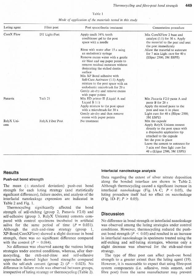
Apply caulk 34% toothconditioner gel to the postspace with a needle
Rinse with water after 15 s usingan endodontic syringe
Remove excess water with a gentleair blast and us!: paper points toremove residual'moisture withoutdesiccating the etched dentinsurface
Mix Xl' Bond adhesive withSelf-Cure Activator (1:1) Applymixture to the post space with anendodontic micro brush for 20 s
Gently air-dry and remove excesswith paper points
Mix ED primer II (Liquid A andLiquid B l: I)
Apply mixture to the post spacewith a microbflk~h for 30 s
Gently air-dry and then removeexcess with paper points
No treatmentRelyX Uni-cem
Mix CoreXFlow 2 base andcatalyst (l: I) for 30 s. Applythe material to the post and seatthe post immediately
Allow the material to autocurefor 7 min Light cure for 40 s(Eli par 2500; 3M ESl'E)
Mix l'anavia F2.0 paste A andpaste B for 20 s
Apply the mixed paste to thepost and seat it in place
Light cure for 40 s (Eli par 2500;3M ESl'E)
Mix the capsuleApply RelyX Unicem cementdirectly to the post space witha disposable application tipattached to the capsule
Set the post in placeLeave the cement to autocure for
5 min and then light cure for40 s (Elipar 2500, 3M ESPE)
Push-out bond strength
The mean (± standard deviation) push-out bondstrength for each luting strategy (and statisticallysignificant differences), failure modes, and analysis of theinterfacial nanoleakage expression are indicated inTable 2 and Fig. 1.
ThemlOcycling significantly affected the bondstrength of self-etching (group 2, Panavia F2.0) andself-adhesive (group 3, RelyX Unicem) cements com-pared with control specimens incubated in artificialsaliva for the same period of time (1' = 0.031).Although the etch-and-rinse strategy (group 1,XP-Bond/CoreXFlow) showed a slight decrease in bondstrength, there was no significant difference comparedwith the control (P = 0.084).
No difference was observed among the various lutingstrategies under control conditions, whereas, after ther-mocyc1ing, the etch-and-rinse and self-adhesiveapproaches showed higher bond strengths comparedwith the self-etch cement (group 2) (1' = 0.023). Nodifference in failure mode was observed between groups,irrespective ofluting strategy or thermocycling (Table 2).
Interfacial nanoleakage analysis
Data regarding the extent of silver nitrate depositionalong the bonded interface are shown in Table 2.Although thermocycling caused a significant increase ininterfacial nanoleakage (Fig. IA-C; P < 0.05), theluting procedure itself had no effect on nanoleakage(Fig. IDF; P > 0.05).
No difference in bond strength or interfacial nanoleakagewas observed among the luting strategies under controlconditions. However, thennocycling reduced the push-out bond strength (P < 0.05) and resulted in an increasein interfacial nanoleakage in specimens treated using theself-etching and self-luting strategies, whereas only aslight decrease was observed for the etch-and-rinsestrategy.
The type of fibre post can affect push-out bondstrength to a greater extent than the luting agent (19).However, it is reasonable to expect that using fibre-postsystem components (i.e. adhesive, resin cement, andfibre post) from the same manufacturer may prevent
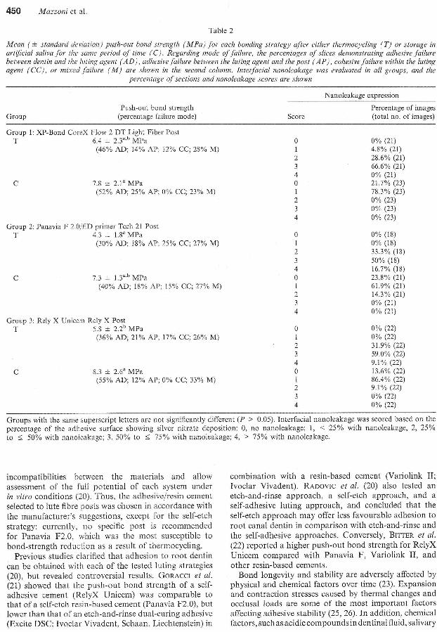
}dean (± standard deviation) push-out bond strength (lvIPa) for each bonding strategy after either thermocycling (T) or storage inartificial saliva for the same period of time (C). Regarding mode o.ffailure, the percentages of slices demonstrating adhesive failurebetween dentin and lhe luting agent (AD), adhesive failure between the luting agent and the post (AP), cohesivefailure within the lUlingagent (CC) , or mixed failure (lvf) are shown in the second column. fnleijacial nanoleakage was evalualed in all groups, and the
percentage of sections and nanoleakage scores are shown
Push-out bond strength(percentage failure mode)
Nanoleakage expression
Percentage of images(total no. of images)
Group 1: XP-Bond CoreX Flow 2 DT Light Fiber PostT 6.4 ± 2.3",b MPa
(46% AD; 14% AP; 12% CC; 28'10 M)
7.8 ± 2.1" MPa(52% AD; 25% AP; 0% CC; 23% M)
Group 2: Panavia F 2JJjED primer Tech 21 PostT 4.3 ± 1.8e MPa
(30% AD; 18% AP; 25% CC; 27% M)
7.3 ± 1.3a,h MPa(40'10 AD; 18% AP; 15% CC; 27% M)
Group 3: Rely X Unicem Rely X PostT 5.8 ± 2.2b MPa
(36% AD; 21 % AP; 17% CC; 26% M)
8.3 ± 2.6a MPa(55% AD; 12% AP; 0% CC; 33% M)
0% (21)4.8% (21)28.6% (21)66.6% (21)0% (21)21.7% (23)78.3% (23)0% (23)0% (23)0% (23)
0% (18)0% (18)33.3% (18)50% (18)16.7% (I8)23.8% (21)61.9% (21)14.3% (21)0% (21)0% (21)
0% (22)0% (22)31.9% (22)59.0% (22)9.1%(22)13.6% (22)86.4% (22)9.1% (22)0% (22)0% (22)
Groups with the same superscript letters are not significantly different (P > 0.05). Interfacialnanoleakage was scored based on thepercentage of the adhesive surface showing silver nitrate deposition: 0, no nanoleakage; I, < 25% with nanoleakage, 2, 25%to ~ 50% with nanoieakage; 3, 50% to ~ 75% with nanoJcakage; 4, > 75% with nanoleakage.
incompatibilities between the materials and allowassessment of the full potential of each system underin vitro conditions (20). Thus, the adhesive/resin cementselected to lute fibre posts was chosen in accordance withthe manufacturer's suggestions, except for the self-etchstrategy: currently, no specific post is recommendedfor Panavia F2.0, which was the most susceptible tobond-strcngth reduction as a result of thermo cycling.
Previous studies clarified that adhesion to root dentincan be obtained with each of the tested luting strategies(20), but revealed controversial results. GORACCIet al.(21) showed that the push-out bond strength of a self-adhesive cement (RelyX Unicem) was comparable tothat of a self-etch resin-based cement (Panavia F2.0), butlower than that of an etch-and-rinse dual-curing adhesive(Excite DSC; !voclar Vivadent, Schaan, Liechtenstein) in
combination with a resin-based cement (Variolink II;!voclar Vivadent), RADOVICet aI, (20) also tested anetch-and-rinse approach, a self-etch approach, and aself-adhesive luting approach, and concluded that theself-etch approach may offer less favourable adhesion toroot canal dentin in comparison with etch-and-rinse andthe self-adhesive approaches. Conversely, BITTERet al.(22) reported a higher push-out bond strength for RelyXUnicem compared with Panavia F, Variolink II, andother resin-based cements.
Bond longevity and stability are adversely affected byphysical and chemical factors over time (23). Expansionand contraction stresses caused by thermal changes andocclusal loads are some of the most important factorsaffecting adhesive stability (25, 26). In addition, chemicalfactors, such as acidic compounds in dentinal fluid, salivary
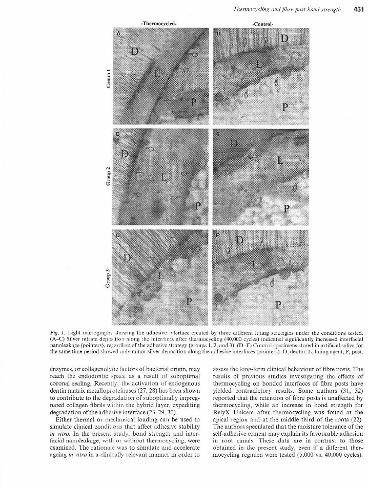
Fig. 1. Light micrographs showing the adhesive interface created by three different luting strategies under the conditions tested.(A-C) Silver nitrate deposition along the interfaces after thermocyding (40,000 cycles) indicated significantly increased interfacialnanoleakage (pointers), regardless of the adhesive strategy (groups 1,2, and 3). (D-F) Control specimens stored in artificial saliva forthe same time-period showed only minor silver deposition along the adhesive interfaces (pointers). D, dentin; L, luting agent; P, post.
enzymes, or collagenolytic factors of bacterial origin, mayreach the endodontic space as a result of suboptimalcoronal sealing. Recently, the activation of endogenousdentin matrix metalloproteinases (27, 28) has been shownto contribute to the degradation of suboptimally impreg-nated collagen fibrils within the hybrid layer, expeditingdegradation of the adhesive interface (23,29,30).
Either thermal or mechanical loading can be used tosimulate clinical conditions that affect adhesive stabilityin vitro. In the present study, bond strength and inter-facial nanoleakage, with or without thermocycling, wereexamined. The rationale was to simulate and accelerateageing in vitro in a clinically relevant manner in order to
assess the long-term clinical behaviour of fibre posts. Theresults of previous studies investigating the effects ofthermocycling on bonded interfaces of fibre posts haveyielded contradictory results. Some authors (31, 32)reported that the retention of fibre posts is unaffected bythermocycling, while an increase in bond strength forRelyX Unicem after thermocycling was found at theapical region and at the middle third of the roots (22).The authors speculated that the moisture tolerance of theself-adhesive cement may explain its favourable adhesionin root canals. These data are in contrast to thoseobtained in the present study, even if a different ther-mocycling regimen were tested (5,000 vs. 40,000 cycles).
In a different study, thermal ageing was found to reducebond strength, as recently reported by BITTERet al. (33),showing a drastic rcduction of bond strcngths regardlessof post type, prctrcatmcnt, or corc matcrial. However,attention should be focuscd on thc conditions undcrwhich the specimens were tested, in partieular the num-ber of thennocycles to which the bonded spccimens weresubjected. According to the ISO TR 11450 (16), 500thermocycles in water between 5°C and 55°C is consid-ered to be an appropriate and essential test for ageingdental materials. In accordance with this recommenda-tion, the majority of previous studies investigated theeffects of thermocycling under these conditipns. How-ever, because no significant decrease in bond-strengtheffectiveness was observed after such limited thermo-cycling (i.e. 3,000-6,000 cycles) (31, 32, 34), the ability ofthe test to simulate intra-oral ageing accurately remainscontroversial. Although some authors (33) have advo-cated placing less emphasis on thermoeycling whenevaluating the retention of root canal posts, EICKet al.(35) supported the ill vitro test, concluding that 10,000cycles was a reasonable approximation of 1 yr of clinicalservice for coronal bonded interfaces. DE MUNCKet al.(15) reported that unsimplified adhesives do not appearto be altered after artificial ageing for up to 20,000 cycles(i.e. 2 yr of simulated clinical service). Based on thesuggestion that an increased number of the1111Ocyc1esshould be performed to properly challenge tbe adhesiveinterface, we examined the effect of 40,000 cycles, rep-resenting approximately 4 yr of functional service (24),in the present study. This increased number of cyclesreduced the adhesive performance of the self-etching andthe self-adhesive approachcs. As thermocyc1ing chal-lenges the adhcsive interface both chcmica]]y (hot watcraccelerates hydrolysis and elution of interface compo-nents) and mechanica]]y (repetitive contraction/expan-sion stresses), increasing the number of cycles impliesextended storage in artificial saliva finally enhancing therole of water-mediated ageing phenomena (15). For thisreason, control specimens were stored in artificial salivaat 37°C for the same period of time needed by thethermocyc1ing speeimens to be tested to failure (approx.7 wk), in order to assay the role of water-mediatedageing phenomena accurately.
However, although thermocycling showed observableeffects on coronal dentin, it should be noted that thesurrounding bone and periodontal tissues minimizethermal changes ill vil'o. Thus, thermocycling can be usedonly as a means to accelerate ageing in vitro in luted fibrepost specimens, which allows us to clarify the possiblerole of thermal contraction and expansion within theendodontic space. In fact, because thennocycling pro-duces a greater degree of heat stress than the amount ofheat that can be transmitted in clinical situations (24),we concluded tbat the acceptable levels of retentionobserved in the present study after artificial ageing maybe even more promising under clinical conditions.
Acknowledgements - The authors wish to thank Mr AurelioValmori for extensive laboratory assistance. The study waspartially supported by MIUR-Italy grants.
!. HAYASCHIM, SUGETAA, TAKAYASHIY, IMAZATOS, Emsu S.Static and fatigue fracture resistances of pulpless teeth restoredwith post-cores. Dent Mater 2008; 24: 1178-1186.
2. FERRARI M. BRESCHI L, GRANDINI S. Fiber Posts andEndodontically treated teeth: a compendium of scientific andclinical perspectives. Wendywood, South Africa: ModernDentistry Media. 2008.
3. CAGIDI,\CO MC, GORACCI C, GARCIA-GODOYF, FERRARI M.Clinical studies of fiber posts: a literature review. Int J Pros-thodont 2008; 21: 328-336.
4. NAKAJIMAM, KANEMURAN, PEREIRAPN, TAGAMIJ, PASHLEYDB. Comparative micro tensile bond strength and SEManalysis of bonding to wet and dry dentin. Am J Dent 2000; 13:324-328.
5. ZHANGL, HUANGL, XIONG Y, FANG M, CHENJH, FERRARIM.Effect of post-space treatment on retention of fiber posts indifferent root regions using two self-etching systems. Eur.J OralSci 2008; 116: 280·286.
6. CHERSONIS, ACQUAVIVAGL, PRATI C, FERRARIM, GRANDINIS, PASHLEYDH, TAY FR. In vivo tJuid movement throughdentin adhesives in endodontically treated teeth . .J Dent Res2005; 84: 223-227.
7. MONTlCELLlF, FERRARI M, TOLEDANOM. Fiber posts lutingprocedure. Med Oral Patol Oral eir Bucal 2008; 13: E214-E22!.
8. RADOVICI, MONTICELLlF, GORACCIC, VULICEVICZR, FERRARIM. Self-adhesive resin cements: a literature review. J AdhesDent 2008; 10: 251-258.
9. SIRMAI S, RllS DN, MORGANO SM. An in vitro study of thefracture resistance and the incidence of vertical root fracture ofpulpless teeth restored with six post-and-core systems. J I'ros-thet Dent 1999: 81: 262-269.
.10. SADEKFT, GORACCIC, MONTICELLIF, GRANDINIS, CURYAH,TAY FR. Immediate and 24-hour evaluation of the interfacialstrengths of fiber posts. J Endod 2006; 32: 1174-1177.
I!. BnTER K, KIELBASSAAM. Post-endodontic restorations withadhesively luted fiber-reinforced composite post systems: areview. Am .TDent 2007; 20: 353··-360.
12. SCHWARTZ RS, FRANSMAN R. Adhesive dentistry andendodontics: materials, clinical strategies and procedures forrestoration of access cavities: a revicw . .J Endod 2005; 31:151-165.
13. BOUlLLAGUETS, TROESCHS, WATAHAJC, KREJCI1, MEYERJM,PASHLEYDH. Microtensile bond strength between adhesivecements and root canal dentin. Dent Mater 2003; 19: 199-205.
14. TAY FR, LOUSHINERJ, LAMBRECHTSP, WELLER RN, PASHLEYDH. Geometric factors affecting dentin bonding in root canals:a theoretical modeling approach . .J Endod 2005; 31: 584-589.
15. DE MUNCK J, VAN LANDUYT K, COUTlNHO E, POITEVINA,PEUMANSN, LAM.BRECHTSP, VAN MEERBEK B. Micro-tensilebond strength of adhesives bonded to Class-1 cavity-bottomdentin after thermo-cycling. Del1l Mater 2005; 21: 999-1007.
16. INTERNATIONALORGANIZATIONFOR STANDARDIZATTON.ISOTR1l405. Dental materials-guidance on testing of adhesion totooth structure. Geneva, Switzerland: International Organiza-tion for Standardization, 1994; 1....15.
17. TAY FR, PASHLEYDH, YOSHIYAMAM. Two modes of nano-leakage expression in single-step adhesives . .J Dent Res 2002; 81:472476.
18. SABOIAVPA, NATO F, MAZZONIA, ORSINI G, PUTIGNANOA,GIANNINIM, BRESCHIL. Adhesion of a two-step eteh-and-rinseadhesiVe on collagen-depleted dentin . .T Adhes Dent, 2008; 10:419-422.
19. KURTZJS, PERDlGAOJ, GERALDELIS, HODGESJS, BOWLESWR.Bond strengths of tooth-colored posts, effect of sealer, dentinadhesive, and root region. Am J Dellt 2003; 16 Spec No: 31A-36A.
20. RADOVIC1, MAZZlTELLIC, CHIEFFI N, FERRARIM. Evaluationof the adhesion of fiber posts cemenled using dirferent adhesiveapproaches. Eur J Oral Sci 2008; 116: 557-63.
21. GORACCIC, SADEK FT. FABIANELLIA, TAY FR, FERRARI M.Evaluation of the adhesion of fiber posts to intraradiculardentin. Oper Delli 2005: 30: 627-635.
22. BITTERK, MEYER-LUECKELH, PRIEHN K, KANJUPARAMBILJP,NEUMANN K, KIELBASSAAM. Effects of luting agent andthermo cycling on bond strengths to root canal dentine. 1mEndod J 2006; 39: 809-818.
23. BRESCHlL, MAZZONIA. RUGGERIA JR, CADENAROM, DI LE-NARDAR, DORIGOE. Dental adhesion review: aging and stabilityof the bonded interface. Delli Mater 2008; 24: 90-101.
24. GALE MS, DARVELL BW. Thermal cycling procedures forlaboratory testing of dental restorations. J Dellt 1999; 27: 89-99.
25. PEUMANS M, HIKITA K, DE MUNCK J, VAN LANDUYT K,POITEVINA, LAMBRECI-ITSP, VAN MEERBEEKB. Bond durabilityof composite luting agenLs Lo ceramic when exposed to long-term thermocycling. Opa Dent 2007; 32: 372····379.
26. DE MUNCK J, VAN MEERBECKB, WEVERSM. LAMBRECHTSP,BRAEMM. Micro-rota ry fatigue of tooth-biomaterial interfaces.Biomaterials 2005; 26: 1145-1153.
27. MAZZONI A, PASHLEYDH. NISHITANI Y, BRESCHI L, TJAD-ERHANEL, TOLEDANO\1, PASHLEYEL, TAY FR. Reactivationof quenched endogenolls proteolytic activities in phosphoricacid-etched dentine by etch-and-rinse adhesives. Biomalerials2006; 27: 4470-4476.
28. PASHLEYDH, TAY FR, Yru CKY, HASHIMOTOM, BRESCHIL,CARVAHLOR, ITo S. Collagen degradation by host-derivedenzymes during aging. j Dellt Res 2004; 83: 216-221.
29. ARM,"TRONGSR, KELLER JC, BOYER DB. The influence ofwater storage and Cofactor on the dentin-resin compositemicrotensile bond strength and debond pathway utilizing afilled and unfilled adhesive resin. Dent Mater 2001; 17: 268-276.
30. CARRIHLO MRO, GERALDELl S, TAY FR, DE GoES MF,CARVAHLORM, TJARDERHANE L, HEBLING J, MAZZONI A,BRESCHI L, PASHLEY DH. III vivo preservation of hybridlayer by chlorhexidine. J Dent Res 2007; 86: 529-533.
31. PURTONDG, CHANDLERNP, QUALTROUGHAJ. Effect of ther-mocycling on Lhe retention of glass-fiber root canal posts.Quintessence Int 2003; 34: 366-369.
32. MONTICELLIF, OSORlO R, TAY FR, SADEK FT, FERRARI M,TOLEDANOM. Resistance to thermo-mechanical stress of dif-ferent coupling agents used as intermediate layer in resin-fiberpost bonds. Am J Dent 2007; 20: 416-420.
33. BITTER K, NEUMANN K, KIELBASSAAM. Effects of pretreat-ment and thermocycling on bond-strength of resin core mate-rials to various fiber-reinforced composite posts. J Ad Dent2008; 10: 481-489.
34. BELL AM, LASSILALV, KANGASMIENlI,VALLlTUPK. Bondingof fibre-reinforced composite post to root canal dentin. J Dent2005; 33: 533-539.
35. EICK JD, GWINNETAJ, PASHLEYDH, ROBINSONSJ. Currentconcepts on adhesion to dentin. Crit Rev Oral Bioi Med 1997; 8:306-335.