oral activity of the antimalarial endoperoxide 6-(1,2,6,7
TRANSCRIPT

RESEARCH ARTICLE
Oral activity of the antimalarial endoperoxide
6-(1,2,6,7-tetraoxaspiro[7.11]nonadec-4-yl)
hexan-1-ol (N-251) against Leishmania
donovani complex
Kofi Dadzie KwofieID1,2, Kai Sato2, Chizu Sanjoba2, Akina Hino1, Rieko Shimogawara1,
Michael Amoa-Bosompem1, Irene Ayi3, Daniel A. Boakye3, Abraham K. Anang3, Kyung-
Soo Chang4, Mitsuko Ohashi1, Hye-Sook Kim5, Nobuo Ohta1, Yoshitsugu Matsumoto2,
Shiroh IwanagaID1*
1 Section of Environmental Parasitology, Graduate School of Medical Dental Sciences, Tokyo Medical Dental
University, Bunkyo-ku, Tokyo, Japan, 2 Laboratory of Molecular Immunology, Department of Animal
Resource Sciences, Graduate School of Agricultural and Life Sciences, The University of Tokyo, Bunkyo-ku,
Tokyo, Japan, 3 Department of Parasitology, Noguchi Memorial Institute for Medical Research, College of
Health Sciences, University of Ghana, Legon, Accra, Ghana, 4 Department of Clinical Laboratory Science,
College of Health Sciences, Catholic University of Pusan, Busan, Republic of Korea, 5 Division of
International Infectious Disease Control, Faculty of Pharmaceutical Sciences, Graduate School of Medicine,
Dentistry Pharmaceutical Sciences, Okayama University, Okayama, Japan
* [email protected], [email protected]
Abstract
Visceral leishmaniasis (VL) is a major problem worldwide and causes significant morbidity
and mortality. Existing drugs against VL have limitations, including their invasive means of
administration long duration of treatment regimens. There are also concerns regarding
increasing treatment relapses as well as the identification of resistant clinical strains with the
use of miltefosine, the sole oral drug for VL. There is, therefore, an urgent need for new alter-
native oral drugs for VL. In the present study, we show the leishmanicidal effect of a novel,
oral antimalarial endoperoxide N-251. In our In vitro studies, N-251 selectively and specifi-
cally killed Leishmania donovani D10 amastigotes with no accompanying toxicity toward the
host cells. In addition, N-251 exhibited comparable activities against promastigotes of L.
donovani D10, as well as other L. donovani complex parasites, suggesting a wide spectrum
of activity. Furthermore, even after a progressive infection was established in mice, N-251
significantly eliminated amastigotes when administered orally. Finally, N-251 suppressed
granuloma formation in mice liver through parasite death. These findings indicate the thera-
peutic effect of N-251 as an oral drug, hence suggest N-251 to be a promising lead com-
pound for the development of a new oral chemotherapy against VL.
Author summary
Visceral Leishmaniasis remains a serious health problem in many developing countries
with thousands of new cases recorded annually. Novel oral therapies are required as
PLOS Neglected Tropical Diseases | https://doi.org/10.1371/journal.pntd.0007235 March 25, 2019 1 / 15
a1111111111
a1111111111
a1111111111
a1111111111
a1111111111
OPEN ACCESS
Citation: Kwofie KD, Sato K, Sanjoba C, Hino A,
Shimogawara R, Amoa-Bosompem M, et al.
(2019) Oral activity of the antimalarial
endoperoxide 6-(1,2,6,7-tetraoxaspiro[7.11]
nonadec-4-yl)hexan-1-ol (N-251) against
Leishmania donovani complex. PLoS Negl Trop Dis
13(3): e0007235. https://doi.org/10.1371/journal.
pntd.0007235
Editor: Laura-Isobel McCall, University of
Oklahoma, UNITED STATES
Received: November 1, 2018
Accepted: February 12, 2019
Published: March 25, 2019
Copyright: © 2019 Kwofie et al. This is an open
access article distributed under the terms of the
Creative Commons Attribution License, which
permits unrestricted use, distribution, and
reproduction in any medium, provided the original
author and source are credited.
Data Availability Statement: All relevant data are
within the manuscript and its Supporting
Information files.
Funding: This work was supported by grant from
the Research Program on Emerging and Re-
emerging Infectious Diseases from Japan Agency
for Medical Research and Development (AMED)
(https://www.amed.go.jp/program/list/01/06/001.
html), awarded to S. I. (15650501) The funders

existing drugs are limited by their invasive means of administration long duration of treat-
ment regimens. Moreover, with miltefosine as the sole oral drug, there are concerns of the
eventual development of parasite resistance with its continuous use. In this study, we
report on the in vitro and in vivo leishmanicidal effect of orally administered N-251 on the
Leishmania donovani complex parasites in mice. Our results suggest that N-251 may be a
potential lead compound for the development of a new oral chemotherapy against VL
Introduction
Visceral leishmaniasis (VL), also known as kala azar, is a disease with a worldwide distribution.
It is endemic in more than 62 countries, with over 90% of cases occurring in Brazil, Ethiopia,
India, Somalia, South Sudan Sudan [1]. An estimated 200,000 to 400,000 new cases are reported
annually [1], making VL a serious public health problem. It is caused by members of the Leish-mania donovani complex, which consists of four species: L. archibaldi, L. chagasi, L. donovani,and L. infantum, which are distinguished by their vectors, reservoir hosts, and their associated
pathology [2,3]. Parasites are transmitted to humans by the female phlebotomine sandfly. VL is
typically presented as a chronic infection, characterized by irregular bouts of fever, weight loss,
splenohepatomegaly, anemia, and a high mortality rate of almost 100% if left untreated [1]. At
present, there is no licensed vaccine, therefore, chemotherapy remains the main form of control.
Currently, treatment for VL relies on the use of drugs such as pentavalent antimonials, par-
omomycin, pentamidine, and amphotericin B. Most of these drugs are, however, either limited
by their invasive methods of administration and/or their long periods of high dose treatment.
Miltefosine remains the sole oral anti-leishmanial drug since it was approved for use in 2002
[4–6]. Recently, the absence of alternate oral drugs has become a major concern, particularly
with recent reports of increasing treatment relapses as well as the identification of miltefosine-
resistant clinical strains [7–9]. There is, therefore, an urgent need for the development of novel
oral chemotherapeutics as alternatives to miltefosine for the effective treatment of VL cases.
Recently, natural and synthetic endoperoxide compounds have received increasing atten-
tion due to their fast action and ability to target drug-resistant parasite strains [10–19]. This
class of compound is known to be activated within host cells, as a result of the iron-mediated
cleavage of the characteristic endoperoxide bridge. Moreover, as anti-infective agents, they are
easily administered via the oral route. Previously, we synthesized a novel endoperoxide com-
pound, 6-(1,2,6,7-tetraoxaspiro[7.11]nonadec-4-yl)hexan-1-ol (N-251) (S1 Fig), that possesses
antimalarial [20,21], anti-Toxoplasma gondii [22], anti-schistosomal [23–25], anti-viral [26,27]
activities in vitro when administered to mice orally. These findings suggest that N-251 pos-
sesses a broad spectrum of anti-infective activities.
In the present study, we report on the in vitro leishmanicidal effects of N-251 against amas-
tigotes of L. donovani, as well as the promastigotes of various L. donovani complex parasites
from different geographical locations. In addition, we report on the in vivo efficacy of N-251 as
an oral drug against L. donovani amastigotes. Results suggest that N-251 may be a promising
lead compound for the development of a new oral chemotherapy against VL.
Methods
Reagents parasites
N-251 was chemically synthesized as described previously [20,21]. It was dissolved in dimethyl
sulfoxide (DMSO) as 100 mM stock solutions stored at −20 ˚C. Miltefosine was purchased
Oral activity of N-251 against L. donovani complex
PLOS Neglected Tropical Diseases | https://doi.org/10.1371/journal.pntd.0007235 March 25, 2019 2 / 15
had no role in study design, data collection and
analysis, decision to publish, or preparation of the
manuscript.
Competing interests: The authors have declared
that no competing interests exist.

from Sigma-Aldrich (MO, USA). Medium 199 was purchased from Nissui Pharmaceuticals
Co., Ltd, Japan. Dulbecco’s Modified Eagle’s Medium (DMEM) Roswell Park Memorial Insti-
tute (RPMI) medium were purchased from Sigma-Aldrich. HEPES Buffer (1 M) was pur-
chased from MP Biomedicals, LLC (Ohio, USA). All reagents were maintained at 4˚C unless
otherwise stated. Five strains of L. donovani complex from different geographical locations
with high VL burden (Brazil, Nepal, India, Sudan Turkey) were used in this study. These
include L. chagasi PP75 (MHOM/BR/74/PP75), L. donovani D10 (MHOM/NP/03/D10), L.
donovani DD8 (MHOM/IN/80/DD8), L. donovani KH (MHOM/SU/43/KH) (ATCC 30503),
and L. infantum EP173. Promastigotes were cultured in vitro in M-199 complete medium
(containing 25 mM HEPES at 25˚C) supplemented with 10% heat-inactivated fetal bovine
serum (Hi-FBS). For in vitro and in vivo infectivity assays, L. donovani D10 was used because
it is constantly maintained in mice in our lab, therefore, highly infective. The usual procedure
is to use freshly isolated parasites that have undergone 1–3 cycles of passages in M-199 com-
plete medium to ensure high infectivity rates in our experiments.
Ethics statement
All animal experiments were reviewed and approved by the Animal Experiment Committee at
the Graduate School of Agricultural and Life Sciences, University of Tokyo (Ref. No. P16-254).
The experiments were performed in accordance with the Regulations for Animal Care Use of
the University of Tokyo, which are based on the Law for the Humane Treatment and Manage-
ment of Animals, Stards Relating to the Care and Management of Laboratory Animals Relief of
Pain (the Ministry of the Environment), Fundamental Guidelines for Proper Conduct of Ani-
mal Experiment Related Activities in Academic Research Institutions (the Ministry of Educa-
tion, Culture, Sports, Science Technology) and the Guidelines for Proper Conduct of Animal
Experiments (the Science Council of Japan). At the end of the experiments, the animals were
euthanized by exsanguination under anesthesia with isoflurane followed by cervical dislocation.
In vitro inhibition activity against intracellular amastigotes
The leishmanicidal effect of N-251 on L. donovani amastigotes was evaluated in murine mac-
rophages. First, 4 x 10⁴ RAW 264.7 macrophage cells in 200 μl of DMEM (containing 1% peni-
cillin/streptomycin, supplemented with 10% Hi-FBS) were seeded in the wells of an 8-well
chamber slide incubated at 37 ˚C in 5% CO2 for 2 hours to allow cell attachment. Next, station-
ary phase L. donovani promastigotes in fresh DMEM were added to the macrophages at a ratio
of 50:1 (parasites:macrophage) and incubated for 6 h at 37 ˚C in 5% CO2. During this period,
the parasites invaded the macrophages and then transformed into amastigotes. Free promasti-
gotes were subsequently removed by successive washes with DMEM. Infected macrophages
were then treated with N-251 at concentrations ranging from 0.78 to 50 μM, and incubated for
24, 48 and 72 h. Miltefosine was used as a reference drug control. Infected treated macro-
phages were then washed with 1x phosphate-buffered saline (PBS) and fixed with methanol for
10 min. Finally, the macrophages were stained with 5% Giemsa in PBS for 25 min and
observed under a light microscope. Anti-leishmanial activity was evaluated by observing 300
macrophages within each treatment group. The percentage of infected macrophages was calcu-
lated using the following formula: [(Number of infected macrophages/300 macrophages
observed) x 100]. Infection index values were then calculated according to the following for-
mula: [Percentage of infected macrophages x average number of intracellular amastigotes per
infected macrophage]. Infection index values were then converted to percentage survival val-
ues relative to the untreated parasite population. IC50 values were eventually obtained by sig-
moidal dose–response curve analysis using the scatter plot option of Microsoft Excel 2016
Oral activity of N-251 against L. donovani complex
PLOS Neglected Tropical Diseases | https://doi.org/10.1371/journal.pntd.0007235 March 25, 2019 3 / 15
(Microsoft Corporation, Washington, USA) expressed as the mean of samples ± stard devia-
tion (SD) from three independent experiments conducted in duplicates.
Cytotoxicity evaluation selectivity index determination
The cytotoxicity of N-251 against murine macrophage cell lines was assessed using the Invitro-
gen alamar blue assay kit (ThermoFisher Scientific, Japan) according to the manufacturer’s
instructions with modifications. Both RAW 264.7 and J774 cell lines were used in this assay.
First, 5 × 10³ cells were seeded into each well of a 96-well plate. Varying concentrations of N-
251, ranging from 0.195 μM to 200 μM, were then added to the cells and incubated for 48 h at
37 ˚C in 5% CO2. Miltefosine was used as a positive control. Next, 10% alamar blue dye was
added to all wells and the plate was incubated for another 24 h in darkness. After a total of 72 h,
fluorescence intensity was measured at a wavelength of 600 nm using the SpectraMax Paradigm
Multi-Mode Detection Platform (Molecular devices LLC, CA, USA). All experiments were car-
ried out 4 times in duplicates. Fluorescence intensity, which is directly proportional to the con-
centration of surviving parasites, was converted to percentage survival. Cytotoxic concentrations
at 50% (CC50) were eventually obtained by sigmoidal dose–response curve analysis using the
scatter plot option of Microsoft Excel 2016 and expressed as mean of samples ± standard devia-
tion (SD). The selectivity index (SI) was calculated as the ratio of the CC50 obtained for both
RAW 264.7 and J774 macrophage cells and the IC50 for Leishmania donovani D10 amastigotes.
In vitro leishmanicidal activity against promastigotes
The effect of N-251 was also evaluated in logarithmic phase promastigotes of the L. donovanicomplex using the Invitrogen alamar blue assay kit. The experimental procedure carried out
was essentially the same as described above. However, 5 × 10⁴ promastigotes were seeded per
well in this case. Treated and untreated promastigotes were incubated at 25 ˚C. Miltefosine
was used a reference compound. IC50 values were also obtained as described above.
Anti-leishmanial efficacy of N-251 in vivoTo evaluate the anti-leishmanial efficacy of N-251, L. donovani D10-infected mice, randomly
allocated into experimental groups of 5 animals each, were treated orally with 68 mg/kg body
weight of N-251 in olive oil. This dose was determined as the maximum concentration at which
no toxicity (ruffled fur, severe weight loss, reduced activity and death) was observed in previous
antimalarial studies [21]. Miltefosine (10 mg/kg body weight) and olive oil were used as positive
and negative controls, respectively. First, 6-week-old BALB/cA mice (CLEA Japan, Inc. Tokyo,
Japan) were infected intraperitoneally with 1x10⁸ stationary phase promastigotes. Four weeks
post-infection, treatment was administered through a feeding gavage at 12-hour intervals for 14
consecutive days. Initially, treatment efficacy was expressed as Leishman-Donovan Units
(LDU), which was determined by sterilely harvesting and weighing the spleen and liver of
euthanized mice. The macrophages in the spleen and liver were imprinted on glass slides and
then fixed with methanol for 10 minutes. Imprints were then stained with 5% Giemsa in PBS
for 25 mins and examined microscopically. LDU was calculated based on the formula: [(Num-
ber of Leishmania amastigotes per 1000 macrophage cells) x organ weight (g)] [28–33]. Percent-
age reduction was also calculated as follows: 100-[(LDU of treatment group/LDU of untreated
group) x 100] Determination of LDU was done before treatment at 1-day post-treatment.
To further evaluate the anti-leishmanial efficacy of N-251, post-treatment parasite burden
levels were also assessed by limiting dilution analysis, modified for L. donovani in our labora-
tory. Briefly, portions of harvested mice liver and spleen were cut, weighed and homogenized
in tissue grinders sterilely. The homogenate was suspended in a final volume of 2 ml of M-199
Oral activity of N-251 against L. donovani complex
PLOS Neglected Tropical Diseases | https://doi.org/10.1371/journal.pntd.0007235 March 25, 2019 4 / 15
complete medium supplemented with 10% Hi-FBS and 1% penicillin/streptomycin. Five-fold
serial dilutions (ranging from 1 to 6.5 x 10⁻¹²) of the homogenate were made in M-199 com-
plete medium plated (100 ul/well) in 96-well flat-bottom tissue culture plates. Plates were
stored in a humidified incubator at 25˚C for 14 days after which wells were visually examined
for growth with an inverted microscope. The presence or absence of motile promastigotes was
recorded in each well. The final titer was the last dilution for which the well contained at least
one parasite. The number of parasites per gram organ (parasite burden) was calculated as fol-
lows: parasite burden = (geometric mean of reciprocal titers from each duplicate/ weight of
homogenized cross section) x 20, where 20 is the reciprocal fraction of the homogenized organ
inoculated into the first well. Percentage reduction was also calculated as follows: 100-[(para-
site burden in treatment group/ parasite burden of control group) x 100]. LDU and limiting
dilution results were analyzed using Microsoft Excel 2016 are expressed as mean ± stard error
of the mean (SEM) from 5 mice per group. Comparison of means was done using two-tailed
Mann-Whitney U-test and differences were considered significant when p� 0.05.
In addition to assessing post-treatment parasite burden levels, samples of harvested mice liver
and spleen were also collected for histological studies. Tissue samples were fixed with formalde-
hyde for 48 hours and gradually dehydrated with increasing concentrations of ethanol. The tis-
sues were then embedded in paraffin 5 μm thick and sections were cut using a microtome. Thin
tissue sections on glass slides were stained with Hematoxylin and Eosin (HE) and analyzed by
visualization under light microscope. The number of granulomas was determined with quantifi-
cation of twenty microscope optic fields using a 40x objective of a light microscope. The experi-
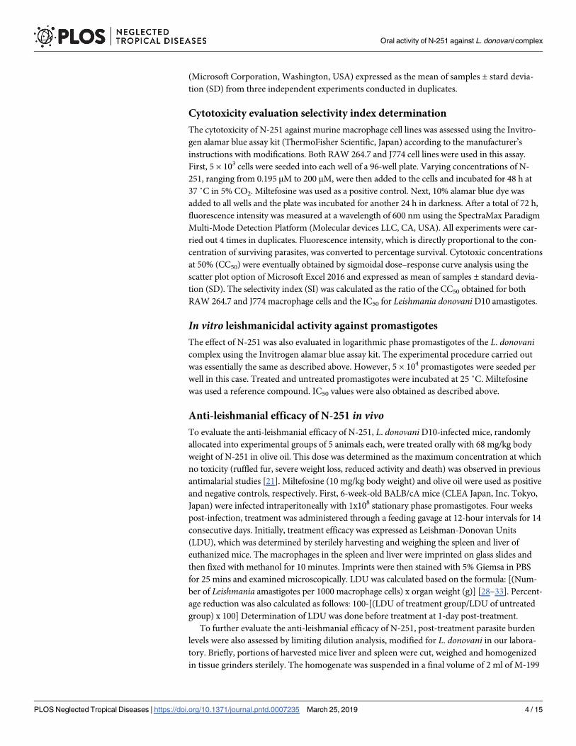
mental plan to evaluate the anti-leishmanial efficacy of N-251 has been summarized in Fig 1
Results
N-251 selectively exhibits leishmanicidal activity against L. donovani D10
amastigotes
To evaluate the leishmanicidal activity of N-251, we used the amastigote form of L. donovaniD10 parasites as the model parasite because the intracellular amastigote, which lives within the
Fig 1. Experimental plan to evaluate the leishmanicidal effect of N-251 in mice.
https://doi.org/10.1371/journal.pntd.0007235.g001
Oral activity of N-251 against L. donovani complex
PLOS Neglected Tropical Diseases | https://doi.org/10.1371/journal.pntd.0007235 March 25, 2019 5 / 15
host macrophage, is the form of the parasite responsible for clinical symptoms of VL observed
in the human host. Therefore, targeting this parasite form in vitro provides a more direct
insight into the effect of N-251 in vivo. Stationary phase promastigotes and RAW 264.7 macro-
phages were incubated for 6 h to allow the invasion and transformation of promastigotes
within the macrophages. Intracellular amastigotes were then challenged with 0–50 μM of N-
251 for 24, 48 and 72 h to investigate the dose-dependent leishmanicidal effect of N-251. In
addition, the IC50 of N-251 was determined at 72 h by microscopy. Our results clearly show
the leishmanicidal effect of N-251 on intracellular amastigotes. When treated with varying
concentrations of N-251, L. donovani amastigotes were eliminated in a dose dependent man-
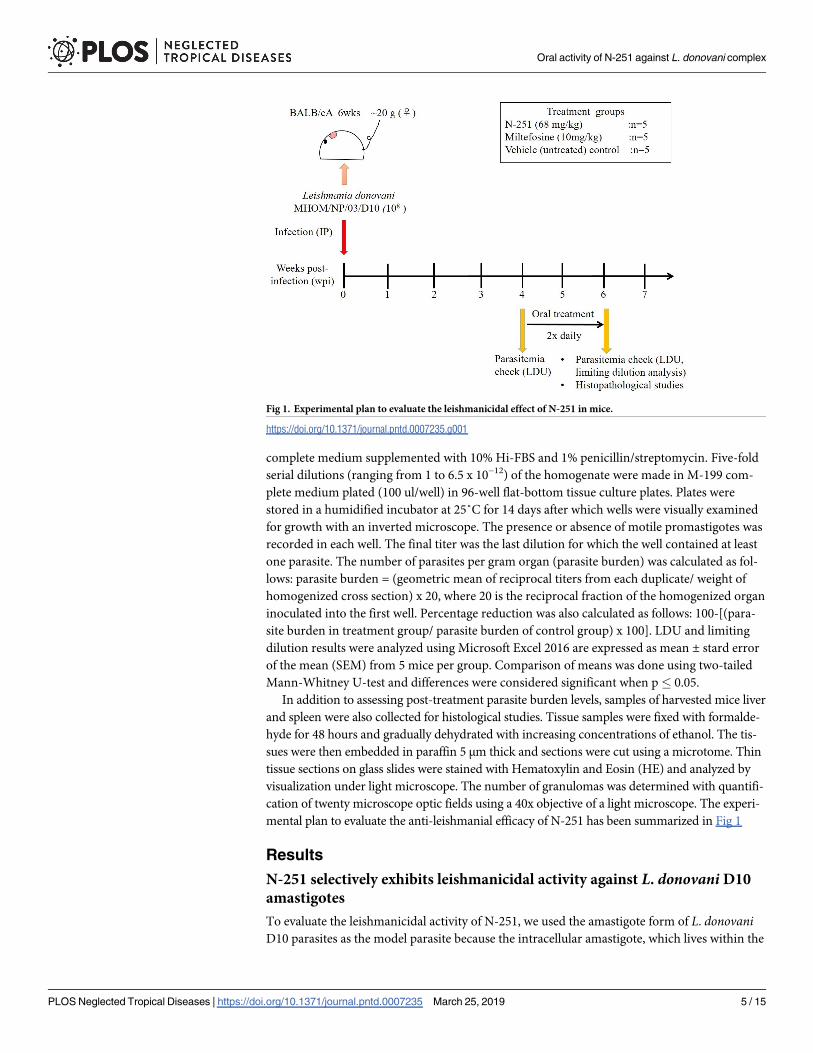
ner (S2 and S3 Figs) at an IC50 of 6.69 ± 0.82, (Table 1). Although most parasites had been
cleared by 24 hours, a few macrophages were observed with about one or two intracellular par-
asites that were probably dead. By 48 and 72 hours, intracellular parasites were completely
cleared from all macrophages (Fig 2). The leishmanicidal activity of N-251 was comparable to
that of miltefosine (reference drug control). The cytotoxicity of N-251 was evaluated on both
Table 1. N-251 selectively exhibits leishmanicidal activity against L. donovani D10 intracellular amastigotes.
Compound Activity (IC50)± μM Toxicity (CC50)� μM SI
L. donovani D10 RAW 264.7 macrophages J774 macrophages RAW 264.7 macrophages J774 macrophages
N-251 6.69 ± 0.826¼ 66.41 ± 4.15 138.25 ± 24.27 9.93 20.67
Miltefosine 1.77 ± 0.05 55.85 ± 5.83 155.75 ± 19.68 31.55 87.99
¶ Selectivity Index (CC50/IC50)±Concentration at which the inhibition of 50% parasites was observed
�Concentration at which the cytotoxicity of 50% cells was observed6¼Values are expressed as the mean ± SD from at least three independent experiments conducted in duplicates.
https://doi.org/10.1371/journal.pntd.0007235.t001
Fig 2. Inhibition of intracellular amastigotes by N-251. Representative Giemsa-stained images (Bar- 20 μm) showing
the leishmanicidal effect of N-251 on L. donovani D10 intracellular amastigotes (A) within RAW 264.7 macrophages
(M) at 24, 48 and 72 hours.
https://doi.org/10.1371/journal.pntd.0007235.g002
Oral activity of N-251 against L. donovani complex
PLOS Neglected Tropical Diseases | https://doi.org/10.1371/journal.pntd.0007235 March 25, 2019 6 / 15
RAW 264.7 and J774 murine macrophage cell lines with an alamar blue assay kit for 72 hours.
The toxicity of the N-251 treatments was very low among both RAW 264.7 and J774 cells;
within the concentration ranges tested, the IC50 values were 66.41 ± 4.15 μM and 138.25 ±24.27 μM, respectively (Table 1). These IC50 values correspond to SI values of�10 for N-251
(Table 1), suggesting that N-251 is highly specific in its activity against L. donovani amasti-
gotes, as biological efficacy cannot be attributed to cytotoxicity when the selectivity index is
�10. The SI values of miltefosine were about twice that of N-251, which were quite comparable
(Table 1). The results therefore suggest that N-251 selectively and specifically exhibits leishma-
nicidal activity by targeting L. donovani intracellular amastigotes within host macrophage
cells, without having any cytotoxic effect on host cells.
N-251 exhibits leishmanicidal activity against various L. donovani complex
parasites
To examine the effect of N-251 on different L. donovani complex parasites, we screened N-251
against the promastigotes of five parasites belonging to the L. donovani complex. The parasites
used in this study were selected specifically from different geographical locations known to be
highly endemic for VL. They include: L. chagasi PP75 (Brazil), L. donovani D10 (Nepal)
[34,35], L. donovani DD8 (India), L. donovani KH (ATCC 30503) (Sudan) and L. infantumEP173 (Turkey). Our results clearly show the leishmanicidal effect of N-251 on various para-
sites of the L. donovani complex. When treated with N-251, leishmanicidal activity was
observed against all three L. donovani (D10, Dd8 KH) parasites, as well as L. chagasi and
L. infantum, in a dose dependent manner (S4 Fig). The IC50 values obtained ranged from
6.12 ± 1.64 μM to 26.90 ± 2.51 μM (Table 2). The results shows that the activity of N-251 is
reproducible in promastigotes of different L. donovani complex parasites including L. dono-vani D10, which had its amastigote forms eliminated by N-251 in our in vitro infection assays.
This suggests that N-251 will probably exhibit leishmanicidal activity against their respective
amastigote forms, regardless of their geographic origin.
Therapeutic effect of N-251 in L. donovani D10-infected BALB/cA mice
The therapeutic efficacy of N-251 was evaluated in L. donovani-infected mice. Briefly, mice
were initially infected intraperitoneally with 108 stationary phase promastigotes, followed by a
4-week incubation period. During this time, parasites invaded the neutrophils and mononu-
clear phagocytes within the spleen and liver, and a chronic infection was established [28,36,37].
Mice were then treated orally through feeding gavages at 12-hour intervals for 14 days. LDU
was then determined in harvested spleen liver tissues before and after treatment. Before
Table 2. N-251 exhibits leishmanicidal activity against different L. donovani complex parasites.
IC50� (μM)
Parasites N-251 Miltefosine
L. chagasi MHOM/BR/74/PP75 23.12 ± 3.06¶ 6.59 ± 1.45
L. donovani MHOM/SU/43/KH 22.95 ± 0.35 5.57 ± 0.11
L. donovani MHOM/NP/03/D10 26.89 ± 1.98 5.9 ± 1.86
L. donovani MHOM/IN/80/DD8 26.90 ± 2.51 9.955 ± 2.62
L. infantum EP-173 6.12 ± 1.64 4.35 ± 0.54
�Concentration at which 50% growth inhibition was observed¶Values are expressed as the mean ± SD from four independent experiments conducted in duplicates.
https://doi.org/10.1371/journal.pntd.0007235.t002
Oral activity of N-251 against L. donovani complex
PLOS Neglected Tropical Diseases | https://doi.org/10.1371/journal.pntd.0007235 March 25, 2019 7 / 15

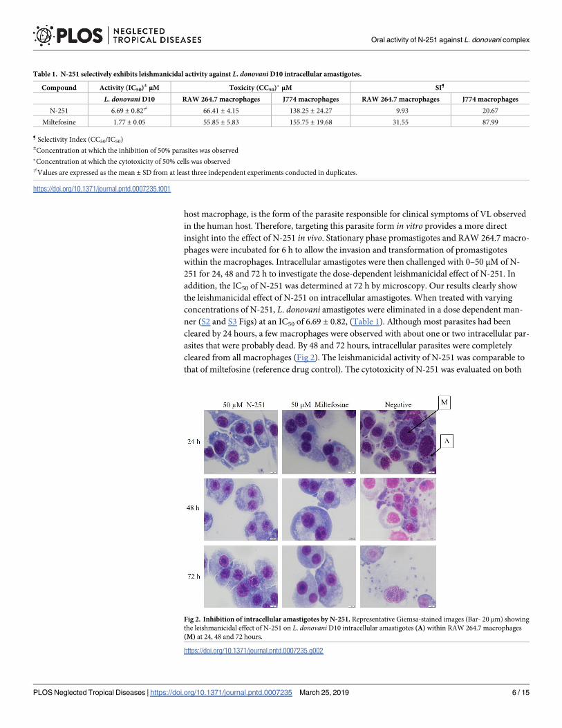
treatment, LDU in the liver and spleen were 857.73 ± 104.05 and 13.94 ± 0.30, respectively.
After treatment, in the vehicle control group that did not receive any drug, LDU increased by
over 100% to 2214.34 ± 426.73 in the liver and 51.38 ± 16.24 in the spleen. In contrast, in N-
251-treated mice, LDU decreased significantly (p = 0.012) by 85.73% and 93.98% to 316.00 ±161.14 and 3.09 ± 1.29 within the liver and spleen, respectively, relative to the untreated group
at 6 weeks post-infection (wpi). These results clearly show that after 14 days of treatment, para-
sites were significantly cleared by N-251(Fig 3a and 3b). Similarly, in miltefosine-treated mice,
parasites were cleared at levels that were comparable to those obtained by N-251.
To further confirm the therapeutic effect of N-251, we also evaluated its efficacy by limited
dilution analysis to determine parasite burden levels post-treatment. Results here also showed
that N-251 significantly cleared L. donovani parasites. After treatment, parasite burden in the
untreated group were determined at 4.15 x 10⁸ and 2.59 x 10⁸ parasites/g organ in liver and
spleen respectively. However, in N-251-treated mice, parasite burden decreased significantly
(p< 0.05) by 85.89% and 97.41% to 5.86 x 10⁷ and 6.68 x 10⁶ parasites/g organ in liver and
spleen respectively (Fig 3c and 3d). In vivo data obtained by both LDU and limiting dilution
analyses therefore support our in vitro observation that N-251 has leishmanicidal activity
against L. donovani amastigotes. Data herein therefore demonstrates the therapeutic effect of
N-251 as an oral drug in L. donovani-infected mice.
One of the major signs of histopathological damage known to be associated with L donovaniinfection in mice humans are granuloma formation in the liver [38]. Hence, we investigated
Fig 3. Therapeutic effect of N-251 in L. donovani D10-infected BALB/cA mice. At 4 wpi, 5 mice were euthanized,
followed by the determination of parasite burden in spleen and liver by LDU. The remaining mice were placed into 3
groups (N-251, Miltefosine and vehicle (untreated) control) consisting of 5 mice per group treated orally at 12-hour
intervals for 14 days. After treatment (6 wpi), mice were euthanized and parasite burden was determined in spleen and
liver tissues by LDU and limiting dilution analyses. Results are represented by bar graphs and are expressed as
mean ± SEM from 5 mice in each group. (A) Liver LDU (B) Spleen LDU (C) Liver (Limiting dilution analysis) (D)
Spleen (Limiting dilution analysis). Blue: 4 wpi. Orange: 6 wpi. Mean values were compared with the untreated group
(6 wpi) and �P� 0.05, ��P� 0.01 were considered significant.
https://doi.org/10.1371/journal.pntd.0007235.g003
Oral activity of N-251 against L. donovani complex
PLOS Neglected Tropical Diseases | https://doi.org/10.1371/journal.pntd.0007235 March 25, 2019 8 / 15
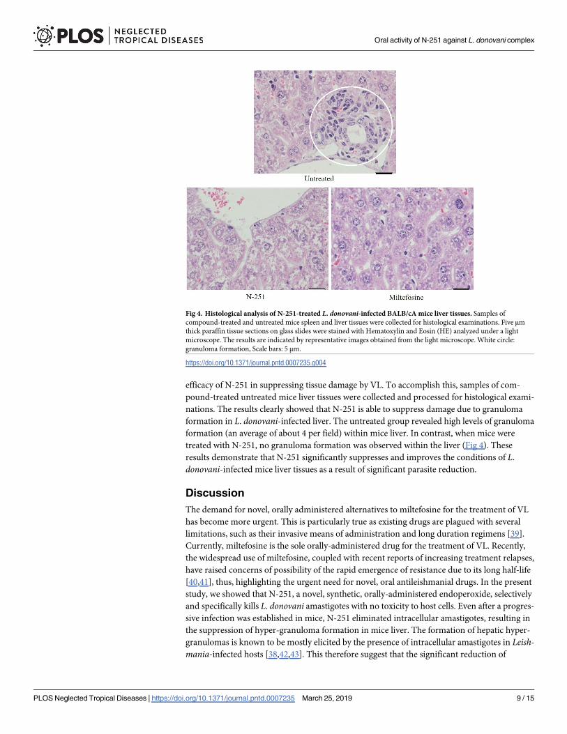
efficacy of N-251 in suppressing tissue damage by VL. To accomplish this, samples of com-
pound-treated untreated mice liver tissues were collected and processed for histological exami-
nations. The results clearly showed that N-251 is able to suppress damage due to granuloma
formation in L. donovani-infected liver. The untreated group revealed high levels of granuloma
formation (an average of about 4 per field) within mice liver. In contrast, when mice were
treated with N-251, no granuloma formation was observed within the liver (Fig 4). These
results demonstrate that N-251 significantly suppresses and improves the conditions of L.
donovani-infected mice liver tissues as a result of significant parasite reduction.
Discussion
The demand for novel, orally administered alternatives to miltefosine for the treatment of VL
has become more urgent. This is particularly true as existing drugs are plagued with several
limitations, such as their invasive means of administration and long duration regimens [39].
Currently, miltefosine is the sole orally-administered drug for the treatment of VL. Recently,
the widespread use of miltefosine, coupled with recent reports of increasing treatment relapses,
have raised concerns of possibility of the rapid emergence of resistance due to its long half-life
[40,41], thus, highlighting the urgent need for novel, oral antileishmanial drugs. In the present
study, we showed that N-251, a novel, synthetic, orally-administered endoperoxide, selectively
and specifically kills L. donovani amastigotes with no toxicity to host cells. Even after a progres-
sive infection was established in mice, N-251 eliminated intracellular amastigotes, resulting in
the suppression of hyper-granuloma formation in mice liver. The formation of hepatic hyper-
granulomas is known to be mostly elicited by the presence of intracellular amastigotes in Leish-mania-infected hosts [38,42,43]. This therefore suggest that the significant reduction of
Fig 4. Histological analysis of N-251-treated L. donovani-infected BALB/cA mice liver tissues. Samples of
compound-treated and untreated mice spleen and liver tissues were collected for histological examinations. Five μm
thick paraffin tissue sections on glass slides were stained with Hematoxylin and Eosin (HE) analyzed under a light
microscope. The results are indicated by representative images obtained from the light microscope. White circle:
granuloma formation, Scale bars: 5 μm.
https://doi.org/10.1371/journal.pntd.0007235.g004
Oral activity of N-251 against L. donovani complex
PLOS Neglected Tropical Diseases | https://doi.org/10.1371/journal.pntd.0007235 March 25, 2019 9 / 15
intracellular amastigotes by N-251 observed in this study, may have resulted in the absence of
granuloma, indicating the potential of N-251 as a lead compound for the development of an
oral chemotherapy against VL.
The cleavage of the endoperoxide bridge (C-O-O-C, S1 Fig) generates short-lived, cytotoxic
oxyradicals in the presence of heme iron or free Fe2+. Fenton degradation of the oxyradical
intermediates can produce hydroxyl radicals (OH) that are highly reactive against a wide vari-
ety of molecules such as amino acids, enzymes, lipids and nucleic acids [44,45]. The released
radicals then oxidize these molecules, thereby inhibiting their functions, which eventually leads
to parasite death. This explains why endoperoxides exhibit a wide spectrum of anti-infective
activity. N-251 is also characterized by a peroxide bridge within its structure that can be cleaved
and activated in the presence of a heme iron or free Fe2+ [20,46,47], suggesting that this is the
basis for its antileishmanial activity observed in both promastigotes and amastigotes screened
in this study. Leishmania are known to forage for iron from host macrophages for their growth
for defense against the macrophage’s oxidative assault by providing iron to the antioxidant
enzyme superoxide dismutase [19,48,49]. The accumulation of iron within the parasite there-
fore results in the selective killing of the parasite by N-251. This also explains the 4-fold increase
in activity observed against intracellular amastigotes relative to that of promastigotes.
Some of the current antileishmanial drugs, including pentavalent antimonials and paromo-
mycin, have been reported to exhibit a limited spectrum of antileishmanial activity, which is
mostly dependent on the species of Leishmania, the geographical location, as well as the clinical
presentation of the disease [50–54]. However, in this study, N-251 exhibited leishmanicidal
effects against various L donovani complex parasites from different parts of the world. This
therefore shows the reproducibility of N-251’s activity in different L. donovani complex para-
sites, suggesting that N-251 may exhibit leishmanicidal activity against different VL parasites,
regardless of their geographical origin. In addition, in previous studies, N-251 was observed to
be active against other non-Leishmania parasites such as Plasmodium, T. gondii and Schisto-soma [20,21,23,47,55]. It is therefore quite clear that N-251 exhibits a wide spectrum of activity.
In general, advantages of wide spectrum compounds include cost effectiveness in using one
drug to treat different infectious diseases as well as serving as good options for Mass drug
administration exercises in poor endemic regions [56,57]. This inherent characteristic of N-251
appears to be one of the several advantages it has over other existing antileishmanial drugs.
Miltefosine, the only oral drug currently available against VL, affects parasites by the dis-
ruption of parasitic Ca2+ homeostasis via opening of the sphingosine-activated plasma mem-
brane Ca2+ channel, together with the impairment of the acidocalcisomes [58]. However, it is
limited by its teratogenicity and several side effects. There are also concerns that its use as a
monotherapy can eventually lead to the rapid emergence of resistant parasites [40,59]. In fact,
treatment relapses among VL patients [60], as well as the identification of miltefosine-resistant
clinical strains, have already been reported [61,62]. Our compound, N-251, is also orally-
administered and acts through a different mode of action from miltefosine, making it a good
candidate for possible combination with miltefosine. According to the WHO, combination
therapy is an important strategy to improve leishmaniasis therapy and also delay the emer-
gence of resistance [63,64], as can be seen in the case of malaria, tuberculosis, and HIV [65–
67]. In future studies, we will therefore evaluate the combinatory effect of N-251 and miltefo-
sine in experimental VL.
In conclusion, results herein demonstrates the in vitro and in vivo antileishmanial effect of
the novel orally administered synthetic endoperoxide, N-251. Also, the broad-spectrum activ-
ity of N-251 against various parasites of L. donovani complex from different geographical loca-
tions was established. More importantly, this study highlights the importance of N-251 as an
oral drug for monotherapy its possible combination with miltefosine. Finally, N-251 may be a
Oral activity of N-251 against L. donovani complex
PLOS Neglected Tropical Diseases | https://doi.org/10.1371/journal.pntd.0007235 March 25, 2019 10 / 15

promising lead compound for the development of new oral chemotherapy against visceral
leishmaniasis.
Supporting information
S1 Fig. Chemical Structure of novel synthetic endoperoxide, 6-(1,2,6,7 -tetraoxaspiro[7.11]
nonadec-4- yl)hexan-1-ol (N-251).
(TIF)
S2 Fig. Dose-dependent leishmanicidal effect of N-251 on intracellular amastigotes. Repre-
sentative Giemsa-stained images (Scale bar-5 μm) showing the dose-dependent effect of N-251
against L. donovani D10 amastigotes within RAW 264.7 macrophages.
(TIF)
S3 Fig. Dose–response curves showing the effect of N-251 on intracellular amastigotes.
(TIF)
S4 Fig. Dose-dependent leishmanicidal activity of N-251 against L. donovani complex pro-
mastigotes. Dose–response curves showing the dose-dependent effect of N-251 against L.
donovani complex promastigotes and macrophage cells.
(TIF)
Acknowledgments
The authors are grateful to the members of the Section of Environmental Parasitology at the
Tokyo Medical Dental University (TMDU), as well as members of the Laboratory of Molecular
Immunology at the Department of Animal Resource Sciences, University of Tokyo. The
authors would also like to thank the staff of the Research Center for Medical Dental Sciences,
TMDU, for technical advice.
Author Contributions
Conceptualization: Kofi Dadzie Kwofie, Nobuo Ohta, Yoshitsugu Matsumoto, Shiroh
Iwanaga.
Data curation: Kofi Dadzie Kwofie.
Formal analysis: Kofi Dadzie Kwofie.
Funding acquisition: Shiroh Iwanaga.
Investigation: Kofi Dadzie Kwofie, Kai Sato, Chizu Sanjoba, Akina Hino, Rieko Shimogawara,
Michael Amoa-Bosompem.
Methodology: Kofi Dadzie Kwofie, Kai Sato, Chizu Sanjoba, Akina Hino, Rieko Shimogawara,
Michael Amoa-Bosompem, Yoshitsugu Matsumoto.
Project administration: Shiroh Iwanaga.
Resources: Chizu Sanjoba, Kyung-Soo Chang, Hye-Sook Kim, Yoshitsugu Matsumoto, Shiroh
Iwanaga.
Supervision: Mitsuko Ohashi, Hye-Sook Kim, Nobuo Ohta, Yoshitsugu Matsumoto, Shiroh
Iwanaga.
Validation: Kai Sato, Yoshitsugu Matsumoto.
Writing – original draft: Kofi Dadzie Kwofie, Shiroh Iwanaga.
Oral activity of N-251 against L. donovani complex
PLOS Neglected Tropical Diseases | https://doi.org/10.1371/journal.pntd.0007235 March 25, 2019 11 / 15

Writing – review & editing: Kofi Dadzie Kwofie, Irene Ayi, Daniel A. Boakye, Abraham K.
Anang, Mitsuko Ohashi, Hye-Sook Kim, Yoshitsugu Matsumoto, Shiroh Iwanaga.
References1. WHO. Visceral Leishmaniasis [Internet]. Programmes, Epidemiology. 2018 [cited 2018 Jan 17]. http://
www.who.int/leishmaniasis/burden/en/
2. Mauricio IL, Scho G, Dujardin J, Soteriadou K, Dedet J, Haralambous C, et al. Evolutionary geographi-
cal history of the Leishmania donovani complex with a revision of current taxonomy. PNAS. 2007;
104(22):9375–80. https://doi.org/10.1073/pnas.0703678104
3. Akhoundi M, Kuhls K, Cannet A, Votypka J, Marty P. A Historical Overview of the Classification, Evolu-
tion, Dispersion of Leishmania Parasites Sflies. PLoS Negl Trop Dis [Internet]. 2016; 10(3):1–40. Avail-
able from: http://dx.doi.org/10.1371/journal.pntd.0004349
4. Sundar S, Jha TK, Thakur CP, Engel J, Sindermann H, Fischer C, et al. Oral Miltefosine for Indian vis-
ceral Leishmaniasis. N Engl J Med. 2002; 347(22):949–55.
5. Jha TK, Sundar S, Thakur CP, Bachmann P, Karbwang J, Fischer C, et al. Miltefosine, an oral agent,
for the treatment of Indian visceral leishmaniasis. N Engl J Med. 1999; 341(24):1795–800. https://doi.
org/10.1056/NEJM199912093412403 PMID: 10588964
6. Sundar S, Olliaro PL. Miltefosine in the treatment of leishmaniasis: Clinical evidence for informed clinical
risk management. Ther Clin Risk Manag. 2007; 3(5):733–40. PMID: 18472998
7. Chappuis F, Sundar S, Hailu A, Ghalib H, Rijal S, Peeling RW, et al. Visceral leishmaniasis: what are
the needs for diagnosis, treatment control? NatRevMicrobiol [Internet]. 2007; 5(1740–1534 (Elec-
tronic)):873–82. Available from: c:%5CKARSTEN%5CPDFs%5CParasitologie-PDFs%5CParasit-
2007%5CChappuis et al.-Visceral leishmaniasis- what are the needs for diagnosis treatment control.
8. WHO. Control of the Leishmaniases. Tech Rep Ser. 2010; 949(March):22–6.
9. Sundar S, Chakravarty J. Leishmaniasis: an update of current pharmacotherapy. Expert Opin Pharmac-
other. 2013;
10. Jefford CW. New developments in synthetic peroxidic drugs as artemisinin mimics. Drug Discov Today.
2007; 12(11–12):487–95. https://doi.org/10.1016/j.drudis.2007.04.009 PMID: 17532534
11. O’Neill PM, Posner GH. A medicinal chemistry perspective on artemisinin related endoperoxides. J
Med Chem. 2004; 47(12):2945–64. https://doi.org/10.1021/jm030571c
12. Sen R, Byopadhyay S, Dutta A, Mal G, Ganguly S, Saha P, et al. Artemisinin triggers induction of cell-
cycle arrest apoptosis in Leishmania donovani promastigotes. J Med Microbiol. 2007; 56(9):1213–8.
13. Sen R, Ganguly S, Saha P, Chatterjee M. Efficacy of artemisinin in experimental visceral leishmaniasis.
Int J Antimicrob Agents [Internet]. 2010; 36(1):43–9. Available from: http://dx.doi.org/10.1016/j.
ijantimicag.2010.03.008 PMID: 20403680
14. Rahaman M, Ghosh S, Chowdhury L, Chatterjee M. Evaluation of anti-leishmanial activity of artemisinin
combined with amphotericin B or miltefosine in Leishmania donovani promastigotes. Int J Basic Clin
Pharmacol [Internet]. 2014; 3(4):1. Available from: http://www.ijbcp.com/?mno=159726
15. Ghaffarifar F, Esav Heydari F, Dalimi A, Hassan ZM, Delavari M, Mikaeiloo H. Evaluation of Apoptotic
Antileishmanial Activities of Artemisinin on Promastigotes BALB/C Mice Infected with Leishmania
major. Iran J Parasitol [Internet]. 2015; 10(2):258–67. Available from: http://www.ncbi.nlm.nih.gov/
pubmed/26246824%5Cnhttp://www.pubmedcentral.nih.gov/articlerender.fcgi?artid=PMC4522302
16. Want MY, Islammudin M, Chouhan G, Ozbak HA, Hemeg HA, Chattopadhyay AP, et al. Nanoliposomal
artemisinin for the treatment of murine visceral leishmaniasis. Int J Nanomedicine. 2017; 12:2189–204.
https://doi.org/10.2147/IJN.S106548 PMID: 28356736
17. Efferth T, R. Romero M, Rita Bilia A, Galal Osman A, ElSohly M, Wink M, et al. Exping the Therapeutic
Spectrum of Artemisinin: Activity Against Infectious Diseases Beyond Malaria Novel Pharmaceutical
Developments. World J Tradit Chinese Med [Internet]. 2016; 2(2):1–23. Available from: http://www.
wjtcm.org:8080/ch/reader/view_abstract.aspx?file_no=20160002&flag=1
18. Sen R, Saha P, Sarkar A, Ganguly S, Chatterjee M. Iron enhances generation of free radicals by Artemi-
sinin causing a caspase-independent, apoptotic death in Leishmania donovani promastigotes. Free
Radic Res [Internet]. 2010; 44(11):1289–95. Available from: http://www.ncbi.nlm.nih.gov/pubmed/
20815780%5Cnhttp://www.tfonline.com/doi/full/10.3109/10715762.2010.498475 PMID: 20815780
19. Das NK, Biswas S, Solanki S, Mukhopadhyay CK. Leishmania donovani depletes labile iron pool to
exploit iron uptake capacity of macrophage for its intracellular growth. Cell Microbiol. 2009; 11(1):83–
94. https://doi.org/10.1111/j.1462-5822.2008.01241.x PMID: 18823384
Oral activity of N-251 against L. donovani complex
PLOS Neglected Tropical Diseases | https://doi.org/10.1371/journal.pntd.0007235 March 25, 2019 12 / 15

20. Kim H-S, Nagai Y, Ono K, Begum K, Wataya Y, Hamada Y, et al. Synthesis Antimalarial Activity of
Novel Medium-Sized 1,2,4,5-Tetraoxacycloalkanes. J Med Chem. 2001; 44(14):2357–61.
21. Sato A, Hiramoto A, Morita M, Matsumoto M, Komich Y, Nakase Y, et al. Antimalarial activity of endo-
peroxide compound 6-(1,2,6,7-tetraoxaspiro[7.11]nonadec-4-yl)hexan-1-ol. Parasitol Int [Internet].
2011; 60(3):270–3. Available from: http://dx.doi.org/10.1016/j.parint.2011.04.001 PMID: 21501696
22. Xin CF, Kim HS, Sato A, Lee HJ, Lee YW, Pyo KH, et al. In vitro inhibition of Toxoplasma gondii by the
anti-malarial cidate, 6-(1,2,6,7-tetraoxaspiro[7.11]nonadec-4-yl)hexan-1-ol. Parasitol Int. 2016; 65
(5):494–9.
23. Taniguchi T, Kumagai T, Shimogawara R, Ichinose S, Hiramoto A, Sato A, et al. Schistosomicidal
antifecundity effects of oral treatment of synthetic endoperoxide N-89. Parasitol Int [Internet]. 2011;
60(3):231–6. Available from: http://dx.doi.org/10.1016/j.parint.2011.02.007
24. Yamabe M, Kumagai T, Shimogawara R, Blay EA, Hino A, Ichimura K, et al. Novel synthetic com-
pounds with endoperoxide structure damage juvenile stage of Schistosoma mansoni by targeting lyso-
some-like organelles. Parasitol Int [Internet]. 2017; 66(1):917–24. Available from: http://dx.doi.org/10.
1016/j.parint.2016.10.013 PMID: 27771462
25. Blay EA, Kumagai T, Yamabe M, Hino A, Shimogawara R, Kim H-S, et al. Insights into the mode of
action of 1,2,6,7-tetraoxaspiro [7.11] nonadecane (N-89) against adult Schistosoma mansoni worms.
Parasitol Int [Internet]. 2018; 67(4):403–12. Available from: http://linkinghub.elsevier.com/retrieve/pii/
S1383576917304956%0Ahttp://www.ncbi.nlm.nih.gov/pubmed/29617630 PMID: 29617630
26. Ueda Y, Gu W, Dansako H, Kim HS, Yoshizaki S, Okumura N, et al. Multiple antiviral activities of the
antimalarial anti-hepatitis C drug cidates N-89 N-251. Biochem Biophys Reports [Internet]. 2018; 15
(November 2017):1–6. Available from: https://doi.org/10.1016/j.bbrep.2018.05.007
27. Ueda Y, Takeda M, Mori K, Dansako H, Wakita T, Kim HS, et al. New Preclinical Antimalarial Drugs
Potently Inhibit Hepatitis C Virus Genotype 1b RNA Replication. PLoS One. 2013; 8(8):1–11.
28. Stauber LA. Host Resistance to the Khartoum Strain of Leishmania donovani. Rice Inst Pam Rice Univ
Stud [Internet]. 1958; 45(1). Available from: http://hdl.hle.net/1911/6279
29. Wasan KM, Wasan EK, Gershkovich P, Zhu X, Tidwell RR, Werbovetz KA, et al. Highly Effective Oral
Amphotericin B Formulation against Murine Visceral Leishmaniasis. J Inifectious Dis. 2009; 200:357–60.
30. Allahverdiyev AM, Koc CR, Melahat B, Elcicek S, Baydar YS, Oztel NO, et al. A new approach for devel-
opment of vaccine against visceral leishmaniasis: Lipophosphoglycan polyacrylic acid conjugates.
Asian Pac J Trop Med. 2017; 10(9):877–86. https://doi.org/10.1016/j.apjtm.2017.09.001
31. Wyllie S, Thomas M, Patterson S, Crouch S, De Rycker M, Lowe R, et al. Cyclin-dependent kinase 12 is
a drug target for visceral leishmaniasis. Nature [Internet]. 2018; 560:192–7. Available from: http://dx.
doi.org/10.1038/s41586-018-0356-z PMID: 30046105
32. Van Den Kerkhof M, Mabille D, Chatelain E, Mowbray CE, Braillard S, Hendrickx S, et al. IJP: Drugs
Drug Resistance In vitro in vivo pharmacodynamics of three novel antileishmanial lead series. Int J
Parasitol Drugs Drug Resist. 2018; 8(January):81–6.
33. Sharma M, Sehgal R, Kaur S. Evaluation of Nephroprotective Immunomodulatory Activities of Antioxi-
dants in Combination with Cisplatin against Murine Visceral Leishmaniasis. PLoS Negl Trop Dis. 2012;
6(5):e1629. https://doi.org/10.1371/journal.pntd.0001629
34. Pey K, Yanagi T, Pey BD, Mallik AK, Sherch JB, Kanbara H. Characterization of Leishmania isolates
from Nepalese patients with visceral leishmaniasis. Parasitol Res. 2007; 100(6):1361–9. https://doi.org/
10.1007/s00436-007-0464-4 PMID: 17310397
35. Morimoto A, Omachi S, Osada Y, Chambers JK, Uchida K, Sanjoba C, et al. Hemophagocytosis in
Experimental Visceral Leishmaniasis by Leishmania donovani. PLoS Negl Trop Dis [Internet]. 2016;
10(3):1–16. Available from: http://dx.doi.org/10.1371/journal.pntd.0004505
36. Mbati PA, Abok K, Orago AS, Anjili C, Githure JI, Kagai J, et al. Establishment of an appropriate inocu-
lum dose of Leishmania donovani promastigotes required to establish a visceral infection in laboratory
animal rodent models. Afr J Health Sci. 1994; 1(4):165–8. PMID: 12153342
37. Silva JM, Zacarias DA, De Figueirêdo LC, Soares MRA, Ishikawa EAY, Costa DL, et al. BOne marrow
parasite burden among patients with new world kala-azar is associated with disease severity. Am J
Trop Med Hyg. 2014; 90(4):621–6. https://doi.org/10.4269/ajtmh.13-0376 PMID: 24615127
38. Gutierrez Y, Maksem JA, Reiner NE. Pathologic changes in murine leishmaniasis (Leishmania dono-
vani) with special reference to the dynamics of granuloma formation in the liver. Am J Pathol [Internet].
1984; 114(2):222–30. Available from: http://www.pubmedcentral.nih.gov/articlerender.fcgi?artid=
1900324&tool=pmcentrez&rendertype=abstract PMID: 6696043
39. Chavez-Fumagalli MA, Ribeiro TG, Castilho RO, Fernes SOA, Cardoso VN, Coelho CSP, et al.
New delivery systems for amphotericin B applied to the improvement of leishmaniasis treatment.
Oral activity of N-251 against L. donovani complex
PLOS Neglected Tropical Diseases | https://doi.org/10.1371/journal.pntd.0007235 March 25, 2019 13 / 15

Rev Soc Bras Med Trop. 2015; 48(3):235–42. https://doi.org/10.1590/0037-8682-0138-2015 PMID:
26107999
40. Bryceson A. A policy for leishmaniasis with respect to the prevention control of drug resistance. Trop
Med Int Heal. 2001; 6(11):928–34.
41. Coelho AC. Miltefosine Susceptibility Resistance in Leishmania: From the Laboratory to the Field. J
Trop Dis [Internet]. 2016; 04(02):2–6. Available from: http://www.esciencecentral.org/journals/
miltefosine-susceptibility-resistance-in-leishmania-from-the-laboratory-to-the-field-2329-891X-
1000203.php?aid=69770
42. Murray HW. Tissue granuloma structure-function in experimental visceral leishmaniasis. Int J Exp
Pathol. 2001; 82(5):249–67. https://doi.org/10.1046/j.1365-2613.2001.00199.x PMID: 11703536
43. Mcelrath MJ, Murray HW, Cohn ZA. THE DYNAMICS OF GRANULOMA FORMATION IN EXPERI-
MENTAL VISCERAL LEISHMANIASIS BY Leishmania donovani is an intracellular parasite that infects
parenchymal macro- phages of susceptible animals (1). Unlike infection with L. major (2) or L. mexicana
(3. 1988;167(June).
44. Antoine T, Fisher N, Amewu R, O’Neill PM, Ward SA, Biagini GA. Rapid kill of malaria parasites by arte-
misinin semi-synthetic endoperoxides involves ROS-dependent depolarization of the membrane poten-
tial. J Antimicrob Chemother. 2014; 69(4):1005–16. https://doi.org/10.1093/jac/dkt486
45. van Agtmael MA, Eggelte TA, van BC. Artemisinin drugs in the treatment of malaria: from medicinal
herb to registered med- ication. Trends Pharmacol Sci. 1999; 20(May 1999):199–205.
46. Morita M, Sanai H, Hiramoto A, Sato A, Hiraoka O, Sakura T, et al. Plasmodium falciparum endoplasmic
reticulum-resident calcium binding protein is a possible target of synthetic antimalarial endoperoxides,
n-89 N-251. J Proteome Res. 2012; 11(12):5704–11. https://doi.org/10.1021/pr3005315
47. Sato A, Kawai S, Hiramoto A, Morita M, Tanigawa N, Nakase Y, et al. Antimalarial activity of 6-(1,2,6,7-
tetraoxaspiro[7.11]nonadec-4-yl)hexan-1-ol (N-251) its carboxylic acid derivatives. Parasitol Int [Inter-
net]. 2011; 60(4):488–92. Available from: http://dx.doi.org/10.1016/j.parint.2011.08.017
48. GHOSH S, GOSWAMI S, ADHYA S. Role of superoxide dismutase in survival of Leishmania within the
macrophage. Biochem J [Internet]. 2003; 369(3):447–52. Available from: http://biochemj.org/lookup/
doi/10.1042/bj20021684
49. Marquis JF, Gros P. Intracellular Leishmania: your iron or mine? Trends Microbiol. 2007; 15(3):93–5.
https://doi.org/10.1016/j.tim.2007.01.001 PMID: 17257847
50. Bhari V, Sundar S, Dujardin JC, Salotra P. Elucidation of cellular mechanisms involved in experimental
paromomycin resistance in leishmania donoVani. Antimicrob Agents Chemother. 2014; 58(5):2580–5.
https://doi.org/10.1128/AAC.01574-13 PMID: 24550335
51. Botero A, Keatley S, Peacock C, Thompson RCA. In vitro drug susceptibility of two strains of the wildlife
trypanosome, Trypanosoma copemani: A comparison with Trypanosoma cruzi. Int J Parasitol Drugs
Drug Resist [Internet]. 2017; 7(1):34–41. Available from: http://linkinghub.elsevier.com/retrieve/pii/
S2211320716301142
52. Laffitte MCN, Leprohon P, Legare D, Ouellette M. Deep-sequencing revealing mutation dynamics in the
miltefosine transporter gene in Leishmania infantum selected for miltefosine resistance. Parasitol Res
[Internet]. 2016; 115(10):3699–703. Available from: http://dx.doi.org/10.1007/s00436-016-5195-y
PMID: 27457482
53. Pink R, Hudson A, Mouriès MA, Bendig M. Opportunities challenges in antiparasitic drug discovery. Nat
Rev Drug Discov. 2005; 4(9):727–40. https://doi.org/10.1038/nrd1824
54. Plourde M, Coelho A, Keynan Y, Larios OE, Ndao M, Ruest A, et al. Genetic polymorphisms drug sus-
ceptibility in four isolates of Leishmania tropica obtained from Canadian soldiers returning from Afghani-
stan. PLoS Negl Trop Dis. 2012; 6(1):1–7.
55. Yamabe M, Kumagai T, Shimogawara R, Blay EA, Hino A, Ichimura K, et al. Novel synthetic com-
pounds with endoperoxide structure damage juvenile stage of Schistosoma mansoni by targeting lyso-
some-like organelles. Parasitol Int. 2017; 66(1):917–24. https://doi.org/10.1016/j.parint.2016.10.013
PMID: 27771462
56. Panic G, Duthaler U, Speich B, Keiser J. Drugs Drug Resistance Repurposing drugs for the treatment
control of helminth infections. Int J Parasitol [Internet]. 2014; 4(3):185–200. Available from: http://dx.doi.
org/10.1016/j.ijpddr.2014.07.002
57. CRUMP A, OMURA S. Ivermectin, ‘Wonder drug’ from Japan: the human use perspective. Proc Japan
Acad Ser B [Internet]. 2011; 87(2):13–28. Available from: http://joi.jlc.jst.go.jp/JST.JSTAGE/pjab/87.
13?from=CrossRef
58. Pinto-Martinez AK, Rodriguez-Duran J, Serrano-Martin X, Hernez-Rodriguez V, Benaim G. Mechanism
of Action of Miltefosine on Leishmania donovani Involves the Impairment of Acidocalcisome Function
Oral activity of N-251 against L. donovani complex
PLOS Neglected Tropical Diseases | https://doi.org/10.1371/journal.pntd.0007235 March 25, 2019 14 / 15
the Activation of the Sphingosine-Dependent Plasma Membrane. Antimicrob Agents Chemother. 2018;
62(1):1–10.
59. Coelho AC. Miltefosine Susceptibility Resistance in Leishmania: From the Laboratory to the Field. J
Trop Dis [Internet]. 2016; 04(02):2–6. Available from: http://www.esciencecentral.org/journals/
miltefosine-susceptibility-resistance-in-leishmania-from-the-laboratory-to-the-field-2329-891X-
1000203.php?aid=69770
60. Rijal S, Ostyn B, Uranw S, Rai K, Bhattarai NR, Dorlo TPC, et al. Increasing failure of miltefosine in the
treatment of kala-azar in nepal the potential role of parasite drug resistance, reinfection, or noncompli-
ance. Clin Infect Dis. 2013; 56(11):1530–8. https://doi.org/10.1093/cid/cit102
61. Cojean S, Houze S, Haouchine D, Huteau F, Lariven S, Hubert V, et al. Leishmania resistance to milte-
fosine associated with genetic marker. Emerg Infect Dis. 2012; 18(4):704–6. https://doi.org/10.3201/
eid1804.110841 PMID: 22469394
62. Bhari V, Kulshrestha A, Deep DK, Stark O, Prajapati VK, Ramesh V, et al. Drug susceptibility in Leish-
mania isolates following Miltefosine treatment in cases of Visceral Leishmaniasis post Kala-Azar dermal
Leishmaniasis. PLoS Negl Trop Dis. 2012; 6(5):1–6.
63. Kremsner PG, Krishna S. Antimalarial combinations. Lancet. 2004; 364(9430):285–94. https://doi.org/
10.1016/S0140-6736(04)16680-4 PMID: 15262108
64. WHO. Research Priorities for Chagas Disease, Human African Trypanosomiasis Leishmaniasis. Tech
Rep TDR Dis Ref Gr Chagas Dis Hum Trypanomiasis Leishmaniasis [Internet]. 2012;100. http://www.
who.int/tdr/publications/research_priorities/en/
65. van Griensven J, Balasegaram M, Meheus F, Alvar J, Lynen L, Boelaert M. Combination therapy for vis-
ceral leishmaniasis. Lancet Infect Dis [Internet]. 2010; 10(3):184–94. Available from: http://dx.doi.org/
10.1016/S1473-3099(10)70011-6 PMID: 20185097
66. Meheus F, Balasegaram M, Olliaro P, Sundar S, Rijal S, Faiz MA, et al. Cost-effectiveness analysis of
combination therapies for visceral leishmaniasis in the Indian subcontinent. PLoS Negl Trop Dis. 2010;
4(9).
67. den Boer M, Davidson RN. Treatment options for visceral leishmaniasis. Expert Rev Anti Inect Ther.
2011; 9(5):555–70.
Oral activity of N-251 against L. donovani complex
PLOS Neglected Tropical Diseases | https://doi.org/10.1371/journal.pntd.0007235 March 25, 2019 15 / 15