optogenetic and pharmacological suppression of …optogenetic and pharmacological suppression of...
TRANSCRIPT

Optogenetic and pharmacological suppression ofspatial clusters of face neurons reveal their causalrole in face gender discriminationArash Afraza,b,1, Edward S. Boydena,b,c,d, and James J. DiCarloa,b
Departments of aBrain and Cognitive Sciences and cBiological Engineering, bMcGovern Institute for Brain Research, and dMIT Media Laboratory,Massachusetts Institute of Technology, Cambridge, MA 02139
Edited by David J. Heeger, New York University, New York, NY, and approved March 27, 2015 (received for review December 8, 2014)
Neurons that respond more to images of faces over nonface ob-jects were identified in the inferior temporal (IT) cortex of primatesthree decades ago. Although it is hypothesized that perceptualdiscrimination between faces depends on the neural activity of ITsubregions enriched with “face neurons,” such a causal link has notbeen directly established. Here, using optogenetic and pharmacolog-ical methods, we reversibly suppressed the neural activity in smallsubregions of IT cortex of macaque monkeys performing a facialgender-discrimination task. Each type of intervention independentlydemonstrated that suppression of IT subregions enriched in face neu-rons induced a contralateral deficit in face gender-discrimination be-havior. The same neural suppression of other IT subregions producedno detectable change in behavior. These results establish a causal linkbetween the neural activity in IT face neuron subregions and facegender-discrimination behavior. Also, the demonstration that briefneural suppression of specific spatial subregions of IT induces behav-ioral effects opens the door for applying the technical advantages ofoptogenetics to a systematic attack on the causal relationship be-tween IT cortex and high-level visual perception.
object recognition | face | gender discrimination | optogenetics |inferior temporal cortex
Face neurons in the inferior temporal (IT) cortex are classi-cally defined as all neurons whose responses discriminate
visual images of faces from images of nonface objects (1). By thespirit of this definition, an ideal “face neuron” would be a unitthat responds to any image containing a face and does not respondto any image containing only nonface objects. Such a hypotheticalface neuron could, in principle, directly support face-detectionbehavior (detecting images of faces with various poses, sizes, po-sitions, and identities among other stimuli) but is not necessarilyuseful for other face-related behaviors such as face discrimination(distinguishing two different faces). Thus, the mechanistic rela-tionship between “face (detector) neurons” and other face-relatedbehaviors remains far from clear.Although previous human neuropsychology (2–4), transcranial
magnetic stimulation (TMS) (5, 6), and electrophysiological (7, 8)work motivates the hypothesis that face (detector) neurons arecausally involved in face discrimination, there is little direct evidencefor it. In this study, we aimed to test this hypothesis for one face-discrimination task: Is face gender* discrimination impaired bytemporary silencing of IT subregions enriched with face (detector)neurons? Because distributed IT cortical populations show thecomputational capacity to support a wide range of invariant objectdiscriminations (9, 10)†, the main alternative hypothesis we consid-ered is that discrimination of different faces (i.e., different objects)can be causally supported by IT neurons outside of the face-detectorneural clusters. Beyond this specific scientific question, our study wasalso motivated by the larger goal of developing better tools for directmanipulation of high-level visual neural activity in primates. That is,although “correlational” analysis of patterns of neural activity canstrongly infer a role for those neurons in supporting a behavior, themost direct way to test the “causal” role of the spiking activity of a
subset of neurons in a given behavioral task is to directly perturb thatneuronal activity and measure its effect on the behavior (11, 12).Previously, direct electrical perturbation of specific IT sub-
regions had been used to show the causal role of face-detectorneurons in face-detection behavior (12), a result that is consistentwith the current operational definition of those neurons. Althoughanecdotal studies in humans reported perceptual distortion offaces after electrical stimulation of large parts of fusiform cortex inhumans (13–15), and TMS studies revealed the impact of large-scale perturbation of functional MRI (fMRI)-defined face-selec-tive cortical regions (16) in face recognition (5, 17), a direct causallink between spiking of IT face neurons and face-discriminationbehavior has not been established.In this study, using standard electrophysiology techniques in
macaque monkeys and the traditional operational definition offace (detector) neurons, we recorded extensively from central ITcortex (CIT) (18) to locate the largest known spatial cluster of faceneurons (also known as middle face patch) (ref. 19; see SI Methodsfor more details). Then, using optogenetic tools, we directly sup-pressed the spiking activity of ∼1-mm (Fig. S1) subregions of ITcortex enriched with face-detector neurons as well as other nearby
Significance
There exist subregions of the primate brain that contain neuronsthat respond more to images of faces over other objects. Thesesubregions are thought to support face-detection and discrimi-nation behaviors. Although the role of these areas in tellingfaces from other objects is supported by direct evidence, theircausal role in distinguishing faces from each other lacks directexperimental evidence. Using optogenetics, here we reveal theircausal role in face-discrimination behavior and provide a mech-anistic explanation for the process. This study is the first docu-mentation of behavioral effects of optogenetic intervention inprimate object-recognition behavior. The methods developedhere facilitate the usage of the technical advantages of opto-genetics for future studies of high-level vision.
Author contributions: A.A. and J.J.D. designed research; A.A. performed research; A.A.and E.S.B. contributed new reagents/analytic tools; A.A. and J.J.D. analyzed data; A.A.and J.J.D. wrote the paper; and E.S.B. contributed with tool development.
Conflict of interest statement: E.S.B. is an inventor on multiple patents coveringoptogenetic tools.
This article is a PNAS Direct Submission.
Freely available online through the PNAS open access option.1To whom correspondence should be addressed. Email: [email protected].
This article contains supporting information online at www.pnas.org/lookup/suppl/doi:10.1073/pnas.1423328112/-/DCSupplemental.
*Following the conventions of the field, the word “gender” here refers to the differencein the visual appearance of male and female faces. We are aware that “sex” is a betterdescriptor of this physical difference in contemporary English.
†Majaj NJHH, Solomon EA, DiCarlo JJ, Computation and Systems Neuroscience (COSYNE),February 23–26, Salt Lake City, UT.
6730–6735 | PNAS | May 26, 2015 | vol. 112 | no. 21 www.pnas.org/cgi/doi/10.1073/pnas.1423328112
IT subregions and assessed the causal contribution of each sub-region in face gender-discrimination behavior.In a separate set of experiments, using pharmacological in-
tervention (muscimol microinjection), we aimed to replicate ourmain optogenetic findings. Although the lower spatial resolutionand much lower temporal resolution of pharmacological toolsdoes not allow fine comparison of small IT subregions (as ispossible with optogenetics), its bigger spatial impact (∼3-mmdiameter) was used to confirm the basic characteristics of ourmain finding with a well-established neural suppression method.
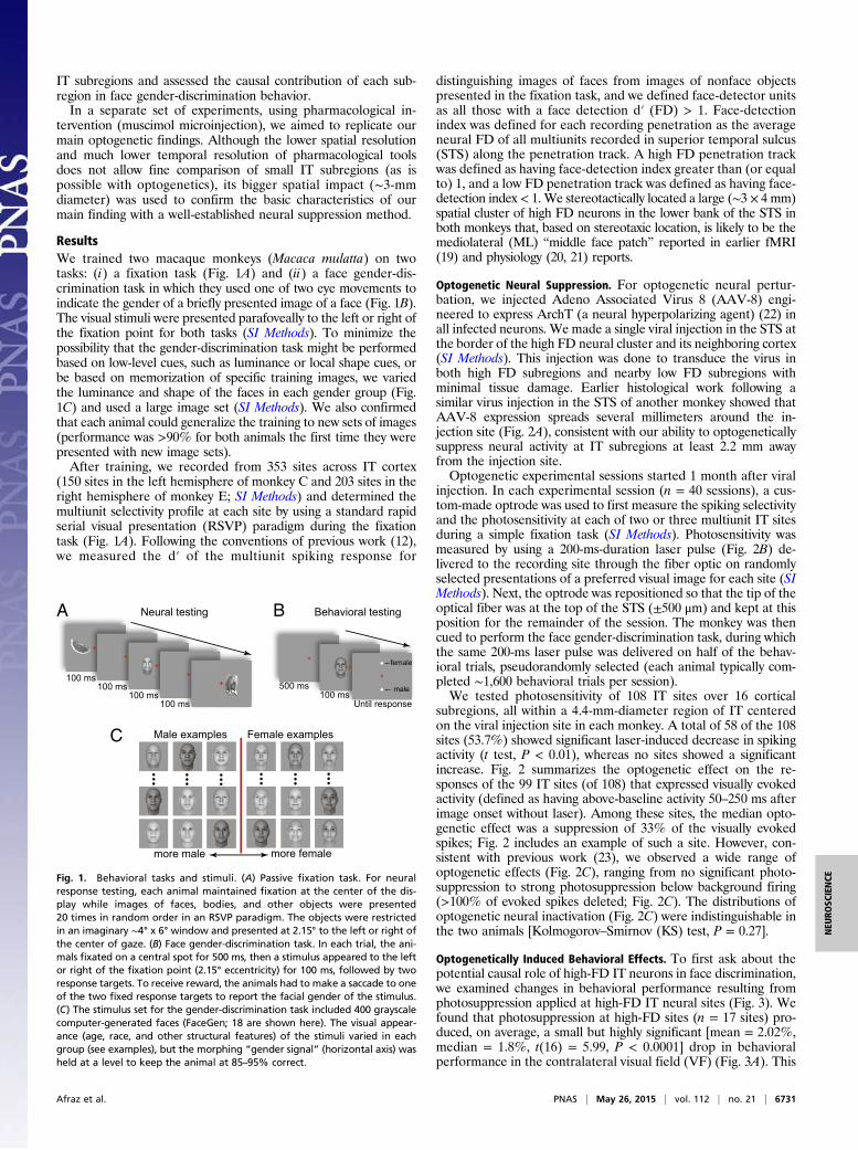
ResultsWe trained two macaque monkeys (Macaca mulatta) on twotasks: (i) a fixation task (Fig. 1A) and (ii) a face gender-dis-crimination task in which they used one of two eye movements toindicate the gender of a briefly presented image of a face (Fig. 1B).The visual stimuli were presented parafoveally to the left or right ofthe fixation point for both tasks (SI Methods). To minimize thepossibility that the gender-discrimination task might be performedbased on low-level cues, such as luminance or local shape cues, orbe based on memorization of specific training images, we variedthe luminance and shape of the faces in each gender group (Fig.1C) and used a large image set (SI Methods). We also confirmedthat each animal could generalize the training to new sets of images(performance was >90% for both animals the first time they werepresented with new image sets).After training, we recorded from 353 sites across IT cortex
(150 sites in the left hemisphere of monkey C and 203 sites in theright hemisphere of monkey E; SI Methods) and determined themultiunit selectivity profile at each site by using a standard rapidserial visual presentation (RSVP) paradigm during the fixationtask (Fig. 1A). Following the conventions of previous work (12),we measured the d′ of the multiunit spiking response for
distinguishing images of faces from images of nonface objectspresented in the fixation task, and we defined face-detector unitsas all those with a face detection d′ (FD) > 1. Face-detectionindex was defined for each recording penetration as the averageneural FD of all multiunits recorded in superior temporal sulcus(STS) along the penetration track. A high FD penetration trackwas defined as having face-detection index greater than (or equalto) 1, and a low FD penetration track was defined as having face-detection index < 1. We stereotactically located a large (∼3 × 4 mm)spatial cluster of high FD neurons in the lower bank of the STS inboth monkeys that, based on stereotaxic location, is likely to be themediolateral (ML) “middle face patch” reported in earlier fMRI(19) and physiology (20, 21) reports.
Optogenetic Neural Suppression. For optogenetic neural pertur-bation, we injected Adeno Associated Virus 8 (AAV-8) engi-neered to express ArchT (a neural hyperpolarizing agent) (22) inall infected neurons. We made a single viral injection in the STS atthe border of the high FD neural cluster and its neighboring cortex(SI Methods). This injection was done to transduce the virus inboth high FD subregions and nearby low FD subregions withminimal tissue damage. Earlier histological work following asimilar virus injection in the STS of another monkey showed thatAAV-8 expression spreads several millimeters around the in-jection site (Fig. 2A), consistent with our ability to optogeneticallysuppress neural activity at IT subregions at least 2.2 mm awayfrom the injection site.Optogenetic experimental sessions started 1 month after viral
injection. In each experimental session (n = 40 sessions), a cus-tom-made optrode was used to first measure the spiking selectivityand the photosensitivity at each of two or three multiunit IT sitesduring a simple fixation task (SI Methods). Photosensitivity wasmeasured by using a 200-ms-duration laser pulse (Fig. 2B) de-livered to the recording site through the fiber optic on randomlyselected presentations of a preferred visual image for each site (SIMethods). Next, the optrode was repositioned so that the tip of theoptical fiber was at the top of the STS (±500 μm) and kept at thisposition for the remainder of the session. The monkey was thencued to perform the face gender-discrimination task, during whichthe same 200-ms laser pulse was delivered on half of the behav-ioral trials, pseudorandomly selected (each animal typically com-pleted ∼1,600 behavioral trials per session).We tested photosensitivity of 108 IT sites over 16 cortical
subregions, all within a 4.4-mm-diameter region of IT centeredon the viral injection site in each monkey. A total of 58 of the 108sites (53.7%) showed significant laser-induced decrease in spikingactivity (t test, P < 0.01), whereas no sites showed a significantincrease. Fig. 2 summarizes the optogenetic effect on the re-sponses of the 99 IT sites (of 108) that expressed visually evokedactivity (defined as having above-baseline activity 50–250 ms afterimage onset without laser). Among these sites, the median opto-genetic effect was a suppression of 33% of the visually evokedspikes; Fig. 2 includes an example of such a site. However, con-sistent with previous work (23), we observed a wide range ofoptogenetic effects (Fig. 2C), ranging from no significant photo-suppression to strong photosuppression below background firing(>100% of evoked spikes deleted; Fig. 2C). The distributions ofoptogenetic neural inactivation (Fig. 2C) were indistinguishable inthe two animals [Kolmogorov–Smirnov (KS) test, P = 0.27].
Optogenetically Induced Behavioral Effects. To first ask about thepotential causal role of high-FD IT neurons in face discrimination,we examined changes in behavioral performance resulting fromphotosuppression applied at high-FD IT neural sites (Fig. 3). Wefound that photosuppression at high-FD sites (n = 17 sites) pro-duced, on average, a small but highly significant [mean = 2.02%,median = 1.8%, t(16) = 5.99, P < 0.0001] drop in behavioralperformance in the contralateral visual field (VF) (Fig. 3A). This
more male more female
Male examples Female examplesC
Neural testing
100 ms100 ms
100 ms100 ms
A Behavioral testing
500 ms100 ms
Until response
female
male
B
Fig. 1. Behavioral tasks and stimuli. (A) Passive fixation task. For neuralresponse testing, each animal maintained fixation at the center of the dis-play while images of faces, bodies, and other objects were presented20 times in random order in an RSVP paradigm. The objects were restrictedin an imaginary ∼4° x 6° window and presented at 2.15° to the left or right ofthe center of gaze. (B) Face gender-discrimination task. In each trial, the ani-mals fixated on a central spot for 500 ms, then a stimulus appeared to the leftor right of the fixation point (2.15° eccentricity) for 100 ms, followed by tworesponse targets. To receive reward, the animals had to make a saccade to oneof the two fixed response targets to report the facial gender of the stimulus.(C) The stimulus set for the gender-discrimination task included 400 grayscalecomputer-generated faces (FaceGen; 18 are shown here). The visual appear-ance (age, race, and other structural features) of the stimuli varied in eachgroup (see examples), but the morphing “gender signal” (horizontal axis) washeld at a level to keep the animal at 85–95% correct.
Afraz et al. PNAS | May 26, 2015 | vol. 112 | no. 21 | 6731
NEU
ROSC
IENCE

effect was significant for each monkey separately (P < 0.02 forboth monkeys; see Fig. S2 for the data presented separately forthe two animals). We also found that photosuppression applied atthese high-FD sites produced no significant change in face-dis-crimination performance for faces presented in the ipsilateral VF[mean = −0.96%, median = −1.35%, t(16) = −1.92, P = 0.07].This lack of a detectable average behavioral effect for the ipsilateralVF is consistent with the well-known contralateral preference ofIT neurons (24–27), which we confirmed within our own recordings(ipsilaterally evoked response was only 45% of contralaterallyevoked response for the preferred stimulus; n = 353 sites).To test the possibility that the laser-induced neural suppres-
sion and behavioral reduction (high-FD neural sites, contralateralVF) was due not to light-gated channels, but to some other effectof tissue illumination (e.g., heating effects), we also performed12 experimental sessions at high-FD neural sites before viral in-jection in one of the animals. No significant neural or behavioraleffects of cortical illumination were observed, even with compa-rable statistical power (Fig. 3B).To compare the causal role of high- and low-FD neurons, we
next looked at the behavioral effect of photosuppression of ITsites with low face selectivity (n = 23 sites with mean FD < 1).We found that photosuppression at these IT sites produced, onaverage, no significant effect on performance for faces presentedin either the contralateral [mean = 0.35%, median = 0.25%,t(22) = 0.89, P = 0.38] or the ipsilateral VF [mean = 0.05%,median = 0.25%, t(22) = 0.16, P = 0.9]. Importantly, this lackof effect cannot be simply explained by a failure to photosuppressneurons at the low FD sites, because the distributions of
photosuppression over low- and high-FD sites were indistin-guishable (KS test, P = 0.33).To determine whether these results reflect a true change in
perceptual gender discriminability (d′) or a change in the animals’choice biases, we calculated the behavioral d′ and the decisioncriterion (C) for each condition. We found that the resultsexpressed in units of d′ largely mirrored those expressed as per-cent correct. Photosuppression at high-FD sites produced a sig-nificant drop [mean = 0.19, median = 0.17, t(16) = 6, P < 0.0001]in behavioral d′ in the contralateral VF, but not the ipsilateral VF[mean = −0.1, median = −0.11, t(16) = −1.9177, P = 0.07]. Noeffect of photosuppression on behavioral d′ was observed for low-FD sites, neither for the contralateral [mean = 0.06, median = 0.06,t(21) = 1.5381, P = 0.14] nor for ipsilateral VF [mean = 0.01,median = 0.02, t(21) = 0.2014, P = 0.84]. We found no evidence ofany significant change in the animals’ decision criterion (choicebias) across different conditions. Fig. S3 summarizes this analysis.Rather than simply lumping the IT sites into two groups (low
and high FD; above), we plotted the FD against the change inbehavioral performance across all 40 experimental sessions. Con-sistent with the results in Fig. 3A, we found a significant negativecorrelation for stimuli presented to the contralateral VF (r = −0.46,P < 0.01; Fig. 3C; the correlation remains significant for the datacollected from each animal: r = −0.73, P < 0.01 and r = −0.56, P <0.05 for monkeys C and E, respectively) but not for the ipsilateral
experimental conditionbe
havi
oral
effe
ct(∆
%co
rrec
t)
**face detector
sitespre-virus
Contra Ipsi C I C I3
0
-3
other IT sites
82
84
86
contra ipsivisual field
beha
vior
al a
ccur
acy
(%)
face detector sitesimageimage+laser
88
** ∆
0 2 4 face detection index (d’)
-4
0
4
beha
vior
al e
ffect
(∆%
corr
ect)
r = –0.46, p = 0.003r = –0.57, p = 0.02
subregion (~1mm2) face detection index
4 2
mea
n be
havi
oral
ef
fect
(∆%
corr
ect)
-2
0
2
-4
0
BA
C D
Fig. 3. Behavioral effects of optogenetic suppression of local IT neural ac-tivity. (A) Face-detector sites. Shown is the mean behavioral effect of pho-tosuppression applied at high-FD cortical sites (penetrations with face-detection index > 1). The ordinate shows the behavioral accuracy of theanimals on the gender-discrimination task. The abscissa indicates the VF inwhich the stimulus was presented. All four trial types were randomly in-terleaved. (B) The behavioral effect of cortical illumination for various exper-imental conditions tested for images presented in the contralateral (C) andipsilateral (I) VF. The ordinate depicts the behavioral effect (change in thebehavioral accuracy for the gender-discrimination task; see A). Face-detectorsites, summarizes the data already shown in A; other IT sites, same for low-FDsites (face-detection index < 1) with equivalent neural suppression (see text);pre-virus, same for high-FD IT sites before viral injection. Error bars show ±1 SE.**P < 0.01. (C) Behavioral effect of photosuppression for all experimentalsessions after the viral injection. The abscissa shows the face-detection index ofthe targeted site. The ordinate indicates the photo-induced behavioral effectfor contralaterally presented images. Each data point shows an experimentsession. (D) Behavioral effect of photosuppression pooled for small (∼1 mm2)subregions of IT cortex (see text). In C and D, open and closed circles depict thedata from monkeys E and C, respectively.
−0.5
0
0.5
1
0
0.5
1
1mm
image
image + laser
laser
time from image onset (ms)
norm
aliz
ed re
spon
se
0 200 400 600
0
0.5
1
image
294% deleted
63% deleted
26% deleted
25
−100 0 100 2000
5
10
15
20
median=33%
250 and above
num
ber o
f site
s
percent of visually evokedspikes deleted
A
C
B
Fig. 2. Neural effects of optogenetic perturbation of the IT cortex. (A) Virustransduction zone. A section of monkey STS transduced (∼8 months beforeeuthanasia) with AAV-delivered ArchT–GFP, stained with anti-GFP antibody(dark region), is shown. The dashed circle shows the size of the estimatedeffective suppression zone for a single optical fiber. (B) Examples of opto-genetic neural suppression observed at different sites in the IT cortex. Bluelines show multiunit response (median of 60 repeated presentations) to thesite’s preferred image (100 ms; black bar) obtained during RSVP. Red linesshow mean response to the same visual image, but with the laser light alsodelivered to the site (200-ms duration; red bar). Laser-on presentations wererandomly interleaved with regular image presentations. Pink and light-blueshadings show ±1 SE. The numbers next to each subplot indicate percentageof visually evoked spikes deleted by light (with respect to the site’s baselinespiking rate; dashed line). (C) Distribution of optogenetic suppression of allvisually driven IT sites that were tested for light sensitivity (n = 99). Percentof visually evoked spikes deleted is defined as 100 x (R − RL)/R, where R is thebaseline subtracted multiunit response to the preferred stimulus (50- to 250-mstemporal window), and RL is the same with laser on.
6732 | www.pnas.org/cgi/doi/10.1073/pnas.1423328112 Afraz et al.

VF (r = 0.074, P = 0.65; absolute r < 0.2 and P > 0.5 in bothanimals). Linear regression suggests that suppression of sites withno face-detection signal (FD = 0) had, on average, a −0.27%impact on performance, whereas sites with the highest observedface-detection signal (mean FD ∼ 3.6) produced a −3.1% impact.The analyses above are presented as if neural activity at single
IT sites should be predictive of the behavioral impact of illumi-nation applied at that site. However, because our optogeneticprotocol is expected to impact all neurons within a ∼1-mm di-ameter spherical region emanating from the optical fiber tip(23), those initial analyses are likely too simplistic because theydo not attempt to account for this 1-mm spread. Thus, we nextsought to summarize the degree of neural face selectivity in eachoptically targeted IT subregion by computing the mean FD of allneural sites recorded within each of the ∼1-mm2 subregionstested (i.e., 16 recording grid locations; median of nine sitesrecorded at each location; SI Methods). We found that 8 of the16 IT subregions had mean FD > 1.0, consistent with previousreports of strong spatial clustering of single neurons with highFD (12, 20, 21). We then plotted the average photo-inducedchange in the animals’ behavioral performance as a function ofthe mean FD of each tested IT subregion. The former wascomputed by averaging the change in behavioral performanceobserved over all experimental sessions performed at that sub-region (median of two sessions). Consistent with the earlieranalyses, these measures were significantly negatively correlatedfor faces presented to the contralateral VF (r = −0.57, P = 0.02)(Fig. 3D) but not for the ipsilateral VF (r = 0.36, P = 0.17).As described in the Introduction, a commonly accepted hy-
pothesis is that high-FD subregions in the IT cortex are causallyinvolved in supporting all face-related tasks (28). The results aboveprovide support for this hypothesis in at least one such task (facegender discrimination). However, this hypothesis is not satisfying inthat it does not offer any computational explanation of whyface-detector neurons should support face discrimination. Thus,we considered a different, computationally motivated hypothesis:Every object-related task depends only on the IT subregions thatconvey explicit (i.e., linearly decodable) information about that task.For instance, under this hypothesis, our face gender-discriminationtask depends on the IT subregions that express the most explicitinformation about gender and does not rely on other IT subregions.This hypothesis predicts that photosuppression of any “explicitgender information” in IT subregions will produce the strongestbehavioral impact, regardless of their ability to support face-detection task (Discussion). To test this prediction, we measured theamount of explicit information (cross-validated linear separability;SI Methods) about our face gender task in each of the tested ITsubregions and plotted that against each subregion’s average photo-induced change in behavioral performance (computed exactly as in
Fig. 3D). This analysis revealed that, over the 16 IT subregions, theamount of explicit information for face gender strongly predictedthe contralateral behavioral deficit resulting from photosuppressionof that subregion (r = −0.6, P = 0.01) (Fig. 4A).
Pharmacological Suppression. We next aimed to compare the be-havioral effects produced by optogenetic methods with thoseproduced by a well-established pharmacological neural sup-pression method (muscimol microinjection) that has less spatialresolution and at least four orders of magnitude less temporalresolution (23, 29) (SI Methods). In six experimental sessions, westereotactically targeted the center of the high-FD neural cluster(in both monkeys) for muscimol microinjection (SI Methods).We used microinjectrodes (30) to record the neural activity be-fore muscimol injection, to measure the FD of each targeted ITsubregion, and to precisely execute small microinjections alongthe entire cortical thickness.We found that injection of muscimol in high-FD subregions of IT
cortex resulted in a significant drop in face gender-discriminationperformance only for faces presented in the contralateral VF,whereas performance remained unchanged in the ipsilateral hemi-field [Fig. 5A; repeated-measures ANOVA interaction test,F(4,20)=7.5, P < 0.001]. Sliding paired t test (including all sessions)showed a significant drop in performance for the contralateralpresentations compared with the ipsilateral presentations for allbehavioral blocks taken 30 min after muscimol injection [t(5) > 3.9;P < 0.007, Bonferroni corrected for all, 5.53% performance dif-ference between the ipsilateral and contralateral VFs averaged forall data points collected after an hour from the end of microinjec-tion; Fig. 5B]. We also performed six experimental sessions injectingmuscimol at neighboring IT loci (at least 2 mm away from theclosest recorded high-FD subregion), and we found no detectablebehavioral impact on the face-discrimination task [repeated-measuresANOVA interaction test, F(4,20)=1.7, P = 0.18]. The pattern of re-sults was similar for both animals (Fig. S4). In addition, we performedtwo experimental sessions injecting saline (1 μL) into high-FDsubregions and found no detectable behavioral effect (Fig. 5B).
DiscussionHere we used neural suppression tools to test the causal linkbetween neural activity in IT subregions enriched with face (de-tector) neurons and one type of face-discrimination behavior. Wefound that neural suppression of these high-FD IT subregionsproduced, on average, a small, but highly significant, reduction inbehavioral performance (for contralaterally presented images). Incontrast, we were unable to detect any average behavioral impactof neural suppression of other IT subregions. These findings wereindependently obtained by using both optogenetic and pharma-cological neural suppression tools.
A B CFig. 4. Explicit encoding of facial genderin CIT. (A) The relationship between explicitneural encoding of facial gender in variousIT subregions and the effect of photo-suppression of those subregions on facegender-discrimination behavior. The abscissashows, for each IT subregion, the genderclassification performance of a linear classi-fier trained and tested (independent imagepresentations) on all sites collected withinthat subregion (SI Methods). The ordinateshows the average impact of photosup-pression of those subregions (pooled across multiple sessions) on the animals’ gender-discrimination performance for contralaterally presented images. (B) Therelationship between explicit neural encoding of facial gender and average face-detection index (FD) over all tested IT subregions. The abscissa is the same as A.The ordinate shows the mean neural FD (d′) for all of the neurons recorded from each cortical subregion. (C) The relationship between neural encoding of facialgender and FD in all recorded IT sites (not pooled). The abscissa represents FD for all individual sites recorded in IT cortex, and the ordinate represents separabilityof the gender signal (defined as absolute d′ of the multiunit response for separating male and female stimuli) in the same sites (n = 353). Seven data points withordinate values > 2 or abscissa values > 6 are not shown here for illustration purposes. Open and closed circles represent monkeys E and C, respectively.
Afraz et al. PNAS | May 26, 2015 | vol. 112 | no. 21 | 6733
NEU
ROSC
IENCE

At broad brush, our results (Figs. 3 and 5) provide, to ourknowledge, the first direct causal support for the idea that face-related tasks rely preferentially on IT spatial subregions that areenriched with face (detector) neurons (16, 28, 31). Althoughprevious studies have revealed that face-detection signals are notuniformly distributed across the IT cortex, those studies providelittle knowledge about the spatial distribution of gender informationacross the IT cortex. Moreover, knowledge of this spatial distributionwould not necessarily dictate the distribution of the causal impact ofdifferent IT subregions on the gender-discrimination task. Thus,in the face of these unknowns, the null hypothesis (H0) is that allIT subregions—including high- and low-FD subregions—equallysupport gender-discrimination behavior. Our results reject thisnull hypothesis (H0). Assuming that face (detector) neuronclusters in the monkey have homology with regions identified inhuman fMRI [fusiform face area and occipital face area (OFA)](31, 32), then our results are consistent with human reports ofdeficits in face discrimination after damage to human fusiformcortex (2, 3, 33) or after temporary disruption of OFA (5, 17).Besides rejection of the hypothesis (H0) stated above, what
other scientific inferences about neural face processing might wedraw from our findings? Tsao and Livingstone (28) proposed thatface processing is a two-stage mechanism that involves separateand sequential “detection” and “discrimination” stages. Accordingto that hypothesis (here termed H1), face-detector neurons(mostly concentrated in the middle face patch), without explicitlyencoding the differences between classes of faces, serve as a“domain-specific gate” for a later discrimination stage that hap-pens at higher processing stages (e.g., more anterior face patches)(28). This hypothesis (H1) predicts that face-detector neurons,even without explicit encoding of facial gender, contribute causallyto face gender discrimination because their activity is required to“gate” the processing of face gender. Our neural suppression re-sults are consistent with this prediction. However, analysis of ourneurophysiological data shows that facial gender is explicitlyencoded in high-FD IT subregions in the middle face patches (Fig.4B), a result that looks inconsistent with H1. More importantly,the fact that neural encoding of gender in IT local neural pop-ulations is the best predictor of their behavioral impact on genderdiscrimination (Fig. 4A) gives birth to an alternative hypothesis(H2). According to H2, every object-related task depends onlyon the IT neurons that contain explicit (linearly decodable)
information about that task (and not on other IT neurons). In thecontext of face processing, H2 states that, unlike H1, face dis-crimination is not gated by the activity of face-detection neurons.Instead, H2, more parsimoniously, states that IT neurons that arebest at face detection support face-detection behavior, whereas ITneurons that are best at face discrimination support face-dis-crimination behavior. Our optogenetic and pharmacological neu-ral suppression experiments are consistent with the predictions ofH2 (Fig. 4). However, because our neurophysiological data revealthat explicit face gender information is most concentrated in high-FD, millimeter-scale IT subregions (Fig. 4B), our neural sup-pression experiments, considered in isolation, cannot distinguishbetween H1 and H2. We currently favor H2 because, unlike H1, itis spatially comprehensive (i.e., speaks to all of IT) and taskcomprehensive (i.e., speaks to all object-related tasks). However,evolution may have settled on special downstream causal linkagesfor socially important stimuli such as faces, so H1 cannot be dis-missed simply because it is less comprehensive.On a related point, note that H2 (as well as H1) is a specific
hypothesis about information readout from the IT cortex. It isabsolutely possible to imagine an upstream area (e.g., V1 or V4)that contains information that is not linearly separable, yet itsinactivation affects the task performance. The correlation of thebehavioral impact of inactivation with linear separability shouldbreak down at such an area. The fact that we actually observe acorrelation with linear separability in IT cortex suggests that ITrepresentations are relatively close to the mechanisms that drivegender-discrimination behavior and that we have approximatedthose mechanisms reasonably well with linear decoders.Are H1 and H2 potentially distinguishable with more spatially
precise neural suppression tools? Our data suggest so. Genderand detection information are moderately (Pearson r = 0.58), butnot perfectly, correlated at the millimeter scale (Fig. 4B), sug-gesting that much larger amounts of data with our current mil-limeter-scale suppression approach could give more weight toH1 or to H2. Moreover, gender and detection information areonly weakly correlated (Pearson r = 0.25) at the single site level(Fig. 4C), which implies that emerging technology that allowslabeling and optogenetic targeting of differently tuned neuralpopulations within cortical subregions (34, 35) should, in prin-ciple, allow us to extend our approach to that finer spatial scale.The behavioral effects of neural suppression reported here are
specific in that, for both optogenetic and pharmacological ex-periments, the effects are limited to the contralateral visualhemifield, and they were not found for other spatial locations inIT where the neural responses were not face selective. However,we have not tested the effect of inactivation of IT neurons inother behavioral tasks. It is possible (and indeed likely if H2 iscorrect) that face selective parts of IT cortex contribute to tasksthat involve discrimination of objects other than faces. Furtherstudies, tailored to study the various task effects, are needed toinvestigate whether perceptual consequences of suppression ofhigh-FD IT subregions are limited to face-related tasks or theyinclude a wider range of perceptual deficits.The finding of contralateral task specificity is against the rel-
atively common intuition that retinotopy and even laterality ofvisual representation decreases dramatically in high-level corticalareas. This common intuition is consistent with large and bi-lateral receptive fields of IT neurons reported in the classicalstudies (24, 25) and has led to a lack of visual-field isolation ofthe stimuli in most of the human neuropsychology studies con-cerning high-level ventral stream damages (2, 3, 33). Our results,however, clearly demonstrate that IT neurons contribute mainlyto visual processing in their contralateral VF. This finding isconsistent with more recent estimations of the size of IT receptivefields (26, 27, 36, 37). Although brain plasticity after temporal lobedamage could mask this contralateral bias, rapidly reversible in-activation methods (as used here) are not subject to this limitation.
Time (minutes)
0 60 120
beha
vior
al a
ccur
acy
(%)
100
80
muscimolmicroinjection
pre recovery
ipsilateral
contralateral
face detector sites
∆
experimental condition
face detectorsites salineother IT
sites
beha
vior
al e
ffect
(∆%
corr
ect)
8
-8
0
4
-4
**
BA
Fig. 5. Behavioral effect of drug microinjection in IT cortex. (A) Face-detector sites. The effect of muscimol injection centered over a high-FD sub-region of IT cortex on gender-discrimination performance is shown. Theordinate represents the animals’ mean behavioral accuracy for the gender-discrimination task. The abscissa represents time. The gray band indicatesthe duration of muscimol microinjection. The red and blue lines show thebehavioral performance for contralaterally and ipsilaterally presented im-ages, respectively. (B) Behavioral effect of drug microinjection for variousexperimental conditions. The ordinate depicts the difference of behavioralaccuracy between the ipsilateral and contralateral VFs averaged for all datapoints collected after 1 h from the end of microinjection. Face-detector sites,summarizes data shown in A (n = 6 microinjections); other IT sites, micro-injections away from high-FD subregions of IT (n = 6); saline, microinjectionof saline in high-FD subregion (n = 2). Error bars show ±1 SE. **P < 0.01.
6734 | www.pnas.org/cgi/doi/10.1073/pnas.1423328112 Afraz et al.

There have been very few reports of behavioral effects of opto-genetic intervention in primate brain (38–40), and induction of sucheffects has not always been straightforward (41). The present studyis, to our knowledge, the first documentation of any behavioralconsequence of optogenetic intervention in a high-level ventralstream cortical area in the primate brain. From a technical point ofview, the demonstrated utility of optogenetic tools for inducingspecific behavioral deficits in face discrimination opens the door forusing the technical advantages of optogenetics (e.g., high temporalprecision and potential neuronal specificity) for future studies ofobject recognition and other high-level vision functions. Giventhat the cortical volume inactivated by 1 μL of muscimol [∼3 mmin diameter (29)] is considerably larger than the volume inacti-vated by a single optical fiber (∼1 mm; Fig. S1) (22, 23), we expectthe behavioral impact of optogenetics to be smaller than themuscimol-induced effect. Our results are consistent with thisintuition (2.0% vs. 5.5% performance change). Although thesmaller effect size of optogenetics may be viewed as a disad-vantage, we see it as an opportunity, because, given the smallervolume of the cortex inactivated, it allows finer spatial dissectionof the role of IT neural populations in visual behaviors. Inaddition, the high statistical power of optogenetic methods(resulting from trial randomization) allows reliable detection ofsmall behavioral effects.
Another remarkable point about the behavioral effect sizesobserved here is that IT is a high-level area in the ventral streamhierarchy. Even massive lesioning or cooling of IT cortex (e.g.,removing an entire gyrus) (42–45) leads to behavioral effectssmaller (∼10–15%) than those after inactivation of early visualareas (46, 47). This finding is consistent with the idea that, in adistributed hierarchical system, inactivation of a low-level corticalsubregion that gates incoming information can produce large be-havioral effects if behavioral tasks are chosen to isolate that gate.Alternatively, the lack of large behavioral effects after massivelesioning or cooling of IT might have resulted from the use ofbehavioral tasks that were not chosen to require the processingvalue added by the ventral stream (e.g., no demand for invariancein the task). Future experiments will hopefully shed more light onsuch questions.
MethodsAll procedures were performed in compliance with standards of MassachusettsInstitute of Technology Animal Care and Use Committee. See SI Methods formore information.
ACKNOWLEDGMENTS. We thank Kailyn Schmidt for her support through-out the project, Chris Stawarz for code support, Aimei Yang for providingthe virus, and Mike Henninger for analysis support. This work was supportedby NIH Grants K99 EY022924 (to A.A.), R21 EY023053 (to J.J.D.), and R01EY14970 (to J.J.D.).
1. Gross CG (2005) Processing the facial image: A brief history. Am Psychol 60(8):755–763.
2. Busigny T, Graf M, Mayer E, Rossion B (2010) Acquired prosopagnosia as a face-spe-cific disorder: Ruling out the general visual similarity account. Neuropsychologia48(7):2051–2067.
3. Susilo T, Duchaine B (2013) Advances in developmental prosopagnosia research. CurrOpin Neurobiol 23(3):423–429.
4. Towler J, Eimer M (2012) Electrophysiological studies of face processing in de-velopmental prosopagnosia: Neuropsychological and neurodevelopmental perspec-tives. Cogn Neuropsychol 29(5-6):503–529.
5. Pitcher D, Garrido L, Walsh V, Duchaine BC (2008) Transcranial magnetic stimulationdisrupts the perception and embodiment of facial expressions. J Neurosci 28(36):8929–8933.
6. Pitcher D, Walsh V, Duchaine B (2011) The role of the occipital face area in the corticalface perception network. Exp Brain Res 209(4):481–493.
7. Rolls ET (1992) Neurophysiological mechanisms underlying face processing within andbeyond the temporal cortical visual areas. Philos Trans R Soc Lond B Biol Sci 335(1273):11–20; discussion 20–21.
8. Leopold DA, Bondar IV, Giese MA (2006) Norm-based face encoding by single neuronsin the monkey inferotemporal cortex. Nature 442(7102):572–575.
9. Hung CP, Kreiman G, Poggio T, DiCarlo JJ (2005) Fast readout of object identity frommacaque inferior temporal cortex. Science 310(5749):863–866.
10. Rust NC, Dicarlo JJ (2010) Selectivity and tolerance (“invariance”) both increase as visualinformation propagates from cortical area V4 to IT. J Neurosci 30(39):12978–12995.
11. Salzman CD, Britten KH, Newsome WT (1990) Cortical microstimulation influencesperceptual judgements of motion direction. Nature 346(6280):174–177.
12. Afraz SR, Kiani R, Esteky H (2006) Microstimulation of inferotemporal cortex in-fluences face categorization. Nature 442(7103):692–695.
13. Allison T, et al. (1994) Face recognition in human extrastriate cortex. J Neurophysiol71(2):821–825.
14. Parvizi J, et al. (2012) Electrical stimulation of human fusiform face-selective regionsdistorts face perception. J Neurosci 32(43):14915–14920.
15. Jonas J, et al. (2014) Self-face hallucination evoked by electrical stimulation of thehuman brain. Neurology 83(4):336–338.
16. Kanwisher N, Yovel G (2006) The fusiform face area: A cortical region specialized forthe perception of faces. Philos Trans R Soc Lond B Biol Sci 361(1476):2109–2128.
17. Pitcher D, Goldhaber T, Duchaine B, Walsh V, Kanwisher N (2012) Two critical andfunctionally distinct stages of face and body perception. J Neurosci 32(45):15877–15885.
18. Felleman DJ, Van Essen DC (1991) Distributed hierarchical processing in the primatecerebral cortex. Cereb Cortex 1(1):1–47.
19. Moeller S, Freiwald WA, Tsao DY (2008) Patches with links: A unified system forprocessing faces in the macaque temporal lobe. Science 320(5881):1355–1359.
20. Tsao DY, Freiwald WA, Tootell RB, Livingstone MS (2006) A cortical region consistingentirely of face-selective cells. Science 311(5761):670–674.
21. Issa EB, Papanastassiou AM, DiCarlo JJ (2013) Large-scale, high-resolution neuro-physiological maps underlying FMRI of macaque temporal lobe. J Neurosci 33(38):15207–15219.
22. Han X, et al. (2011) A high-light sensitivity optical neural silencer: Developmentand application to optogenetic control of non-human primate cortex. Front SystNeurosci 5:18.
23. Chow BY, et al. (2010) High-performance genetically targetable optical neural si-lencing by light-driven proton pumps. Nature 463(7277):98–102.
24. Desimone R, Albright TD, Gross CG, Bruce C (1984) Stimulus-selective properties ofinferior temporal neurons in the macaque. J Neurosci 4(8):2051–2062.
25. Ito M, Tamura H, Fujita I, Tanaka K (1995) Size and position invariance of neuronalresponses in monkey inferotemporal cortex. J Neurophysiol 73(1):218–226.
26. DiCarlo JJ, Maunsell JH (2003) Anterior inferotemporal neurons of monkeys engagedin object recognition can be highly sensitive to object retinal position. J Neurophysiol89(6):3264–3278.
27. Op De Beeck H, Vogels R (2000) Spatial sensitivity of macaque inferior temporalneurons. J Comp Neurol 426(4):505–518.
28. Tsao DY, Livingstone MS (2008) Mechanisms of face perception. Annu Rev Neurosci31:411–437.
29. Arikan R, et al. (2002) A method to measure the effective spread of focally injectedmuscimol into the central nervous system with electrophysiology and light micros-copy. J Neurosci Methods 118(1):51–57.
30. Noudoost B, Moore T (2011) A reliable microinjectrode system for use in behavingmonkeys. J Neurosci Methods 194(2):218–223.
31. Kanwisher N (2000) Domain specificity in face perception. Nat Neurosci 3(8):759–763.32. Grill-Spector K, Knouf N, Kanwisher N (2004) The fusiform face area subserves face
perception, not generic within-category identification. Nat Neurosci 7(5):555–562.33. Chatterjee A, Farah MJ (2001) Face module, face network: The cognitive architecture
of the brain revealed through studies of face processing. Neurology 57(7):1151–1152.34. Ramirez S, et al. (2013) Creating a false memory in the hippocampus. Science
341(6144):387–391.35. Redondo RL, et al. (2014) Bidirectional switch of the valence associated with a hip-
pocampal contextual memory engram. Nature 513(7518):426–430.36. Afraz SR, Cavanagh P (2008) Retinotopy of the face aftereffect. Vision Res 48(1):
42–54.37. Afraz A, Pashkam MV, Cavanagh P (2010) Spatial heterogeneity in the perception of
face and form attributes. Curr Biol 20(23):2112–2116.38. Dai J, Brooks DI, Sheinberg DL (2014) Optogenetic and electrical microstimulation
systematically bias visuospatial choice in primates. Curr Biol 24(1):63–69.39. Cavanaugh J, et al. (2012) Optogenetic inactivation modifies monkey visuomotor
behavior. Neuron 76(5):901–907.40. Jazayeri M, Lindbloom-Brown Z, Horwitz GD (2012) Saccadic eye movements evoked
by optogenetic activation of primate V1. Nat Neurosci 15(10):1368–1370.41. Ohayon S, Grimaldi P, Schweers N, Tsao DY (2013) Saccade modulation by optical and
electrical stimulation in the macaque frontal eye field. J Neurosci 33(42):16684–16697.42. Weiskrantz L, Saunders RC (1984) Impairments of visual object transforms in monkeys.
Brain 107(Pt 4):1033–1072.43. Horel JA, Pytko-Joiner DE, Voytko ML, Salsbury K (1987) The performance of visual
tasks while segments of the inferotemporal cortex are suppressed by cold. BehavBrain Res 23(1):29–42.
44. Heywood CA, Cowey A (1992) The role of the ‘face-cell’ area in the discrimination andrecognition of faces by monkeys. Philos Trans R Soc Lond B Biol Sci 335(1273):31–37,discussion 37–38.
45. Buckley MJ, Gaffan D, Murray EA (1997) Functional double dissociation between twoinferior temporal cortical areas: Perirhinal cortex versus middle temporal gyrus.J Neurophysiol 77(2):587–598.
46. Schiller PH, Tehovnik EJ (2008) Visual prosthesis. Perception 37(10):1529–1559.47. Newsome WT, Paré EB (1988) A selective impairment of motion perception following
lesions of the middle temporal visual area (MT). J Neurosci 8(6):2201–2211.
Afraz et al. PNAS | May 26, 2015 | vol. 112 | no. 21 | 6735
NEU
ROSC
IENCE