optivista - pentax medical (global) · in addition to the already established i-scan modes, the...
TRANSCRIPT
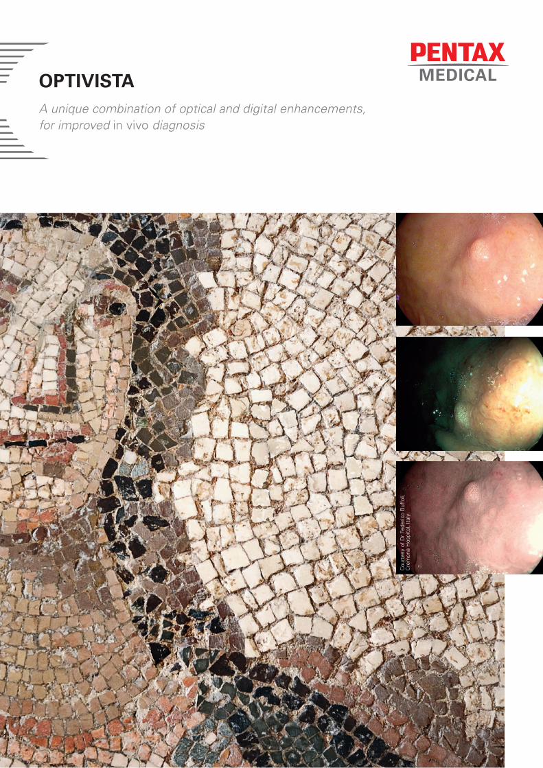
OPTIVISTA A unique combination of optical and digital enhancements, for improved in vivo diagnosis
Cou
rtes
y of
Dr
Fede
rico
Buf
foli,
C
rem
ona
Hos
pita
l, It
aly
The new premium solution for advanced endoscopy
The success of a procedure can hinge on your ability to clearly visualize the epithelial surface pit-pattern and vascular pattern. In addition to the already established i-scan modes, the OPTIVISTA now features i-scan OE (Optical Enhancement), creating a very unique platform where both digital and optical enhancements are available. This unique combination provides extra information for a more accurate in vivo diagnosis through improved vessel and mucosal pattern characterization.
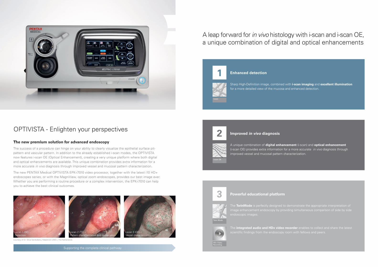
The new PENTAX Medical OPTIVISTA EPK-i7010 video processor, together with the latest i10 HD+ endoscopes series, or with the MagniView, optical zoom endoscopes, provides our best image ever. Whether you are performing a routine procedure or a complex intervention, the EPK-i7010 can help you to achieve the best clinical outcomes.
Supporting the complete clinical pathway
The new premium solution for advanced endoscopy
OPTIVISTA - Enlighten your perspectives
Powerful educational platform3
Twin Mode
HD+ Video Recording
The TwinMode is perfectly designed to demonstrate the appropriate interpretation of image enhancement endoscopy by providing simultaneous comparison of side by side endoscopic images.
The integrated audio and HD+ video recorder enables to collect and share the latest scientifi c fi ndings from the endoscopy room with fellows and peers.
Enhanced detection1Sharp High-Defi nition image, combined with i-scan imaging and excellent illumination for a more detailed view of the mucosa and enhanced detection.
i-scan
Improved in vivo diagnosis2A unique combination of digital enhancement (i-scan) and optical enhancement (i-scan OE) provides extra information for a more accurate in vivo diagnosis through improved vessel and mucosal pattern characterization.
i-scan 1 (SE) – Detection
i-scan 2 (TE) – Pattern characterization and demarcation
i-scan 3 (OE) –Vessel characterization
i-scan OE
Courtesy of Dr. Silvia Sanduleanu, Maastricht UMC+, The Netherlands
A leap forward for in vivo histology with i-scan and i-scan OE,a unique combination of digital and optical enhancements
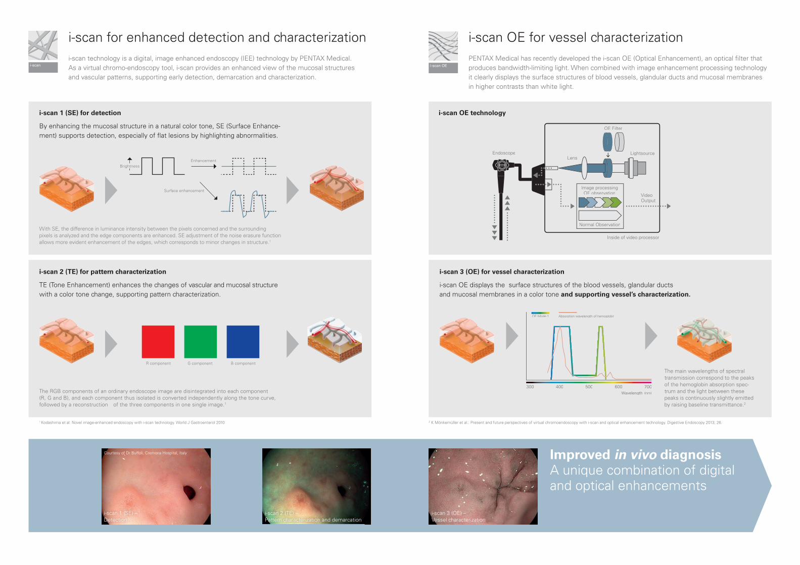
i-scan for enhanced detection and characterizationi-scan technology is a digital, image enhanced endoscopy (IEE) technology by PENTAX Medical. As a virtual chromo-endoscopy tool, i-scan provides an enhanced view of the mucosal structures and vascular patterns, supporting early detection, demarcation and characterization.
i-scan OE for vessel characterizationPENTAX Medical has recently developed the i-scan OE (Optical Enhancement), an optical fi lter that produces bandwidth-limiting light. When combined with image enhancement processing technology it clearly displays the surface structures of blood vessels, glandular ducts and mucosal membranes in higher contrasts than white light.
The RGB components of an ordinary endoscope image are disintegrated into each component (R, G and B), and each component thus isolated is converted independently along the tone curve, followed by a reconstruction of the three components in one single image.1
OE Filter
VideoOutput
LightsourceLens
Inside of video processor
Image processing OE observation
Normal Observation
Endoscope
300 400 500 600Wavelength (nm)
700
OE Mode 1 Absorption wavelength of hemoglobin
The main wavelengths of spectral transmission correspond to the peaks of the hemoglobin absorption spec-trum and the light between these peaks is continuously slightly emitted by raising baseline transmittance.2
i-scan 3 (OE) –In vivo diagnosis
i-scan 2 (TE) for pattern characterization
TE (Tone Enhancement) enhances the changes of vascular and mucosal structure with a color tone change, supporting pattern characterization.
i-scan 3 (OE) for vessel characterization
i-scan OE displays the surface structures of the blood vessels, glandular ducts and mucosal membranes in a color tone and supporting vessel’s characterization.
i-scan 1 (SE) – Detection
Improved in vivo diagnosisA unique combination of digital and optical enhancements
1 Kodashima et al: Novel image-enhanced endoscopy with i-scan technology. World J Gastroenterol 2010
i-scan 1 (SE) for detection
By enhancing the mucosal structure in a natural color tone, SE (Surface Enhance-ment) supports detection, especially of fl at lesions by highlighting abnormalities.
i-scan OE technology
With SE, the difference in luminance intensity between the pixels concerned and the surrounding pixels is analyzed and the edge components are enhanced. SE adjustment of the noise erasure function allows more evident enhancement of the edges, which corresponds to minor changes in structure.1
2 K Mönkemüller et al.: Present and future perspectives of virtual chromoendoscopy with i-scan and optical enhancement technology. Digestive Endoscopy 2013; 26.
Courtesy of Dr Buffoli, Cremona Hospital, Italy
i-scan 2 (TE) – Pattern characterization and demarcation
i-scan 3 (OE) –Vessel characterization
Surface enhancement
R component G component B component
EnhancementBrightness
i-scan i-scan OE
This brochure is intended for distribution in EMEA.
i-scan HD+ solutions: A range that meets all needs OPTIVISTA, a powerful educational platformThe OPTIVISTA EPK-i7010 video processor is a state-of-the-art educational tool with customizable functions operated easily by the touch screen, or via the endoscope buttons.
Pre
miu
mE
PK
-i701
0 –
OP
TIV
ISTA
Cla
ssic
EP
Ki-5
000
Perf
orm
ance
E
PK
-300
0 - D
EFI
NA
HD processors Endoscopes
A complete diagnostic tool, bringing together the best of digital and optical enhancement for complimentary use in pattern and vessel visualization.
A High-Defi nition video processor offering standard functionality supported by state-of-the-art imaging technology.
The video processor to enter the High-Defi nition and i-scan world with an excellent price performance effi ciency.
Slim caliber, bigger working channel and Close Focus, all combined in an i10 HD+ endoscope, the high perfor-mance endoscopes series, for the day-to-day advanced endoscopy practice.
With the 90i endoscope series, you can experience HD+ images combined with exceptional illumination, during routine examinations.
90K endoscope series are the optimal choice for an exceptional high resolution image combined with out-standing illumination.
Collect and share with the unique integrated video and audio recording
• The video recording function enables capture of HD+ video fi les through a USB storage device, for fast and easy sharing of fi ndings with peers. Audio recording for video is captured through external microphone.
• Contributes to cost savings in the endo-scopy room by avoiding unnecessary external HD recording devices.
• For best image collection, the OPTIVISTA also incorporates freeze scan technology which automatically selects the sharpest picture.
0:04 0:12
HD+ Video Recording
HD White Light
Teach and assess with the unique TwinMode
• TwinMode is useful in teaching the appropriate interpretation of image enhanced endoscopy, providing simultaneous comparison of side by side endoscopic images.
• Among experts, TwinMode is respected appreciated as an educational tool for
non-experts in “building the bridge” between HD+ white light images and the different i-scan modes and fi ndings.
• The simultaneous comparison of enhanced clinical images is useful in teaching the appropriate characterization of lesions.
Twin Mode
Colon adenoma
Exchange and expand your knowledge of i-scan by visiting the i-scan website
www.i-scanimaging.com
i-scan 2 (TE)
LCM
/03/
05/1
6/32
0143
/02
TÜV Süd CE0123 • Medical device class: IIa • This product must be used only by Healthcare professionals. Before usage and for detailed product specifications, please refer to the instructions for use. In the interest of technical process, specifications may change without notice.
Netherlands PENTAX Nederland B.V.Edisonring 66669NB DodewaardTel.: +31 88 / 5 30 30 30Fax: +31 88 / 5 30 30 40E-mail: [email protected]
Italy PENTAX Italia S.r.l.Via Dione Cassio, 1520138 MilanoTel.: +39 / 02 50 99 58 1Fax: +39 / 02 50 99 58 60E-mail: [email protected]
Spain SIMMEDICA – Sistemas Integralesde Medicina, S.A.Avenida del Sistema Solar 2528830 San Fernando de Henares · MadridTel.: +34 91 / 301 62 40Fax: +34 91 / 751 31 15E-mail: [email protected]
RussiaPENTAX Europe GmbHRepresentative office in MoscowSadovnicheskaya str, 82, build 2, entrance 6Regus, Business center „Aurora“Office 2012, 2013Moscow, 115035Tel.: +7 495 / 792 52 00Fax: +7 495 / 792 35 66
Turkey PENTAX TurkeyVeko Giz Plaza, Meydan SokakNo:3/43 343396Maslak – IstanbulTel.: +90 212 / 705 05 26Fax: +90 212 / 705 05 00
Japan HOYA Corporation
6-10-1 Nishi-shinjuku Shinjuku-kuTokyo 160-00231
EMEA HeadquarterGermany
PENTAX Europe GmbHJulius-Vosseler-Straße 10422527 HamburgTel.: +49 40 / 5 61 92 - 0Fax: +49 40 / 5 60 42 13E-mail: [email protected]
United Kingdom PENTAX U.K. LimitedPENTAX HouseHeron Drive, LangleySlough SL3 8PNTel.: +44 17 53 / 79 27 33Fax: +44 17 53 / 79 27 94E-mail: [email protected]
France PENTAX France Life Care S.A.S.116 quai de BezonsB.P. 20495106 ARGENTEUIL CEDEXTel: +33 1 / 3025 7575Fax: +33 1 / 3025 7445E-mail: [email protected]