ophthalmology in rhinology ( basics )rhinologychair.org/assets/eye-in-ent-.pdf · legal blindness...
TRANSCRIPT

Ophthalmology in
Rhinology
( Basics )
Ibrahim AlAwadh
MBBS , SB & KSUF Resident , ORL-H&N
Rhinology Chair
KSU – KAUH
13\3\2017

Objectives
– Anatomy of the orbit in relation to nose and PNS
– Basics of Visual Acuity examination
– Basics of Color Vision examination
– Basics of Visual Field examination
– Basics of Intra Ocular Pressure measurement
– Basics of Extra Ocular Muscles examination
– Basics of Exophthalmos measurement
Terminology
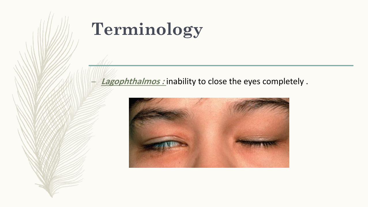
– Lagophthalmos : inability to close the eyes completely .

Terminology
– Chemosis : swelling ( edema ) of the conjunctiva .

Terminology
– Ptosis : drooping of upper eyelid.

Terminology
– Proptosis : Forward displacement of an organ , esp, eye ( pushing
of the globe ) .
– Exophthalmos : protrusion of eyeballs in their sockets ( uniform
expansion of orbital contents ) .

Terminology
– Proptosis : Forward displacement of an organ , esp, eye ( pushing
of the globe ) .
– Exophthalmos : protrusion of eyeballs in their sockets ( uniform
expansion of orbital contents ) .
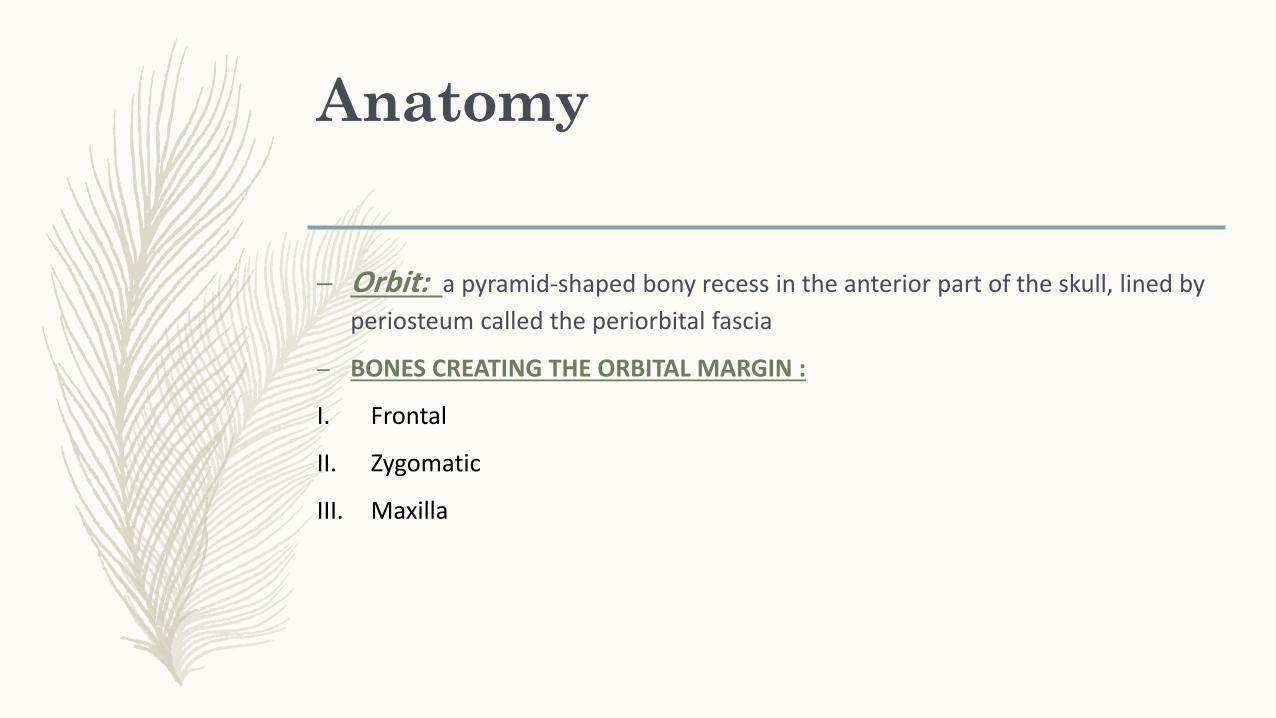
Anatomy
– Orbit: a pyramid-shaped bony recess in the anterior part of the skull, lined by
periosteum called the periorbital fascia
– BONES CREATING THE ORBITAL MARGIN :
I. Frontal
II. Zygomatic
III. Maxilla

Anatomy

Anatomy

Anatomy
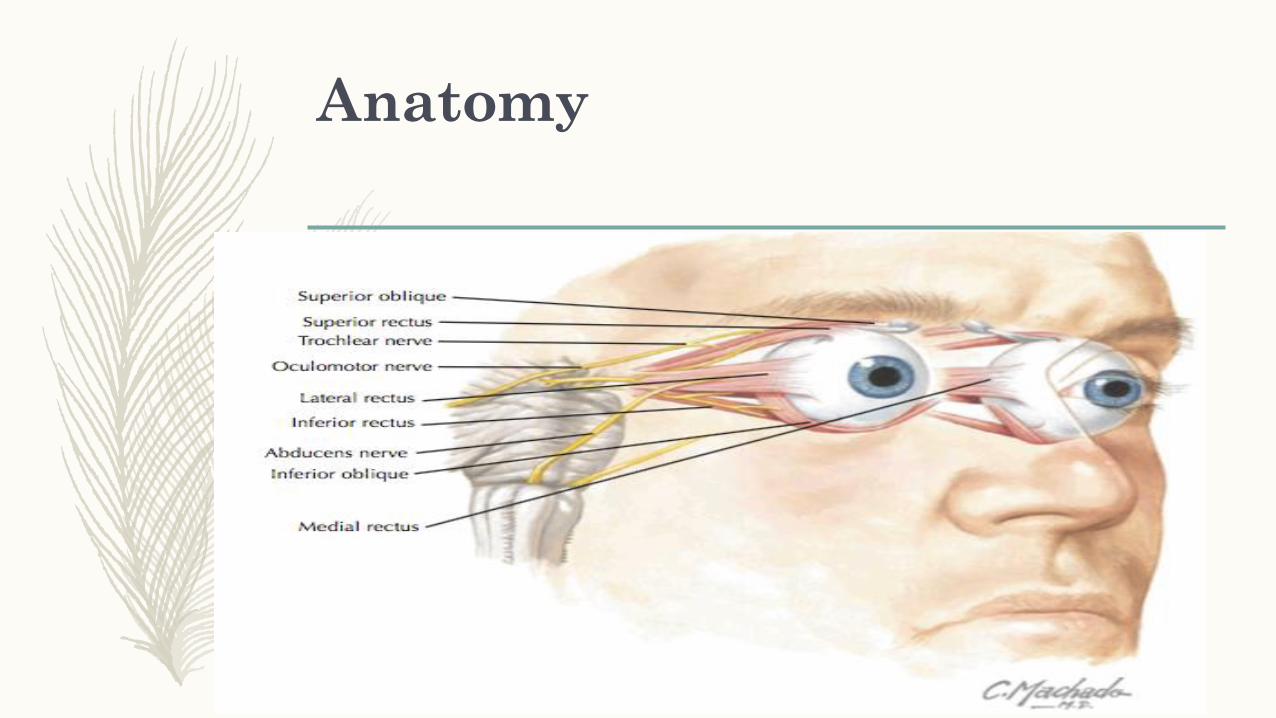
– Contents of orbit include:❖ Eye—organ associated with vision
❖ Extrinsic muscles
❖ CN : II , III , IV , V 1 , VI
❖ Ciliary ganglion
❖ Ophthalmic artery and branches
❖ Superior and inferior ophthalmic veins
❖ Lacrimal apparatus
❖ Much fatty tissue

Anatomy

Anatomy

Anatomy

Anatomy
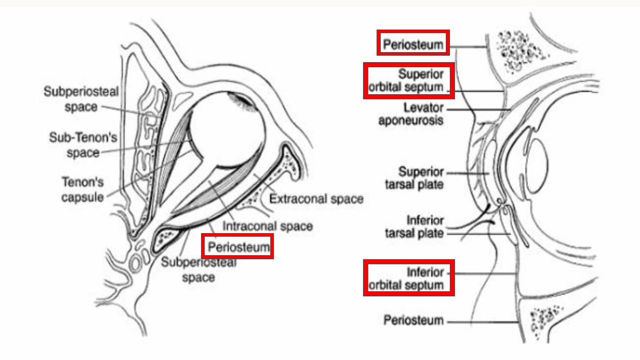
Main anatomical barriers of spreading the infections into the orbit:
1. Lamina Papyracea:
❖Paper-thin bony plate separate ethmoid cells from orbit.
❖Contains several perforations through which valveless
blood vessels and nerves travel.

Anatomy
Main anatomical barriers of spreading the infections into the orbit:
2. Periorbita:
❖Periosteum of the internal orbit and covers the bony orbital walls from
the anterior aperture of the orbital cavity back to the cone enveloping
the optic canal.
❖Only soft tissue barrier between the sinuses and the orbital contents.

Anatomy
Main anatomical barriers of spreading the infections into the orbit:
3. Orbital septum:
❖ Reflection of the periorbita at the margins of the orbit and attaches into the upper and
lower eyelids at the levator aponeurosis superiorly and the tarsal plate inferiorly.
❖ Lacks lymphatic channels and forms a barrier limiting infections from passing directly
through eyelids into orbit.
❖ Forms a boundary between pre and post-septal infections.
Anatomy

Anatomy

Anatomy
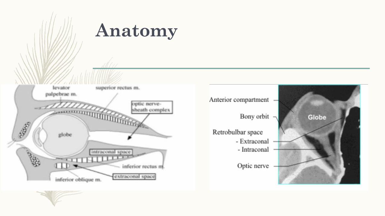
Important anatomical spaces related to the orbit:
1. Sub-periosteal space:
❖ Potential space lying between the orbital bones and the periorbita.
❖Can be fill with blood after orbital fracture or with abscesses when
associated with a paranasal sinusitis.
Anatomy
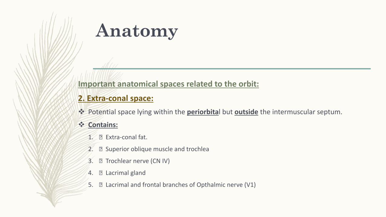
Important anatomical spaces related to the orbit:
2. Extra-conal space:
❖ Potential space lying within the periorbital but outside the intermuscular septum.
❖ Contains:
1. Extra-conal fat.
2. Superior oblique muscle and trochlea
3. Trochlear nerve (CN IV)
4. Lacrimal gland
5. Lacrimal and frontal branches of Opthalmic nerve (V1)

Anatomy
Important anatomical spaces related to the orbit:
3. Intra-conal (Retro-bulbar) space:
❖ Potential space lying within the periorbital but inside the intermuscular septum.
❖ Contains:
1. Intra-conal fat
2. Optic nerve (CN II)
3. Oculomotor nerve (CN III)
4. Abducens nerve (CN VI)
5. Nasociliary branch of Opthalmic nerve (V1)
6. Ophthalmic artery

Anatomy
Anatomy
Anatomy
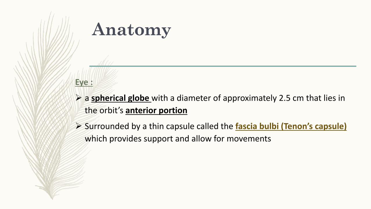
Eye :
➢ a spherical globe with a diameter of approximately 2.5 cm that lies in
the orbit’s anterior portion
➢ Surrounded by a thin capsule called the fascia bulbi (Tenon’s capsule)
which provides support and allow for movements

Anatomy
Eye :
➢ Composed of 3 coats: Sclera, Uveal tract, Retina
➢ Divided into an anterior and a posterior segment

Anatomy

Anatomy

Anatomy
Anatomy

Anatomy

Vision Assessment
❖ Always test visual acuity first
❖ Test best corrected visual acuity (BCVA) whenever possible (i.e. corrective lenses)
❖ Test each eye individually, starting with the right eye, and covering the untested eye
❖ Assess distance and near vision

Visual Acuity
– Visual acuity is the measurement of spatial resolution of the eye (
function of RETINA )
– Components :
1. Minimum VISIBLE VA
2. Minimum RESOLVABLE VA (ordinary VA)
3. Minimum DISCRIMINABLE (hyperacuity)

Visual Acuity
1. Minimum VISIBLE VA
o Determines presence or absence of target

Visual Acuity
2. Minimum RESOLVABLE VA (ordinary VA)
Determines presence of identifying/ distinguishing feature in visible
target
Visual Acuity
2. Minimum RESOLVABLE VA (ordinary VA)
Snellen VA:
• Measures minimum resolvable visual acuity

Visual Acuity
2. Minimum RESOLVABLE VA (ordinary VA)
Snellen VA:
• Letters are made of different sizes and designated by distance at which
letter subtends 5 min of ARC, e.g. letters on 20/20 line subtend 5 min
of ARC when viewed at 20 feet
• Patient asked to recognize progressively smaller letters or forms
Visual Acuity
3. Minimum DISCRIMINABLE (hyperacuity)
➢ Spatial distinctions can be made at lower than ordinary VA
➢ Determines positions of 2 or more visible features relative to each
other

Visual Acuity – Distance
Snellen Fraction =
testing distance (usually 20 feet or 6 metres) smallest line patient can
read on the chart / smallest line patient can read on the chart
e.g. 20/40 = what the patient can see at 20 feet (numerator), a “normal”
person can see at 40 feet (denominator)

Visual Acuity – Distance
Testing hierarchy for low vision:
Snellen acuity (20/x) counting fingers at x distance (CF) hand motion (HM) light perception with projection (LP with projection) light perception (LP) no light perception (NLP)
Legal blindness is BCVA that is ≤20/200 in the better eye, or a limit to the binocular central field of vision <20 degrees

Visual Acuity – Distance

Optotypes for measuring
visual acuity✓Optotype cards
Letter Cards
Snellen

Optotypes for measuring
visual acuity✓Optotype Cards
Letter Cards
Bailey-Lovie
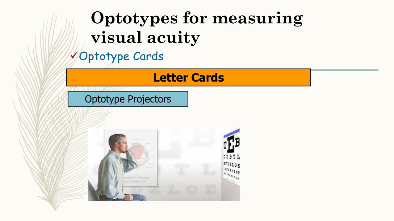
✓Optotype Cards
Letter Cards
Optotype Projectors
Optotypes for measuring
visual acuity

✓Optotype Cards
Number Cards
Optotype Projectors
Optotypes for measuring
visual acuity

Optotypes for measuring
visual acuity✓Optotype Cards
One Character Optotype Cards
Landolt Rings

Optotypes for measuring
visual acuity✓Optotype Cards
One Character Optotype Cards
Snellen “E”

Optotypes for measuring
visual acuity✓Optotype Cards
Other Cards
Infant Visual Acuity

Visual Acuity – Near
❖ use pocket vision chart (Rosenbaum Pocket Vision Screener)
❖ record Jaeger (J) or Point number and testing distance (usually 30 cm)
e.g. J2 @ 30 cm
❖ conversion to distance visual acuity possible (e.g. immobile patient, no
distance chart available)

Visual Acuity – Near
❖ use pocket vision chart (Rosenbaum Pocket Vision Screener)
❖ record Jaeger (J) or Point number and testing distance (usually 30 cm)
e.g. J2 @ 30 cm
❖ conversion to distance visual acuity possible (e.g. immobile patient, no
distance chart available)

Factors that affect Visual
Acuity
✓ Refractive error
✓ Size of the pupil
✓ Illumination
✓ Time exposure of target
✓ Area of retina stimulated
✓ State of adaptation of the eye
✓ Eye movements

Good to Know …

Color Vision
Color vision is the ability to perceive and differentiate colors.
It is the sensory response to stimulation of cones ( in retina ) by light of wavelength 400–700 nm.
The physiological basis is the relative absorption of different wavelengths by the three cones (cone outer segment visual pigments )
Color itself can be described in terms of its hue, saturation and brightness
Very important for optic nerve function

Color Vision - 2 Basic
Theories
1- Trichromatic theory = selective wavelength absorption
❖ Three types of photolabile visual pigments:
➢ Short wavelength: Absorbed by “blue” cones
➢ Middle wavelength: Absorbed by “green” cones
➢ Long wavelength: Absorbed by “red” cones

Color Vision - 2 Basic
Theories
2- Opponent color theory = stimulation and inhibition of different
“receptive fields”
– “Receptive fields” of color sensitive cells have regions that compare
intensity of:
– Red vs green
– Blue vs yellow

Color Vision – Description of
Colors
➢ Hue (“color”): Refers to wavelength
➢ Saturation: Refers to depth of color, purity or richness of color
➢ Brightness: Refers to intensity or radiant flux

Color Vision – Tests
1. Quantitative (both sensitive and specific )
I. Farnsworth-Munsell 100 hue test
II. Nagel’s anomaloscope
2. Qualitative (more sensitive, but less specific)
I. Farnsworth 15 panel
II. Pseudoisochromatic color plate tests e.g : Ishihara Plates

Color Vision – Quantitative

Color Vision – Quantitative
➢ Based on matching hues/color
➢ Consists of 84 colored discs
➢ Discs arranged in sequence (increasing levels of hue)
➢ Test is then scored
➢ Difference in hues between adjacent tablets is 1–4 nm
➢ Accurate in classifying color deficiency
➢ Very sensitive
➢ Time consuming and tiring

Color Vision – Quantitative
Nagel’s anomaloscope
❖ Based on matching luminance or brightness
❖ Good for congenital red-green color defects
❖ Sensitive

Color Vision – Qualitative
1.Farnsworth 15 panel

Color Vision – Qualitative
• More rapid and convenient to use than 100 hue test
• 15 colored tablets
• Hues more saturated than 100 hue test
• Tablets arranged in sequence
• Errors plotted very quickly on a simple circular diagram to define nature of color
deficiency

Color Vision – Qualitative
• Not very sensitive
• Useful in judging practical significance of color deficiency
• Desaturated versions available to recognize more subtle degrees of color
deficiency Discriminates well between congenital and acquired defects
• Congenital defects: Very precise protan/deutan pattern
• Acquired defects: Irregular pattern or errors ,Shows tritan errors very clearly

Color Vision – Qualitative
2. Pseudoisochromatic color plate tests e.g : Ishihara Plates
❖ Gross estimate of acquired color loss
❖ Quick, available, usefulcentral visual dysfunction
❖ Test congenital red-green defects

Color Vision – Qualitative
1. Test in well-illuminated room
2. Held 75 cm from subject and perpendicular to
line of sight
3. Literate patients use plates 1–17
• Answer given within 3 seconds
4. Illiterate patients use plate 18–24
• Lines traced with a brush within 10 seconds

Color Vision – Qualitative
5. Results:
• 13 plates correct: Normal color vision• < 9 plates correct: Deficient color vision
• Only reads “12”: Total color blindness• Reads first 7 plates (except “12”) incorrectly and unable to read the rest: Red-green deficiency
• Reads “26” as 6 and “42” as 2: Protan defect
• Reads “26” as 2 and “42” as 4: Deutan defect
• Unable to read all plates, including “12” (despite good VA): Suspect functional color blindness

Visual Field
❖ The visual field (VF) is one of the functional components of vision.
❖ It is defined as the area that is perceived simultaneously by a fixating eye.
❖ Not 2- but 3-dimensional
❖ “Island of vision in a sea of darkness” (Traquair’s definition)
❖ Limits:
• 60° nasally, 50° superiorly, 90–110° temporally, 70° inferiorly
• Blind spot 15° temporal to fixation

Visual Field
❖ Test “visual fields by confrontation” (4 quadrants, each eye tested separately)
for estimate of visual field loss
❖ Accurate, quantifiable assessment with automated visual field testing ,
perimetry (Humphrey or Goldmann) or Tangent Screen , done by ophthalmolgy

Visual Field – Uses
1. Diagnosis of
I. Glaucoma
II. Optic nerve diseases (optic neuritis, anterior ischemic optic neuropathy, toxic neuropathy )
III. Unexplained visual loss
IV. Malingering patients
2. Follow-up of
I. Glaucoma
II. Tumors (pituitary adenoma)

Aqueous Humor and Intraocular
Pressure
❖ The aqueous humor is the fluid in the anterior (AC) and posterior chamber (PC).
❖ Produced from ciliary body and outflow trabecular meshwork/pressure
dependent flow ( 90% ) ( others like retinal and iris veins )
❖ Three functions
I. Maintains volume and IOP
II. Nutrition for avascular ocular tissue: Posterior cornea, trabecular meshwork, lens
and anterior vitreous
III. Optical Role

Aqueous Humor and Intraocular
Pressure
❖Properties
▪ Clear fluid
▪ Composition:
o No cells and less than 1% of proteins compared to plasma
o Same sodium and chloride, slightly lower potassium and 30% lower bicarbonate than
plasma
o Thirty times higher ascorbate than plasma

Aqueous Humor and Intraocular
Pressure
❖Properties
▪ Volume in AC and PC = 0.30 ml
0.25 ml in AC
0.05 ml in PC
▪ Rate of secretion = 3 ul/min (therefore takes 100 min to completely reform AC
and PC!)

IOP Variation

IOP Variation
IOP Variation

Tonometry - measurement of the IOP
❖Normal range is 10-21.5 mmHg (average 15 mmHg)
❖ Types :
1. Applanation tonometry (Goldman tonometer , Air puff tonometer )
2. Indentation tonometry (Schiotz tonometer )
3. Combined applanation–indentation tonometry (Mackay-Marg
tonometer)

Applanation tonometry-Determines force necessary to flatten a fixed area of cornea
1. Goldman tonometer ( gold standard ) : performed using the slit-
lamp with special tip (prism)
2. Air puff tonometer : Non-contact , Pressurized air current
directed against a fixed area of cornea ,Not reliable in extremes

Indentation tonometry Determines extent of indentation of cornea by fixed weight
❖ Schiotz tonometer : Plunger with known weight indents cornea (Weights of 5.5, 7.5 or 10 g )
Method:
– Patient in supine position
– Topical anesthetic
– Tip of plunger allowed to rest on surface of eye forcing an indentation
– Depth of indentation registered on scale in mm
– IOP in mmHg read off from conversion chart
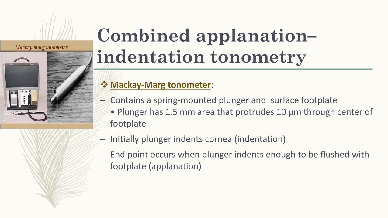
Combined applanation–
indentation tonometry
❖Mackay-Marg tonometer:
– Contains a spring-mounted plunger and surface footplate• Plunger has 1.5 mm area that protrudes 10 μm through center of footplate
– Initially plunger indents cornea (indentation)
– End point occurs when plunger indents enough to be flushed with footplate (applanation)
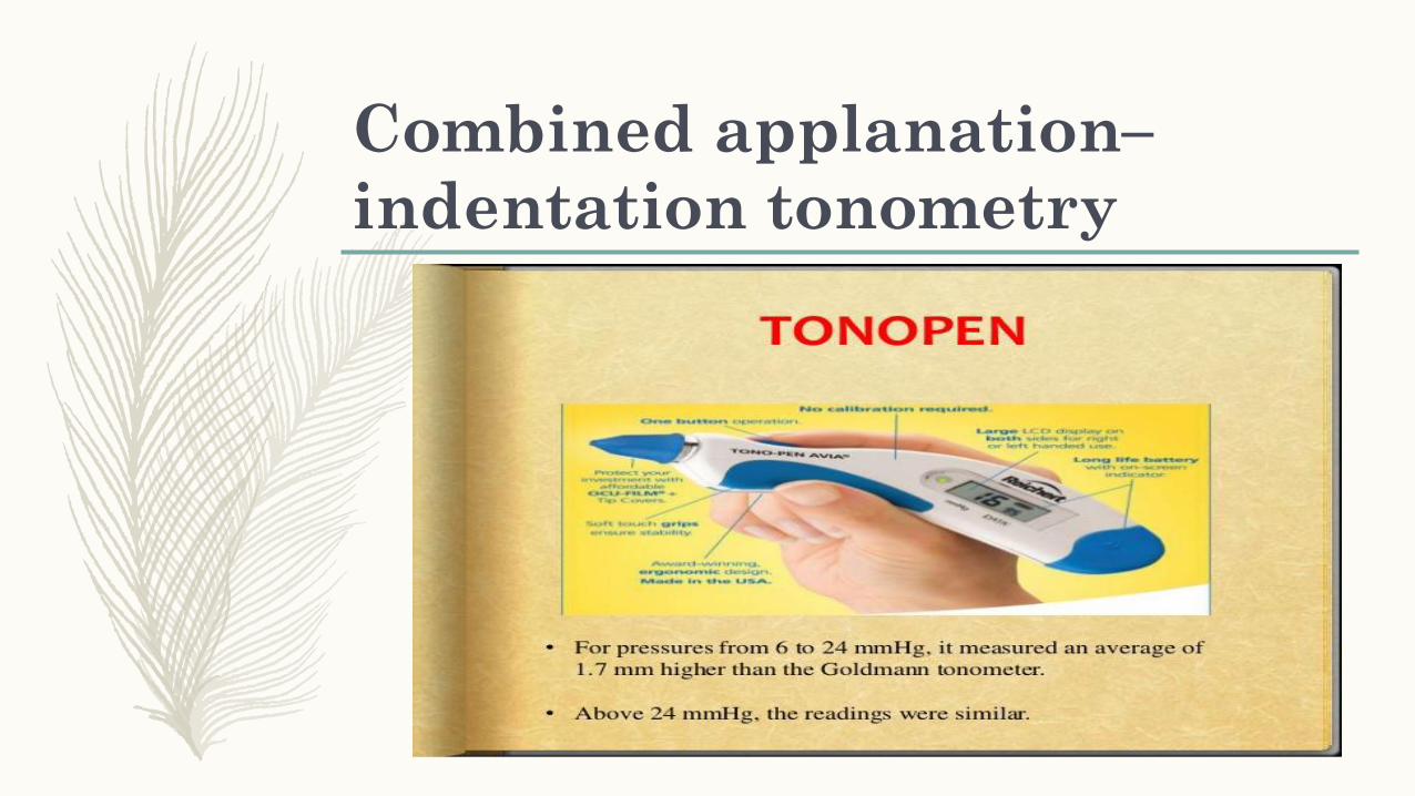
Combined applanation–
indentation tonometry

Tonometry

Extra Ocular Muscles’
Examination
Alignment
▪ Hirschberg corneal reflex test
✓ Examine in primary position of gaze (e.g. straight ahead) with
patient focusing on distant object
✓ Shine light into patient’s eyes from ~30 cm away
✓ Corneal light reflex should be symmetric and at the same position
on each cornea

Extra Ocular Muscles’
Examination
Movement
✓ Examine movement of eyeball through six cardinal positions of
gaze
✓ Ask patient if diplopia is present in any position of gaze or observe
for any restricted movement

Extra Ocular Muscles’
Examination
Movement
✓ Examine movement of eyeball through six cardinal positions of
gaze
✓ Ask patient if diplopia is present in any position of gaze or observe
for any restricted movement

Exophthalmous measurement
1- History and PE including full local examination
2- Visual acuity
3- Pupil reaction ( Marcus Gunn pupil optic nerve compression)
4- Fundoscopy
5- EOM restricted in thyroid ophthalmopathy, extensive tumour
growths and neurological deficit
6- Labs

Exophthalmous measurement
7- Exophthalmometry
❖ It measures protrusion of the apex of cornea from the outer orbital margin (with
the eyes looking straight ahead).
❖ Normal values vary between 10 and 21 mm and are symmetrical in both eyes.
❖ A difference of more than 2 mm between the two eyes is considered significant.
❖ The simplest instrument to measure proptosis is Luedde’s exophthalmometer
❖ Hertel’s exophthalmometer ( is the most commonly used instrument)
✓ Its advantage is that it measures the two eyes simultaneously.

❖Luedde’s exophthalmometer Hertel’s exophthalmometer

Conclusion
❖ It is very important for otorhinolaryngologist to know the basic anatomy and basic simple bed side investigations of ophthalmology related ENT diseases to deal with many conditions
❖ One of these is visual acuity to start with in initial assessment
❖ EOM , visual field , and color vision should be encountered with simple examinations
❖ Exophthalmos is very important sign to be diagnosed as it could be related to underlying conditions
❖ Always ask for help from your colleagues in ophthalmology to create safe and professional environment for the patients

Referrences
• Oxford Medical Dictionary• Manitoba Notes• Riyadh et al notes • Google images

Thank You …