open ventral hernia repair - ucsf medical · pdf fileopen ventral hernia repair ... (what was...
TRANSCRIPT

Open Ventral Hernia Repair
UCSF Postgraduate Course in General SurgeryMaui, HI
March 21, 2011
Hobart W. Harris, MD, MPH
Ventral Hernias
Ventral Hernias: National Experience
• Occur following 11-23% of laparotomies, resulting in ~250,000 ventral hernia repairs/year in the U.S.;
• Average patient in their 5th decade of life;
• Risk factors include obesity, diabetes, lung disease, smoking, wound infection, and steroids;
• No universally accepted classification system or evidence-based management guidelines;
• Wide variation in surgical techniques and strategies for repair.
Complex Ventral Hernias: Definition
A ventral hernia that;
• involves a compromised surgical field in which gastrointestinal, biliaryor genitourinary procedures are performed or frank infection is present;
• includes enterocutaneous or enteroatmospheric fistulas;
• includes an infected prosthetic mesh;
• is large (>10 cm in any dimension) +/- loss of domain
• has recurred (what was “plan A,” and why did it fail?)

Complex Ventral Hernias: Pre-operative Evaluation
• Complete history and physical examination
• Optimize patient’s physiology- nutritional status (alb>3.0)- control serum glucose levels- smoking cessation- taper immunosuppressants, anticoagulants, NSAIDs- cardiac & pulmonary evaluations, if indicated- exercise program & weight loss
• Abdominopelvic CT scan
• Review most recent operative reports, if available
Complex Ventral Hernias: Operative Goals
• Enter the abdomen with minimal iatrogenic injury;
• Define the relevant anatomy;
• Re-establish / certify intestinal continuity
• Develop a repair strategy;
• Reconstruct the abdominal wall
• Close the abdominal wound
Enter the abdomen with minimal iatrogenic injury.
Point of Emphasis #1:A careful & meticulous dissection is essential
• Allow adequate time for the surgery; reduce external stressors;
• Sharp v. electrocautery dissection;
• A thorough adhesiolysis as indicated;
• Scrutinize potential bowel injuries (first tag and then repair).
Define the relevant anatomy.
Point of Emphasis #2:Optimize treatment of bowel injuries and fistulas
• Quantify intra-op findings, e.g., location & number of bowel injuries, length of involved small bowel;
• Minimize length of bowel resected & number of anastomoses;
• Avoid short-gut syndrome & anastomotic leaks.

• Primary suture v. prosthetic repair;
N = 200 patients randomized to primary v. mesh repair of a midline incisional hernia (<6 cm), followed for 3 years for hernia recurrence.
The 3-year cumulative rates of recurrence were 43% v. 24% (p=0.02) among patients who had suture repair v. mesh repairThe risk factors for recurrence weresuture repair, infection, prostatism (in men), and previous surgery for AAA.
The size of the hernia did not affect the rate of recurrence.
New Engl J Med 2000;343:392-8
Repair Principles & Techniques Repair Principles & Techniques
• Primary suture v. prosthetic repair;
• Prosthetic-fascia interface options;
Onlay
Inlay
Underlay
Repair Principles & Techniques
• Primary suture v. prosthetic repair;
• Prosthetic-fascia interface options;
• Component separation;
• Suture & points of fixation;+ suture-to-wound length ratio >4+ debride fascial edges/hernia sac to healthy tissue+ continuous v. interrupted suture technique+ heavy, permanent, monofilament suture
• Synthetic v. biologic prosthetics
Develop a repair strategy.
Point of Emphasis #3:Standardize your approach & surgical technique
• Collect follow up data on your patients;
• Determine what techniques & materials work best in your practice;
• Read the literature with a critical (skeptical) eye.
Reconstruct the abdominal wall.
• Case A: ventral hernia repair with wound contamination
• Case B: incisional hernia with mesh infection
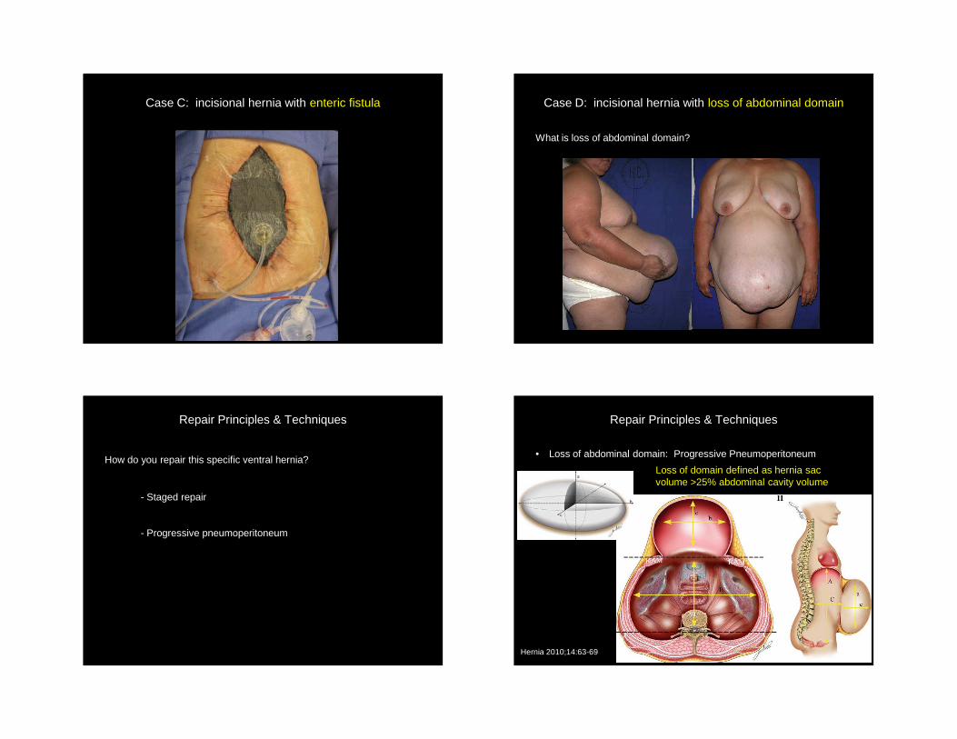
• Case C: incisional hernia with enteric fistula
• Case D: large ventral hernia with loss of domain
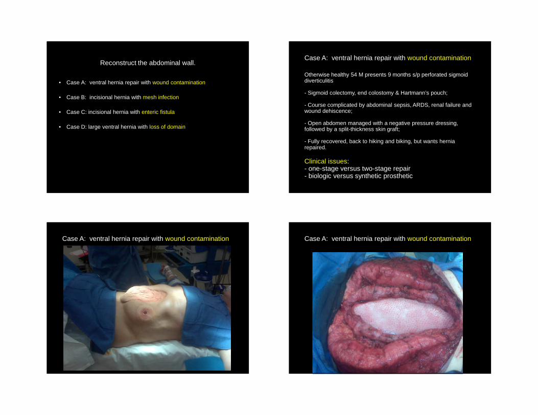
Case A: ventral hernia repair with wound contamination
Otherwise healthy 54 M presents 9 months s/p perforated sigmoid diverticulitis
- Sigmoid colectomy, end colostomy & Hartmann’s pouch;
- Course complicated by abdominal sepsis, ARDS, renal failure and wound dehiscence;
- Open abdomen managed with a negative pressure dressing, followed by a split-thickness skin graft;
- Fully recovered, back to hiking and biking, but wants hernia repaired.
Clinical issues:- one-stage versus two-stage repair- biologic versus synthetic prosthetic
Case A: ventral hernia repair with wound contamination Case A: ventral hernia repair with wound contamination
Case A: ventral hernia repair with wound contamination Case B: incisional hernia with mesh infection
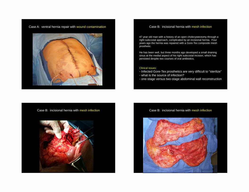
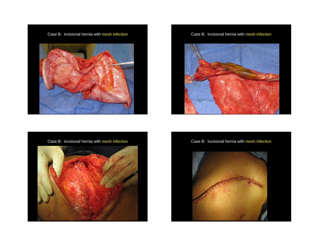
47 year old man with a history of an open cholecystectomy through a right subcostal approach, complicated by an incisional hernia. Four years ago the hernia was repaired with a Gore-Tex composite mesh prosthetic.
He has been well, but three months ago developed a small draining sinus at the medial aspect of his right subcostal incision, which has persisted despite two courses of oral antibiotics.
Clinical issues:- Infected Gore-Tex prosthetics are very difficult to “sterilize”- what is the source of infection?- one-stage versus two-stage abdominal wall reconstruction
Case B: incisional hernia with mesh infection Case B: incisional hernia with mesh infection
Case B: incisional hernia with mesh infection Case B: incisional hernia with mesh infection
Case B: incisional hernia with mesh infection Case B: incisional hernia with mesh infection
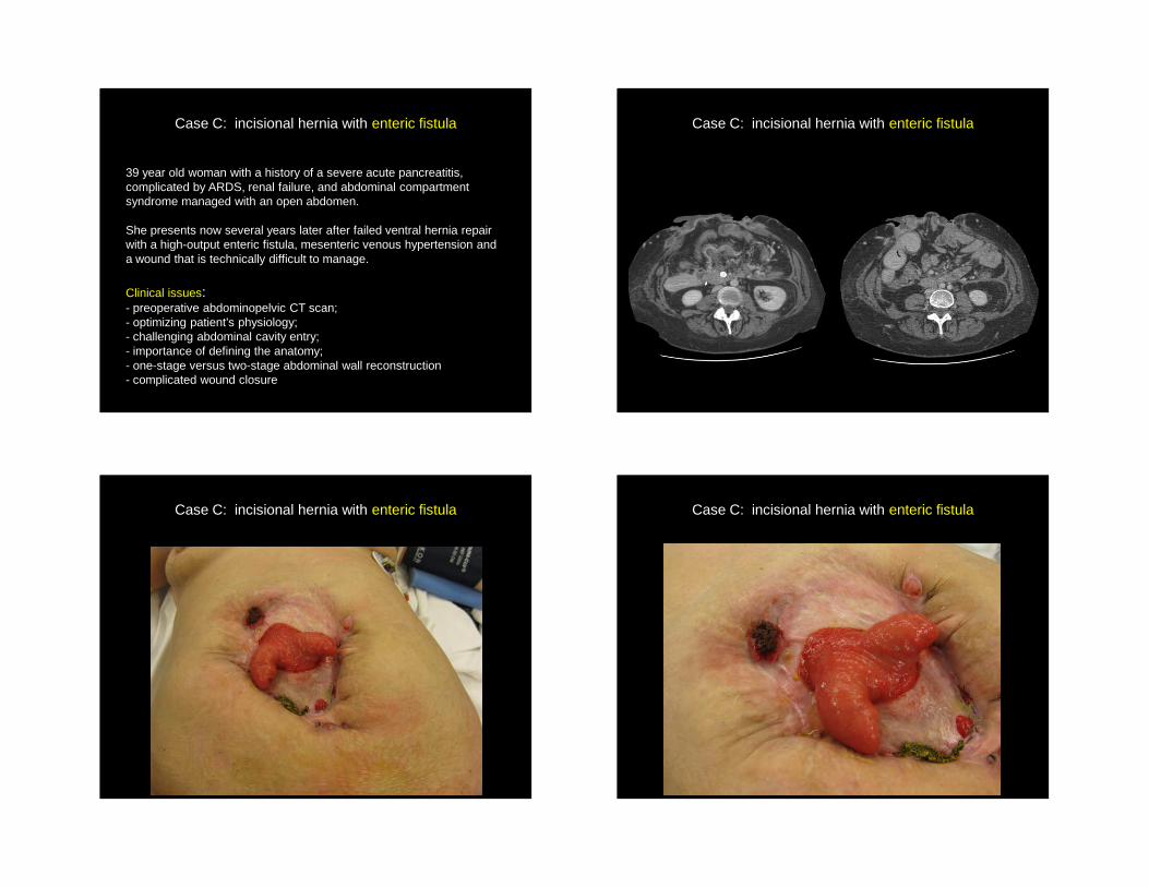
39 year old woman with a history of a severe acute pancreatitis, complicated by ARDS, renal failure, and abdominal compartment syndrome managed with an open abdomen.
She presents now several years later after failed ventral hernia repair with a high-output enteric fistula, mesenteric venous hypertension and a wound that is technically difficult to manage.
Clinical issues:- preoperative abdominopelvic CT scan;- optimizing patient’s physiology;- challenging abdominal cavity entry;- importance of defining the anatomy;- one-stage versus two-stage abdominal wall reconstruction- complicated wound closure
Case C: incisional hernia with enteric fistula Case C: incisional hernia with enteric fistula
Case C: incisional hernia with enteric fistula Case C: incisional hernia with enteric fistula

Case C: incisional hernia with enteric fistula Case C: incisional hernia with enteric fistula
Case C: incisional hernia with enteric fistula Case C: incisional hernia with enteric fistula
Case C: incisional hernia with enteric fistula
What is loss of abdominal domain?
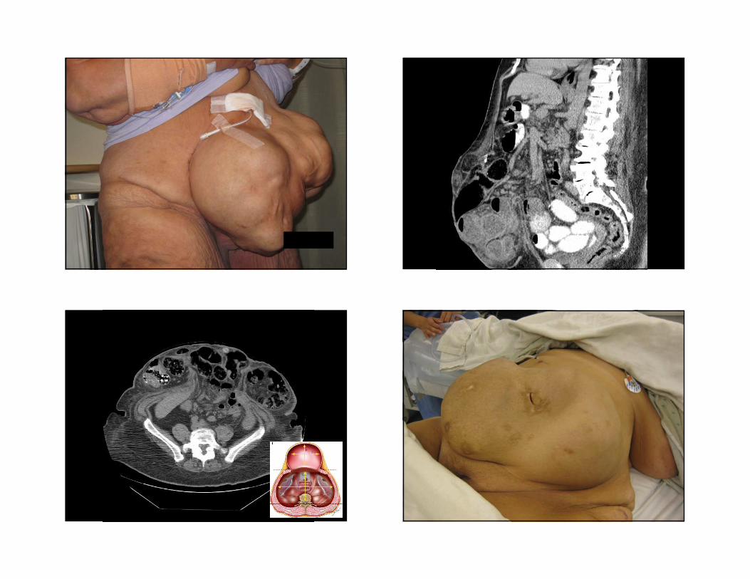
Case D: incisional hernia with loss of abdominal domain
Repair Principles & Techniques
How do you repair this specific ventral hernia?
- Staged repair
- Progressive pneumoperitoneum
Loss of domain defined as hernia sac volume >25% abdominal cavity volume
Hernia 2010;14:63-69
Repair Principles & Techniques
• Loss of abdominal domain: Progressive Pneumoperitoneum

Repair Principles & Techniques
• Loss of abdominal domain: Progressive Pneumoperitoneum
- initial stage involves 3-D abdominal CT scan with volumetric analysis;- insertion of a peritoneal dialysis catheter;- daily insulflation with CO2 increasing the total volume inserted in 500 ml increments each day;
N = 23 patients, mean age 56 years (range 31 – 83), mean BMI = 38.5- Avg. hernia sac volume 4,500 ml (1,850 – 6,600 ml)- Avg. volume ratio 36% (26 – 73%)- Avg. pneumoperitoneum sessions 10 (4 – 18)- 26 % wound infection rate- 4 % 30-day mortality rate- 4 % recurrence rate at 2 years (82% follow up)
Hernia 2010;14:63-69
55 year old woman with a history of morbid obesity, osteoarthritis and depression, who presents s/p multiple failed umbilical hernia repairs.
She presents now with a large ventral hernia that is becoming progressively more symptomatic, including interference with acts of daily living and work.
Clinical issues:- staged versus progressive pneumoperitoneum;- avoidance of iatrogenic abdominal compartment syndrome;- evaluation for pulmonary hypertension;- one-stage versus two-stage abdominal wall reconstruction- set clear clinical goals
Case D: incisional hernia with loss of abdominal domain
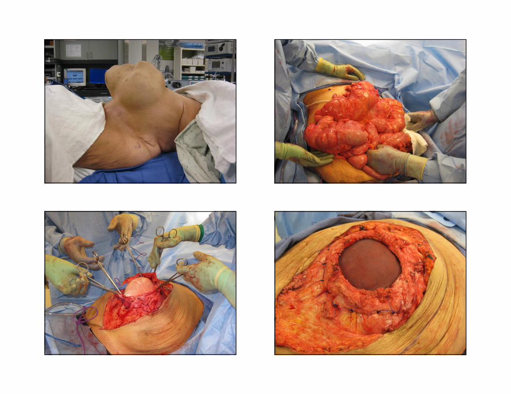

• Multiple options / strategies for reconstruction of the abdominal wall:
- primary suture repair (rarely indicated)
- synthetic prosthetics (absorbable v. permanent)
- biological prosthetics (porcine, bovine, human)
- onlay, inlay and underlay
- component separation
- one stage versus two-stage repair
- negative pressure wound therapy dressings
Open Ventral Hernia Repairs: Summary
• Points of Emphasis:
- a careful and meticulous dissection
- optimize treatment of fistulas and bowel injuries
- standardize your approach & surgical technique
- establish clear and reasonable clinical goals
Open Ventral Hernia Repairs: Summary