open-label allopregnanolone treatment of men with fragile
TRANSCRIPT

ORIGINAL ARTICLE
Open-Label Allopregnanolone Treatment of Men with FragileX-Associated Tremor/Ataxia Syndrome
J. Y. Wang1,2 & A. M. Trivedi3 & N. R. Carrillo3 & J. Yang4,5 & A. Schneider1,6 &
C.Giulivi1,7 & P. Adams1,6 & F. Tassone1,2 &K.Kim1,8& S.M. Rivera1,5,9 &N. Lubarr10 &C.-
Y. Wu4,11& R. W. Irwin12
& R. D. Brinton12,13& J. M. Olichney4,5 & M. A. Rogawski4,14 &
R. J. Hagerman1,6
# The American Society for Experimental NeuroTherapeutics, Inc. 2017
Abstract Fragile X-associated tremor/ataxia syndrome(FXTAS) is a late-onset neurodegenerative disorder affectingapproximately 45% of male and 16% of female carriers of theFMR1 premutation over the age of 50 years. Currently, noeffective treatment is available. We performed an open-labelintervention study to assess whether allopregnanolone, aneurosteroid promoting regeneration and repair, can improveclinical symptoms, brain activity, and magnetic resonanceimaging (MRI) measurements in patients with FXTAS.Six patients underwent weekly intravenous infusions ofallopregnanolone (2–6 mg over 30 min) for 12 weeks. Allpatients completed baseline and follow-up studies, thoughMRI scans were not collected from 1 patient because of MRIcontraindications. The MRI scans from previous visits, along
with scans from 8 age-matchedmale controls, were also includ-ed to establish patients’ baseline condition as a reference.Functional outcomes included quantitative measurements oftremor and ataxia and neuropsychological evaluations. Brainactivity consisted of event-related potential N400 wordrepetition effect during a semantic memory processing task.Structural MRI outcomes comprised volumes of thehippocampus, amygdala, and fluid-attenuated inversionrecovery hyperintensities, and microstructural integrity of thecorpus callosum. The results of the study showed thatallopregnanolone infusions were well tolerated in all subjects.Before treatment, the patients disclosed impairment inexecutive function, verbal fluency and learning, and progres-sive deterioration of all MRI measurements. After treatment,
* R. J. [email protected]
1 UC Davis MIND Institute, UC Davis Health, Sacramento, CA, USA2 Department of Biochemistry and Molecular Medicine, School of
Medicine, University of California, Davis, Sacramento, CA, USA3 School of Medicine, University of California, Davis,
Sacramento, CA, USA4 Department of Neurology, School of Medicine, University of
California, Davis, Sacramento, CA, USA5 Center for Mind and Brain, University of California, Davis,
Davis, CA, USA6 Department of Pediatrics, School of Medicine, University of
California, Davis, Sacramento, CA, USA7 Department of Molecular Biosciences, University of California
Davis, School of Veterinary Medicine, Davis, CA, USA
8 Department of Public Health Sciences, University of California,Davis, Davis, CA, USA
9 Department of Psychology, University of California Davis,Davis, CA, USA
10 Department of Neurology, Mount Sinai Beth Israel Hospital, NewYork, NY, USA
11 PK/PD Bioanalytical Core Facility, UC Davis Health,Sacramento, CA, USA
12 Department of Pharmacology and Pharmaceutical Sciences, Schoolof Pharmacy, University of Southern California, Los Angeles, CA,USA
13 Center for Innovation in Brain Science, School of Medicine,Departments of Pharmacology and Neurology, University ofArizona, Tucson, AZ, USA
14 Department of Pharmacology, School of Medicine, University ofCalifornia, Davis, Sacramento, CA, USA
NeurotherapeuticsDOI 10.1007/s13311-017-0555-6
the patients demonstrated improvement in executive function-ing, episodic memory and learning, and increased N400 repe-tition effect amplitude. AlthoughMRI changes were not signif-icant as a group, both improved and deterioratedMRI measure-ments occurred in individual patients in contrast to uniformdeterioration before the treatment. Significant correlationsbetween baselineMRImeasurements and changes in neuropsy-chological test scores indicated the effects of allopregnanoloneon improving executive function, learning, and memory forpatients with relatively preserved hippocampus and corpuscallosum, while reducing psychological symptoms for patientswith small hippocampi and amygdalae. The findings show thepromise of allopregnanolone in improving cognitive function-ing in patients with FXTAS and in partially alleviating someaspects of neurodegeneration. Further studies are needed toverify the efficacy of allopregnanolone for treating FXTAS.
Keywords Fragile X premutation . FMR1 .
Neurodegeneration . Allopregnanolone . FXTAS .
Neurogenesis
Introduction
Fragile X-associated tremor/ataxia syndrome (FXTAS) is alate-adult–onset neurodegenerative disorder that affects, withage- and sex-specific penetrance, carriers of premutation al-leles (55–200 CGG repeats) of the fragile Xmental retardation1 (FMR1) gene. Defining clinical features of FXTAS includeprogressive kinetic tremor, gait ataxia, executive function andmemory deficits, peripheral neuropathy, and parkinsonism.Associated variable features include cognitive impairmentand dementia; psychiatric symptoms, such as depression;and dysautonomia [1–4]. FXTAS is characterized by progres-sive global brain atrophy and white matter disease of both thecerebellum and cerebrum [5–8]. Cerebellar and brainstem at-rophy and ventricular enlargement are detectable even beforethe onset of tremor or ataxia [6, 9, 10]. Currently, no specifictreatment for FXTAS can slow the progression of neurodegen-eration, though symptomatic treatments exist for some of thesymptoms, such as tremor [11].
Allopregnanolone is a naturally occurring neurosteroidwith activity as a positive modulator of γ-aminobutryic acidA receptors that can stimulate hippocampal neurogenesis andreverse hippocampal-dependent learning and memory dys-func t ion [12] . Al lopregnanolone a lso promotesoligodendrogenesis and myelination [13] and confers neuro-protection by reducing the expression of the proapoptotic pro-tein caspase 3 [14, 15].We recently reported that hippocampalneurons cultured from premutationmice exhibit abnormal net-work bursting [16]. Such persistent bursting is a plausiblemechanism for the neurodegeneration that leads to the neuro-logical symptoms in FXTAS. We have further demonstrated
that allopregnanolone, by potentiating γ-aminobutryic acid A-receptor signaling, eliminates the abnormal bursting in thepremutation neurons, providing a mechanism for neuroprotec-tion [16].
Allopregnanolone is endogenously present in the peripheryand brain, and thus is expected to be safe for long-term treat-ment. Intravenous infusion of allopregnanolone in healthysubjects [17] and in the treatment of status epilepticus [18,19] has not revealed treatment-related adverse effects otherthan sedation and mild nausea. Given the neuroregenerativeproperties of allopregnanolone and the preclinical evidence ofefficacy in the FXTAS model, we sought to conduct a prelim-inary, open-label investigation of the therapeutic potential ofallopregnanolone for carriers of the premutation with FXTAS.We assessed the impact of allopregnanolone on workingmemory deficits and neurological symptoms. In addition, weexplored whether improvements in mitochondrial functionand magnetic resonance imaging (MRI) measurements canbe observed that reflect neuroregeneration and alleviation ofwhite matter disease.
Methods
Study Design and Patients
This study was a preliminary, 3-month, open-label, uncon-trolled trial of allopregnanolone treatment in 6 men withFXTAS who received weekly intravenous infusions for aperiod of 12 weeks. Patients with FXTAS were recruitedat the University of California, Davis (UC Davis), MINDInstitute’s Fragile X Research and Treatment Center be-tween December 2015 and March 2016, with the last par-ticipant completing treatment in July 2016. Notification ofthe study was provided on ClinicalTrials.gov (Identifier:NCT02603926).
Individuals aged 50–85 years with the premutation and adiagnosis of FXTAS were eligible for the study. Exclusioncriteria were other serious systemic disease, alcohol or drugabuse, and current use of phenytoin. To establish patients’baseline condition, we included scans acquired before thetreatment. Table 1 summarizes demographic information forthe enrollees. Serial brain MRI scans from 8 age-matchedhealthy men carrying normal FMR1 alleles (CGG repeatlength 20–32) were used as a null reference group forassessing MRI parameters (age 61 ± 6 years, range 52–72years, 2–3 scans/individual).
CGG-repeat sizing was carried out using a polymerasechain reaction-based approach [20]. Mitochondrial outcomesin lymphocytes were measured, as previously described [21].Lactate and pyruvate ratios from lymphocytes were detectedby utilizing a metabolomic approach.
Wang et al.

Intervention
The study was conducted under US Food and DrugAdministration-approved Investigational New DrugApplications 111,085 (to MAR) and 125,502 (to RJH) andwith the consent of the UC Davis Institutional ReviewBoard. Written informed consent was also obtained from eachpatient prior to the study. Allopregnanolone intravenous injec-tion was formulated at the UC Davis Good ManufacturingPractices Laboratory and dispensed by the UC DavisInvestigational Drug Pharmacy. The formulation consistedof 99.9% pure GMP-grade allopregnanolone (3α-hydroxy-5α-pregnan-20-one) at a concentration of 0.5 mg/ml in 0.9%sodium chloride injection, United States Pharmacopeia, con-taining 6% sulfobutyl ether-β-cyclodextrin sodium salt,United States Pharmacopeia (Dexolve; CycloLab, Budapest,Hungary). The weekly intravenous infusions and follow-upobservations were performed at the Infusion Center at theUC Davis CTSC Clinical Research Center (CCRC) exceptfor 2 patients residing in the New York area who receivedtheir first 3 infusions at the CCRC Infusion Center and theremainder of their infusions at the Mount Sinai Beth IsraelHospital Therapeutic Infusion Center (New York, NY, USA)under the supervision of one of the co-authors of this study(NL).
All infusions were administered over 30min, followed by aflush for 30 min. The dose for the first infusion was 2.0 mg;the subsequent week, the dose was increased to 4.0 mg; andfinally increased to 6.0 mg in the third week. The RichmondAgitation Sedation Scale (RASS) was administered before,during, and after each infusion. Infusions were to be terminat-ed if the score fell below –1 (drowsy) during the infusion.Each subject had a RASS score of 0 (alert and calm) beforebeginning infusion and maintained the RASS score of 0throughout each infusion. Since the 6.0-mg dose was toleratedby all subjects, this dose was administered on weeks 3 to 12 inall cases.
Each participant was required to be accompanied by achaperone, who drove the participant home from the CCRCInfusion Center and remained with the participant for at least20 h, to monitor for adverse effects.
Blood Level Measurements and PharmacokineticAnalysis
Plasma allopregnanolone levels were determined by liquidchromatography–tandem mass spectrometry in 1 subject inblood samples drawn at intervals during and after a 6-mginfusion from the opposite arm of that receiving the intrave-nous infusion. Blood was collected in heparinized tubes be-fore infusion (0 time), at 15 and 30 min during the infusion,and at 30, 90, and 150 min after the infusion. The blood wascentrifuged at 1500 g for 10 min to separate the plasma, whichwas stored at −80 °C until the time of analysis. D4-allopregnanolone was added as internal standard, and theanalytes were extracted using the solid phase extraction meth-od and injected into a Waters ACQUITY UPLC System(Waters, Milford, MA, USA). The effluent was directed to aXevo TQ-S triple quadrupole mass spectrometer, which wasused to ionize target molecules and monitor the ion m/z tran-sitions from 319.20→ 283.30 for allopregnanolone quantifi-cation and 323.3→ 287.3 for D4-allopregnanolone quantifi-cation. Two-compartment pharmacokinetic parameters wereestimated with Phoenix WinNonlin 7.0 (Certara, Princeton,NJ, USA).
Assessments and Follow-Up
Clinical Assessment
All participants completed baseline and follow-up clinical ex-aminations and neuropsychological assessments. A detailedphysical and neurological examination was performed andvideotaped [22], emphasizing the major features of FXTAS:cerebellar ataxia, intention tremor, and neuropathy findings,such as vibration sense and pinprick sensation.
Neuropsychological and Emotional Assessment
Learning and memory was measured through the CaliforniaVerbal Learning Test 2 [23]. Working memory was assessedusing Weschler Memory Scale-IV (WMS-IV) [24]. Othercognitive measures included Mini Mental Status Exam
Table 1 Demographicinformation Participants Age Total no. of previous
visitsCGG-repeatlength
FXTASstage
Education(years)
Handedness
Patient 1 57 1 104 5 16 Right
Patient 2 68 3 88 4 27 Right
Patient 3 79 0 83 3 20 Right
Patient 4 64 2 105 4 20 Right
Patient 5 68 2 85 3 17 Right
Patient 6 74 2 98 5 20 Right
FXTAS = Fragile X-associated tremor/ataxia syndrome
Open-Label Allopregnanolone Treatment of Men with FXTAS

(MMSE) [25], Behavior Dyscontrol Scale (BDS-2) [26, 27],Cambridge Neuropsychological Test Automated Battery(CANTAB) [28], and Controlled Oral Word AssociationTest [29]. The Symptom Checklist-90-Revised (SCL-90-R)[30], and Beck Anxiety Inventory [31] were used to assessneuropsychological symptoms. In addition, CATSYS [32, 33]tremor and sway assessments evaluated improvements inhand tremor and balance. Table 2 shows a brief descriptionof functional measures used in the study.
MRI Acquisitions and Analyses
MRI scans were acquired on a Siemens Trio 3 T MRI scanner(Siemens Medical Solutions, Erlangen, Germany) equippedwith a 32-channel head coil. High-resolution T1-weightedMagnetization Prepared Rapid Gradient Echo (MPRAGE)images were acquired in 192 sagittal slices of 1-mm thicknesswith field of view (FOV) 256 mm, 256 × 256 matrix, repeti-tion time (TR) of 2170 ms, echo time (TE) of 4.82 ms, and 7°flip angle. Fluid attenuated inversion recovery (FLAIR) im-ages for quantifying subcortical lesions were acquired in 104sagittal slices of 1.9-mm thickness with FOV 243 mm, 512 ×512 matrix, TR of 5000 ms, TE of 456 ms, and inversion time1700 ms. Diffusion tensor imaging (DTI) scans for assessingmicrostructural integrity were obtained using a single shotspin-echo echo planar imaging (EPI) sequence in 68 axialslices of 2-mm thickness with FOV 224mm, 112 × 112matrixzero-filled to 224 × 224 pixels, TR of 7100 ms, TE of 73 ms,number of excitations of 2, 30 gradient encoding directions forb = 800 s/mm2, along with 4 interleaved b = 0 s/mm2 images.
MRI preprocessing included automated anterior commis-sure–posterior commissure alignment in T1-weighted scansusing acpcdetect (http://www.nitrc.org/projects/art) [34]. Forfailed cases, manual anterior commissure–posterior
commissure alignment was performed using DTI Studio(www.mristudio.org) [35]. MRI bias field correction in bothT1 and FLAIR scans were performed using N4 (http://stnava.github.io/ANTs/) [36]. For DTI scans, motion and eddy currentwere corrected using FSL (http://fsl.fmrib.ox.ac.uk/fsl/fslwiki/)[37] prior to the generation of fractional anisotropy (FA) andmean diffusivity (MD) maps using DTI Studio.
Hippocampus, amygdala, and corpus callosum were seg-mented automatically using MRI Cloud (http://mricloud.org)[38], followed by automated machine learning-based error cor-rection implemented in SegAdapter (https://www.nitrc.org/projects/segadapter/) [39, 40] and manual corrections. DTI FAand MD values of the corpus callosum were calculated basedon previously published method [41], with modifications.Within-subject co-registration of MD maps with T1 was per-formed automatically, and then manually adjusted to maximizethe match of the corpus callosum using FreeSurfer (https://surfer.nmr.mgh.harvard.edu/fswiki/bbregister) [42]. Theresulting transformation was then applied to the FA maps. Tofurther reduce the effect of mismatch, we generated masks bythresholding the MD maps at 2 μm2/ms and applied the masksto both FA and MD maps using the FSL command, fslmaths.The generation of regions-of-interests for the 3 brain structures(i.e., hippocampus, amygdala, and corpus callosum), and cal-culation of volume, FA, andMD values were also performed inFSL using the commands fslmaths and fslstats.
Segmentation of subcortical hyperintensities was per-formed on FLAIR scans using a fully automated tool, lesionprediction algorithm (LPA) (http://www.applied-statistics.de/lst.html) [43] from SPM12. LPA has shown high performancerelative to other publically available tools for automatedFLAIR lesion segmentation [44]. To calculate FLAIRhyperintensity volume (HV) in the corpus callosum, we ap-plied the threshold of 0.7 to LPA-generated lesion probability
Table 2 Description of neuropsychological and emotional tests
Outcome measures Description
Beck Anxiety Inventory (BAI) Self-report questionnaire to obtain measurement of anxiety
Behavioral Dyscontrol Scale (BDS-2) Nine-item measurements assessing frontal lobe integrity, includingexecutive function and cognitive control over behaviors
Cambridge Neuropsychological Test Automated Battery (CANTAB) Computerized test measuring memory, attention and executive function
CATSYS Test measuring postural sway, tremor, and gait using force plate andtremor pen
Controlled Oral Word Association Test (COWAT) Assessment measuring verbal fluency or cognitive flexibility that canbe correlated with executive functioning
California Verbal Learning Test (CVLT-II) Assessment measuring working memory and memory subtypes
Mini Mental Status Exam (MMSE) Clinician-administered assessment measuring cognitive function, mentalstate, orientation, and memory
Symptom Checklist-90-Revised (SCL-90-R) Ninety-item self-reporting measure of psychological symptoms
Weschler Memory Scale Fourth Edition (WMS-IV) Assessment measuring immediate, delayed, visual and auditory memory
Wang et al.

maps, followed by applying corpus callosum masks usingfslmaths.
Event-Related Potential Word Repetition Effect Assessment
Electroencephalogram (EEG) recordings were obtained during asemantic category decision/word repetition paradigm at bothvisits [45]. Categorical statements were read aloud by an exper-imenter, with each phrase followed (~1 s later) by a target wordvisually presented in the center of the monitor (duration =300 ms, visual angle = 0.4°). Participants were instructed to notmove or respond for 3 s following the target word. Their re-sponse was to repeat the spoken word, followed by a Byes^ orBno^ judgment indicating whether the word was congruent withthe preceding statement (congruous trial, 50%) or not (incongru-ous trial, 50%). An EEG session consisted of 2 blocks of 144trials, each block lasting approximately 20min. In each block, 12congruous category–target pairswere presented once, 12 present-ed twice, and 12 presented 3 times. Equal numbers of incongru-ous pairs were presented in the same repetition conditions.
Thirty-two-channel EEG was recorded (Nicolet SM 2000amplifier, bandpass 0.016–100 Hz, sampling rate 250 Hz).Interelectrode impedance was maintained below 5 kΩ. All scalpelectrodes were referenced online to the left mastoid and re-referenced offline to the average of both mastoids. Eye move-ments were captured by electro-oculogram recorded with 4 elec-trodes (1 beneath and 1 at the outer canthus of each eye). Alltrials free of EEG/electro-oculogram artifact were averaged toproduce the event-related potential (ERP) for each participantsession.
Statistical Analysis
All statistical analyses were conducted in R 3.2.5 language andenvironment [46]. To establish baseline MRI measurements forpatients with FXTAS, group means and change rates beforetreatment were compared with the healthy controls using mixedeffectsmodels [47]. Improvements in primary outcomemeasuresbefore and after treatment were tested for significance using 1-sided paired t tests. Pearson correlation coefficients were utilizedto identify pretreatment MRI data predictive of functional im-provements after the treatment. The threshold for statistical sig-nificance was set at 0.05.
Results
Patient 3 (body weight 76.3 kg) received an intravenous infu-sion of 6 mg allopregnanolone over 30 min. Blood was col-lected in heparinized tubes before infusion (0 time), at 15 and30 min during the infusion, and at 30, 90, and 150 min afterthe infusion. Table 3 shows the estimated 2-compartmentpharmacokinetic parameters. The peak allopregnanolone
concentration achieved at the end of the 30-min infusionwas 35.5 ng/ml (112 nM). The plasma concentration valuesare shown in Figure 1.
Functional Outcomes
At baseline, all patients exhibited impairment in executivefunction assessed using BDS-2 (range 8–17). Four patientsshowed impairment in learning and memory (CaliforniaVerbal Learning Test-II T-score 24–37) and 4 (with 3 overlap-ping) disclosed compromised verbal fluency (Controlled OralWord Association Test-total raw score 16–29). In addition, 2patients showed borderline scores of WMS visual memory(73 and 79) and 1 with borderline MMSE score at 24.
Table 4 shows the comparisons of functional outcomemea-sures before and after the treatment. The majority of the pa-tients showed improvement in mental state (MMSE), execu-tive function (BDS-2 and CANTAB One Touch Stockings),memory (WMS-IV and CANTAB Paired AssociatesLearning), anxiety (SCL-90-R), and intention tremor(CATSYS Dot-to-Dot left-hand tremor intensity); improve-ment in BDS-2 and CANTAB Paired Associates Learningtotal error adjusted achieved statistical significance (p =0.009 and p = 0.042, respectively).
Table 3 Estimated 2-compartment pharmaco-kinetic parameters
Parameter Estimate Units
V 1.11 l/kg
V2 1.61 l/kg
CLx 2.12 l/h/kg
CL2 1.87 l/h/kg
Tmax = 30 min. Cmax = 35.5 ng/ml. Thefalling phase was fit to a single exponentialwith half-time of 34.7 min
V = volume of distribution of central com-partment; V2 = volume of distribution ofperipheral compartment; CL = centralcompartment clearance; CL2 = peripheralcompartment clearance
Fig. 1 Allopregnanolone plasma concentrations and pharmacokineticanalysis in patient 3
Open-Label Allopregnanolone Treatment of Men with FXTAS
MRI Findings
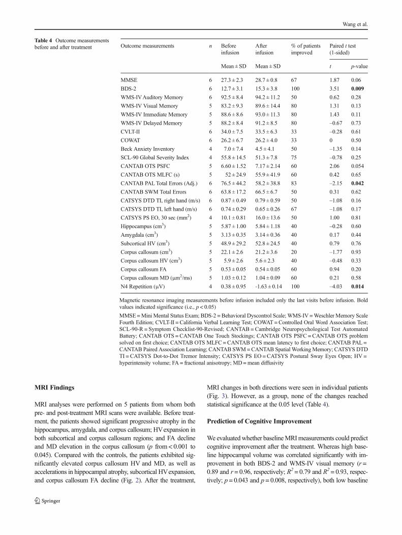
MRI analyses were performed on 5 patients from whom bothpre- and post-treatment MRI scans were available. Before treat-ment, the patients showed significant progressive atrophy in thehippocampus, amygdala, and corpus callosum; HVexpansion inboth subcortical and corpus callosum regions; and FA declineand MD elevation in the corpus callosum (p from < 0.001 to0.045). Compared with the controls, the patients exhibited sig-nificantly elevated corpus callosum HV and MD, as well asaccelerations in hippocampal atrophy, subcortical HVexpansion,and corpus callosum FA decline (Fig. 2). After the treatment,
MRI changes in both directions were seen in individual patients(Fig. 3). However, as a group, none of the changes reachedstatistical significance at the 0.05 level (Table 4).
Prediction of Cognitive Improvement
We evaluatedwhether baselineMRImeasurements could predictcognitive improvement after the treatment. Whereas high base-line hippocampal volume was correlated significantly with im-provement in both BDS-2 and WMS-IV visual memory (r =0.89 and r= 0.96, respectively; R2= 0.79 and R2 = 0.93, respec-tively; p= 0.043 and p = 0.008, respectively), both low baseline
Table 4 Outcome measurementsbefore and after treatment Outcome measurements n Before
infusionAfterinfusion
% of patientsimproved
Paired t test(1-sided)
Mean ± SD Mean ± SD t p-value
MMSE 6 27.3 ± 2.3 28.7 ± 0.8 67 1.87 0.06
BDS-2 6 12.7 ± 3.1 15.3 ± 3.8 100 3.51 0.009
WMS-IVAuditory Memory 6 92.5 ± 8.4 94.2 ± 11.2 50 0.62 0.28
WMS-IV Visual Memory 5 83.2 ± 9.3 89.6 ± 14.4 80 1.31 0.13
WMS-IV Immediate Memory 5 88.6 ± 8.6 93.0 ± 11.3 80 1.43 0.11
WMS-IV Delayed Memory 5 88.2 ± 8.4 91.2 ± 8.5 80 –0.67 0.73
CVLT-II 6 34.0 ± 7.5 33.5 ± 6.3 33 –0.28 0.61
COWAT 6 26.2 ± 6.7 26.2 ± 4.0 33 0 0.50
Beck Anxiety Inventory 4 7.0 ± 7.4 4.5 ± 4.1 50 –1.35 0.14
SCL-90 Global Severity Index 4 55.8 ± 14.5 51.3 ± 7.8 75 –0.78 0.25
CANTAB OTS PSFC 5 6.60 ± 1.52 7.17 ± 2.14 60 2.06 0.054
CANTAB OTS MLFC (s) 5 52 ± 24.9 55.9 ± 41.9 60 0.42 0.65
CANTAB PALTotal Errors (Adj.) 6 76.5 ± 44.2 58.2 ± 38.8 83 –2.15 0.042
CANTAB SWM Total Errors 6 63.8 ± 17.2 66.5 ± 6.7 50 0.31 0.62
CATSYS DTD TI, right hand (m/s) 6 0.87 ± 0.49 0.79 ± 0.59 50 –1.08 0.16
CATSYS DTD TI, left hand (m/s) 6 0.74 ± 0.29 0.65 ± 0.26 67 –1.08 0.17
CATSYS PS EO, 30 sec (mm2) 4 10.1 ± 0.81 16.0 ± 13.6 50 1.00 0.81
Hippocampus (cm3) 5 5.87 ± 1.00 5.84 ± 1.18 40 –0.28 0.60
Amygdala (cm3) 5 3.13 ± 0.35 3.14 ± 0.36 40 0.17 0.44
Subcortical HV (cm3) 5 48.9 ± 29.2 52.8 ± 24.5 40 0.79 0.76
Corpus callosum (cm3) 5 22.1 ± 2.6 21.2 ± 3.6 20 –1.77 0.93
Corpus callosum HV (cm3) 5 5.9 ± 2.6 5.6 ± 2.3 40 –0.48 0.33
Corpus callosum FA 5 0.53 ± 0.05 0.54 ± 0.05 60 0.94 0.20
Corpus callosum MD (μm2/ms) 5 1.03 ± 0.12 1.04 ± 0.09 60 0.21 0.58
N4 Repetition (μV) 4 0.38 ± 0.95 -1.63 ± 0.14 100 –4.03 0.014
Magnetic resonance imaging measurements before infusion included only the last visits before infusion. Boldvalues indicated significance (i.e., p < 0.05)
MMSE=Mini Mental Status Exam; BDS-2 = Behavioral Dyscontrol Scale; WMS-IV =Weschler Memory ScaleFourth Edition; CVLT-II = California Verbal Learning Test; COWAT =Controlled Oral Word Association Test;SCL-90-R = Symptom Checklist-90-Revised; CANTAB = Cambridge Neuropsychological Test AutomatedBattery; CANTAB OTS = CANTAB One Touch Stockings; CANTAB OTS PSFC = CANTAB OTS problemsolved on first choice; CANTAB OTS MLFC=CANTAB OTS mean latency to first choice; CANTAB PAL =CANTAB Paired Association Learning; CANTAB SWM=CANTAB Spatial WorkingMemory; CATSYS DTDTI = CATSYS Dot-to-Dot Tremor Intensity; CATSYS PS EO = CATSYS Postural Sway Eyes Open; HV =hyperintensity volume; FA = fractional anisotropy; MD=mean diffusivity
Wang et al.

hippocampal and amygdala volumes were correlated significant-ly with reduced SCL-90-R symptom severity (r= 0.98 and r=0.95, respectively; R2 = 0.95 and R2 = 0.91, respectively; p =0.023 and p = 0.046, respectively). In addition, low baseline sub-cortical HV volume was correlated with improvement in WMS-IV immediate memory (r = –0.94, R2 = 0.88, p = 0.018), andboth low baseline corpus callosum HV fraction (i.e., HV/vol-ume) and MD values showed correlations with reduction inCANTAB One Touch Stockings mean latency to first choice(r= 1.00 and r= 0.99, respectively; R2= 1.00 and R2 = 0.97, re-spectively; p < 0.001 and p = 0.013, respectively) (Fig. 4).
ERP Results
Figure 5 shows the grand averaged ERP difference waveforms(Incongruous New minus Incongruous Old) for the baseline andfollow-up based on 4 patients. At the posterior electrodes illus-trated, ERPs at the follow-up visits weremore negative in generalthan those at the baseline. Paired t tests demonstrated that mean
amplitudes of theN400 repetition effect at POzwere larger (morenegative) at the follow-up comparedwith the baseline (p = 0.014;Table 4), suggesting beneficial intervention effects on verballearning and memory.
Molecular Measurements
None of the mitochondrial outcomes tested (Complex I, II–III,IV, V, and citrate synthase activities; ratios of lactate-to-pyruvate,lactate, pyruvate, ratio of alanine and lactate to pyruvate; short-chain organic acids levels associated with mitochondrial respira-tory chain disorders) were significantly different before and afterthe treatment (as determined by paired t test).
Discussion
This open-label study demonstrates that intravenous infusionof allopregnanolone at doses up to 6 mg are well tolerated and
Am
yg
da
la (
cm
3)
Su
bco
rtica
l H
V (
cm
3)
mc
( s
up
ma
co
ppi
H3)
CC
(cm
3)
CC
FA
2/m
s)
Age Age Age
Age Age Age
Fig. 2 Spaghetti plots showinglongitudinal changes in magneticresonance imaging measurementsfor premutation carriers beforetreatment and controls. Orangedenotes patients with Fragile X-associated tremor/ataxia syn-drome; blue indicates controls.HV = hyperintensity volume;CC= corpus callosum; FA = frac-tional anisotropy; MD=meandiffusivity
Before After
Am
yg
da
la (
cm
3)
mc
( s
up
ma
co
ppi
H3)
Before After
CC
FA
Before After
Su
bco
rtica
l H
V (
cm
3)
Before After
CC
(cm
3)
Before After
CC
HV
(cm
3)
Before After
CC
No
n-L
esio
n (
cm
3)
Before After
2/m
s)
Before After
PA1
PA2
PA3
PA4
PA5
PA1
PA2
PA3
PA4
PA5
Fig. 3 Changes in magneticresonance imaging measurementsbefore and after allopregnanolonetreatment. HV = hyperintensityvolume; CC = corpus callosum;FA = fractional anisotropy; MD =mean diffusivity
Open-Label Allopregnanolone Treatment of Men with FXTAS

without apparent adverse effects in men with FXTAS. In aprevious study of healthy women, a similar dose ofallopregnanolone was associated with sedation and, in somesubjects, mild nausea [17]; changes in saccadic eye move-ments were also consistent with sedation. While the peak con-centration of allopregnanolone in patient 3 was modestlyhigher than achieved in this prior study, our subject did notexhibit sedation or nausea. The half-life of allopregnanolonefollowing termination of the infusion was approximately35 min. Therefore, the peak level was only present for a briefperiod of time, thus likely limiting the occurrence of sideeffects. In contrast, in the prior study, subjects were dosed with
3 successive boluses so that levels were elevated over a moreprolonged time, increasing the likelihood that side effectswould be perceived.
To date, no treatment has been demonstrated to slow symp-tom progression or neurodegeneration in FXTAS. In the presentstudy, only patient 5, the least affected patient in our cohort,experienced subjective improvement. Following theallopregnanolone treatment, he reported improved balance andrestored ability to walk down stairs. He also experienced a reso-lution of symptoms of peripheral neuropathy, including numb-ness and pain. Interestingly, studies in animal neuropathic painmodels have demonstrated that allopregnanolone can exert a
ΔO
TS
MLF
C (
sec)
r = 0.99,
R2 = 0.97
2/ms)Δ
OT
S M
LF
C (
se
c)
r = 1.00,
R2 = 1.00
CC HV Fraction (%)
ΔIm
media
te M
em
ory
r = 0.94,
R2 = 0.88
HV (cm3)
ΔV
isual M
em
ory r = 0.96,
R2 = 0.93
Hippocampus (cm3)
ΔB
DS
-2
r = 0.89,
R2 = 0.79
Hippocampus (cm3)
ΔS
CL-90-R
r = 0.95,
R2 = 0.91
Amygdala (cm3)
Fig. 4 Correlations between baseline magnetic resonance imagingmeasurements and changes in cognitive outcomes before and afterallopregnanolone treatment. Both correlation of coefficient (r) andcoefficient of determination of goodness of fit (R2) are provided.Shaded areas represent the 95% confidence intervals on the linear
regression lines. BDS-2 = Behavioral Dyscontrol Scale; SCL-90-R =Symptom Checklist-90-Revised; HV = hyperintensity volume; OTSMLFC =Cambridge Neuropsychological Test Automated Battery OneTouch Stocking mean latency to first choice; CC = corpus callosum;MD=mean diffusivity
Fig. 5 Changes in event-relatedpotential N400 repetition effectbefore and after allopregnanolonetreatment. 41 L, 41R = approxi-mately over Brodmann area 41;WL, WR= over Wernicke’s areaand right hemisphere homologue
Wang et al.

reparative effect [48, 49]. In addition, in human studies, oraladministration of synthetic allopregnanolone-like compoundsexhibited antinociceptive properties. The other 5 patients didnot experience improvement in their ataxia or balance symptoms,and none of the patients experienced improvement in their trem-or. These subjective experiences are validated by the lack ofsignificant improvement in the CATSYS measures of tremor orbalance.
Cognitively, the patients demonstrated significant improve-ment in executive function and episodic memory and learning,the most affected domains in the 6 patients at baseline and inprior studies of patients with FXTAS [50]. The results supportanecdotal reports from the patients and their families that cogni-tive processing and conversational abilities were improved fol-lowing the allopregnanolone treatment. Since this was not a ran-domized controlled study, we cannot rule out the possibility thatthese improvements were due to a placebo effect, although theimprovements seen in ERP results, neuropsychological testing,and MRI findings suggest that allopregnanolone could have hada disease-modifying effect.
We evaluated brain structures that are affected in FXTAS andcould potentially have shown effects of allopregnanolone, in-cluding the hippocampus, the site of adult neurogenesis [51];the amygdala, where neurosteroid exerts anxiolytic effect [52,53]; and the corpus callosum, where the white matter structureis severely damaged in FXTAS [54–59]. Examination of serialMRI scans acquired before the treatment confirmed progressiveatrophy in all 3 structures, structural deterioration in the corpuscallosum, and expansion of subcortical FLAIR hyperintensities.Importantly, the directions of the pretreatment changes generallydisplayed consistency across the data points. Compared with thecontrols, the patients exhibited acceleration of hippocampal atro-phy, subcortical hyperintensity expansion, and structural deterio-ration in the corpus callosum before the treatment. After thetreatment, individual patients showed improvement in some ofthe MRI measurements, while showing no change or worseningin others. None of the changes in MRI measurements reachedstatistical significance at the 0.05 level, most likely because ofsignificant interpatient variability. In light of variabilities in pa-tients’ baseline conditions, as well as in changes after the treat-ment, we performed correlational analyses to identify baselineMRI measurements that could predict functional changes. Thesignificant correlations between baselineMRImeasurements andfunctional improvements indicate that allopregnanolone mayshow effects in improving executive function in patients withrelatively healthy hippocampus and corpus callosum and in alle-viating psychological symptoms in patients with small hippo-campus and amygdala.
Only one previous controlled trial has been performed inindividuals with FXTAS, a memantine trial lasting 1 year,but this treatment did not improve tremor, ataxia, or execu-tive function [22]. However, we found that the N400 repeti-tion effect amplitude, thought to index semantic processing
and verbal learning/memory, displayed improvements afterthe memantine treatment compared with placebo, and pre-dicted verbal learning and memory performance in theFXTAS group (but not in a group with prodromal/earlyAlzheimer disease) [60, 61]. The results suggest that theN400 repetition effect, primarily originating from the tempo-ral neocortex [62, 63], is sensitive to memory changes inFXTAS. In the current open-label study, ERP assessmentswere conducted in 4 patients, all of whom demonstratedimprovements in the N400 repetition effect.
Notably, this preliminary trial was conducted to ob-tain evidence of treatment benefits in a small sample,and thus the study was not powered to detect statisticalsignificance while controlling for multiple comparisons.The substantial amount of interpatient variability is like-ly another contributing factor for the nonsignificant re-sults, given the small sample size. Despite all statisticalanalyses provided in the study, it should be noted thatall analyses were based on a small sample size as toprovide preliminary evidence on the clinical efficacy ofallopregnanolone; thus, an extra level of caution is war-ranted in using the results. The lack of a placebo groupis an additional study limitation. Despite these limita-tions, our findings demonstrate the promise ofallopregnanolone in treating FXTAS, which will be sub-ject to future confirmatory studies with larger numbersof patients.
In summary, this open-label treatment trial has provided pre-liminary evidence that weekly infusion of allopregnanolone inindividuals with FXTASmay improve deficits in executive func-tion, learning, and memory. We did not, however, obtain evi-dence of a benefit for the hallmark neurological symptoms oftremor and ataxia. Subtle improvements on someMRI measure-ments were also noted for some patients, suggesting thatallopregnanolonecouldbedisease-modifyingafteronly12weeksof treatment, possibly through an effect on neurogenesis or vianeuroprotection. These preliminary results indicate that con-trolled trials, possibly over a longer treatment period, arewarranted.
Acknowledgments We thank Gerhard Bauer for GMP manufacturing.This study was made possible by private donations to Fragile X-associated tremor/ataxia research and by National Institute of ChildHealth and Human Development (NICHD) grant R01 HD036071 toR.J.H. Additional support was provided by the MIND InstituteIntellectual and Developmental Disabilities Research Center (NICHDgrant U54 HD079125) and by the Office of the Assistant Secretary ofDefense for Health Affairs under Award No. W81XWH-09-1-0746 toM.A.R. Opinions, interpretations, conclusions, and recommendationsare those of the authors and are not necessarily endorsed by theDepartment of Defense. The event-related potential work received sup-port from National Institute on Aging grant R01 AG04825 to J.M.O.
Required Author Forms Disclosure forms provided by the authors areavailable with the online version of this article.
Open-Label Allopregnanolone Treatment of Men with FXTAS

References
1. Hagerman RJ, Leehey M, Heinrichs W, et al. Intention tremor,parkinsonism, and generalized brain atrophy in male carriers offragile X. Neurology 2001;57(1):127-130.
2. Berry-Kravis E, Abrams L, Coffey SM, et al. Fragile X-associatedtremor/ataxia syndrome: clinical features, genetics, and testingguidelines. Mov Disord 2007;22(14):2018-1030.
3. Jacquemont S, Hagerman RJ, Leehey M, et al. Fragile Xpremutation tremor/ataxia syndrome: molecular, clinical, and neu-roimaging correlates. Am J Hum Genet 2003;72(4):869-878.
4. Tassone F, Berry-Kravis EM, editors. Fragile X-associated tremorataxia syndrome (FXTAS). New York, NY: Springer; 2010.
5. Loesch DZ, Cook M, Litewka L, et al. A low symptomatic form ofneurodegeneration in younger carriers of the FMR1 premutation, man-ifesting typical radiological changes. JMedGenet 2008;45(3):179-181.
6. Cohen S, Masyn K, Adams J, et al. Molecular and imaging corre-lates of the fragile X-associated tremor/ataxia syndrome. Neurology2006;67(8):1426-1431.
7. Brunberg JA, Jacquemont S, Hagerman RJ, et al. Fragile Xpremutation carriers: characteristic MR imaging findings of adultmale patients with progressive cerebellar and cognitive dysfunc-tion. AJNR Am J Neuroradiol 2002;23(10):1757-1766.
8. Hashimoto R, Javan AK, Tassone F, Hagerman RJ, Rivera SM. Avoxel-based morphometry study of grey matter loss in fragile X-associated tremor/ataxia syndrome. Brain 2011;134(Pt 3):863-878.
9. LeowA, Harvey D, Goodrich-Hunsaker NJ, et al. Altered structuralbrain connectome in young adult fragile X premutation carriers.Hum Brain Mapp 2014;35(9):4518-4530.
10. Wang JY, Hessl D, Hagerman RJ, et al. Abnormal trajectories incerebellum and brainstem volumes in carriers of the fragile Xpremutation. Neurobiol Aging 2017;55:11-19.
11. Hall DA, Leehey MA, Berry-Kravis E, Hagerman RJ. Treatmentand management of FXTAS. In: Tassone F, Hall DA, editors.FXTAS, FXPOI, and Other Premutation Disorders. AGSwitzerland: Springer International Publishing; 2016. p. 181-98.
12. Singh C, Liu L, Wang JM, et al. Allopregnanolone restoreshippocampal-dependent learning and memory and neural progeni-tor survival in aging 3xTgAD and nonTg mice. Neurobiol Aging2012;33(8):1493-1506.
13. Brinton RD. Neurosteroids as regenerative agents in the brain: ther-apeutic implications. Nat Rev Endocrinol 2013;9(4):241-250.
14. Djebaili M, Hoffman SW, Stein DG. Allopregnanolone and proges-terone decrease cell death and cognitive deficits after a contusion ofthe rat pre-frontal cortex. Neuroscience 2004;123(2):349-359.
15. Djebaili M, Guo Q, Pettus EH, Hoffman SW, Stein DG. Theneurosteroids progesterone and allopregnanolone reduce cell death,gliosis, and functional deficits after traumatic brain injury in rats. JNeurotrauma 2005;22(1):106-118.
16. Cao Z, Hulsizer S, Tassone F, et al. Clustered burst firing in FMR1premutation hippocampal neurons: ameliorat ion withallopregnanolone. Hum Mol Genet 2012;21(13):2923-2935.
17. Timby E, Balgard M, Nyberg S, et al. Pharmacokinetic and behav-ioral effects of al lopregnanolone in healthy women.Psychopharmacology (Berl) 2006;186(3):414-424.
18. Broomall E, Natale JE, Grimason M, et al. Pediatric super-refractory status epilepticus treated with allopregnanolone. AnnNeurol 2014;76(6):911-915.
19. Vaitkevicius H, Bobb W, Rosenthal ES, et al. First-in-manallopregnanolone use in super-refractory status epilepticus. AnnClin Transl Neurol 2017;4:411-414.
20. Filipovic-Sadic S, Sah S, Chen L, et al. A novel FMR1 PCR methodfor the routine detection of low abundance expanded alleles and fullmutations in fragile X syndrome. Clin Chem 2010;56(3):399-408.
21. Giulivi C, Zhang YF, Omanska-Klusek A, et al. Mitochondrialdysfunction in autism. JAMA 2010;304(21):2389-2396.
22. Seritan AL, Nguyen DV, Mu Y, et al. Memantine for fragile X-associated tremor/ataxia syndrome: a randomized, double-blind,placebo-controlled trial. J Clin Psychiatry 2014;75:264-271.
23. Delis DC, Kramer JH, Kaplan E, Ober B. California verballearning test-second edition. San Antonio, TX: PsychologicalCorporation; 2000.
24. Wechsler D.Wechsler Memory Scale-Fourth Edition. San Antonio,TX: Pearson; 2009.
25. Folstein MF, Folstein SE, McHugh PR. "Mini-mental state". Apractical method for grading the cognitive state of patients for theclinician. J Psychiatr Res 1975;12(3):189-198.
26. Grigsby J. The Behavioral Dyscontrol Scale: Manual (2nd Ed.).Denver, CO: Authors; 1996.
27. Kaye K, Grigsby J, Robbins LJ, Korzun B. Prediction of indepen-dent functioning and behavior problems in geriatric patients. J AmGeriatr Soc 1990;38(12):1304-1310.
28. Fray PJ, Robbins TW. CANTAB battery: proposed utility inneurotoxicology. Neurotoxicol Teratol 1996;18(4):499-504.
29. Spreen O, Strauss E. A compendium of neuropsychological tests:administration, norms and commentary. New York: OxfordUniversity Press; 1998.
30. Derogatis LR. Symptom Checklist-90-R: administration, scoringand procedures manual. 3rd ed. Minneapolis, MN: NationalComputer Systems, Inc.; 1994.
31. Beck AT, Epstein N, Brown G, Steer RA. An inventory for mea-suring clinical anxiety: psychometric properties. J Consult ClinPsychol 1988;56(6):893-897.
32. Aguilar D, Sigford KE, Soontarapornchai K, et al. A quantitativeassessment of tremor and ataxia in FMR1 premutation carriersusing CATSYS. Am J Med Genet A 2008;146A:629-635.
33. Narcisa V, Aguilar D, Nguyen DV, et al. A quantitative assessmentof tremor and ataxia in female FMR1 premutation carriers usingCATSYS. Curr Gerontol Geriatr Res 2011;2011:484713.
34. Ardekani BA, Bachman AH. Model-based automatic detection ofthe anterior and posterior commissures on MRI scans. Neuroimage2009;46(3):677-682.
35. Mori S, Crain BJ, Chacko VP, van Zijl PC. Three-dimensionaltracking of axonal projections in the brain by magnetic resonanceimaging. Ann Neurol 1999;45(2):265-269.
36. Tustison NJ, Avants BB, Cook PA, et al. N4ITK: improved N3 biascorrection. IEEE Trans Med Imaging 2010;29(6):1310-1320.
37. Smith SM, Jenkinson M, Woolrich MW, et al. Advances in func-tional and structural MR image analysis and implementation asFSL. Neuroimage 2004;23(Suppl. 1):S208-S219.
38. Wang H, Pouch A, Takabe M, et al. Multi-atlas segmentation withrobust label transfer and label fusion. Inform Process Med Imaging2013;23:548-559.
39. Wang H, Das SR, Suh JW, et al. A learning-based wrapper methodto correct systematic errors in automatic image segmentation: con-sistently improved performance in hippocampus, cortex and brainsegmentation. Neuroimage 2011;55(3):968-985.
40. Wang JY, Ngo MM, Hessl D, Hagerman RJ, Rivera SM. RobustMachine learning-based correction on automatic segmentation ofthe cerebellum and brainstem. PLOS ONE 2016;11(5):e0156123.
41. Wang JY, Hagerman RJ, Rivera SM. A multimodal imaging anal-ysis of subcortical gray matter in fragile X premutation carriers.Mov Disord 2013;28(9):1278-1284.
42. Greve DN, Fischl B. Accurate and robust brain image alignmentusing boundary-based registration. Neuroimage 2009;48(1):63-72.
43. Schmidt P, Gaser C, Arsic M, et al. An automated tool for detectionof FLAIR-hyperintense white-matter lesions in multiple sclerosis.Neuroimage 2012;59(4):3774-3783.
44. Griffanti L, Zamboni G, Khan A, et al. BIANCA (Brain IntensityAbNormality Classification Algorithm): a new tool for automated
Wang et al.

segmentation of white matter hyperintensities. Neuroimage2016;141:191-205.
45. Olichney JM, Chan S, Wong LM, et al. Abnormal N400 wordrepetition effects in fragile X-associated tremor/ataxia syndrome.Brain 2010;133(Pt 5):1438-1450.
46. Team RDC. R: a language and environment for statistical comput-ing. Vienna, Austria: R Foundation for Statistical Computing; 2014.
47. Winter B. Linear models and linear mixed effects models in R withlinguistic applications. Available at: http://arxiv.org/pdf/1308.5499.pdf. Accessed June 14, 2017.
48. Meyer L, Patte-Mensah C, Taleb O, Mensah-Nyagan AG.Allopregnanolone prevents and suppresses oxaliplatin-evokedpainful neuropathy: multi-parametric assessment and direct evi-dence. Pain 2011;152(1):170-181.
49. Patte-Mensah C, Meyer L, Taleb O, Mensah-Nyagan AG. Potentialrole of allopregnanolone for a safe and effective therapy of neuro-pathic pain. Prog Neurobiol 2014;113:70-78.
50. Grigsby J, Cornish K, Hocking D, et al. The cognitive neuropsy-chological phenotype of carriers of the FMR1 premutation. JNeurodevelop Disord 2014;6(1):28.
51. Lieberwirth C, Pan Y, Liu Y, Zhang Z, Wang Z. Hippocampal adultneurogenesis: Its regulation and potential role in spatial learningand memory. Brain Res 2016;1644:127-140.
52. Akwa Y, Purdy RH, Koob GF, Britton KT. The amygdala mediatesthe anxiolytic-like effect of the neurosteroid allopregnanolone inrat. Behav Brain Res 1999;106(1-2):119-125.
53. Wang C, Marx CE, Morrow AL, Wilson WA, Moore SD.Neurosteroid modulation of GABAergic neurotransmission in thecentral amygdala: a role for NMDA receptors. Neurosci Lett2007;415(2):118-123.
54. Adams PE, Adams JS, Nguyen DV, et al. Psychological symptomscorrelate with reduced hippocampal volume in fragile X
premutation carriers. Am J Med Genet B Neuropsychiatr Genet2009;153B(3):775-785.
55. Hessl D, Rivera S, Koldewyn K, et al. Amygdala dysfunction inmen with the fragile X premutation. Brain 2007;130(Pt 2):404-416.
56. Koldewyn K, Hessl D, Adams J, et al. Reduced hippocampal acti-vation during recall is associated with elevated FMR1 mRNA andpsychiatric symptoms in men with the Fragile X premutation. BrainImaging Behav 2008;2(2):105-116.
57. Wang JY, Hessl DH, Hagerman RJ, Tassone F, Rivera SM. Age-dependent structural connectivity effects in fragile x premutation.Arch Neurol 2012;69(4):482-489.
58. Wang JY, Hessl D, Schneider A, Tassone F, Hagerman RJ, RiveraSM. Fragile X-associated tremor/ataxia syndrome: influence of theFMR1 gene on motor fiber tracts in males with normal andpremutation alleles. JAMA Neurol 2013;70(8):1022-1029.
59. Greco CM, Berman RF, Martin RM, et al. Neuropathology of frag-ile X-associated tremor/ataxia syndrome (FXTAS). Brain2006;129(Pt 1):243-255.
60. Yang JC, Chi L, Teichholtz S, et al. ERP abnormalities elicited byword repetition in fragile X-associated tremor/ataxia syndrome(FXTAS) and amnestic MCI. Neuropsychologia 2014;63:34-42.
61. Yang JC, Niu YQ, Simon C, et al. Memantine effects on verbal mem-ory in fragile X-associated tremor/ataxia syndrome (FXTAS): a double-blind brain potential study. Neuropsychopharmacology 2014;39(12):2760-2768.
62. Halgren E, Dhond RP, Christensen N, et al. N400-like magnetoen-cephalography responses modulated by semantic context, word fre-quency, and lexical class in sentences. Neuroimage 2002;17(3):1101-1116.
63. Nobre AC, Allison T, McCarthy G. Word recognition in the humaninferior temporal lobe. Nature 1994;372(6503):260-263.
Open-Label Allopregnanolone Treatment of Men with FXTAS