open access research articleevotech® endoscope cleaner …
TRANSCRIPT

Alfa et al. BMC Infectious Diseases 2010, 10:200http://www.biomedcentral.com/1471-2334/10/200
Open AccessR E S E A R C H A R T I C L E
Research articleEVOTECH® endoscope cleaner and reprocessor (ECR) simulated-use and clinical-use evaluation of cleaning efficacyMichelle J Alfa*1,2,3, Pat DeGagne2, Nancy Olson3 and Iram Fatima3
AbstractBackground: The objective of this study was to perform simulated-use testing as well as a clinical study to assess the efficacy of the EVOTECH® Endoscope Cleaner and Reprocessor (ECR) cleaning for flexible colonoscopes, duodenoscopes, gastroscopes and bronchoscopes. The main aim was to determine if the cleaning achieved using the ECR was at least equivalent to that achieved using optimal manual cleaning.
Methods: Simulated-use testing consisted of inoculating all scope channels and two surface sites with Artificial Test Soil (ATS) containing 108 cfu/mL of Enterococcus faecalis, Pseudomonas aeruginosa and Candida albicans. Duodenoscopes, colonoscopes, and bronchoscopes (all Olympus endoscopes) were included in the simulated use testing. Each endoscope type was tested in triplicate and all channels and two surface sites were sampled for each scope. The clinical study evaluated patient-used duodenoscopes, bronchoscopes, colonoscopes, and gastroscopes (scopes used for emergency procedures were excluded) that had only a bedside flush prior to being processed in the ECR (i.e. no manual cleaning). There were 10 to 15 endoscopes evaluated post-cleaning and to ensure the entire ECR cycle was effective, 5 endoscopes were evaluated post-cleaning and post-high level disinfection. All channels and two external surface locations were sampled to evaluate the residual organic and microbial load. Effective cleaning of endoscope surfaces and channels was deemed to have been achieved if there was < 6.4 μg/cm2 of residual protein, < 1.8 μg/cm2 of residual hemoglobin and < 4 Log10 viable bacteria/cm2. Published data indicate that routine manual cleaning can achieve these endpoints so the ECR cleaning efficacy must meet or exceed these to establish that the ECR cleaning cycle could replace manual cleaning
Results: In the clinical study 75 patient-used scopes were evaluated post cleaning and 98.8% of surfaces and 99.7% of lumens met or surpassed the cleaning endpoints set for protein, hemoglobin and bioburden residuals. In the simulated-use study 100% of the Olympus colonoscopes, duodenoscopes and bronchoscopes evaluated met or surpassed the cleaning endpoints set for protein, and bioburden residuals (hemoglobin was not evaluated).
Conclusions: The ECR cleaning cycle provides an effective automated approach that ensures surfaces and channels of flexible endoscopes are adequately cleaned after having only a bedside flush but no manual cleaning. It is crucial to note that endoscopes used for emergency procedures or where reprocessing is delayed for more than one hour MUST still be manually cleaned prior to placing them in the ECR.
BackgroundCleaning and disinfection of flexible endoscopes presentsa significant challenge to reprocessing personnel becauseof the pressure to quickly turn around the patient-used
endoscope so that it is ready for the next patient. In addi-tion to pressure on staff to rush the process there is alsothe challenge of how well trained and competent thereprocessing personnel are. Current guidelines [1-3] rec-ommend that specific and thorough training be providedinitially as well as on an ongoing basis as technology israpidly changing and adequate training of reprocessingstaff on newly acquired models is often overlooked. The
* Correspondence: [email protected] Diagnostic Services of Manitoba, 409 Tache Avenue, Winnipeg, MB, R2H 2A6, CanadaFull list of author information is available at the end of the article
© 2010 Alfa et al; licensee BioMed Central Ltd. This is an Open Access article distributed under the terms of the Creative Commons At-tribution License (http://creativecommons.org/licenses/by/2.0), which permits unrestricted use, distribution, and reproduction in anymedium, provided the original work is properly cited.

Alfa et al. BMC Infectious Diseases 2010, 10:200http://www.biomedcentral.com/1471-2334/10/200
Page 2 of 14
manual cleaning phase is a critical part of the reprocess-ing protocol and is prone to errors [1-11]. These humanerrors may include; failing to clean channels because staffwere not aware of them, failing to properly assess if chan-nels are blocked or leaking, or not flushing adequate fluidvolumes through all channels. Puszko [12] demonstratedthat manual flushing of channels during the cleaningphase results in potential for repetitive strain injuries inreprocessing personnel and that using flushing pumpsprovided a more reliable way to ensure adequate fluidvolumes were consistently used. The greatest concernwhen errors are made is that high-level-disinfection(HLD) may be inadequate thereby allowing infectiousorganisms to survive and be transmitted to the nextpatient that the endoscope is used on. Infection transmis-sion and chemical colitis associated with improper repro-cessing of flexible endoscopes still is a concern [5,13-17].Despite the advent of automated endoscope reprocessorsthat have "wash cycles" as part of the whole process, thereare a number of studies that question the cleaning effi-cacy of such cycles [18-21]. Recent simulated-use evalua-tion of the washing provided by the Reliance EndoscopeProcessing System indicated that it provided cleaningthat was equivalent to manual cleaning [22] but that clini-cal testing was needed.
The EVOTECH® Endoscope Cleaner and Reprocessor(ECR) has received FDA clearance for cleaning claimsthat eliminate the need for manual cleaning prior to HighLevel Disinfection (HLD). Despite this, the Society ofGastrointestinal Nurses and Associates (SGNA) hasalerted users in 2007 and again in 2009 that manualcleaning should be continued until clinical testing data isavailable to confirm that the ECR can provide reliablecleaning without the full manual cleaning process [23,24].
The primary objective of this study was to use simu-lated-use testing as well as a clinical study to evaluate theefficacy of the ECR in removing bioburden and organicmaterial from the channels and surfaces of flexible endo-scopes.
MethodsEVOTECH® Endoscope Cleaner and Reprocessor (Hereafter referred to as the ECR)The ECR (Advanced Sterilization Products, Irvine, CA)was located in the endoscopy clinic of a large tertiary carehospital. The ECR consists of two independently oper-ated endoscope reprocessing basins controlled by a singleprocessor, with sub-systems that monitor and controltemperature, pressure, fluid flow rate and the water,detergent, disinfectant and alcohol delivery systems. TheECR was fed with softened tap water as per the manufac-turer's instructions. As per the manufacturer's instruc-tions the valves were manually cleared prior to placing inthe ECR. A unique software program was made available
in the ECR that allowed the cycle to be stopped at the endof the cleaning phase such that samples could be col-lected from the processed endoscopes prior to HLD(Note; this cycle is not commercially available)CIDEZYME®-GI Detergent (Advanced Sterilization Prod-ucts Irvine, CA) was the enzymatic detergent used in theECR cleaning cycle at 1:185 dilution (as per manufac-turer's recommended use-dilution). As indicated in theECR manufacturer's cycle parameters, once the repro-cessing cycle has been initiated, the first step is the leaktest. If the pressure does not reach the target and stabilizeor if a substantial leak is detected, the cycle cancels. Thenext step is a pre-rinse where the basin is filled withambient temperature filtered water (all filtered water inthe cycle is achieved using 0.2 μm filtration) to removeany gross contamination in the channels. The water ispumped through the endoscope channels and drained.The pre-rinse consists of cool water followed by a warmwater rinse. This is followed by the wash stage where thebasin is filled with filtered water to which CIDEZYME®-GI detergent is added. The water temperature during theentire cleaning phase is held at 35°C (if the water temper-ature falls below this the cycle will be aborted and anerror message displayed). The detergent solution is circu-lated for 3 minutes and then drained and followed by arinse. Filtered air is blown through the channels. An auto-mated blockage test in then performed on each individualchannel with specific criteria for each channel to deter-mine pass or fail. After the blockage test the rinse stagebegins during which the basin is filled with filtered waterat 45°C and circulated through the channels for at least 30seconds then drained. The ECR then disinfects the endo-scope by perfusing all lumens with a 0.055% in-use con-centration of CIDEX® OPA concentrate solution that isheld at 50-55°C and circulated through the channels for 5minutes. At the end of the HLD cycle the disinfectantsolution is drained and filtered air is blown through thechannels. After HLD, two rinses are performed using fil-tered water. After the final rinse, filtered air is blownthrough the channels. Following the cleaning and HLDcycle, there is an optional flush with 70% isopropyl alco-hol. Finally the leak test is repeated.
The entire cleaning phase (includes rinses and deter-gent exposure phase) is approximately 10 to12 minuteslong. CIDEZYME®-GI detergent is the only one recom-mended by the ECR manufacturer and the resultsobtained in this study apply to the ECR only when thisdetergent is used.
Cleaning Study overviewThe study was carried out in two phases with Phase Ibeing a clinical in-use study and Phase II being a simu-lated-use study. These two phases were necessary toensure that the ECR cleaning cycle was thoroughly evalu-
Alfa et al. BMC Infectious Diseases 2010, 10:200http://www.biomedcentral.com/1471-2334/10/200
Page 3 of 14
ated under actual patient-use conditions (where soilinglevels were reflective of what happens in actual usage butwhere the level of soiling cannot be controlled, i.e. worst-case clinical conditions) as well as under controlled inoc-ulation studies where all channels and surface test sitescould be reproducibly inoculated with high bioburdenand organic levels (i.e. worst-case simulated-use condi-tions that are representative of worst-case clinical condi-tions).
The accepted benchmark(s) for adequate cleaning arestill under debate and the only currently publishedbenchmarks for residual organic and bioburden levels inflexible endoscopes that are based on clinical measure-ments are those referred to in the AAMI ST35 [8] andTIR30 [25] which are based on the work published byAlfa et al [26,27]. These published benchmarks were usedin this current study to assess the adequacy of cleaningfor both Phase I and II studies and were set at; < 6.4 ug/cm2 of residual protein, < 1.8 ug/cm2 of hemoglobin and <4 log10 viable bacteria/cm2 [25,28]. As such those samplescollected from the endoscope channel or surface afterECR cleaning in the current study that had organic andbioburden levels lower than the stated benchmarks wereconsidered to have been adequately cleaned (i.e. equiva-lent to routine manual cleaning). Previously reportedtesting has shown that manual cleaning of endoscopestested after routine cleaning of patient-used endoscopes[27] as well as those tested after inoculation using ATS insimulated-use studies do achieve these benchmarks [26].The step by step manual cleaning protocol that was usedfor comparison to the ECR cleaning cycle took experi-enced research personnel between 14 minutes (broncho-scopes) to 25 minutes (side-viewing duodenoscope) tocomplete. The 14 step manual cleaning process (no flush-ing pumps used in this protocol) is clearly described inAlfa et al [26].
Microbial culturesThe bacterial strains used for Phase II simulated-use test-ing included; Enterococcus faecalis (EF) ATCC 29212,Pseudomonas aeruginosa (PA) ATCC 27853, and Can-dida albicans (CA) ATCC 14053. All microorganismswere stored as frozen skim milk stocks at -70°C and werepassaged three times before use in any testing. All micro-organisms were grown on Tryptone Soya Agar + 5%Sheep Blood (BA) (Oxoid Inc. cat #MP2012).
Organic Test SoilThe organic challenge was provided using Artificial Testsoil-thickened (ATS-T; US Patent # 6,447,990; inventorDr. Michelle Alfa) which was prepared fresh, stored at4°C and used within one month of preparation asdescribed by Alfa et al [26,22]. This test soil was formu-lated to reflect the worst-case levels of organic material
based on data obtained from patient-used flexible endo-scopes [26], Patent # 6,447,990] prior to bedside pre-cleaning or manual cleaning. The three test organismswere added to achieve a final concentration of approxi-mately 108 cfu/mL in ATS-T. The main components inATS-T include; whole sheep blood, serum, physiologicalsolution and a thickening agent. The protein, carbohy-drate, haemoglobin and bioburden levels in ATS-T wererepresentative of the worst-case levels found in patient-used flexible endoscopes [22].
Effect of CIDEZYME®-GI detergent on viability of test microorganismsCIDEZYME®-GI was prepared at its use-dilution (1:185dilution) using sterile tap water as well as sterile tap watercontrols were held in a heating block at 35°C. Test organ-isms were added to achieve a final concentration ofapproximately 106 cfu/mL of each organism. Sampleswere collected after 1, 3, 5 and 10 minutes exposure andviable counts performed using the spread-plate tech-nique. Testing over the 10 minutes timeframe wasselected because the length of exposure to enzymaticdetergent in the ECR is approximately 3 minutes.
Phase I Clinical Use studyThe clinical use (CU) study consisted of patient-usedflexible endoscopes used for a variety of non-emergencyprocedures. Normally flexible endoscopes used onpatients receive "bedside pre-cleaning" and then aretransported to the reprocessing area where they are leaktested and then have "manual cleaning" followed by HLD.Bedside pre-cleaning consisted of flushing enzymaticdetergent solution (Renuzyme used at 1:32 use-dilution)through all channels and wiping off the exterior of theinsertion tube using a cloth moistened with the sameenzymatic detergent. The manual cleaning consisted ofimmersing the scope in use-dilution enzymatic detergent(Renuzyme used at 1:64 use-dilution), brushing of thesuction-biopsy channel (and any other channel that theendoscope manufacturer recommended brushing), flush-ing all channels with enzymatic detergent whileimmersed followed by transfer and immersion in a basinof tap water and flushing all channels while immersed.
The ECR manufacturer states that flexible endoscopesused for emergency procedures or that have transit timesbeyond one hour require full manual cleaning prior toplacement in the ECR. For this study flexible endoscopesused for emergency procedures were excluded and allpatient-used endoscopes evaluated had transit times ofless than one hour. The patient-used endoscopes evalu-ated in this study were given the usual bedside pre-clean-ing with enzymatic detergent solution and then placeddirectly into the ECR. The CU study involved 15 bron-choscopes where 10 received cleaning only, 5 received

Alfa et al. BMC Infectious Diseases 2010, 10:200http://www.biomedcentral.com/1471-2334/10/200
Page 4 of 14
cleaning and high level disinfection (HLD). In additionthere were 20 colonoscopes, 20 duodenoscopes and 20gastroscopes with 15 of each scope type receiving clean-ing only and an additional 5 that received cleaning andHLD (Table 1). Both FDA guidance documents [28] andAAMI [25] recommend that testing be performed toassess each phase of the washer-disinfector cycle as wellas the entire process. Therefore, this approach was usedfor the current study and samples were collected to evalu-ate the cleaning phase as well as the entire cleaning andHLD cycle of the ECR.
The flexible video endoscopes evaluated in this studywere all Olympus scopes including; bronchscope modelsBF-P60 and BF-P40, duodenoscope model TJF-160VF,colonoscope models CF-Q180AL and CF-H180AL andgastroscope models GIF-Q180 and GIF-H180 (moredetails of the dimensions of these specific models ofendoscopes can be found on the manufacturer's website).Since the internal surface area of flexible endoscopes isnot provided by the endoscope manufacturers, the chan-nel length was measured by threading fishing wirethrough the channel and this measurement along withthe channel inner diameter (which is provided by thescope manufacturers) was used to calculate the surfacearea for each channel.
The patient-used endoscopes that were processedthrough the ECR were then fully reprocessed using theroutine process of manual cleaning followed by HLD.This additional reprocessing was required by the site asthis was considered to be a research study. Research andEthics approval was obtained and since each scope wasdouble-processed (i.e. research sample collected and thenreprocessed by usual endoscopy clinic protocol)informed consent from the patients was not required.
Phase II Simulated-use StudyThe simulated-use (SU) study was performed using allOlympus scopes including; bronchoscope BF P40, duo-denoscope JF 1T30 and colonoscope CF 40L. Table 1 out-lines the channels and surface sites tested (more details ofthe dimensions of these specific models of endoscopescan be found on the manufacturer's website). The innersurface area of the channels was calculated as describedfor the clinical endoscopes. These scopes were only usedfor research purposes and were not used on any patients.Every channel in the scope being studied was perfusedcompletely with ATS containing 108 cfu/mL of each testorganism as described in Alfa et al [26] Once the channelwas perfused, excess inoculum was flushed out using air.Two surface sites (1 cm2 surface area) were then inocu-lated with 50 μL of ATS-microbe mixture that was spreadover a 1cm2 surface area. A template was used to indicatethe surface area to inoculate. The inoculated endoscopewas allowed to dry for one hour. Negative controls (unin-
oculated endoscopes) and positive controls (inoculatedendoscopes that did not receive any cleaning) wereincluded in the study. This inoculation process has beenutilized in the authors research lab since 1999 [26].
Endoscope sample collectionAll channels from SU and CU endoscopes were evaluatedby collecting samples using the "flush only" method or the"flush-brush-flush" method described by Alfa et al [22].The total sample volume was 40 mLs for L1, 20 mLs forL2, 10 mLs for L3 and 5 mLs for L4. The surface siteswere sampled as described by Alfa et al [22] by using apre-moistened swab that was rubbed vigorously over theinoculated 1 cm2 surface area (a template was used toindicate the surface area to sample) and then placed into2 mLs of sterile reverse osmosis (RO) water. The samplesfrom lumens and surfaces were mixed for 1 min on a vor-tex mixer, sonicated (Bransonic 1200 ultrasonic bath,Bransonic Canada, Pickering, ON) for three 5 secondpulses, and then mixed a second time for 1 min on a vor-tex mixer. The sampling method was validated usingrepeated rounds of sample collection to demonstrate that≥ 95% of the organic and bioburden residuals were recov-ered in the first round of sample collection and that therewas no added value in more extensive sampling. Thesedata are not shown as the testing was done prior to thisstudy and this sampling method has been in use sincethen.
Quantitative assays for endoscope samplesThe endoscope samples collected were each assayed todetermine the protein, and hemoglobin concentrations aswell as the bioburden level. The QunatiPro BCA Assay kitwhich includes a bovine serum albumin protein standardand uses bicinchoninic acid (Sigma, St. Louis, Missouri)was used for protein quantitation. The TMB (3,3',5,5'Tetramethylbenzadine) Liquid substrate system forELISA (Sigma) was used in conjunction with a 80 mg/dLcyanonethemoglobin standard (Stanbio Laboratory,Boerne, texas) for haemoglobin quantitation. Both theprotein and haemoglobin assay were used as per the man-ufacturer's instructions. The limit of detection (LD) forthe protein assay was 0.5 μg/mL and for the haemoglobinassay the LD was 5 μg/mL. These samples were alsoassayed to determine the level of bioburden using stan-dard spread plate technique. Briefly, viable counts usingserial 1:10 dilutions with triplicate spread plates inocu-lated with 0.1 mLs were performed using BBL™CHROMagar™ Orientation media as this facilitated dif-ferentiating the three different types of microorganismsas they produced different colored colonies in this media.The limit of detection for the viable count assay was 10cfu/mL. For more detailed description of the methodsused please see Alfa et al [26].

Alfa et al. BMC Infectious Diseases 2010, 10:200http://www.biomedcentral.com/1471-2334/10/200
Page 5 of 14
Table 1: Summary of scopes and samples tested for Clinical-use and Simulated-use evaluations
Site Colonoscope Gastroscope Bronchoscope Duodenoscope
(N = 20)** (N = 20)** (N = 15)** (N = 20)**
Clinical-use Testing:
Surface
S1 (insertion tube*) √ √ √ √
S2 (control head*) √ √ √ √
L1: Suction biopsy channel (umbilical to distal end) √ √ √ √
[364.5 cm2]1 [255.7 cm2] [53.2 (or 58.7) cm2] [402 cm2]
L2: Air/water channel (umbilical to distal end) √ √ √
[377 cm2] [ 269 (or 254) cm2] [363 cm2]
L3: Auxiliary water channel √
[175.3 cm2]
L4 Elevator wire channel √
[43.4 cm2]
Samples collected per endoscope 5 4 3 5
Simulated-use Testing; each scope type tested in triplicate***
Site Colonoscope Gastroscope Bronchoscope Duodenoscope
(N = 3) Not done (N = 3) (N = 3)
Surface
S1 (insertion tube)**** √ √ √
S2 (control head)**** √ √ √
L1: Suction biopsy channel (From umbilical to distal end) √ √ √
[358.7 cm2] [53.2 cm2] [307.5 cm2]
L2: Air/water channel (From umbilical to distal end) √ √
[345 cm2] [213.9 cm2]
L4 Elevator wire channel √
[34 cm2]
Samples per scope 4 3 5
* Surface area sampled was 1 cm2 for each site** Clinical use testing: Colonoscope, Gastroscope and Duodenoscope: 15 tested after cleaning only, 5 tested after cleaning and HLD, Bronchoscope: 10 tested after cleaning only, 5 tested after cleaning and HLD*** Simulated use testing: Colonoscope, Bronchoscope and Duodenoscope: positive and negative controls as well as ECR cleaning was tested for each scope type in triplicate.**** Surface area inoculated and sampled was 1 cm2 for each site1 The calculated surface area was determined using the manufacturer's published lumen diameter and the length of the channel as determined by threading a fishing wire down the channel lumen and measuring the length. Where two values are stated these refer to the two different scope models used for this study.

Alfa et al. BMC Infectious Diseases 2010, 10:200http://www.biomedcentral.com/1471-2334/10/200
Page 6 of 14
Reprocessing of simulated-use endoscopesAfter sample collection was completed all endoscopeswere leak tested (Olympus Model MU-1 leak tester) andif no leaks detected they were then fully manually cleaned(following the manufacturer's instructions). A suctionPump (Olympus model SSU-2) and flushing pump (PCIMedical Model EFP-250) were used for this manualcleaning process as per the manufacturer's instructions.Once fully cleaned the research endoscopes were pro-cessed through the ASP Automated Endoscope Repro-cessor (AER) for HLD using CIDEX®OPA Solution.
Data analysisAs has been done in previously published analysis of AERcleaning [22] efficacy negative controls (baseline) con-sisted of samples taken from the flexible endoscopes thathad been cleaned and high level disinfected (i.e. patientready endoscopes in the Clinical study and non-soiledendoscopes in the Simulated-use study). All data pre-sented in the tables and figures has been normalizedagainst the corresponding baseline samples (i.e. the aver-age value obtained for clean, disinfected endoscopes wassubtracted from the each test value). This is representedby the following equation:
Where:[XPN] represents the "normalized" amount of residual
protein, haemoglobin or bioburden/cm2 for the patient-used scope tested.
XP represents the amount of residual protein, haemo-globin or bioburdern/cm2 after the patient-used endo-scope had been cleaned or cleaned and HLD.
BP represents the average amount of residual protein,haemoglobin or bioburden/cm2 for five fully reprocessedendoscopes that were patient-ready but had not yet beenused.
The same calculations described for normalizationwere done for the samples collected during the Phase IISimulated-use study.
This approach was used to account for any low levels oforganic residuals that may be present due to routine han-dling as flexible endoscopes are not stored under sterileconditions. In addition this approach helps confirm thatthere was no "accumulation" of material over the durationof the study.
ResultsSince viable counts were one of the benchmarks beingassessed, it is necessary to ensure that the exposure todetergent alone did not contribute significantly to thekilling of the test microorganisms. Figure 1 shows the
results of this testing. P. aeruginosa was the only microor-ganism affected by exposure to the enzymatic detergentand the net effect was a 1.0 Log10 reduction in viabilityafter 3 minutes exposure (length of detergent exposure inECR). The viability of the other two test organisms wasnot affected even after 10 minutes of detergent exposureat 35°C.
The clinical-use evaluation was carried out over aneight month period. Bronchoscopes, colonoscopes, duo-denoscopes and gastroscopes were evaluated after ECRcleaning only as well as after a full cycle that includedHLD using CIDEX® OPA Concentrate in-use (OPAC)Solution. The range of negative control values confirmedthat there were no abnormally high levels or protein, hae-moglobin or bioburden in any of the patient-ready endo-scopes tested. The highest average level detected fornegative controls was 9.248 μg/cm2 protein for the S1(surface) of bronchoscopes, and 0.346 Log10cfu/cm2 forbioburden in the L4 channels of duodenoscopes. Thehaemoglobin level in all negative scopes for all sites was <LD. The protein detected most likely would reflect han-dling and storage under non-sterile conditions as theseprotein levels were only detected on surfaces (lumen lev-els for negative controls for all scope types were all < 6.4μg/cm2 protein). There were only two surface sites (bothS2) from ECR cleaned patient-used endoscopes that were> 6.4 μg/cm2 protein before subtracting the average nega-tive control (these were 8.990 and 6.962 μg/cm2 protein).The ECR cleaning actually removed some of the pre-existing residual surface protein remaining from routinemanual cleaning for bronchoscopes at this facility as 5 of10 negative controls (i.e. 50% of patient-ready broncho-scopes not cleaned by ECR (i.e. received regular manualcleaning) had > 6.4 μg/cm2 protein residuals). The sum-mary of results after the cleaning phase are shown inTable 2 (all data has been normalized against the negativecontrol values as outlined in the Materials and Methodssection). There were five replicates of each scope typethat were fully processed (ECR cleaning and HLD) afterpatient use (results not shown in Table 2). Of the 20endoscopes given full reprocessing there were only twothat showed any residuals above the LD. The L1 channelonly from one of the five colonoscopes and the L1 chan-nel only from one of the five bronchoscopes had residualbioburden of 0.35 Log10cfu/cm2 and 0.30 Log10cfu/cm2,respectively. All other surfaces and lumens from all fullyreprocessed scopes evaluated showed less than the limitof detection for hemoglobin, protein and bioburden.
To demonstrate how many were totally compliant withthe cleaning benchmarks, Table 3 provides a summary ofall the patient-used scopes and all the sites tested thatwere cleaned only by the ECR. The overall compliance of
XPN[ ] = −XP BP

Alfa et al. BMC Infectious Diseases 2010, 10:200http://www.biomedcentral.com/1471-2334/10/200
Page 7 of 14
the ECR cleaning with all benchmarks for surfaces andlumens was > 99%.
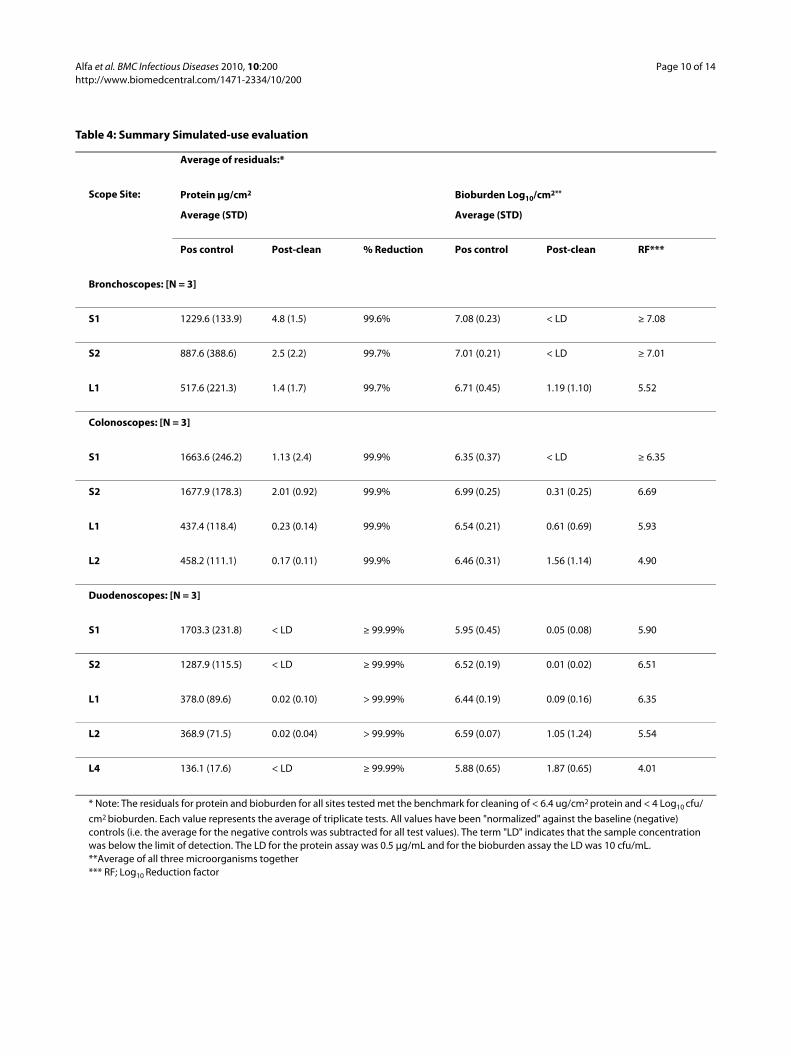
The range of negative control values confirmed thatthere were no abnormally high levels or protein, haemo-globin or bioburden in any of the simulated-use endo-scopes tested. The highest average level detected fornegative controls was 4.767 μg/cm2 protein for the S1(surface) of duodenoscopes, and 0.342 Log10cfu/cm2 forbioburden in the S1 sites of duodenoscopes. These dataconfirmed that despite the extensive soiling used for allchannels in this Phase II study that there was no detect-able "accumulation" of organic material or bioburdenwithin the channels over the course of the study. Table 4provides a summary of the simulated-use evaluationusing bronchoscopes, colonoscopes and duodenoscopes(all values have been normalized against the negativecontrols as described in the Materials and Methods Sec-tion). Simulated-use testing provides reproducible inocu-lation of all channels as well as the surface sites to betested. Unlike clinical-use testing it is possible to havepositive controls and to determine the percentage reduc-tion for protein residuals as well as the Log10 ReductionFactor (RF) for bioburden. The overall protein reduction
achieved by the ECR cleaning was > 99% and the overallbioburden RF was > 4.
Figure 2 provides a summary of the removal of biobur-den stratified by organism tested. Although the impact ofthe detergent may partly explain why P. aeruginosa wasthe most susceptible to removal, the data for the othertwo test organisms demonstrated that there was signifi-cant removal of microorganisms from both lumens andsurfaces as neither E. faecalis or C. albicans were affectedby the detergent yet they were effectively removed by theECR cleaning (4 to 7 Log10 reduction in bioburden).
DiscussionWhen the Endoscope Cleaner and Reprocessor (ECR)was cleared by the United States Food and Drug Admin-stration (FDA) and first marketed, it was the first com-mercially available endoscope cleaner and reprocessorthat had label claims regarding the efficacy of the auto-mated cleaning cycle that allowed elimination of manualcleaning. Despite the desire for automated cleaning ofpatient-used flexible endoscopes, there was concern thatthere were no published clinical-use studies for the ECR.The American Society for Gastrointestinal Endoscopy(ASGE) and Society of Gastroenterology Nurses and
Figure 1 Effect of CIDEZYME®-GI detergent on viability of E.faecalis, P.aeruginosa and C. albicans. The organisms were suspended in the use-dilution of the detergent of in tap water controls and at the timed intervals noted samples were taken to determine the level of viable bacteria.
Cidezyme-GI Killing Effect
0
1
2
3
4
5
6
7
Time 0 3 minute 5 minute 10 minute
Exposure Time
Lo
g10 c
fu/m
L
E.faecalis Tap water control P.aeruginosa Tap water control C.albicans Tap water control
E.faecalis Cidezyme-GI P.aeruginosa Cidezyme-GI C.albicans Cidezyme-GI

Alfa et al. BMC Infectious Diseases 2010, 10:200http://www.biomedcentral.com/1471-2334/10/200
Page 8 of 14
Table 2: Summary Clinical Use Evaluation
Scope Site: Average of residuals remaining post-cleaning*Average (Standard Deviation) [Range]
Protein μg/cm2 Hemoglobin μg/cm2 Bioburden Log10 cfu/cm2
Bronchoscopes: [N = 10]
S1 < LD < LD < LD
S2 0.32 (0.83) < LD 0.12 (0.26)
[0 - 2.62] [0 - 0.67]
L1 < LD < LD 0.03 (0.09)
[0-0.29]
Colonoscopes: [N = 15]
S1 0.26 (0.79) < LD 0.12 (0.20)
[0-2.94] [0 - 0.67]
S2 1.04 (2.47) < LD 0.02 (0.08)
[0-9.41] [0-0.33]
L1 0.12 (0.12) < LD 0.04 (0.07)
[0-0.32] [0-0.25]
L2 < LD < LD 0.0007 (0.022)
[0-0.008]
L3 < LD < LD 0.007 (0.023)
[0-0.09]
Duodenoscopes: [N = 15]
S1 0.03 (0.11) < LD 0.37 (0.94)
[0-0.44] [0-3.34]
S2 1.28 (3.80) < LD 0.14 (0.53)
[0-14. [0-2.04]
L1 0.21 (0.67) < LD 0.26 (0.97)
[0-2.60] [0-3.78]
L2 0.043 (0.09) < LD 0.11 (0.41)
[0-0.33] [0-1.58]
L4 0.06 (0.11) < LD 0.81 (1.35)
[0-0.31] [0-4.47]
Alfa et al. BMC Infectious Diseases 2010, 10:200http://www.biomedcentral.com/1471-2334/10/200
Page 9 of 14
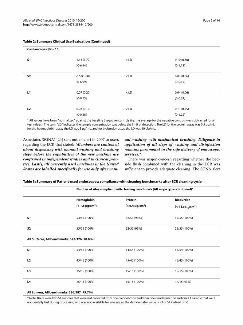
Associates (SGNA) [24] sent out an alert in 2007 to usersregarding the ECR that stated: "Members are cautionedabout dispensing with manual washing and brushingsteps before the capabilities of the new machine areconfirmed in independent studies and in clinical prac-tice. Lastly, all currently used machines in the UnitedStates are labelled specifically for use only after man-
ual washing with mechanical brushing. Diligence inapplication of all steps of washing and disinfectionremains paramount in the safe delivery of endoscopicservices."
There was major concern regarding whether the bed-side flush combined with the cleaning in the ECR wassufficient to provide adequate cleaning. The SGNA alert
Gastroscopes: [N = 15]
S1 1.14 (1.77) < LD 0.10 (0.30)
[0-6.44] [0-1.13]
S2 0.63(1.80) < LD 0.03 (0.06)
[0-6.99] [0-0.15]
L1 0.07 (0.20) < LD 0.04 (0.06)
[0-0.75] [0-0.24]
L2 0.03 (0.10) < LD 0.11 (0.35)
[0-0.38] [0-1.22]
* All values have been "normalized" against the baseline (negative) controls (i.e. the average for the negative controls was subtracted for all test values). The term "LD" indicates the sample concentration was below the limit of detection. The LD for the protein assay was 0.5 μg/mL, for the haemoglobin assay the LD was 5 μg/mL, and for bioburden assay the LD was 10 cfu/mL.
Table 2: Summary Clinical Use Evaluation (Continued)
Table 3: Summary of Patient-used endoscopes; compliance with cleaning benchmarks after ECR cleaning cycle
Number of sites compliant with cleaning benchmark (All scope types combined)*
Hemoglobin Protein Bioburden
(< 1.8 μg/cm2) (< 6.4 μg/cm2) (< 4 Log10/cm2)
S1 53/53 (100%) 52/53 (98%) 55/55 (100%)
S2 55/55 (100%) 52/55 (95%) 55/55 (100%)
All Surfaces, All benchmarks: 322/326 (98.8%)
L1 54/54 (100%) 54/54 (100%) 54/54 (100%)
L2 45/45 (100%) 45/45 (100%) 45/45 (100%)
L3 15/15 (100%) 15/15 (100%) 15/15 (100%)
L4 15/15 (100%) 15/15 (100%) 14/15 (93%)
All Lumens, All benchmarks: 386/387 (99.7%)
* Note: there were two S1 samples that were not collected from one colonoscope and from one duodenoscope and one L1 sample that were accidentally lost during processing and was not available for analysis so the demoninator value is 53 or 54 instead of 55.
Alfa et al. BMC Infectious Diseases 2010, 10:200http://www.biomedcentral.com/1471-2334/10/200
Page 10 of 14
Table 4: Summary Simulated-use evaluation
Average of residuals:*
Scope Site: Protein μg/cm2 Bioburden Log10/cm2**
Average (STD) Average (STD)
Pos control Post-clean % Reduction Pos control Post-clean RF***
Bronchoscopes: [N = 3]
S1 1229.6 (133.9) 4.8 (1.5) 99.6% 7.08 (0.23) < LD ≥ 7.08
S2 887.6 (388.6) 2.5 (2.2) 99.7% 7.01 (0.21) < LD ≥ 7.01
L1 517.6 (221.3) 1.4 (1.7) 99.7% 6.71 (0.45) 1.19 (1.10) 5.52
Colonoscopes: [N = 3]
S1 1663.6 (246.2) 1.13 (2.4) 99.9% 6.35 (0.37) < LD ≥ 6.35
S2 1677.9 (178.3) 2.01 (0.92) 99.9% 6.99 (0.25) 0.31 (0.25) 6.69
L1 437.4 (118.4) 0.23 (0.14) 99.9% 6.54 (0.21) 0.61 (0.69) 5.93
L2 458.2 (111.1) 0.17 (0.11) 99.9% 6.46 (0.31) 1.56 (1.14) 4.90
Duodenoscopes: [N = 3]
S1 1703.3 (231.8) < LD ≥ 99.99% 5.95 (0.45) 0.05 (0.08) 5.90
S2 1287.9 (115.5) < LD ≥ 99.99% 6.52 (0.19) 0.01 (0.02) 6.51
L1 378.0 (89.6) 0.02 (0.10) > 99.99% 6.44 (0.19) 0.09 (0.16) 6.35
L2 368.9 (71.5) 0.02 (0.04) > 99.99% 6.59 (0.07) 1.05 (1.24) 5.54
L4 136.1 (17.6) < LD ≥ 99.99% 5.88 (0.65) 1.87 (0.65) 4.01
* Note: The residuals for protein and bioburden for all sites tested met the benchmark for cleaning of < 6.4 ug/cm2 protein and < 4 Log10 cfu/cm2 bioburden. Each value represents the average of triplicate tests. All values have been "normalized" against the baseline (negative) controls (i.e. the average for the negative controls was subtracted for all test values). The term "LD" indicates that the sample concentration was below the limit of detection. The LD for the protein assay was 0.5 μg/mL and for the bioburden assay the LD was 10 cfu/mL.**Average of all three microorganisms together*** RF; Log10 Reduction factor

Alfa et al. BMC Infectious Diseases 2010, 10:200http://www.biomedcentral.com/1471-2334/10/200
Page 11 of 14
Figure 2 A summary of the simulated-use testing to assess the reduction in E. faecalis, P. aeruginosa and C. albicans after ECR cleaning. All results represent the average and standard deviation for three replicate experiments. S1 and S2 represent samples from 1 cm2 surface sites on the insertion tube and control head, respectively. L1, L2, L4 represent samples taken from the suction-biopsy, air-water and elevator guidewire channels, respectively. [Note; there were no Auxiliary water channels (L3) on the endoscopes used for simulated-use testing.]
Colonoscope - Simulated Use Data
0
1
2
3
4
5
6
7
8
S1 S2 L1 L2
Lo
g 1
0 c
fu/c
m2
E.faecalis Pos control P.aeruginosa Pos control C.albicans Pos control
E. faecalis clean only P. aeruginosa clean only C.albicans clean only
A)
Duodenoscope - Simulated Use Data
0
1
2
3
4
5
6
7
8
S1 S2 L1 L2 L4
Lo
g 1
0 c
fu/c
m2
E.faecalis Pos control P.aeruginosa Pos control C.albicans Pos control
E. faecalis clean only P. aeruginosa clean only C.albicans clean only
B)
B r o n c h o s c o p e - S i m u l a t e d U s e D a t a
0
1
2
3
4
5
6
7
8
S1 S2 L1
Lo
g10cfu/cm
2
E.�faecalis�Pos�control P.�aeruginosa�Pos�control C.�albicans�Pos�control
E.�faecalis�clean�only P.�aeruginosa�clean�only C.�albicans�clean�only
C)

Alfa et al. BMC Infectious Diseases 2010, 10:200http://www.biomedcentral.com/1471-2334/10/200
Page 12 of 14
recommended that sites continue performing manualcleaning until clinical-use data was published. Indeed thisconcern was reiterated in 2009 by SGNA [23] where theystated: "It is necessary to follow all steps for the manualcleaning of the endoscope prior to using an automatedreprocessor. No independent confirmatory data arecurrently available to show that automated reproces-sors are able to provide cleaning of endoscopes that iscomparable to that of manual washing and brushing."
The results of our study are the first published data toaddress this issue. Our clinical use study and simulated-use study clearly demonstrated that the cleaning processused by the ECR provided excellent removal of bothorganic material (protein and hemoglobin) and biobur-den from all flexible endoscopes evaluated. The clinicalstudy by Alfa et al [27] indicated that after complete man-ual cleaning (before HLD) the range of residual materialin the suction/biopsy channels of Olympus broncho-scopes, duodenoscopes and colonoscopes was 0 μg/cm2
to 4.40 μg/cm2 for hemoglobin, 3.51 to 8.55 μg/cm2 forprotein and 2.17 to 4.05 Log10/cm2 for bioburden.Although the endoscope reprocessing staff in the clinic atthe time the 1999 study [27] was conducted were follow-ing the Olympus flexible endoscope manufacturer'scleaning instructions they were likely not adhering to itcompletely as the average time required in the endoscopyclinic for cleaning flexible endoscopes ranged from 5minutes to 6.5 minutes [27] whereas research personnelwho timed all stages took between 14 minutes to 25 min-utes to clean the same scope types. At the time of the1999 [27] study, the endoscopy clinic cleaning protocolwas totally manual with no flushing pumps being used.
For simulated-use testing the residuals reported by Alfaet al [22] for optimal manual cleaning by research person-nel were lower (maximum residual protein was 4.46 μg/cm2 ) compared to the residuals (up to 8.55 μg/cm2 ) leftafter routine manual cleaning of patient-used flexibleendoscopes in a busy endoscopy clinic [27]. The datafrom the current study demonstrated that for patient-used flexible endoscopes the residuals for protein, hemo-globin and bioburden in the suction channel (L1) afterthe ECR cleaning are substantially better (99.7% met allbenchmarks) compared to optimal manual cleaning.Indeed it is clear from the data on manual cleaning ofpatient-used flexible endoscopes presented by Alfa et al[27] that after routine manual cleaning in an endoscopyclinic there are scopes that may have > 6.4 μg/cm2 of pro-tein, > 1.8 μg/cm2 of hemoglobin and > 4 Log10 cfu/cm2
bioburden remaining.The volume of fluid to be flushed through channels that
is required by endoscope manufacturers for the detergentand rinse stages of manual cleaning is large. Olympus rec-ommends 90 mLs per suction-biopsy channel for colono-
scopes and according to Puszko [12] 150 mLs per channelis recommended by the Australian endoscope reprocess-ing guidelines. For manual cleaning this requires staff touse syringes filled with fluid and repeated manual flush-ing that often leads to repetitive strain injuries [12]. Theuse of stand alone pumps to facilitate the flushing ofcleaning and rinsing fluid has become widely accepted incentres that can afford these pumps [12]. The use ofendoscope reprocessors with validated cleaning cycleswould greatly reduce the strain on staff and would stan-dardize the fluid volume being flushed.
Despite the overall low rates of infection associatedwith flexible endoscopy procedures, flexible endoscopesare still the most common cause of healthcare deviceassociated outbreaks [29]. Bisset et al [4] have showedthat the incidence of residual microbial contaminationfor patient ready gastroscopes and duodenoscopes was1.9% and 1.8% respectively. Furthermore, they indicatedthat the incidence of bioburden contamination increasedwith the number of times the endoscope was used. Thissuggests that biofilm may form over repeated uses andthis may be related to the wide variation in the manualcleaning performed [6,4]. The recent study by Alfa andHowie [30] modelling build-up biofilm as well as the oneby Vickery et al [31] using endoscope tubing both con-firm that repeated rounds of exposure to organic mate-rial, cleaning and HLD (complete reprocessing) result inan accumulation of material within the channel. The rateof accumulation is modulated by organic load [30] andtype of detergent used [17]. The review by Moses and Lee[6] reveal that only 70% of centers surveyed flusheddetergent through channels and used brushes for thechannels and valves (30% omitted manual cleaning andrelied on the AER to perform cleaning even though therewere no AERs at that time with FDA clearance for clean-ing cycles). Marion et al [32] suggest that periodic use ofbiofilm detaching agent may be needed to reduce the riskof biofilm accumulation in scope channels. These studiesall suggest that in healthcare there is widespread diffi-culty in achieving the endoscope manufacturers recom-mended manual cleaning and that great variability existsin the manual cleaning currently being performed.
The value of AERs with validated cleaning combinedwith efficient HLD and final rinsing cannot be overem-phasized. However, not all AER cleaning cycles are equiv-alent as many of the "cleaning" cycles in commerciallyavailable AERs do not have FDA cleared cleaning claimsand therefore still require manual cleaning prior to pro-cessing in the AER. As indicated by Alfa and Howie [30]when an aldehyde is used for HLD (as is the case for ECR)it becomes critical that the cleaning needs to be thoroughand reproducible to reduce the risk of microbial survivalin accumulated organic material. Our data suggest that

Alfa et al. BMC Infectious Diseases 2010, 10:200http://www.biomedcentral.com/1471-2334/10/200
Page 13 of 14
because of the efficacy of the ECR cleaning cycle and thefact that the cycle cannot be "shortened" by the user, thepotential for build-up biofilm would likely be reducedcompared to manual cleaning where short-cuts in clean-ing are common.
The cleaning efficiencies achieved by the AdaptaScopeand LS 2000 AERs tested by Kircheis and Martiny [33]using endoscope channel tubing indicated that efficientmicrobial removal was only achieved when direct chan-nel connection was used. The two "pressure chamber"models of AERs evaluated could not achieve flow throughof the inoculated tubing therefore there was no biobur-den reduction. The ECR evaluated in the current studyuses direct channel connection and the data from oursimulated-use study demonstrated that the RF achievedby the cleaning cycle alone was > 6 for all surface sites and> 4.5 for all air/water, suction/biopsy and auxillary waterchannels for all types of flexible endoscopes. This furtherextends Kircheis and Martiny's [33] work on tubing seg-ments to demonstrate how efficient cleaning can beachieved in the various channels and surface of flexibleendoscopes. The ECR was also able to achieve an entero-coccus RF of > 4 for the elevator guidewire channel whichpresents unique challenges to cleaning which were notaddressed in Kircheis and Martiny's [33] study. Indeedthis RF of 4.01 achieved by the ECR cleaning cycle issuperior to a previous report [22] for the Reliance AER(RF of 2.10) cleaning as well as for complete manualcleaning (RF of 3.22). The cleaning efficacy achievedusing an AER with "boot pressure chamber" technologyversus "direct channel connection" technology willdepend entirely on the manufacturer's specifications forthat particular machine. Both technologies have advan-tages and disadvantages. Direct channel connectionallows for monitoring of individual channel flow(although not all AERs with channel connections providethis option) whereas this is not currently available for"boot pressure chambers". Boot pressure chamber over-comes possible user errors with channel connection mix-ups that may occur with traditional AERs that use directchannel connections. Users need to determine whichAER process best suites their needs and should thor-oughly assess the validation data provided for any AERthey are considering.
It should be noted that it is difficult to assess the effectsof lumen anomalies such as scratches or biofilm buildupon the efficacy of either manual or ECR cleaning. How-ever, the in-use study using patient-used endoscopes doesprovide valuable data on cleaning efficacy for devicesknown to have been exposed to normal wear and tearwithin the clinical setting.
Although only Olympus flexible endoscopes wereincluded in the current study, the data from this study
provides "proof of concept" that the cleaning technologyof the ECR is adequate. In the study by Alfa et al [22]where Olympus, Pentax and Fujinon endoscopes wereevaluated [22] the AER cleaning cycle showed similarcleaning efficiencies regardless of which manufacturer'sendoscopes were tested. In addition, as the ECR manu-facturer completes validation of different scope types andmodels they will be added to their list of validated endo-scopes that can be processed through the ECR.
ConclusionsIn summary the data presented for the simulated-usetesting and clinical use testing clearly demonstrate thatthe cleaning cycle does provide reliable organic andbioburden removal from 98.8% of surfaces and 99.7% oflumens for the bronchoscopes, duodenoscopes, gastro-scopes and colonoscopes that were tested. The ECRcleaning for endoscope surfaces and channels is superiorto optimal manual cleaning providing all the ECR manu-facturer's specific instructions are followed (including thetype of enzymatic detergent used in the ECR). Althoughonly Olympus flexible endoscopes were evaluated in thisstudy it does provide stringent proof of concept for thecleaning phase of the ECR complete reprocessing cycle(cleaning and HLD). This cleaning efficiency for patient-used endoscopes is only validated for elective procedures(not emergency endoscopies) and only if the bedside pre-cleaning is done and the transit time is less than an hourprior to placing the endoscope into the ECR.
Competing interestsThe ECR and all funds for this study were provided by Advanced SterilizationProducts, a Division of Ethicon, a Johnson and Johnson company. MJA hasundertaken contract research projects (unrelated to the current publication)for a variety of companies including; 3M, bioMerieux, Olympus, Case Medical,Intelligent Hospital Systems, Johnson & Jonson, Novaflux, STERIS and Virox. Nomonies from the current or past research contracts have gone to MJA, theywere administered through the St. Boniface Research Centre and were used forresearch related expenses only. MJA has been a sponsored conference speakerand she has acted as a paid consultant for preparation of a one time literaturereview for STERIS in 2003 and for providing a one time educational microbiol-ogy workshop for 3M staff in 2006.PD, NO and IF have not acted as consultants and have no financial or other linkwith any company.
Authors' contributionsAll authors have read and approved the final manuscript. MJA was responsiblefor the conception of the research project and provided direction for theexperimental testing. Writing of the protocols, performing sample collection,all testing and data entry were performed by PD, NO and IF. MJA was responsi-ble for data analysis and manuscript preparation with input from PD, NO and IF.No company played any role in the gathering or preparation of data. AdvancedSterilization Products provided input on the study design and editing of themanuscript.
AcknowledgementsThe staff (in particular Brice Morin) in the endoscopy clinic and the OR repro-cessing area at the St. Boniface General Hospital are acknowledged for their role in accommodating the clinical use testing.
Alfa et al. BMC Infectious Diseases 2010, 10:200http://www.biomedcentral.com/1471-2334/10/200
Page 14 of 14
Author Details1Diagnostic Services of Manitoba, 409 Tache Avenue, Winnipeg, MB, R2H 2A6, Canada, 2St. Boniface Research Centre, 351 Tache Avenue, Winnipeg, MB, R2H 2A6, Canada and 3University of Manitoba, Winnipeg, MB, 745 Bannatyne Avenue, Room 543 Basic Medical Sciences Building, R3E 0J9, Canada
References1. (AORN) AORN: Perioperative Standards and Recommended Practices.
American Operating Room Nurses (AORN) 2008:421-445.2. Rutala WA, Weber DJ: Reprocessing endoscopes: United States
perspective. J Hosp Infect 2004, 56(Suppl 2):S27-39.3. Canadian Standards Association Inc. Publishers M, ON: CSA Z314.8-08
Decontamination of Reusable Medical Devices. 2008.4. Bisset L, Cossart YE, Selby W, West R, Catterson D, O'Hara K, Vickery K: A
prospective study of the efficacy of routine decontamination for gastrointestinal endoscopes and the risk factors for failure. Am J Infect Control 2006, 34(5):274-280.
5. Mehta AC, Prakash UB, Garland R, Haponik E, Moses L, Schaffner W, Silvestri G: American College of Chest Physicians and American Association for Bronchology [corrected] consensus statement: prevention of flexible bronchoscopy-associated infection. Chest 2005, 128(3):1742-1755.
6. Moses FM, Lee JS: Current GI endoscope disinfection and QA practices. Dig Dis Sci 2004, 49(11-12):1791-1797.
7. Nelson DB, Muscarella LF: Current issues in endoscope reprocessing and infection control during gastrointestinal endoscopy. World J Gastroenterol 2006, 12(25):3953-3964.
8. Association for the Advancement of Medical Instrumentation: ANSI/AAMI ST35: Safe handling and biolgoical decontamination of medical devices in health care facilities and in nonclinical settings. 1996.
9. Pajkos A, Vickery K, Cossart Y: Is biofilm accumulation on endoscope tubing a contributor to the failure of cleaning and decontamination? J Hosp Infect 2004, 58(3):224-229.
10. Rutala WW, DJ; and the Healthcare Infection Control Practices Advisory Committee (HICPAC): Guideline for Disinfection and Sterilization in Healthcare facilities. Centre for Disease Control 2008.
11. Spach DH, Silverstein FE, Stamm WE: Transmission of infection by gastrointestinal endoscopy and bronchoscopy. Ann Intern Med 1993, 118(2):117-128.
12. Puszko GB: Manual cleaning of endoscopes: a comparison study of syringe versus suction methods using the endo-suction cleaning system. Gastroenterol Nurs 2001, 24(2):69-74.
13. Agerton T, Valway S, Gore B, Pozsik C, Plikaytis B, Woodley C, Onorato I: Transmission of a highly drug-resistant strain (strain W1) of Mycobacterium tuberculosis. Community outbreak and nosocomial transmission via a contaminated bronchoscope. JAMA 1997, 278(13):1073-1077.
14. Bronowicki JP, Venard V, Botte C, Monhoven N, Gastin I, Chone L, Hudziak H, Rihn B, Delanoe C, LeFaou A, et al.: Patient-to-patient transmission of hepatitis C virus during colonoscopy. N Engl J Med 1997, 337(4):237-240.
15. Ahishali E U-BO, Dalopcioglu D: Chemical colistis due to glutaraldehyde: Case series and review of the literature. Dig Dis Sci 2008 in press.
16. Nelson DB: Recent advances in epidemiology and prevention of gastrointestinal endoscopy related infections. Curr Opin Infect Dis 2005, 18(4):326-330.
17. Vickery K, Ngo QD, Zou J, Cossart YE: The effect of multiple cycles of contamination, detergent washing, and disinfection on the development of biofilm in endoscope tubing. Am J Infect Control 2009, 37(6):470-475.
18. Zuhlsdorf B, Emmrich M, Floss H, Martiny H: Cleaning efficacy of nine different cleaners in a washer-disinfector designed for flexible endoscopes. J Hosp Infect 2002, 52(3):206-211.
19. Zuhlsdorf B, Martiny H: Intralaboratory reproducibility of the German test method of prEN ISO 15883-1 for determination of the cleaning efficacy of washer-disinfectors for flexible endoscopes. J Hosp Infect 2005, 59(4):286-291.
20. Zuhlsdorf B, Winkler A, Dietze B, Floss H, Martiny H: Gastroscope processing in washer-disinfectors at three different temperatures. J Hosp Infect 2003, 55(4):276-282.
21. Mean M, Mallaret MR, Bichard P, Shum J, Zarski JP: Gastrointestinal endoscopes cleaned without detergent substance following an automated endoscope washer/disinfector dysfunction. Gastroenterol Clin Biol 2006, 30(5):665-668.
22. Alfa MJ, Olson N, DeGagne P: Automated washing with the Reliance Endoscope Processing System and its equivalence to optimal manual cleaning. Am J Infect Control 2006, 34(9):561-570.
23. Society of Gastrointestinal and Associates IP: SGNA Standards and Guidelines: Standards of Infection Control in Reprocessing of Flexible Gastrointestinal Endoscopes. 2009.
24. Society of Gastrointestinal and Associates IP: SGNA Standards and Guidelines: Standards of Infection Control in Reprocessing of Flexible Gastrointestinal Endoscopes. 2007.
25. Association for the Advancement of Medical Instrumentation: AAMI TIR30: A compendium of processes, materials, test methods, and acceptance criteria for cleaning reusable medical devices. 2003.
26. Alfa M, DeGagne P, Olson N: Validation of ATS as an appropriate test soil. Zentr Steril (Central Service) 1999, 7:68-382.
27. Alfa MJ, Degagne P, Olson N: Worst-case soiling levels for patient-used flexible endoscopes before and after cleaning. Am J Infect Control 1999, 27(5):392-401.
28. US Department of Health and Human Services PHS, Food and Drug Administration Center for Devices and Radiologic Health: FDA Reviewer Guidance on the content and format of premarket notification 510(k) submissions for washers and washer-disinfectors. 1998.
29. Gillespie EE, Kotsanas D, Stuart RL: Microbiological monitoring of endoscopes: 5-year review. J Gastroenterol Hepatol 2008, 23(7 Pt 1):1069-1074.
30. Alfa MJ, Howie R: Modeling microbial survival in buildup biofilm for complex medical devices. BMC Infect Dis 2009, 9:56.
31. Vickery K, Pajkos A, Cossart Y: Removal of biofilm from endoscopes: evaluation of detergent efficiency. Am J Infect Control 2004, 32(3):170-176.
32. Marion K, Freney J, James G, Bergeron E, Renaud FN, Costerton JW: Using an efficient biofilm detaching agent: an essential step for the improvement of endoscope reprocessing protocols. J Hosp Infect 2006, 64(2):136-142.
33. Kircheis U, Martiny H: Comparison of the cleaning and disinfecting efficacy of four washer-disinfectors for flexible endoscopes. J Hosp Infect 2007, 66(3):255-261.
Pre-publication historyThe pre-publication history for this paper can be accessed here:http://www.biomedcentral.com/1471-2334/10/200/prepub
doi: 10.1186/1471-2334-10-200Cite this article as: Alfa et al., EVOTECH® endoscope cleaner and reprocessor (ECR) simulated-use and clinical-use evaluation of cleaning efficacy BMC Infectious Diseases 2010, 10:200
Received: 19 October 2009 Accepted: 9 July 2010 Published: 9 July 2010This article is available from: http://www.biomedcentral.com/1471-2334/10/200© 2010 Alfa et al; licensee BioMed Central Ltd. This is an Open Access article distributed under the terms of the Creative Commons Attribution License (http://creativecommons.org/licenses/by/2.0), which permits unrestricted use, distribution, and reproduction in any medium, provided the original work is properly cited.BMC Infectious Diseases 2010, 10:200