open access journal of agricultural research · open access journal of agricultural research ......
TRANSCRIPT

Open Access Journal of Agricultural Research ISSN: 2474-8846
The Functional State of Animals Blood under the Electromagnetic Radiation Influence at Millimeter Range J Agri Res
The Functional State of Animals Blood under the
Electromagnetic Radiation Influence at Millimeter Range
Avdeenko VS*, Krenitskiy AP, Avdeenko AV, Molchanov, Rykhlov
AS and Kyzerjvenkov MA
Federal State Budgetary Educational Institution of Higher Professional Education,
Vavilov Saratov State Agrarian University
*Corresponding author: Avdeenko VS, Federal State Budgetary Educational Institution of Higher Professional
Education, Vavilov Saratov State Agrarian University, Email: [email protected]; [email protected]
Abstract
The electromagnetic EHF field at frequencies of molecular absorption atmospheric oxygen spectrum in vitro that effects
on the functional activity of animals’ blood was first studied. The change in the number of erythrocytes and their
properties was founded. The homogenate effect of electromagnetic radiation at EHF-mm range (129 - 150 GHz) on
erythrocytes which contact with atmospheric oxygen was founded. Electromagnetic EHF waves at frequencies of
molecular spectrum of oxygen absorption and emission that influence on the functional activity of animals’ blood
homeostasis in vitro were considered. Significant changes of rheological and electrolytic blood properties under the
electromagnetic radiation influence were studied. The mechanisms of the therapeutic EHF effects which influence on
animals body through peripheral blood are discussing.
Keywords: Electromagnetic radiation at EHF-mm range; Neutrophils; Erythrocytes; Homeostasis
Introduction
Theoretical and experimental research on the physics of information interaction of EHF waves with biological environments show the effectiveness of their use in the EHF therapy [1]. Functioning features of the receptor structures and intracellular links coupling are largely determined by the cell homeostasis and the nature of its response to endogenous and exogenous incentives [2]. It is known that neutrophils express different receptors on their membrane [3]. From this standpoint the system of polymorphonuclear leukocytes is of great interest. Their role is not limited in phagocytosis participation and nonspecific resistance ensuring [4] but is regarded as universal homeostasis effector [5]. The possible use of EHF electromagnetic radiation at frequencies of molecular absorption spectrum of atmospheric gases in EHF therapy proposed in the project [6].
It became possible to discover changes in the noise signal structure interacting with the bio object due to additional amplitude, frequency and phase fluctuations [3-6] when we were analyzing the EHF mechanisms influenced on bio objects and their interaction with electromagnetic fields. The fluctuations which influence on the individual body systems allow identifying direct participation of studied systems in the EHF influence effect realization [7-9]. Currently, the data that show the possibility of EHF generation by body cells are obtained [10,11] and convincing evidence of "information interaction" in the biological objects system [12,13] are presented. It was proved that electromagnetic radiation of millimeter range effects on the hemorheology of patients with stable
Research Article
Volume 1 Issue 3
Received Date: August 30, 2016
Published Date: December 09, 2016

Open Access Journal of Agricultural Research
Avdeenko VS, et al. The Functional State of Animals Blood under the Electromagnetic Radiation Influence at Millimeter Range. J Agri Res 2016, 1(3): 000119.
Copyright© Avdeenko VS, et al.
2
angina forms [14-16]. The most convenient method for animals’ treatment is to use special EHF therapy. This method based on the selective increase of gases reactivity (including medicinal aerosols and atmospheric gases such as oxygen) in the livestock buildings. A pumping through the EHF radiation at frequencies of their molecular emission and absorption spectra is used for molecular excitation gases. The introduction of these gases in the respiratory system occurs when animals breathing gases excited by EHF radiation. This treatment method requires special quasi-optical EHF - AERO equipment. In addition, it is necessary to carry out research of EHF waves affects on biological systems and air gases contacting with them. OJSC “Central Research Institute of Measuring Equipment” (Saratov) and Federal State Budgetary Educational Institution of Higher Professional Education “Saratov State Agrarian University named after N.I.Vavilov” carried out work that show how EHF waves at frequencies of molecular absorption spectrum of atmospheric oxygen affect on animal blood functional parameters. Experiments were carried out in vitro through the oxygen and blood contact. When we were studying the interaction of electromagnetic EHF radiation and erythrocytes in vitro we got clear evidence of its effect on oxygen transport erythrocytes` function [2,3,6]. The quasi-optical generator working at the frequency of molecular spectra of atmospheric gases emission was made by OJSC “Central Research Institute of Measuring Equipment”. This generator operates at the frequency range from 53.7 to 270 GHz and allows simulating the interaction between physical and biological environments and electromagnetic fields with different structure (modes and polarizations, spectral composition, types of amplitude and frequency modulation) [7]. The quasi-optical programmable generator allows exciting ТМоm, ТЕоm, ЕНnm waves in the space and creating linearly polarized E, H waves and waves with circular (right or left) polarization. The generator can automatically form the frequencies’ arrays and molecular amplitudes spectra of atmospheric gases according to the Poisson and Gauss laws or the frontal law "1/f". We chose the frequency of 129 and 150 GHz as the frequency of atmospheric oxygen intense absorption and resonant interaction with the water component of animals’ blood. We proposed to use the quasi-optical generator which forms the fundamental mode on a hybrid ЕН11wave providing rotation of field E and H vectors for the EHF field formation which affecting on erythrocytes. All this is
used for more effective information impact on molecular processes in blood in vitro which generates molecular emission and absorption spectrum of substances involved in metabolism the structure of which includes waves of different types and polarization. In this regard, our goal was to study the electromagnetic EHF vibrations at the frequencies of molecular spectrum of atmospheric oxygen absorption that influence on the animals’ blood functional state (in vitro) upon contact with oxygen.
Material and Methods
Experiments were carried out on Wistar rats to investigate the effect of EHF electromagnetic field at the molecular absorption and emission spectrum of atmospheric oxygen on the enzymatic status, metabolic profile and neutrophils bactericidal systems’ status. Metabolic enzyme activity and microbicidal systems state by means of cytochemical methods were determined by the following indicators in neutrophils: glycogen content by J. McManus; lipid by Hil. Sbeeban; lysosomal cationic proteins (LCP) by VE Pigarevskiy; ATPase activity by M. Wachstein; myeloperoxidase (MPO) by R. Graham; succinate dehydrogenase (SD) by R.P.Nartsissov; NBT test by BS Nagoeva’s modification. Smears were prepared from control and experimental blood samples in each experiment’s series next they were fixed and stained using a binocular microscope (15x40) and cytochemical parameters were studied. In assessing the results of cytochemical study we used the G. Astaldi and L. Verga’s principle based on the identification of varying specific color intensity. One hundred polymorphonuclear leukocytes were counted in each blood smear. The results were analyzed as a percentage of positively reacting cells (PRC) and medium cytochemical coefficient (MCC). Samples of whole blood and blood serum of 15 clinically healthy lactating cows were used to investigate how electromagnetic EHF fields of molecular absorption and emission spectrum of atmospheric oxygen affect on the erythrocyte membrane’s behavior using in vitro technology. Arterial blood was taken with heparin syringe balanced by electrolyte in an amount of 5.0 ml per animal which was stabilized by trisodium citrate (3.8%). One blood dose was centrifuged at 1500 rpm for 15 min supernatant layer was removed and the remaining erythrocyte mass was twice washed and resuspended in isotonic saline (0.8 % NaСl) in the amount providing the

Open Access Journal of Agricultural Research
Avdeenko VS, et al. The Functional State of Animals Blood under the Electromagnetic Radiation Influence at Millimeter Range. J Agri Res 2016, 1(3): 000119.
Copyright© Avdeenko VS, et al.
3
same concentration of erythrocytes as well as in intact blood. Erythrocyte suspension was divided into two parts: one was used for control and the second was subjected to electromagnetic EHF influence on the 129 - 150 GHz frequency. Pilot samples were placed in a quasi-optical load of pan-spectrometric quasi-optical measurement system with the access of atmospheric oxygen to the samples through a quasi-optical path. The accuracy of frequency setting by using wave meter measurement system was ± 0.1 GHz. Quasi-optical beam guide with tested blood was excited by field with the rotating field vector IN to enhance the molecular interaction. The power density of 10-3 W/cm² of the irradiation was set to within ± 15%. Whole blood and erythrocyte suspension was placed in a cone-shaped cell made of EHF transparent material. The cell was attached to cone quasi-optical matched load shielded from external fields absorbing all incoming power through beam guide and power’s VSWR <1.05 (voltage standing wave ratio). The inner load surface consisted of absorbent material. The cell was facing its conical surface toward the power supply ensuring minimal reflection [3]. Contact time with the blood samples was 15, 30 and 90 minutes. Shielding from external electromagnetic radiation was carried out by using a metal foil. Number of erythrocytes, erythrocyte sedimentation rate, hemoglobin concentration, hematocrit, osmotic resistance of erythrocytes, electrophoretic mobility of erythrocytes (EME), lipid peroxidation (LPO) in the level of malondialdehyde, yield strength, erythrocyte aggregation and its coefficient counted according to conventional techniques were determined in control and experimental blood samples. Determination of metabolic parameters was carried out using the CIBA-CORNING Z88 BLOOD CAS SYSTEM analyzer (made in USA). Hematologic studies were carried using the НС1020 НУGЕL НD27 НУСЕL (USA). The statistical analysis was performed by using parametric (t-Student's test) and nonparametric methods (Uilkonson - Manny - Whitney, X²). The calculation was performed using the software Stat graphics package installed on a personal computer.
Results and Discussion
When ND Devyatkov and co-authors (1987) were analyzing the available data about changes in the cell under the influence of electromagnetic radiation of millimeter range they concluded that structural and functional changes of membrane structures of cells and
intracellular organelles underline the biological action of this effect. It is known that normally neutrophil working in a limited metabolic support, glucose comes from outside and the content of glycogen granules is practically unchanged [17]. Oxidase activation is the biochemical basis of respiratory burst. In this case, nicotinamide adenine dinucleotide phosphate unsubstituted oxidase (NADPHU-oxidase) of phagocytes is a multi component membrane binding enzyme that catalyzes the single-electron cleavage of molecular oxygen to the superoxide anion [18]. 202 + НАДФН2 - 02 + НАДФ+ + Н+ (1) It consumes NADPHU, which is used by neutrophils oxidase as a physiological substrate for respiratory burst. Glucose is oxidized in reactions and again desoxydate the NADPH and NADPHU [19]. Our studies show that 15 minute irradiation of rats’ blood (Table 1) reduces glycogen and lipids content that indicating increased cells’ energy expenditure.
Cytochemical indicator
Control Contact time, min 15 30
Glycogen 1.40 ± 0.06 1.15 ± 0.05** 1.90 ± 0.03*
Lipids 1.88 ± 0.05 1.68 ± 0.02 2.12 ± 0.05*
ATPase 1.74 ± 0.06 1.96 ± 0.05 2.00 ± 0.06*
SD 1.42 ± 0.03 1.98 ± 0.06* 1.88 ± 0.05
MPO 1.36 ± 0.09 1.40 ± 0.05 2.03 ± 0.08*
LSP 1.45 ± 1.06 2.26 ± 0.05** 2.00 ± 0.05*
NBT test 1.60 ± 0.05 1.92 ± 0.05 1.58 ± 0.05
Note: р<0, 05; ** р<0<01. Here and further.
Table 1: Changes of neutriphils’ cytochemical indicators obtained by using electromagnetic irradiation in peripheral rats’ blood in vitro (MCC)
Increased SD activity in the Krebs cycle can be regarded as a marker of neutrophil metabolism amplification of synthetic processes requiring energy supply (Table 2) [20].
Cytochemical indicator
Control Contact time, min
15 30
Glycogen 96.36 ± 1.40 88.21 ± 1.52* 94.16 ± 1.52
Lipids 97.52 ± 0.32 93.22 ± 0.91 97.39 ± 0.64
ATPase 98.82 ± 0.46 97.22 ± 0.99 98.22 ± 0.62
SD 79.80 ± 2.02 90.49 ± 2.03* 83.92 ± 2.07
MPO 100 99.89 ± 0.44 100

Open Access Journal of Agricultural Research
Avdeenko VS, et al. The Functional State of Animals Blood under the Electromagnetic Radiation Influence at Millimeter Range. J Agri Res 2016, 1(3): 000119.
Copyright© Avdeenko VS, et al.
4
LSP 74.92 ± 2.82 88.22 ± 2.86 90.99 ± 1.92*
NBT test 82.70 ± 1.30 86.88 ± 1.62 74.0 ± 1.95*
Table 2: Changes of neutriphils’ cytochemical indicators obtained by using irradiation EMR EHF Molecular Absorption and Emission Spectrum (MAES) of O₂ in peripheral rats’ blood in vitro (PRC). LSP rise indicates an increase in leukocytes’ microbial capacity. Registered rise of NBT test indicates the activation of membrane oxygen dependent processes associated with activation of NADPHU-oxidase under the influence of electromagnetic radiation of millimeter range and reactions interfaced with it. During 30 minute irradiation of rats’ blood in vitro NBT test parameters changed as follows: the number of positive cells was significantly increased, and the average cytochemical coefficient was not different from the control blood samples. ATPase activity did not change. All other investigated cytochemical neutrophils’ indicators increased after 30 minutes irradiation that promoted the accumulation of intracellular energy substrates such as glycogen and lipids and enzyme activation of SD and MP and cationic protein content increased neutrophil lysosomes. Consequently, the experiments showed that the 15 minutes rats’ blood irradiation caused a decrease in intracellular glycogen reserves as evidenced by decline of MCC by 19.5% (p<0.05) and PRC by 12.7% (p<0.01). Neutrophils’ saturation by intracellular lipids also reduced by 10.9 % (p<0.05) while positively reacting cells percentage was reducing. The SD activity increased sharply by 141% (p<0.001) from the control level.MPO and ATPase activity remained unchanged after 15 min irradiation of rats’ blood. LCP content in the cell increased by almost 1.5 times (p<0.001) and the percentage of positively reacting cells increased by 17.5% (p<0.01). NBT test parameters increased: the average cytochemical coefficient increased by 22.3% (p<0.01) and the number of positively reacting cells increased by 6.2% (p<0.05) while oxygen independent mechanisms of neutrophil bactericidal activity were stepping up. Doubling time (30 minutes irradiation of rats’ blood in vitro) increased the number of glycogen and lipids in the cell (Figure1). The lipid content in neutrophils was increased by 28.7% (p<0.01) after electromagnetic irradiation of blood in vitro, and the intracellular glycogen content increased by 27.2% (p<0.01) at a constant percentage of positively reacting cells. Dehydrogenation
process of succinic acid in the Krebs cycle significantly activated by 30 minute irradiation of rats’ blood: SD activity increased by 37.2% (p<0.01). At the same time myeloperoxidase activity increased at the 100% constant number of positively reactive cells and the mean value of medium cytochemical coefficient increased by more than 1.5 times (p<0.01). Rats’ blood irradiation causes a sharp activation of oxygen independent systems of neutrophil bactericidal activity as evidenced by increased number of cells containing lysosomal cationic proteins granules by 40,0% (p<0.001). The number of cells giving a positive reaction to NBT test at constant medium cytochemical coefficient reduced by 9.0% (p<0.05) after 30 minutes irradiation [21-23]. Peripheral blood neutrophils are highly reactive cells and respond quickly to the impact of electromagnetic irradiation at frequencies of molecular absorption and emission spectra of atmospheric oxygen [24]. Irradiation of animals’ blood in vitro alters the content of energy substrates, enzyme activity and state of oxygen dependent and oxygen independent bactericidal systems in polymorphonuclear leukocytes. The influence of electromagnetic waves on neutrophils depends on exposure: 15 minute blood irradiation in vitro causes stimulation of glycogen metabolism and lipid levels, increased activity of succinate dehydrogenase, an increase in lysosomal cationic proteins content and activation of oxygen dependent processes in the membrane of neutrophils; 30 minute irradiation followed by increasing levels of energy substrates and lysosomal cationic proteins, increased activity of succinate dehydrogenase and myeloperoxidase in neutrophils and reducing number of NBT positive cells. Our experiments with animal blood (in vitro) show that erythrocytes’ number significantly reduced from 6, 88 ± 0,36 to 4,97 ± 0,49 -10¹²/L (p<0,05 ) after experimental electromagnetic irradiation effect. Erythrocytes’ number varies depending on contact time reaching a minimum (5,02 ± 0,47 1012/L) during 90 minutes (Table 3).
Groups Erythrocytes’
number 1012/L
Erythrocytes’ diameter, µm
Мin Мах
Control 6.88 ± 0.36 3.51 ± 0.41 5.35 ± 0.29
Experimental, 15 min
4.97 ± 0.49 ** 4.56 ± 0.37 * 7.97 ± 0.68 **
Experimental, 30 min
5.02 ± 0.47** 4.71 ±0.29* 8.05 ± 0.47**
Open Access Journal of Agricultural Research
Avdeenko VS, et al. The Functional State of Animals Blood under the Electromagnetic Radiation Influence at Millimeter Range. J Agri Res 2016, 1(3): 000119.
Copyright© Avdeenko VS, et al.
5
Experimental, 90 min
3.98 ± 0.62** 4.96 ± 0.35* 8.34 ± 0.53**
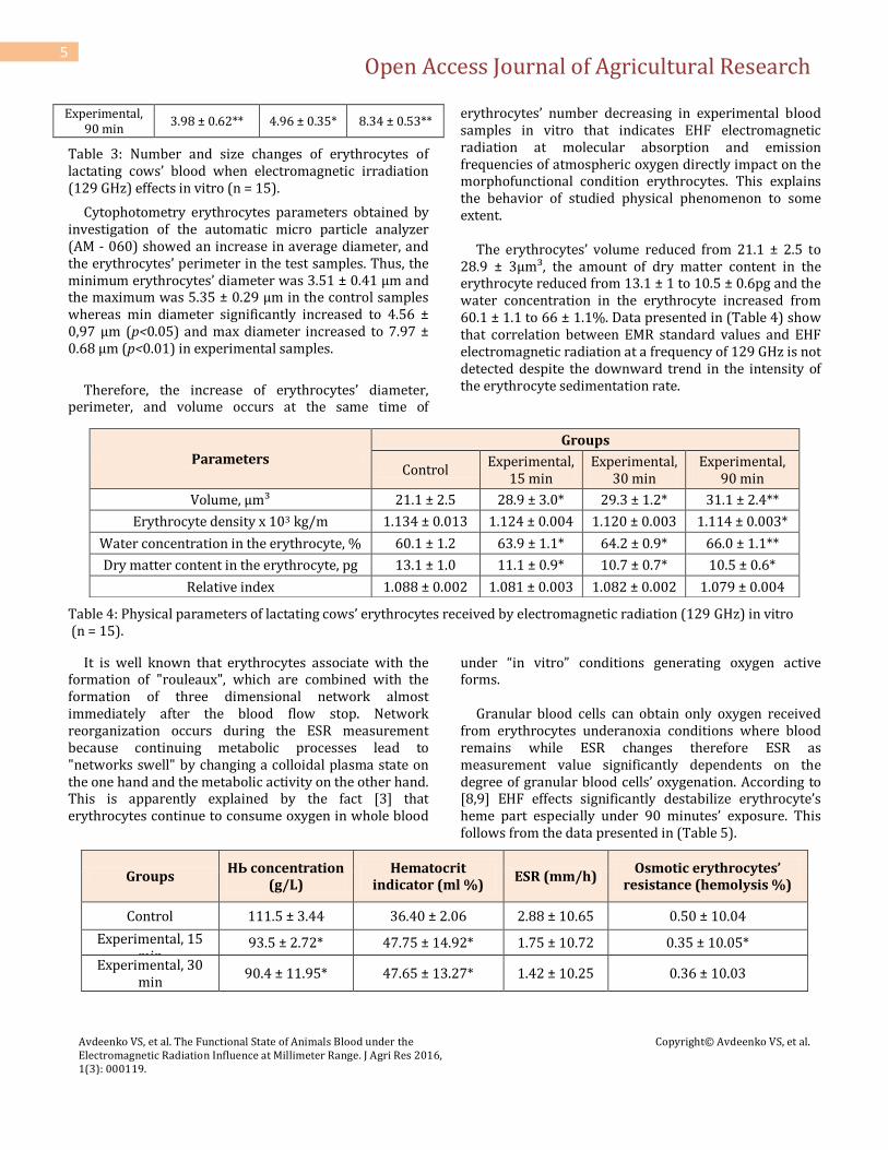
Table 3: Number and size changes of erythrocytes of lactating cows’ blood when electromagnetic irradiation (129 GHz) effects in vitro (n = 15).
Cytophotometry erythrocytes parameters obtained by investigation of the automatic micro particle analyzer (AM - 060) showed an increase in average diameter, and the erythrocytes’ perimeter in the test samples. Thus, the minimum erythrocytes’ diameter was 3.51 ± 0.41 µm and the maximum was 5.35 ± 0.29 µm in the control samples whereas min diameter significantly increased to 4.56 ± 0,97 µm (p<0.05) and max diameter increased to 7.97 ± 0.68 µm (p<0.01) in experimental samples.
Therefore, the increase of erythrocytes’ diameter, perimeter, and volume occurs at the same time of
erythrocytes’ number decreasing in experimental blood samples in vitro that indicates EHF electromagnetic radiation at molecular absorption and emission frequencies of atmospheric oxygen directly impact on the morphofunctional condition erythrocytes. This explains the behavior of studied physical phenomenon to some extent. The erythrocytes’ volume reduced from 21.1 ± 2.5 to 28.9 ± 3µm³, the amount of dry matter content in the erythrocyte reduced from 13.1 ± 1 to 10.5 ± 0.6pg and the water concentration in the erythrocyte increased from 60.1 ± 1.1 to 66 ± 1.1%. Data presented in (Table 4) show that correlation between EMR standard values and EHF electromagnetic radiation at a frequency of 129 GHz is not detected despite the downward trend in the intensity of the erythrocyte sedimentation rate.
Table 4: Physical parameters of lactating cows’ erythrocytes received by electromagnetic radiation (129 GHz) in vitro (n = 15).
It is well known that erythrocytes associate with the formation of "rouleaux", which are combined with the formation of three dimensional network almost immediately after the blood flow stop. Network reorganization occurs during the ESR measurement because continuing metabolic processes lead to "networks swell" by changing a colloidal plasma state on the one hand and the metabolic activity on the other hand. This is apparently explained by the fact [3] that erythrocytes continue to consume oxygen in whole blood
under “in vitro” conditions generating oxygen active forms. Granular blood cells can obtain only oxygen received from erythrocytes underanoxia conditions where blood remains while ESR changes therefore ESR as measurement value significantly dependents on the degree of granular blood cells’ oxygenation. According to [8,9] EHF effects significantly destabilize erythrocyte’s heme part especially under 90 minutes’ exposure. This follows from the data presented in (Table 5).
Groups НЬ concentration
(g/L) Hematocrit
indicator (ml %) ESR (mm/h)
Osmotic erythrocytes’ resistance (hemolysis %)
Control 111.5 ± 3.44 36.40 ± 2.06 2.88 ± 10.65 0.50 ± 10.04
Experimental, 15 min
93.5 ± 2.72* 47.75 ± 14.92* 1.75 ± 10.72 0.35 ± 10.05*
Experimental, 30 min
90.4 ± 11.95* 47.65 ± 13.27* 1.42 ± 10.25 0.36 ± 10.03
Parameters
Groups
Control Experimental,
15 min Experimental,
30 min Experimental,
90 min
Volume, µm³ 21.1 ± 2.5 28.9 ± 3.0* 29.3 ± 1.2* 31.1 ± 2.4**
Erythrocyte density х 103 kg/m 1.134 ± 0.013 1.124 ± 0.004 1.120 ± 0.003 1.114 ± 0.003*
Water concentration in the erythrocyte, % 60.1 ± 1.2 63.9 ± 1.1* 64.2 ± 0.9* 66.0 ± 1.1**
Dry matter content in the erythrocyte, pg 13.1 ± 1.0 11.1 ± 0.9* 10.7 ± 0.7* 10.5 ± 0.6*
Relative index 1.088 ± 0.002 1.081 ± 0.003 1.082 ± 0.002 1.079 ± 0.004

Open Access Journal of Agricultural Research
Avdeenko VS, et al. The Functional State of Animals Blood under the Electromagnetic Radiation Influence at Millimeter Range. J Agri Res 2016, 1(3): 000119.
Copyright© Avdeenko VS, et al.
6
Experimental, 90 min
87.8 ± 12.05** 46.51 ± 12.95** 1.77 ± 10.67 0.32 ± 10.05*
Table 5: Changes of blood parameters of lactating cows when electromagnetic irradiation (129 GHz) effects in vitro (n = 15).
Researches show that electromagnetic irradiation at frequencies of molecular absorption and emission spectrum of atmospheric oxygen affects erythrocytes’ suspension significantly influencing the osmotic erythrocytes’ resistance.
It was found (Figure 1) that 100.0 % hemolysis in experimental blood samples in vitro was practically observed in NaСl solution of 0.15 % concentration.
Figure 1: Change of erythrocytes’ osmotic resistance.
At the same time different resistance types of experimental and control blood samples was identified in vitro to the hemolytic effect of hypotonic solutions. So if hemolysis was observed at NaСl concentration of 0.25-0.30% solution in the control samples then it was observed at 0.49-0.55% solution in the pilot samples that indicates erythrocytes’ stability to hypotonic solutions’ hemolysing action after electromagnetic irradiation at 129 GHz frequency. Osmotic resistance of erythrocytes fallen significantly when it was exposed to electromagnetic radiation at a frequency of 129 GHz. This is probably due to the fact that electromagnetic waves lead to unbalanced arrangement of lipids’ molecules in the erythrocyte decreasing the force of hydrophobic interactions. Apparently, the electromagnetic radiation of EHF molecular absorption
and emission spectrum of oxygen at 129 GHz frequency increases the glutathione oxidation by increasing the activity of oxidative enzymes which is accompanied by a decrease in hemoglobin concentrations by an average of 16.14% (p<0.05) and an increase in hematocrit to 30.9% (p<0.05). The data presented in (Table 6) show that the erythrocytes exposed to electromagnetic irradiation at 129 GHz frequency release coagulation factors because recalcification time is reduced by 15.3% compared with subtract plasma and reduction occurs at 15.3% compared to control supernatant samples.

Open Access Journal of Agricultural Research
Avdeenko VS, et al. The Functional State of Animals Blood under the Electromagnetic Radiation Influence at Millimeter Range. J Agri Res 2016, 1(3): 000119.
Copyright© Avdeenko VS, et al.
7
Parameters EHF contact time
Control 15' 30' 90'
Recalcification time, sec 119.0 ± 6.75 137.2 ± 7.24* 156.2 ± 3.27* 162.3 ± 3.21**
Prothrombin consumption, sec 130.7 ± 5.34 138.2 ± 4.09 119.2 ± 2.97** 103.4 ± 3.08**
Thrombin time, sec 35.2 ±1.02 30.06 ± 2.04* 29.2 ± 1.07* 2.7 ± 1.21**
Table 6: Change of blood coagulation properties when electromagnetic irradiation (129 GHz) effects in vitro (n = 15). Prothrombin time is increased by 15 minute exposure thereby coagulation factors realized better. However prothrombin consumption significantly reduced at 90 min exposure. Blood viscosity significantly (p<0,05) decreases at high shear rates (Table 7). This was revealed in a study of blood rheological properties before and after exposure to electromagnetic radiation at 129 GHz frequency.
Moreover, a decrease in blood viscosity properties observed upon exposure to 90 min. Blood viscosity decreased to 2.99 ± 0.02 cPs with 90 minutes’ exposure at a shear rate of 200 s' and decreased to 3.08 ± 0.03 cPs at 90 minutes’ exposure at a shear rate of 100 s". Value of blood viscosity was 4.25 ± 0.02 cPs (p<0.05) at 50 s', and 4.45 ± 0.02 cPs at 20 s" with 5 minutes’ exposure.
Parameters Control EHF contact time
15 30 90
Structural viscosity, cPs 28.4 ± 0.4 25.2 ± 0.024* 24.1 ± 0.62* 20.1 ± 0.42**
Yield limit, dyne/cm 0.57 ± 0.09 0.55 ± 0.08 0.42 ± 0.08* 0.23 ± 0.08**
Erythrocyte aggregation, dyne/cm 30.0 ± 1.40 35.3 ± 2.23* 40.1 ± 1.42** 55.1 ± 1.3***
Erythrocyte aggregation coefficient, % 1.55 ± 0.12 2.02 ± 0.4* 02.45 ± 10.14** 1.84 ± 0.34
Table 7: Parameters of blood rheological properties when electromagnetic irradiation (129 GHz) effects in vitro (n = 15). Activation of phospholipase A₂ is particularly important processes occurring at the membrane level. This process hydrolyzes the ester group between glycerin remain and unsaturated acyl radical of phospholipids of lipid biolayer of erythrocyte membrane. Firstly the formation of free unsaturated fatty acids (primarily arachidonic acid) is transformed into prostaglandins, thromboxanes and leukotrienes and secondly the lysophosphatides accumulation which possessing a strong chaotropic action are the result of hydrolysis. Capillary viscosimetry suspensions of erythrocytes and their "shadows" was carried out depending on the temperature and detecting the thermotropic phase transition (Δɳ) in the T=35°C-41°C. Materials represented graphically in (Figure 3) indicate that the extreme high viscosity occurs at T=39, 5°C in irradiated blood samples instead of the expected exponential function which corresponds to a temperature phase transition in erythrocyte membranes. Original change in erythrocytes’ structure which is manifested as a shift of the phase transition point T – 39.5 °C to T – 37.5 °C was founded in the study of erythrocytes’ suspensions temperature dependence while electromagnetic radiation influenced at
a frequency of molecular absorption spectrum of atmospheric O₂ that is, the phase transition was shifted to "low-temperature" area. Thus "blurring" of the phase transition was characterized depending on exposure of electromagnetic radiation on the erythrocyte mass. Consequently, the curves differ significantly from the control samples, and time-dependent effects of electromagnetic radiation at a frequency of atmospheric oxygen absorption on the erythrocytes’ suspension. These differences appear not only on the critical temperature but also on the shape and number of thermotropic phase transitions. Data are presented as hyperbole for the control erythrocytes’ samples and indicate that the curve corresponding to T = 40°C has an extreme minimum of two or more superior to all others. Data are graphically depicted in Figure 4. The point is the location of the temperature optimum is TcΔɳ = 39,5°C /КМ39,5 = 6,654•10-5 ± 1,328•10-7мАТФ (1); V max39,5 Δɳ = 1,026•10-5 ± 6,095 •10-7мАТФ/8•10-6

Open Access Journal of Agricultural Research
Avdeenko VS, et al. The Functional State of Animals Blood under the Electromagnetic Radiation Influence at Millimeter Range. J Agri Res 2016, 1(3): 000119.
Copyright© Avdeenko VS, et al.
8
However, research results of Na, K, Ca ++ and ATPase activity in test samples after exposure to electromagnetic waves at 129 GHz showed three types of positions (Pl) points of temperature optimum of enzyme activity (Figure 5,6). When EHF EMR exposure within 15 minutes TcΔɳ = 38°С/КМ39 = 7,099•10-5 ± 1,488•10-7 мАТФ (2); V max 38Δɳ = 1,199•10-5 ± 6,859•10-7мАТФ/7•106 Within 30 minutes TcΔɳ = 37°С/КМ37 = 7,948•10-5 ± 1,009•10-7 мАТФ (3); V max 37Δɳ = 1,589•10-5 ± 1,369•10-6мАТФ/7•106 Within 90 minutes TcΔɳ = 36°C /КМ365 = 7,960•10-6 ± 5,889•10-8мАТФ (4); V max 36Δɳ = 1,939•10-5 ± 1,678 •10-7мАТФ/6•10-6
The data (2-4) show that the enzyme activity significantly shifted to the low temperature area. As data presented the activity of Na+, К+, Са-- and ATPase increases as compared with control samples of erythrocytes’ suspensions in 1,5 – 1,8 times Δɳ КМ in temperature points of electromagnetic radiation exposure in erythrocytes. Phospholipids’ activation occurs through using of cytochromoxidase and dehydrogenase to the acetal phosphatide and glycerol followed by the fructose formation because of the stabilization of oxygen absorption and emission in the mm-range of 129 GHz.
О2— СзН6Оз —► С6Н12О6
EHFEMR 129 GHz However, the critical temperature [Δɳ(Тс)] correlate well with data on the position of the point of the phase transition temperature for the activity of Na+, К+, Са--and ATPase depending on the results of Δɳ(Тс) research. The dependence of enzyme activity by the temperature range of 38°C - 40 °C has a characteristic "bell" shape. However, the top of the "bell" no longer corresponds to 39,5°C as in the control samples and corresponds to 39°C under irradiation exposure during 15 minutes and at 37°C during 90 minutes exposure. Break of the refractive index Δɳ(Тс) is also well correlated with the position of the thermotropic phase transition in erythrocyte membranes. What is more the oxidative deamination of amino acids (tyrosine, phenylalanine and tryptophan) enhanced in experimental samples.
R - СН2 - СН - СООН + О2 + Н2О = R - СН2 - СО - СООН + Н2О2 + NH3
N2 EMR (129 GHz) The neutralization of hydrogen peroxide and the reaction of energy formation occur under the influence of blood and hemoglobin catalysis. ATF—>Na+, К+, Са-- and ATPase = 2 Н2РО2 + АДF + 5- 10'3mole Са Adenosine triphosphatase Glucose oxidation by specific erythrocyte suspension in the temperature optimum (t°с = 39°-40°C) occurs within 190 sec. Vmax39,5 Δɳ = 4,925•10-5 ± 2,590•10-6, при t° нас. = 190 с. for control blood samples There are two variants of Δɳ(Тс) location for glucose utilization by erythrocytes depending on radiation exposure which are formed when electromagnetic radiation exposes at 129 GHz frequency. Thus, the rate of glucose oxidation-d11 Δɳ(Тс) 39,0° – 38,5°C is within the Vmax39, 0Δɳ = 3,250•10-5 ± 2,340•10-6, при t° нас = 200 sec. While the rate of glucose oxidation occurs within 220°C when the exposure is 90 minutes and Δɳ(Тс) 37,0° – 37,5 °C are within the V max 37,0Δɳ = 2,540•10-5 ± 2,490•10-6, при t° of sat. = 220 sec. Moreover, the oxidation of glucose erythrocyte membranes involves the formation of significant energy amounts at lower temperatures. С6Н12О6 + 602 = 6 С02 + Н20 + 680000 of energy per mole of glucose Oxydase |сarboxylase The shift of optimum temperature to the "low-temperature" area is typical for glucose utilization by erythrocyte suspension when electromagnetic radiation exposes at a frequency of molecular absorption of atmospheric 0₂. Thus time of saturation increases and the amount of recoverable glucose (50% of the optimum temperature of control samples) decreases. Temperature optimum of Na+, К+, Са-- and ATPase Δɳ(Тс) activity for glucose oxidation well correlate with the position of the transition point in erythrocytes’ membranes when irradiation exposures during 30 minutes and especially during 90 minutes [25].

Open Access Journal of Agricultural Research
Avdeenko VS, et al. The Functional State of Animals Blood under the Electromagnetic Radiation Influence at Millimeter Range. J Agri Res 2016, 1(3): 000119.
Copyright© Avdeenko VS, et al.
9
There are no significant differences in the rate of glucose utilization by erythrocytes when irradiation exposures during 15 minutes compared with the control blood samples in the area of 39.0 – 39.5°C where extra maximum and optimum temperature Na+, К+, Са-- and ATPase activity are founded according capillary viscometry. Thus, the electromagnetic radiation in the millimeter wave frequency of molecular absorption of atmospheric O₂ causes a decrease of erythrocytes’ number and increase of erythrocytes’ diameter, reduction of erythrocyte membranes’ rigidity and leads to gipotocytes’ destruction, and releases the clotting as evidenced by the results of our morphological, biochemical and biophysical observations.
Changes in erythrocytes’ structure occur in test samples that are seen as shift of temperature phase transition point to the "low-temperature" area. EHF electromagnetic field significantly reduces Na+, К+, Са-- and ATPase activity in the 129 GHz range. Saturation time of disposable glucose by erythrocyte membranes increases. Oxygen reactivity increases therefore oxidative processes are enhanced in erythrocyte membranes and their permeability increases that finally lead to loosening
due to electromagnetic waves at a frequency of molecular absorption of atmospheric O₂. So the impinging field of inertial forces must spend less energy for maximum membrane compression and cell fragments’ release into surrounding blood plasma. Metabolic and physiological effects lead to structure disruption of erythrocytes’ bi-lipid layer and changes in conformation and architectonics as well as a decrease in functional activity associated with erythrocyte s’ proteins. These effects are caused by electromagnetic radiation exposed at 129 GHz frequency along with the detergent action of fatty acids penetrating into erythrocytes from plasma. The uncoupling of oxidative phosphorylation is one of the main reasons for the increase in the generation of active oxygen metabolites cells (AMK - superoxide anion radical (O₂), hydrogen peroxide (H₂O₂), hydroxyl radical (OH), perhydroxyl radical (OH₂), singlet oxygen (NO), organic radicals and peroxides (KO₂, KO), nitrogen oxide (NO). Nitrogen oxide (NO) forms extremely aggressive peroxynitrite interacting with relatively little activity of superoxide anion radical. The intensity of free radical oxidation is limited by the functioning of tissue-specific inhibitors’ system (Table 8).
Enzyme activity, mcmol/ml Control Contact time (min)
15 30 90
Fructose-1,6-diphosphate aldolase 9.8 ± 1.3 10.6 ± 1.5 9.8 ± 1.4 7.3 ± 0.8
Transketolase 0.26 ± 0.04 0.34 ± 0.05 0.32 ± 0.004 0.33 ± 0.004*
2,3-biphosphoglycerate 4.3 ± 0.6 5.6 ± 0.8* 5.2 ± 0.7 4.2 ± 0.5
Adenosine triphosphate 1.0 ± 0.08 1.4 ± 0.1* 1.5 ± 0.1 0.7 ± 0.008
Table 8: Changes in erythrocytes’ energy status when electromagnetic irradiation (129 GHz) effects in vitro (n = 15). Oxidative stress occurs due to tissue balance shift in favor of prooxidants’ activity increasing. Disruption of antioxidant protection is characterized by the development of free-radical damage of macromolecular
and supramolecular erythrocytes’ structures when electromagnetic radiation exposes during 30-90 minutes (Table 9).
Enzymes’ activity Control Contact time (min)
15 30 90
Superoxide dismutase, U/ml 50.8 ± 2.3 56.5 ± 2.4 54.9 ± 3.0 41.3 ± 1.9*
Catalase, U/ml 12.5 ± 0.5 13.2 ± 0.6 10.4 ± 0.5 7.4 ± 0.4**
Glutathione peroxidase, mmol/L 78.1 ± 4.8 82.5 ± 5.3 80.4 ± 5.2 68.5 ± 4.4*
Glutathione reductase, µmol/ml 102.5 ± 11.6 116.5 ± 12.6 137.5 ± 15.2* 104.5 ± 12.2
Table 9: Changes in the enzymes’ activity of erythrocytes when electromagnetic irradiation (129 GHz) effects in vitro (n = 15).

Open Access Journal of Agricultural Research
Avdeenko VS, et al. The Functional State of Animals Blood under the Electromagnetic Radiation Influence at Millimeter Range. J Agri Res 2016, 1(3): 000119.
Copyright© Avdeenko VS, et al.
10
Antioxidant enzymes work mainly cooperative thus, erythrocyte superoxide dismutase accelerates significantly (2000 times) the reaction of oxygen dismutase.
02 + 02 +2Н>Н202 + 02 (1) The erythrocyte glutathione peroxidase catalyzes H₂O₂ reduction by glutathione turning H₂O₂ in H₂O by catalase.
Н₂O₂+ 2 ГSН —»2Н₂O + ГSSГ (2)
The glutathione reductase restores oxidized glutathione and ensures the continuity of glutathione peroxidase action.
ГSSГ +NADPHU (Н4)—>2 ГSН+NADPH (3)
Hence, antioxidant enzyme activity of erythrocytes reinforced with polyunsaturated phospholipids. The recalcification reduction, increased prothrombin consumption, the reduction of thrombin time proportionally to exposure time confirm made conclusion. At the same time the thromb-yielding compounds and substances with anti heparin activity released from erythrocytes after experimental exposure as evidenced by an elongation of recalcification time (p<0.05) and a reduction of prothrombin consumption in comparison with control samples of whole blood (p<0.01) [26]. According to data presented in (Table 10), it follows that the gas composition of whole blood samples under the influence of electromagnetic radiation exposed to fluctuations and depends on exposure degree.
Parameters EHF contact time
Control 15' 30' 90'
РСО2, mmНg 43.4 ± 2.39 39.7 ± 1.36 36.9 ± 2.07 35.5 ± 2.35*
НСО3, mmol/1 25.8 ± 2.14 27.2 ± 2.40 29.3 ± 2.77 35.5 ± 2.50*
ВЕ , mmol/l 0.15 3.33 4.4 4.5
Table 10: Changes of blood gasometric properties (in vitro) when electromagnetic irradiation effects. Irradiation experienced in whole blood samples within 30 'and 90' leads to changes which are produced as a result of inorganic and organic acids and carbon dioxide which is a source of hydrogen ions in metabolic processes. Emerged competition for keeping the hydrogen ion concentration in the experimental samples of whole blood within the boundaries of 38 - 44 nmol/l needs direct hydrogen ions binding by blood buffers (bicarbonate - 53%, hemoglobin buffer - 35% and others - 12% of the buffer capacity of whole blood test sample). In this regard intracellular changes associated with the formation of hydrogen ions occur under the influence of electromagnetic radiation during30'and 90'. Carbon dioxide pressure (pCO₂) occurs under the influence of electromagnetic radiation and reflects the concentration of carbon dioxide in the blood samples. In this case carbon dioxide (HCO₃) male in bicarbonate buffer comes into equilibrium with the carbon dioxide (CO₂) which increases proportionally to carbon dioxide reduction under the influence of electromagnetic radiation. The removing volatile carbon dioxide takes place, the accumulation of acid in the blood occurs; the amount of buffering anion concentration is reduced, resulting in
increased alkaline reserve to form a base current buffer during exposure to electromagnetic radiation experimental samples. However, pO₂ (oxygen tension) located in test samples of whole blood under the influence of electromagnetic radiation excites the dissolved oxygen fraction. Thus there is a violation of dynamic equilibrium between oxygen in erythrocytes and surrounding blood plasma. The actual degree hemoglobin saturation by oxygen under the influence of electromagnetic radiation on the blood samples in vitro resulting in a reduction of relative total hemoglobin capacity for binding oxygen. Where in the total oxygen content increases and the total amount of dissolved oxygen decreases and the amount of bound oxygen increases in blood. As we discovered standard deviation changed in control (non-irradiated) samples compared with irradiated (prototypes) in the electrolyte composition of blood. Significant decrease occurs in test samples of whole blood p - inorganic from 2,24 ± 0,73 to 1,22 ± 0,27 mmol/l (p<0,01); Na from 165,3±7,26 to 123,8 ± 6,34 mmol/l (p<0,01); K from 3,52 ± 0,42 to 2,95 ± 0,43 mmol/l (p<0,01); Ca from 2,68 ± 0,42 to 1,64 ± 0,17 mmol/l (p<0,01) under the influence of electromagnetic radiation.

Open Access Journal of Agricultural Research
Avdeenko VS, et al. The Functional State of Animals Blood under the Electromagnetic Radiation Influence at Millimeter Range. J Agri Res 2016, 1(3): 000119.
Copyright© Avdeenko VS, et al.
11
The data indicate that the dispersion viscosity of whole blood decreases as a result of rheological response when electromagnetic radiation exposures. The level of electrolyte dispersion is inversely related to the dynamic equilibrium of oxygen, erythrocytes and plasma gases. Such dependence is likely a reflection of adaptive
response of blood biosystem and indicates the presence of "informational interaction" in the system "EHF - blood." The most dynamic changes were observed in intracellular К+ cation and extracellular Na+ which leads to activation of the carbohydrate, lipid and protein metabolism levels (Table 11).
EHF contact time
Parameters Control 15' 30' 90'
Glucose, mmо1/1 5.13 ± 0.75 4.40 ± 0.39 3.36 ± 0.47* 2.93 ± 0.72**
Urea, mmо1/1 6.25 ± 00.42 5.88 ± 0.38 5.19 ± 0.29* 2.99 ± 0.15**
Total protein, g/l 90.0 ± 8.80 92.8 ± 9.71 87.2 ± 7.40 88.8 ± 8.88
Albumins, g/l Globulins, g/l 0.32 ± 0.02 0.30 ± 0.04 0.29 ± 0.03 0.23 ± 0.02*
α 0.17 ± 0.02 0.16 ± 0.01 0.19 ± 0.01 0.20 ± 0.02
ß 0.21 ± 0.02 0.20 ± 0.01 0.16 ± 0.02* 0.15 ± 0.01**
γ 0.30 ± 0.03 0.34 ± 0.02 0.35 ± 0.01* 0.40 ± 0.02**
Table 11: Biochemical blood parameters (in vitro) when electromagnetic irradiation effects. Presented data indicate that glucose utilization by blood cells increases in 1,17 times at 15 'exposure; in 1,93 times at 30'; and in 1,75 times at 90 'exposure (p <0,01) when electromagnetic radiation effects. Reduction of protein occurs slightly and statistically insignificant (p<0,05).Apparently, exposure of whole blood samples during 30 'and 90' leads to interaction of less stable products of protein metabolism confirmed that the change of urea which is 5,19 ± 0,29 and 2,99 ± 0,15 mmol against background control samples of whole blood (0,25 ± 0,42 mmol, p<0,01).
Dependence of the severity of EHF - effects observed from baseline albumin levels (p<0,05) while a clear decrease is 13 - globulins (p<0,01) in considered exposure mode. Therefore, we can assume a primary contribution to the process of "informational interaction" by blood extracellular components: albumin, globulin, and other high-molecular compounds. This suggests an initial interest of macromolecular compounds of blood plasma prototypes to electromagnetic radiation which is confirmed by the materials presented in (Table 12).
Parameters EHF contact time, minutes
Control 15' 30' 90'
АлАт, µmol/h-ml 0.60 ± 0.06 1.87 ± 0.23 1.67 ± 0.35* 1.22 ± 0.27*
АсАт, µmol/h-ml 0.34 ± 0.03 0.64 ± 0.12 0.73 ± 0.17** 0.74 ± 0.13*
Alkaline phosphatase, u/е 6.74 ± 0.38 96.7 ± 3.03 82.3 ± 2.88* 71.4 ± 3.43*
Cholinesterase, u/е 0.25 ± 0.02 151.7 ± 6.05 144.4 ± 5.97** 123.8 ± 6.34*
α-amylase mg/s-l 10.23 ± 0.88 3.40 ± 0.32 3.29 ± 0.23* 2.95 ± 0.43
МДА, nmol/ml 2.00 ± 0.23 2.57 ± 0.33 1.95 ± 0.24 1.64 ± 0.17
Table 12: Changes in blood enzyme activity (in vitro) when electromagnetic irradiation effects. We found a statistically significant increase of АлАТ in 2,03 times, АсАТ in 2,18 times, alkaline phosphatase in 10,59 times, and cholinesterase in 495,2 times when we were investigating the influence of different irradiation modes in blood in vitro at 129 GHz frequency [27]. At the
same time there is a significant decrease in α-amylase in 3,47 times, and non significant reduction malonic dialdehyde in 1,22 times which characterizes the level of lipid peroxidation.

Open Access Journal of Agricultural Research
Avdeenko VS, et al. The Functional State of Animals Blood under the Electromagnetic Radiation Influence at Millimeter Range. J Agri Res 2016, 1(3): 000119.
Copyright© Avdeenko VS, et al.
12
Thus, the analysis of phase portraits indicates greater stability of the blood cell system to electromagnetic radiation in comparison with plasma components; macromolecular compounds are particularly susceptible to "information impact".
Conclusion
A comparative analysis of the results leads to the following generalizations. The electromagnetic radiation during 15 minutes causes a stimulating effect on the metabolism and functions of polymorphonuclear leukocytes. The electromagnetic irradiation in EHF millimeter range at O₂ frequencies promotes the formation of adaptive reactions causing an increase in cells’ potential, provides modifying effect on the metabolism and neutrophil function and depends on the contact time which could serve as a basis explaining the action mechanism of EHF electromagnetic radiation at O₂ frequencies. A large amount of experimental data of recent years obtained on different types of cells allow considering the oxygen active forms as molecules that regulate various aspects of erythrocytes’ functional activity and intracellular signaling. The electromagnetic irradiations causes a decrease of erythrocytes’ number and increase of their diameter, reduce the erythrocyte membranes’ stiffness, leads to hypocytes destruction and releases clotting factors according to the results of our morphological, biochemical and biophysical observations. It should also be noted that the conditions of blood saturation and desaturation by oxygen are completely different under the real conditions in animals’ body. Research of blood respiratory function of oxygen transport was carried in enclosed space where the blood samples contacted with atmospheric gases. Under these conditions, the oxygen diffusion was maximally simplified in the hemoglobin molecule. The contact time with the hemoglobin oxygen is practically infinitely up to complete saturation. The blood flows through the capillaries which limit the contact surface between the blood and the gas medium in alveoli and tissues of animals’ different organs in real conditions. The contact time between the blood, gas medium and tissues is limited. It is determined by the rate of blood flow in the microvasculature’s (where the gas exchange occurs) and by their length.
Some parameters of the blood respiratory function become meaningless in animal vessels. For example, blood hematocrit in vitro determining the blood oxygen content is not matter in the capillary wherein the plasma moves slower then erythrocytes. Therefore, the gas exchange between erythrocytes and the sphere in capillars is determined by such complex parameters as contact time, the erythrocytes’ number that crosses a transverse section of the capillary, the linear speed of erythrocytes, the reaction rate of hemoglobin oxygenation in the lungs and deoxygenation in tissue. There are completely new physiological problems [10]. Their decision depends on understanding of physiological norm of implementation of the blood respiratory function and different pathological processes associated with impaired gas exchange between the blood and the environment in the form of oxygen excited by EHF field at a frequency of molecular absorption of atmospheric oxygen in the animal tissues’ alveolus. Therefore, the results of experiments conducted on the animals’ blood are of a purely scientific importance as the first step in the knowledge of the interaction biophysics of living cells and oxygen with the EHF waves at a frequency of molecular absorption of atmospheric oxygen (129 GHz). The problem of EHF – waves interaction at frequencies of oxygen molecular spectra is attractive if we consider the interesting fact that a liter of blood contains as much oxygen as it is in a liter of air (~ 20%), that means the body is transparent to oxygen [11].
References
1. Devyatkov N, Galant MB, Betsky OV (1991), Millimeter waves and their role in life processes [Millimetrovye volny i ih rol’ v processe zhiznedeyatel’ nosti], M.: Radio i svyaz’.
2. Betskiy OV, YAremenko UG (2002) Millimeter waves and promising areas of their application [Millimetrovye volny i perspectivnye oblasti ih primeneniya], Zarybezhnaya Electronika no 5.
3. Logvinov VV, Rysyaev VF, Tymanyants EN (1999) Influence of EHF electromagnetic radiation on human erythrocytes (t VI (ro) [Vliyanie electromagnitnogo izlycheniya KVCH na erytrocity cheloveka (t VI (ro)], Millimetrovye volny v biologii i medicine no 1(13): 17-19.

Open Access Journal of Agricultural Research
Avdeenko VS, et al. The Functional State of Animals Blood under the Electromagnetic Radiation Influence at Millimeter Range. J Agri Res 2016, 1(3): 000119.
Copyright© Avdeenko VS, et al.
13
4. Mayborodin AV, Krenitskiy AP, Betskiy OV (2002) Electrodynamic model of interaction of EHF-waves and atmospheric air in the respiratory system [Electrodinamicheskaya model’ vzaimodeistviya KVCH-voln i atmosfernogo vozdukha v dykhatel’noy sisteme], Biomeditsinskie technologii i radioelektronikano: 5-6.
5. Inozemtsev VP (1999) Quantum therapy of cows with inflammatory diseases of the uterus and lacteal gland [Kvantovaya terapiya korov pri vospalitel’nykh zabolevaniyakh matki i molochnoy zhelezy] Synopsis-dissertation for the degree of Doctor of Veterinary Science, St.-Petersburg: p 45.
6. Mayborodin AV, Krenitskiy AP, Typikin Troshin OF (2002) Quasi-optical EHF generator of molecular emission spectra of atmospheric gases [Kvaziopticheskiy KVCH-generator molekylyarnykh spectrov izlycheniya atmosfernykh gazov], Electronnaya promyshlennost’no 1.
7. Fanshtein DE, Kozinets TN, Bakhramov Khokhlova MP (1987) Diseases of the blood system [Bolezni sistemy krovi], Izdatel’ stvo “Medicina”, Tashkent: p 670.
8. Cameby IA, Klyubin IV (1999) Rolus of rective oxygen species signaling and regulation of cellulac functions. Int Kec Cytol 188: 203-255.
9. Ivanov KP (1992) Modern problems of blood respiratory function and gas exchange in the lungs [Sovremennye problemy dykhatel’noy funkcii krovi i gazoobmena v legkih], Fiziologicheskiy zurnal im. Sechenova 11: 78-82.
10. Ivanytskiy GR (2001) Biophysics at the new Millennium: perfluorocarbon environment and gas-transmission blood substitutes [Biofizika na poroge novogo tysyacheletiya: perftoryglerodnye sredy i gazotransportnye krovozamenitely], Biofizika 1: 25-33.
11. Avdeenko VS (2003) EHF - generator of molecular emission spectra of atmospheric gases for studying biological objects [KVCH-generator molekylyarnyh spektrov izlycheniya atmosfernyh gazov dlya issledovaniya biologicheskih ob’ektov], Millimetrovye volny v biologii i medicine 3(31): 3-5.
12. Shahov AG (2007) Ecological-adaptation strategy to protect the health and productivity of animals in modern conditions [Ecolo-adaptacionnaya strategiya zashchity zdorov’ya i prodyktivnosty zhivotnyh v sovremennyh ysloviyah], Voronezh: Voronezh state university: 207.
13. Kirichyk VF et al. (2001) The influence of a millimeter wave electromagnetic radiation on the rheological properties of whole blood and some functional parameters of erythrocytes [Vliyanie electromagnitnogo izlycheniya millimetrovogo diapazona na reologicheskie svoistva tsel’noy krovi i nekotorye functsional’nye parametry eritrocitov ], Data obtained from I International congress and “Novye medicinskie tehnologii”, St Petersburg: 128-129.
14. Galant MB, SHaahlov VA (1986) The use of low-intensity millimeter radiation in biology and medicine [Primenenie millimetrovogo izlycheniya nizkoy intensivnosti v biologii i medicine], Izdatel’stvo IRE AN USSR: 127-132.
15. YAshin AL (1999) Stochastic resonance in the noise spectrum of cellular aggregations initiated by external electromagnetic radiation of the body as a mechanism of activation of free energy regulation [Stohasticheskii rezonans v shymovom spectre kletochnyh agregacii, iniciirovannyi vneshnym elektromagnytnym izlycheniem organizma, kak mehanizm aktivacii processov regylyacii svobodnoy energii], Fizika volnovyh processov i radiotechnicheskie sistemy, part II, no 3,4.
16. Sinytsin NI, Petrosyan VI, Elkin VA, Devyatkov NA, Gylyaev UV, et al. (1999) The special role of the system "millimeter waves - the aquatic environment" in nature [Osobaya rol’ sistemy “millimetrovye volny – vodnaya sreda” v prirode], Biomedecinskaya radioelektronika, no1, pp 3-21.
17. Walter H, Krob EJ (1989) Fixation with even smoll guantities of glutaraldehyde effects red bloods cell surface properties in a cell – Bioscience reports 9: 727-735.
18. Petrosyan VI, Gylyaev UV, ZHiteneva EA, Sinitsyn NI (1998) Resonance of the molecular system as a thermodynamic phase state [Rezonans molekylyarnoi sistemy kak termodinamicheskoe fazovoe sostoyanie], Radiotechnika i elektronika 43(7).

Open Access Journal of Agricultural Research
Avdeenko VS, et al. The Functional State of Animals Blood under the Electromagnetic Radiation Influence at Millimeter Range. J Agri Res 2016, 1(3): 000119.
Copyright© Avdeenko VS, et al.
14
19. Malinova AN (2002) The influence of electromagnetic radiation of millimeter range on hemorheology of patients with stable angina [Vliyanie elektromagnitnogo izlycheniya millimetrovogo diapazona na gemoreologiu stabol’noy stenokardiei], Synopsis-dissertation holder Doctorate degree in Medicine holder, Saratov 22.
20. Berezhnaya NM (1988) Neutrophils and immunological homeostasis [Neitrofily i immunologicheskii gomeostaz], Naukova Dumka 190.
21. Gesprach C, Abita S (1982) Role of the anitibody Fc in the immunoclearance - Mol. Farmacol. 21: 78.
22. Mayanskiy AN (1983) Neutrophil as effector in antibody-dependent cellular cytotoxicity reactions [Nitrofiya kak effector v reakciyah antitelozavisimoi kletochnoi tsititoksichnosti], Immunologiya 2: 21-26.
23. Mayanovskiy AN (1989) Pathological aspects of neutrophil-dependent reactions [Patologicheskie aspekty neitrofizavisimyh reaktsii], Pat Fiziol 6: 66-72.
24. Kirichyk VF (2001) Influence of electromagnetic radiation of extremely high frequency on the rheological parameters of blood in patients with stable angina [Vliyanie electromagnitnogo izlycheniya kraine vysokoi chastoty na reologicheskie pokazateli krovi y bol’nyh stabil’noi stenokardiei], The data of the Russian National Congress of Cardiology 241.
25. Zemskov VM (1984) Phagocytosis: Advances in modern biology [Fagocitoz: ycpekhi sovremennoi biologii], part 28, 2 (5): 219-234.
26. Babtor BM (1992) The respiratory burst oxidase. Adv Enzymol Relat Areas Mol Biol 65: 49-95.
27. Segal AW, Coade SB (1978) Kinetics of oxugen consumption by phagocytosig human neutrophils - Bioclem. Biophys. Res. Commun 84(3): 611-617.