open access discussions immersion freezing of ice ... access discussions immersion freezing of ice...
TRANSCRIPT

Atmos. Chem. Phys., 13, 5751–5766, 2013www.atmos-chem-phys.net/13/5751/2013/doi:10.5194/acp-13-5751-2013© Author(s) 2013. CC Attribution 3.0 License.
EGU Journal Logos (RGB)
Advances in Geosciences
Open A
ccess
Natural Hazards and Earth System
Sciences
Open A
ccess
Annales Geophysicae
Open A
ccessNonlinear Processes
in Geophysics
Open A
ccess
Atmospheric Chemistry
and PhysicsO
pen Access
Atmospheric Chemistry
and Physics
Open A
ccess
Discussions
Atmospheric Measurement
Techniques
Open A
ccess
Atmospheric Measurement
Techniques
Open A
ccess
Discussions
Biogeosciences
Open A
ccess
Open A
ccess
BiogeosciencesDiscussions
Climate of the Past
Open A
ccess
Open A
ccess
Climate of the Past
Discussions
Earth System Dynamics
Open A
ccess
Open A
ccess
Earth System Dynamics
Discussions
GeoscientificInstrumentation
Methods andData Systems
Open A
ccess
GeoscientificInstrumentation
Methods andData Systems
Open A
ccess
Discussions
GeoscientificModel Development
Open A
ccess
Open A
ccess
GeoscientificModel Development
Discussions
Hydrology and Earth System
Sciences
Open A
ccess
Hydrology and Earth System
Sciences
Open A
ccess
Discussions
Ocean Science
Open A
ccess
Open A
ccess
Ocean ScienceDiscussions
Solid Earth
Open A
ccess
Open A
ccess
Solid EarthDiscussions
The Cryosphere
Open A
ccess
Open A
ccess
The CryosphereDiscussions
Natural Hazards and Earth System
Sciences
Open A
ccess
Discussions
Immersion freezing of ice nucleation active protein complexes
S. Hartmann1, S. Augustin1, T. Clauss1, H. Wex1, T. Santl-Temkiv2, J. Voigtlander1, D. Niedermeier1, andF. Stratmann1
1Leibniz Institute for Tropospheric Research, Permoserstr. 15, 04318 Leipzig, Germany2Aarhus University, Stellar Astrophysics Centre, Department of Physics and Astronomy, 120 Ny Munkegade,8000 Aarhus, Denmark
Correspondence to:F. Stratmann ([email protected]), S. Hartmann ([email protected])
Received: 25 July 2012 – Published in Atmos. Chem. Phys. Discuss.: 21 August 2012Revised: 19 April 2013 – Accepted: 5 May 2013 – Published: 14 June 2013
Abstract. Utilising the Leipzig Aerosol Cloud InteractionSimulator (LACIS), the immersion freezing behaviour ofdroplet ensembles containing monodisperse particles, gen-erated from a SnomaxTM solution/suspension, was investi-gated. Thereto ice fractions were measured in the tempera-ture range between−5◦C to −38◦C. SnomaxTM is an in-dustrial product applied for artificial snow production andcontainsPseudomonas syringaebacteria which have longbeen used as model organism for atmospheric relevant icenucleation active (INA) bacteria. The ice nucleation activityof such bacteria is controlled by INA protein complexes intheir outer membrane.
In our experiments, ice fractions increased steeply in thetemperature range from about−6◦C to about−10◦C andthen levelled off at ice fractions smaller than one. The plateauimplies that not all examined droplets contained an INA pro-tein complex. Assuming the INA protein complexes to bePoisson distributed over the investigated droplet populations,we developed the CHESS model (stoCHastic modEl of sim-ilar and poiSSon distributed ice nuclei) which allows for thecalculation of ice fractions as function of temperature andtime for a given nucleation rate. Matching calculated andmeasured ice fractions, we determined and parameterised thenucleation rate of INA protein complexes exhibiting classIII ice nucleation behaviour. Utilising the CHESS model, to-gether with the determined nucleation rate, we compared pre-dictions from the model to experimental data from the litera-ture and found good agreement.
We found that (a) the heterogeneous ice nucleation rateexpression quantifying the ice nucleation behaviour of theINA protein complex is capable of describing the ice nucle-ation behaviour observed in various experiments for both,
SnomaxTM and P. syringaebacteria, (b) the ice nucleationrate, and its temperature dependence, seem to be very similarregardless of whether the INA protein complexes inducingice nucleation are attached to the outer membrane of intactbacteria or membrane fragments, (c) the temperature rangein which heterogeneous droplet freezing occurs, and the frac-tion of droplets being able to freeze, both depend on the ac-tual number of INA protein complexes present in the dropletensemble, and (d) possible artifacts suspected to occur inconnection with the drop freezing method, i.e., the methodfrequently used by biologist for quantifying ice nucleationbehaviour, are of minor importance, at least for substancessuch asP. syringae, which induce freezing at comparablyhigh temperatures. The last statement implies that for sin-gle ice nucleation entities such as INA protein complexes, itis the number of entities present in the droplet population,and the entities’ nucleation rate, which control the freezingbehaviour of the droplet population. Quantities such as iceactive surface site density are not suitable in this context.
The results obtained in this study allow a different per-spective on the quantification of the immersion freezing be-haviour of bacterial ice nucleation.
1 Introduction
Ice formation in clouds influences the formation of precipita-tion and cloud radiative properties and therefore impacts bothweather and climate (DeMott et al., 2003; Lohmann, 2006).Ice formation in clouds occurs either through homogeneousor heterogeneous ice nucleation. For the latter, an insolubleparticle called ice nucleus lowers the energy barrier for the
Published by Copernicus Publications on behalf of the European Geosciences Union.

5752 S. Hartmann et al.: Immersion freezing of INA protein complexes
phase transition from liquid water to ice, causing freezing athigher temperatures than those observed in the case of ho-mogeneous ice nucleation.
Atmospheric observations indicate that heterogeneous icenucleation in mixed-phase clouds can already occur at tem-peratures higher than−20◦C (Ansmann et al., 2009; Seifertet al., 2010; Kanitz et al., 2011). In contrast, laboratory stud-ies showed that the majority of non-biological substancesfound in atmospheric ice crystal residues, i.e., natural min-eral dust (e.g.,Twohy and Poellot, 2005; Pratt et al., 2009;Kamphus et al., 2010), are only ice active at lower temper-atures (e.g.,Hoose and Mohler, 2012; Murray et al., 2012).One possible explanation for the observed temperature dif-ferences might be the presence of biological material (e.g.,bacteria) initiating freezing already at temperatures above−10◦C. This biological material may be internally or ex-ternally mixed with other substances in atmospheric aerosolparticles (Pratt et al., 2009).
Within the framework of this paper, we will focus on bac-teria induced ice nucleation specifically considering the bac-terial speciesPseudomonas syringae. The importance ofP.syringae for atmospheric ice nucleation was already sug-gested in the 1970s (Schnell and Vali, 1972). Maki et al.(1974) andVali et al. (1976) related the ice nucleation ac-tivity of organic matter from decomposing plant leaves to thepresence of this bacterial species. BesidesP. syringae, otherice nucleation active (INA) bacteria species, such asPseu-domonas fluorescens, Pseudomonas viridiflava, Pantoea ag-glomeransor Xanthomas campestris, have been described.These INA bacteria can induce heterogeneous freezing attemperatures as high as−1◦C (Morris et al., 2004) and are,therefore, among the most efficient ice nuclei (IN) known(e.g., Yankofsky et al., 1981; Levin and Yankofsky, 1983;Mohler et al., 2008). Most of the INA bacterial species typi-cally grow on plant surfaces, where they cause massive frostdamage at high subzero temperatures. In the 1980s, signif-icant research onP. syringaeand other INA bacteria wasperformed in the context of reducing frost damage on crops(Skirvin et al., 2000, gives an historical overview). Asidefrom residing on plant surfaces,P. syringaeis commonlyfound on decaying plant material (Maki et al., 1974; vanOverbeek et al., 2010; Monteil et al., 2012). Also soils har-bouring dead plant material were suggested to represent acommon environment for this INA bacterium (Monteil et al.,2012).
Due to natural (e.g., wind erosion) and anthropogenic (e.g,harvesting) processes, bacteria in general, and consequentlyalso INA bacteria, can be dispersed into the atmosphere.Bur-rows et al.(2009) estimated that the mean bacterial concen-trations in air over land are> 104 cells per m3, which is inthe range of total ice nuclei (IN) concentrations in the atmo-sphere (102−105 per m3, Murray et al., 2012). Over agricul-turally used areas, the number concentration of bacteria canbe even higher (106–109 bacteria m−3 given inMorris et al.,2004, and references therein), but the exact fraction of INA
bacteria among all airborne bacteria remains poorly quanti-fied. However, species belonging toPseudomonashave oftenbeen found dominant among bacteria from fog and cloud wa-ter (Fuzzi et al., 1997; Amato et al., 2006; Ahern et al., 2007).Christner et al.(2008) reported that about 0.4 % of bacterialcells in snowfall were ice nucleating active between−4◦Cand−7◦C. Morris et al.(2008) found thatP. syringaewasubiquitously present in precipitation and freshwater and hassuggested that INA bacteria are being disseminated as partof the water cycle. And most recently,DeLeon-Rodriguezet al. (2013) found that in particle samples from the mid-dle to upper troposphere around 20% of the total particles inthe diameter range from 250 nm to 1 µm were viable bacte-rial cells, and based on that they claim that bacteria are animportant and underestimated fraction of micrometer-sizedatmospheric aerosol particles.
Concerning their ice nucleation ability, different INA bac-teria may feature different behaviour. Even in the same popu-lation ofP. syringae, Yankofsky et al.(1981) and laterTurneret al.(1991) identified three different types/classes of bacte-ria. The most abundant one is class III, found in INA bacte-rial cells that nucleate ice in the range from about−7◦C to−10◦C, and occurring in about “1 of 300 cells” to “almostall cells” (Yankofsky et al., 1981; Turner et al., 1990). Theclass I and II bacterial IN are associated with bacterial cellswhich induce freezing already at temperatures around−2◦Cand−5◦C. These two classes occur much more rarely, anddifferent publications give numbers of 1 in 103 to 1 in 106 forclasses II and I. (Yankofsky et al., 1981; Govindarajan andLindow, 1988; Turner et al., 1990, 1991; Cochet and Wide-hem, 2000).
The ability of INA bacteria to nucleate ice is caused by aspecial protein anchored in the outer membrane of the bacte-rial cell wall (Wolber et al., 1986; Warren and Wolber, 1987).This INA protein is encoded by a single gene (Orser et al.,1985; Green and Warren, 1985), which is highly conservedamong the INA bacterial species (Warren and Wolber, 1991).Thereby, different bacterial species and strains may featuredifferent patterns of gene expression (for an overview seeDespres et al., 2012). The apparent molecular weight of anINA protein was estimated to be 120–180 kDa (Kawahara,2002) and its active surface area was given byGarnham et al.(2011) as 4 nm× 32 nm.
The INA proteins have been repeatedly shown to aggre-gate in bacterial outer membranes (e.g.,Govindarajan andLindow, 1988; Southworth et al., 1988; Mueller et al., 1990;Schmid et al., 1997). Govindarajan and Lindow(1988) es-timated that two to more than a hundred proteins can befound in such an aggregate.Southworth et al.(1988) re-ported that INA proteins were found in groups of two tothree. Through use ofγ -radiation analysis, i.e., by grad-ually decreasing biological activity in aP. syringaesam-ple, Govindarajan and Lindow(1988) derived that a singleINA protein induces freezing at−12◦C. Recently,Garnhamet al. (2011) proposed that the INA proteins naturally form
Atmos. Chem. Phys., 13, 5751–5766, 2013 www.atmos-chem-phys.net/13/5751/2013/

S. Hartmann et al.: Immersion freezing of INA protein complexes 5753
oligomers of at least two, i.e., dimers. It is concluded thatoligomers consisting of two up to a few single proteins couldcorrespond to class III IN, i.e., initiate freezing in the tem-perature range from about−7◦C to −10◦C (Govindarajanand Lindow, 1988; Garnham et al., 2011), where the major-ity of the INA bacteria can be ice nucleation active. Largerprotein oligomers are considered to be active at higher tem-peratures and are thought to be related to class I and II bac-terial IN (Schmid et al., 1997). Up to now, the structure andfunctionality of the INA protein oligomers has not been fi-nally clarified and is still the object of contemporary research(Garnham et al., 2011).
Within the scope of this paper, we will adopt the conceptof aggregates of INA proteins as the smallest ice nucleatingentity and we will from here on call this entity INA proteincomplex as done in e.g.,Morris and Sands(2012).
Next, we will deal with the state-of-the-art concerningbacterial ice nucleation experiments so far and the needsfor further research. Following the drop freezing methodas described inVali (1971), since the 1970s, many studiesinvestigated the ice nucleation behaviour of bacteria (e.g.,Maki et al., 1974; Vali et al., 1976; Maki and Willoughby,1978; Yankofsky et al., 1981; Lindow et al., 1982; Levinand Yankofsky, 1983; Orser et al., 1985; Turner et al., 1990;Cochet and Widehem, 2000). In most of these studies, thefreezing of tens of droplets, which contained multiple (102
to 108) immersed bacteria cells per droplet and were placedon a substrate, was analysed and onset temperatures of freez-ing were determined. Such techniques feature two potentialproblems, i.e., (a) possible interferences by the substrate can-not be ruled out, and (b) the reported onset temperatures maynot be the proper parameter for quantifying the ice nucle-ation behaviour. Concerning (a), inMurray et al.(2012) itis stated that “to date, relatively few experiments have ex-amined the [ice] activity of bacteria using techniques vali-dated to be free from potential interference such as nucle-ation by substrates”. With respect to (b), fromNiedermeieret al.(2011a), it can be concluded that both, the onset temper-ature of freezing, and the apparent temperature dependenciesmay be affected by the number of ice nuclei present in thefreezing droplets. Such effects become obvious especiallyif the number of IN per droplet is large. Consequently, ex-periments should be carried out that examine the freezing offree-floating droplets, which contain only a small and definednumber of bacteria and/or INA protein complexes. Such ex-periments are also closer to atmospheric conditions, underwhich more than one IN per droplet is rather unlikely.
Since 2000, some studies (e.g.,Wood et al., 2002; Mohleret al., 2008) investigated the ice nucleation behaviour of airsuspended droplets containing SnomaxTM as a convenientsurrogate for living bacteria. SnomaxTM consists of non-viableP. syringaecells. However, in these studies, investiga-tions were performed for polydisperse IN distributions, mak-ing the interpretation and quantification of the ice nucleationbehaviour difficult, because polydisperse measurements usu-
ally do not allow for the separation of size, composition andmixture (external vs. internal) effects.
Finally, almost all studies mentioned above used frozendroplet fractions and/or onset temperatures of freezing forquantifying the ice nucleation behaviour. This implies thatthe reported results could be experiment specific. In thepresent paper, we will use a different approach, i.e., the con-cept of temperature dependent nucleation rates for the quan-tification of the observed ice nucleation behaviour. We doso because (a) nucleation rate is not an experiment specificproperty, i.e., nucleation rates from different experiments candirectly be compared, and (b) the concept of nucleation ratesis consistent with the stochastic nature of the heterogeneousice nucleation process (Niedermeier et al., 2011a).
Altogether, compared to former investigations, we use anadmittedly more complicated, however, also more power-ful technique for investigating bacterial ice nucleation be-haviour. Our approach is free of potential interferences fromsubstrates, allows for the consideration of a statistically rel-evant number of droplets, which contain only a small num-ber of bacteria and/or bacteria fragments, includes particlesize segregated measurements and therefore makes possiblea more detailed investigation of bacterial ice nucleation be-haviour.
Finally, we take a different perspective in quantifying thebacteria’s ice nucleation behaviour, i.e., we are not lookingat the number of bacteria, but the number of ice nucleationactive protein complexes present in the investigated dropletsand we use ice nucleation rates instead of onset temperaturesand frozen fractions.
2 Measurements
2.1 SnomaxTM and particle generation
Particles examined in our study were produced fromSnomaxTM (Johnson Controls Snow, Colorado, USA).SnomaxTM consists of pellets that contain non-viablePseu-domonas syringaebacteria, their fragments, i.e., cell con-stituents and fragments of the cell membrane with or with-out attached INA protein complex, remnants of the nutritionmedium used for bacterial cultivation, and some other un-known byproducts. Usually, SnomaxTM is applied for arti-ficial snow production purposes. However, as it containsP.syringaebacteria, which have long been used as a modelorganism for atmospheric relevant INA bacteria, we utiliseSnomaxTM as a convenient source for these bacteria, or bet-ter, for providing INA protein complexes as occurring onP.syrinageand other INA bacteria.
Particle generation for our study was carried out using con-ventional particle generation technology for the productionof particles from liquids. In a first step, 1.6g SnomaxTM pel-lets were dissolved/suspended in 1 L double deionized water.An atomizer (design similar to TSI 3076) was used to spray
www.atmos-chem-phys.net/13/5751/2013/ Atmos. Chem. Phys., 13, 5751–5766, 2013

5754 S. Hartmann et al.: Immersion freezing of INA protein complexes
the solution/suspension (Fig.1). The generated droplets weredried in a diffusion dryer, and dry particles were provided forfurther analysis. The resulting number size distribution, asmeasured using a Differential Mobility Analyser (DMA, type“Vienna medium”,Knutson and Whitby, 1975) together witha Condensation Particle Counter (CPC, TSI 3010, TSI Inc.,St. Paul, Minnesota, USA) is shown in Fig.6 (red curve). Ascan be seen, particles are distributed over the entire diameterrange (20 nm to 800 nm) examined. This indicates that notonly intact bacteria cells are present in the generated aerosol.Intact bacteria cells appear in the size range around 800 nm,visible as small bump in the size distribution. This is consis-tent with the size range in which bacteria cells for SnomaxTM
andP. syringaewere found inMohler et al.(2008). The gen-erated particles at sizes below the small bump are not madeup of intact bacteria, but contain other SnomaxTM ingredientssuch as e.g., cell constituents and nutrition remnants and also,as will later follow from our results, fragments of cell mem-brane with intact INA protein complexes. It should be notedthat there is a possibility that during particle generation bac-terial cells are destroyed and/or INA protein complexes maylose their ice nucleation ability. However, this will not affectour results, as we are interested in quantifying the ice nucle-ation ability of the smallest INA entity, which in our view isthe intact INA protein complex, regardless of whether it isattached to an intact cell or a cell membrane fragment. Theapplicability of the gained results to intact non-viable and vi-ableP. syringaebacteria will be discussed below.
In the course of our ice nucleation experiments, size-segregated particles were used. The DMA was used for sizeselection and the CPC for monitoring the number concentra-tion of the particles provided for the measurements.
2.2 LACIS
LACIS (Leipzig Aerosol Cloud Interaction Simulator,Strat-mann et al., 2004) is a multi-purpose instrument for inves-tigating aerosol cloud interactions and cloud microphysicalprocesses such as aerosol particle hygroscopic growth, clouddroplet activation, growth and freezing. In the framework ofthe present paper, it is used for investigating the immersionfreezing behaviour of well-characterised, size-selected par-ticles generated from a SnomaxTM solution/suspension as afunction of temperature with only one particle immersed ineach droplet. In this section, the setup and operating principlewill be described briefly, especially with respect to technicalinnovations, compared to former experiments as described inNiedermeier et al.(2010, 2011b) andHartmann et al.(2011).
The schematic setup of the particle generation, the LACISflow tube, and the optical detection system are shown inFig. 1. Particles were generated as described above andthen fed into LACIS. Additionally, a particle free sheath airflow was conditioned with respect to its humidity by mix-ing a dry and a humidified (humidifier, PH-30T-24KS, PermaPur, Toms River, New Jersey) air flow prior to entering the
NEUTRALIZER
INLET
TOPS-ICE
TH
1T
H 1
TH
1T
H 1
TH
1T
H 1
TH
3T
H 2
TH
1T
H 1
TH
1T
H 1
TH
1T
H 4
TH
6T
H 1
TH
1T
H 1
TH
1T
H 1
TH
5T
H 7
SATURATOR
TH
SHEATH AIR
DPM
D
M
A
L
A
C
I
S
DPM
CPC
DRYER
ATOMIZER
CPC
Fig. 1.Setup of the particle generation, the LACIS flow tube and op-tical detection system. The abbreviations stand for: DMA – Differ-ential Mobility Analyser, DPM – chilled mirror dew point hygrome-ter, TH – thermostat, TOPS-Ice – Thermally Stabilized Optical Par-ticle Spectrometer, and CPC – condensational particle counter.
flow tube. A chilled mirror dew point hygrometer (DPM,DPM 973, MBW Calibration Ltd., Wettingen, Switzerland)was used to monitor the dew point of the sheath air flow.The aerosol and sheath air flows were combined in the inletsection of LACIS, which serves as heat exchanger for harmo-nizing the inlet temperatures of both air flows. The sheath airenters the laminar flow tube isokinetically with the aerosolflow, with the latter forming an aerosol beam with a diameterof roughly 2 mm in the centre of the flow tube. The flow tubeconsists of seven connected one-metre tube sections with aninner diameter of 15 mm. The wall temperature of each tubesection is set separately by different thermostats (TH).
At the outlet of LACIS, the self-build Thermally StabilizedOptical Particle Spectrometer (TOPS-Ice,Clauss et al., 2013)is used to determine number and phase state of the hydrom-eteors exiting LACIS. The phase state discrimination takesplace via the evaluation of the polarization state of scatteredlight in 42.5◦ forward direction. By use of TOPS-Ice, we ob-tain ice fractions, i.e., the number of frozen droplets dividedby the total number of hydrometeors (unfrozen droplets andice particles), together with droplet sizes. At the outlet ofLACIS, a CPC and a DPM are installed for monitoring pur-poses only.
For investigating the immersion freezing behaviour of bi-ological particles, temperatures ranging from 0◦C to−35◦Cwere considered. This is an innovation compared to olderstudies (Niedermeier et al., 2010, 2011b; Hartmann et al.,
Atmos. Chem. Phys., 13, 5751–5766, 2013 www.atmos-chem-phys.net/13/5751/2013/
S. Hartmann et al.: Immersion freezing of INA protein complexes 5755
0.0 0.5 1.0 1.5 2.0 2.5 3.0
0.90
0.95
1.00
1.05
1.10
1.15
Sw,
Si
t [s]
Sw
Si
-15-10-505
10152025
Tw6= 0.0°C; Tw
7= -5.0°C; Td
IN= 24.2°C
Tw6= 0.0°C; Tw
7= -10.0°C; Td
IN= 24.2°C
Tw6= 0.0°C; Tw
7= -15.0°C; Td
IN= 23.7°C
T
[°C]
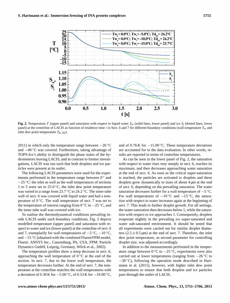
Fig. 2. TemperatureT (upper panel) and saturation with respect to liquid waterSw (solid lines, lower panel) and iceSi (dotted lines, lowerpanel) at the centerline of LACIS as function of residence timet in Sect. 6 and 7 for different boundary conditions (wall temperatureTw andinlet dew point temperatureTd, IN ).
2011) in which only the temperature range between−26◦Cand−40◦C was covered. Furthermore, taking advantage ofTOPS-Ice’s ability to distinguish the phase states of the hy-drometeors leaving LACIS, and in contrast to former investi-gations, LACIS was run such that both droplets and ice par-ticles were present at its outlet.
The following LACIS parameters were used for the exper-iments performed in the temperature range between 0◦ and−25◦C: the inlet as well as the wall temperatures of sections1 to 5 were set to 25.0◦C, the inlet dew point temperaturewas varied in a range from 23.7◦C to 24.2◦C. The inner tubewall of sect. 6 was covered with liquid water and had a tem-perature of 0◦C. The wall temperature of sect. 7 was set tothe temperature of interest ranging from 0◦C to−25◦C, andthe inner tube wall was covered with ice.
To outline the thermodynamical conditions prevailing in-side LACIS under such boundary conditions, Fig.2 depictsmodelled temperature (upper panel) and saturation with re-spect to water and ice (lower panel) at the centerline of sect. 6and 7, exemplarily for wall temperatures of−5◦C, −10◦C,and−15◦C (obtained with the combined Fluent/FPM model,Fluent: ANSYS Inc., Canonsburg, PA, USA, FPM: ParticleDynamics GmbH, Leipzig, Germany,Wilck et al., 2002).
The temperature profiles show a steep decrease in sect. 6,approaching the wall temperature of 0◦C at the end of thesection. In sect. 7, due to the lower wall temperature, thetemperature decreases further. At the end of sect. 7, the tem-perature at the centerline matches the wall temperatures witha deviation of 0.30 K for −5.00◦C, of 0.53 K for −10.00◦C,
and of 0.76 K for −15.00◦C. These temperature deviationsare accounted for in the data evaluation. In other words, re-sults are reported in terms of centerline temperatures.
As can be seen in the lower panel of Fig.2, the saturationwith respect to water rises very steeply in sect. 6, reaches itsmaximum, and then decreases approaching water saturationat the end of sect. 6. As soon as the critical super-saturationis reached, the particles are activated to droplets and thesedroplets grow dynamically to sizes of about 4 µm at the endof sect. 6, depending on the prevailing saturation. The watersaturation decreases further for a wall temperature of−5◦C.For wall temperatures of−10◦C and−15◦C, the satura-tion with respect to water increases again at the beginning ofsect. 7. This leads to further droplet growth. For all settings,the water-saturation then decreases below 1, while the satura-tion with respect to ice approaches 1. Consequently, dropletsevaporate slightly in the prevailing ice super-saturated andwater sub-saturated environment. It should be noted thatall experiments were carried out for similar droplet diame-ters (2.5± 0.5 µm) at the end of sect. 7. Therefore, the inletdew point temperature, as second parameter for controllingdroplet size, was adjusted accordingly.
In addition to the measurements performed in the temper-ature range between 0◦C to −25◦C, experiments were alsocarried out at lower temperatures (ranging from−26◦C to−38◦C), following the operation mode described inHart-mann et al.(2011), however, with higher inlet dew pointtemperatures to ensure that both droplets and ice particlespass through the outlet of LACIS.
www.atmos-chem-phys.net/13/5751/2013/ Atmos. Chem. Phys., 13, 5751–5766, 2013

5756 S. Hartmann et al.: Immersion freezing of INA protein complexes
0 -5 -10 -15 -20 -35 -40
10-3
10-2
10-1
100
fice
T [°C]
Snomax
650 nm
800 nm
TM
Fig. 3. Ice fractionfice as function of temperatureT for 650 nm and800 nm particles generated from a SnomaxTM solution/suspension.
3 Results and discussion
3.1 Experimental results
The immersion freezing behaviour of size-selected par-ticles (mobility diameterDP: 100 nm, 300 nm, 400 nm,650 nm, and 800 nm) generated from a SnomaxTM suspen-sion/solution was studied. For 650 nm and 800 nm particles,the ice fractionfice was analysed as a function of temper-ature in the range from−6.6◦C to −38◦C. The results areshown in Fig.3. The ice fraction curves are very steep, al-most linear for temperatures between−6.6◦C and−9.5◦C.The respective measurement uncertainties are represented bysingle standard deviations determined from at least 3 inde-pendent measurements performed under identical conditions.
As can be seen, forT ≤ −15◦C, the ice fraction curveslevel off to constant values of about 0.2 and 0.4 for the650 nm and 800 nm particles, respectively. As the ice frac-tions do not increase any further, we conclude that a satura-tion regime is reached, i.e., there are no further IN presentin the droplet population which can initiate freezing evenat the low temperatures considered. This suggests that only20 % of 650 nm and 40 % of 800 nm particles generated fromSnomaxTM solution/suspension are ice active. As describedin the Introduction, the ice nucleation ability of SnomaxTM
or better theP. syringaeis due to INA protein complexesattached to the bacterias’ outer cell membrane or fragmentsof cell membrane. Consequently, our results imply that only20 % of the 650 nm and 40 % of the 800 nm particles gen-erated from SnomaxTM solution/suspension contain one ormore INA protein complexes, and that the unfrozen dropletsdo not even contain a single INA protein complex.
Before looking into the data in more detail, it should beclarified to which class (I, II, or III) of ice nucleation ac-
tivity, observed forP. syringae, the majority of INA proteincomplexes observed in our experiments belong. For the per-formed experiments, droplets contained no or only a verysmall number of INA protein complexes. Between 1000 to10 000 frozen droplets, i.e., droplets containing INA proteincomplexes, were counted for determining an ice fraction.Considering the respective probabilities given in the Intro-duction (1 in 103 to 1 in 106 for class II and I), it is un-likely that significant and detectable numbers of class I andII proteins are present in the droplets. Additionally, (a) thelogarithm office increases linearly in the temperature rangefrom −6.6◦C to −9.5◦C, suggesting that only one class ofIN is present, and (b) freezing occurs in a temperature rangesimilar to that reported byYankofsky et al.(1981) andTurneret al.(1991) for class III ice nucleation activity. This suggeststhat we observed class III bacterial ice nucleation activity inour experiments, which, according to literature (see Introduc-tion) corresponds to protein complexes consisting of two upto a few single proteins.
Having identified the ice nucleation activity class, we nowreturn to the measured ice fractions, their saturation be-haviour, and the resulting consequences. The left panel ofFig. 4 shows ice fractions in the saturation range, denoted asf ?
ice and measured at about−15◦C, as a function of the sizeof the particles fed into LACIS. For 100 nm particles we ob-serve almost no freezing. In general,f ?
ice, and consequentlythe number of INA protein complexes present in the particles,increases with increasing mobility diameterDP, but not in alinear manner. As mentioned before, not all droplets containan INA protein complex, and it can be seen in Fig.4 that thenumber of protein complexes present in a droplet is related toparticle size. This offers the possibilities to determine the av-erage number of INA protein complexes distributed over thedroplet ensemble for each specific experiment (i.e., for eachof the examined particle sizes), and also the determination ofthe IN size distribution underlaying the particle size distri-bution resulting from atomization of the SnomaxTM suspen-sion/solution. These topics will be dealt with next.
3.2 Distribution of INA protein complexes over dropletensembles and IN size distribution
As not all droplets contain an INA protein complex, the num-ber of INA protein complexes distributed over the dropletensemble is small compared to the number of droplets form-ing the population. Consequentely, it can be assumed thatthe INA protein complexes are Poisson distributed over thedroplet population (a similar approach can be found for otherice nucleating components inVali, 1971; Yankofsky et al.,1981). The probability that a droplet containsk INA proteincomplexes (k varies in the range ofk = 0, . . . ,n) is then givenby
Pλ(X = k) =λk
k!exp(−λ), (1)
Atmos. Chem. Phys., 13, 5751–5766, 2013 www.atmos-chem-phys.net/13/5751/2013/

S. Hartmann et al.: Immersion freezing of INA protein complexes 5757
100 200 300 400 500 600 700 80010
-3
10-2
10-1
100
fice
DP [nm]
1x10⁸
2x10⁸
3x10⁸
4x10⁸
5x10⁸
0.0
0.1
0.2
0.3
0.4
0.5
0.6
measurement
fit: λ= 9.995 10-10 nm
-3 D
P
3
λ
DP
3 [nm
3]
Fig. 4. Ice fractionf ?ice vs. mobility diameterDp (left panel) atT = −15◦C (saturation range). Measuredλ and fit function describing the
average number of INA protein complexes per particle generated from a SnomaxTM solution/suspension, as function of particle volume (D3P)
(right panel).
with parameterλ, the average number of INA protein com-plexes per droplet. The measured ice fractionfice, and con-sequently alsof ?
ice, correspond to the probability of freezing.Assuming that a droplet can only freeze if it contains at leastone INA protein complex, or the other way around that itdoes not freeze if it does not contain an INA protein complex,the probability that a droplet does not contain an INA proteincomplex can be written as
P(X = 0) = exp(−λ) = 1− f ?ice (2)
and consequently,
λ = − ln(1− f ?ice). (3)
This equation offers the possibility of determiningλ, the ex-pected value for the number of INA protein complexes perdroplet, from the experimentally determined ice fractions inthe saturation rangef ?
ice. Equation (3) is defined only forf ?
ice < 1, i.e., in other casesλ cannot be unambiguously de-termined.
As each droplet is grown on a single particle generatedfrom the SnomaxTM suspension/solution, and as during eachmeasurement all droplets contain particles of the same size,the parameterλ corresponds also to the expected number ofINA protein complexes per particle fed into LACIS. Con-sequently,λ can be related to particle size. The right panelof Fig. 4 showsλ values as a function ofD3
P, i.e., particlevolume. As can be seen,λ is a linear function ofD3
P, illus-trated by the linear fit (red line). This volume dependenceof λ can be explained as follows. The number of INA pro-tein complexes in the atomized solution/suspension is pro-portional to the mass of SnomaxTM used for producing the
solution/suspension. Assuming that no demixing takes placeduring atomization of the solution/suspension, the number ofINA protein complexes per generated particle is proportionalto the particle mass. Furthermore, assuming that in course ofthe droplet activation process inside LACIS all INA proteincomplexes become accessible (i.e., the soluble particle frac-tion dissolves completely), a mass, and considering a size in-dependent particle density, a particle volume dependence ofthe average number of INA protein complexes per particle isexpected. In this context it should be mentioned that the num-ber of INA protein complexes per particle volume or mass isa fundamentally different property compared to, for example,active site density (DeMott, 1995). While active site densityassociates ice nucleating sites to a particle surface area, thenumber of INA complexes relates ice nucleation ability tothe number of complexes present, regardless whether theyare attached to a particle surface or not. In general one couldimage active site surface density being a suitable propertyfor characterising insoluble ice nuclei such as dust particles,while number of INA entities is better suited for e.g., solu-ble particles containing small ice nucleating entities such asINA protein complexes.
Knowing λ as a function of particle volume, the distribu-tion of INA protein complexes over a monodisperse particlepopulation can be calculated by means of Eq. (1). The re-spective results are shown in Fig.5, depicting the probabilityof k INA protein complexes occurring in particles of differentsizes. Considering a population of 100 nm particles, the ma-jority of particles contain no INA protein complex (k = 0),and only a very small fraction of 10−3 includes one protein
www.atmos-chem-phys.net/13/5751/2013/ Atmos. Chem. Phys., 13, 5751–5766, 2013

5758 S. Hartmann et al.: Immersion freezing of INA protein complexes
0 1 2 3 4 5 610
-4
10-3
10-2
10-1
5 10-1
100
Pλ
k
100 nm; λ=0.001
300 nm; λ=0.027
400 nm; λ=0.064
650 nm; λ=0.274
800 nm; λ=0.512
Fig. 5.ProbabilityPλ for k = 0,1,2,3,4,5 INA protein complexesper particle/droplet (Poisson distributed) for different average num-bers of INA protein complexesλ.
complex (k = 1). For 300 nm particles, the fraction of parti-cles containing one INA protein complex is higher, and fur-thermore, a minor fraction of particles has two INA proteincomplexes. For a particle diameter of 800 nm, 40 % of allparticles contain basically either 1, 2, 3, 4 or 5 INA proteincomplexes. With this, the IN number size distributionnIN canbe determined from the number size distribution of the par-ticles generated from the SnomaxTM suspension/solutionn0:
nIN = (1− exp(−λ(D3P))n0. (4)
This is illustrated in Fig.6. This Figure shows clearly that themajority of the particles do not contain an INA protein com-plex, and therefore do not function as ice nuclei, whereby thefraction of particles with IN to those without IN increaseswith increasing particle diameter. However, the presence ofIN in the size range of a few 100 nm also shows that frag-ments of bacterial cell membrane with intact INA proteincomplexes are present.
The IN number size distribution shown in Fig.6 is in con-tradiction toMohler et al.(2008) who speculated, based onpolydisperse measurements, that only particles in the largermode (DP ≥ 600 nm) of the bimodal size distribution areice active. Results obtained in the study byWood et al.(2002) support our finding of INA protein complexes be-ing present also in smaller size classes. They analysed a fil-tered SnomaxTM solution/suspension containing only parti-cles with diameters smaller than 200 nm and observed im-mersion freezing.
For particles in the size range around 800 nm, where weexpect to find the intactP. syringaecells, less than 50 % ofthe particles functioned as ice nuclei. Under the assumptionthat all particles in this size range are intact bacteria cells,this would imply that although theP. syringaecontained in
10 100 100010
0
101
102
103
104
number size distribution n
IN size distribution n
d N/
d log Dp
[#/cm3]
Dp
[nm]
0
IN
Fig. 6. Number size distribution of particles generated from aSnomaxTM solution/suspensionn0 and derived IN size distributionnIN
SnomaxTM are grown under idealized conditions to maxi-mize their ice nucleation ability, apparently less than everyother bacterium induces ice nucleation.
Knowing the IN number size distribution, the ice fractionto be expected in an immersion freezing experiment con-sidering a polydisperse particle population, can be derivedby dividing the integral over the IN size distribution by theintegral over the particle size distribution. For the distribu-tions shown in Fig.6, we calculate an expected ice frac-tion of 0.4 % atT = −15◦C. This agrees well with the re-sult of a polydisperse measurement performed at LACIS atT = −15◦C, which yielded an ice fraction of 0.55 %. Theslightly higher measured value can be explained by the factthat for the prediction only particle sizes up to 800 nm couldbe considered, while it is likely that during the experimentsome particles with sizes larger than 800 nm (probably up toabout 1300 nm) were present.
Up to here, we determined the average/expected numberof INA protein complexes per particle. As each droplet inLACIS contains only a single particle, the average/expectednumber of INA protein complexes per particle is equal to theaverage/expected number of complexes per droplet. Know-ing the average/expected number of INA protein complexesper droplet, we can now deal with the quantification of theice nucleation behaviour of the INA protein complexes.
3.3 Determination of nucleation rate
Knowing the number ofP. syringae’s class III INA proteincomplexes per particle/droplet, we quantify their ice nucle-ation behaviour in terms of a temperature dependent hetero-geneous ice nucleation rate. Thereto, combining the assump-tions that (a) heterogeneous ice nucleation is a stochasticprocess (Niedermeier et al., 2011a), and (b) the distribution
Atmos. Chem. Phys., 13, 5751–5766, 2013 www.atmos-chem-phys.net/13/5751/2013/

S. Hartmann et al.: Immersion freezing of INA protein complexes 5759
of INA protein complexes over the droplet population canbe described by a Poisson distribution, we developed theCHESS model (stoCHastic modEl of similar and poiSSondistributed ice nuclei). The CHESS model, at this stage,should be considered a tool for quantifying the nucleationrate of the INA protein complexes based on the ice frac-tions measured at LACIS. In the model, it is assumed that allINA protein complexes are similar and that only the INA pro-tein complexes initiate droplet freezing. Hence, the probabil-ity of freezing of a droplet containing one INA protein com-plexp1 at temperatureT can be described as:
p1 = 1− exp(−Ssitejhet(T )t) (5)
with the surface area of an INA protein complexSsite, thetemperature dependent heterogeneous ice nucleation rate co-efficientjhet, and the ice nucleation timet . If a droplet pos-sessesk > 1 immersed INA protein complexes, the probabil-ity of freezing is increased due to an increased ice nucleationactive surface areakSsite. Following the assumption that allINA protein complexes have similar properties, and hencesimilar surface areas, the probability of freezing of a dropletcontainingk INA protein complexes is given by:
pk = 1− exp(−kSsitejhet(T )t) (6)
Considering, as a next step, a droplet ensemble at a certaintemperature, and containing Poisson distributed INA proteincomplexes, the probability of droplet freezing is determinedby the product of the probability that a droplet contains atleast one INA protein complexPλ(k ≥ 1) and the probabilitypk that one of this INA protein complexes induces freezing:
fice(T ) =
n∑k=1
pkPλ(X = k). (7)
Inserting Eq. (1) and Eq. (6), after several conversion steps,we obtain the expression:
fice(T ) = 1− exp(−λ(1− exp(−Ssitejhet(T )t))). (8)
It should be noted thatfice corresponds to the probabilitythat one droplet out of a population freezes at temperatureT after timet , where the population contains on averageλ
INA protein complexes, with each complex featuring an icenucleation active surfaceSsite, and a temperature dependentheterogeneous ice nucleation rate coefficientjhet(T ).
All quantities of Eq. (8) are known except forSsite andjhet.Consequently, the product of these quantities, which corre-sponds to the heterogeneous ice nucleation rateJhet(T ) =
Ssitejhet(T ) with the unit # s−1, can be derived from the icefractions measured at LACIS, for a given ice nucleation time.The ice nucleation timet used for the determination ofJhetis defined as time period in which the temperature at the cen-terline of LACIS is in the range ofTend+ 0.3 K ≥ T ≥ Tend,
-6 -7 -8 -9 -1010
-3
10-2
10-1
100
101
SnomaxTM
650 nm
800 nm
fit
Jhet
[s-1]
T [°C]
Fig. 7. Heterogeneous ice nucleation rate of single INA proteincomplexesJhet as function of temperatureT derived for 650 nmand 800 nm particles.
wherebyTend corresponds to the temperature at the center-line at the outlet of LACIS.Jhet should therefore be con-sidered as the average nucleation rate in the temperature in-terval Tend+ 0.3 K to Tend. The results gained for 650 nmand 800 nm particles are shown in Fig.7. Here, we onlyuse the range in which the logarithms of the ice fractionsincrease linearly with temperature (−6.6◦C≥ T ≥ −9.5◦C),i.e., well outside the temperature range in which the ice frac-tions become saturated.Jhet derived for both, 650 nm and800 nm sized particles fall together within the level of un-certainty. In other words, the nucleation rate is not a func-tion of initial particle size or number of INA protein com-plexes present in the droplets. This is expected and confirmsthat we are indeed determining the nucleation rates of singleINA protein complexes.
Next, we parameterise the ice nucleation rateJhet as afunction of temperature:
Jhet(T ) = Ssitejhet(T ) = A exp(B T ) . (9)
with the coefficients A = 9.99× 10⁻¹⁰ s−1 and B =
−2.34◦C−1 being valid forP. syringae’s class III INA pro-tein complexes (red line in Fig. 6).
In the experiments, temperatures in the nucleation zonevary by 0.3 K. The above derivedJhet, however, was derivedfor the centerline temperature at the LACIS outlet. Modelsimulations with Fluent/FPM were carried out in which weaccounted for this temperature variation of 0.3 K. Accordingto these simulations, coefficientB is not affected and differ-ences in coefficientA are less than 10 %. These differencesare well within the measurement uncertainty and hence theassumption of a constant temperature in the nucleation zoneis feasible.
Comparing the temperature dependence of the derivedheterogeneous ice nucleation rate parameterisation (Eq.9)
www.atmos-chem-phys.net/13/5751/2013/ Atmos. Chem. Phys., 13, 5751–5766, 2013

5760 S. Hartmann et al.: Immersion freezing of INA protein complexes
0 -2 -4 -6 -8 -10 -12 -14 -16 -18 -20
10-3
10-2
10-1
100
Pseudomonas syringae:Yankofsky et al. (1981):
5E+6 - 2E+7 cells/drop λ = 170.12
Maki et al. (1974): unknown cell conc. λ = 2.98E+4 unknown cell conc. λ = 7.81E+5
Lindow et al. (1982): 1.2E+3 cells/drop λ = 2.80E+5 1.2E+6 cells/drop λ = 2.30E+6
SnomaxTM
:this study - LACIS:
650 nm λ=0.228 800 nm λ=0.536 polydisperse λ=0.0055
Wood et al. (2002): polydisperse, <200 nm λ=28.59
fice
T [°C]
Fig. 8. A comparison of measured and calculated ice fractions obtained from the CHESS model with data from literature, see legend.Measured (data points) and calculated (lines) ice fractionsfice are given as function of temperatureT .
with that determined applying Classical Nucleation Theory(CNT) using only one contact angle (not shown here), wefound slightly less pronounced temperature dependence forthe parameterised nucleation rate. FromNiedermeier et al.(2011a) it can be concluded that this is most likely due to theinvestigated INA protein complexes not having fully identi-cal properties. This heterogeneity of the INA protein com-plexes could imply both, variations in size and/or structure.In other words, although we are investigating the ice nucle-ation behaviour of single INA protein complexes, that doesnot necessarily imply that all INA protein complexes arefully identical. Therefore, the given parameterisation for thenucleation rate has to be considered as the average rate forthe class III INA protein complexes. A higher level, and moreaccurate, parameterisation could be developed based on theSoccer ball model, and the assumption of a contact angle dis-tribution (Niedermeier et al., 2011a). However, this goes be-yond the scope of the present paper and is left for future in-vestigations.
In summary, based on measured ice fractions, and utilisingthe newly developed CHESS model, we were able to quan-tify the ice nucleation behaviour of the INA protein complexwhich controls the ice nucleation ability ofP. syringae. Asresult we provide ice nucleation rates as a function of tem-perature.
3.4 Application of the nucleation rate to experimentaldata
We will now investigate the applicability of the determinednucleation rate, and thereto we will try to reproduce resultsfrom different experiments reported in literature, using theCHESS model (Eq.8). We implemented the above derived
parameterisation for the INA protein complexes’ nucleationrate (Eq.9) into the model. For our comparison, we con-sider the experimental data gained in our measurements withLACIS as well as data from earlier experiments (Maki et al.,1974; Yankofsky et al., 1981; Lindow et al., 1982; Woodet al., 2002). Results are shown in Fig.8, depicting ice frac-tions as a function of temperature.
First, considering the results found at LACIS, the CHESSmodel reproduces the ice fractions measured (blue and blacksquares) for the 650 nm (λ = 0.228, blue curve) and 800 nm(λ = 0.536, black curve) particles very well. Both, slope ofthe ice fraction curve, and ice fraction at saturation, are nicelyreproduced by the model. However, this is not surprising,as the model parameters (λ and Jhet) were derived fromthis data. For the polydisperse measurements performed atLACIS (light blue square),λ has a value of 0.0055. TheCHESS model (light blue line) predicts an increase of theice fraction from 5×10⁻⁴ to its maximum value for temper-atures ranging from−8.5◦C to−10◦C. Theλ value for thispolydisperse experiment is quite low because most of the par-ticles generated from the SnomaxTM solution/suspension donot contain an INA protein complex initiating ice nucleation,as already shown in Fig.6.
Next, we apply the CHESS model to other experimentaldata. Thereto, we use again the above-determined and pa-rameterised heterogeneous ice nucleation rate (Eq.9). Withthis, λ and the ice nucleation timet are the only free pa-rameters in the model, which have to be adjusted for re-producing the experimental data. By doing so, we assumethat in all considered experiments, ice nucleation is initiatedby similar INA protein complexes, featuring similar nucle-ation rates (Eq.9). Under this assumption, varyingλ and
Atmos. Chem. Phys., 13, 5751–5766, 2013 www.atmos-chem-phys.net/13/5751/2013/
S. Hartmann et al.: Immersion freezing of INA protein complexes 5761
keepingt constant, an increase inλ, which corresponds toan increase of the number of INA protein complexes presentin the droplet population, will result in an increase of the sat-uration value of the ice fraction (f ?
ice). As a consequence, thefice curve moves upwards while its slope does not change.This also results in an apparent increase of the temperatureat which the ice fractions start to rise, or in other words, ofthe onset temperature of freezing. By changingt and keepingλ constant, the ice fraction curves are shifted similarly alongthe temperature axis, but again the slope office, and also themaximum ice fraction is not affected. Ifλ is large enough,i.e., if f ?
ice is equal to one, increasingλ or t will both result ina shift of thefice curve towards higher temperatures. In fact,in this range, there is a relation betweenλ and t , implyingthat when analysing experimental data from that range withthe CHESS model, a decrease inλ corresponds to an increasein t , and vice versa. This allows for analysing experimentaldata even if the exact nucleation time is unknown as it is thecase for the investigations presented below. Therefore, in thefollowing considerations an ice nucleation time of 1s can beassumed for convenience.
We start with ice fractions determined in the study byWood et al.(2002) (green diamonds in Fig.8), in whichthe immersion freezing of freely falling droplets contain-ing particles smaller than 200 nm from a SnomaxTM so-lution/suspension filtrate was analysed. By means of theCHESS model an average number of INA protein complexesof λ ≈ 28.6 (green line) is calculated. The slopes of the mea-sured and predictedfice curves agree very well. From this, itfollows that inWood et al.(2002) a temperature dependenceof the heterogeneous ice nucleation rate similar to that in ourstudy was found.
Interestingly, the CHESS model is able to reproduce theice nucleation behaviour ofP. syringaebacteria as well. Thiscan be seen when looking at the results given inYankof-sky et al. (1981), who analysed the freezing behaviourof droplets with multiple immersedP. syringaecells (sta-tionary phase of bacteria growth). For a cell concentra-tion of about 1–4× 10⁹ cells ml−1, corresponding to about5× 106
− 2× 10⁷ cells drop−1 (dark brown hexagons), theslopes of measured and calculated ice fraction curves alsoagree with a reasonably small deviation, presumably withinthe measurement uncertainties. In this case, the CHESSmodel predicts an average number of INA protein complexesof 170.1, i.e., a number much smaller than the given bacte-ria cell concentration per droplet. This indicates that only aminor fraction of the bacteria was ice active.
Unfortunately in many studies (Orser et al., 1985; Wolberet al., 1986; Lindow et al., 1989; Attard et al., 2012), the con-centrations of bacterial cells in the examined droplets are notreported, and therefore the cumulative ice nuclei concentra-tions given in these studies cannot be converted to ice frac-tions (fice), and hence they cannot be included in our com-parison.
However,Lindow et al. (1982) directly reported frozenfractions. They analysed the freezing ofP. syringaecells asfunction of temperature and cell concentration. For a lowerP.syringaecell concentration (3.5×10⁴ cells ml−1 correspond-ing to about 1.2× 10³ cells drop−1, dark green triangle) themodel predicts aλ value of about 2.8×10⁵ (dark green line)and for a higher cell concentration (3.5× 10⁷ cells ml−1 cor-responding to about 1.2× 10⁶ cells drop−1, dark cyan trian-gle) aλ value of about 2.3×10⁶ (dark cyan line), respectively.The shift of the ice fraction curve towards higher tempera-tures with increasing cell concentration is nicely reproduced.However, agreement betweenλ values and cell concentra-tions is only qualitative. Possible reasons for that are, thatthe fraction of cells being ice nucleation active was differ-ent for the different experiments, or that the number of cellsper droplet was so large that class I and II ice nucleation ac-tivities started to become important. Additionally, as statedabove, we use an ice nucleation time of 1 s to determine theλ
values. For the drop freezing method however, ice nucleationtimes are usually much longer (on the order of minutes). Butas nucleation times are usually not reported, a quantitativecomparison ofλ values with cell number per droplet cannotbe obtained. Therefore, further research is needed to explorethe relation between the number of INA protein complexesand the number of cells present, especially in the range ofhigh cell concentrations.
Another study dealing with the immersion freezing ofdroplets containing multipleP. syringaebacteria cells (redand purple dots) is described inMaki et al. (1974). In thispublication, ice fractions at two different temperatures aregiven for different samples with different but unknown cellconcentrations. A deviation between the observed and mod-elled temperature dependencies of the ice fractions couldbe inferred from the comparison, however considering thesparseness of the experimental data, a meaningful compari-son is difficult.
Generally, applying the CHESS model together with thenucleation rate expression for INA protein complexes as sug-gested in this work, we observe an overall good agreementbetween the calculated and measured slopes of the ice frac-tion curves, and a shift of the ice fraction curves to highertemperatures (higher onset temperatures) with an increasingnumber of INA protein complexes and/or cells. This shift ofthe ice fraction curves to higher freezing temperatures withincreasing number of INA protein complexes/bacteria cellswas observed in e.g.,Ward and DeMott(1989) andCochetand Widehem(2000) as well.
Summarising the results presented above, it can be statedthat the CHESS model, together with the heterogeneous icenucleation rate expression for single class III INA proteincomplexes, is capable of describing the ice nucleation be-haviour observed in various experiments for both SnomaxTM
andP. syringaebacteria. This implies that the heterogeneousice nucleation rate, and its temperature dependence, seemto be very similar, regardless of whether the INA protein
www.atmos-chem-phys.net/13/5751/2013/ Atmos. Chem. Phys., 13, 5751–5766, 2013

5762 S. Hartmann et al.: Immersion freezing of INA protein complexes
complex causing the ice nucleation ability is attached to theouter cell membrane of intact bacteria or to cell membranefragments. In this context, it is worth mentioning again that inour experiments, we mainly investigated class III nucleationactivity while some of the experiments shown in the abovecomparison may feature cell concentrations which make thepresence of class I and II ice nucleation activity likely. Thegood agreement between the measured and calculated icefractions then suggests that the temperature dependence ofthe nucleation rates is similar, regardless of which class ofactivity is considered. However, this is highly speculative andrequires further investigations. Furthermore, it can be statedthat the temperature range in which heterogeneous dropletfreezing occurs, and the fraction of droplets able to freeze,both depend on the actual number of INA protein complexespresent in the droplet ensemble. Consequently, ice fractionsand onset temperatures may not be the proper parameters toquantify ice nucleation behaviour. Finally, the fact that weare able to consistently describe a variety of different experi-ments with the same nucleation rate indicates that this nucle-ation rate is a suitable property for quantifying ice nucleationbehaviour. It also suggests that possible artifacts suspectedto occur in connection with the drop freezing method intro-duced byVali (1971), as mentioned in the Introduction, areof minor importance, at least for substances which inducefreezing at such high temperatures asP. syringae.
4 Summary and conclusions
At LACIS, the immersion freezing behaviour of monodis-perse particles generated from a SnomaxTM solu-tion/suspension was investigated by measuring ice fractionsas a function of temperature in the range between−6.6◦Cand −38◦C. SnomaxTM is an industrial product appliedfor artificial snow production and containsPseudomonassyringae bacteria which have long been used as modelorganism for atmospheric relevant INA bacteria. The icenucleation activity of such bacteria is controlled by INA pro-tein complexes in the outer membrane of the bacterial cellwall.
From the facts, that (a) the measured ice fraction curvesfeature a saturation behaviour, suggesting that not all gen-erated particles contained an ice nucleus, (b) the number ofparticles/droplets containing IN was found to be a functionof particle volume, and (c) already 100 nm particles exhib-ited a detectable ice nucleation ability, we concluded that theice nucleating entity we were examining in our experimentswas an INA protein complex attached to either intact cells orfragments of outer membrane of the bacterial cell wall. Both,statistical analysis and temperature range in which freezingwas observed, are indicative for the examined INA proteincomplexes featuring a class III ice nucleation activity. Forparticles in the size range where intact bacteria are to beexpected (> 600 nm), we found that less than 50 % of the
particles were ice nucleation active despite the low temper-atures (down to−38◦C), indicating that maybe only half ofthe bacteria were able to nucleate ice, i.e., were carrying anINA protein complex. This finding supports data available inthe literature suggesting that the majority ofP. syringaecellshave none, one or only little more than one ice forming siteon their surface (e.g.,Orser et al., 1985; Wolber et al., 1986;Lindow et al., 1989; Attard et al., 2012).
In order to derive the heterogeneous ice nucleation rateof the INA protein complexes, a simple model, the CHESSmodel, was developed. The CHESS model is based on theassumptions that the INA protein complexes are Poisson dis-tributed over a droplet ensemble, and that the heterogeneousice nucleation on similar INA protein complexes is a stochas-tic process. Under these assumptions, the average nucleationrate for class III INA protein complexes was determined andparameterised as function of temperature.
Uitilising the parameterised nucleation rate implementedinto the CHESS model, we compared predictions from themodel to other experimental data. In these investigations, thenumber of INA protein complexes present in the droplets,and the ice nucleation time were the only free parameters.
We found that:
– The heterogeneous ice nucleation rate expression quan-tifying the ice nucleation behaviour of single INA pro-tein complexes is capable of describing the ice nucle-ation behaviour observed in various experiments forboth, SnomaxTM andP. syringaebacteria.
– The ice nucleation rate, and its temperature depen-dence, seem to be very similar regardless of whether theINA protein complexes are attached to the outer mem-brane of intact bacteria or cell membrane fragments.
– The temperature range in which heterogeneous dropletfreezing occurs, and the fraction of droplets beingable to freeze, both depend on the average number ofINA protein complexes present in the droplet ensemble.
– Possible artifacts suspected to occur in connection withthe drop freezing method introduced byVali (1971) areof minor importance, at least for substances such asP.syringae.
The last two statements require additional remarks. It shouldbe noted that in case of single ice nucleation entities suchas INA protein complexes, which are not necessarily at-tached to a solid particle, it is the average number of entitiesin the droplet population, and the entities’ nucleation rate,which control the freezing behaviour of the droplet popula-tion. Quantities such as active site density (DeMott, 1995)are not suitable in this context.
The last statement in the bulleted list is based on thefact that our LACIS measurements and the droplet freezingmethod yield similar results concerning the nucleation rate ofthe INA protein complexes. This implies that in the droplet
Atmos. Chem. Phys., 13, 5751–5766, 2013 www.atmos-chem-phys.net/13/5751/2013/

S. Hartmann et al.: Immersion freezing of INA protein complexes 5763
freezing method according toVali (1971), possible interfer-ences such as nucleation by substrates seem to be of minorimportance, at least when examining substances which in-duce freezing at temperatures as high asP. syringae. Fur-thermore, it should be noted that when applying the dropletfreezing method, reporting the specific concentrations used,sizes of the examined droplets, the cooling rates applied, andtime of freezing observed, as well as the frozen fractions de-termined, would be very valuable. Presenting only the finalinformation about number of ice nuclei per cell, as done, forexample, in a lot of the above mentioned publications, limitsthe possibility for further use of the data, e.g., for calculatingthe parameters described in this study.
At the end, some additional remarks, suggestions, and hy-potheses, based on the results gained in the present study:firstly, it should be noted that the presented nucleation rate onits own already allows for the dynamic simulation of bacteriainduced heterogeneous ice nucleation in cloud or larger scalemodels. Here the nucleation rate could e.g., represent thesource term in the differential equation describing the timeevolution of the number concentration of frozen droplets. Al-ternatively, CHESS model and nucleation rate together couldbe used for parameterising bacterial ice nucleation in largerscale models. This approach allows for the determination offrozen droplet fractions for a given temperature and time,without solving a differential equation. In addition to tem-perature and time, both approaches require information con-cerning the number of INA protein complexes present in thedroplet population. This number could be determined havingparticle number and mass concentrations as model variables,and e.g., number of INA protein complexes per particle massas a parameter. However, it has to be noted that the num-ber of INA protein complexes per particle mass is currentlya parameter that is difficult to quantify and which needs tobe constrained by atmospheric measurements. As a first ap-proximation the average number of ice nucleating bacteriacould be used. However, even data concerning the numberof ice nucleating bacteria per droplet or volume of precipita-tion is still sparse as already mentioned in the Introduction.Looking into densities of total bacterial IN larger than 0.2 µmin precipitation,Christner et al.(2008) found between 4 and120 bacterial IN per litre of precipitation. Also in precipi-tation, two studies have found that between 2 % and 19 %of bacteria, which could be cultivated under laboratory con-ditions, were INA (Stephanie and Waturangi, 2011; Santl-Temkiv et al., 2013). Joly et al.(2013) studied INA bacte-ria in cloud water and could estimate that clouds over Puyde Dome contain between around 0 and 500 INA bacteriaper ml of cloud water, which corresponds to a single INAcell in every 107–1015 of cloud droplets (assuming a diam-eter of 10µm). However, all these studies, exceptChristneret al. (2008), only investigated the fraction of INA bacteriathat was alive and could grow under laboratory conditions.Thus, the actual atmospheric densities of bacterial IN are still
largely unknown and the currently accepted densities mayonly represent the lower bound as will be discussed below.
The application of the CHESS model is not limited toINA protein complexes only. In fact, it could be applied forother IN types, e.g., ice nucleation active protein complexesfrom other bacteria or macro-molecules from pollen (Pum-mer et al., 2012; Augustin et al., 2012), under the conditionthat the IN considered as one type all feature similar ice nu-cleation properties and that these IN are Poisson distributedover the droplet population.
Concerning the relevance of INA protein complexes for at-mospheric ice nucleation, it could be possible that INA pro-tein complexes exist separately from intact bacterial cells,even in nature, which then would imply (a) a decoupling ofthe protein complex and bacteria cell numbers, and (b) thepossible enrichment of INA protein complexes on, e.g., plantsurfaces or in soil. In this context, it is worth noting thatKle-ber et al.(2007) found INA protein complexes to be wellpreserved – and maybe accumulated – when being connectedto mineral surfaces. AlsoConen et al.(2011) found that or-ganic matter consisting of remains of micro-organisms thattook part in the degradation of plant debris is responsible forenhancing the ice nucleation of atmospheric mineral dust.
Such an enhancement of the ice nucleation ability of min-eral and soil dusts by bacterial IN could increase the impor-tance of bacterial IN compared to the results presented inHoose et al.(2010). However, this is highly speculative andneeds further investigations.
Acknowledgements.This research project was performed in theframework of the German Research Foundation (DFG project WE4722/1-1) research unit INUIT. Funding for T.S.-T. was providedby The Danish National Research Foundation (Grant agreementno.: DNRF106) and the ASTERISK project (ASTERoseismicInvestigations with SONG and Kepler) funded by the EuropeanResearch Council (Grant agreement no.: 267864).
Edited by: B. Ervens
References
Ahern, H. E., Walsh, K. A., Hill, T. C. J., and Moffett, B. F.: Fluores-cent pseudomonads isolated from Hebridean cloud and rain wa-ter produce biosurfactants but do not cause ice nucleation, Bio-geosciences, 4, 115–124, doi:10.5194/bg-4-115-2007, 2007.
Amato, P., Parazols, M., Sancelme, M., Laj, P., Mailhot, G., andDelort, A. M.: Microorganisms isolated from the water phaseof tropospheric clouds at the Puy de Dome: major groups andgrowth abilities at low temperatures, Fems Microbiol. Ecol., 59,242–254, 2007.
Ansmann, A., Tesche, M., Seifert, P., Althausen, D., Engelmann, R.,Fruntke, J., Wandinger, U., Mattis, I., and Muller, D.: Evolutionof the ice phase in tropical altocumulus: SAMUM lidar observa-tions over Cape Verde, J. Geophys. Res.-Atmos., 114, D17208,doi:10.1029/2008JD011659, 2009.
www.atmos-chem-phys.net/13/5751/2013/ Atmos. Chem. Phys., 13, 5751–5766, 2013

5764 S. Hartmann et al.: Immersion freezing of INA protein complexes
Attard, E., Yang, H., Delort, A.-M., Amato, P., Poschl, U.,Glaux, C., Koop, T., and Morris, C. E.: Effects of atmo-spheric conditions on ice nucleation activity of Pseudomonas,Atmos. Chem. Phys., 12, 10667–10677, doi:10.5194/acp-12-10667-2012, 2012.
Augustin, S., Hartmann, S., Pummer, B., Grothe, H., Nieder-meier, D., Clauss, T., Voigtlander, J., Tomsche, L, Wex, H.and Stratmann, F.: Immersion freezing of birch pollen wash-ing water, Atmos. Chem. Phys. Discuss., 12, 32911–32943,doi:10.5194/acpd-12-32911-2012, 2012.
Burrows, S. M., Elbert, W., Lawrence, M. G., and Poschl, U.: Bac-teria in the global atmosphere – Part 1: Review and synthesis ofliterature data for different ecosystems, Atmos. Chem. Phys., 9,9263–9280, doi:10.5194/acp-9-9263-2009, 2009.
Christner, B. C., Morris, C. E., Foreman, C. M., Cai, R. M. andSands, D. C.: Ubiquity of biological ice nucleators in snowfall,Science, 319, 1214–1214, 2008.
Clauss, T., Kiselev, A., Hartmann, S., Augustin, S., Pfeifer, S., Nie-dermeier, D., Wex, H., and Stratmann, F.: Application of lin-ear polarized light for the discrimination of frozen and liquiddroplets in ice nucleation experiments, Atmos. Meas. Tech., 6,1041–1052, doi:10.5194/amt-6-1041-2013, 2013.
Cochet, N. and Widehem, P.: Ice crystallization byPseu-domonas syringae, Appl. Microbiol. Biotech., 54, 153–161,doi:10.1007/s002530000377, 2000.
Conen, F., Morris, C. E., Leifeld, J., Yakutin, M. V., and Alewell,C.: Biological residues define the ice nucleation properties of soildust, Atmos. Chem. Phys., 11, 9643–9648, doi:10.5194/acp-11-9643-2011, 2011.
DeLeon-Rodriguez, N., Lathem, T. L., Rodriguez-Rojas, L. M.,Barazesh, J. M., Anderson, B. E., Beyersdorf, A. J.,Ziemba, L. D., Bergin, M., Nenes, A., and Konstantini-dis, K. T.: Microbiome of the upper troposphere: Species com-position and prevalence, effects of tropical storms, and atmo-spheric implications, Proc. Natl. Acad. Sci., 110, 2575–2580,doi:10.1073/pnas.1212089110, 2013.
DeMott, P. J.: Quantitative descriptions of ice formation mecha-nisms of silver iodide-type aerosols. Atmos. Res., 38, 63–99,1995.
DeMott, P. J., Sassen, K., Poellot, M. R., Baumgardner, D.,Rogers, D. C., Brooks, S. D., Prenni, A. J. and Kreiden-weis, S. M.: African dust aerosols as atmospheric ice nu-clei. Geophys. Res. Lett., 30, 1732, doi:10.1029/2003GL017410,2003.
Despres, V. R., Huffman, J. A., Burrows, S. M., Hoose, C., Safa-tov, A. S., Buryak, G., Frohlich-Nowoisky, J., Elbert, W., An-dreae, M. O., Poschl, U. and Jaenicke, R.: Primary biologicalaerosol particles in the atmosphere: a review, Tellus B, 64, 15598,doi:10.3402/tellusb.v64i0.15598, 2012.
Fuzzi, S., Mandrioli, P., and Perfetto, A.: Fog droplets – An atmo-spheric source of secondary biological aerosol particles, Atmos.Environ., 31, 287–290, 1997.
Garnham, C. P., Campbell, R. L., Walker, V. K. and Davies, P. L.:Novel dimeric beta-helical model of an ice nucleation pro-tein with bridged active sites, BMC Struct. Biol., 11,doi:10.1186/1472-6807-11-36, 2011.
Green, R. L. and Warren, G. J.: Physical and functional repetitionin an bacterial ice nucleation gene, Nature, 317, 645–648, 1985.
Govindarajan, A. G. and Lindow, S. E.: Size of bacterial ice-nucleation sites measured in-situ by radiation inactivation analy-sis, Proc. Nat. Acad. Sci. USA, 85, 1334–1338, 1988.
Hartmann, S., Niedermeier, D., Voigtlander, J., Clauss, T.,Shaw, R. A., Wex, H., Kiselev, A., and Stratmann, F.: Homo-geneous and heterogeneous ice nucleation at LACIS: operatingprinciple and theoretical studies, Atmos. Chem. Phys., 11, 1753–1767, doi:10.5194/acp-11-1753-2011, 2011.
Hoose, C., Kristjansson, J. E. and Burrows, S. M.: How importantis biological ice nucleation in clouds on a global scale?, Environ.Res. Lett., 5, 024009, doi:10.1088/1748-9326/5/2/024009, 2010.
Hoose, C. and Mohler, O.: Heterogeneous ice nucleation on at-mospheric aerosols: A review of results from laboratory exper-iments, Atmos. Chem. Phys., 12, 9817–9854, doi:10.5194/acp-12-9817-2012, 2012.
Joly, M., Attard, E., Sancelme, M., Deguillaume, L., Guilbaud, C.,Morris, C. E., Amato, P., and Delort, A.-M.: Ice nucleation ac-tivity of bacteria isolated from cloud water, Atmos. Environ., 70,392–400, doi:10.1016/j.atmosenv.2013.01.027, 2013.
Kamphus, M., Ettner-Mahl, M., Klimach, T., Drewnick, F.,Keller, L., Cziczo, D. J., Mertes, S., Borrmann, S., and Cur-tius, J.: Chemical composition of ambient aerosol, ice residuesand cloud droplet residues in mixed-phase clouds: single par-ticle analysis during the Cloud and Aerosol CharacterizationExperiment (CLACE 6), Atmos. Chem. Phys., 10, 8077–8095,doi:10.5194/acp-10-8077-2010, 2010.
Kanitz, T., Seifert, P., Ansmann, A., Engelmann, R., Althausen, D.,Casiccia, C., and Rohwer, E. G.: Contrasting the impactof aerosols at northern and southern midlatitudes on het-erogeneous ice formation, Geophys. Res. Lett., 38, L17802,doi:10.1029/2011GL048532, 2011.
Kawahara, H.: The structures and functions of ice crystal-controlling proteins from bacteria, J. Biosci. Bioeng., 94, 492–496, 2002.
Kleber, M., Sollins, P. and Sutton, R.: A conceptual model oforgano-mineral interactions in soils: self-assembly of organicmolecular fragments into zonal structures on mineral surfaces,Biogeochemistry, 85, 9–24, 2007.
Knutson, E. O. and Whitby, K. T.: Aerosol classification by electricmobility: Apparatus, theory and applications, J. Aerosol Sci., 6,443–451, 1975.
Levin, Z. and Yankofsky, S. A.: Contact Versus Immersion Freez-ing Of Freely Suspended Droplets By Bacterial Ice Nuclei, J.Climate Appl. Meteorol., 22, 1964–1966, 1983.
Lindow, S. E., Arny, D. C., and Upper, C. D.: Bacterial ice nucle-ation – a factor in frost injury to plants, Plant Physiol., 70, 1084–1089, 1982.
Lindow, S. E., Lahue, E., Govindarajan, A. G., Panopoulos, N. J.,and Gies, D.: Localization of ice nuleation activity and the iceCgene product inPseudomonas syringaeand Escherichia-coli,Mol. Plant Microbe In., 2, 262–272, 1989.
Lohmann, U.: Aerosol effects on clouds and climate, Space Sci.Rev., 125, 129–137, 2006.
Maki, L. R. and Willoughby, K. J.: Bacteria as biogenic sources offreezing nuclei, J. Appl. Meteorol., 17, 1049–1053, 1978.
Maki, L. R., Galyan, E. L., Chang-Chien, M.-M., and Cald-well, D. R.: Ice nucleation induced byPseudomonas syringae,Appl. Microbiol., 28, 456–459, 1974.
Atmos. Chem. Phys., 13, 5751–5766, 2013 www.atmos-chem-phys.net/13/5751/2013/
S. Hartmann et al.: Immersion freezing of INA protein complexes 5765
Mohler, O., Georgakopoulos, D. G., Morris, C. E., Benz, S., Ebert,V., Hunsmann, S., Saathoff, H., Schnaiter, M., and Wagner, R.:Heterogeneous ice nucleation activity of bacteria: new labora-tory experiments at simulated cloud conditions, Biogeosciences,5, 1425–1435, doi:10.5194/bg-5-1425-2008, 2008.
Monteil, C. L., Guilbaud, C., Glaux, C., Lafolie, F., Soubeyrand, S.,and Morris, C. E.: Emigration of the plant pathogenPseu-domonas syringaefrom leaf litter contributes to its populationdynamics in alpine snowpack, Environ. Microbiol., 14, 2099-2112, 2012.
Morris, C. E. and Sands, D. C.: From Grains to Rain: the linkbetween landscape, airborne microorganisms and climate pro-cesses, e-book, accessed 26 April 2012,http://bioice.wordpress.com/, 2012.
Morris, C. E., Georgakopoulos, D. G., and Sands, D. C.: Ice nucle-ation active bacteria and their potential role in precipitation. J.Phys. IV, 121, 87–103, 2004.
Morris, C. E., Sands, D. C., Vinatzer, B. A., Glaux, C., Guilbaud, C.,Buffiere, A., Yan, S. C., Dominguez, H., and Thompson, B. M.:The life history of the plant pathogenPseudomonas syringaeislinked to the water cycle, ISME J., 2, 321–334, 2008.
Mueller, G. M., Wolber, P. K., and Warren, G. J.: Clustering of icenucleation protein correlates with ice nucleation activity, Cryobi-ology, 27, 416–422, 1990.
Murray, B. J., O’Sullivan, D., Atkinson, J. D., and Webb, M. E.: Icenucleation by particles immersed in supercooled cloud droplets.Chem. Soc. Rev., 41, 6519-6554, 2012.
Niedermeier, D., Hartmann, S., Shaw, R. A., Covert, D.,Mentel, T. F., Schneider, J., Poulain, L., Reitz, P., Spindler, C.,Clauss, T., Kiselev, A., Hallbauer, E., Wex, H., Mildenberger, K.,and Stratmann, F.: Heterogeneous freezing of droplets with im-mersed mineral dust particles – measurements and parameteriza-tion, Atmos. Chem. Phys., 10, 3601–3614, doi:10.5194/acp-10-3601-2010, 2010.
Niedermeier, D., Shaw, R. A., Hartmann, S., Wex, H., Clauss, T.,Voigtlander, J., and Stratmann, F.: Heterogeneous ice nu-cleation: exploring the transition from stochastic to singularfreezing behavior, Atmos. Chem. and Phys., 11, 8767–8775,doi:10.5194/acp-11-8767-2011, 2011a.
Niedermeier, D., Hartmann, S., Clauss, T., Wex, H., Kiselev, A.,Sullivan, R. C., DeMott, P. J., Petters, M. D., Reitz, P., Schnei-der, J., Mikhailov, E., Sierau, B., Stetzer, O., Reimann, B.,Bundke, U., Shaw, R. A., Buchholz, A., Mentel, T. F., andStratmann, F.: Experimental study of the role of physicochem-ical surface processing on the IN ability of mineral dust parti-cles, Atmos. Chem. Phys., 11, 11131–11144, doi:10.5194/acp-11-11131-2011, 2011b.
Orser, C., Staskawicz, B. J., Panopoulos, N. J., Dahlbeck, D., andLindow, S. E.: Cloning and expression of bacterial ice nucleationgenes in Escherichia-coli, J. Bacteriol., 164, 359–366, 1985.
Pratt, K. A., DeMott, P. J., French, J. R., Wang, Z., Westphal, D. L.,Heymsfield, A. J., Twohy, C. H., Prenni, A. J., and Prather, K. A.:In situ detection of biological particles in cloud ice-crystals, Nat.Geosci., 2, 397–400, doi:10.1038/ngeo521, 2009.
Pummer, B. G., Bauer, H., Bernardi, J., Bleicher, S., and Grothe,H.: Suspendable macromolecules are responsible for ice nucle-ation activity of birch and conifer pollen. Atmos Chem Phys.,12, 2541–2550, doi:10.5194/acp-12-2541-2012, 2012.
Santl-Temkiv, T., Finster, K., Sahyoun, M., Hartmann, S., Au-gustin, S., Stratmann, F., Wex, H., Clauss, T., Korsholm, U. S.,Nielsen, N. W., Sorensen, J. H., and Karlson, U. G.: AirbornePseudomonas sp.may increase the number of biological ice nu-clei in the atmosphere by excreting ice nucleation active proteins,in preparation for Environ. Microbiol., 2013.
Schnell, R. C. and Vali, G.: Atmospheric ice nuclei from decompos-ing vegetation, Nature, 236, 163–165, 1972.
Schmid, D., Pridmore, D., Capitani, G., Battistutta, R., Neeser, J.R., and Jann, A.: Molecular organisation of the ice nucleationprotein InaV fromPseudomonas syringae, Febs Lett., 414, 590–594, 1997.
Seifert, P., Ansmann, A., Mattis, I., Wandinger, U., Tesche, M., En-gelmann, R., Muller, D., Perez, C., and Haustein, K.: Saharandust and heterogeneous ice formation: eleven years of cloud ob-servations at a Central European EARLINET site, J. Geophys.Res.-Atmos., 115, D20201, doi:10.1029/2009JD013222, 2010.
Skirvin, R. M., Kohler, E., Steiner, H., Ayers, D., Laughnan, A.,Norton, M. A., and Warmund, M.: The use of genetically en-gineered bacteria to control frost on strawberries and potatoes.Whatever happened to all of that research?, Sci. Hortic., 84, 179–189, 2000.
Southworth, M. W., Wolber, P. K., and Warren, G. J.: Nonlinearrelationship between concentration and activity of a bacterial icenucleation protein, J. Biol. Chem., 263, 15211–15216, 1988.
Stephanie, D. and Waturangi, E.: Distribution of ice nucleation-active (INA) bacteria from rain-water and rain, HAYATI Journalof Biosciences, 18, 108–112, 2011.
Stratmann, F., Kiselev, A., Wurzler, S., Wendisch, M., Heintzen-berg, J., Charlson, R. J., Diehl, K., Wex, H., and Schmidt, S.:Laboratory studies and numerical simulations of cloud dropletformation under realistic supersaturation conditions, J. Atmos.Ocean. Tech., 21, 876–887, 2004.
Turner, M. A., Arellano, F., and Kozloff, L. M.: Three separateclasses of bacterial ice nucleation structures, J. Bacteriol., 172,2521–2526, 1990.
Turner, M. A., Arellano, F., and Kozloff, L. M.: Components of icenucleation structures of bacteria, J. Bacteriol., 173, 6515–6527,1991.
Twohy, C. H. and Poellot, M. R.: Chemical characteristics of iceresidual nuclei in anvil cirrus clouds: evidence for homogeneousand heterogeneous ice formation, Atmos. Chem. Phys., 5, 2289–2297, doi:10.5194/acp-5-2289-2005, 2005.
Vali, G.: Quantitative Evaluation Of Experimental Results On Het-erogeneous Freezing Nucleation Of Supercooled Liquids, J. At-mos. Sci., 28, 402–409, 1971.
Vali, G., Christensen, M., Fresh, R. W., Galyan, E. L., Maki, L. R.,and Schnell, R. C.: Biogenic ice nuclei. Part II: Bacterial sources,J. Atmos. Sci., 33, 1565–1570, 1976.
van Overbeek, L. S., Nijhuis, E. H. M., Koenraadt, H., Visser, J.,and van Kruistum, G.: The role of crop waste and soil inPseu-domonas syringaepathovar porri infection of leek (Allium por-rum), Appl. Soil Ecol., 46, 457–463, 2010.
Ward, P. J. and DeMott, P. J.: Preliminary Experimental Evaluationof SnomaxTM Snow Inducer,Pseudomonas syringae, as an Arti-ficial Ice Nucleus, J. Weather Mod., 21, 9–13, 1989.
Warren, G. and Wolber, P.: Molecular aspects of microbial ice nu-cleation, Mol. Microbiol., 5, 239–243, 1991.
www.atmos-chem-phys.net/13/5751/2013/ Atmos. Chem. Phys., 13, 5751–5766, 2013

5766 S. Hartmann et al.: Immersion freezing of INA protein complexes
Warren, G. J. and Wolber, P. K.: Heterogeneous ice nucleation bybacteria, Cryo. Lett., 8, 204–217, 1987.
Wilck, M., Stratmann, F., and Whitby, E.: A fine particle model forfluent: Description and application, in: Proc. Sixth Int. AerosolConf., 1269–1270, Chinese Association for Aerosol Researchin Taiwan/International Aerosol Research Assembly, Taipei, Tai-wan, 2002.
Wolber, P. K., Deininger, C. A., Southworth, M. W., Vandekerck-hove, J., Vanmontagu, M., and Warren, G. J.: Identification andpurification of a bacterial ice-nucleation protein, P. Natl. A. Sci.USA, 83, 7256–7260, 1986.
Wood, S. E., Baker, M. B., and Swanson, B. D.: Instrument for stud-ies of homogeneous and heterogeneous ice nucleation in free-falling supercooled water droplets, Rev. Sci. Inst., 73, 3988–3996, 2002.
Yankofsky, S. A., Levin, Z., Bertold, T., and Sandlerman, N.: Somebasic characteristics of bacterial freezing nuclei, J. Appl. Meteo-rol., 20, 1013–1019, 1981.
Atmos. Chem. Phys., 13, 5751–5766, 2013 www.atmos-chem-phys.net/13/5751/2013/