open access anatomy and histology of the spiral valve intestine in juvenile australian ...€¦ ·...
TRANSCRIPT
62 The Open Zoology Journal, 2009, 2, 62-85
1874-3366/09 2009 Bentham Open
Open Access
Anatomy and Histology of the Spiral Valve Intestine in Juvenile Australian Lungfish, Neoceratodus forsteri
Masoud Hassanpour and Jean Joss*
Department of Biological Sciences, Macquarie University, Sydney NSW 2109, Australia
Abstract: The Australian lungfish, Neoceratodus forsteri, is the only vertebrate that possesses a complete spiral valve
intestine with pre-pyloric coiling. This study describes the anatomy and histology of the spiral valve intestine in juvenile
N. forsteri and compares it to a previous study of adult N. forsteri, thus providing a broader picture and better
understanding of the intestine of the Australian lungfish. Not surprisingly, most features of the spiral valve intestine in
juvenile and adult N. forsteri are similar. However, our study goes further to show that, unlike most other vertebrates, the
stomach (pre-pyloris) is non-distensible (lacks rugae). Rugae are confined to the post-pyloric duodenum. The epithelium
of the pyloric fold, between foregut and midgut, is ciliated and the presence of lymphoid tissue in the pyloric fold implies
the involvement of this region in the immune system. Lymphoid tissue is also present around the posterior spleen in the
medial axis, which indicates a broader gut-associated lymphoid tissue (GALT) in juvenile Neoceratodus than has been
previously recognized in adult Neoceratodus. This study also found some node-like structures in the epithelium of the
mucosal tissue, which resemble the Peyer’s patches of other more advanced vertebrates. Furthermore, a previously
unreported parasite was found in the spleen encased in fibrous tissue, indicating an immune response had been mounted
by the host against it. These latter observations suggest that a thorough investigation of GALT in Neoceratodus is
warranted.
Keywords: Australian lungfish, Neoceratodus forsteri, spiral valve intestine, anatomy and histology, pyloric fold.
INTRODUCTION
Lungfish belong to class Osteichthyes, subclass Sarcopterygii, order Dipnoi, a group that has survived since the early Devonian. Living lungfish belong to only three genera and are considered to be living fossils [1, 2] . Due to the phylogenetic relationship between tetrapods and lungfish [2-4], lungfish are being extensively studied as the closest living relative of tetrapods [5, 6]. Among three genera of lungfish, N. forsteri is believed to be phylogenetically the most ancient species, which has survived unchanged for more than 100 million years [7]. The gastrointestinal tract in lungfish comprises a spiral valve intestine, which is also characteristic for cartilaginous fish like sharks [8, 9] and sawfish [10] and some of the non-teleost actinopterygians, such as sturgeons [11] and paddlefish [12], and the only other living sarcoptergian fish, the coelacanths [13]. The spiral valve intestine or valvular intestine [8, 9], which is relatively short and located in the post duodenal intestine in most fish taxa, provides a large surface area of mucosal tissue for absorption of nutrients. In the lungfish, Neoceratodus, spiralling begins immediately after the esophagus.
The most recent study of the spiral valve intestine of the Australian lungfish is that by Rafn and Wingstrand [14], in which they compare the anatomical and histological features of adult Neoceratodus forsteri with the other two living lungfish genera, Lepidosiren and Protopterus. However, the
*Address correspondence to this author at the Department of Biological
Sciences, Macquarie University, Sydney NSW 2109, Australia;
E-mail: [email protected]
anatomy and histology of the gastrointestinal tract of juvenile N. forsteri, which may ontogenetically present some variations has not been reported previously. Therefore, the current study was based on Rafn and Wingstrand [14], targeting the anatomy and histology of the spiral valve intestine in juvenile N. forsteri. Further, we have added to Rafn and Wingstrand’s [14] diagrams, by presenting whole mount images of the intestine that we hope will assist the reader’s understanding of this complicated spiral valve intestine.
MATERIALS AND METHODOLOGY
The animals used in this study were obtained from the Australian lungfish breeding facility at Macquarie University, Sydney, Australia1. Depending on fish age, they are fed with different commercial sinking pellets like Silver Perch (from Grobest) or Brine Shrimp (from Aquasonic). Animals first were anaesthetized in clove oil (Sigma), 1-3 ml of clove oil added to 5 L of water for 7-10 min, prior to being euthanized by spinal severance. The Macquarie University Animal Ethics Committee has approved these procedures (approval number 2006/020-2).
Dissection of Gut
Before starting the dissection, 1-2 ml blood was taken directly from the heart. This reduced the blood flow into the intestinal vessels, which consequently enhanced the visibility of the dissection. The abdominal cavity was opened longitudinally, by first inserting forceps into the abdominal cavity, between the external wall of the intestine and that of
1http://www.bio.mq.edu.au/dept/centres/lungfish/index.html

Anatomy and Histology of the Spiral Valve Intestine The Open Zoology Journal, 2009, Volume 2 63
the abdominal cavity to guide the body wall cut and avoid perforating the external wall of the gut. Starting the dissection from the head end and cutting towards the cloaca is beneficial as the liver covers the foregut and protects the gut wall from damage during the cutting of the abdominal wall. Anteriorly, the oesophagus was severed and the intestine separated from the lung in the abdominal cavity as far as the rectum, which was also cut where it enters the cloaca. The lung is located dorsally and tightly attached to the intestine. Separation of gut from the lung is challenging but can be achieved with care. Finally, the liver was cut away and the gut removed from the abdominal cavity. It was flushed with phosphate buffered saline (PBS) to remove gut contents and then filled with fresh PBS and placed in a petri dish with PBS.
Anatomy
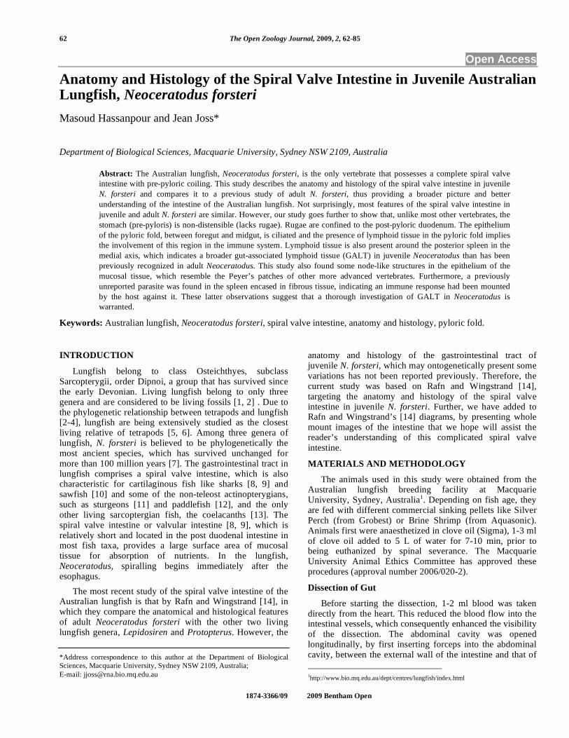
Three juvenile lungfish (2-4 years old), of different sizes were selected for gross anatomical analysis of the spiral valve and its components (Table 1). Following the removal of intestine from the abdominal cavity and flushing with PBS, the oesophagus end was clamped and the spiral valve filled with Bouin’s fixative solution [15] after which the rectum was also clamped (Fig. 1A). These specimens were kept in Bouin’s fixative for 48 hours to one week depending on the size of the gut being fixed. The three intestines (samples 1-3, Table 1) were dehydrated to 70% ethanol over 24-48 hours depending on their size, followed by several further changes of 70% alcohol until the tissue was effectively cleared of Bouin’s. These samples were cut and imaged in different orientations and investigated under a dissecting microscope for the different morphological aspects of the spiral valve. The bigger size spiral valve (sample 1) was cut longitudinally twice on left surface (Fig. 1B) and the spiral valve intestine was divided into three longitudinal sections (Fig. 1C, D) of middle section (Fig. 1E), dorsal section (Fig. 1F), and ventral section (Fig. 1G). Four transverse cuts were applied to the smaller spiral valve (sample 3) in different locations along the spiral valve (Fig. 2A), providing transverse sectional views of foregut-midgut area on the anterior end of the spiral valve (Fig. 2B), the anterior part of midgut (Fig. 2C), the middle part of midgut (Fig. 2D), and the posterior end of midgut (Fig. 2E). For the medium size gut (sample 2) the external wall of the intestine
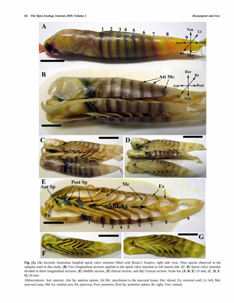
was peeled off (Fig. 3A, B) except in the pyloric fold area (Fig. 3C). Keeping the external wall in the pyloric fold area simplifies the orientation of the spiral valve, as the curve (arch) of the pyloric fold is on the left side. Therefore, the main anatomical features of the spiral valve intestine, which are dorsal (Fig. 4A), ventral (Fig. 4D), left lateral (Fig. 4C) and right lateral can be easily identified. Fig. (4B) shows the left-ventral view of the intestine. Further, the anterior (Fig. 5A, B), posterior (Fig. 5D), and middle parts of midgut (Fig. 5C) can be determined by their distance from the pyloric fold.
Histology
Three juvenile lungfish (2-4 years old) were used for histological analysis of the intestine (Table 1). The bigger and smaller specimen (sample 1 and 3), which were previously used for anatomical study were also used for histology. The 4th juvenile lungfish (sample 4) was dissected similarly, but the intestine was fixed in 10% neutral buffered formalin (NBF). This sample was also used for histological study. These specimens were used to provide longitudinal and transverse sections of the spiral valve in various locations. The tissue samples were processed for embedding in paraffin and serially sectioned at 5-8 μ. All even numbered slides were stained with Harris’ hematoxylin and eosin (H & E). The stained slides were examined and imaged under a BX50 Olympus light microscope equipped with iMac computer and Visicapture software. Our observations show that Bouin’s solution is preferred fixative for morpho-logy, while 10% NBF is better for histological analysis of the spiral valve intestine of Neoceratodus forsteri.
RESULTS
The Australian lungfish intestine is fusiform in shape, which is narrower at both ends and thicker in the middle (Figs. 1A, 6A). At its entrance into the peritoneal cavity the diameter increases dramatically from that of the esophagus and reaches the maximum thickness in the foregut. From the midgut on it decreases gradually until it opens into the cloaca. This straight structure anteriorly and ventrally is surrounded by the lobes of the liver, the lung dorsally, and the gonads laterally. Posteriorly, the paired kidneys are found dorsal to the gut. The intestine weight, after cleaning of any waste, varies from 3.5 to 7 g while its length is 10-13
Table 1. Four juvenile Australian lungfish (2-4 years old) of different sizes were used in this study. Three of them fixed in Bouin’s
solution and one sample fixed in 10% NBF. It’s worth saying that the Australian lungfish size is better determined by its
nutrition and ecosystem rather than by age.
Australian
Lungfish/Characteristics
Fish
Weight (g)
Fish Length
(cm)
Intestinal
Weight (g)
Intestinal
Length (cm)
Number
of Spirals
Fixed in Used for
Big fish (1st sample) 192 29 6.9 13 9 Bouin’s
fixative
Longitudinal sections
(morphology, histology)
Medium size fish (2nd
sample)
159 27 5.6 11.9 9 Bouin’s
fixative
Internal structure of spiral
valve (morphology) post
removal of external wall
Small fish (3rd sample) 100 23 3.5 10 9 Bouin’s
fixative
Transverse sections
(morphology, histology)
4th sample 116 34 - - 9 10% NBF Histology
64 The Open Zoology Journal, 2009, Volume 2 Hassanpour and Joss
Fig. (1). (A) Juvenile Australian lungfish spiral valve intestine filled with Bouin’s fixative, right side view. Nine spirals observed in the
samples used in this study. (B) Two longitudinal sections applied to the spiral valve intestine in left lateral side. (C, D) Spiral valve intestine
divided in three longitudinal sections. (E) Middle section, (F) Dorsal section, and (G) Ventral section. Scale bar (A, B, E) 10 mm, (C, D, F,
G) 20 mm.
Abbreviations: Ant: anterior, Ant Sp: anterior spleen, Att Mc: attachment to the mucosal tissue, Dor: dorsal, Ex: external wall, Lt: left, Mc:
mucosal issue, Md Ax: medial axis, Pn: pancreas, Post: posterior, Post Sp: posterior spleen, Rt: right, Ven: ventral.

Anatomy and Histology of the Spiral Valve Intestine The Open Zoology Journal, 2009, Volume 2 65
Fig. (2). Transverse sections of the spiral valve intestine of Australian lungfish post fixation in Bouin’s solution. (A) Four transverse sections
in different locations along the spiral valve intestine applied. (B) Anterior view of the transverse section of anterior end of the spiral valve
intestine (foregut-midgut area). (C) Posterior view of the transverse section of anterior part of midgut. (D) Anterior view of the transverse
section of middle part of the midgut. (E) Posterior view of the transverse section of posterior end of midgut (hindgut). Scale bar (A) 10 mm,
(B, C) 5 mm, (D, E) 3 mm.
Abbreviations: Ex: external wall, Mc: mucosal issue, Pn: pancreas, Py: pyloric fold, Sp: spleen.
66 The Open Zoology Journal, 2009, Volume 2 Hassanpour and Joss
Fig. (3). Removal of the external wall of spiral valve intestine of N. forsteri post fixation in Bouin’s solution. (A) (Mirror image) Right lateral
view, (B) Ventral view, (C) Right lateral view, Scale bar (A, B, C) 10 mm.
Abbreviations: Ant: anterior, Dor: dorsal, Ex: external wall, Lt: left, Mc: mucosal issue, Post: posterior, Py: pyloric fold, Rt: right, Ven:
ventral.

Anatomy and Histology of the Spiral Valve Intestine The Open Zoology Journal, 2009, Volume 2 67
Fig. (4). The main orientations of the spiral valve intestine after removal of external wall post fixation in Bouin’s solution. (A) (Mirror image)
Dorsal view, (B) (Mirror image) Left-ventral view, (C) (Mirror image) Left lateral view, (D) (Mirror image) Ventral view. Coiling process is
pre-pyloric and clockwise in anterior-posterior direction. The diameter of infolding tissue (mucosal tissue) is greater than the diameter of the
actual intestinal lumen. This makes the spirals form cornet-like coils (cones), which overlap each other in midgut. The apices of these cones
point in anterior direction. The first coil is wide, about half of which is foregut. Scale bar (A, B, C, D) 10 mm.
Abbreviations: Ant: anterior, Dor: dorsal, Lt: left, Lt-Dor: left-dorsal, Lt-Ven: left-ventral, Pn: pancreas, Post: posterior, Py: pyloric fold, Rg:
rugae, Rt: right, Rt-Dor: right-dorsal, Rt-Ven: right-ventral, Ven: ventral.
68 The Open Zoology Journal, 2009, Volume 2 Hassanpour and Joss
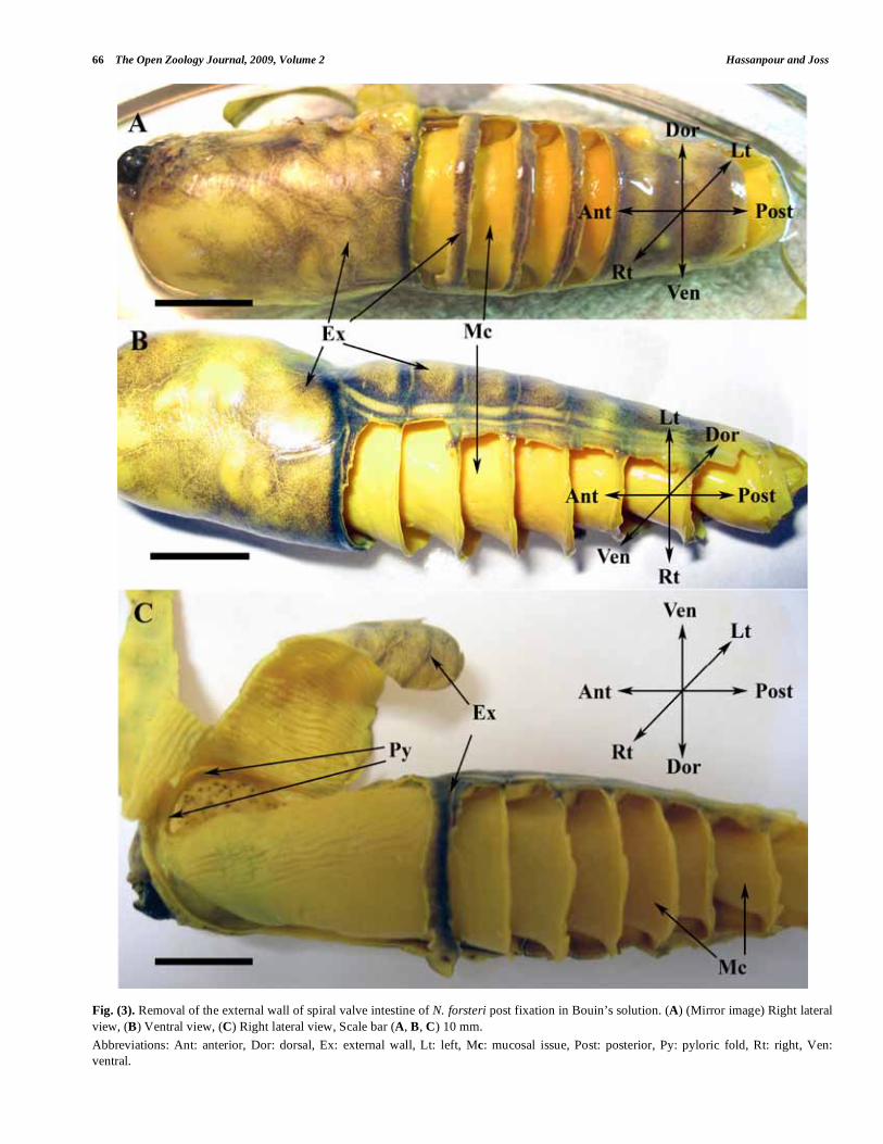
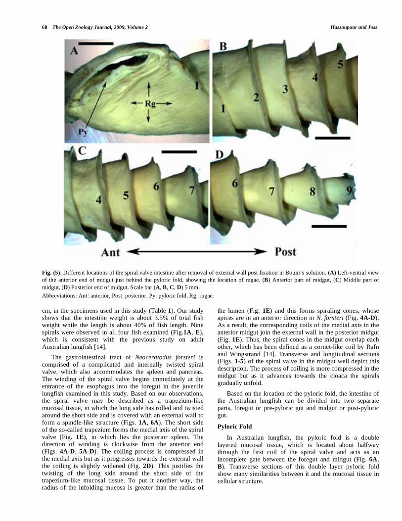
Fig. (5). Different locations of the spiral valve intestine after removal of external wall post fixation in Bouin’s solution. (A) Left-ventral view
of the anterior end of midgut just behind the pyloric fold, showing the location of rugae. (B) Anterior part of midgut, (C) Middle part of
midgut, (D) Posterior end of midgut. Scale bar (A, B, C, D) 5 mm.
Abbreviations: Ant: anterior, Post: posterior, Py: pyloric fold, Rg: rugae.
cm, in the specimens used in this study (Table 1). Our study shows that the intestine weight is about 3.5% of total fish weight while the length is about 40% of fish length. Nine spirals were observed in all four fish examined (Fig.1A, E), which is consistent with the previous study on adult Australian lungfish [14].
The gastrointestinal tract of Neoceratodus forsteri is comprised of a complicated and internally twisted spiral valve, which also accommodates the spleen and pancreas. The winding of the spiral valve begins immediately at the entrance of the esophagus into the foregut in the juvenile lungfish examined in this study. Based on our observations, the spiral valve may be described as a trapezium-like mucosal tissue, in which the long side has rolled and twisted around the short side and is covered with an external wall to form a spindle-like structure (Figs. 1A, 6A). The short side of the so-called trapezium forms the medial axis of the spiral valve (Fig. 1E), in which lies the posterior spleen. The direction of winding is clockwise from the anterior end (Figs. 4A-D, 5A-D). The coiling process is compressed in the medial axis but as it progresses towards the external wall the coiling is slightly widened (Fig. 2D). This justifies the twisting of the long side around the short side of the trapezium-like mucosal tissue. To put it another way, the radius of the infolding mucosa is greater than the radius of
the lumen (Fig. 1E) and this forms spiraling cones, whose apices are in an anterior direction in N. forsteri (Fig. 4A-D). As a result, the corresponding coils of the medial axis in the anterior midgut join the external wall in the posterior midgut (Fig. 1E). Thus, the spiral cones in the midgut overlap each other, which has been defined as a cornet-like coil by Rafn and Wingstrand [14]. Transverse and longitudinal sections (Figs. 1-5) of the spiral valve in the midgut well depict this description. The process of coiling is more compressed in the midgut but as it advances towards the cloaca the spirals gradually unfold.
Based on the location of the pyloric fold, the intestine of the Australian lungfish can be divided into two separate parts, foregut or pre-pyloric gut and midgut or post-pyloric gut.
Pyloric Fold
In Australian lungfish, the pyloric fold is a double layered mucosal tissue, which is located about halfway through the first coil of the spiral valve and acts as an incomplete gate between the foregut and midgut (Fig. 6A, B). Transverse sections of this double layer pyloric fold show many similarities between it and the mucosal tissue in cellular structure.

Anatomy and Histology of the Spiral Valve Intestine The Open Zoology Journal, 2009, Volume 2 69
The pyloric fold may be described as a deep and straight evagination of the epithelium of the mucosal tissue between the foregut and midgut inside the lumen of spiral valve, which can be compared to venous valves (Fig. 6C). However, these pyloric mucosal flaps are too small to act as a complete valve (Fig. 6D). The pyloric fold only partially obstructs the movement of ingesta in the intestinal lumen, acting as an incomplete gate. Nevertheless, the pyloric fold
flaps are directed towards the midgut in anterior-posterior direction, which implies that the pyloric fold can at least partially prevent the backflow of the ingesta from midgut to foregut.
Histologically the pyloric fold consists of an epithelial layer, a lamina propria, and a submucosa. Like mucosal tissue either side of the pyloric fold is covered with an
Fig. (6). Pyloric fold in juvenile Australian lungfish. (A) Ventral view of the spiral valve intestine filled with Bouin’s fixative. Small arrows
show the attachment of the pyloric fold to the external wall. (B) Right-ventral view of the anterior part of midgut, where external wall is
peeled off. Pyloric fold looks like an oval-shape ring, after which rugae are located. (C) Transverse section in foregut-midgut region. Pyloric
fold is a double-layer mucosal tissue separating midgut from foregut, which resembles a venous valve. (D) Ventral view of the anterior end
of midgut just behind pyloric fold, where rugae are present. Pyloric fold acts as an incomplete gate between foregut and midgut. The entry of
pancreatic and common bile duct is just behind the pyloric fold. Scale bar (A, B) 10 mm, (C) 5 mm, (D) 3 mm.
Abbreviations: Ant: anterior, Dor: dorsal, En P/CBD: entries of pancreatic and common bile ducts, Lt: left, Mc: mucosal tissue, Pn: pancreas,
Post: posterior, Py: pyloric fold, Rg: rugae, Rt: right, Sp: spleen, Ven: ventral.
70 The Open Zoology Journal, 2009, Volume 2 Hassanpour and Joss
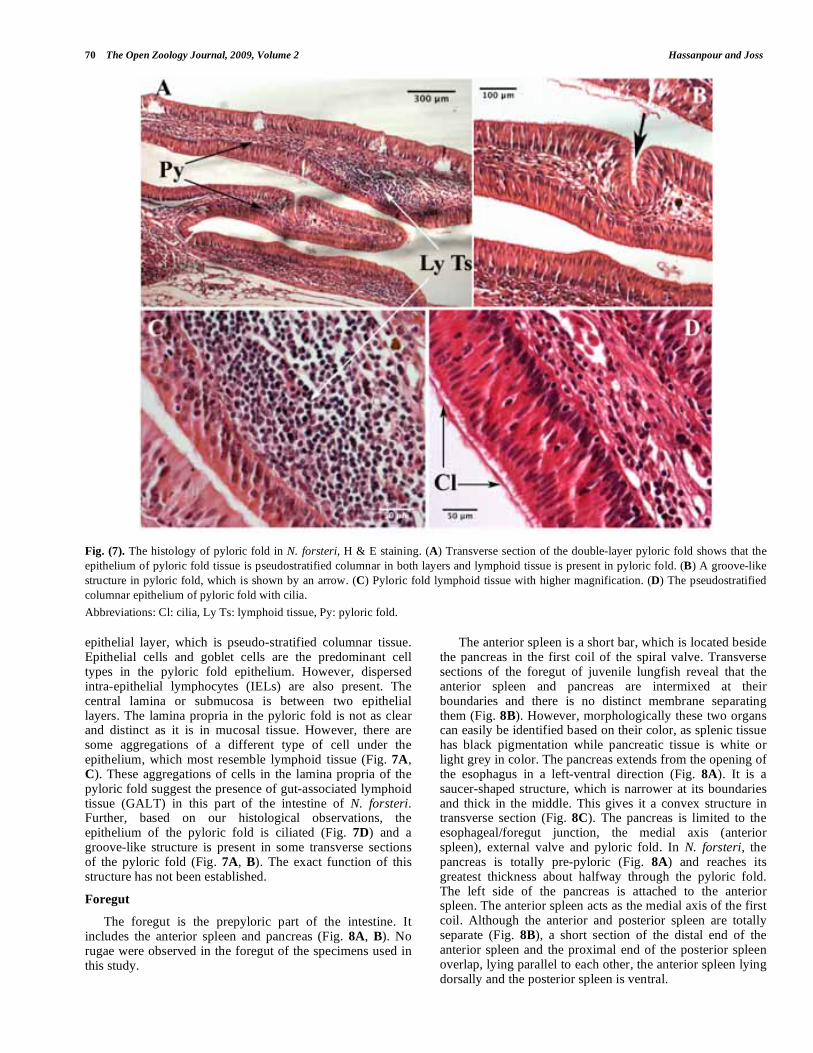
Fig. (7). The histology of pyloric fold in N. forsteri, H & E staining. (A) Transverse section of the double-layer pyloric fold shows that the
epithelium of pyloric fold tissue is pseudostratified columnar in both layers and lymphoid tissue is present in pyloric fold. (B) A groove-like
structure in pyloric fold, which is shown by an arrow. (C) Pyloric fold lymphoid tissue with higher magnification. (D) The pseudostratified
columnar epithelium of pyloric fold with cilia.
Abbreviations: Cl: cilia, Ly Ts: lymphoid tissue, Py: pyloric fold.
epithelial layer, which is pseudo-stratified columnar tissue. Epithelial cells and goblet cells are the predominant cell types in the pyloric fold epithelium. However, dispersed intra-epithelial lymphocytes (IELs) are also present. The central lamina or submucosa is between two epithelial layers. The lamina propria in the pyloric fold is not as clear and distinct as it is in mucosal tissue. However, there are some aggregations of a different type of cell under the epithelium, which most resemble lymphoid tissue (Fig. 7A, C). These aggregations of cells in the lamina propria of the pyloric fold suggest the presence of gut-associated lymphoid tissue (GALT) in this part of the intestine of N. forsteri. Further, based on our histological observations, the epithelium of the pyloric fold is ciliated (Fig. 7D) and a groove-like structure is present in some transverse sections of the pyloric fold (Fig. 7A, B). The exact function of this structure has not been established.
Foregut
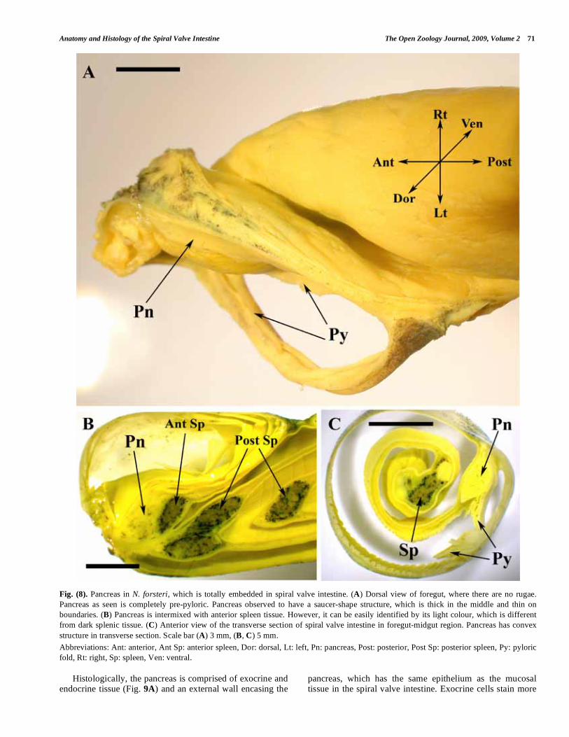
The foregut is the prepyloric part of the intestine. It includes the anterior spleen and pancreas (Fig. 8A, B). No rugae were observed in the foregut of the specimens used in this study.
The anterior spleen is a short bar, which is located beside the pancreas in the first coil of the spiral valve. Transverse sections of the foregut of juvenile lungfish reveal that the anterior spleen and pancreas are intermixed at their boundaries and there is no distinct membrane separating them (Fig. 8B). However, morphologically these two organs can easily be identified based on their color, as splenic tissue has black pigmentation while pancreatic tissue is white or light grey in color. The pancreas extends from the opening of the esophagus in a left-ventral direction (Fig. 8A). It is a saucer-shaped structure, which is narrower at its boundaries and thick in the middle. This gives it a convex structure in transverse section (Fig. 8C). The pancreas is limited to the esophageal/foregut junction, the medial axis (anterior spleen), external valve and pyloric fold. In N. forsteri, the pancreas is totally pre-pyloric (Fig. 8A) and reaches its greatest thickness about halfway through the pyloric fold. The left side of the pancreas is attached to the anterior spleen. The anterior spleen acts as the medial axis of the first coil. Although the anterior and posterior spleen are totally separate (Fig. 8B), a short section of the distal end of the anterior spleen and the proximal end of the posterior spleen overlap, lying parallel to each other, the anterior spleen lying dorsally and the posterior spleen is ventral.
Anatomy and Histology of the Spiral Valve Intestine The Open Zoology Journal, 2009, Volume 2 71
Fig. (8). Pancreas in N. forsteri, which is totally embedded in spiral valve intestine. (A) Dorsal view of foregut, where there are no rugae.
Pancreas as seen is completely pre-pyloric. Pancreas observed to have a saucer-shape structure, which is thick in the middle and thin on
boundaries. (B) Pancreas is intermixed with anterior spleen tissue. However, it can be easily identified by its light colour, which is different
from dark splenic tissue. (C) Anterior view of the transverse section of spiral valve intestine in foregut-midgut region. Pancreas has convex
structure in transverse section. Scale bar (A) 3 mm, (B, C) 5 mm.
Abbreviations: Ant: anterior, Ant Sp: anterior spleen, Dor: dorsal, Lt: left, Pn: pancreas, Post: posterior, Post Sp: posterior spleen, Py: pyloric
fold, Rt: right, Sp: spleen, Ven: ventral.
Histologically, the pancreas is comprised of exocrine and endocrine tissue (Fig. 9A) and an external wall encasing the
pancreas, which has the same epithelium as the mucosal tissue in the spiral valve intestine. Exocrine cells stain more
72 The Open Zoology Journal, 2009, Volume 2 Hassanpour and Joss
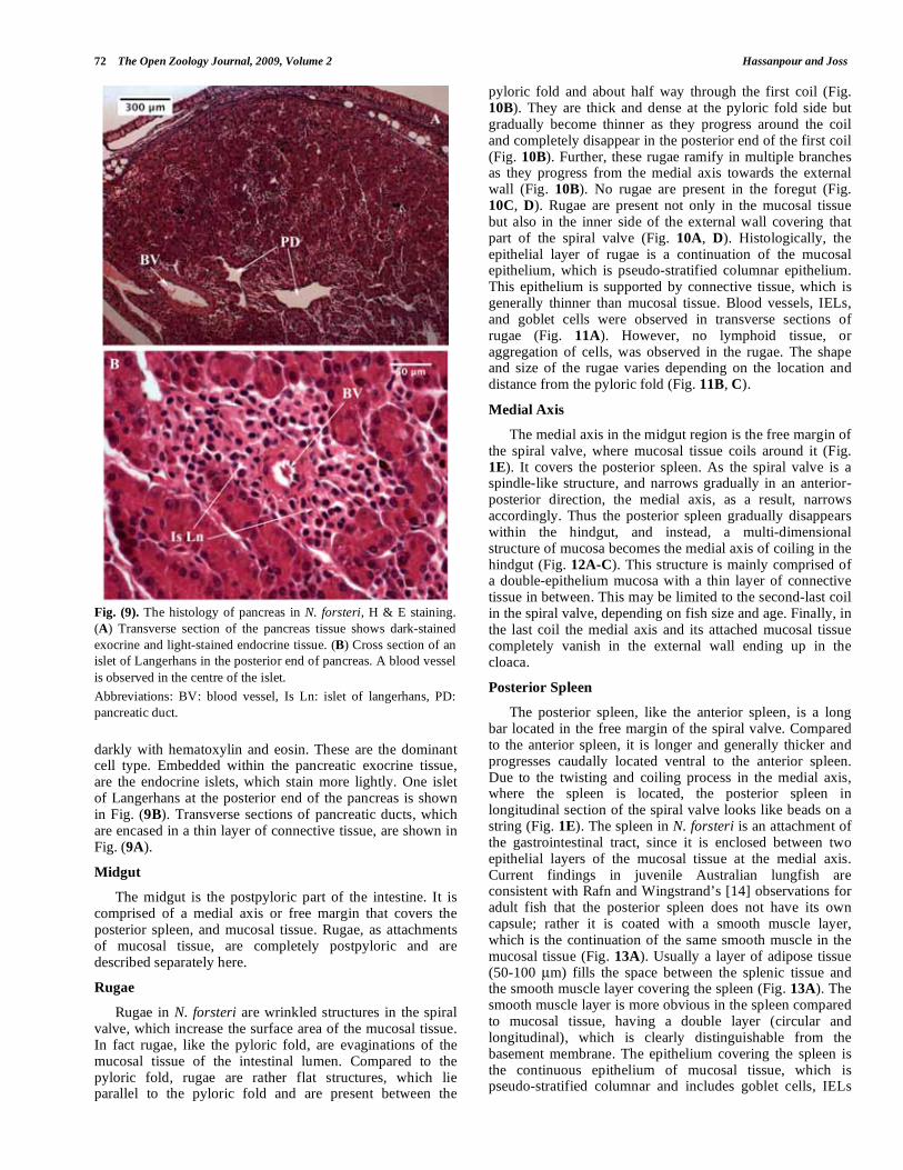
darkly with hematoxylin and eosin. These are the dominant cell type. Embedded within the pancreatic exocrine tissue, are the endocrine islets, which stain more lightly. One islet of Langerhans at the posterior end of the pancreas is shown in Fig. (9B). Transverse sections of pancreatic ducts, which are encased in a thin layer of connective tissue, are shown in Fig. (9A).
Midgut
The midgut is the postpyloric part of the intestine. It is comprised of a medial axis or free margin that covers the posterior spleen, and mucosal tissue. Rugae, as attachments of mucosal tissue, are completely postpyloric and are described separately here.
Rugae
Rugae in N. forsteri are wrinkled structures in the spiral valve, which increase the surface area of the mucosal tissue. In fact rugae, like the pyloric fold, are evaginations of the mucosal tissue of the intestinal lumen. Compared to the pyloric fold, rugae are rather flat structures, which lie parallel to the pyloric fold and are present between the
pyloric fold and about half way through the first coil (Fig. 10B). They are thick and dense at the pyloric fold side but gradually become thinner as they progress around the coil and completely disappear in the posterior end of the first coil (Fig. 10B). Further, these rugae ramify in multiple branches as they progress from the medial axis towards the external wall (Fig. 10B). No rugae are present in the foregut (Fig. 10C, D). Rugae are present not only in the mucosal tissue but also in the inner side of the external wall covering that part of the spiral valve (Fig. 10A, D). Histologically, the epithelial layer of rugae is a continuation of the mucosal epithelium, which is pseudo-stratified columnar epithelium. This epithelium is supported by connective tissue, which is generally thinner than mucosal tissue. Blood vessels, IELs, and goblet cells were observed in transverse sections of rugae (Fig. 11A). However, no lymphoid tissue, or aggregation of cells, was observed in the rugae. The shape and size of the rugae varies depending on the location and distance from the pyloric fold (Fig. 11B, C).
Medial Axis
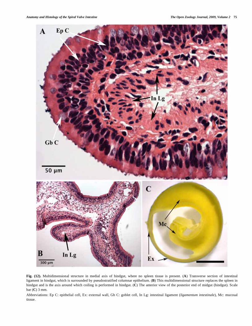
The medial axis in the midgut region is the free margin of the spiral valve, where mucosal tissue coils around it (Fig. 1E). It covers the posterior spleen. As the spiral valve is a spindle-like structure, and narrows gradually in an anterior-posterior direction, the medial axis, as a result, narrows accordingly. Thus the posterior spleen gradually disappears within the hindgut, and instead, a multi-dimensional structure of mucosa becomes the medial axis of coiling in the hindgut (Fig. 12A-C). This structure is mainly comprised of a double-epithelium mucosa with a thin layer of connective tissue in between. This may be limited to the second-last coil in the spiral valve, depending on fish size and age. Finally, in the last coil the medial axis and its attached mucosal tissue completely vanish in the external wall ending up in the cloaca.
Posterior Spleen
The posterior spleen, like the anterior spleen, is a long bar located in the free margin of the spiral valve. Compared to the anterior spleen, it is longer and generally thicker and progresses caudally located ventral to the anterior spleen. Due to the twisting and coiling process in the medial axis, where the spleen is located, the posterior spleen in longitudinal section of the spiral valve looks like beads on a string (Fig. 1E). The spleen in N. forsteri is an attachment of the gastrointestinal tract, since it is enclosed between two epithelial layers of the mucosal tissue at the medial axis. Current findings in juvenile Australian lungfish are consistent with Rafn and Wingstrand’s [14] observations for adult fish that the posterior spleen does not have its own capsule; rather it is coated with a smooth muscle layer, which is the continuation of the same smooth muscle in the mucosal tissue (Fig. 13A). Usually a layer of adipose tissue (50-100 μm) fills the space between the splenic tissue and the smooth muscle layer covering the spleen (Fig. 13A). The smooth muscle layer is more obvious in the spleen compared to mucosal tissue, having a double layer (circular and longitudinal), which is clearly distinguishable from the basement membrane. The epithelium covering the spleen is the continuous epithelium of mucosal tissue, which is pseudo-stratified columnar and includes goblet cells, IELs
Fig. (9). The histology of pancreas in N. forsteri, H & E staining.
(A) Transverse section of the pancreas tissue shows dark-stained
exocrine and light-stained endocrine tissue. (B) Cross section of an
islet of Langerhans in the posterior end of pancreas. A blood vessel
is observed in the centre of the islet.
Abbreviations: BV: blood vessel, Is Ln: islet of langerhans, PD:
pancreatic duct.

Anatomy and Histology of the Spiral Valve Intestine The Open Zoology Journal, 2009, Volume 2 73
Fig. (10). In N. forsteri, rugae are present in the anterior end of the first coil in midgut. (A) Rugae are present in the inner side of the external
wall that covers the anterior end of the first coil in midgut. (B) (Mirror image) Ventral view of the anterior end of midgut. Rugae are present
in the first half of the first coil in midgut. Rugae are finger-like villi that are parallel to the pyloric fold and ramify into multiple branches from
medial axis to the external wall direction. Pancreatic and common bile ducts (CBD) enter into midgut just behind and on ventral end of
pyloric fold. (C) (Mirror image) Left lateral view of foregut-midgut region. Rugae are absent in pre-pyloric gut (foregut). (D) Posterior view
of the transverse section in foregut-midgut region. Rugae are absent in foregut but present beyond the double-layer pyloric fold. Scale bar (A,
B, C, D) 5 mm.
Abbreviations: Ant: anterior, Dor: dorsal, En P/CBD: entries of pancreatic and common bile ducts, FG: foregut, Lt: left, MG: midgut, Pn:
pancreas, Post: posterior, Py: pyloric fold, Rg: rugae, Rt: right, Ven: ventral.

74 The Open Zoology Journal, 2009, Volume 2 Hassanpour and Joss
Fig. (11). The histology of rugae in N. forsteri, H & E staining. (A) Rugae like mucosal tissue, have pseudostratified columnar epithelium,
which is mainly comprised of epithelial cells and goblet cells. However, IELs are also seen in rugae. (B) Rugae are observed in the inner side
of external wall. (C) Rugae have different shapes depending on their location.
Abbreviations: Ep C: epithelial cell, Ex: external wall, Gb C: goblet cell, IEL: intraepithelial lymphocyte.
and epithelial cells. The number of goblet cells is greater in the corners where the tissue turns to follow the coiling process. Also some aggregations of cells are found between the epithelial layer and the spleen’s smooth muscle layer (Fig. 13B, C). These are lymphoid tissue (GALT), not reported by Rafn and Wingstrand [14] in adult lungfish. Pigment cells observed scattered in different areas across the spleen are most abundant in the cortical area (Fig. 8B, C). Our histological observations also confirm the presence of a cord of connective tissue at the apex of the posterior spleen along the free margin of the spiral valve, which is oval in transverse section (Fig. 13A, D). Rafn and Wingstrand [14]
referred to this structure as the ligamentum intestinale (intestinal ligament). The intestinal ligament extends further in the posterior end of the hindgut, where there is no splenic tissue (Fig. 12A, B) and seems to act as a ‘backbone’ for the spiral valve.
Mucosal Tissue
Mucosal tissue (Fig. 14A-E) is the main part of the intestine in N. forsteri. It is limited to the medial axis and external wall on both sides (Fig. 14B-D), and the pyloric fold and cloaca at either end, respectively. The epithelium layer that covers other components of the spiral valve includes the
Anatomy and Histology of the Spiral Valve Intestine The Open Zoology Journal, 2009, Volume 2 75
Fig. (12). Multidimensional structure in medial axis of hindgut, where no spleen tissue is present. (A) Transverse section of intestinal
ligament in hindgut, which is surrounded by pseudostratified columnar epithelium. (B) This multidimensional structure replaces the spleen in
hindgut and is the axis around which coiling is performed in hindgut. (C) The anterior view of the posterior end of midgut (hindgut). Scale
bar (C) 3 mm.
Abbreviations: Ep C: epithelial cell, Ex: external wall, Gb C: goblet cell, In Lg: intestinal ligament (ligamentum intestinale), Mc: mucosal
tissue.
76 The Open Zoology Journal, 2009, Volume 2 Hassanpour and Joss
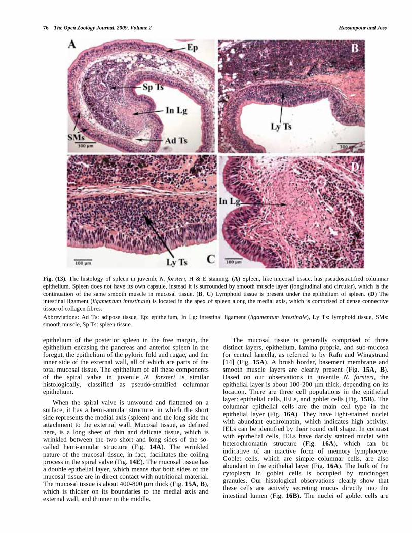
Fig. (13). The histology of spleen in juvenile N. forsteri, H & E staining. (A) Spleen, like mucosal tissue, has pseudostratified columnar
epithelium. Spleen does not have its own capsule, instead it is surrounded by smooth muscle layer (longitudinal and circular), which is the
continuation of the same smooth muscle in mucosal tissue. (B, C) Lymphoid tissue is present under the epithelium of spleen. (D) The
intestinal ligament (ligamentum intestinale) is located in the apex of spleen along the medial axis, which is comprised of dense connective
tissue of collagen fibres.
Abbreviations: Ad Ts: adipose tissue, Ep: epithelium, In Lg: intestinal ligament (ligamentum intestinale), Ly Ts: lymphoid tissue, SMs:
smooth muscle, Sp Ts: spleen tissue.
epithelium of the posterior spleen in the free margin, the epithelium encasing the pancreas and anterior spleen in the foregut, the epithelium of the pyloric fold and rugae, and the inner side of the external wall, all of which are parts of the total mucosal tissue. The epithelium of all these components of the spiral valve in juvenile N. forsteri is similar histologically, classified as pseudo-stratified columnar epithelium.
When the spiral valve is unwound and flattened on a surface, it has a hemi-annular structure, in which the short side represents the medial axis (spleen) and the long side the attachment to the external wall. Mucosal tissue, as defined here, is a long sheet of thin and delicate tissue, which is wrinkled between the two short and long sides of the so-called hemi-annular structure (Fig. 14A). The wrinkled nature of the mucosal tissue, in fact, facilitates the coiling process in the spiral valve (Fig. 14E). The mucosal tissue has a double epithelial layer, which means that both sides of the mucosal tissue are in direct contact with nutritional material. The mucosal tissue is about 400-800 μm thick (Fig. 15A, B), which is thicker on its boundaries to the medial axis and external wall, and thinner in the middle.
The mucosal tissue is generally comprised of three distinct layers, epithelium, lamina propria, and sub-mucosa (or central lamella, as referred to by Rafn and Wingstrand [14] (Fig. 15A). A brush border, basement membrane and smooth muscle layers are clearly present (Fig. 15A, B). Based on our observations in juvenile N. forsteri, the epithelial layer is about 100-200 μm thick, depending on its location. There are three cell populations in the epithelial layer: epithelial cells, IELs, and goblet cells (Fig. 15B). The columnar epithelial cells are the main cell type in the epithelial layer (Fig. 16A). They have light-stained nuclei with abundant euchromatin, which indicates high activity. IELs can be identified by their round cell shape. In contrast with epithelial cells, IELs have darkly stained nuclei with heterochromatin structure (Fig. 16A), which can be indicative of an inactive form of memory lymphocyte. Goblet cells, which are simple columnar cells, are also abundant in the epithelial layer (Fig. 16A). The bulk of the cytoplasm in goblet cells is occupied by mucinogen granules. Our histological observations clearly show that these cells are actively secreting mucus directly into the intestinal lumen (Fig. 16B). The nuclei of goblet cells are

Anatomy and Histology of the Spiral Valve Intestine The Open Zoology Journal, 2009, Volume 2 77
Fig. (14). The mucosal tissue of the spiral valve intestine in Australian lungfish. (A) The hemi-annular structure of unwound and flattened
spiral valve intestine. The mucosal tissue is wrinkled between medial axis and external wall. (B) Ventral view of midgut, where external wall
is removed from right lateral side. Mucosal tissue is densely coiled in midgut and is limited to the medial axis and external wall. (C) Anterior
view of the transverse section of the middle part of midgut, where mucosal tissue is twisted around spleen. (D) Longitudinal section of
midgut. The mucosal tissue is limited to the medial axis and external wall. (E) (Mirror image) The actual appearance of the mucosal tissue in
midgut where external wall is removed. The infolding mucosal tissue forms spiral cones, whose apexes face in anterior direction and they
overlap each other. Scale bar (A, B) 10 mm, (C) 3mm, (D, E) 5 mm.
Abbreviations: Ant: anterior, Att Ex: attachment to the external wall, Ex: external wall, Mc: mucosal issue, Md Ax: medial axis, Post:
posterior, Sp: spleen.
78 The Open Zoology Journal, 2009, Volume 2 Hassanpour and Joss
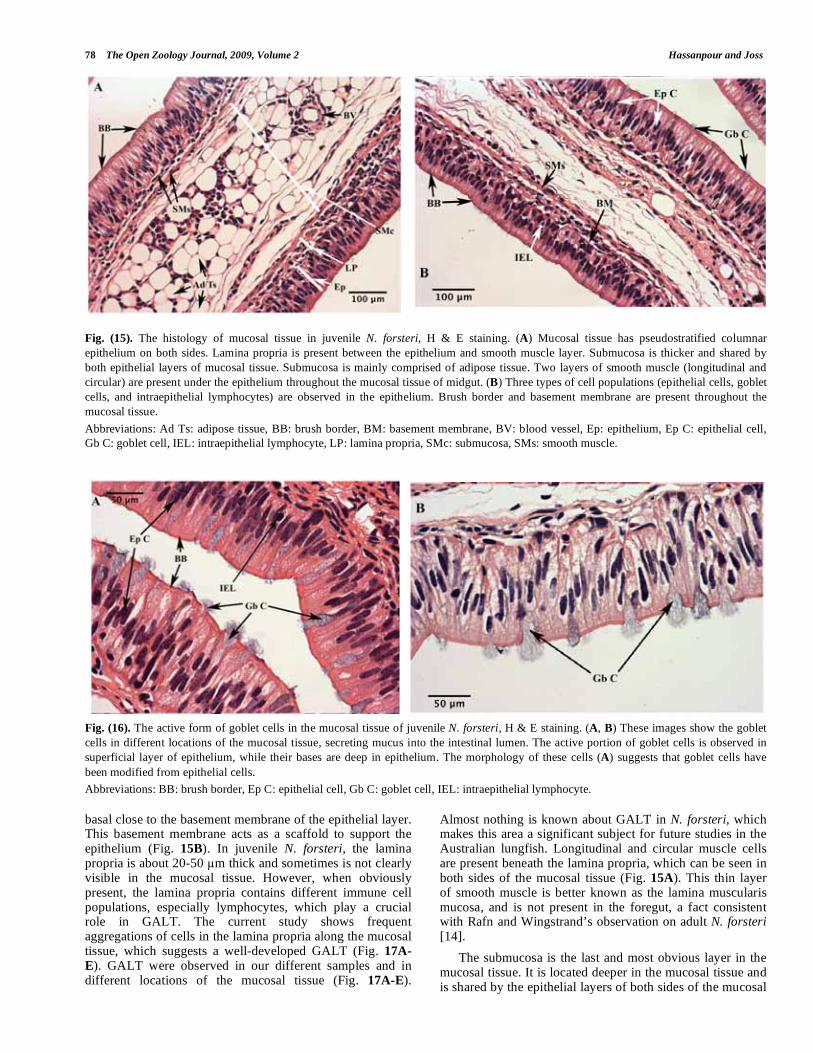
Fig. (15). The histology of mucosal tissue in juvenile N. forsteri, H & E staining. (A) Mucosal tissue has pseudostratified columnar
epithelium on both sides. Lamina propria is present between the epithelium and smooth muscle layer. Submucosa is thicker and shared by
both epithelial layers of mucosal tissue. Submucosa is mainly comprised of adipose tissue. Two layers of smooth muscle (longitudinal and
circular) are present under the epithelium throughout the mucosal tissue of midgut. (B) Three types of cell populations (epithelial cells, goblet
cells, and intraepithelial lymphocytes) are observed in the epithelium. Brush border and basement membrane are present throughout the
mucosal tissue.
Abbreviations: Ad Ts: adipose tissue, BB: brush border, BM: basement membrane, BV: blood vessel, Ep: epithelium, Ep C: epithelial cell,
Gb C: goblet cell, IEL: intraepithelial lymphocyte, LP: lamina propria, SMc: submucosa, SMs: smooth muscle.
Fig. (16). The active form of goblet cells in the mucosal tissue of juvenile N. forsteri, H & E staining. (A, B) These images show the goblet
cells in different locations of the mucosal tissue, secreting mucus into the intestinal lumen. The active portion of goblet cells is observed in
superficial layer of epithelium, while their bases are deep in epithelium. The morphology of these cells (A) suggests that goblet cells have
been modified from epithelial cells.
Abbreviations: BB: brush border, Ep C: epithelial cell, Gb C: goblet cell, IEL: intraepithelial lymphocyte.
basal close to the basement membrane of the epithelial layer. This basement membrane acts as a scaffold to support the epithelium (Fig. 15B). In juvenile N. forsteri, the lamina propria is about 20-50 m thick and sometimes is not clearly visible in the mucosal tissue. However, when obviously present, the lamina propria contains different immune cell populations, especially lymphocytes, which play a crucial role in GALT. The current study shows frequent aggregations of cells in the lamina propria along the mucosal tissue, which suggests a well-developed GALT (Fig. 17A-
E). GALT were observed in our different samples and in different locations of the mucosal tissue (Fig. 17A-E).
Almost nothing is known about GALT in N. forsteri, which makes this area a significant subject for future studies in the Australian lungfish. Longitudinal and circular muscle cells are present beneath the lamina propria, which can be seen in both sides of the mucosal tissue (Fig. 15A). This thin layer of smooth muscle is better known as the lamina muscularis mucosa, and is not present in the foregut, a fact consistent with Rafn and Wingstrand’s observation on adult N. forsteri [14].
The submucosa is the last and most obvious layer in the mucosal tissue. It is located deeper in the mucosal tissue and is shared by the epithelial layers of both sides of the mucosal

Anatomy and Histology of the Spiral Valve Intestine The Open Zoology Journal, 2009, Volume 2 79
Fig. (17). An aggregation of cells (lymphoid tissue) in the mucosal tissue of juvenile N. forsteri, H & E staining. (A) The aggregation of cells
is observed under the epithelium (lamina propria), which is associated with lymphoid tissue. (B, C, D, E) Lymphoid tissue is present in
different samples and different locations of mucosal tissue – several examples.
Abbreviations: Ad Ts: adipose tissue, BV: blood vessel, Ep: epithelium, Ly Ts: lymphoid tissue, SMs: smooth muscle, Sp: spleen.
80 The Open Zoology Journal, 2009, Volume 2 Hassanpour and Joss
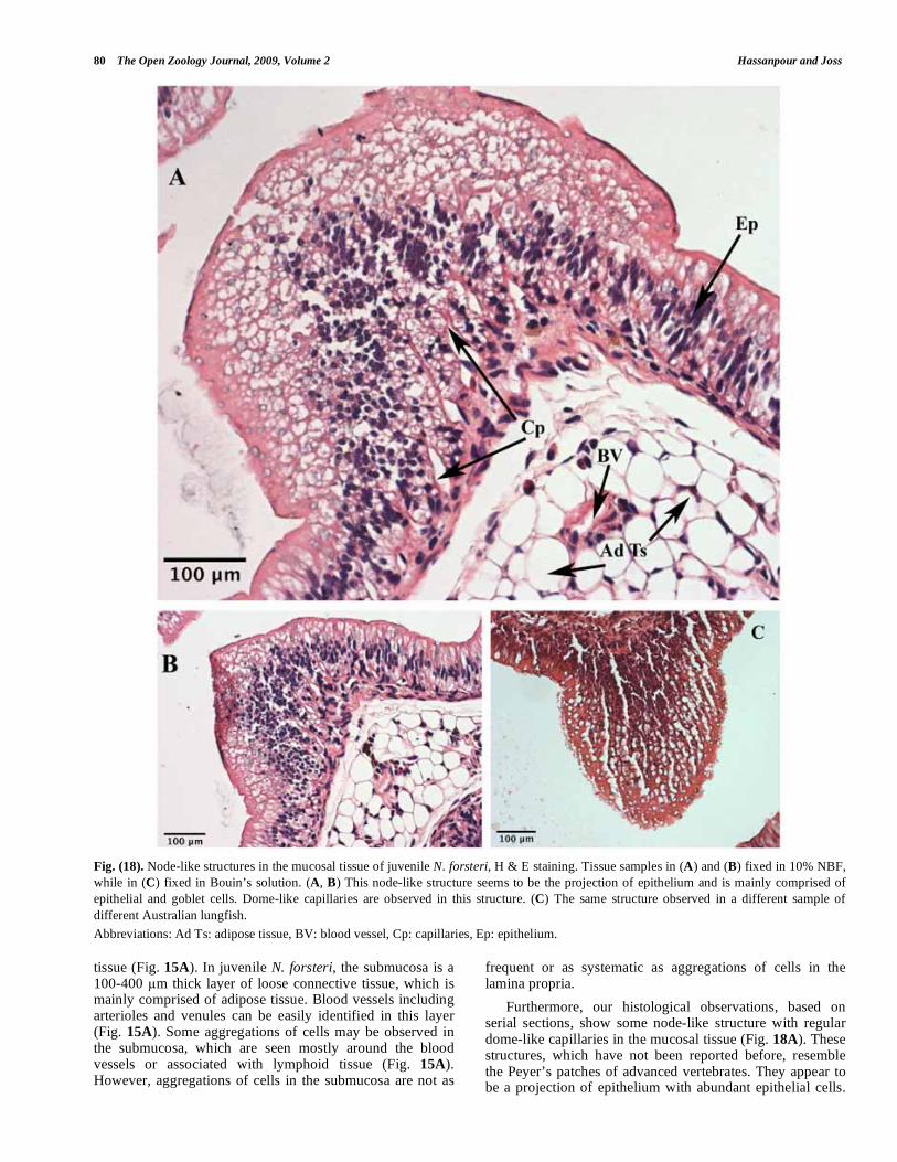
Fig. (18). Node-like structures in the mucosal tissue of juvenile N. forsteri, H & E staining. Tissue samples in (A) and (B) fixed in 10% NBF,
while in (C) fixed in Bouin’s solution. (A, B) This node-like structure seems to be the projection of epithelium and is mainly comprised of
epithelial and goblet cells. Dome-like capillaries are observed in this structure. (C) The same structure observed in a different sample of
different Australian lungfish.
Abbreviations: Ad Ts: adipose tissue, BV: blood vessel, Cp: capillaries, Ep: epithelium.
tissue (Fig. 15A). In juvenile N. forsteri, the submucosa is a 100-400 m thick layer of loose connective tissue, which is mainly comprised of adipose tissue. Blood vessels including arterioles and venules can be easily identified in this layer (Fig. 15A). Some aggregations of cells may be observed in the submucosa, which are seen mostly around the blood vessels or associated with lymphoid tissue (Fig. 15A). However, aggregations of cells in the submucosa are not as
frequent or as systematic as aggregations of cells in the lamina propria.
Furthermore, our histological observations, based on serial sections, show some node-like structure with regular dome-like capillaries in the mucosal tissue (Fig. 18A). These structures, which have not been reported before, resemble the Peyer’s patches of advanced vertebrates. They appear to be a projection of epithelium with abundant epithelial cells.
Anatomy and Histology of the Spiral Valve Intestine The Open Zoology Journal, 2009, Volume 2 81
However goblet cells are frequent in the superficial layer of these structures as well. A basement membrane is not very clear but a smooth muscle layer is clearly visible. A big arteriole was also observed beneath this structure deep in the submucosal layer. These structures were seen in two different samples of juvenile N. forsteri during this study (Fig. 18B, C).
External Wall
The external wall, as defined in this study, is a thin layer, which encloses the spiral valve and gives it a spindle-like structure (Figs. 3A-C, 14B-C).
From an outside view, the external wall is grey in color with some patchy dark pigmentation. Where the external side of the mucosal tissue, opposite to the medial axis side, joins the external wall, it appears as a ring in the outside view, one for each coil of the spiral valve (Fig. 1A, B). These rings are continuous across the spiral valve and represent the attachment of mucosal tissue to the external wall.
Based on histological differences, the external wall of the midgut can be divided into an inner side and an outer side layer, separated by the outer layer of smooth muscle (Fig. 19). The inner side is comprised of a serous membrane and a connective tissue layer. The serous membrane is the outermost layer of the spiral valve, which is directly exposed
to the abdominal cavity. The connective tissue layer of the outer side carries blood vessels that nourish the serous membrane cells. The inner side of the external wall has the same histological structure as mucosal tissue; i.e., it is comprised of epithelium, basement membrane, and lamina propria. The smooth muscle layer beneath the lamina propria and submucosa are readily apparent. Compared to the mucosal epithelium, the goblet cells are more common here.
Gut Parasite
In our specimens there were frequent node-like structures in the external wall of the spiral valve intestine (Fig. 20A, C). On dissection they appear to be nematode parasites as yet undescribed. In addition, sections of the spleen reveal the presence of a parasite, also undescribed, which has been confined with a thick layer of fibrous tissue (Fig. 20B, D-G).
DISCUSSION
Prior to the above descriptions, the most recent study of the spiral valve intestine of the Australian lungfish was that by Rafn and Wingstrand [14], in which they described anatomical and histological features of the intestine of adult Neoceratodus forsteri which they compared with the other two genera, Lepidosiren and Protopterus. The current study targeted the anatomy and histology of the intestine of juvenile Neoceratodus forsteri and by using different preparatory methods and imaging techniques reveals some new features: the location of the stomach, of the rugae, of the pyloric fold, and of gut-associated lymphoid tissue, and intestinal parasites. The anatomy and histology of the spiral valve intestine in juvenile N. forsteri is otherwise essentially similar to that of adult fish.
The location of the stomach in N. forsteri is rather ambiguous and controversial. Rugae, which are associated with distention and therefore of storage are located in the stomach of most vertebrates. However in N. forsteri, rugae are absent in the foregut region immediately following the esophagus. Rather, they are limited to the midgut between the pyloric fold and about halfway through the end of the first coil of the spiral valve. The presence of rugae just beyond the pyloric fold implies that this region of the intestine may be capable of distension, rather than the pre-pyloric region more usually associated with distension and storage in vertebrates. On the other hand, the entries of the common bile duct and pancreatic duct into this region, just beyond the pyloric fold (14), (current study, Figs. 6D, 10B), define this region with rugae as the duodenum (a distensible duodenum!). Rafn and Wingstrand (14) referred to this region as the bursa entiana as it is often defined in sharks and rays. However, a bursa entiana is a chamber-like enlargement of the pyloric part of the stomach that adds more space for storage [16]. Therefore, due to its structure and location, it is not appropriate to refer to the distensible duodenum in lungfish as a bursa entiana because it is beyond the pyloric fold and receives the ducts of the gallbladder and pancreas. Rafn and Wingstrand’s manuscript [14] lacks a clear explanation of the location and relation of the rugae and stomach. They appear to have regarded the foregut as the stomach. They did report the absence of muscularis mucosa (circular and longitudinal smooth muscles) in the foregut, which our study confirms, but they did not clearly state whether rugae were present or absent in the foregut.
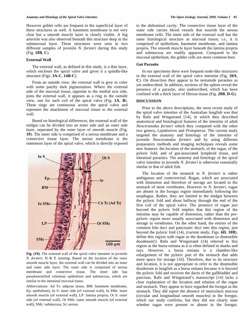
Fig. (19). The external wall of the spiral valve intestine in juvenile
N. forsteri, H & E staining. Based on the location of the outer
smooth muscle layer, the external wall can be divided into an inner
and outer side layer. The outer side is comprised of serous
membrane and connective tissue. The inner side has
pseudostratified columnar epithelium and submucosa, which are
similar to the intestinal mucosal tissue.
Abbreviations: Ad Ts: adipose tissue, BM: basement membrane,
Ep: epithelium), In S: inner side (of external wall), In SMs: inner
smooth muscle (of external wall), LP: lamina propria, Ot S: outer
side (of external wall), Ot SMs: outer smooth muscle (of external
wall), SMc: submucosa, Sr: serous.

82 The Open Zoology Journal, 2009, Volume 2 Hassanpour and Joss
Fig. (20). Parasites in spiral valve intestine of juvenile N. forsteri. (A, C) Multiple white nodes are observed in external wall, which home
some parasitic worms. (B) Transverse section of spleen in midgut, containing a parasite. (D, E, F, G) (Different magnifications) H & E
staining. Parasite is observed in splenic tissue surrounded by a thick layer of fibrous tissue, which indicates an immune response has been
activated against this parasite. Scale bar (A) 10 mm, (B) 5 mm, and (C) 20 mm.
Abbreviations: Fb Ts: fibrous tissue, Pr: parasite, Sp: spleen.
Liu et al. [7] found a slow and long-lasting tonic contraction in the anterior part of intestine in N. forsteri in response to substance P (SP), which is a tachykinin found in the mucosal cells. Further, a thin external wall on one side and a solid pancreas on the other side would restrict the ability of the
foregut to distend. The most logical explanation is that the foregut in N. forsteri is not a storage organ, although it may carry out the enzymatic function of a vertebrate stomach. The pyloric fold then allows stomach ingesta to move into the midgut more easily and it is this region that distends to
Anatomy and Histology of the Spiral Valve Intestine The Open Zoology Journal, 2009, Volume 2 83
allow the ingesta a longer exposure to bile and the pancreatic enzymes before moving into the rest of the spiral valve intestine for absorption of nutrients. An unpublished study by Van Noorden and Joss [17] found gastrin and cholecystokinin (CCK) in the anterior part of the intestine, but it did not specify the exact location as being either foregut or midgut, for these endocrine hormones. However, the presence of gastrin is associated with parietal cells, which secrete hydrochloric acid (HCl) in the stomach. Putting all current data together, it seems more appropriate to consider the foregut as a non-storage stomach in N. forsteri, with a distensible duodenum. Compared to other vertebrates, this is an unusual characteristic of N. forsteri, since even the other genera of living lungfish have more conventional spiral valves occurring, postpylorically [14].
Our observations on the pyloric fold in Neoceratodus forsteri suggest that in addition to its role in controlling the movement of the ingesta, it also plays a role in the immune system. In most vertebrates, including many fish taxa the pylorus is a region of the stomach connected to the duodenum [18, 19]. However, in lungfish there is no such region, instead there is a single or double layer of tissue, which is referred to as the pyloric fold. This structure is neither a stomach region, nor is it a complete sphincter. Rather, the pyloric fold acts as an incomplete gate that is located about half way through the first coil of the spiral valve intestine, separating the fore- and midgut regions. According to Rafn and Wingstrand [14], the pyloric fold in N. forsteri has a double, strongly oblique oval fold, while in Lepidosiren and Protopterus it is a simple ring-shaped pyloric fold. Due to its double layer structure in N. forsteri, which extends in an anterior to posterior direction, the pyloric fold might have an important role in preventing regurgitation of ingesta from the midgut to the foregut. Moreover, the epithelium of the pyloric fold is ciliated, a primitive characteristic, used to assist in the movement of the ingesta along the spiral valve. Further, our study has found lymphoid tissue in the pyloric fold. This is a significant immunological strategy in N. forsteri, which enables the pyloric fold to act as a “security gate” to prevent any harmful microorganisms entering via the gastrointestinal tract. Therefore, it is reasonable to regard the pyloric fold as the first battlefield between lymphocytes and hostile antigens. Another significant feature of the pyloric fold is that it is the only anatomical boundary between two regions of the spiral valve. On the other hand, based on the histological similarities between the epithelium of the pyloric fold and mucosal tissue, the pyloric fold may also have a physiolo-gical function as mucosal tissue.
Our findings indicate that the immune system in the gastrointestinal tract of juvenile N. forsteri is broader or more active compared to that described by Rafn and Wingstrand [14] in adult fish. These authors reported gut associated lymphoid tissue (GALT) throughout the mucosal tissue in adult N. forsteri. Our observations on juvenile N. forsteri not only confirm the presence of GALT in mucosal tissue, but extend these observations to the pyloric fold and also the region around the posterior spleen. Moreover, our histological observations describe an active immune response to a parasite in the spleen. Rafn and Wingstrand [14] go on to report that GALT is more extensive in the gastrointestinal tract of Lepidosiren than it is in
Neoceratodus, an observation we can neither confirm nor deny. We have found that juvenile N. forsteri have some node-like structures in their midgut epithelium that resemble Peyer’s patches. Peyer’s patches are aggregations of lymphoid tissue in the gut [20], which have specialized cells called Follicle Associated Epithelial (FAE) cells or M cells [21]. Peyer’s patches, as secondary lymphoid tissue, provide immune surveillance in the intestine and generate immune responses within the mucosa. Prior to our finding, the paddlefish (primitive ray-finned fish) was the only fish recorded as having Peyer’s patches [12, 22, 23]. Petrie-Hanson and Peterman [23] described the Peyer’s patches in paddlefish as being very similar to mammalian Peyer’s patches, located prominently in the spiral valve of older specimens. A broader study needs to be done to confirm whether the node-like structures found in the mucosal tissue of N. forsteri are equivalent to the Peyer’s patches associated with the intestine in amniote vertebrates.
The spleen in N. forsteri is an attachment of the gastrointestinal tract and contains abundant pigment cells. It is totally embedded in the spiral valve intestine and can be easily missed if the spiral valve is not dissected carefully. The coiling process of the mucosal tissue diverges the medial axis from a straight structure and the posterior spleen is narrowly twisted in the free margin. Longitudinal section of the spiral valve, especially in the midgut region, showed the posterior spleen as beads on a string. This may easily mislead the examiner to think that there are multiple spleens in the spiral valve. But in fact these ‘beads on a string’ are multiple sections of the twisted rod-like posterior spleen. Our findings in juvenile Australian lungfish are consistent with Rafn and Wingstrand’s [14] results for adult fish, namely, that the posterior spleen does not have its own capsule; rather it is coated with a smooth muscle layer, which is the continuation of the same smooth muscle (muscularis mucosa) of the mucosal tissue. The presence of lymphoid tissue around the posterior spleen in N. forsteri shows that the immune system extends throughout the spiral valve intestine. Pigment cells are abundant in the anterior and posterior spleen. Rafn and Wingstrand [14] found that these pigment cells are rich in iron. Gisbert et al. [24] showed that black-brown pigment granules in the alimentary tract of Siberian sturgeon larvae during the lecithotrophic stage correspond to melanin. Another study by Scalia et al. [25] demonstrated that spleen pigment cells in Rana esculenta (edible frog) are macrophages and are able to synthesize melanin during phagocytosis. The presence of iron in splenic pigment cells may indicate phagocytosis of red blood cells and degradation of their hemoglobin. Therefore, based on ours and previous studies, we assume that the splenic pigment cells of N. forsteri are Kupffer-like cells, which are melanogenic. However, the exact structure and function of these pigment cells remains unexplained.
The mucosal tissue in the spiral valve intestine of N. forsteri is a double layered epithelium, which shares the same submucosa and is primarily involved in absorption of nutrients. The same nutritional material, while passing through the spiral valve intestine, will reach the same area of mucosal tissue, but is exposed to the epithelium of the opposite side, after exactly one turn in the spiral valve. This rule, along with the fact that there are usually nine spirals in the intestine, may be used to determine some physiological
84 The Open Zoology Journal, 2009, Volume 2 Hassanpour and Joss
aspects of the spiral valve intestine. The morphology and histology of the mucosa in juvenile and adult N. forsteri [14] are mostly the same. However, goblet cells, which secrete mucus, look to be more common in juvenile fish due to the smaller spiral valve structure. Abundant mucus, especially in the boundaries between the medial axis and the external wall, would ease the mobilization and digestion processes of any ingesta in the blind spots of the spiral valve intestine. Further, based on our histological observation (Fig. 16A), we believe that goblet cells are modified from epithelial cells, which is consistent with Irving’s [18] findings in sea bass.
The spiral valve intestine in N. forsteri shelters some macro-parasites. Multiple white nodes containing parasitic worms (most likely nematodes) are seen in the external wall [14] (current study). These parasites look to be harmless and their relationship with their host seems to be commensal. However, there has been no detailed description of this parasite. The only parasite reported for Neoceratodus is a monogenean of the family Polystomatidae (Concinnocotyla australiensis), which lives exclusively in the oral cavity and gills [26, 27]. The current study also found a parasite in the spleen of one individual. It was surrounded by a thick layer of fibrotic tissue, indicating that it had activated an immune response. Further studies would be required to characterize intestinal parasites in N. forsteri.
CONCLUSION
To sum up, the gastrointestinal tract of N. forsteri presents many unusual and even seeming bizarre charac-teristics, which may be advantageous in food manipulation. Their unique dentition [28] accompanied by their mode of feeding [29] appears to complement these features, and confirm the basal phylogenetic position of N. forsteri.
ACKNOWLEDGMENTS
We are indebted to Mohammad G. Mohammad for his kindly support in tissue preparation. We appreciate Rolf Ericsson for his instructions on Adobe Photoshop software. Our many thanks to Debra Birch and Nicole Vella from the microscopy lab in the Department of Biological Sciences, Macquarie University, for their technical assistance with staining and imaging facilities. We are also grateful to Seied Mohammad Moosavi and Anahita Rezaei for their suggestions on the histology image analyses. This study was supported by the Department of Biological Sciences, Macquarie University, and funded by Macquarie University Higher Degree Research Office.
ABBREVIATIONS
GALT = Gut-associated lymphoid tissue
IEL = Intraepithelial lymphocyte
H & E = Hematoxylin and eosin
PBS = Phosphate buffered saline
NBF = Neutral buffered formalin
CCK = Cholecystokinin
REFERENCES
[1] Meyer A, Dolven SI. Molecules, fossils, and the origin of
tetrapods. J Mol Evol 1992; 35(2): 102-13.
[2] Joss JM. Lungfish evolution and development. Gen Comp
Endocrinol 2006; 148(3): 285-9. [3] Takezaki N, Figueroa F, Zaleska-Rutczynska Z, Takahata N, Klein
J. The phylogenetic relationship of tetrapod, coelacanth, and lungfish revealed by the sequences of forty-four nuclear genes. Mol
Biol Evol 2004; 21(8): 1512-24. [4] Metcalf VJ, George PM, Brennan SO. Lungfish albumin is more
similar to tetrapod than to teleost albumins: purification and characterisation of albumin from the Australian lungfish,
neoceratodus forsteri. Comp Biochem Physiol B Biochem Mol Biol 2007; 147(3): 428-37.
[5] Zardoya R, Cao Y, Hasegawa M, Meyer A. Searching for the closest living relative(s) of tetrapods through evolutionary analysis
of mitochondrial and nuclear data. Mol Biol Evol 1998; 15(5): 506-17.
[6] Venkatesh B, Erdmann MV, Brenner S. Molecular synapomorphies resolve evolutionary relationships of extant jawed vertebrates. Proc
Natl Acad Sci USA 2001; 98(20): 1382-7. [7] Liu L, Conlon JM, Joss JMP, Burcher E. Purification,
characterization, and biological activity of a substance P-related peptide from the gut of the Australian lungfish, neoceratodus
forsteri. Gen Comp Endocrinol 2002; 125(1): 104-12. [8] McAllister JA. Reevaluation of the formation of spiral coprolites.
Journal [serial on the Internet]. 1985 Date [cited 2009 21/01/2009]; Paper 114: Available from: http: //hdl.handle.net/1808/3667.
[9] Crow GL, Howe JC, Uchida S, Kamolnick S, Wisner MG, Caira JN. Protrusion of the valvular intestine through the cloaca in sharks
of the family carcharhinidae. Copeia 1990; 1: 226-9. [10] Henningsen AD, Whitaker BR, Walker ID. Protrusion of the
valvular intestine in captive small tooth sawfish and comments on pristid gastrointestinal anatomy and intestinal valve types. J Aquat
Anim Health 2005; 17: 289-95. [11] Cataldi E, Albano C, Boglione C, et al. Acipenser naccarii: fine
structure of the alimentary canal with references to its ontogenesis. J Appl Ichthyol 2002; 18(4-6): 329-37.
[12] Peterman AE, Petrie HL. Ontogeny of American paddlefish lymphoid tissues. J Fish Biol 2006; 69(sa): 72-88.
[13] Suyehiro Y. Some views on the dissected specimens of coelacanth. Bienn Report Keikyu Aburatsubo Mar Park Aquar 1985; (12): 12.
[14] Rafn S, Wingstrand KG. Structure of intestine, pancreas, and spleen of the Australian lungfish, Neoceratodus forsteri (Krefft).
Zool Scr 1981; 10(3): 223-39. [15] Humason GL. Animal Tissue Techniques. 2nd ed. Ralph Emerson
DK, Roderick BP, Eds. San Francisco and London: W. H. Freeman and Company 1967.
[16] Hamlett WC. Sharks, Skates, and Rays: The Biology of Elasmobranch Fishes. Baltimore: John Hopkins University Press
1999. [17] Van Noorden S, Joss J. Lungfish gastroentero-pancreatic peptides:
an immunocytochemical study. 2009; (Unpublished). [18] Irving HB. Studies on the comparative histology of the digestive
tube of certain teleost fishes. III. A bottom-feeding fish, the sea robin (Prionotus carolinus). J Morphol 1936; 60(1): 77-102.
[19] Weinreb EL, Bilstad NM. Histology of the digestive tract and adjacent structures of the rainbow trout, salmo gairdneri irideus.
Copeia 1955; 3: 194-204. [20] Owen RL. Sequential uptake of horseradish peroxidase by
lymphoid follicle epithelium of Peyer's patches in the normal unobstructed mouse intestine: an ultrastructural study.
Gastroenterology 1977; 72(3): 440-51. [21] Burns RB, Maxwell MH. Ultrastructure of Peyer's patches in the
domestic fowl and turkey. J Anat 1986; 147 : 235-43. [22] Weisel GF. Anatomy and histology of the digestive system of the
paddlefish (Polyodon spathula). J Morphol 1973; 140(2): 243-55. [23] Petrie HL, Peterman AE. American paddlefish leukocytes
demonstrate mammalian-like cytochemical staining characteristics in lymphoid tissues. J Fish Biol 2005; 66(4): 1101-15.
[24] Gisbert E, Sarasquete C. Histochemical identification of the black-brown pigment granules found in the alimentary canal of Siberian
sturgeon (Acipenser baeri) during the lecitotrophic stage. Fish Physiol Biochem 2000; 22(4): 349-54.
[25] Scalia M, Pietro CD, Poma M, Ragusa M, Sichel G, Corsaro C. The spleen pigment cells in some amphibia. Pigment Cell Res
2004; 17: 119-27. [26] Whittington ID, Pichelin S. Attachment of eggs by concinnocotyla
australensis (monogenea: polystomatidae) to the tooth plates of the

Anatomy and Histology of the Spiral Valve Intestine The Open Zoology Journal, 2009, Volume 2 85
Australian lungfish, Neoceratodus forsteri (Dipnoi). Int J Parasitol
1991; 21(3): 341-6. [27] Pichelin S, Whittington I, Pearson J. Concinnocotyla (Monogenea:
Polystomatidae), a new genus for the polystome from the Australian lungfish Neoceratodus forsteri. Syst Parasitol 1991;
18(2): 81-93.
[28] Reisz RR, Smith MM. Developmental biology: lungfish dental
pattern conserved for 360 Myr. Nature 2001; 411(6837): 548. [29] Bemis WEaL, George V. Morphology and function of the feeding
apparatus of the lungfish, Lepidosiren paradoxa (Dipnoi). J Morphol 1986; 187(1): 81-108.
Received: February 25, 2009 Revised: March 30, 2009 Accepted: April 08, 2009
© Hassanpour and Joss; Licensee Bentham Open.
This is an open access article licensed under the terms of the Creative Commons Attribution Non-Commercial License
(http://creativecommons.org/licenses/by-nc/3.0/) which permits unrestricted, non-commercial use, distribution and reproduction in any medium, provided the work is properly cited.