online detection and extraction of fecg signals using ica
TRANSCRIPT
_____________________________________________________________________________________________________ *Corresponding author: Email: [email protected];
Journal of Engineering Research and Reports
15(2): 10-18, 2020; Article no.JERR.59138 ISSN: 2582-2926
Online Detection and Extraction of FECG Signals Using ICA: A Comparative Study
Mohammed Sheikh1*, Majdi Marai2 and Roiss Alhutaish3
1Aden University, Aden, Yemen.
2Hadhramout University, Al Mukalla, Yemen. 3Aden Community College, Aden, Yemen.
Authors’ contributions
This work was carried out in collaboration among all authors. Author MS designed the study,
performed the statistical analysis, wrote the protocol and wrote the first draft of the manuscript. Author MM managed the analyses of the study. Author RA managed the literature searches. All authors read
and approved the final manuscript.
Article Information
DOI: 10.9734/JERR/2020/v15i217140 Editor(s):
(1) Dr. Guang Yih Sheu, Chang-Jung Christian University, Taiwan. Reviewers:
(1) H. K. Shreedhar, Global Academy of Technology, India. (2) Yousef Farhaoui, University of Moulay Ismail, Morocco.
Complete Peer review History: http://www.sdiarticle4.com/review-history/59138
Received 12 May 2020 Accepted 19 July 2020
Published 04 August 2020
ABSTRACT
In this paper a new study to detect fetal heart rate (F H R) online from abdominal electrocardiogram (ECG) signal, which are extracted by three different algorithms of independent component analysis ICA (AMUSE, EVD2 and SOBI) is presented. Four stages for fetal electrocardiogram (FECG) extraction and detection is proposed. After preprocessing and (FECG) extraction by ICA, maternal QRS complex removal window is used to remove or scale down the maternal remaining peaks, and smoothed by II notch filter. 25 data sets are used to validate this method of study for fetal peak detection online from signals extracted by ICA. Two ways are used to test 25 signals firstly off line and secondly online. The average sensitivity of the ICA (AMUSE, EVD2 and SOBI) based method are 72.3%, 66.2% and 75.1% off line respectively, and 55%, 53% and 059% online respectively, while average positive predictivity are 61.4%, 61.3% and 69.7% off line respectively, while 43%, 41% and 46% online respectively. These show that the ICA based method is more successful in detecting the FHR off line than online, which is more complicated, where the automatic selection of the output signals is not a trivial task.
Original Research Article

Sheikh et al.; JERR, 15(2): 10-18, 2020; Article no.JERR.59138
11
Keywords: Independent component analysis; fetal peak detection and on line monitoring. 1. INTRODUCTION Fetal heart rate (F H R) monitoring is one of known methodologies to test the fetal situation and diagnose for possible abnormalities. FECG can be derived from the abdominal ECG (AECG) which recorded by placing several leads on the abdomen of the mother. Fetal monitoring throughout the pregnancies enables the clinician to recognize and diagnose some pathologic conditions such as asphyxia [1]. Although Doppler ultrasound device is currently used for FHR monitoring, it is not suitable for long term monitoring due to its sensitivity to movement and its safety for long term exposure has yet to be established [2]. The abdominal ECG (AECG) is always corrupted with power line interference, maternal ECG (MECG) and electromyogram where its variability is influenced by the gestational age, position of the electrodes and the skin impedance [3]. Therefore, appropriate signal processing techniques are required to reveal the fetal ECG (FECG) from the AECG. Various research efforts have been proposed to extract the FECG from the AECG such as, correlation techniques [4], a combination of wavelet analysis and blind source separation methods [5] Fast ICA extraction [6] and adaptive filtering [7]. FHR can be calculated by determining the R-R intervals from the extracted FECG. However, the extracted FECG is still corrupted by the residual peaks of MECG (especially its QRS complexes) hence the FECG detection remains difficult.
In this study 25 recorded data are used to evaluate the ability of ICA algorithms to be used for extracting FECG signal and detect its peaks on line.
2. METHODOLOGY AECG signals were recorded in UKM hospital {UKM university – Malaysia} from 25 healthy pregnant women (at 35 to 38 weeks of gestation), which are corrupted with different levels of noises, using the lead system as shown in Fig. 1. The AECG signals,
1 2 , , ..., T
pX n X n X n X n , where n denotes a discrete-time index, and T is the transpose operator, were simultaneously recorded from maternal abdomen using six
electrodes (five electrodes, , 2, 3, 4, 5p l,
with a single common) using high gain amplifiers (BIOPAC- MP 100A). The AECG signals were digitized at 1000 Hz with 12 bit resolution and resembled to 256Hz. The total recording time during each session was about one minute. Electrode p1 is located in such a way that only MECG signals are acquired while the electrodes p2, p3, p4 and p5 acquired the mixture of MECG and FECG. Therefore, X1(n) is defined as the reference. Three signals only are selected from the four of the acquired AECG signals,
2 3 , ,..., 2, 3 4, 5[ ,pX n X n X n X n p ], and fed into the ICA algorithm.
Fig. 1. Abdominal electrodes position

Sheikh et al.; JERR, 15(2): 10-18, 2020; Article no.JERR.59138
12
2.1 Algorithms The proposed algorithm consists of the following stages:
a-: Pre-processing stage b-: FECG extraction using 3 ICA algorithms
stage c-: Removal window and post processing
stage d-: FECG detection stage
2.1.1 Preprocessing stage The preprocessing stage consists of stages that are applied to the observation signals each of these signals is made zero mean by subtracting its mean as follows:
( ) ( )( ) ( ) n nx n x mean x (1)
Baseline wander is caused by the patient's breathing or movements during recording. A FIR band-pass filter with cut-off frequencies at 4 Hz and 90 Hz is used for filtering the frequency of the baseline wander due to breathing, which is in the range of 1 Hz and the EMG noise (artifacts of muscular contractions) is characterized by relatively high frequency noise. A notch filter centered at 50 Hz is used to eliminate the interference of the power line, which consists of 50 Hz sine wave and its harmonics
2.1.2 FECG extraction technique
Three ICA algorithms namely (AMUSE, SOBI and EVD and) have been implemented in this work to evaluate their performance for FECG extraction and peak detection’s
2.1.2.1 Independent component analysis
ICA is a method to find underlying factors or components from multivariate (multidimensional) statistical data. It looks for components that are both statistically independent and non-gaussian. Although an excellent review has been given by Cichocki & Amari [8], a brief description is given here.Given a set of p mixed signals
1 2 , , , T
pX n X n X n X n which are linear mixed with q (p≥ q) unknown mutually statistically independent, zero-mean source
signals 1 2 , , ,
T
qS n s n s n s n and
noise contaminated.
This can be written as:
1, 2, ,
i ij j ciX n A s n g n i
p
(2)
or in the matrix notation
CX AS g (3)
where X = X(n) is the vector of sensor signals, S = S(n) is the source signal vector,
, , , T
c c c c pg g n g n g n is the
additive noise vector, A is an unknown p×q mixing matrix and n is the discrete time index.
The noise vector, cg is assumed Gaussian and independent.
The mixing matrix A is determined by the body geometry and conductivity, as well as the electrode-source relative positions [9]., maximum likelihood, minimization of mutual information [10] Criteria based on maximization of non-gaussianity [11], non-linear decorrelation [12] may be used to estimate the mixing matrix A and the source signal vector S, and tensorial
methods [13].In the noise-free model, 0cg , the identification of the mixing matrix A and the sources signal, S can be estimated if the sources are independent and non-Gaussian, and the number of sensors is equal or larger than the number of independent sources to be estimated. However, a noisy estimates of the sources signal
may obtain, 1 , 0c cS A X g i f g
. Therefore, pre-processing before applying ICA may improve the performance of the ICA. An example of an input signals is shown in Fig. 2.
After the preprocessing step, the three ICA algorithms are applied for extracting FECG signal, each algorithm is fed with 25 signals each signal consist of 3 components, (input signals
are denoted by 2 3 4, ,X X X , while the resulting extracted signals are denoted by
2 3 4, ,Y Y Y . After extraction the three extracted signals from every input signals are the maternal, fetal and noise signals. All these signals are saved in a file in addition to the signal (Y1), which will be used as input signals for the FECG peak detection algorithm on line Fig. 3.

Fig. 2. input signals to ICA for extracting
Fig. 3. The block diagram the steps of the proposed algorithm After extraction, different signals are resulted from each output, some examples of output signals can be seen in Fig. 4, which are corrupted with different levels of noise. Other signals may be appeared in different deflection (positions). For example MECG or
Sheikh et al.; JERR, 15(2): 10-18, 2020; Article no.
13
Fig. 2. input signals to ICA for extracting
Fig. 3. The block diagram the steps of the proposed algorithm
After extraction, different signals are resulted some examples of output
signals can be seen in Fig. 4, which are corrupted with different levels of noise.
Other signals may be appeared in different deflection (positions). For example MECG or
FECG are upside down or both signals are upside down. In these signals some maternal peaks are upward or down ward deflection (MPu or MPd) and other fetal peak are upward or down ward deflection (FPu or FPd), while some maternal and fetal peak have down ward deflection (MPd and FPd) as shown in Fig. 5.
; Article no.JERR.59138
FECG are upside down or both signals are se signals some maternal
peaks are upward or down ward deflection (MPu or MPd) and other fetal peak are upward or down ward deflection (FPu or FPd), while some maternal and fetal peak have down ward deflection (MPd and FPd) as shown in Fig. 5.

Fig. 4. Noisy extracted signals by ICA
Fig. 5. extracted signals by ICA 25 signals are fed to each algorithm; the result is three outputs each output has 25 different extracted signals (maternal, noise, fetal mixed with maternal or maternal and fetal with different levels of noise) in addition to that some extracted signals are upside down. All the fetal extracted signals still corrupted with maternal peaks. Table 1 shows an example of the deflectifor mixed MECG and FECG only.
2.2 Post Processing Before Post processing Stage, MQRS removal window is applied to signal Y1 only As shown in
Sheikh et al.; JERR, 15(2): 10-18, 2020; Article no.
14
Fig. 4. Noisy extracted signals by ICA
Fig. 5. extracted signals by ICA
25 signals are fed to each algorithm; the result is three outputs each output has 25 different
noise, fetal mixed with maternal or maternal and fetal with different levels of noise) in addition to that some extracted signals are upside down. All the fetal extracted signals still corrupted with maternal peaks. Table 1 shows an example of the deflection direction
Before Post processing Stage, MQRS removal window is applied to signal Y1 only As shown in
Fig. 6 To improve the performance of the detection, [14]. The post processing stage, consist of steps, which are implemented to scale down and adjustment the maternal remaining peaks and to enhance the fetal peak for detection. All the 3 extracted signals (Y2,Y3,Y4 ) are fed to this stage with the same arrangement as these signals after extraction. At the end the resulted signals are fed to the detection stage, to evaluate the detection of fetal peaks of FECG extracted by the 3 ICA algorithms SOBI, AMUSE and EVD2.
; Article no.JERR.59138
.
Fig. 6 To improve the performance of the
The post processing stage, consist of three steps, which are implemented to scale down and adjustment the maternal remaining peaks and to enhance the fetal peak for detection. All the 3 extracted signals (Y2,Y3,Y4 ) are fed to this stage with the same arrangement as these
tion. At the end the resulted signals are fed to the detection stage, to evaluate the detection of fetal peaks of FECG extracted by the 3 ICA algorithms SOBI, AMUSE and

Table 1. Direction of deflection for mixed MECG and FECG
Algorithm signal No of sig Y1 25EVD Y2 25 Y3 25 Y1 25SOBI Y2 25 Y3 25 Y1 25AMUSE Y2 25 Y3 25
Fig
2.3 Fetal Peak Detection In this work, the recorded signals are stored in PC and applied in two steps. First step: after preprocessing ICA algorithms are used to extract FECG signal and detect its peaks off line. Second step: after preprocessing ICA algorithms are used to extract FECG signal and detect its peaks on line. Peak detection is performed under Matlab®/Simulink® platform. The model is
Sheikh et al.; JERR, 15(2): 10-18, 2020; Article no.
15
Direction of deflection for mixed MECG and FECG
No of sig MPd+FPd MPd+FPu MPu+FPd25 3.20% 3.20% 25.8%25 6.50% 9.67% 22.58%25 5% 3.20% 25.80%25 4% 32% 16%25 30% 32% 28%25 4% 16% 34%25 16% 8% 36%25 24% 20% 44%25 6% 20% 36%
Fig. 6. MQRS removal window
In this work, the recorded signals are stored in
after preprocessing ICA algorithms are used to extract FECG signal and detect its
after preprocessing ICA algorithms are used to extract FECG signal and
Peak detection is performed under mulink® platform. The model is
developed for system simulation towards realizing real time FHR, which is built by connecting the embedded Matlab function blocks and the available required blocks in the software library. The parameters of the blocks are entwhile designing them for simulation. 25 recorded data which were between 36 and 38th week of singleton pregnancy are used to test for fetal QRS detection.
The FECG extracted signals in addition to signal (Y1) in a personal computer are “From file” block, as *.mat file. Then these signals were, used as input signals as shown in Fig. 7.
; Article no.JERR.59138
MPu+FPd 25.8% 22.58% 25.80% 16% 28% 34% 36% 44% 36%
developed for system simulation towards realizing real time FHR, which is built by connecting the embedded Matlab function blocks and the available required blocks in the software library. The parameters of the blocks are entered while designing them for simulation. 25 recorded data which were between 36 and 38th week of singleton pregnancy are used to test for fetal
The FECG extracted signals in addition to signal (Y1) in a personal computer are stored in “From file” block, as *.mat file. Then these signals were, used as input signals as shown in

Fig. 3. RESULTS AND DISCUSSION In this study 3 ICA algorithms (AMUSE, EVD2 and SOBI) are applied to 25 recoded signals in 2 steps as follow:
a) Off line: each algorithm is applied for
extracting signals. After that all the 25 signal are evaluated by fetal peak detection.
b) On line: each algorithm is applied for extracting signals. After that all the 25 signal are evaluated by fetal peak detection.
The aim of this study is to test the ability of ICA algorithms to be used in FECG signal extraction and detection its peaks on line.
3.1 Performance Evaluation The proposed algorithms have been implemented in Matlab codes using Matlab(The Math-works Inc.). The performances of the algorithms were then evaluated based on their sensitivities and positive productivities’, when applied to recorded AECG signals. The sensitivity is the fraction of real events that are correctly detected and it is defined by:
Table 2.
Week of gestation
No of signals
AMUSE
Se(%) 35 7 68.4 36 12 73.3 37 6 75.2 72.2(%)
Sheikh et al.; JERR, 15(2): 10-18, 2020; Article no.
16
7. model for fetal peak detection
DISCUSSION
algorithms (AMUSE, EVD2 and SOBI) are applied to 25 recoded signals in 2
: each algorithm is applied for extracting signals. After that all the 25 signal are evaluated by fetal peak
each algorithm is applied for extracting signals. After that all the 25 signal are evaluated by fetal peak
The aim of this study is to test the ability of ICA algorithms to be used in FECG
d detection its peaks on line.
The proposed algorithms have been implemented in Matlab codes using Matlab-7.4
works Inc.). The performances of the algorithms were then evaluated based on their
ve productivities’, when applied to recorded AECG signals. The sensitivity is the fraction of real events that are correctly detected and it is defined by:
se = TP / (TP + FN)
The Positive Predictively is the fraction of detections that are real events and it is defined by:
+P = TP / (TP + FP)
Where FN (False Negatives) denotes the number of missed detections, FP (False Positives) represents the number of extra detections and TP (True Positives) is the number of detected QRS complexes.
Table 2 off line shows the performance using ICA algorithms based methods at signal extraction stage. The average sensitivity of theEVD2 and SOBI with Se% are 72.3%, 66.2% and 75.1% respectively, The average positpredictively of the AMUSE, EVD2 +P% are 61.4%, 61.3% and 69.7% respectively,. It shows that the SOBI based method was more successful in detecting the FHR than AMUSE based method and EVD2.
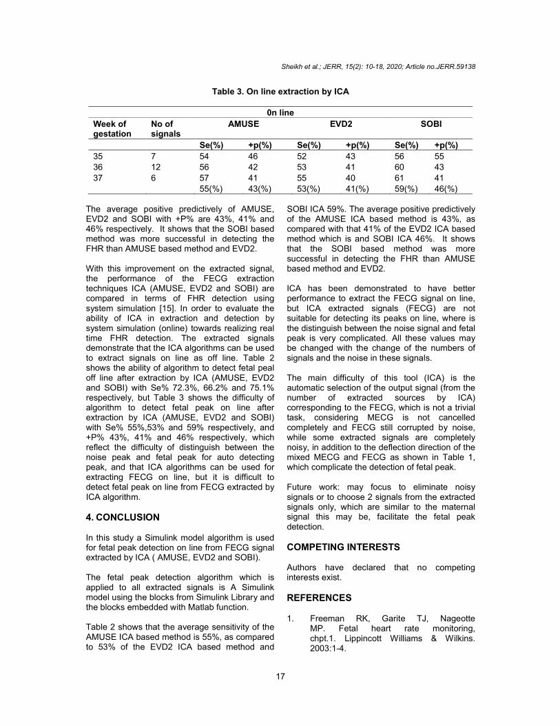
Table 3 on line shows the performance of 3 ICA algorithms based methods at signal extraction stage. The average sensitivity of the AMUSE, EVD2 and SOBI with Se% are 55%, 53% and 59% respectively.
Table 2. off line extraction by ICA
Off line extraction AMUSE EVD2 SOBI
+p(%) Se(%) +p(%) Se(%) 47.8 55.5 45.7 69.6 71.2 70.2 68.2 77.4 65.4 73.3 70.1 78.5
72.2(%) 61.4(%) 66.3(%) 61.3(%) 75.1(%)
; Article no.JERR.59138
(4)
The Positive Predictively is the fraction of are real events and it is defined
(5)
Where FN (False Negatives) denotes the number of missed detections, FP (False Positives) represents the number of extra detections and TP (True Positives) is the number of correctly
Table 2 off line shows the performance using ICA algorithms based methods at signal extraction stage. The average sensitivity of the AMUSE, EVD2 and SOBI with Se% are 72.3%, 66.2% and 75.1% respectively, The average positive
and SOBI with +P% are 61.4%, 61.3% and 69.7% respectively,. It shows that the SOBI based method was more
detecting the FHR than AMUSE
Table 3 on line shows the performance of 3 ICA algorithms based methods at signal extraction stage. The average sensitivity of the AMUSE, EVD2 and SOBI with Se% are 55%, 53% and
SOBI
+p(%) 67.6 71.5 70.1
69.7(%)
Sheikh et al.; JERR, 15(2): 10-18, 2020; Article no.JERR.59138
17
Table 3. On line extraction by ICA
0n line
Week of gestation
No of signals
AMUSE EVD2 SOBI
Se(%) +p(%) Se(%) +p(%) Se(%) +p(%)
35 7 54 46 52 43 56 55
36 12 56 42 53 41 60 43
37 6 57 41 55 40 61 41
55(%) 43(%) 53(%) 41(%) 59(%) 46(%)
The average positive predictively of AMUSE, EVD2 and SOBI with +P% are 43%, 41% and 46% respectively. It shows that the SOBI based method was more successful in detecting the FHR than AMUSE based method and EVD2. With this improvement on the extracted signal, the performance of the FECG extraction techniques ICA (AMUSE, EVD2 and SOBI) are compared in terms of FHR detection using system simulation [15]. In order to evaluate the ability of ICA in extraction and detection by system simulation (online) towards realizing real time FHR detection. The extracted signals demonstrate that the ICA algorithms can be used to extract signals on line as off line. Table 2 shows the ability of algorithm to detect fetal peal off line after extraction by ICA (AMUSE, EVD2 and SOBI) with Se% 72.3%, 66.2% and 75.1% respectively, but Table 3 shows the difficulty of algorithm to detect fetal peak on line after extraction by ICA (AMUSE, EVD2 and SOBI) with Se% 55%,53% and 59% respectively, and +P% 43%, 41% and 46% respectively, which reflect the difficulty of distinguish between the noise peak and fetal peak for auto detecting peak, and that ICA algorithms can be used for extracting FECG on line, but it is difficult to detect fetal peak on line from FECG extracted by ICA algorithm. 4. CONCLUSION In this study a Simulink model algorithm is used for fetal peak detection on line from FECG signal extracted by ICA ( AMUSE, EVD2 and SOBI). The fetal peak detection algorithm which is applied to all extracted signals is A Simulink model using the blocks from Simulink Library and the blocks embedded with Matlab function. Table 2 shows that the average sensitivity of the AMUSE ICA based method is 55%, as compared to 53% of the EVD2 ICA based method and
SOBI ICA 59%. The average positive predictively of the AMUSE ICA based method is 43%, as compared with that 41% of the EVD2 ICA based method which is and SOBI ICA 46%. It shows that the SOBI based method was more successful in detecting the FHR than AMUSE based method and EVD2. ICA has been demonstrated to have better performance to extract the FECG signal on line, but ICA extracted signals (FECG) are not suitable for detecting its peaks on line, where is the distinguish between the noise signal and fetal peak is very complicated. All these values may be changed with the change of the numbers of signals and the noise in these signals. The main difficulty of this tool (ICA) is the automatic selection of the output signal (from the number of extracted sources by ICA) corresponding to the FECG, which is not a trivial task, considering MECG is not cancelled completely and FECG still corrupted by noise, while some extracted signals are completely noisy, in addition to the deflection direction of the mixed MECG and FECG as shown in Table 1, which complicate the detection of fetal peak. Future work: may focus to eliminate noisy signals or to choose 2 signals from the extracted signals only, which are similar to the maternal signal this may be, facilitate the fetal peak detection.
COMPETING INTERESTS Authors have declared that no competing interests exist.
REFERENCES 1. Freeman RK, Garite TJ, Nageotte
MP. Fetal heart rate monitoring, chpt.1. Lippincott Williams & Wilkins. 2003:1-4.

Sheikh et al.; JERR, 15(2): 10-18, 2020; Article no.JERR.59138
18
2. Friesen GM, et al. A comparison of the noise sensitivity of nine QRS detection algorithms. IEEE Trans. Biomed. Engineering. 1990;37:85–98.
3. Goodlin RC. History of fetal monitoring, Am. J. Obstet, Gynecol. 1989;133:323-352.
4. Abboud S, Alaluf A, Einav S, Sadeh D, Real time abdominal fetal ECG recording using hardware correlator. Comput. Biol. Med. 1992;22:32–335.
5. Maria JG, Jonathon CA, Fetal electrocardiogram extraction by sequential source separation in the wavelet domain. IEEE Trans Biomed Eng, b. Azzerboni, F.L. Foresta, N. Mammone, and F.C. Morabito, A New Approach Based on Wavelet-ICA Algorithms for Fetal Electrocardiogram Extraction, in Proc. 13th. 2005;52: 390-400.
6. Li Yuan, Zhuhuang Zhou, Yanchao Yuan, Shuicai Wu. An improved fast ICA method for fetal ECG extraction. Computational and Mathematical Methods in Medicine; 2018.
7. Ferrara ER, Widrow B, Fetal electrocardiogram enhancement by time-sequenced adaptive filtering. IEEE Trans. Biomed. Eng. 1982;29:458-460.
8. A Cichocki, Amari S. Adaptive blind signal and image processing. Wiley. 2002;157-
175. European Symposium of Artificial Neural Networks. 2005;27-29.
9. P Comon. Independent component analysis: A new concept. Signal Process. 1894;36:287-314.
10. Jutten, A. Taleb, Source separation: From dusk till dawn. Proceedings of the second International Workshop on Independent Component Analysis and Blind Source Separation. 2000:15-26.
11. Cardoso JF. Source separation using higher order moments. Proc. ICASSP. 1989;2:2109-2112. DOI: 10.1109/ICASSP.1989.266878
12. Gustavo Deco, Wilfried Brauer. Nonlinear higher-order statistical decorrelation by volume-conserving neural architectures. 1995;8(4):525-535.
13. Geng Jun. Find peak value of datas. USTB, Beijing, China for Dr. Ma Zheng, [Online].
14. Sheikh M, Mohd Ali MA. Threshold-free detection of maternal heart rate from abdominal electrocardiogram. European Journal of Scientific Research. 2009;27(4):565-575. ISSN 1450-216X.
15. Sheikh M, Mohd Ali MA. Evaluation of an improved algorithm for fetal QRS detection. International Journal of the Physical Sciences. 2011;6(2):213-220.
© 2020 Sheikh et al.; This is an Open Access article distributed under the terms of the Creative Commons Attribution License (http://creativecommons.org/licenses/by/4.0), which permits unrestricted use, distribution, and reproduction in any medium, provided the original work is properly cited.
Peer-review history: The peer review history for this paper can be accessed here:
http://www.sdiarticle4.com/review-history/59138