online correction of setup error in prostate cancer: a comparison of an ultrasound system and...
TRANSCRIPT
Proceedings of the 49th Annual ASTRO Meeting S661
the beams-eye-view (BEV) perspective. This study introduces the novel dual-energy subtraction imaging technique for radiother-apy localization in the pelvis, using a commercially available gantry-mounted kV x-ray imaging system. Proof of concept isdemonstrated in an anthropomorphic pelvis phantom.
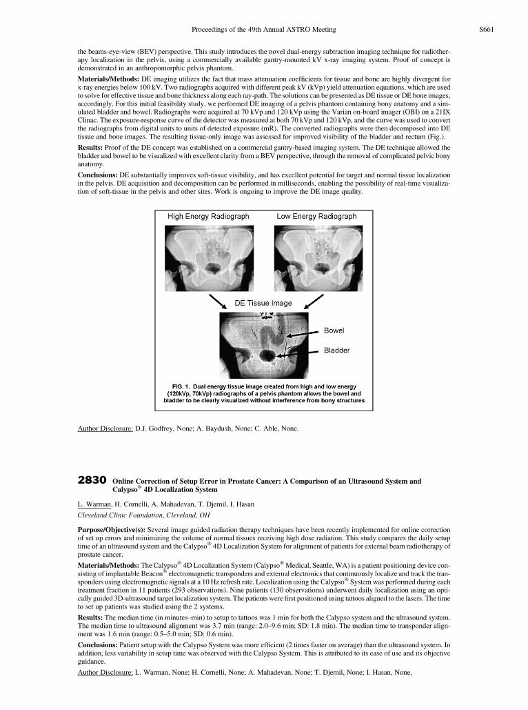
Materials/Methods: DE imaging utilizes the fact that mass attenuation coefficients for tissue and bone are highly divergent forx-ray energies below 100 kV. Two radiographs acquired with different peak kV (kVp) yield attenuation equations, which are usedto solve for effective tissue and bone thickness along each ray-path. The solutions can be presented as DE tissue or DE bone images,accordingly. For this initial feasibility study, we performed DE imaging of a pelvis phantom containing bony anatomy and a sim-ulated bladder and bowel. Radiographs were acquired at 70 kVp and 120 kVp using the Varian on-board imager (OBI) on a 21IXClinac. The exposure-response curve of the detector was measured at both 70 kVp and 120 kVp, and the curve was used to convertthe radiographs from digital units to units of detected exposure (mR). The converted radiographs were then decomposed into DEtissue and bone images. The resulting tissue-only image was assessed for improved visibility of the bladder and rectum (Fig.).
Results: Proof of the DE concept was established on a commercial gantry-based imaging system. The DE technique allowed thebladder and bowel to be visualized with excellent clarity from a BEV perspective, through the removal of complicated pelvic bonyanatomy.
Conclusions: DE substantially improves soft-tissue visibility, and has excellent potential for target and normal tissue localizationin the pelvis. DE acquisition and decomposition can be performed in milliseconds, enabling the possibility of real-time visualiza-tion of soft-tissue in the pelvis and other sites. Work is ongoing to improve the DE image quality.
Author Disclosure: D.J. Godfrey, None; A. Baydush, None; C. Able, None.
2830 Online Correction of Setup Error in Prostate Cancer: A Comparison of an Ultrasound System and�
Calypso 4D Localization SystemL. Warman, H. Cornelli, A. Mahadevan, T. Djemil, I. Hasan
Cleveland Clinic Foundation, Cleveland, OH
Purpose/Objective(s): Several image guided radiation therapy techniques have been recently implemented for online correctionof set up errors and minimizing the volume of normal tissues receiving high dose radiation. This study compares the daily setuptime of an ultrasound system and the Calypso� 4D Localization System for alignment of patients for external beam radiotherapy ofprostate cancer.
Materials/Methods: The Calypso� 4D Localization System (Calypso� Medical, Seattle, WA) is a patient positioning device con-sisting of implantable Beacon� electromagnetic transponders and external electronics that continuously localize and track the tran-sponders using electromagnetic signals at a 10 Hz refresh rate. Localization using the Calypso� System was performed during eachtreatment fraction in 11 patients (293 observations). Nine patients (130 observations) underwent daily localization using an opti-cally guided 3D-ultrasound target localization system. The patients were first positioned using tattoos aligned to the lasers. The timeto set up patients was studied using the 2 systems.
Results: The median time (in minutes–min) to setup to tattoos was 1 min for both the Calypso system and the ultrasound system.The median time to ultrasound alignment was 3.7 min (range: 2.0–9.6 min; SD: 1.8 min). The median time to transponder align-ment was 1.6 min (range: 0.5–5.0 min; SD: 0.6 min).
Conclusions: Patient setup with the Calypso System was more efficient (2 times faster on average) than the ultrasound system. Inaddition, less variability in setup time was observed with the Calypso System. This is attributed to its ease of use and its objectiveguidance.
Author Disclosure: L. Warman, None; H. Cornelli, None; A. Mahadevan, None; T. Djemil, None; I. Hasan, None.