oncogenic osteomalacia as a harbinger of recurrent...
TRANSCRIPT

Sarcoma (1999) 3, 95± 99
ORIGINAL ARTICLE
Oncogenic osteomalacia as a harbinger of recurrent osteosarcoma
ELIZABETH B. LAMONT,1
MELISSA K. CAVAGHAN2
& BRUCE E. BROCKSTEIN1
1Section of Hematology-Oncology and
2Section of Endocr inology, Department of Medicine, University of Chicago, Chicago,
IL 60637, USA
Abstract
Discussion. Oncogenic osteomalacia is a rare paraneoplastic syndrome of skeletal demineralization from renal phosphateloss. Patients with this disorder have the characteristic clinical, laboratory, and radiographic ® ndings of hyperphosphaturicosteomalacia. Although the pathophysiology has not yet been clearly delineated, a humoral factor produced by the tumor issuspected to be the cause.Purpose. We report the ® rst case of oncogenic osteomalacia that improved with chemotherapy, discuss this paraneoplasticsyndrome, and review the medical literature regarding its etiology.
Key words: oncogenic osteom alacia, etiolog y, chemotherapy
Introduction
With fewer than 100 cases reported in the medical
literature, oncogenic osteomalacia is a rare endocrino-
logical paraneoplastic syndrome characterized by
defective bone mineralization from renal phosphate
loss. Tumor elaboration of a phosphaturic factor is
the putative mechanism. Although occasionally
reported in other tumor types, oncogenic osteoma-
lacia is almost exclusively described in patients with
benign tumors of mesenchymal origin. Characteristi-
cally, patients present with signs and symptoms of
osteomalacia, i.e., waddling gait, joint deformities,
bone pain, muscle weakness, anorexia, fatigue and
occasionally long bone fractures. Laboratory examina-
tion is notable for hypophosphatemia in the setting of
inappropriate phosphaturia, but usually normal PTH
levels. 1,25-Vitamin D is inappropriately normal rela-
tive to the coexisting hypophosphatemia and is frankly
depressed in many cases. Evaluation with plain ® lms
usually reveals osteopenia, and occasionally the pseu-
dofractures (Looser zones) and subperiosteal erosions
seen in osteomalacia. Bone scans also show the
characteristic ® ndings of osteomalacia, which is
uptake that is diffuse and/or focal. Because the tumors
associated with this syndrome are typically quite small
( £ 1 cm), the clinical symptoms often precede the
identi® cation of the tumor. In an otherwise healthy
patient who presents with osteomalacia due to hyper-
phosphaturia, evaluation for an occult tumor should
be considered. For patients with known tumors,
surgical resection usually cures them of the syndrome.
Case presentation
CB is a 68-year-old Caucasian woman diagnosed in
September 1993 with stage II B osteosarcoma of the
left femur. She was treated with neoadjuvant cisplatin,
adriamycin and ifosfam ide, followed by a limb-
spar ing resection revealing nearly 100% tumor
necro sis. Post-operatively, she began adju van t
chemotherapy, but this treatment was terminated in
August 1994 after only one cycle of carboplatin
because of persistent difficulties with wound healing
and declining performance status. She resumed a
vigorous lifestyle including a 60-h work week and
daily exercise. She was well until May 1996 when she
developed a single pulmonary metastasis which was
then surgically resected. After a normal routine
tumor surveillance CT scan of the chest and upper
abdom en in Janua r y 1997, she p resen ted in
February 1997 to her oncolog ist complaining of
painful lower extremity edema and bilateral ankle
pain. Her examination was notable for 2+ bilateral
tender pitting edema, tender ankles, but no stigmata
of heart failu re, hepatic failure, nephrotic syndrome,
or deep venous thromboses. Additionally, she had
painless proximal interphalangeal joint swelling and
signi® cant ulnar deformity of her metacarpal phalan-
geal joints. Laboratory studies revealed signi® cant
hypophosphatem ia (1.5 m g/dl; no rm al range,
2.6± 4.4), borderline hypocalcemia (8.3 mg/dl; normal
Correspondence to: Elizabeth Lamont, University of Chicago, 5841 S. Maryland Avenue, MC2115, Chicago, IL 60637, USA. Tel: +1 773702 4143; Fax: +1 773 702 3163; E-mail: [email protected]
1357-714 X print/1369-164 3 online/99/020095-0 5 ½ 1999 Taylor & Francis Ltd

range, 8.1 ± 10.2), and a mildly elevated alkaline phos-
phatase (200 U/l; normal range, 51± 153). Of note,
her serum sodium (136 m eq/l; norm al range,
134 ± 149), bicarbonate (25 meq/l; normal range,
23± 30), potassium (4.1 mg/dl; normal range, 3.5± 5.2),
magnesium (2.2 meq/l; normal range, 1.6± 2.5), and
creatinine (0.5 mg/dl; normal range, 0.5 ± 1.4) were
normal. Additionally, both her parathyroid hormone
(PTH) and 1,25(OH)2-vitamin D levels were normal
(57 pg/ml; normal range, <60; and 27 pg/ml; normal
range, 15± 60, respectively). Plain ® lms of the ankles
revealed osteopenia, and a bone scan (see Fig. 1)
revealed symmetrical uptake in the joints of the hands,
feet and spine. Her clinical symptoms and hypophos-
phatemia were relatively refractory to high doses of
oral phosphate (500 mg elemental phosphorus daily)
but her edema improved with lasix (20 mg daily).
In M arch, the patient was seen at the Endo-
crinology Clinic where a 24-h urine revealed both
inappropriate phosphaturia (777 mg; normal range,
500± 1500) and calciuria (262 mg; normal range,
100± 200) given her serum values. Additionally, there
was mild glycosuria (1.05 g; normal range, 0.0 ± 0.5),
mild proteinuria (0.15 g; normal range, 0.05± 0.1),
and trace elevation of urinary glycine. No other amino
acids were detected. Notably, a review of the patient’s
extensive laboratory evaluation prior to and after
surgery, and chemotherapy at her initial diagnosis of
osteosarcoma in 1993, revealed no similar abnormali-
ties. Bone densitometry of her lumbar spine (L2± L4)
and her femoral neck revealed severe osteoporosis.
The diagnosis of osteomalacia was assumed and, given
her sarcoma history, the etiologies considered were
oncogenic osteomalacia or, less likely, a renal tubular
Fig. 1. The whole body bone scan of our patient with oncogenic osteom alacia reveals the multi-foca l radiotracer avidity of osteoma-
lacia.
96 E. B. Lamont et al.

defect (e.g., Fanconi syndrome or chronic toxicity)
from prior chemotherapy (ifosfam ide). The patient’s
course was not consistent with the Fanconi syndrome,
pr imar ily because her hypophosphatemia was
temporally distant from ifosfamide treatment. Further,
her aminoaciduria was negligible, and she had no
evidence of potassium, magnesium or bicarbonate
wasting that would accompany a more generalized
proximal tubular defect.The patient’s course was also
not consistent with a chronic tubulopathy from ifos-
famide, since her severe phosphate wasting was out
of proportion to other evidence of proximal tubu-
lopathy and was unresponsive to phosphate reple-
tion , uncharacter ist ic of prev ious ly descr ibed
ifosfamide-induced renal toxicity.1While some degree
of chronic tubular damage from prior ifosfamide and
cisplatin was almost certain, ifosfam ide toxicity alone
was felt unlikely to explain the acute onset and refrac-
tory hypophosphatemia.
Given the working diagnosis of oncogenic osteoma-
lacia, the endocrinology consultants recommended
evaluation for possible recurrent tumor, increased
her dose of phosphate supplement to 1500 mg of
elemental phosphorous daily and added vitamin D
(800 U daily) and calcium carbonate (1500 mg daily).
Her hypophosphatemia did not improve and a chest
CT obtained in May revealed a left hilar mass and a
single large liver lesion, both suspicious for recurrent
or a second malignancy. In June multiple liver lesions
and an obstructing pancreatic head lesion were noted,
and biopsies of both locations were consistent with
metastatic osteosarcoma. Within 2 weeks of a single
dose of adriamycin* (30 mg/m2) given in July 1997,
the patient ’ s serum phosphate increased into
the normal range for the ® rst time since diagnosis
of the syndrome 6 months previously. However, the
normalization was ¯ eeting as it declined within the
subsequent 2 weeks. Later that month the patient
developed thoracic spinal cord compression and
expired in August 1997 while enrolled in a home
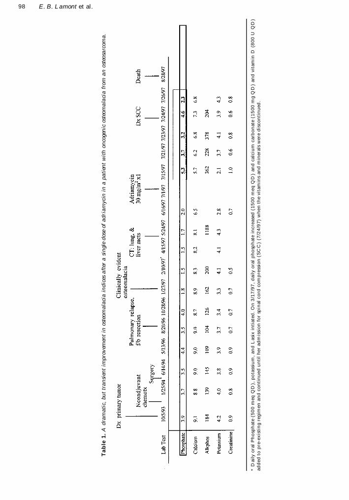
hospice. Table 1 contains a timeline of her disease,
electrolyte values and medical interventions. While
the patient’s mean phosphate level during her period
of osteomalacia (preceding the dose of adriamycin)
was 1.76 mg/dl, the mean phosphate level after the
single dose of adriamycin until death was 3.82 mg/dl.
A post-adriamycin 24-h urine for phosphate was not
collected.
Discussion
Osteomalacia is a disorder of defective mineraliza-
tion of osteoid, organic bone matrix. Since osteoid is
mineralized to bone through deposition of crystals
composed of calcium and phosphate, prolonged
de® ciency of phosphate, calcium, or both, can beget
osteomalacia. Chronic hypophosphatemia results from
inadequate intake or excessive loss and presents in
inherited or acquired forms. Inadequate phosphate
intake can be the result of extensive intestinal disease,
the presence of phosphate binders in the diet, or
secondary to vitamin D de® ciency (due to low dietary
intake, lack of sun exposure, renal or hepatic disease,
and inherited disorders of vitamin D metabolism).
Excessive urinary loss due to renal tubular dysfunc-
tion can be inherited (e.g., autosomal recessive and
X-linked hypophosphatemic r ickets, Fanconi
syndrome) or acquired (e.g., renal tubular acidosis,
toxic damage by heavy metals or certain chemothera-
peutic agents, and tumor-induced osteomalacia).
Chronic hypocalcemia as a result of intestinal disease,
abnormalities of vitamin D and parathyroid hormone,
or systemic acidosis is an unusual cause of osteoma-
lacia since symptoms of hypocalcemia often result in
earlier medical attention and are only rarely seen in
hypoparathyroidism and pseudohypoparathyroidism.
There are several hypotheses for the mechanism of
urinary phosphate loss in oncogenic osteomalacia,
with a humoral mechanism by far the most compel-
ling based on clinical observations and experimental
data.The clinical observations that support a humoral
mechanism are that the culprit tumors are often small
and remote from the kidney, that tumor removal
results in both rapid resolution of phosphaturia and
in rapid skeletal remineralization, and that some
culprit tumors appear to have neurosecretory granules
on EM suggesting peptide hormone production.2,3
In vivo and in vitro experiments suggest the presence
of a transmittable phosphaturic factor. Investigators
report phosphaturia in dogs and rats injected with
tumor extracts4± 6 and in athymic mice transplanted
with tumor.7 In single cell assays, investigators have
described inhibition of both renal tubular sodium-
dependent phosphate transport8,9 and renal tubular
25(OH)D-1 a -hydroxylase when media from tumor
cell cultures were added to cultures of animal renal
tubule cells.7 These experiments resonate with the
clinical ® ndings of phosphatur ia and decreased
1,25(O H )2-vitamin D in patients w ith these
neoplasms. However, whether the circulating factor
has PTH-like activity is still unclear: tumor extracts
have stimulated cAMP in some cases8,10 but not in
others.4,9
The mainstay of management of oncogenic osteo-
malacia has been removal of tumor through complete
surgical resection. Since the condition is usually
associated with localized benign tumors and not
metastatic malignant disease, this approach is usually
successful. Unfortunately, our patient had metastatic
malignant disease that was not resectable. However,
the fact that tumor-speci® c chemotherapy was effec-
tive in at least transiently improving her hypophos-
phatemia may support the theory that oncogenic
osteomalacia is paraneoplastic in origin. However,
why the patien t’ s tum or progressed while her
phosphate was still in the normal range is not clear. It
* Dose of adriamycin reduced from 60 mg/m2 because of hyperbilirubinemia.
Osteomalacia 97
Ta
ble
1.
Ad
ram
ati
c,bu
ttr
an
sien
tim
pro
vem
ent
inos
teom
ala
cia
ind
ices
aft
era
sin
gle
dos
eof
ad
ria
myci
nin
apa
tien
tw
ith
on
coge
nic
oste
om
ala
cia
from
an
ost
eosa
rcom
a.
*D
ail
yo
ral
Ph
osp
hate
(50
0m
eqQ
D),
po
tass
ium
,an
dL
asi
xin
tiate
d.
On
3/1
7/9
7,
dail
yo
ral
ph
osp
hat
ein
cre
ase
d(1
50
0m
eq
QD
)an
dca
lciu
mcarb
on
ate
(15
00
mg
QD
)an
dvit
am
inD
(80
0U
QD
)ad
ded
top
re-e
xis
tin
gre
gim
en
an
dco
nti
nu
ed
un
til
her
ad
mis
sio
nfo
rsp
inal
co
rdco
mp
ress
ion
(SC
C)
(7/2
4/9
7)
wh
en
the
vit
am
ins
an
dm
iner
als
wer
ed
isco
nti
nu
ed.
98 E. B. Lamont et al.

may be that the kinetics of the putative paraneo-
plastic phosphaturic factor and the kinetics of the
tumor are not equal.
Conclusion
We report an atypical case of the rare entity onco-
genic osteomalacia. The case was atypical because it
was associated with a malignant tumor, because the
syndrome presented only with a second recurrence of
the tumor, not with its initial presentation or ® rst
relapse, and because it transiently improved with
chemotherapy. Like others with this paraneoplastic
syndrome, our patient had characteristic clinical,
laboratory and radiographic ® ndings of hyperphos-
phaturic osteomalacia. While the pathophysiology of
oncogenic osteomalacia is still not entirely clear, a
paraneoplastic etiology has been proposed. Through
our report of a patient’s transient resolution of hypo-
phosphatemia coincident with chemotherapy, we
provide further evidence to suggest that this syndrome
may be paraneoplastic in origin.
References
1 Rossi R, Godde A, Kleinebrand A, et al. Unilateralnephrectomy and cisplatin as risk factors of ifosfamide-induced nephrotoxicity: analysis of 120 patients. J Clin
Oncol 1994; 12:159± 65.
2 Stone MD, Quincey C, Hosking DJ. A neuroendocrinecause of oncogenic osteomalacia. J Pathol 1992;167:181 ± 5.
3 Wilkins GE, Granleese S, Hegele RG, et al. Oncogenicosteomalacia: evidence for a humoral phosphaturicfactor. J Clin Endocr inol Metab 1995; 80(5):1628± 34.
4 Aschinberg LC, Solomon LM, Zeis PM, et al. VitaminD-resistant rickets associated with epidermal nevussyndrome: demonstration of a phosphaturic substancein the dermal lesions. J Pediatr 1977; 91:56± 60.
5 Popotvtzer MM. Tumor-induced hypophosphatemicosteomalacia: evidence for a phosphaturic cyclicAMP-independent action of tumor extract. Clin Res
1981; 29:418A. abstract.6 Lau K, Stom MC, Goldberg M, et al. Evidence of a
humoral phosphaturic factor in oncogenic hypophos-phatemic osteomalacia. C lin Res 1979; 27:421A(Abstract)
7 Cai Qiang, Hodgson SF, Kao PC, et al. Brief report:inhibition of renal phosphate transport by a tumorproduct in a patient with oncogenic osteomalacia. New
Engl J Med 1994; 330(23):1645± 9.8 Nelson AE, Namkung HJ, Patava J, et al. Characteristics
of tumor cell bioactivity in oncogenic osteomalacia.M ol Cell Endocr inol 1996; 124:17± 23.
9 Miyauchi A, Fukase M, Tsutsumi M, Fujita T.Hemangiopericytoma-induced osteomalacia: tumortransplantation in nude mice causes hypophosphatemiaand tumor extracts inhibit renal 25-hydroxyvitaminD1-hydroxylase activity. J Clin Endocrinol M etab 1988;67:46± 53.
10 Seshadri MS, Cornish CJ, Mason RS, Posen S. Parathy-roid hormone like bioactivity in tumours from patientswith oncogenic osteomalacia. Clin Endocrinol 1985;23:689± 97.
Osteomalacia 99

Submit your manuscripts athttp://www.hindawi.com
Stem CellsInternational
Hindawi Publishing Corporationhttp://www.hindawi.com Volume 2014
Hindawi Publishing Corporationhttp://www.hindawi.com Volume 2014
MEDIATORSINFLAMMATION
of
Hindawi Publishing Corporationhttp://www.hindawi.com Volume 2014
Behavioural Neurology
EndocrinologyInternational Journal of
Hindawi Publishing Corporationhttp://www.hindawi.com Volume 2014
Hindawi Publishing Corporationhttp://www.hindawi.com Volume 2014
Disease Markers
Hindawi Publishing Corporationhttp://www.hindawi.com Volume 2014
BioMed Research International
OncologyJournal of
Hindawi Publishing Corporationhttp://www.hindawi.com Volume 2014
Hindawi Publishing Corporationhttp://www.hindawi.com Volume 2014
Oxidative Medicine and Cellular Longevity
Hindawi Publishing Corporationhttp://www.hindawi.com Volume 2014
PPAR Research
The Scientific World JournalHindawi Publishing Corporation http://www.hindawi.com Volume 2014
Immunology ResearchHindawi Publishing Corporationhttp://www.hindawi.com Volume 2014
Journal of
ObesityJournal of
Hindawi Publishing Corporationhttp://www.hindawi.com Volume 2014
Hindawi Publishing Corporationhttp://www.hindawi.com Volume 2014
Computational and Mathematical Methods in Medicine
OphthalmologyJournal of
Hindawi Publishing Corporationhttp://www.hindawi.com Volume 2014
Diabetes ResearchJournal of
Hindawi Publishing Corporationhttp://www.hindawi.com Volume 2014
Hindawi Publishing Corporationhttp://www.hindawi.com Volume 2014
Research and TreatmentAIDS
Hindawi Publishing Corporationhttp://www.hindawi.com Volume 2014
Gastroenterology Research and Practice
Hindawi Publishing Corporationhttp://www.hindawi.com Volume 2014
Parkinson’s Disease
Evidence-Based Complementary and Alternative Medicine
Volume 2014Hindawi Publishing Corporationhttp://www.hindawi.com