on the mouth-parts of some blattidæ
TRANSCRIPT

On the Mouth-Parts of Some BlattidæAuthor(s): Joseph ManganSource: Proceedings of the Royal Irish Academy. Section B: Biological, Geological, andChemical Science, Vol. 27 (1908/1909), pp. 1-10Published by: Royal Irish AcademyStable URL: http://www.jstor.org/stable/20516949 .
Accessed: 18/06/2014 16:55
Your use of the JSTOR archive indicates your acceptance of the Terms & Conditions of Use, available at .http://www.jstor.org/page/info/about/policies/terms.jsp
.JSTOR is a not-for-profit service that helps scholars, researchers, and students discover, use, and build upon a wide range ofcontent in a trusted digital archive. We use information technology and tools to increase productivity and facilitate new formsof scholarship. For more information about JSTOR, please contact [email protected].
.
Royal Irish Academy is collaborating with JSTOR to digitize, preserve and extend access to Proceedings of theRoyal Irish Academy. Section B: Biological, Geological, and Chemical Science.
http://www.jstor.org
This content downloaded from 62.122.73.86 on Wed, 18 Jun 2014 16:55:30 PMAll use subject to JSTOR Terms and Conditions

PROCEEDINGS
THE RO-YAL IRISH ACADEMY
PAPERS READ BEFORE THE ACADEMY
I.
ON THE MOUTH-PARTS OF SOME BLATTIDZE.
By JOSEPH MANGAN, B.A., A.R.C.Sc.
[COMMUNICATED BY PROFESSOR G. H. CARPENTER, B.SC., M.R.I.A.]
(PLATES I.-III.)
[Read APRIL 13. Ordered for Puiblication APRIL 15. Publisbhed MAY 23, 1908.]
COCKROACHES occupy a peculiar position amongst insects. The comparative
ease with which they may be dissected, the readiness with which they
can be procured, together with generalized structure, mark them out
as the "type*" par excellence of the Hexapoda, furnishing as they do
a most suitable ground-work for further systematic study of the group.
Hence it is not surprising that some of the conmmon species have been
the subject of many careful descriptions in text-books, and have been
employed therein for comparison with the higher members of the class.
In this last respect, the mouth-parts are possibly of greatest interest,
yet they appear to have received but little detailed study. The precise
contour of the brain has even been recorded by piecing together the drawings
of consecutive sections; but referring to the maxilli or labium the student will experience a very certain sense of dissatisfaction, both diagrams and
descriptive matter showing the scant attention accorded to these parts. To appreciate the novel views of Hansen ('93) as to the jointing of the maxille
and his belief in the presence of homologuies of the Thysanurani maxilluhle in
some Orthoptera, a more careful examination is necessary. R. I. A. PROC., VOL. XXVII., SECT. B. [B]
This content downloaded from 62.122.73.86 on Wed, 18 Jun 2014 16:55:30 PMAll use subject to JSTOR Terms and Conditions

2 Proceedinigs of the Royal Irish Academy.
The following attempt accurately to review the parts in question in Periplaneta australasice may be of use as a starting-point to those possessing the necessary material, and desirous of seeing how far Hanseni's views find support amongst forms allied to Periplaneta, attention moreover being directed to onie or two points of especial interest which have hitherto escaped comment in this well-known genus.
The work has been carried out in the Zoological Laboratory of the Royal College of Science for Ireland; and I am indebted to Professor G. H. Carpenter for his guidance and advice during this undertaking, and to Mr. F. W. Moore of the Royal Botanic Gardenis for supplying me with
abundant material.
THE MANDIBLES (Plate I.)
The mandible articulates with the head by means of the "condyle " (e)
and the " ginglymus" (g). The former is a knob-like projection on the posterior
surface, proximal and external in position, which works in a socket afforded by the epicranial plate; the latter, a shallow groove, plays upon a ridge of
the clypeus, and is situated anteriorly, some distance from the outer border of the mandible. The axis of revolution of the jaw is thus directed forwards, inclining slightly towards the middle line. The inner border bears some distal teeth or blades, and a proximal truncated process, the " pars
molaris" (mp), the right mandible bearing three distinct blades, the left having five such. When the jaws close, the processes are said to interlock, which is certainly true with respect to the two molar surfaces, but the blades
of the right mandible all come to lie behind and across those of the left, the
third, or most proximal, on the right, being supported by the two extra
processes on the left (fig. 2). During mastication, I imagine that the molar
surfaces may, by closing uipon and supporting the more resistinig food-stuffs,
enable the overlapping blades to cut with better effect; the slight inward
inclination of the axes of rotation of the jaws tending to the same end.
Below the pars molaris there is a well-marked process (la) projecting
freely inwards, doubtless a homologue of the lacinia mobilis recorded by Hansen ('93) and others as occurring in certain Coleoptera. Though apparently figured by Muhr ('77), he makes no comment upon it. Miall and
Denny ('86) speak of a flexible chitinouis flap, in Blatta orientalis, extending from the innier border of the mandible to the labrum. As certainly no such
flap exists, those authors evidently refer to the lacinia mobilis, though mistaken as to its true nature.
The abductor, or extensor muscle of the mandible (Ex), arises from the
upper portion of the side of the external head skeleton, and is inserted by a
This content downloaded from 62.122.73.86 on Wed, 18 Jun 2014 16:55:30 PMAll use subject to JSTOR Terms and Conditions

MANGAN- On the Mlouth-Parts of some Blattidcr. 3
slender tendoil on the outer edge of the jaw. Adduction or flexion is brought about by two powerful muscles. The long flexor (L) is a very large muscle arisinig from the roof and back of the head, its fibres converging to a very
strong chitinous tendon which is inserted on the posterior surface, close to the lacinia mobilis, being therefore quite removed from the ginglymus, near
which it is said, by Miall and Denny, to be inserted. The short flexor (S)
arises from the crus of the tentorium, and is inserted directly upon the
posterior surface of the jaw. A third muscle (In), which has not been recorded by the above authors, lies within the mandible, and might also act as a flexor, though more probably it moves the tongue. The fibres spring directly from the outer surface of the mandible and form an elongate tapering bundle, which merges into a thin, round, chitinous tendoln, this latter passing to the side of the tongue. Basch ('65) figures a similar muscle in Termes, terming it the levator linguwe.
The accompanying drawings were made from the adult male P. austrolasic, but I could detect no differences in the female, or in specimens of P. americana,
Blatta orientalis, or Phyllodromaia germanica. The parts in a specimen of B. orientalis 4 mm. in length were essentially as in the adult.
THE HYPOPHARYNX. (Plate I.)
The hypopharynx, or tongue (Iy), though partially connected with the labium, arises between the mandibles, and is best considered with them. The proximal portion (hypopharynx of Huxley) is a broad fold of the hinder surface of the mouth-cavity, smooth and flat. The free distal portion (lingua of Huxley) tapers slightly, and presents an arched surface, densely covered with hairs. The hypopharynx is strengthened basally by two chitinous plates (x and y), the distal of which (y) bears a number of strong bristles, and is
continued along the edge of the anterior surface as a chitinous rod. In contact with this laterally is situated the smaller proximal plate (x), which ends basally, close to the tendon of the interior muscle of the mandible. The free tip is furnished at the sides with a pair of elongate plates (z), which
carry bristles, and are continuous behind, as thin rods, round the opening of the salivary duct; posteriorly the distal surface of the hypopharynx exhibits a pair of less decided chitinous thickenings.
The position of the above plates (z) is conformable with the idea that they may represent a pair of m:iaxillulie (see Hansen, '93), which have become com
pletely fused with the tongue, since in the Apterygota these latter are shown to originate, at least in some cases, between the mandibles. On each side a
[B*J
This content downloaded from 62.122.73.86 on Wed, 18 Jun 2014 16:55:30 PMAll use subject to JSTOR Terms and Conditions

4 Proceedings of the Royal Irish Acadeny.
ligulate muscle ( V) passes from the base of the plate (z) to the posterior por tion of the tentorium (ten). Soine muscular fibres, which are inserted very basally on the anterior surface of the tongue, converge to two tendons which pass over the upper surface of the tentorial plate, and take their origin from the posterior edge of its circular aperture. A pair of muscles (not depicted on drawing) pass from the labium, above the mentum, to the region of inser tion of the muscle V at the base of z.
The salivary duct (sal) opens to the exterior at the back of the tongue, between it and the labium. At the sides, and somiewhat in front of this opening, there are a pair of pit-like depressions, which I take to be salivary receptacles; the left receptacle (rcep) is seen in fig. 1.
The view has been put forward that the hypopharynx represents the appendages of a head-segment, while Heymons ('95) entertains the idea that it represents the sterna of the segments which bear the mandibles, maxilke, and labium; the majority of zoologists, however, regard it as a secondary outgrowth from the mouth region. That it stands in close relation to the
mandibles is, perhaps, suggested by the muscles (In) passing from the interior of those jaws to its sides.
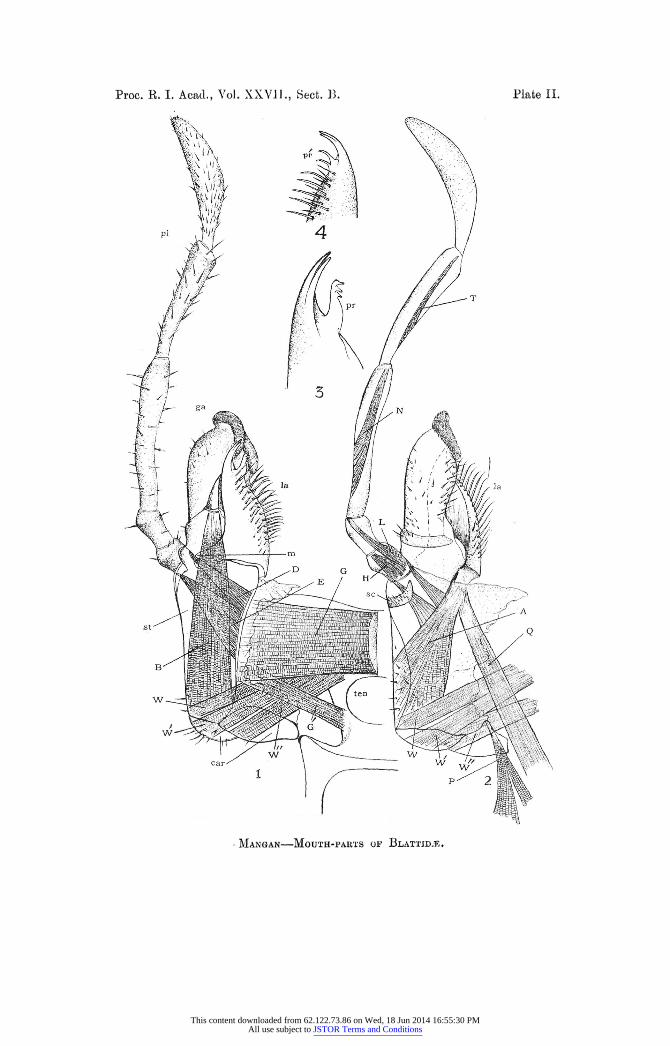
THE MAXILLIE. (Plate II.)
For descriptive purposes the maxilloe are generally considered as composed of a horizontal basal segment, the cardo, succeeded by a vertical segment, the stipes, the latter carrying a galea, lacinia, and palp; Hansen ('93), however, extending the term 'stipes' to the appendages which it bears. They are placed widely apart, so that the fused second maxillke, or labium, coming between them, meet the root of the tongue. The cardo (car) is an outwardly convex
plate, and is distinctly divided into two portions, a strong internal ridge projecting i-nwards along the line of demarcation. Proximally it articulates with the epicranial plate, and with the lower corner of the chitinous frame
which surrounds the occipital foramen ; distally it supports at right angles the stipes (st). The stipes presents a strongly thickened posterior surface, the sclerite lapping round the outer border, and extending for a short distance upon the anterior surface, which elsewhere is covered but by a thin cuticle. Front and back, flexible flaps extend from the inner edges of both
cardo and stipes to the head anid to the labium, offering, however, no impedi
ment to the free inotion of these segments. Near the distal end of thie stipes,
close to the outer edge, there arises on the anterior surface the five-segmented
palp (pl). A strong setiferous sclerite (so) at its base suggests a sixth segment.
This content downloaded from 62.122.73.86 on Wed, 18 Jun 2014 16:55:30 PMAll use subject to JSTOR Terms and Conditions

MANGAN-On the Mouth-Parts oa/some Blattidwe. 5
The galea (ga) consists of two segments, a basal portion continuous with the stipes and articulatiing by a well-marked joint with a distal hood-like segmnent. The lacinia (lta) is posteriorly decidedly segmented off from the plate of the stipes, with the exception of its outer corner, which at nv sends back a connecting plate. For a little distance the lacinia is united to the basal
segment of the galea, the two appearing to move as a whole upon the stipes,
with some deg,ree of backwards and forwards motion, but with no lateral freedom. The lacinia ends in two strongly chitinized prongs, and along its inner edge bears several rows of stiff setw. Just below the tip there is a singular process (pr, fig. 3), which arises anteriorly from the inner edge; although mentioned by Rolleston ('88), it is not recorded on any drawing of orthopteran maxillh known to me. It is present to my knowledge in P. australacsice, P. iamericana, and B. orientalis. In Phyllodromia germanica it differs; the two processes (pr', fig. 4) which in that species occupy an exactly similar position being doubtless homiologous with it. They resemble curved seta- for the outer portion of their length, but, broadening basally, they
merge gradually into the surface of the lacinia without exhibiting any of the thickenings or constrictions peculiar to the articulation of hairs. The above projections are probably homologous with some of the terminal processes to be found on the lacinia of forms like Machilis. They certainly correspond with the " comb-processes" on the lacinia of the Lepismatidae (Escherich, '05).
Their conditioln in the Japygide (Verhoeff, '04) is intermediate between their condition in the Machilidae on the one hand, and in the Lepismatida3 and Blattidae on the other.
The cardo is lowered by a tripartite muscle, which has its origin on the under surface of the tentorium, at the side of the central keel, a bundle of fibres ( W) being inserted at the base of the stipes, and also ( W') on the outer,
and (TW") on the ininer segment of the cardo. The same arrangement exists in Forficula (VerhoeW, '05), and I think it supports the idea that the cardo is not, as usiially stated, a single segment. The cardo is raised, or abducted, by
a muscle (P) inserted in front on a slight process of its inner sclerite. This
muscle arises in two distinct portions from the posterior region of the epi cranium. The stipes is adducted by a powerful muscle, inserted upon an
internal chitinous ridge which extends along the posterior inner edge of that
segmaent. This muscle has its origin, in part (G) upon the keel of the ten
torium, in part (C') lower down upon the central plate. The smaller portioni
(G') probably assists in raising the cardo. The stipes is apparently restored
to the vertical by the elasticity of the hinge between it and the cardo. The
same internal ridge of the stipes gives purchase to the two muscles D and
E which move the basal segmenalit of the palp, the succeeding segments being
This content downloaded from 62.122.73.86 on Wed, 18 Jun 2014 16:55:30 PMAll use subject to JSTOR Terms and Conditions

6 Proceedings of the Royal Irish Academy.
moved respectively by the muscles H, L, N, and 7. Two nerves pass
up the centre of the palp, and in places are apt to be mistaken for delicate
muscles. A muiscle (B) fromn the base of the stipes moves the galea, and
perhaps bends back both galea and lacinia as a whole. Anteriorly a stronger
muscle (A), arising also from the base of the stipes, is inserted by a broad
tendon into the initernal basal corner of the lacinia; it is joined by a slender
muscle (Q) from the epicranium. These muscles bend the lacinia, and with it
the galea, forwards. The strange suggestion of Verhoeff ('05), that the labium is really anterior
to the maxillae, finds no support in the musculature of those parts, as the
muscles of the maxillae that come from the tentorium all originate anteriorly
to those passing from there to the labiumn.
The theoretical interpretations of the jointing of the maxilla are numerous.
All appear to regard the cardo as the basal segment, though, as has been
pointed out above, it might perhaps be composed of two segments. Marshall
and Hurst ('99) regard the stipes and cardo as homologous with the protopodite
of the crustacea, the palp as an exopodite, the galea and lacinia as a divided
endopodite. Henneguy ('04) regards the stipes as a second segment, which is
followed by a third bearing as an internal ridge, the lacinia, the galea forming
a fourth and terminal segment; segments three and four constituting the
endopodite, the palp the exopodite. Lang ('91) and Boas ('96) regard the
galea and lacinia as mere masticatory ridges of the stipes segment, and I am
not acquainted with their views with respect to the succeeding palp segments.
Chatin, in his comparative account of the jaws of biting insects ('84), adopts a
somewhat empirical threefold division of the maxilla into basal, central, and
appendicular portions. Hansen ('93), who appears to have very carefully gone
into the matter, regards the lacinia as the masticatory lobe of the second or
stipes segment, while the third segment, which is cut off from this very
obliquely, bears the palp and galea. On mere examination of forms like
Periplaneta, one would, perhaps, accept this view with extreme hesitation; but in a specimen of Praemachilis which I examined, the galea certainly
appeared to be but an internal appendage of a third segment which carried
the palp. Hansen, who holds that insect appendages are directly comparable
with those of Malacostraca, regards the palp as endopodite. His extended observations do not apparently give any support to the theory that the galea
is homologous with the crustacean endopodite; indeed, the only fact that favours that theory seems to be the segmentation of the galea in certain forms less generalized than the Orthoptera-i.e., in the Adephaga, or carnivorous
Coleoptera. Moreover, Verhoeff ('04) points out that the galea and laciniia are very possibly homologues of the coxal organs present upon the basal segmiient
This content downloaded from 62.122.73.86 on Wed, 18 Jun 2014 16:55:30 PMAll use subject to JSTOR Terms and Conditions

MANGAN-On the Mouth-Parts of some Blattidw. 7
of the abdominal appendages in Thysanura and Myriopoda. The evidence is on the whole, distinctly favourable to the homology of the palp with the jointed ambulatory thoracic leg in the Insecta, and, consequently, with the endopodite of the typical crustacean appendage.
THE LABIUM. (Plate II.)
Authors, with the possible exception of Verhoeff, very generally regard the labium as the fused appendages of the segment coming next to that bearing the maxillae-a segmient which, according to Huxley ('77), is represented by the cervical sclerites. Notwithstanding its juxtaposition to the tongue, its parts in the cockroach are distinctly free from, and in no way directly con nected to, the head-skeleton.
The cervical sclerites, which we may provisionally regard as belonging to the segment bearing the labium, are eight in number. The two dorsal are
triangular, and meet in the middle line; at the sides are the lateral sclerites
(v and u), while the two narrow setiferous bands (t) are the ventral elements.
From u, the largest sclerite, a pair of muscles (S) converge, to be inserted on
the epieranial plate. The squarish submentum (sin) is the basal piece of the labium, and, despite
its relatively large size, is believed to result from the union of the first seg
ments, or cardines, of the constituent appendages. The mentum (me) is much
shorter and a little narrower, and its distal border overlaps to some extent
the succeeding surface-a point which is not evident when the labium has
been removed and mounted. Some regard the mentum as composed of the
entire stipites; but it is obvious, I think, to those who believe that the
lacinia or galea is a masticatory ridge of the stipes segment, that the mentum
contains but portion of the stipites. To me it appears that there is no joint
in the maxilla corresponding to the distal articulation of the mentum. If
viewed from the back, the remainder of the labium seems to consist of a
strongly chitinized piece, with which, on each side, are very distinctly
articulated a lacinia (la), a galea (ga), and a palp (pl) of three segments.
Moreover, a little distance from the end of the mentum is the furthest point
to which fusion of the primitively separate appendages has advanced. Viewed
from in front, the cuticle in this region is seen to be thin and flexible, bearing
fine scale-like markings, and the galea and lacinia exhibit no jointing with
the main part; while on this side there is decided indication of an additional
segment, the palpiger (pgr). The cuticle of the anterior surface merges on
to the hypopharynx, on a level with the distal border of the submentum.
This content downloaded from 62.122.73.86 on Wed, 18 Jun 2014 16:55:30 PMAll use subject to JSTOR Terms and Conditions

8 Proceedings of the Royal Irish Academy.
A muscle (R), which has its origin upon the plate of the submentum, is
inserted upon a slight ridge, which is coincident with the distal edge of the
mentum, its action being to pull the latter forwards. A muscle (D') from the
mentum and a muscle (E') from the posterior edge of the central plate of the
tentorium (beside the origini of V, Plate I) are both inserted at the base of
the palpiger, moving it slightly perhaps, but more probably working the end
of the labium as a whole. A long, slender muscle (F), coming from the ten
torium with Et, also the muscles K and X, move the first segment of the
palp. The muscle L' moves its second segment, and N' and X its ter
minal segment. The lacinia is bent back by the muscle A', being restored
by the elasticity of the unjointed cuticle in front; muscles B'and C bend
back the galea, which is restored in a like manner. As has been mentioned
in the account of the tongue, a pair of muscles pass from the labium, above
the mentum, to the sides of the hypopharynx.
Verhoeff ('05), as previously stated, regards the labium as the second pair
of mouth appendages, the maxillae, according to him, belonging to a succeeding
segment. His views are based to a great extent upon the nature of the
mentum and submentum. These he regards, not as fused portions of the
labial appendages, but as the sterna of two of the cephalic segments. He is
convinced that the mentum represents the sternum of the labial segment, the submentum that of the maxillary segment. To account for this he supposes
that a shifting of the maxillae has occurred, from their primitive position
behind the labial appendages to their present situation anterior to the latter.
He lays stress upon the close relations which appear to him to exist between
the cardo and the submentum; but even if they were united, it would hardly
be safe to draw conclusions as to their primitive connexion, as fusion between
neighbouring segments is of such common occurrence. Then all the muscles passing to the maxilla from the tentorium and
epicranial vault are anterior to the two pairs of muscles that go from the
tentorium to the labial palpiger and palp. This demands the almost total
disappearance of those primitive labial muscles which it is reasonable to sup
pose, on Verhoeff's theory, at one time did pass to the head in front of those
from the maxillae. Judging from the figures given by Miall and Denny, there is nothing in
the arrangement of the tracheal or nerve supply suggestive of such a profound
disturbance in the primitive arrangement of the limbs. Though the views that have hitherto been put forward regarding the homologies of mentum and
submentum may well be criticized, yet the theory substituted by Verhoeff appears to have far less basis in actual fact, and, by reason of its highly specu
lative character, it will most probably be adopted by few, if any, zoologists.
This content downloaded from 62.122.73.86 on Wed, 18 Jun 2014 16:55:30 PMAll use subject to JSTOR Terms and Conditions

MANGAN-OU the Mouth-Parts of some Blattidw. 9
Eeferences.
1865. Basch, S.?Untersuchungen ?ber das Skelet und die Muskeln des
Kopfes von Termes. Zeitschr. f. wissensch. Zoologie, 15 Band, 1865.
1896. Boas, J. E. V.?Text-book of Zoology. Transi. J. W. Kirkaldy and
E. C. Pollard. London, 1896.
1884. Chatin, J.?Sur le sous-maxillaire, le maxillaire, le palpig?re, le sous
galea, et les appendices de la m?choire chez les Insectes Broyeurs.
Comptes Eendus, vol. xcix., pp. 51-53, 285-288, 939-942. 1884.
1905. Escherich, K.?Das System der Lepismatiden. Zool?gica, xviii. 1905.
1893. Hansen, H. J.?A Contribution to the Morphology of the Limbs and
Mouth-parts of Crustaceans and Insects. Ann. Mag. Nat. Hist. (6), vol. xii. 1893 (from Zoolog. Anz., 1893).
1904. Henneguy, L. F.?Les Insectes. Paris, 1904.
1895. Heymons, E.?Die Embryonalentwickelung von Dermapteren und
Orthopteren. Jena, 1895.
1877. Huxley, T. H.?Manual of the Anatomy of Invertebrated Animals.
1877.
1891. Lang, A.?Text-book of Comparative Anatomy. Transi. H. and M.
Bernard. Part I. London, 1891.
1899. Marshall, A. M., and Hurst, C. H.?Practical Zoology. (Fifth ed.) London, 1899.
1886. Miall, L. C, and Denny, A.?The Cockroach. London, 1886.
1877. Muhr, J.?lieber die Mundtheile der Orthoptera. Prag, 1877.
1888. Eolleston, G? Forms of Animal Life. (Second ed.), Oxford, 1888.
(pp. 138-147.)
1904. Verhoeff, K. W.?Zur vergleichenden Morphologie und Systematik der Japygiden. Arch. f. Naturgeschichte. Jahrg. lxx., i. Band, 1904.
1905. Verhoeff, K. W.?Ueber vergleichende Morphologie des Kopfes niederer Insekten. Abhand. K. Leopold. Carolin. Akad., 84. Band, 1905.
R. I. A. PROC., VOL. XXVII., SECT. B. [C]
This content downloaded from 62.122.73.86 on Wed, 18 Jun 2014 16:55:30 PMAll use subject to JSTOR Terms and Conditions

10 Proceedings of the Royal Irish Academy.
EXPLANATION OF PLATES.
PLATE 1. Fig. l.Periplan eta australasiw. Mandibles and tongue viewed from behind. x 31.
2. Mandibles closed. x 19.
c, condyle; g, ginglymus; mp, pars molaris; la, lacinia mobilis; hy, hypopharynx; ten, tentorium; x, y, z, sclerites on hypopharynx; Ex, extensor muscle; L, long flexor; S, short flexor; In, muscle in
interior of mandible; V, posterior muscle of tongue; sal, salivary duct; reep, left salivary receptacle.
PLATE II.
1. Periplaneta australasiw. Right maxilla seen from behind. x 28.
2. P. australasice. Left maxilla seen from in front. x 28.
3. P. australasiwe. Apex of right lacinia. x 56.
4. Phyllodromia germanica. Apex of left lacinia, drawn to same scale as
Fig. 3.
ear, cardo; st, stipes; pl, palp; so, sclerite at base of palp;
ga, galea; la, lacinia; pr, pr', processes on apex of lacinia; m, connect
ing plate between lacinia and stipes ; ten, tentorium; W, W', WI", cardo
muscles; P, abductor of cardo; G, G', stipes muscles; 1, E, H, L, N, 7,
muscles of the palp; B, muscle of galea; A, Q, muscles of lacinia.
PLATE III.
1. Periplaneta amstralasiw. Left side of labium viewed from behind. x 31.
2. P. australasim. Left side of labium: anterior view. x 31.
a v, I, cervical selerites; sn, submentumn; me, iimentum; ga, galea;
lca, laciiiia; pri palp; pyr, palpiger; S, muscle front u to epicrainial
plate; B, muscle from submentum to mentum; D', E', muscles of
palpiger; F, K, H', muscles supplying basal segment of palp; L', muscle
of second segnment of palp,; N, M, muscles of terminal segment of
palp; A', muscle of lacinia; B', C, muscles of galea.
This content downloaded from 62.122.73.86 on Wed, 18 Jun 2014 16:55:30 PMAll use subject to JSTOR Terms and Conditions

Proc. R. I. Acad, Vol. XXVII., Sect. B. Plate L
In
M M
MANGAN MOUTH-PARTS OF BLATTIDSE.
This content downloaded from 62.122.73.86 on Wed, 18 Jun 2014 16:55:30 PMAll use subject to JSTOR Terms and Conditions
Proc. R. I. Acad., Vlol. XXVIL., Sect. 13. Plate II.
Sr~ ~~~~~~~p N 'f
l Ja
viL
1MAN GAN MOU TH-P1ART1S OF~ BLATT1IRY-E
This content downloaded from 62.122.73.86 on Wed, 18 Jun 2014 16:55:30 PMAll use subject to JSTOR Terms and Conditions

Proc. R. I. Acad., Vol XXVII., Sect. B. PlaUte III
.A ATu~~~~~~~~~~~~~~~~
MANGA&N---MOIJTH-PARTS OF BLATTIDJE.
This content downloaded from 62.122.73.86 on Wed, 18 Jun 2014 16:55:30 PMAll use subject to JSTOR Terms and Conditions