omega ground calibration b. gondet,saint louis, 21/05/2008
TRANSCRIPT

OMEGA GROUND CALIBRATION
B. Gondet,Saint Louis, 21/05/2008

Optical test bench
• OMEGA was mounted on a two axis platform 0.1 mrad in both site and azimuth (0.1 IFOV) Cold sink for regulating the operating temperatures
• light sources: - monochromator (spectraPro 275) the grating can be set as a mirror - tungsten ribbon lamp (Russia) brightness temperature from 2500 K to 2850 K five filters with different transmission profiles pinhole point source (steps of 0.05 IFOV) sample holder - black body, from 325 K to 1400 K (Galaï – 1200 BB) circular aperture covering 4 IFOV’s - grid of 4 x 4 IFOV-sized black bodies (Galileo) - cold plate, from 150 K to 293 K
OMEGA calibration plan
• spectral calibration • photometric calibration influence of detector temperature and spectrometer temperature linearity • geometric calibration co-alignment, registration stray light
• reference targets

OMEGA IN THE CALIBRATION TANK

THE OPTICAL BENCH

1. Spectral calibration (lambda.dat)

Tspectro T det (IR) Position in the FOV
-90°C 20°C 5
-90°C
20°C 1 (center)
-80°C -195°C 5
-80°C -195°C 1 (center)
-70°C -195°C 3
-95°C 20°C 3
VNIR (100 &200) msTspectro Tdet (IR) Position in the FOV
-90°C -195°C 3
-80°C -195°C center
-70°C -195°C 3
-90°C -191°C center
-90°C -198°C center
-90°C -200°C center
-90°C -197°C center
-90°C -193°C center
-90°C -188°C center
SWIR (2,5 ms & 5ms)
List of spectral calibration performed using the monochromator

Procedure and examples of results for SWIR
Tspectro = -90°C Tdet = -195°CTspectro = -70°C Tdet = -195°C
Spectel n° 60 (C)
Nanomètres
Dig
its
14nm
Spectel N°188 (L)
Nanomètres
Dig
its 23nm

Spectel number
Nan
omet
e rs
SWIR-L spectral calibration as a function of position in the FOV
right of FOV
left of FOV

Spectral match between OMEGA Earth spectrum, and "MODTRAN" simulated
SWIR-C SWIR-L

2. radiometric calibration
mtfYYMM_25.dat (2.5 msec, release of year YY, month MM)mtfYYMM_50.dat ( 50 msec, release of year YY, month MM)

Operational parameters
Hot Black body source temperature, varying from 9°C to 1175°CCold BB temperature from 10°C down -120°CIR detector temperature from –201°C to –188°C (-195°C nominal)Integration time = 2,5 ms, 5 ms & 10ms for SWIR = 100 ms & 200 ms for VNIRTest at several positions in the FOV

197°C
295°C
368°C523°C
saturation
low S/N
Signal in DN divided by the corresponding black body flux
DETERMINATION OF THE PHOTOMETRIC FUNCTION (C CHANNEL)

Photometric response of OMEGA
VIS
SWIR C
SWIR L
Bright region
Dark region
S/N ratio for observations of MarsPhase = 0°, surface temperature = 250 K
Wavelength (µm)
Wavelength (µm)

INFLUENCE OF THE TEMPERATURE OF THE SPECTROMETER
For OMEGA, photons remove electrons from pre-charged wells The available dynamics decreases rapidly with the temperature of the spectrometerbeyond 4.5 µm, in particular for long integration times (5 msec)
This is essential for the detection of sulfates
200 K 190 K175 K
Solid line: 5 msecDashed lines: 2.5 msec
Wavelength (µm)

3. geometric calibration

"AZIMUTH" = pitch: spacecraft motion
"SITE" = yaw: SCANNING MIRROR motion
SLIT
Point Source(ribbon lamp, grid Officine Galileo)
Geometric calibration

"Site", scanning mirror
Azimuth, spacecraft motion

1. IFOV for each spectel, for each of the 128 spatial directions
2. perpendicular to the slit ("azimuth") direction
- for each channel, relative variation of the viewing direction along the spectrum, for each IFOV within the entire 128 px FOV
- spectral registration for the 3 channels, and each of the 128 positions
3. along the slit ("site") direction
- relation between the IR swath position and the VNIR column number- spectral registration for the 3 channels, and each of the 128 directions
Geometric calibration files

"VIS" SPECTELS (0.35 – 1.1 µm)
Arc
min
utes
IR SPECTELS (1.0 – 2.7 µm)
Arc
min
ute
s
IFOV = f(wavelength), at the center of the FOV

12 pixels
l l l l.45 µm 1. µm 2.5 µm 5 µm
(Y,λ) maps of a point source scanned in azimuth
VNIR
SWIR-LSWIR-C

IRC IRL VIS
(i,j) (i,j-/+n) (i-/+m,j-/+n)

REGISTRATION : creation of false feature
in case the viewing direction of # spectels would be as follows…
…we would end up with a misinterpretated spectral feature

PIX
ELS
IR spectels (1 to 2.7 µm)
# positions along the slit64/12832/12896/128
5/128125/128
3/128
2/128
1/128
122/128
121/128
120/128
59/128
60/128
9, 124, 62 /128
Pix
els
VIS and IR spectels (0.35 to 2.7 µm)
# positions (pixels) along the slit
IR spectels (1 to 4.5 µm)
Pix
els
Center of the field
REGISTRATION EFFECTS along the azimuth


4. Stray light

Measurements made:± 30° ("site")± 40° ("azimuth")
Source: ribbon lamp, full power
No stray light detected

5. Reference samples
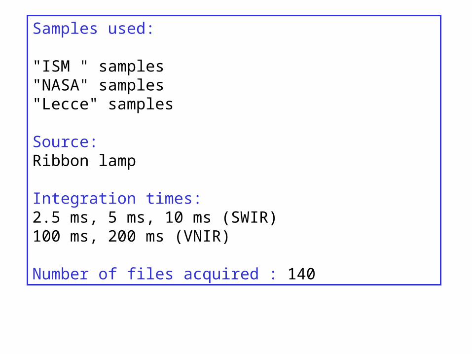
Samples used:
"ISM " samples"NASA" samples"Lecce" samples
Source: Ribbon lamp
Integration times:2.5 ms, 5 ms, 10 ms (SWIR)100 ms, 200 ms (VNIR)
Number of files acquired : 140

HYPERSTENE
μm
GYPSUM
μm Nanomètres
20% calcite 80% Palagonite5% calcite 95% Palagonite1% calcite 99% Palagonite
μm
Calcite10% Calcite + 90% Montmorillonite4% Calcite + 96% Montmorillonite

CalciteMontmorilloniteGypsumDiopsiteFayeliteLabradoriteMagnetite
wavelength (micrometers)