olfactory transfer of analgesic drugs after nasal ...intranasal.net/anatomyphysiology/westin...
TRANSCRIPT

ACTAUNIVERSITATISUPSALIENSISUPPSALA2007
Digital Comprehensive Summaries of Uppsala Dissertationsfrom the Faculty of Pharmacy 55
Olfactory Transfer of AnalgesicDrugs After Nasal Administration
ULRIKA ESPEFÄLT WESTIN
ISSN 1651-6192ISBN 978-91-554-6871-2urn:nbn:se:uu:diva-7829

I rörelse
Den mätta dagen, den är aldrig störst. Den bästa dagen är en dag av törst.
Nog finns det mål och mening i vår färd – men det är vägen, som är mödan värd.
Det bästa målet är en nattlång rast, där elden tänds och brödet bryts i hast.
På ställen, där man sover blott en gång, blir sömnen trygg och drömmen full av sång.
Bryt upp, bryt upp! Den nya dagen gryr. Oändligt är vårt stora äventyr.
Karin Boye
Till Anders och Alfred


Papers discussed
This thesis is based on the following papers, which will be referred to by the Roman numerals assigned below:
I. Westin, U., Piras, E., Jansson, B., Bergström, U., Dahlin, M., Brittebo, E. and Björk, E.: Transfer of morphine along the olfactory pathway to the central nervous system after nasal administration to rodents. Eur J Pharm Sci. 2005, 24(5): 565-573Reproduced with permission. ©2005 Elsevier
II. Espefält Westin, U., Boström, E., Gråsjö, J., Hammarlund-Udenaes, M. and Erik Björk.:Direct nose-to-brain transfer of morphine after nasal administration to rats.Pharm Res. 2006, 23(3): 565-572. Reproduced with permission. ©2006 Springer
III. Fransén, N., Espefält Westin., U. Nyström C. and Björk E.: The in vitro transport of dihydroergotamine across porcine nasal respiratory and olfactory mucosa and the effect of a novel powder formulation Submitted
IV. Espefält Westin, U. and Björk E.: Morphine transport across porcine nasal respiratory and olfactory mucosa studied in horizontal Ussing chambers Submitted
My contribution: I contributed to all parts of the above papers except for the choice of statistical methods and simulations in Paper II, the preparation and characterisation of the powder formulation and the drug analysis in Paper III.

Contents
1 Introduction................................................................................................111.1 Nasal systemic delivery of analgesics ................................................11
1.1.1 Nasal absorption into the systemic blood circulation .................121.1.2 Drug transport across the blood-brain barrier.............................13
1.2 Olfactory transfer of drugs after nasal administration........................131.2.1 Transfer mechanisms and target areas ........................................141.2.2 Selected methods for studying olfactory transfer .......................17
1.3 Defence mechanisms affecting nasal drug delivery ...........................181.4 Improving nasal drug delivery ...........................................................191.5 Nasal administration of morphine and dihydroergotamine ................21
2 Aims of the thesis.......................................................................................24
3 Materials and methods ...............................................................................253.1 In vivo olfactory transfer of morphine in rodents (Papers I and II) ....25
3.1.1 Materials .....................................................................................253.1.2 Animals.......................................................................................253.1.3 Experimental set-up ....................................................................253.1.4 Drug analysis ..............................................................................283.1.5 Data analysis and statistics .........................................................29
3.2 In vitro transport of dihydroergotamine and morphine across porcine nasal respiratory and olfactory mucosa (Papers III and IV).....................30
3.2.1 Materials .....................................................................................303.2.2 Isolation of nasal mucosa............................................................303.2.3 The horizontal Ussing chamber ..................................................313.2.4 Drug analysis ..............................................................................343.2.5 Data analysis and statistics .........................................................34
4 Results and discussion ...............................................................................354.1 In vivo olfactory transfer of morphine in rodents (Papers I and II) ....35
4.1.1 Screening for olfactory transfer of morphine (Paper I) ..............354.1.2 Visualising the olfactory transfer of morphine (Paper I) ............374.1.3 Quantification of the olfactory transfer of morphine to the brain (Paper II)..............................................................................................39
4.2 In vitro transport of dihydroergotamine and morphine across porcine nasal respiratory and olfactory mucosa (Papers III and IV).....................42
4.2.1 Development of the horizontal Ussing chamber method............424.2.2 Stability and drug adsorption studies..........................................454.2.3 Drug transport studies.................................................................45
4.3 Implications of olfactory transfer of analgesics (Papers I-IV) ...........48
5 Conclusions................................................................................................51
6 Perspectives ...............................................................................................52
7 Populärvetenskaplig sammanfattning ........................................................53
8 Acknowledgements....................................................................................54
9 References..................................................................................................57

Abbreviations
ANOVA analysis of variance AUC area under the concentration-time curve BBB blood-brain barrier BCSFB blood-CSF barrier CNS central nervous system CSF cerebrospinal fluid CV coefficient of variation CYP cytochrome P450 DHE dihydroergotamine EDTA ethylene diamine tetra-acetic acid HPLC high-performance liquid chromatography HPLC rats rats whose results were analysed using HPLC i.v. rats rats receiving study drug by intravenous route Isc short circuit current KRB Krebs-Ringer bicarbonate buffer LOB left olfactory bulb LOT lateral olfactory tract LS liquid scintillation LS rats rats whose results were analysed using LS M3G morphine-3-glucuronide M6G morphine-6-glucuronide nasal rats rats receiving study drug by nasal route Papp apparent permeability PBS phosphate-buffered saline PD potential difference P-gp P-glycoprotein R transmucosal electrical resistance RH relative humidity ROB right olfactory bulb rpm revolutions per minute SD standard deviation SEM standard error of the mean SSG sodium starch glycolate Tmax time to peak plasma concentration UDP uridine diphosphate UGT UDP-glucuronosyl transferase


11
1 Introduction
“Pain is an awareness created by the brain” Professor Helen Crawford
Rapid and reliable drug delivery to the central nervous system (CNS) is es-sential for successful pain treatment. Fast onset of pain relief and good pain control lowers the total perception of pain and decreases the risk of develop-ing chronic pain. Some analgesics are rapidly absorbed into the systemic blood circulation after nasal administration, with resultant high bioavailabil-ity because of the absence of first-pass hepatic metabolism (Dale et al.,2002; Rapoport et al., 2004). However, the blood-brain barrier (BBB) se-verely limits delivery of several central-acting drugs from the systemic cir-culation to the CNS (Terasaki and Pardridge 2000). Nonetheless, direct drug transfer to the CNS after nasal administration, via olfactory pathways that bypass the BBB, has been demonstrated in both animals and humans (Mathison et al., 1998; Illum 2004). The potential for both interesting treat-ment possibilities and the risk of unwanted side effects associated with olfac-tory transfer of drugs will increase as more effective formulations and deliv-ery devices are developed. The focus of this thesis is the olfactory transfer of analgesics, specifically morphine and dihydroergotamine (DHE), to the CNS.
1.1 Nasal systemic delivery of analgesics Nasal drug delivery is easy, well-tolerated and noninvasive. In children, nasal administration of fentanyl or diamorphine is preferable to alternative routes for treatment of acute pain, for example after a bone fracture (Kendallet al., 2001; Borland et al., 2007). Similarly, nasal butorphanol offered better and longer analgesia in women with post-caesarean section pain than i.v. administration (Abboud et al., 1991). Nasal morphine, ketamine and fentanyl have also been evaluated in the treatment of breakthrough pain in patients with cancer, with promising results (Zeppetella 2000; Pavis et al., 2002; Fitzgibbon et al., 2003; Carr et al., 2004). The patient can self-administer the drug and control the dosage when appropriate, which enables home treat-ment and a cheaper alternative to the various pump systems used for patient-controlled analgesia (Striebel et al., 1996; Dale et al., 2002). In their review of nasal administration of opioids for acute pain, Anez Simon et al. (2006) reported that systemic adverse effects were similar to those after

12
i.v. administration (drowsiness, nausea and vomiting). The local adverse effects were a burning sensation and a bad taste.
1.1.1 Nasal absorption into the systemic blood circulation Nasal drug absorption is influenced by anatomical and physiological factors as well as by the properties of the drug, the drug formulation and delivery device. Nasal drug absorption into the systemic blood circulation takes place across the nasal respiratory mucosa. In humans, this mucosa covers most of the total 150-180 cm2 nasal surface area, which is larger than might be ex-pected because of surface-increasing structures such as the turbinates (con-chae). Absorption is rapid because the mucosa is richly vascularised and blood flow is high, so as to warm, humidify and filter the incoming air.
Figure 1. Cell types found in the nasal respiratory mucosa (Hägerström 2003).
The mucosa contains four main cell types: ciliated and non-ciliated columnar cells, goblet cells and basal cells (Figure 1). On average throughout the nasal cavity, twenty percent of the respiratory epithelial cells are ciliated, and each cell also bears approximately 300 microvilli (Mathison et al., 1998; Illum 2004). Nasally administered drugs can passively diffuse trans- or paracellu-larly, or be subjected to active carrier/receptor-mediated transport or endocy-tosis across the nasal respiratory mucosa. The physicochemical factors of the drug molecule, such as charge, lipophilicity and molecular weight, will also affect its transport. Small lipophilic drugs are readily transported transcellu-larly; for example, the bioavailability of fentanyl is 71 % after nasal admini-stration (Striebel et al., 1993). The tight junctions, however, limit the paracellular route for hydrophilic drugs, often resulting in bioavailabilities of less than 10% for small polar drugs and larger hydrophilic drugs such as peptides (Illum 2003).

13
1.1.2 Drug transport across the blood-brain barrier Drug concentrations in the brain after nasal administration are usually the result of absorption into the systemic blood circulation and subsequent transport across the BBB, i.e. the drug is transported to the CNS systemi-cally. However, delivery of drugs from the systemic circulation to the CNS is heavily limited by the BBB. The BBB is an endothelium of capillaries with epithelial-like high resistance tight junctions that perfuses the mammal-ian brain (Pardridge 1999). Penetration of a drug through the BBB depends on characteristics such as the lipophilicity and size of the molecule and its specificity for a variety of ATP-dependent transport systems (Graff and Pol-lack 2004). Small lipophilic drugs are transported across the BBB via free diffusion, but other drugs require the active carrier- or receptor-mediated transport mechanisms that exist for transfer of endogenous substances such as nutrients or vitamins (Pardridge 1999). Efflux proteins such as P-glycoprotein (P-gp) in the BBB protect the brain from potentially harmful substances. There is also a barrier between the blood and the cerebrospinal fluid (CSF), the blood-CSF barrier (BCSFB), which consists of a single con-tinuous layer of polarised epithelial cells with tight junctions that line the choroid plexus. This barrier is not as restrictive as the BBB and has a 1000-fold smaller surface area, but has a wider range of enzymes than the BBB (Graff and Pollack 2004; Loscher and Potschka 2005).
1.2 Olfactory transfer of drugs after nasal administration Individuals have reported euphoria as soon as 3-5 min after sniffing the ille-gal drug cocaine, and the initiation of the behavioural or physiological ef-fects of cocaine precedes the rise in plasma cocaine concentrations after a single nasal dose (Perez-Reyes and Jeffcoat 1992; Farre et al., 1993; McCance-Katz et al., 1993). Thus, early cocaine effects are not all due to nasal or oral absorption into the systemic blood circulation and subsequent transport across the BBB; some cocaine has been directly transferred to the CNS, presumably via the olfactory pathways, bypassing the BBB. An animal study demonstrated three times higher levels of cocaine in the olfactory bulbs one minute after a nasal dose compared with i.v. administration (Chowet al., 1999), which indicates that cocaine can be transferred to the CNS via olfactory pathways.
The olfactory transfer route would be of particular interest for very potent drugs with poor blood-brain permeability such as neuropeptides for treat-ment of Alzheimer’s or Parkinson’s disease (Born et al., 2002), or when high brain concentrations are needed very rapidly, as in pain medication. Indica-tions of olfactory transfer in humans have for example been shown for the following compounds: arginine-vasopressin, 99mTc-DTPA-hyaluronidase,

14
insulin, melancortin and adrenocorticotropin (Pietrowsky et al., 1996; Oku-yama 1997; Derad et al., 1998; Born et al., 2002).
Figure 2. Location of the olfactory bulb, the olfactory mucosa and the olfactory nerve cells in humans. Modified picture from the Nobel Prize official homepage (Nobelprize.org).
1.2.1 Transfer mechanisms and target areas One of the main functions of the nose is olfaction. The olfactory mucosa is located posteriorly in the nose, at the roof of the nasal cavity, just underneath the cribriform plate of the ethmoid bone (Figure 2). The olfactory mucosa covers 3-10 % of the total nasal surface area, including parts of the nasal septum, the roof of the nasal cavity and the superior turbinate. This mucosa is a modified respiratory mucosa and contains three main types of cells: ol-factory nerve cells, supporting cells and basal cells (Mathison et al., 1998; Illum 2004) (Figure 3). Tight junctions are present between the supporting cells and between supporting cells and olfactory nerve cells. Further, the depth of the olfactory epithelium is more than twice that of the nasal respira-tory epithelium (Morrison and Costanzo 1992).
The olfactory nerve cells are bipolar neurons with dendrites projecting into the surface of the olfactory mucosa; the axon ends in the olfactory bulb where it synapses with second order neurons. The olfactory nerves are, therefore, in direct contact with both the environment and the CNS. The dendrite terminates in a knob with 10-20 very long immobile cilia containing receptors for olfaction. Several axons are grouped to form olfactory nerve bundles (Cranial nerve I) in the lamina propria, which pass through the cri-briform plate to the olfactory bulb. The bundles are surrounded by Schwann’s cells and perineural cells (Jackson et al., 1979)(Figure 3).

15
Adult humans have 6-7 million mature olfactory nerve cells on each side of the nasal cavity (Moran et al., 1982). Since, in adults, olfactory nerve cells only survive for approximately one month, they are regenerated throughout life; this process is controlled by neurogenesis and apoptosis (Cowan and Roskams 2002; Suzuki 2004). The new olfactory nerve cells are derived from globose basal cells, which are considered to be stem cells for the olfactory neurons (Calof et al., 1998). It has also been suggested that neural apoptosis acts as a defence mechanism to protect the CNS against viral infections. For example, studies in mice indicated protection of the brain by virus-induced apoptosis of neurones after an infection of neuroviru-lent influenza (Mori et al., 2002; Brauchi et al., 2006; Mori et al., 2006).
Figure 3. A schematic representation of the anatomical connections between the olfactory mucosa, the CSF and the olfactory bulb. Modified from Mathison et al.(1998).
The olfactory transfer of drugs to the CNS is generally thought to follow two routes: slow transfer within the olfactory nerve cell to the olfactory bulbs and from there to the brain, or more rapid transfer across the olfactory mucosa and thence via pathways outside the olfactory nerve cell direct to the CSF, the olfactory bulbs and/or the brain (Mathison et al., 1998; Illum 2004; Graff and Pollack 2005).
For transfer within the olfactory nerve cell, the drug would enter the cell via a mechanism such as endocytosis and then travel via mechanisms for anterograde axonal transport of endogenous substances to the olfactory bulb (Figure 3). Transport by this route is time consuming, varying from a

16
relatively fast 20-400 mm/day or the slower 0.1-4 mm/day, depending on the substance (Valle and Bloom, 1991). A study by Kristensson and Olsson (1971) showed that horseradish peroxidase was transferred via the olfactory nerves by axonal transport for up to 24 hours. After entering the olfactory bulb, the olfactory nerve cells synapse in glomeruli with second order neu-rons such as mitral cells. The drug may then be transferred into the brain. The nearest brain structures are the lateral olfactory tract, the olfactory tu-bercle, the amygdala, the prepyriform cortex, the anterior olfactory nucleus, the entorhinal cortex, the hippocampus, the hypothalamus and the thalamus (Illum 2004) (Figure 2 and Figure 4).
For transfer outside the nerve cell, the drug is transported across the olfac-tory epithelium by the same transport mechanisms as across nasal respiratory epithelium. Then if the drug is not absorbed into the lymphatic or systemic blood circulation in the lamina propria, it may be further transferred via various olfactory pathways. One option is that the drug is transported trans- or paracellularly via the perineural and Schwann cells into the CSF, the ol-factory bulbs and the brain. Drugs may also enter the perineural space, i.e. the extracellular space outside the neuron surrounded by the perineural cells, which is continuous with the subarachnoid space of the brain and filled with CSF. Once in the CSF, drugs may reach the olfactory bulbs, the brain or the spinal cord or be eliminated (Thorne et al., 2004) (Figure 3 and Figure 4).
Most drugs shown to be transferred via olfactory pathways are relatively hydrophilic, for example dopamine and picolinic acid (Dahlin et al., 2000; Bergström et al., 2002). Lipophilic drugs, such as the substances NXX-066 and S-UH-301(Dahlin and Björk 2000; Dahlin and Björk 2001), can also be transferred via olfactory pathways but, since transport to the systemic blood circulation and across the BBB is rapid, olfactory transfer is difficult to de-tect and may be of less importance. Preliminary evidence of direct transfer from the nasal cavity via the trigeminal nerve to the brain has been published (Thorne et al., 2004) but this pathway has not been investigated in this the-sis.
Figure 4. Schematic drawing of possible drug transfer after nasal administration

17
1.2.2 Selected methods for studying olfactory transfer Studies of the olfactory transfer of drugs, most in animals but some in hu-mans, have focused on monitoring CNS effects or visualising drug transfer; in animals, drug concentrations in the CNS have also been measured (Illum 2004; Jansson 2004). Vertical side-by-side diffusion chambers have been used for investigating in vitro drug transport across (bovine) olfactory mu-cosa (Kandimalla and Donovan 2005), but no comparisons with nasal respi-ratory mucosa were made.
In this thesis, the olfactory transfer of drugs in mice and rats was tracked by measuring drug concentrations in individual brain regions and collecting blood samples after nasal and i.v. administration. Two detection methods were used: liquid scintillation (LS) and high-performance liquid chromatog-raphy (HPLC). The low level of work-up before LS analysis makes this a suitable and rapid technique for initial screening experiments, but it is lim-ited in that the results are presented in terms of drug-derived radioactivity. HPLC analysis results are presented as drug concentrations and this method enables detection of metabolites, but it is time consuming because of the necessity for more complicated sample work up before analysis and the re-quirement for larger tissue samples. Hence, mouse olfactory bulbs were too small for HPLC detection of morphine. An autoradiographic study was also performed on rats; this enables visualisation of the olfactory transfer of drug-derived radioactivity in an intact brain and excludes the possibility of con-tamination of brain samples from the nasal cavity. Rats were used because of their larger size, resulting in autoradiograms with better resolution.
For the quantification of the olfactory transfer of morphine it was impor-tant to use an appropriate pharmacokinetic study design. The brain concen-trations was determined several timepoints after nasal and i.v. administra-tion, enabling the calculation of AUC values instead of comparison of con-centrations at various time points. The parameters of the i.v. administration was adjusted to match the nasal administration, i.e. the same morphine dose was used for both routes and the infusion rate for the i.v. dose was chosen to give a similar plasma morphine concentration-time profile to that after nasal administration. This approach had the advantage of allowing direct compari-sons between brain:plasma AUC ratios after nasal and i.v. administration, while avoiding the potentially erroneous conclusions arising from, say, a comparison of the nasal results with those after an i.v. bolus dose.
The in vitro transport studies in this thesis employed horizontal Ussing chambers. Comparisons were also made between drug transport across nasal respiratory mucosa and that across olfactory mucosa, which is not possible in vivo. The air interface of the horizontal Ussing chamber on the mucosal side gives a more physiologically realistic environment for the nasal mucosa than vertical side-by-side diffusion chambers. Further, small quantities of

18
drugs and powder formulations can be applied to the horizontally mounted mucosae, mimicking the in vivo situation.
1.3 Defence mechanisms affecting nasal drug delivery
Mucociliary clearance Mucus covers both nasal respiratory and the olfactory mucosae. The nasal respiratory mucus layer is produced by goblet cells and seromucosal glands in the underlying lamina propria. The mucus consists of a low viscosity solu-tion layer that surrounds the cilia and a more viscous layer on top of the cilia. The normal pH of nasal secretions ranges from 5.5 to 6.5 in adults, but changes with inflammation and disease. A pH of 6.5 or below is believed to prevent the growth of pathogenic bacteria (Chien et al., 1989). The cilia remove mucus along with trapped particles from the nasal respiratory mu-cosa to the nasopharynx where it is swallowed; it takes approximately 20 min for mucociliary clearance (Schipper et al., 1991). The mucus layer of the olfactory mucosa is produced mainly by Bowman’s glands and the sup-porting cells. The mucus layer is thicker, more dense and more viscous than the nasal respiratory mucus layer and is not cleared by mucociliary clearance as the cilia are non-motile. Instead the mucus is overproduced and moved by gravity to the nasal respiratory mucosa, from whence it is cleared (Mathisonet al., 1998; Illum 2004).
The metabolic barrier First-pass hepatic metabolism is avoided after nasal administration, but both nasal respiratory and olfactory mucosae contain Phase I and II drug metabo-lising enzymes, providing pre-systemic metabolism (Brittebo 1997). The specific content (nmol/mg tissue) of cytochrome P450 (CYP) is higher in the nasal respiratory and olfactory mucosae than in any other tissue, except for the liver (Sarkar 1992). In almost every species examined, the olfactory mu-cosa had a greater metabolising ability than that of the nasal respiratory mu-cosa; in some cases it was even comparable to or even higher than that of the liver (Thornton-Manning and Dahl 1997; Franzen et al., 2006). The enzymes in the nasal respiratory mucosa are thought to protect the respiratory tract against toxicity from inhalants, and the high metabolic activity of the olfac-tory mucosa is believed to be important for the termination of odorant sig-nals and protection of the brain (Lazard et al., 1991).
However, drug metabolism in the nasal mucosa has not been proven to be a major barrier against nasal absorption of drugs, possibly because the drug:enzyme ratio is high in the nasal cavity. For example, metabolism does not appear to greatly affect absorption of degradation-sensitive peptides (Illum 2003).

19
The active barrier The nasal respiratory mucosa contains efflux proteins, for example P-gp and multi resistance-associated protein were expressed in the nasal epithelium and nasal glands in a study by Wioland et al. (2000). P-gp was also shown to be present in the olfactory mucosa, where it lowered the brain uptake of substrates after nasal administration to mice (Graff and Pollack 2003; 2005). An effective, specialised mucosal immune defence system is also present in the nasal mucosa, as part of the body’s protection against micro-organisms (Barackman et al., 1999).
1.4 Improving nasal drug delivery
Nasal formulations Isotonic physiological nasal formulations without irritating vehicles or ex-cipients are important in order to avoid elimination reactions by the nose, such as sneezing and production of excessive mucus secretions. A particle or droplet size of 10-50 m is suitable for nasal administration. A large propor-tion of any nasally administered formulation may be swallowed, and aerosol particles smaller than 10 m may be inhaled (Chien et al., 1989). This can result in oral or pulmonary absorption to the systemic circulation after nasal administration. For example, the drug plasma concentration-time profiles for the anti-migraine drug sumatriptan (Imigran®) are similar after nasal and oral administration, suggesting that most of the nasally administered drug is ab-sorbed orally (Duquesnoy et al., 1998).
The three main principles for improving nasal drug delivery are to in-crease the solubility of the drug, to enhance absorption and to prolong the residence time in the nasal cavity. Most nasal formulations are water-based, with an administration volume of 150 µl or less per nostril. High drug solu-bility is thus important for these small volumes. Drug absorption can be en-hanced by using more lipophilic pro-drugs or by adding absorption enhan-cers. Residence times can be prolonged by using powder or gel formulations or mucoadhesive systems.
A new way of producing dry powder delivery systems with mucoadhesive and absorption-enhancing properties for nasal administration has been sug-gested (Fransén et al., 2007). In this study, mucoadhesive carrier particles of sodium starch glycolate (SSG) of a suitable size for nasal administration, i.e.down to a particle size of 30 µm could be used to form interactive mixtures, in which micronised particles of the substance were adhered to the surface of the carriers after dry mixing. SSG adheres to mucus through its capacity for absorbing water from the mucus layer. This could then cause temporary de-hydration of the mucosa with subsequent opening of the tight junctions

20
between the epithelial cells (Björk et al., 1995), thus facilitating the paracel-lular transport of hydrophilic substances. In addition, immediate absorption is favoured since the carrier particles do not need to be completely hydrated before the drug can be released. The in vitro absorption of dihydroergo-tamine from this novel powder formulation is investigated in this thesis.
Devices for drug delivery Many delivery techniques and devices for nasal administration have been developed over the years, starting from simple nasal drops and progressing to the current novel nasal sprays and powder devices. The challenge for new delivery techniques and devices is to be able to direct the formulation more specifically, either reaching or avoiding the olfactory mucosa. Normally, spray pumps, nasal pressurised metered-dose inhalers and powder inhalers deposit a large proportion of the drug in the anterior non-ciliated region of the nose and no drug is deposited on the olfactory mucosa itself.
The developers of two new nasal devices have claimed that OptiMist™ and ViaNase™ can target the olfactory mucosa. OptiMist™ is a bidirectional nasal spray device with both a mouthpiece and a nosepiece. When the patient exhales through the mouthpiece, the soft palate closes to establish a bidirec-tional air flow that enters through one nostril and exits through the other. Gamma-scintigraphy studies of this device have shown improved deposition patterns in the nasal cavity, including successful targeting of the olfactory mucosa (Djupesland et al., 2006). However, the bioavailability of nasally administrated midazolam was not increased and the time to peak plasma concentrations (Tmax) was not decreased compared with a traditional spray pump device in a human volunteer study by Dale et al. (2006). In contrast, Charlton et al. (2007) have shown that it is possible to deposit a formulation directly onto the olfactory mucosa in the nasal cavity of human volunteers by means of a simple drop device.

21
1.5 Nasal administration of morphine and dihydroergotamine
a) b)
Figure 5. Chemical structures of (a) morphine and (b) dihydroergotamine.
MorphineMorphine is a natural opium alkaloid that is the drug of choice for moderate-to-severe pain (Figure 5) (Hanks et al., 2001). Parenteral, oral and rectal dosage forms are on the market, but oral administration is recommended because of its simplicity and convenience (Walsh and Saunders 1981). How-ever, oral morphine is associated with low bioavailability (20-32 %; (Bourget et al., 1995; Westerling et al., 1995) because of intestinal and first-pass hepatic metabolism, and a slow onset of pain relief (it can take 20-30 min for onset, with peak analgesia after one hour or more; (Säwe et al.,1983; Collins et al., 1998). Buccal, pulmonary and nasal delivery of mor-phine have been investigated in endeavours to achieve a more rapid onset of pain relief (Beyssac et al., 1998; Pavis et al., 2002; Fitzgibbon et al., 2003; Farr and Otulana 2006).
Some indications for nasal morphine are trauma pain, post-surgical and post-myocardial infarction analgesia and breakthrough cancer pain. Several patents on nasal morphine formulations have been published and Javelin Pharmaceuticals and Nastech Pharmaceutical Co. have nasal morphine for-mulations in their development pipelines; Javelin’s chitosan delivery system for morphine (Rylomine™) is in Phase III and Nastech has a morphine glu-conate molecule in Phase II.
Early studies using a simple morphine solution achieved only 10% bioavailability after nasal administration. Significant improvements were subsequently seen after making changes to the formulation, resulting in the morphine chitosan solution and powder formulations and the morphine glu-conate formulation above. In early clinical studies, the morphine chitosan formulation resulted in a bioavailability of nearly 60% with a Tmax of 15 min

22
in healthy volunteers (Illum et al., 2002). The same formulation was accept-able to cancer patients, was well tolerated and had an onset of pain relief 5 min after administration (Pavis et al., 2002). An efficacy and safety evalua-tion study of morphine gluconate in cancer patients resulted in an absolute bioavailability of 22%; however, the nasal to oral bioavailability ratio was 226% and Tmax was 15 min. Patients reported rapid onset of pain relief (per-ceptible pain relief in 2.4 ± 2.1 min and meaningful pain relief began after 6.8±7.3 min), and adverse effects were limited to nasal irritation (Fitzgibbonet al., 2003). Nasal administration of analgesics with higher lipophilicity has an even faster onset of pain relief (Dale et al., 2002), but if the effect is too rapid, the risk of misuse by drug addicts may increase. Further, the in vitrotransport of primary microparticles and agglomerates of morphine for nasal isufflation have been investigated across rabbit nasal mucosa in a vertical side-by-side diffusion cell. The results demonstrated that the in vitro trans-port through rabbit nasal mucosa was faster using the powders, than using a saturated solution (Russo et al., 2006).
Morphine is a cation, is relatively hydrophilic at physiological pH and is a substrate for the efflux protein P-gp, which results in an effect delay mainly due to limited transport across the BBB (Letrent et al., 1999; Bouw et al.,2000). Morphine may therefore be a candidate for olfactory transfer after nasal administration.
Morphine is metabolised via glucuronidation into the more hydrophilic metabolites, which in humans are active morphine-6-glucuronide (M6G) and inactive morphine-3-glucuronide (M3G) (Christrup 1997). Glucuronidation is generally considered a detoxification reaction that terminates the biologi-cal activity of the drug and facilitates its elimination from the body (Mulder 1992). Rodents only metabolise morphine into M3G (Kuo et al., 1991). High levels of morphine-metabolising enzymes, uridine diphosphate (UDP)-glucuronosyl transferases (UGTs), have been detected in rodent, porcine and human olfactory mucosa, but these enzymes are present to a lower extent or not detectable in nasal respiratory mucosa of rodents, pigs and humans (Gervasi et al., 1991; Lazard et al., 1991; Jedlitschky et al., 1999) (Marini et al., 1998). The UGT2A1 isoform, olfactory UGT, is expressed in the olfac-tory bulbs as well as the olfactory mucosa, and may play a role in the protec-tion of the brain against airborne hazardous chemicals entering the brain via olfactory pathways (Heydel et al., 2001).
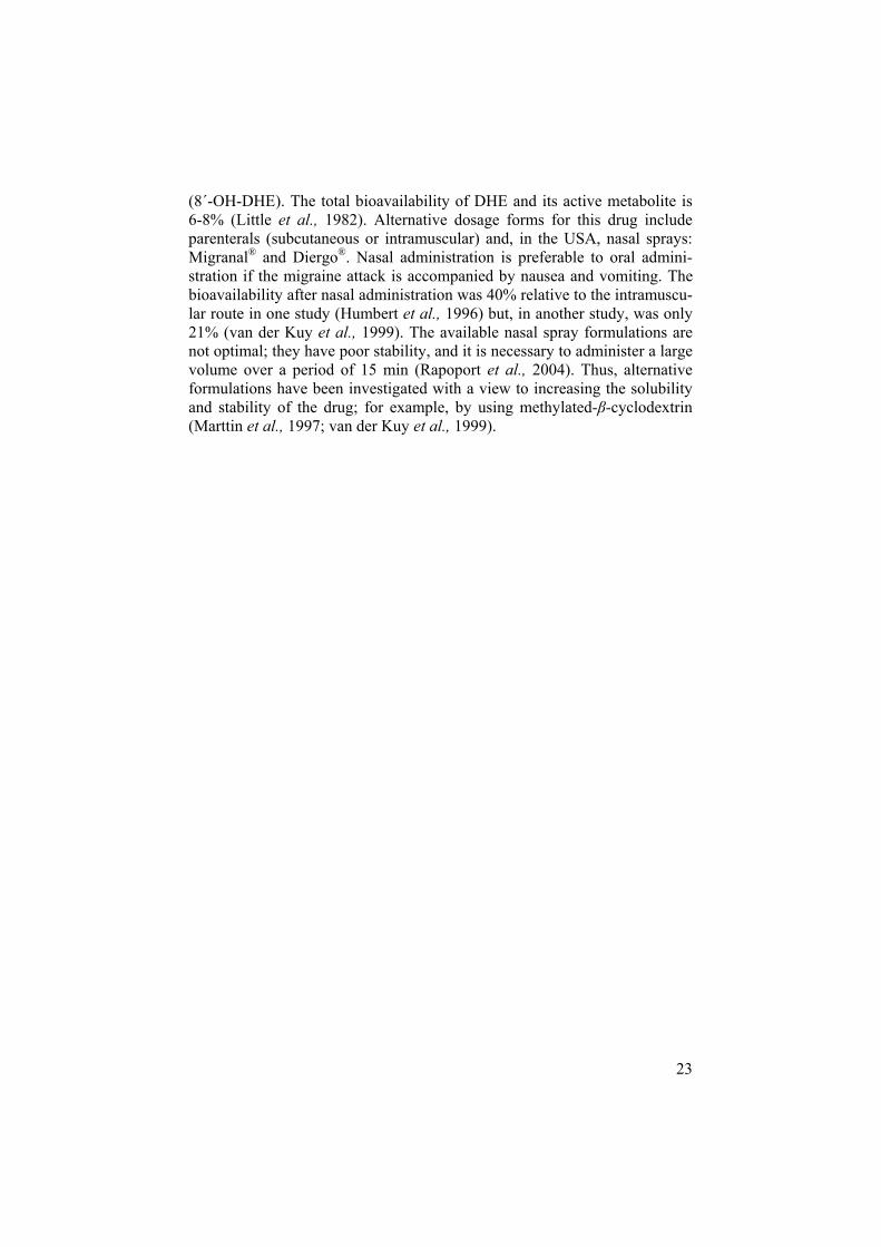
Dihydroergotamine DHE is an ergot peptide alkaloid mainly used for the treatment of migraine and orthostatic hypertension (Figure 5) (Callaham and Raskin 1986) (Thulesius and Berlin 1986). The oral bioavailability of DHE is very low, less than 1%, because of incomplete absorption and first-pass hepatic me-tabolism. DHE is metabolised by CYP 3A4, a cytochrome P450 enzyme; the main active metabolite in humans is 8´-hydroxy-dihydroergotamine
23
(8´-OH-DHE). The total bioavailability of DHE and its active metabolite is 6-8% (Little et al., 1982). Alternative dosage forms for this drug include parenterals (subcutaneous or intramuscular) and, in the USA, nasal sprays: Migranal® and Diergo®. Nasal administration is preferable to oral admini-stration if the migraine attack is accompanied by nausea and vomiting. The bioavailability after nasal administration was 40% relative to the intramuscu-lar route in one study (Humbert et al., 1996) but, in another study, was only 21% (van der Kuy et al., 1999). The available nasal spray formulations are not optimal; they have poor stability, and it is necessary to administer a large volume over a period of 15 min (Rapoport et al., 2004). Thus, alternative formulations have been investigated with a view to increasing the solubility and stability of the drug; for example, by using methylated- -cyclodextrin (Marttin et al., 1997; van der Kuy et al., 1999).

24
2 Aims of the thesis
The primary objective of this thesis was to study the olfactory transfer of morphine to the CNS after nasal administration, and to compare the transport of analgesic drugs across nasal respiratory and olfactory mucosa.
A secondary objective was to develop the horizontal Ussing chamber method for in vitro drug transport studies across olfactory mucosa.
The specific aims were:
To investigate whether morphine is transferred via olfactory path-ways to the CNS after nasal administration to mice and rats;
To quantify the olfactory transfer of morphine to the brain hemi-spheres in rats by comparing brain and plasma levels after nasal ad-ministration with those after i.v. administration;
To develop a technique for reliable isolation of porcine olfactory mucosa and to assure the viability of olfactory mucosa when mounted in the horizontal Ussing chamber;
To study the in vitro transport of dihydroergotamine across porcine nasal respiratory and olfactory mucosae and to evaluate its absorp-tion from a dry powder formulation in horizontal Ussing chambers;
To compare the in vitro transport of morphine across porcine nasal respiratory and olfactory mucosae in horizontal Ussing chambers.
25
3 Materials and methods
3.1 In vivo olfactory transfer of morphine in rodents (Papers I and II)
3.1.1 Materials [N-Methyl-3H]-morphine, dissolved in ethanol, with a specific activity of 85.5 Ci/mmol and radiochemical purity greater than 97% was obtained from Perkin Elmer, USA. Morphine hydrochloride trihydrate and heparin were purchased from Apoteket AB, Sweden. The morphine metabolite morphine-3-glucuronide (M3G) was purchased from Lipomed, Arlesheim, Switzer-land. All chemicals and solvents used were of analytical grade.
3.1.2 Animals Female Balb/c mice weighing 16.5-18.0 g and male Sprague-Dawley rats weighing 230-324 g on the day of the experiment were obtained from B&K Universal, Sweden. The animals were acclimatised for one week prior to the experiments and group housed under a 12-hour light-dark cycle with free access to food and water. The Uppsala Ethics committee for Animal Re-search approved the study protocols (Mice: C153/98 and C199/1, Rats: C 211/99 and C 223/2).
3.1.3 Experimental set-up Anaesthesia Mice were anaesthetised with an intraperitoneal injection of 0.1 ml per 10 g bodyweight of a 4: 1: 15 mixture by volume of Ketalar® (ketamine 50 mg/ml, Pfizer AB, Sweden), Rompun® vet (xylazine 20 mg/ml, Bayer AG Animal Health Business Group, Germany) and MilliQ water. Rats were an-aesthetised with an intraperitoneal injection of 0.27 ml per 100 g bodyweight of a 1: 1: 2 mixture by volume of Hypnorm (fentanyl citrate 0.315 mg/ml and fluanisone 10 mg/ml, Janssen Animal Health, Belgium), Dormicum®
(midazolam 5 mg/ml, Roche AB, Sweden) and Milli-Q water. All animals

26
were placed on a heated pad (37-38ºC) from the time of anaesthesia until euthanasia.
Drug administration For details of drug administration see Table 1. The mice were laid on their right sides during nasal administration to avoid leakage to the other nasal cavity and the nasal formulation was placed approximately 3 mm into the right nostril using a polyethylene tube (PE 10) attached to a micropipette.
All nasal rats were placed on their backs (to keep the formulation in con-tact with the olfactory mucosa). The nasal formulation was applied approxi-mately 5 mm into the right nostril using a polyethylene tube (PE 50) attached to a micropipette.
Indwelling polyethylene catheters were inserted under anaesthesia into the arteria carotis and vena jugularis of i.v. rats in the HPLC study, for the col-lection of blood and administration of morphine, respectively. The rats were allowed to recover from surgery under anaesthesia for 30 min before the start of the experiment.
Table 1 Drug administration, euthanasia and analysisPa-per Species Admini-
stration Volume Dose Euthanasia timepoints (min)
Ana-lysis
I Mouse Nasal 5 µl [3H]-morphine 5 µCi in PBS
15, 60, 240 (n=3/timepoint)
LS
I Rat Nasal 50 µl [3H]-morphine 25 µCi + 0.25 mg morphine*
in PBS
15, 60, 240 (n=3/timepoint)
LS
I Rat Nasal 50 µl [3H]-morphine 40 µCi + 0.25 mg morphine*
in PBS
5, 15, 60, 240 (n=1/timepoint) TSA
I Rat I.v. bolus in tail vein
100 µl 0.25 mg morphine*
in physiological saline 15(n=3/timepoint)
HPLC
I-II Rat Nasal 50 µl 0.25 mg morphine*
in PBS 5, 15, 60, 240 (n=3/timepoint)
HPLC
II Rat I.v. 15 min constantrate infu-sion
100 µl/min 1.5 ml in total
1 mg morphine/kg bodyweight in physiological saline
5, 15, 60, 240 (n=3/timepoint)
HPLC
*Equivalent to 1 mg morphine/kg bodyweight. Abbreviations: LS, liquid scintillation; TSA, Tape section autoradiography; HPLC, high-performance chromatography; PBS, phosphate-buffered saline.

27
Tissue and blood collection The animals were euthanised at scheduled times after drug administration (see Table 1) by exposure to gaseous CO2. After decapitation, the skulls were cut open and brain tissue samples were excised in the following order: cerebellum (rats), small portions of posterior and anterior cortex (mice and LS rats), cerebrum and cerebellum in one piece (mice), left and right brain hemispheres (rats), right lateral olfactory tract (LOT), left olfactory bulb (LOB) and right olfactory bulb (ROB), according to Figure 6. The oesopha-gus and trachea were also dissected from the mice and LS rats.
Blood (250 µl) from the mice, nasal rats and L.S./i.v. rats was collected with a syringe and a coarse needle from the neck directly after decapitation. Blood (250 µl) from the HPLC/i.v. rats was collected from the arteria caro-tis; the volume removed was replaced by physiological saline. Catheterisa-tion of the HPLC/i.v. rats enabled blood sampling at all time points until the scheduled sacrifice of the rat. All blood samples were put in heparinised tubes, and the blood samples for HPLC detection were thereafter centrifuged for 5 min at 7200 g and the plasma was transferred to new tubes; tissue and plasma samples were frozen at -20ºC until analysis.
Figure 6. Outline of the rat brain from a dorsal view, showing the areas collected, including the left olfactory bulb (LOB), right olfactory bulb (ROB), anterior cortex (A), posterior cortex (P), left and right brain hemispheres and the cerebellum. The lateral olfactory tract (LOT) was excised from underneath the brain.

28
3.1.4 Drug analysis Liquid scintillation (LS) All tissue samples were weighed and dissolved in 1 or 2 ml of tissue solubi-liser (Soluene-350®), depending on the size of the sample, and incubated overnight at 50ºC, after which 10 or 20 ml of scintillation cocktail (Hionic-FluorTM) was added. The blood samples were transferred to scintillation vials (150 µl blood) and 1 ml tissue solubiliser (Soluene-350®) was added. After incubation (50ºC, 15 min) followed by cooling, 0.4 ml of hydrogen peroxide (30%) was added to bleach the sample. After further incubation, 10 ml of scintillation cocktail was added (Hionic-FluorTM). The radioactivity in the tissue and blood samples was measured for 10 min/sample in an LS analyser (Tri-Carb® Liquid Scintillation Analyzers, Model 1900CA Packard Instru-ment Company, IL) after a night in the dark. Olfactory bulbs, brain and blood samples obtained from control animals were analysed to correct for apparent background.
Tape section autoradiography (TSA) The autoradiography experiments were performed as previously described (Ullberg 1977). Briefly, the skulls were immediately frozen with cold isopentane, embedded in a semiliquid gel of carboxymethyl cellulose in wa-ter and frozen in hexane, cooled with solid carbon dioxide. Series of hori-zontal sections of the head (20 µm) were collected on tape at various levels and processed for autoradiography using Hyperfilm-[3H] (CEA Amersham, Sweden). The film was exposed at –20ºC for approximately 10 weeks.
High-performance liquid chromatography (HPLC) The brain samples and olfactory bulbs were homogenised with 5-fold and 10-fold larger volumes of 0.1 M perchloric acid, respectively. The homoge-nates were centrifuged for 10 min at 1000 g. The supernatant (100 µl) was pre-treated using a slight modification of the method by Joel et al. (1988). Plasma samples (100µl) were pre-treated using the same method. Morphine and M3G were eluted with 3 ml of methanol and the solution was evaporated under a stream of nitrogen at 45ºC. The residue was dissolved in 150 µl of the mobile phase, and 55 µl was injected into the HPLC system using a Tri-athlon auto-injector (Spark Holland, the Netherlands).
Morphine and M3G concentrations in brain and plasma were determined using HPLC (Nucleosil C18 column; 150 × 4.6 mm i.d.; 5µm particles; Chrompack, Sweden). Morphine was detected using an electrochemical de-tector (Coulochem II, ESA Inc., Chelmsford, USA) with a guard cell (ESA 5020, ESA Inc.; potential at 600mV) and two analytical cells (ESA 5011, ESA Inc.; potentials at 300 and 450mV). M3G was analysed by fluorescence detection (Jasco 821-FP, Japan; excitation and emission wavelengths 212 and 340 nm) coupled in series with electrochemical detection. The mobile
29
phase [720 ml 0.01 M phosphate buffer (pH 2.1), containing 0.2 mM SDS, 280 ml methanol, and 50 ml tetrahydrofuran] was delivered at 1 ml/min (ESA 580, ESA Inc.). The peak height was compared with a standard curve to quantify the content of morphine and M3G in each sample. Olfactory bulbs, brain and plasma samples obtained from blank animals were also ana-lysed.
3.1.5 Data analysis and statistics Results are presented as means ± standard deviations (SD). A value of p<0.05 was considered statistically significant. In paper I, a one-way analy-sis of variance (ANOVA) test followed by Bonferroni’s multiple compari-sons test was used for statistical testing of the collected brain tissue samples within one timepoint after nasal administration.
In paper II, the area under the concentration-time curve (AUC) values for olfactory bulbs, brain hemispheres and plasma were calculated using the trapezoidal rule (Yuan 1993) from the mean drug concentrations at 5, 15, 60 and 240 min after administration, because only one set of brain tissue sam-ples per animal per time point could be collected. The variance for the AUC values and AUC ratios was therefore calculated according to Yuan (1993) and Bevington and Robinson (1992), respectively. All AUC values are pre-sented as values from 0-t min.
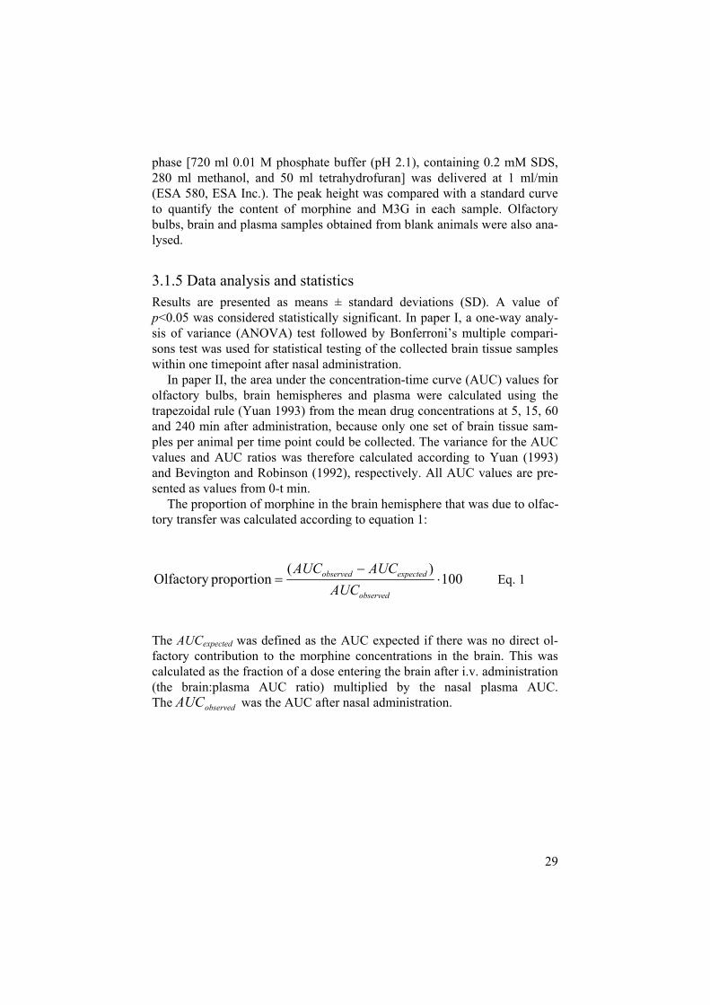
The proportion of morphine in the brain hemisphere that was due to olfac-tory transfer was calculated according to equation 1:
100)(
proportionOlfactoryobserved
expectedobserved
AUCAUCAUC
Eq. 1
The AUCexpected was defined as the AUC expected if there was no direct ol-factory contribution to the morphine concentrations in the brain. This was calculated as the fraction of a dose entering the brain after i.v. administration (the brain:plasma AUC ratio) multiplied by the nasal plasma AUC. The observedAUC was the AUC after nasal administration.
30
3.2 In vitro transport of dihydroergotamine and morphine across porcine nasal respiratory and olfactory mucosa (Papers III and IV)
3.2.1 Materials Krebs-Ringer Bicarbonate buffer (KRB) from Sigma Aldrich (Sweden) was supplemented with 15 mM NaHCO3, 1.2 mM CaCl2 and 138 mM NaCl. The stabilising agents sodium ethylene diamine tetra-acetic acid (EDTA) (1 mg/mL) and sodium metabisulfite (0.05 mg/mL) were added to all buffer solutions that were to contain DHE. Morphine hydrochloride trihydrate was purchased from Apoteket AB, Sweden. Morphine-6-glucuronide (M6G) and morphine-3-glucuronide (M3G) were purchased from Lipomed, Arlesheim, Switzerland. All chemicals were of analytical grade.
Sodium starch glycolate (SSG; Primojel®, DMV International GmbH, the Netherlands) was used as the carrier material. The appropriate size fraction of the carrier particles was obtained using an air classifier (100 MZR, Al-pine, Germany). Dihydroergotamine mesylate (DHE; Boehringer Ingelheim, Germany) was milled in a centrifugal ball mill (S1, Retsch GmbH, Ger-many) and used as the fine-particulate drug. DHE was then dry-mixed with SSG to form an interactive mixture. All materials and mixtures were stored in desiccators below 18% relative humidity (RH) and were also refrigerated and protected from light after addition of DHE.
3.2.2 Isolation of nasal mucosa Nasal respiratory and olfactory mucosae from healthy 6-month-old domestic pigs were isolated at the local slaughterhouse (Swedish Meats, Uppsala, Sweden). The snout was separated from the cranium with a frontal incision just anterior to the eyes. For the isolation of olfactory mucosa, an additional incision was made approximately 8 cm anterior to the previous incision and the nasal cavity was then divided into upper and lower halves by a horizontal incision. The olfactory mucosa was carefully removed from the upper part of the septum, the roof of the nasal cavity and the superior turbinate. Respira-tory mucosa was obtained from the ventral nasal concha (turbinate) at the anterior part of the nasal cavity after exposing this area with a sagittal inci-sion along the septum (Figure 7). The mucosae were transported to the labo-ratory in preoxygenated ice-cold KRB.

31
Figure 7. Frontal section of the porcine snout. (1) septum; (2) area of excised nasal respiratory mucosa; (3) area of excised nasal olfactory mucosa; and (4) horizontal incision required for excision of olfactory mucosa.
3.2.3 The horizontal Ussing chamber Unless otherwise stated, all experiments were performed according to the horizontal Ussing chamber method in studies of nasal drug delivery as de-scribed by Östh et al. (2002). In brief, six horizontal Ussing chambers were placed side-by-side on a waterheated block (Horizontal diffusion chamber System, Costar, Cambridge, MA) to bring the temperature of the liquid in the chamber to 37º C. Oxygenation in the receiver chamber was provided via silicone tubing (Sikema, AB Sweden) and the solution was stirred by placing the equipment on a circular shaker set at 155±1 rpm (Unimax, Wernerglas, Sweden).
Binding of the drug to the receiver chamber wall Some drugs bind to the Plexiglas® surface of the horizontal Ussing chamber walls, resulting in lower than expected receiver chamber concentrations (Östh et al., 2002). Binding of the drug to the receiver chamber walls was, therefore, investigated at concentrations representative of the experimental concentrations in the chamber. After adding 1.2 ml of each morphine solu-tion to the receiver chambers (n=3 for each concentration), 10 µl samples were taken at 0, 5, 10, 20, 40, 60 and 90 min and frozen at -20º C until analysis. The binding experiments were performed under the same condi-tions as the transport experiments.

32
Viability of the nasal respiratory and olfactory mucosa A circular piece of either nasal respiratory or olfactory mucosa was excised and mounted with the mucosal side upward in the horizontal Ussing cham-ber, resulting in an exposed surface area of 0.55 cm2. The closed top of the horizontal Ussing chamber was used to measure the electrophysiological parameters (Figure 8). After adding 1.2 ml KRB to both the donor and re-ceiver chambers, the tissue was allowed approximately 10 min to equilibrate before viability measurements commenced. The viability of the tissue was investigated before (for 90 min) and after the transport experiments by measuring the following electrophysiological parameters: resistance (R), potential difference (PD) and short circuit current (Isc), as described by Wikman Larhed et al. (1995). The resistance reflects the integrity of the tight junctions, although a low resistance may also indicate a considerable amount of damage to the mucosa. The potential difference and short circuit current reflect the integrity of the cell membranes and the activity of the ion pumps.
The nasal respiratory mucosa was judged to be viable when R 30 cm2, PD -1 mV and Isc 30 µA/cm2. Similarly, the olfactory mu-cosa was considered to be viable when R 30 cm2, PD -1 mV and Isc 20 µA/cm2. These electrophysiological criteria were selected to exclude non-viable mucosae; the criteria for nasal respiratory mucosa were taken from Östh et al. (2002) whereas the criteria for the olfactory mucosa had been screened for prior to conducting this investigation.
Figure 8. The horizontal Ussing chamber (modified from Costar Corporation, 1994). (A) Tissue mounting ring, (B) Closed top, (C) Electrode port, (D) Open top.

33
Transport experiments Only mucosae judged viable according to the chosen electrophysiological criteria were used. The open top of the horizontal Ussing chamber was used to create an air interface during the transport experiments (Figure 8). The KRB in the donor chamber was replaced with 50 µl of the liquid formulation or the DHE powder formulation. Samples of 100 µl were taken from the receiver chamber 0, 5, 10, 15, 30, 45, 60, 75 and 90 min after application of the donor solution or powder formulation. To keep a constant volume of 1.2 ml in the receiver chamber, equal amounts of KRB were added after each sampling. All samples were kept at -20º C until analysis.
Histology of mucosae Fresh nasal respiratory and olfactory mucosae, which had not been mounted in the Ussing chambers, were placed in Bouin’s fluid for a maximum of 24 h. The tissues were then dehydrated and embedded in plastic according to the manufacturer’s instructions (Technovit 7100, Leica Microsystems, Ger-many). Sections (of thickness 3 µm) were cut from the middle part of the tissues using a motorised rotary microtome (Leica RM 2165, Leica Micro-systems, Germany) and transferred to microscope slides by first stretching them out on the surface of deionised water. Dry sections were stained with toluidine blue and examined in a light microscope (Olympus BX-51) equipped with a digital camera (Olympus DP50) and software (Olympus DP-soft). The presence of elements specific to olfactory mucosa, i.e., Bow-man’s glands, dendritic knobs with bundle formations of long cilia and non-ciliated epithelial cells was investigated to determine whether the mucosa had been successfully isolated. Isolation of respiratory mucosa from the ven-tral nasal concha has been evaluated previously (Östh et al., 2002). The pieces of fresh mucosa were also used as reference to a representative num-ber of correspondingly plastic embedded and toluidine stained pieces of mu-cosa that had been used in the Ussing chamber experiments, to detect any detrimental effects the formulations might have had on the epithelial cell layers.
34
3.2.4 Drug analysis In paper III, the samples were analysed using UV-HPLC by Mikrokemi AB, Uppsala, Sweden (SWEDAC accredited).
In paper IV, the donor and receiver chamber samples that had been in contact with nasal mucosa contained biological material which would disturb an HPLC analysis. Thus, these were cleaned by adding 200 µl of acetonitrile to each sample after it had been thawed, whereupon the mixtures were vor-texed, and centrifuged for 5 min at 7200 g. Subsequently, 150 µl of the su-pernatant was evaporated with N2 at 45º C, dissolved in 50 µl of ultrapure water, vortexed, placed in an ultrasonic bath for 5 min, vortexed again and centrifuged for 2 min at 7200 g; 15 µl was then injected onto the HPLC sys-tem for all samples from the binding and transport studies.
In Paper IV the mucosae from a total of four chambers (two containing nasal respiratory mucosa and two olfactory mucosa, with one low and one high donor morphine concentration per type of mucosa) were collected and washed in KRB after the 90 min transport experiment to study the content of morphine and metabolites.The nasal mucosae samples were homogenised with 5-fold larger volumes of 0.1 M perchloric acid. The homogenates were centrifuged for 10 min at 1000 g and 100 µl of the supernatant was pre-treated using the same method as for brain tissue and plasma in Papers I-II. In Paper IV, all samples were analysed by the same HPLC-method as in Papers I-II, except that also M6G was measured for using the electrochemi-cal detector, and that the potential of the first cell was set at 0 or 300 mV for chamber or mucosa samples, respectively.
3.2.5 Data analysis and statistics The results are given as means ± S.D. or means ± S.E.M. The binding kinet-ics for morphine were calculated as described by Östh et al. (2002). The drug concentrations on the receiver side were corrected for sampling losses and those on the donor side were corrected for the amount transferred to the receiver side. Apparent permeability coefficients (Papp cm/s) were calculated in accordance with the model for non-sink conditions suggested by Palm et al. (1996). Paired t-tests were used to compare the pre- and post- electro-physiological periods for nasal respiratory and olfactory mucosa. Unpaired t-tests were used to compare the electrophysiological values for nasal respira-tory vs olfactory mucosa and for viable vs non-viable mucosa. A two-way ANOVA was used to test for statistically significant differences in the Pappvalues for the two factors. If the two-way ANOVA revealed that there was a significant interaction between the two factors, a one-way ANOVA was car-ried out, followed by Bonferroni’s multiple comparison test when necessary to identify the origin of the statistical difference. A p-value of <0.05 was considered statistically significant.
35
4 Results and discussion
4.1 In vivo olfactory transfer of morphine in rodents (Papers I and II)
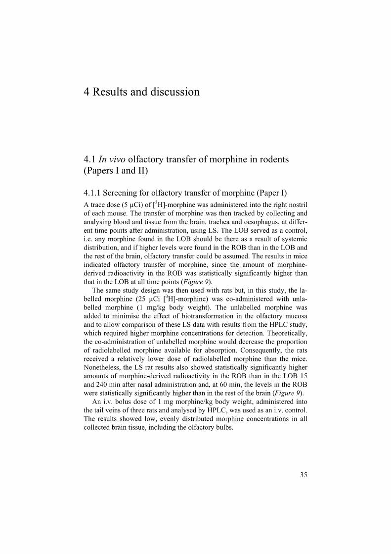
4.1.1 Screening for olfactory transfer of morphine (Paper I)A trace dose (5 µCi) of [3H]-morphine was administered into the right nostril of each mouse. The transfer of morphine was then tracked by collecting and analysing blood and tissue from the brain, trachea and oesophagus, at differ-ent time points after administration, using LS. The LOB served as a control, i.e. any morphine found in the LOB should be there as a result of systemic distribution, and if higher levels were found in the ROB than in the LOB and the rest of the brain, olfactory transfer could be assumed. The results in mice indicated olfactory transfer of morphine, since the amount of morphine-derived radioactivity in the ROB was statistically significantly higher than that in the LOB at all time points (Figure 9).
The same study design was then used with rats but, in this study, the la-belled morphine (25 µCi [3H]-morphine) was co-administered with unla-belled morphine (1 mg/kg body weight). The unlabelled morphine was added to minimise the effect of biotransformation in the olfactory mucosa and to allow comparison of these LS data with results from the HPLC study, which required higher morphine concentrations for detection. Theoretically, the co-administration of unlabelled morphine would decrease the proportion of radiolabelled morphine available for absorption. Consequently, the rats received a relatively lower dose of radiolabelled morphine than the mice. Nonetheless, the LS rat results also showed statistically significantly higher amounts of morphine-derived radioactivity in the ROB than in the LOB 15 and 240 min after nasal administration and, at 60 min, the levels in the ROB were statistically significantly higher than in the rest of the brain (Figure 9).
An i.v. bolus dose of 1 mg morphine/kg body weight, administered into the tail veins of three rats and analysed by HPLC, was used as an i.v. control. The results showed low, evenly distributed morphine concentrations in all collected brain tissue, including the olfactory bulbs.
36
Figure 9. Mean levels of morphine-derived radioactivity in brain tissue 15, 60 and 240 min after right-sided nasal administration of (a) 5 µCi [3H]-morphine to mice and (b) 25 µCi of [3H]-morphine plus 0.25 mg unlabelled morphine to rats. The ROB/LOB ratios are displayed above the columns. n=3, * p<0.05 for ROB vs LOB.

37
Table 2 Amount of radioactivity (mean ± S.D., n=3) in the oesophagus and trachea after right-sided nasal administration of morphine to mice and rats using liquid scin-tillation analysis Species Dose [3H-morphine]
+ unlabelled morphine Time (min)
Oesophagus(dpm/mg tissue)
Trachea (dpm/mg tissue)
Mice 5 Ci (0.02 g) 15 166 ± 15.2 193 ± 19.2 Mice 5 Ci (0.02 g) 60 4594 ± 5249 227 ± 70.7 Mice 5 Ci (0.02 g) 240 36.1 ± 14.2 15.3 ± 0.9 Rats 25 Ci (0.08 g) + 0.25 mg 15 203 ± 274 103 ± 58.1 Rats 25 Ci (0.08 g) + 0.25 mg 60 1878 ± 3121 204 ± 122 Rats 25 Ci (0.08 g) + 0.25 mg 240 981 ± 1316 33.1 ± 27.5
Radioactivity in mice and rat oesophagus increased steeply from 15 to 60 min after nasal delivery, which indicates that most of the formulation re-mained in the nasal cavity during the first 15 min, draining to the oesopha-gus thereafter (Table 2). There was a strong negative correlation in the indi-vidual animals between radioactivity levels in the oesophagus/trachea and those in the ROB. Low levels of morphine-derived radioactivity in the ROBs was accompanied by high levels of radioactivity in oesophagus/trachea due to swallowing, leaving less drug available for olfactory drug transfer.
4.1.2 Visualising the olfactory transfer of morphine (Paper I) Autoradiography was used to visualise the olfactory transfer of morphine in the intact rat brain. 40 µCi of [3H]-morphine co-administered with unlabelled morphine (1 mg/kg body weight) was administered into the right nostril of rats and the experiments were terminated 5, 15, 60 and 240 min after ad-ministration. The nasal cavity, cribriform plate, olfactory bulbs and brain were easily distinguished in the corresponding tissue sections.
The autoradiographic study demonstrated successful right-sided nasal administration, with high levels of morphine-derived radioactivity through-out the right nasal cavity, including the olfactory mucosa, 5, 15 and 60 min after administration. However, after 240 min, radioactivity was located only in the nasal mucosa. Only negligible amounts of radioactivity were seen in the left nasal cavity, except in the 15 min autoradiogram where some radio-activity was seen at the opening of the left nostril (data not shown).
An enlarged view of the 5 min autoradiogram displays radioactivity sur-rounding the ROB, most likely in the cerebrospinal fluid (CSF) of the su-barachnoid space, and spreading to the longitudinal cerebral fissure (Figure10). This result indicates very rapid olfactory transfer of morphine-derived radioactivity from the olfactory mucosa to the CSF.
At 60 min (Figure 11), radioactivity was distinctly located in the ROB, with decreasing levels towards the centre. This result demonstrates that mor-phine is also transferred to and diffuses within the olfactory bulb after nasal administration, but that olfactory transfer to this area is slower than that to

38
the CSF. A fraction of the morphine present in the olfactory bulbs may also be a consequence of distribution from the CSF.
Selective uptake of radioactivity was not observed in brain regions other than the right olfactory bulb and the longitudinal cerebral fissure, at any time point.
Figure 10. (A) Autoradiogram of a rat skull (horizontal section) 5 min after right-sided nasal administration of 40 µCi (0.13 µg) [3H]-morphine and 0.25 mg mor-phine. Radioactivity is present in the right nasal cavity, surrounding the right olfac-tory bulb, and reaching the longitudinal cerebral fissure. The radioactivity is pre-sented in white colour. (B) Corresponding tissue section. The arrows indicate the longitudinal cerebral fissure. n=nasal cavity, e= eye, ROB=right olfactory bulb, LOB= left olfactory bulb, b=brain.
Figure 11. Autoradiogram of a rat skull (horizontal section) 60 min after right-sided nasal administration of 40 µCi (0.13 µg) [3H]-morphine and 0.25 mg morphine, superimposed on the corresponding tissue section. Radioactivity is present in the right nasal cavity and in the right olfactory bulb. e=eye, ROB=right olfactory bulb, LOB= left olfactory bulb, b=brain. The radioactivity is presented in black colour.

39
4.1.3 Quantification of the olfactory transfer of morphine to the brain (Paper II) To quantify the olfactory transfer of morphine in rats, brain and plasma mor-phine AUC values were compared after nasal administration or a 15 min constant rate i.v. infusion of 1 mg morphine/kg body weight. The infusion rate for the i.v. dose was chosen to give a similar plasma morphine concen-tration-time profile to that after nasal administration. The study showed simi-lar brain hemisphere morphine concentrations at 5 or 15 minutes after nasal and i.v. administration, despite lower plasma concentrations after nasal ad-ministration (Figure 12). That is, there were no statistically significant dif-ferences in the brain hemisphere morphine AUC0-5 min or AUC0-15 min values between the nasal and i.v. groups (Table 3).
Figure 12. Morphine concentration-time profiles in (A) brain hemispheres and (B) plasma following right-sided nasal administration or a 15-min i.v. infusion of 1.0 mg morphine/kg body weight to rats. Each point represents the mean from three rats ± S.D. in (A) and from 3-12 rats in (B).
After nasal administration, M3G was detected in the ROB at 15 and 60 min (0.8± 0.3 and 1.0± 0.4 nmol/g tissue, respectively) but was not detected elsewhere in the olfactory bulbs or brain hemispheres. After i.v. administra-tion, M3G was detected in neither olfactory bulbs nor brain hemispheres. This indicates that morphine was either metabolised in the nasal cavity into M3G and then transferred via the olfactory pathways to the olfactory bulbs, or metabolised into M3G in the olfactory bulb. Morphine metabolising en-zymes have been demonstrated in both the rat nasal cavity and the rat olfac-tory bulbs (Lazard et al., 1991; Heydel et al., 2001).
The M3G concentrations in plasma were higher after nasal administration than after i.v administration (Table 3). The plasma M3G:morphine AUC0-240 min ratio was 5.3 after nasal administration and statistically signifi-cantly higher than the ratio of 1.2 after i.v. administration, which indicates that morphine was more extensively metabolised after nasal administration than after i.v. administration (Table 3).

40
Table 3. AUC values (mean ± S.D.) for morphine in brain tissue (nmol*min/g) and for morphine and M3G in plasma (nmol*min/ml) following right-sided nasal ad-ministration and a 15 min i.v. infusion of 1.0 mg morphine/kg body weight to rats (n=3 for each collection time). ROB=right olfactory bulb, LOB=left olfactory bulb, RH=right hemisphere, LH= left hemisphere.
After i.v. administration, the morphine concentration in the brain is the result of distribution from the systemic blood circulation across the BBB to the brain. After nasal administration, the concentrations of morphine in the brain could be the result of both distribution from the systemic blood circula-tion across the BBB and transfer via direct olfactory pathways. Thus, higher brain tissue:plasma morphine AUC ratios after nasal administration than after i.v. administration can be attributed to olfactory transfer.
The brain hemispheres:plasma morphine AUC0-5min ratios were approxi-mately 3 and 0.1 after nasal and i.v. administration, respectively, demonstrat-ing early distribution of morphine to the brain hemispheres via the nasal route (Figure 13.). At 240 min, these ratios had evened out to approximately 0.5 for both administration routes (Figure 13). The reason for early distance-independent elevation of the brain concentrations may be distribution of morphine via the local CSF after olfactory transfer, as indicated in the autoradiographic study.
41
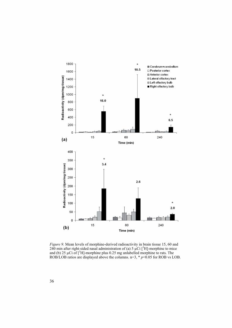
Figure 13. Brain hemisphere:plasma morphine AUC ratio as a function of time following right-sided nasal administration to twelve rats or a 15 min i.v. infusion to twelve rats of 1.0 mg morphine/kg body weight. The AUC ratios represent the val-ues from time 0 for all points (0-5, 0-15, 0-60 and 0-240 min) and are presented as means ± S.D. *Significant difference between the brain hemisphere:plasma ratios after nasal and i.v. administration, p<0.0125 (Bonferroni corrected).
The proportion of morphine in the right brain hemisphere that was due to olfactory transfer was calculated according to equation 1 (3.1.5 Data analysis and statistics) and were 95, 71, 48 and 10% for the 0-5, 0-15, 0-60 and 0-240 min intervals, respectively. Hence the impact of olfactory transfer decreased with time, and the early contribution to brain morphine concentrations from direct olfactory transfer would have been overlooked if the investigation had been confined to later in the process. Further, the contribution of olfactory transfer to the brain is easier to differentiate from that of systemic distribu-tion for drugs like morphine, which permeate the brain relatively poorly, and olfactory transfer of morphine may also be of greater clinical importance than that of permeable lipophilic drugs.
42
4.2 In vitro transport of dihydroergotamine and morphine across porcine nasal respiratory and olfactory mucosa (Papers III and IV)
4.2.1 Development of the horizontal Ussing chamber method The horizontal Ussing chamber method was developed by Östh et al. (2002) for transport studies across porcine nasal respiratory mucosa. Jansson devel-oped the method further for transport studies across olfactory mucosa (Jansson 2004). In this thesis, the horizontal Ussing chamber method was again modified for the olfactory mucosa by performing one histological ex-amination and one viability evaluation of both nasal respiratory and olfac-tory mucosae from the pig.
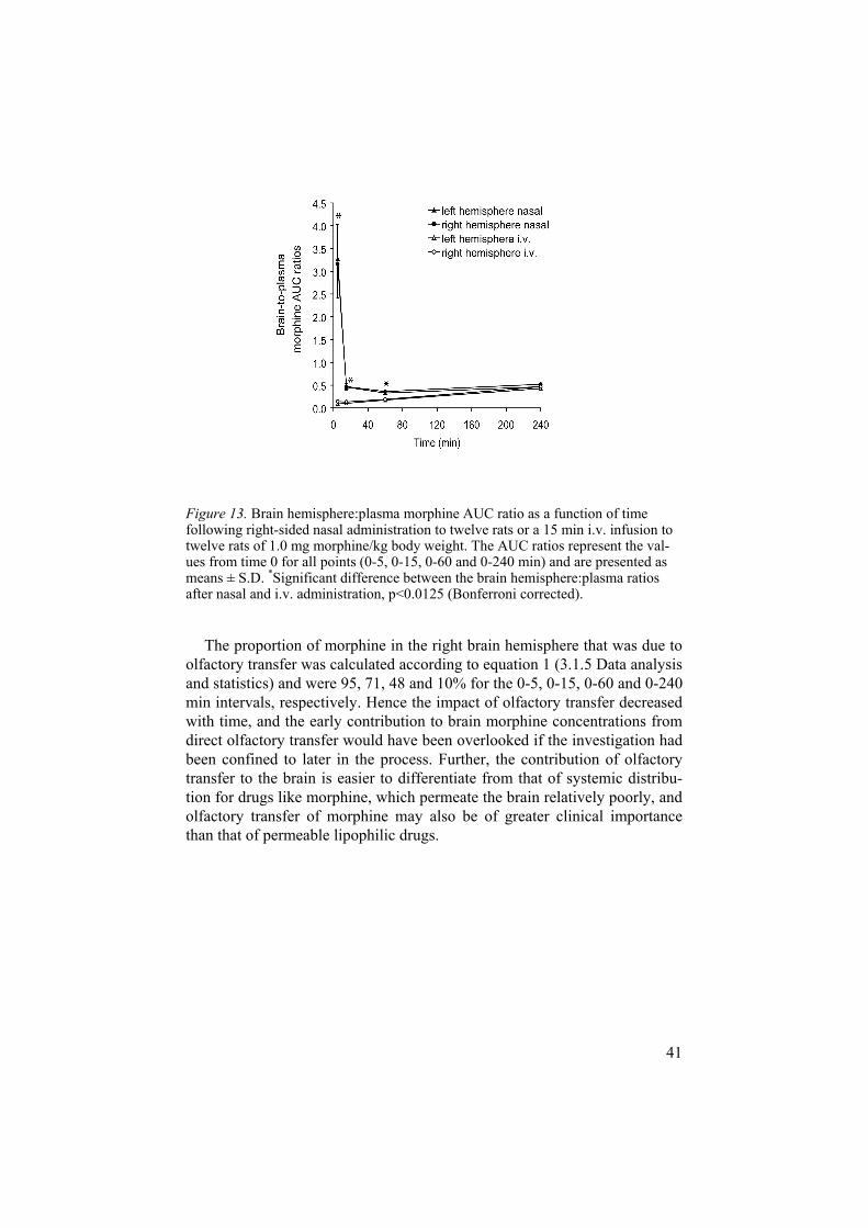
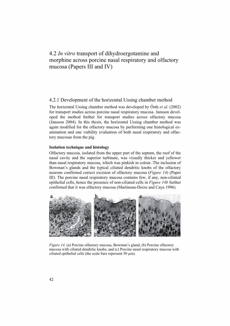
Isolation technique and histology Olfactory mucosa, isolated from the upper part of the septum, the roof of the nasal cavity and the superior turbinate, was visually thicker and yellower than nasal respiratory mucosa, which was pinkish in colour. The inclusion of Bowman’s glands and the typical ciliated dendritic knobs of the olfactory neurons confirmed correct excision of olfactory mucosa (Figure 14) (Paper III). The porcine nasal respiratory mucosa contains few, if any, non-ciliated epithelial cells, hence the presence of non-ciliated cells in Figure 14b further confirmed that it was olfactory mucosa (Martineau-Doize and Caya 1996).
Figure 14. (a) Porcine olfactory mucosa, Bowman’s gland; (b) Porcine olfactory mucosa with ciliated dendritic knobs; and (c) Porcine nasal respiratory mucosa with ciliated epithelial cells (the scale bars represent 50 µm).

43
The comparison of untreated mucosae with those having been in contact with the reference solution or powder formulation for 90 min, revealed that there was no visible detrimental effect on the epithelial cells.
Two pieces of either nasal respiratory mucosa or olfactory mucosa were obtained from each snout. The nasal respiratory mucosa was collected ac-cording to Östh et al. (2002) in Papers III-IV, i.e. respiratory mucosa was obtained from the ventral nasal concha after exposing this area with a sagit-tal incision along the septum. The possibility of obtaining both nasal respira-tory and olfactory mucosae from the same pig was investigated, i.e. collect-ing respiratory mucosa from the left lower half and olfactory mucosa from the upper half, after the transverse incision made according to Figure 7. This would have enabled intra-individual comparisons of drug transport across nasal respiratory and olfactory mucosae. Unfortunately, both the success rate and the electrophysiological results were much poorer when both types of mucosa were collected from the same pig, possibly because the isolation process took longer and the respiratory mucosa was collected from a more posterior location in the nasal cavity than was outlined by Östh et al. (2002).
Viability of nasal respiratory and olfactory mucosae In Papers III-IV, estimation of tissue viability was based on the measurement of three electrophysiological parameters: R, PD and Isc. A study of the com-parative viability of nasal respiratory and olfactory mucosae was performed in Paper IV. The electrophysiological parameters were measured before the start of the transport experiment for approximately 90 min, a period that was considered to be adequate for achieving bioelectrical stability of the muco-sae. The values chosen for PD and Isc were suitable for readily distinguishing viable from non-viable mucosae for both nasal respiratory mucosa (PD -1 mV and Isc 30 µA/cm2) and olfactory mucosa (PD -1 mV and Isc 20 µA/cm2) (Figure 15). However, the resistance parameter (R 30 cm2) was not sufficient to distinguish viable from non-viable mucosae, although there was a tendency for viable olfactory mucosa to have somewhat higher values than those of the other mucosae. The resistance parameter alone has com-monly been used as a viability marker, but the results of this investigation demonstrate that the Isc and PD parameters should also be included when investigating viability. The resistance parameter is, however, a useful tool in drug transport mechanism evaluation (Jansson 2004).
The electrophysiological values for olfactory mucosa were similar to those previously reported for nasal respiratory mucosa (Östh et al., 2002) and to those for viable nasal respiratory mucosa in this study. The selection criteria for olfactory mucosa can, therefore, be changed to an Isc value 30 µA/cm2, i.e. the same as that for nasal respiratory mucosa.

44
Figure 15. Mean electrophysiological values for viable and non-viable porcine nasal respiratory and olfactory mucosae (n = 6-8). The legend explaining the symbols applies to all three graphs. In 1b and 1c are the 90 min values for viable vs. non-viable mucosa statistically significantly different, p<0.05.
The changes in the electrophysiological values indicated that the viability of the olfactory mucosa, as predicted by the selection criteria, was retained. This suggests that the anterior part of the olfactory nerve cells in the

45
epithelium plus the supporting cells and basal cells of the olfactory mucosa are capable of maintaining the integrity and viability of these tissues (Figure15). Further, it indicates that the trauma of isolating the posterior part of the olfactory nerve cells from the nasal cavity does not markedly affect the elec-trophysiological values.
4.2.2 Stability and drug adsorption studies As expected (Vermeire and Remon 1999), morphine remained stable in the horizontal Ussing chamber. However, it was necessary to add stabilising agents (sodium EDTA and sodium metabisulfite) to the KRB in the receiver chamber to retain the stability of the DHE. Some degradation of DHE still occurred in the 90 min samples, and these samples were therefore not in-cluded in the Papp calculations.
Both DHE and morphine were <10% bound to the receiver chamber wall. However, it appeared that lower drug concentrations were more affected by binding. All calculated morphine Papp values were, therefore, tested for losses attributable to binding to the chamber wall, using the equations devel-oped by Östh et al.(2002); however, the overall outcome was not affected.
4.2.3 Drug transport studies Mucosa-dependent transport Permeabilities for DHE were significantly higher across nasal olfactory mu-cosa than across nasal respiratory mucosa and there were no statistically significant differences in morphine Papp values between the two types of mucosa (Table 4). Thus, the olfactory mucosa was no greater barrier than the nasal respiratory mucosa. This is interesting, as the defence system of olfac-tory mucosa against xenobiotics is believed to be more rigorous, with a wider enzymatic capacity, than that of nasal respiratory mucosa. Further, morphine and DHE are metabolised by UGTs and CYP 3A4, respectively, both of which, according to Marini et al. (1998), are present in porcine olfac-tory mucosa.
In Paper IV, the main metabolites of morphine, M6G and M3G, were not detected in the samples, which indicate that there is a low in vitro morphine metabolism in both porcine nasal respiratory and olfactory mucosa if such a metabolism occurs in these mucosae at all. In humans, UGTs have been de-tected olfactory mucosa, but not in nasal respiratory mucosa, but in (Gervasiet al., 1991; Jedlitschky et al., 1999).

46
Table 4. Papp values (cm/s) · 10-6 (mean ± SD) for nasal respiratory and olfactory mucosa after application of two formulations of dihydroergotamine (DHE) and two concentrations of morphine
Nasal mucosa DHELiquid (n=4)
DHEPowder (n=5)
Morphine0.2 mg/ml (n=3)
Morphine20 mg/ml (n=4)
Respiratory 0.210 ± 0.221 0.0982 ± 0.0706 3.06 ± 2.98 3.08 ± 0.73 Olfactory 0.664 ± 0.441 0.377 ± 0.481 10.6 ± 6.26 1.43 ± 1.10
Östh et al. (2002) studied the transport across nasal respiratory mucosa with the horizontal Ussing chamber method for testosterone and mannitol and received Papp values off 10.23 ± 12.26 · 10-6 and 2.35 ± 1.47 · 10-6 cm/s,respectively. When compared to these results, our Papp values for DHE and morphine (Table 4) across nasal respiratory mucosa were as expected, with respect to their physicochemical properties and molecular sizes. This indi-cates that this in vitro method produces consistent results.
Formulation-dependent transport In Paper III, transport studies in the horizontal Ussing chambers revealed no statistically significant difference in the calculated Papp coefficients between DHE in solution and in a powder formulation (Table 4). The concentrations in the first samples were not detectable and a more sensitive analytical method would have been necessary to show any initial absorption-enhancing effect of the powder formulation through widening of the tight junctions. The indication towards an inferior absorption from the powder formulation may have several explanations: The mucoadhesive action of this particular system is exerted by hydration of the carrier particles upon contact with the mucosa. In the nasal cavity, the amount of powder per square centimetre will be more diluted and the access to fluid greater than in the horizontal Ussing chamber. The surfaces of the mucosae in the chambers became visibly dry after powder application, which will most likely have resulted in a decreased dissolution of DHE.
The reference solution contained EDTA, which was required to keep the drug stable. As EDTA can be used as an absorption enhancer (Cassidy and Tidball 1967), it is likely to have had a positive effect on absorption of the drugs, which would explain the slightly more extensive transport of DHE from the reference solution than from the powder. This was verified by the fact that the resistance of the mucosae, which can be used as a measure of the integrity of the tight junctions, changed more after contact with the liquid than after the powder (having dropped to 69.7 5.2 and 84.2 4.2 % of the initial resistance values, respectively).
In Paper IV, the effect of two different morphine concentrations (0.2 and 20 mg/ml) on drug transport was evaluated. Transport over the nasal respira-tory mucosa was not concentration-dependent for the concentrations

47
investigated but, for olfactory mucosa, the Papp values were statistically sig-nificantly higher for the lower concentration than for the higher. However, the Papp values for the higher concentration were in the same range as those for the nasal respiratory mucosa. The results indicate that the drug transport capacity across the olfactory mucosa decreased with higher drug concentra-tions.
The inverse drug concentration dependence seen with the olfactory mu-cosa and the absence of concentration dependence for the nasal respiratory mucosa indicate that morphine is not greatly affected by efflux proteins, such as P-gp, or by nasal metabolism.

48
4.3 Implications of olfactory transfer of analgesics (Papers I-IV)
Overall, the results of Papers I-II demonstrated that morphine was trans-ferred via the olfactory pathways to the olfactory bulbs and a CSF-filled fissure of the rodent brain and that olfactory transfer contributed signifi-cantly to the early brain morphine concentrations seen after nasal administra-tion to rats. The fact that morphine was transferred via olfactory pathways directly to a CSF-filled fissure of the brain is interesting from a clinical point of view, as the presence of the drug in CSF may influence analgesia, and as the half life of morphine is longer in both CSF and the brain compared to plasma (Wolff et al., 1995; Dennis et al., 1999). There is, however, a func-tional barrier between the CSF and the brain tissue, which prevents drug concentration equilibration between these compartments (Graff and Pollack 2004; 2005). The result of elevated morphine concentrations in the olfactory bulbs due to olfactory transfer may have clinical repercussions. For example, µ-opioid receptors have been detected in mice and rat olfactory bulbs (Mansour et al., 1995). Further, µ-opioid agonists appear to interfere with sensory transmission at the level of second-order neurons in the olfactory pathway (Perez et al., 1989), in which case nasal administration of morphine may result in associated adverse effects.
However, conclusions regarding the impact of olfactory transfer of mor-phine in humans cannot be reliably drawn from studies in rodents. The struc-ture of the rodent nasal passages is more complex than that in humans, and the olfactory mucosa covers approximately 50 % of the total rodent nasal area compared with only approximately 3-10 % of the total in humans (Illum 2004).
It is often argued in the context of the significance of olfactory drug trans-fer in humans that the fraction of the administered dose present in various brain regions or CSF due to olfactory transfer after nasal administration (Chen et al., 1998; Kumbale et al., 1999; Dahlin et al., 2000) is very small and would, in fact, be insignificant with respect to exerting pharmacological effects (Illum 2004; Graff and Pollack 2005). However, these fractions have to be compared with corresponding fractions present after i.v. administra-tion, and the size of the investigated brain region has also to be taken into account. For example, only a small amount of intravenous morphine enters the brain relative to the administered dose (Way and Adler 1961). In Paper II, the drug concentrations in the brain were in the order of thousandths of the administered dose, but they were equal after nasal and i.v. administra-tion, even though the driving force from the plasma was significantly lower after nasal administration.

49
Figure 16. Fraction of the administered dose (1.0 mg morphine/kg body weight) present in various brain regions 15 min after nasal or i.v. administration (nasal value/i.v. value; n=3)
Further, the onset of perceptible pain relief (2.4± 2.1 min) after nasal ad-ministration of morphine to cancer patients is remarkably rapid (Fitzgibbonet al., 2003), and Illum et al. (2002) registered higher sedation scores among the volunteers in their clinical trial at the earliest time point after nasal ad-ministration than seen after i.v. infusion over 30 min. These findings may well be the result of olfactory transfer of morphine in humans.
The olfactory transfer of morphine seems to follow two main routes: 1) rapid olfactory transfer of morphine into the CSF surrounding the olfactory bulbs (Paper I; autoradiography), followed by further transfer to the brain hemispheres (independent of distance; Paper II) and probably also to both olfactory bulbs, which would explain why the LOB contained more mor-phine than expected; and 2) slower diffusion of morphine within the olfac-tory bulb, presumably targeting the near brain areas (Paper I; autoradiogra-phy). In fact, in Paper I there was a tendency for higher levels in the right lateral olfactory tract after 15 min (rats) and 60 min (mice and rats) and in the anterior cortex after 60 min (mice and rats) than in the cerebrum and cerebellum, indicating further transfer of radioactivity towards the brain (Figure 9).
Paper IV demonstrated that the in vitro transport of morphine was no more restricted across porcine olfactory mucosa than across porcine nasal respiratory mucosa. In this thesis nasal respiratory and olfactory mucosa from the pig was used. The pig have many features that appear similar in scientific comparison to humans (Balk 1987; Sachs 1994). The porcine nasal cavity consists of upper olfactory and lower respiratory areas, and the areas of nasal respiratory and olfactory mucosa are large enough to be isolated and mounted in the horizontal Ussing chamber (Dyce et al., 1987). As olfactory

50
transfer of morphine was demonstrated in Papers I-II, this in vitro result is also indicative of olfactory transfer of morphine. However, when comparing in vitro drug transport across nasal respiratory and olfactory mucosae with respect to olfactory transfer, it is important to remember that only transport across the first barrier to olfactory transfer, i.e. the olfactory mucosa, has been investigated. This in vitro method cannot predict olfactory drug transfer all the way to the CNS. Nonetheless, the olfactory mucosal barrier is an im-portant barrier to olfactory transfer.
Differences in morphology and available drug transport mechanisms be-tween nasal respiratory and olfactory mucosa should be taken into considera-tion when evaluating the transport of drugs across the mucosal barrier (Pa-pers III-IV). For systemic nasal absorption in vivo, the drug molecule is ini-tially absorbed by blood vessels present in the nasal respiratory mucosa; however, in the in vitro system used in these studies, the drug molecule has to pass through the nasal respiratory epithelium and submucosa before reaching the receiver chamber. As a result, the in vivo transport capacity of morphine and DHE across the nasal respiratory mucosa may be underesti-mated in these studies. The transport of morphine and DHE could also be facilitated across the olfactory mucosa through paracellular transfer via the olfactory nerves, which would take place to a certain extent in vivo, while some of the drug may have been taken up into the systemic circulation from the submucosa.

51
5 Conclusions
Morphine is transferred via olfactory pathways to the CNS (rapid transfer to the longitudinal cerebral fissure and relatively slow diffusion within the olfactory bulbs) after nasal administration to mice and rats.
Olfactory transfer of morphine contributes significantly to the early high brain hemisphere morphine concentrations seen after nasal administration to rats.
Olfactory mucosa correctly isolated from the pig can produce similar electrophysiological values, when mounted in horizontal Ussing cham-bers, to those reported in the literature for viable nasal respiratory mu-cosa.
The in vitro transport of morphine was no more restricted and for dihy-droergotamine (DHE) even higher across the olfactory mucosa than across the nasal respiratory mucosa. Thus, the olfactory mucosa should not be a major barrier to olfactory transfer of DHE or morphine.
Absorption of DHE from the powder formulation was similar to that from the reference solution in the horizontal Ussing chambers.
The potential for olfactory transfer of drugs in humans deserves further investigation, especially for drugs with central effects currently under de-velopment for nasal administration to patients.

52
6 Perspectives
In the search for new analgesic drugs with a rapid onset of pain relief, nasal administration has become an interesting option. Evidence on the direct ol-factory transfer of drugs from the nasal cavity, via olfactory pathways, to the CNS has recently been presented (Mathison et al., 1998; Illum 2004). For-mulations of both of the drugs investigated in this thesis, morphine and di-hydroergotamine, are currently being developed for nasal administration. Both drugs have shown indications of direct olfactory transfer to the CNS in animal studies (Wang et al., 1998; Betbeder et al., 2000). The focus of this thesis was the propensity for olfactory transfer of these drugs, an important topic since olfactory transfer may lead to both positive and negative clinical effects.
The results of the thesis showed that morphine is transferred via olfactory pathways to the olfactory bulbs, a cerebral fissure and the brain hemispheres in rats after “best case scenario” nasal administration. That is, the rats were sedated and placed on their backs during nasal administration, and the for-mulation was kept in contact with the olfactory mucosa for at least 15 min. Future investigations could study the significance of olfactory trans-fer of morphine in awake rats and investigate possible early differences in nociceptive responses after nasal and i.v. administration. The olfactory trans-fer of morphine should also be investigated in animals (e.g. monkeys) with smaller areas of olfactory mucosa, more closely resembling the human situa-tion.
The horizontal Ussing chamber method was further developed in this the-sis, which offers a unique opportunity to compare drug transport across the nasal respiratory and olfactory mucosae, a comparison which cannot be achieved in an in vivo model. The studies showed that the in vitro transport of morphine and DHE was no more restricted across the olfactory mucosa than across the nasal respiratory mucosa. This is in accordance with previous results on olfactory transfer for both drugs. However, future studies should examine the in vitro-in vivo correlation for the horizontal Ussing chamber method. For example, the transport of a drug that is known not to be trans-ferred via olfactory pathways could be studied across both nasal respiratory and olfactory mucosa in the horizontal Ussing chamber.
53
7 Populärvetenskaplig sammanfattning
I mitt avhandlingsarbete har jag studerat absorptionen av smärtstillande sub-stanser efter att läkemedlet sprejats i näsan, med fokus på direkttransport till hjärnan via luktnerverna (olfaktorisk transport). Att ge läkemedel via näsan är ett bra alternativ till injektioner och tabletter när snabb smärtlindring samt låg nedbrytning av läkemedlet i levern eftersträvas. Till exempel undersöks effekten av nasalt morfin mot genombrottsmärta hos cancerpatienter i klinis-ka studier. Med nya effektiva nässprejer ökar förutsättningen för ett upptag till blodcirkulationen, men också potentialen för direkttransport till hjärnan via luktnerverna.
I avhandlingens första arbete fastställdes att morfin transporteras via lukt-nerverna till luktloberna och ett vätskefyllt hålrum mellan luktloberna och hjärnan. Den olfaktoriska transporten följdes genom att ta ut och analysera hjärnstrukturer vid olika tidpunkter efter nasal administrering till möss och råttor (arbete I). Studien kompletterades med en teknik där den olfaktoriska transporten visualiserades i hjärnan med hjälp av att morfin var radioaktivt märkt och snittning av hjärnan (autoradiografi). I nästa arbete var målet att ta reda på vilka mängder som kunde transporteras olfaktoriskt i råttor. Hjärn-koncentrationer efter nasal administrering jämfördes med de efter intravenös administrering, och resultaten visade att den olfaktoriska transporten signifi-kant bidrog till de initialt höga hjärnkoncentrationerna efter nasal administre-ring (arbete II).
I nästa steg utnyttjades en modell av näsan, den horisontella Ussingkam-maren, och i den monterades både vanlig nässlemhinna och luktslemhinna från gris. Transportförsök med migränläkemedlet dihydroergotamin (DHE) samt morfin genomfördes (arbete III-IV). Effekten av en ny pulverberedning med DHE utvärderades också i den horisontella Ussingkammaren. Resulta-ten visade ingen skillnad i transport av DHE mellan pulverberedningen och referenslösningen. Vidare så transporterades DHE i signifikant högre ut-sträckning över luktslemhinnan jämfört med den vanliga nässlemhinnan och för morfin var det ingen skillnad i transport mellan båda slemhinnetyperna. Sammantaget innebär detta att luktslemhinnan inte utgör någon större barriär mot olfaktorisk transport (arbete III-IV).
54
8 Acknowledgements
The studies in this thesis were carried out at the Department of Pharmacy, Uppsala University.
The financial support from the Swedish Foundation for Strategic Research (SSF) and Orexo AB is gratefully acknowledged. The travel grants from SSF, ULLA and Apotekare CD Carlssons stiftelse, which enabled attending courses or conferences in London, Barcelona, Kyoto, Copenhagen and Lake Tahoe are very much appreciated.
I wish to express my sincere gratitude to:
Dr Erik Björk for your kind ways and for being the best supervisor for me! You really knew when to support, encourage or push me and I always felt that you believed in me as a scientist.
My co-supervisors: Professor Eva Brittebo and Professor Margareta Ham-marlund-Udenaes. For giving me the opportunity to do research at your divisions, and for interesting scientific discussions, which broadened this thesis.
My co-authors: Nelly Fransén, Emma Boström, Johan Gråsjö, Lic. ElenaPiras, Dr Björn Jansson, Dr Ulrika Bergström, Dr Maria Dahlin, Prof. Eva Brittebo, Prof. Margareta Hammarlund-Udenaes, Prof. and Christer Nyström for all in different ways contributing to the papers! It has been a pleasure working with you.
Professor Göran Alderborn and Professor Martin Malmsten for providing excellent working facilities at the Department of Pharmacy.
Antona Wagstaff and Suzanne Lidström for excellent linguistic revision of the thesis and Papers, and for all nice comments that cheered my up on the way.
Apotekarsocieteten, Sektionen för galenisk farmaci och biofarmaci, med Göran Lidgren i spetsen för en lärorik och rolig tid som doktorandrespre-sentant.
55
Eva Nises-Ahlgren, Harriet Pettersson och Ulla Wästberg-Galik för er omtanke och allehanda hjälp.
Christin Magnusson, för våra pratstunder och för att jag fick bli salvtant samt tack till salvtanterna och salvfarbrodern för roliga timmar på kurslabb.
Britt Jansson och Jessica Strömgren på farmakokinetiken för värdefull analys- och labbhjälp. Raili Engdahl och Lena Norgren på toxikologen för snabb hjälp med autoradiografi.
Mina examensarbetare: Sara Pettersson, Rima Issa och Marcus Söder-berg. Det var verkligen roligt jobba ihop med er. Jag glömmer aldrig hur bra Sara klarade sig själv på labb när jag bröt armen, eller hur gullig Rima var när jag fick mitt första pek accepterat eller Marcus ”fuldans” på labb!
Johan Gråsjö, för att du med gott humör och rolig humor hjälpte mig med allt ifrån att reparera elektrofysiologisk apparatur, komplicerad statistik, till byten av innanmäten i volvobildörrar.
Grabbarna på Swedish Meats i Uppsala, speciellt Erik Lindberg, för glada tillrop samt skicklig isolering av grisnässlemhinna.
Till alla nuvarande och dåvarande doktorander vid Institutionen för farma-ci, för allt roligt vi har haft när vi inte har jobbat: under resor, mö- och fruhippor, Borrel, WC-träffar, fisketävlingar och spexande på fester.
Till den eminenta ”näsgruppen”: Alltid lika glada Dr Karin Östh för kloka svar på Ussingfrågor. Min ”fadder” Dr Björn Jansson, för ditt lugn och för all visdom du har delat med dig av, samt ett stort tack för dina relevanta kommentarer på avhandlingen. Gulliga Nelly Fransén, du har verkligen ställt upp för mig under avhandlingsskrivandet! Hade det inte varit för din tacopaj, vet jag inte vad det blivit av det här… ulriak
Mina rumskamrater: Dr Gunilla Englund, för en trevlig start på doktorand-tiden. Lic. Gustaf Jederström för intressanta diskussioner och för besöket på Möja. Framförallt vill jag tacka min nuvarande rumskamrat Dr Eva Ragnarsson, för vår vänskap och din otroliga omtänksamhet. Kram!
Till alla djur och människor i Krycklingestallet, Bälinge, med en extra klapp på mulen till min fina häst Birkir. För mysiga ridturer, mycket fikande och roliga fester. Ett alldeles speciellt tack till min vän Jessica Lindroth för att du har tagit så väl hand om Birkir, nu när inte jag har hunnit!
56
Mina väninnor i Alingsås, Liselott Lindström och Anna Frid Johnsen. För att vi alltid har så kul ihop när vi väl träffas!
Farmor Ruth, farfar Birger Westin och morfar Birger Dahlberg för att ni bryr er om mig!
Till alla i Anders familj, speciellt mina otroligt snälla svärföräldrar Ralf och Birgitta Espefält. Ett extra stort tack till Birgitta, för all hjälp med Alfred! Jag hade aldrig kunnat disputera så här snart efter föräldraledigheten om det inte hade varit för dig!
Min älsklingslillebror Herman Westin, för din vänskap, roliga humor och för våra långa telefonsamtal.
Min söta lillasyster Åsa Agerson Westin med familj, tack för allt stöd och uppmuntran samt för att Alfred har fått vara hos er! Min svåger Fredrik Agerson, för att du alltid bjuder på så god mat, din köttfärssås är bäst! Många kramar till mosters älsklingar Isak och Irma.
Min kära muminmamma Ulla-Britt och kära muminpappa Bobby Westin,ni är de goaste och snällaste föräldrar man kan tänka sig! Tusen tack för hjälpen när jag skrev på avhandlingen hos er i Kärtared. Voffhälsningar till Arnold och Ebba.
Min älskade man Anders Espefält, för att du vidgar mina vyer! Tack för CDn med fransk musik i bilen, resan till Svalbard och sökandet i Sveriges alla hörn efter den gäckande Myrliljan. För att du är en så snäll pappa till Alfred, och för att du alltid tror på mig - utan dig hade jag nog aldrig börjat doktorera.
Min älskade son Alfred – du är solen i mitt liv!
Uppsala den 30 mars 2007
Ulrika
57
9 References
Abboud, T. K., J. Zhu, J. Gangolly, M. Longhitano, F. Swart, A. Makar, G. Chu, M. Cool, M. Mantilla, N. Kurtz and L. Reich (1991). "Transna-sal butorphanol: a new method for pain relief in post-cesarean sec-tion pain." Acta Anaesthesiol Scand 35(1): 14-8.
Anez Simon, C., M. Rull Bartomeu, A. Rodriguez Perez and A. Fuentes Baena (2006). "Intranasal opioids for acute pain." Rev Esp Aneste-siol Reanim 53(10): 643-52.
Balk, M. W. (1987). "Emerging models in the U.S.A.: swine, woodchucks, and the hairless guinea pig." Prog Clin Biol Res 229: 311-26.
Barackman, J. D., G. Ott and D. T. O'Hagan (1999). "Intranasal immuniza-tion of mice with influenza vaccine in combination with the adjuvant LT-R72 induces potent mucosal and serum immunity which is stronger than that with traditional intramuscular immunization." In-fect Immun 67(8): 4276-9.
Bergström, U., A. Franzen, C. Eriksson, C. Lindh and E. B. Brittebo (2002). "Drug targeting to the brain: transfer of picolinic acid along the ol-factory pathways." J Drug Target 10(6): 469-78.
Betbeder, D., S. Sperandio, J. P. Latapie, J. de Nadai, A. Etienne, J. M. Zajac and B. Frances (2000). "Biovector nanoparticles improve antino-ciceptive efficacy of nasal morphine." Pharm Res 17(6): 743-8.
Bevington, P. R. and D. K. Robinson (1992). Data reduction and error analy-sis for the physical sciences. New York, McGraw-Hill, Inc.
Beyssac, E., F. Touaref, M. Meyer, L. Jacob, P. Sandouk and J. M. Aiache (1998). "Bioavailability of morphine after administration of a new bioadhesive buccal tablet." Biopharm Drug Dispos 19(6): 401-5.
Björk, E., U. Isaksson, P. Edman and P. Artursson (1995). "Starch micro-spheres induce pulsatile delivery of drugs and peptides across the epithelial barrier by reversible separation of the tight junctions." J Drug Target 2(6): 501-7.
Borland, M., I. Jacobs, B. King and D. O'Brien (2007). "A randomized con-trolled trial comparing intranasal fentanyl to intravenous morphine for managing acute pain in children in the emergency department." Ann Emerg Med 49(3): 335-40.
Born, J., T. Lange, W. Kern, G. P. McGregor, U. Bickel and H. L. Fehm (2002). "Sniffing neuropeptides: a transnasal approach to the human brain." Nat Neurosci 5(6): 514-6.
Bourget, P., A. Lesne-Hulin and V. Quinquis-Desmaris (1995). "Study of the bioequivalence of two controlled-release formulations of morphine." Int J Clin Pharmacol Ther 33(11): 588-94.
58
Bouw, M. R., M. Gårdmark and M. Hammarlund-Udenaes (2000). "Pharma-cokinetic-pharmacodynamic modelling of morphine transport across the blood-brain barrier as a cause of the antinociceptive effect delay in rats - a microdialysis study." Pharm Res 17(10): 1220-7.
Brauchi, S., C. Cea, J. G. Farias, J. Bacigalupo and J. G. Reyes (2006). "Apoptosis induced by prolonged exposure to odorants in cultured cells from rat olfactory epithelium." Brain Res 1103(1): 114-22.
Brittebo, E. B. (1997). "Metabolism-dependent activation and toxicity of chemicals in nasal glands." Mutat Res 380(1-2): 61-75.
Callaham, M. and N. Raskin (1986). "A controlled study of dihydroergo-tamine in the treatment of acute migraine headache." Headache26(4): 168-71.
Calof, A. L., J. S. Mumm, P. C. Rim and J. Shou (1998). "The neuronal stem cell of the olfactory epithelium." J Neurobiol 36(2): 190-205.
Carr, D. B., L. C. Goudas, W. T. Denman, D. Brookoff, P. S. Staats, L. Brennen, G. Green, R. Albin, D. Hamilton, M. C. Rogers, L. Fire-stone, P. T. Lavin and F. Mermelstein (2004). "Safety and efficacy of intranasal ketamine for the treatment of breakthrough pain in pa-tients with chronic pain: a randomized, double-blind, placebo-controlled, crossover study." Pain 108(1-2): 17-27.
Cassidy, M. M. and C. S. Tidball (1967). "Cellular mechanism of intestinal permeability alterations produced by chelation depletion." J Cell Biol 32(3): 685-98.
Charlton, S., N. S. Jones, S. S. Davis and L. Illum (2007). "Distribution and clearance of bioadhesive formulations from the olfactory region in man: Effect of polymer type and nasal delivery device." Eur J Pharm Sci 30(3-4): 295-302.
Chen, X. Q., J. R. Fawcett, Y. E. Rahman, T. A. Ala and I. W. Frey (1998). "Delivery of Nerve Growth Factor to the Brain via the Olfactory Pathway." J Alzheimers Dis 1(1): 35-44.
Chien, Y. W., K. S. E. Su and S. F. Chang (1989). Nasal Systemic Drug Delivery. New York, Marcel Dekker.
Chow, H. H. S., Z. Chen and G. T. Matsuura (1999). "Direct transport of cocaine from the nasal cavity to the brain following intranasal co-caine administration in rats." J Pharm Sci 88(8): 754-8.
Christrup, L. L. (1997). "Morphine metabolites." Acta Anaesthesiol Scand41(1 Pt 2): 116-22.
Collins, S. L., C. C. Faura, R. A. Moore and H. J. McQuay (1998). "Peak plasma concentrations after oral morphine: a systematic review." J Pain Symptom Manage 16(6): 388-402.
Cowan, C. M. and A. J. Roskams (2002). "Apoptosis in the mature and de-veloping olfactory neuroepithelium." Microsc Res Tech 58(3): 204-15.
Dahlin, M., U. Bergman, B. Jansson, E. Björk and E. Brittebo (2000). "Transfer of dopamine in the olfactory pathway following nasal ad-ministration in mice." Pharm Res 17(6): 737-42.
59
Dahlin, M. and E. Björk (2000). "Nasal absorption of (S)-UH-301 and its transport into the cerebrospinal fluid of rats." Int J Pharm 195(1-2): 197-205.
Dahlin, M. and E. Björk (2001). "Nasal administration of a physostigmine analogue (NXX-066) for Alzheimer's disease to rats." Int J Pharm212(2): 267-74.
Dale, O., R. Hjortkjaer and E. D. Kharasch (2002). "Nasal administration of opioids for pain management in adults." Acta Anaesthesiol Scand46(7): 759-70.
Dale, O., T. Nilsen, T. Loftsson, H. Hjorth Tonnesen, P. Klepstad, S. Kaasa, T. Holand and P. G. Djupesland (2006). "Intranasal midazolam: a comparison of two delivery devices in human volunteers." J Pharm Pharmacol 58(10): 1311-8.
Dennis, G. C., D. Soni, O. Dehkordi, R. M. Millis, H. James, W. L. West and R. E. Taylor (1999). "Analgesic responses to intrathecal mor-phine in relation to CSF concentrations of morphine-3,beta-glucuronide and morphine-6,beta-glucuronide." Life Sci 64(19):1725-31.
Derad, I., K. Willeke, R. Pietrowsky, J. Born and H. L. Fehm (1998). "Intra-nasal angiotensin II directly influences central nervous regulation of blood pressure." Am J Hypertens 11(8 Pt 1): 971-7.
Djupesland, P. G., A. Skretting, M. Winderen and T. Holand (2006). "Breath actuated device improves delivery to target sites beyond the nasal valve." Laryngoscope 116(3): 466-72.
Duquesnoy, C., J. P. Mamet, D. Sumner and E. Fuseau (1998). "Compara-tive clinical pharmacokinetics of single doses of sumatriptan follow-ing subcutaneous, oral, rectal and intranasal administration." Eur J Pharm Sci 6(2): 99-104.
Dyce, K. N., W. O. Sack and C. J. G. Wensing (1987). The head and neck of the pig. Textbook of Veterinary Anatomy. Philadelphia, Saunders:733-35.
Farr, S. J. and B. A. Otulana (2006). "Pulmonary delivery of opioids as pain therapeutics." Adv Drug Deliv Rev 58(9-10): 1076-88.
Farre, M., R. de la Torre, M. Llorente, X. Lamas, B. Ugena, J. Segura and J. Cami (1993). "Alcohol and cocaine interactions in humans." J Pharmacol Exp Ther 266(3): 1364-73.
Fitzgibbon, D., D. Morgan, D. Dockter, C. Barry and E. D. Kharasch (2003). "Initial pharmacokinetic, safety and efficacy evaluation of nasal morphine gluconate for breakthrough pain in cancer patients." Pain106(3): 309-15.
Fransén, N., E. Björk and C. Nyström (2007). "Development and characteri-zation of mucoadhesive interactive mixtures for nasal delivery." Eur. J. Pharm. Biopharm. Accepted.
Franzen, A., C. Carlsson, V. Hermansson, M. Lang and E. B. Brittebo (2006). "CYP2A5-mediated activation and early ultrastructural changes in the olfactory mucosa: studies on 2,6-dichlorophenyl me-thylsulfone." Drug Metab Dispos 34(1): 61-8.
60
Gervasi, P. G., V. Longo, F. Naldi, G. Panattoni and F. Ursino (1991). "Xenobiotic-metabolizing enzymes in human respiratory nasal mu-cosa." Biochem Pharmacol 41(2): 177-84.
Graff, C. L. and G. M. Pollack (2003). "P-Glycoprotein attenuates brain uptake of substrates after nasal instillation." Pharm Res 20(8): 1225-30.
Graff, C. L. and G. M. Pollack (2004). "Drug transport at the blood-brain barrier and the choroid plexus." Curr Drug Metab 5(1): 95-108.
Graff, C. L. and G. M. Pollack (2005). "Functional evidence for P-glycoprotein at the nose-brain barrier." Pharm Res 22(1): 86-93.
Graff, C. L. and G. M. Pollack (2005). "Nasal drug administration: potential for targeted central nervous system delivery." J Pharm Sci 94(6):1187-95.
Hanks, G. W., F. Conno, N. Cherny, M. Hanna, E. Kalso, H. J. McQuay, S. Mercadante, J. Meynadier, P. Poulain, C. Ripamonti, L. Radbruch, J. R. Casas, J. Säwe, R. G. Twycross and V. Ventafridda (2001). "Morphine and alternative opioids in cancer pain: the EAPC recom-mendations." Br J Cancer 84(5): 587-93.
Heydel, J., S. Leclerc, P. Bernard, H. Pelczar, D. Gradinaru, J. Magdalou, A. Minn, Y. Artur and H. Goudonnet (2001). "Rat olfactory bulb and epithelium UDP-glucuronosyltransferase 2A1 (UGT2A1) expres-sion: in situ mRNA localization and quantitative analysis." BrainRes Mol Brain Res 90(1): 83-92.
Humbert, H., M. D. Cabiac, C. Dubray and D. Lavene (1996). "Human pharmacokinetics of dihydroergotamine administered by nasal spray." Clin Pharmacol Ther 60(3): 265-75.
Hägerström, H. (2003). Polymer Gels as Pharmaceutical Dosage Forms: Rheological Performance and Physicochemical Interactions at the Gel-Mucus Interface for Formulations Intended for Mucosal Drug Delivery. Faculty of Pharmacy. Uppsala, Uppsala University: 76.
Illum, L. (2003). "Nasal drug delivery-possibilities, problems and solutions." J Control Release 87(1-3): 187-98.
Illum, L. (2004). "Is nose-to-brain transport of drugs in man a reality?" J Pharm Pharmacol 56(1): 3-17.
Illum, L., P. Watts, A. N. Fisher, M. Hinchcliffe, H. Norbury, I. Jabbal-Gill, R. Nankervis and S. S. Davis (2002). "Intranasal delivery of mor-phine." J Pharmacol Exp Ther 301(1): 391-400.
Jackson, R. T., J. Tigges and W. Arnold (1979). "Subarachnoid space of the CNS, nasal mucosa, and lymphatic system." Arch Otolaryngol105(4): 180-4.
Jansson, B. (2004). Models for the transfer of drugs from the nasal cavity to the central nervous system. Faculty of Pharmacy. Uppsala, Uppsala University: 46.
Jedlitschky, G., A. J. Cassidy, M. Sales, N. Pratt and B. Burchell (1999). "Cloning and characterization of a novel human olfactory UDP-glucuronosyltransferase." Biochem J 340 ( Pt 3): 837-43.

61
Joel, S. P., R. J. Osborne and M. L. Slevin (1988). "An improved method for the simultaneous determination of morphine and its principal glu-curonide metabolites." J Chromatogr 430(2): 394-9.
Kandimalla, K. K. and M. D. Donovan (2005). "Carrier mediated transport of chlorpheniramine and chlorcyclizine across bovine olfactory mu-cosa: implications on nose-to-brain transport." J Pharm Sci 94(3):613-24.
Kendall, J. M., B. C. Reeves and V. S. Latter (2001). "Multicentre random-ised controlled trial of nasal diamorphine for analgesia in children and teenagers with clinical fractures." BMJ 322(7281): 261-5.
Kristensson, K. and Y. Olsson (1971). "Uptake of exogenous proteins in mouse olfactory cells." Acta Neuropathologica 19: 145-54.
Kumbale, R., W. H. Frey, S. Wilson and Y. E. Rahman (1999). "GM1 deliv-ery to the CSF via the olfactory pathway." Drug Deliv 6: 23-30.
Kuo, C. K., N. Hanioka, Y. Hoshikawa, K. Oguri and H. Yoshimura (1991). "Species difference of site-selective glucuronidation of morphine." J Pharmacobiodyn 14(4): 187-93.
Lazard, D., K. Zupko, Y. Poria, P. Nef, J. Lazarovits, S. Horn, M. Khen and D. Lancet (1991). "Odorant signal termination by olfactory UDP glucuronosyl transferase." Nature 349(6312): 790-3.
Letrent, S. P., G. M. Pollack, K. R. Brouwer and K. L. Brouwer (1999). "Ef-fects of a potent and specific P-glycoprotein inhibitor on the blood- brain barrier distribution and antinociceptive effect of morphine in the rat." Drug Metab Dispos 27(7): 827-34.
Little, P. J., G. L. Jennings, H. Skews and A. Bobik (1982). "Bioavailability of dihydroergotamine in man." Br J Clin Pharmacol 13(6): 785-90.
Loscher, W. and H. Potschka (2005). "Role of drug efflux transporters in the brain for drug disposition and treatment of brain diseases." Prog Neurobiol 76(1): 22-76.
Mansour, A., C. A. Fox, H. Akil and S. J. Watson (1995). "Opioid-receptor mRNA expression in the rat CNS: anatomical and functional impli-cations." Trends Neurosci 18(1): 22-9.
Marini, S., V. Longo, A. Mazzaccaro and P. G. Gervasi (1998). "Xenobiotic-metabolizing enzymes in pig nasal and hepatic tissues." Xenobiotica28(10): 923-35.
Martineau-Doize, B. and I. Caya (1996). "Ultrastructural characterization of the nasal respiratory epithelium in the piglet." Anat Rec 246(2): 169-75.
Marttin, E., S. G. Romeijn, J. C. Verhoef and F. W. Merkus (1997). "Nasal absorption of dihydroergotamine from liquid and powder formula-tions in rabbits." J Pharm Sci 86(7): 802-7.
Mathison, S., R. Nagilla and U. B. Kompella (1998). "Nasal route for direct delivery of solutes to the central nervous system: fact or fiction?" JDrug Target 5(6): 415-41.
McCance-Katz, E. F., L. H. Price, C. J. McDougle, T. R. Kosten, J. E. Black and P. I. Jatlow (1993). "Concurrent cocaine-ethanol ingestion in
62
humans: pharmacology, physiology, behavior, and the role of co-caethylene." Psychopharmacology (Berl) 111(1): 39-46.
Moran, D. T., J. C. Rowley, 3rd, B. W. Jafek and M. A. Lovell (1982). "The fine structure of the olfactory mucosa in man." J Neurocytol 11(5):721-46.
Mori, I., F. Goshima, Y. Imai, S. Kohsaka, T. Sugiyama, T. Yoshida, T. Yokochi, Y. Nishiyama and Y. Kimura (2002). "Olfactory receptor neurons prevent dissemination of neurovirulent influenza A virus into the brain by undergoing virus-induced apoptosis." J Gen Virol83(Pt 9): 2109-16.
Mori, I., F. Goshima, D. Watanabe, H. Ito, N. Koide, T. Yoshida, B. Liu, Y. Kimura, T. Yokochi and Y. Nishiyama (2006). "Herpes simplex vi-rus US3 protein kinase regulates virus-induced apoptosis in olfac-tory and vomeronasal chemosensory neurons in vivo." Microbes In-fect 8(7): 1806-12.
Morrison, E. E. and R. M. Costanzo (1992). "Morphology of olfactory epi-thelium in humans and other vertebrates." Microsc Res Tech 23(1):49-61.
Mulder, G. J. (1992). "Glucuronidation and its role in regulation of biologi-cal activity of drugs." Annu Rev Pharmacol Toxicol 32: 25-49.
Okuyama, S. (1997). "The first attempt at radioisotopic evaluation of the integrity of the nose-brain barrier." Life Sci 60(21): 1881-4.
Palm, K., K. Luthman, A. L. Ungell, G. Strandlund and P. Artursson (1996). "Correlation of drug absorption with molecular surface properties." J Pharm Sci 85(1): 32-9.
Pardridge, W. M. (1999). "Blood-brain barrier biology and methodology." J Neurovirol 5(6): 556-69.
Pavis, H., A. Wilcock, J. Edgecombe, D. Carr, C. Manderson, A. Church and A. Fisher (2002). "Pilot study of nasal morphine-chitosan for the re-lief of breakthrough pain in patients with cancer." J Pain Symptom Manage 24(6): 598-602.
Perez-Reyes, M. and A. R. Jeffcoat (1992). "Ethanol/cocaine interaction: cocaine and cocaethylene plasma concentrations and their relation-ship to subjective and cardiovascular effects." Life Sci 51(8): 553-63.
Perez, H., A. Hernandez and H. Inostroza (1989). "Mu and kappa opioid modulation of olfactory bulb evoked potentials." Int J Neurosci49(3-4): 329-32.
Pietrowsky, R., C. Struben, M. Molle, H. L. Fehm and J. Born (1996). "Brain potential changes after intranasal vs. intravenous administra-tion of vasopressin: evidence for a direct nose-brain pathway for peptide effects in humans." Biol Psychiatry 39(5): 332-40.
Rapoport, A. M., M. E. Bigal, S. J. Tepper and F. D. Sheftell (2004). "Intra-nasal medications for the treatment of migraine and cluster head-ache." CNS Drugs 18(10): 671-85.
Russo, P., C. Sacchetti, I. Pasquali, R. Bettini, G. Massimo, P. Colombo and A. Rossi (2006). "Primary microparticles and agglomerates of mor-phine for nasal insufflation." J Pharm Sci 95(12): 2553-61.
63
Sachs, D. H. (1994). "The pig as a xenograft donor." Pathol Biol (Paris)42(3): 217-9.
Sarkar, M. A. (1992). "Drug metabolism in the nasal mucosa." Pharm Res9(1): 1-9.
Schipper, N. G., J. C. Verhoef and F. W. Merkus (1991). "The nasal muco-ciliary clearance: relevance to nasal drug delivery." Pharm Res 8(7): 807-14.
Striebel, H., J. Kramer, I. Luhmann, I. Rohierse-Hohler and A. Rieger (1993). "Pharmakokinetische Studie zur intranasalen Gabe von Fen-tanyl." Der Schmerz 7: 122–25.
Striebel, H. W., T. Olmann, C. Spies and G. Brummer (1996). "Patient-controlled intranasal analgesia (PCINA) for the management of postoperative pain: a pilot study." J Clin Anesth 8(1): 4-8.
Suzuki, Y. (2004). "Fine structural aspects of apoptosis in the olfactory epi-thelium." J Neurocytol 33(6): 693-702.
Säwe, J., B. Dahlström and A. Rane (1983). "Steady-state kinetics and anal-gesic effect of oral morphine in cancer patients." Eur J Clin Phar-macol 24(4): 537-42.
Terasaki, T. and W. M. Pardridge (2000). "Targeted drug delivery to the brain; (blood-brain barrier, efflux, endothelium, biological trans-port)." J Drug Target 8(6): 353-5.
Thorne, R. G., G. J. Pronk, V. Padmanabhan and W. H. Frey, 2nd (2004). "Delivery of insulin-like growth factor-I to the rat brain and spinal cord along olfactory and trigeminal pathways following intranasal administration." Neurosci 127(2): 481-96.
Thornton-Manning, J. R. and A. R. Dahl (1997). "Metabolic capacity of nasal tissue interspecies comparisons of xenobiotic-metabolizing en-zymes." Mutat Res 380(1-2): 43-59.
Thulesius, O. and E. Berlin (1986). "Dihydroergotamine therapy in or-thostatic hypotension due to psychotropic drugs." Int J Clin Phar-macol Ther Toxicol 24(9): 465-7.
Ullberg, S. (1977). "The technique of whole body autoradiography. Cryosec-tioning of large specimen." Science tools. The LKB Instrumental Journal.(Special issue on whole body autoradiography): 1-29.
Walsh, T. D. and C. M. Saunders (1981). "Oral morphine for relief of chronic pain from cancer." N Engl J Med 305(23): 1417-8.
van der Kuy, P. H., J. J. Lohman, P. M. Hooymans, J. W. Ter Berg and F. W. Merkus (1999). "Bioavailability of intranasal formulations of di-hydroergotamine." Eur J Clin Pharmacol 55(9): 677-80.
Wang, Y., R. Aun and F. L. Tse (1998). "Brain uptake of dihydroergotamine after intravenous and nasal administration in the rat." Biopharm Drug Dispos 19(9): 571-5.
Way, E. L. and T. K. Adler (1961). "The biological disposition of morphine and its surrogates. I." Bull World Health Organ 25: 227-62.
Vermeire, A. and J. P. Remon (1999). "Stability and compatibility of mor-phine." Int J Pharm 187(1): 17-51.

64
Westerling, D., C. Persson and P. Höglund (1995). "Plasma concentrations of morphine, morphine-3-glucuronide, and morphine-6-glucuronide after intravenous and oral administration to healthy volunteers: rela-tionship to nonanalgesic actions." Ther Drug Monit 17(3): 287-301.
Wikman Larhed, A. and P. Artursson (1995). "Co-cultures of human intesti-nal goblet (HT29-H) and absorptive (Caco-2) cells for studies of drug and peptide absorption." Eur J Pharm Sci 3: 171-83.
Wioland, M. A., J. Fleury-Feith, P. Corlieu, F. Commo, G. Monceaux, J. Lacau-St-Guily and J. F. Bernaudin (2000). "CFTR, MDR1, and MRP1 immunolocalization in normal human nasal respiratory mu-cosa." J Histochem Cytochem 48(9): 1215-22.
Wolff, T., H. Samuelsson and T. Hedner (1995). "Morphine and morphine metabolite concentrations in cerebrospinal fluid and plasma in can-cer pain patients after slow-release oral morphine administration." Pain 62(2): 147-54.
Yuan, J. (1993). "Estimation of variance for AUC in animal studies." J Pharm Sci 82(7): 761-3.
Zeppetella, G. (2000). "An assessment of the safety, efficacy, and acceptabil-ity of intranasal fentanyl citrate in the management of cancer-related breakthrough pain: a pilot study." J Pain Symptom Manage 20(4): 253-8.
Östh, K., J. Gråsjö and E. Björk (2002). "A new method for drug transport studies on pig nasal mucosa using a horizontal Ussing chamber." J Pharm Sci 91(5): 1259-73.


Acta Universitatis UpsaliensisDigital Comprehensive Summaries of Uppsala Dissertationsfrom the Faculty of Pharmacy 55
Editor: The Dean of the Faculty of Pharmacy
A doctoral dissertation from the Faculty of Pharmacy, UppsalaUniversity, is usually a summary of a number of papers. A fewcopies of the complete dissertation are kept at major Swedishresearch libraries, while the summary alone is distributedinternationally through the series Digital ComprehensiveSummaries of Uppsala Dissertations from the Faculty ofPharmacy. (Prior to January, 2005, the series was publishedunder the title “Comprehensive Summaries of UppsalaDissertations from the Faculty of Pharmacy”.)
Distribution: publications.uu.seurn:nbn:se:uu:diva-7829
ACTA
UNIVERSITATIS
UPSALIENSIS
UPPSALA
2007