of may, vol. in yaba tumor poxvirms synthesis in vitrojb.asm.org/content/91/5/1986.full.pdf · yaba...
TRANSCRIPT

JOURNAL OF BACTERIOLCGY, May, 1966 Vol. 91, No. 5Copyright ( 1966 American Society for Microbiology Printed In U.S.A.
Yaba Tumor Poxvirms Synthesis In VitroIII. Growth Kinetics
DAVID S. YOHN, VICTORIA A. HAENDIGES, AND ETIENNE DE HARVEN
Viral Oncology Section, Roswell Park Memorial Institute, Buffalo, New York, anld Division ofCytology, Sloan-Kettering Institute for Cancer Research, New York, New York
Received for publication 2 December 1965
ABSTRACTYOHN, DAVID S. (Roswell Park Memorial Institute, Buffalo, N.Y.), VICTORIA A.
HAENDIGES, AND ETIENNE DE HARVEN. Yaba tumor poxvirus synthesis in vitro. III.Growth kinetics. J. Bacteriol. 91:1986-1991. 1966.-The infectious synthetic cycleof Yaba poxvirus by BSC-1 cells at 35 C is described and discussed together withrelated observations obtained by immunofluorescence and electron microscopy.After adsorption of virus at 25 C, at least 6 hr of incubation at 35 C was requiredbefore viral eclipse was demonstrable. Infectious progeny appeared after an addi-tional 48 hr of incubation at 35 C. Newly synthesized virus particles were seen at68 hr but not at 44 hr by electron microscopy. Maximal maturation occurred be-tween 100 and 120 hr. Maximal yields were obtained from the 5th through the 8thday. The differences in kinetics between Yaba and vaccinia poxviruses, as describedfor the latter in the literature, are discussed. Possible factors accounting for thesedifferences are enumerated.
Previous descriptions of Yaba virus propaga-tion in primary rhesus monkey kidney cells (2),in cercopithecus kidney cell line, BSC-1 (23, 24),and in primary human fetal kidney and adultgrivet monkey kidney cells (14), provided pre-sumptive evidence that Yaba virus replicationoccurred at a rate much slower than replication ofother poxviruses such as vaccinia (5, 12, 15),rabbit poxvirus (3), and Shope fibroma virus (10,11). The previous reports on Yaba virus werebased largely on either microscopic observationsof cytopathic effects alone or in combination withhistochemical or immunofluorescent staining, orboth. Analysis of the results obtained with thecombined techniques indicated that, although 36to 48 hr are required before Yaba virus antigensynthesis is detected in BSC-1 cells (24), thecomplete cycle requires 50 to 60 hr. The presentstudies were undertaken to establish moreprecisely the time relationships of Yaba poxvirussynthesis in BSC-1 monolayers. It is anticipatedthat the results will provide baseline informationfor detailed analyses of the biochemical eventsassociated with Yaba virus synthesis in vitro.
MATERIALS AND METHODS
Virus and assay. The virus lot employed in thesestudies was obtained from saline-Genetron extracts oftumors obtained from a 3-year-old rhesus monkey,no. 10. Extraction of these tumors was described pre-
viously (24). The virus assay and titration methodsalso were described in detail previously (25). Briefly,virus was diluted in medium 199 containing 50%bovine amniotic fluid (BAF) adjusted to 2.0 mmcalcium, 1.0 mm magnesium, and to pH 7.0. Amountsof 0.4 ml of each dilution were added to six 80-mmLeighton tubes per dilution and incubated for 18hr at 25 C. A 1-ml amount of Eagle's basal medium(EBM) containing 50% BAF was added, and thetubes were incubated at 35 C for 19 to 21 days. Freshmaintenance medium was added every 3rd or 4th day.
Cell cultures. BSC-1 monolayer cultures weregrown in EBM containing 10% agamma calf serumand were maintained in EBM containing 50% BAF.All media contained 100 units of penicillin and 20 ,ugof streptomycin per ml.
Electron microscopy. Yaba virus-infected BSC-1cells were gently scraped from glass surfaces with arubber policeman and centrifuged at 800 X g for3 min. The supernatant fluid was decanted, and thecell pellet was fixed for 10 min at 0 C with glutaralde-hyde diluted to 1% in 0.1 M phosphate buffer (pH7.5). The glutaraldehyde was removed by rinsing thepellet three times with buffer containing 0.33 Msucrose, and the pellet was fixed for 1 hr at 0 C inbuffered 2% OS04. The pellet was dehydrated throughabsolute ethyl alcohol and embedded in Epon (16).Sections were examined with a Siemens Elmiskop Ielectron microscope, with the use of the double con-densor system, 80-kv accelerating voltage, and 50-Mpmolybdenum objective apertures. Ilfor N.60 "photo-mechanical" plates were employed.
1986
on May 15, 2018 by guest
http://jb.asm.org/
Dow
nloaded from
YABA TUMOR POXVIRUS SYNTHESIS. III
RESULTS
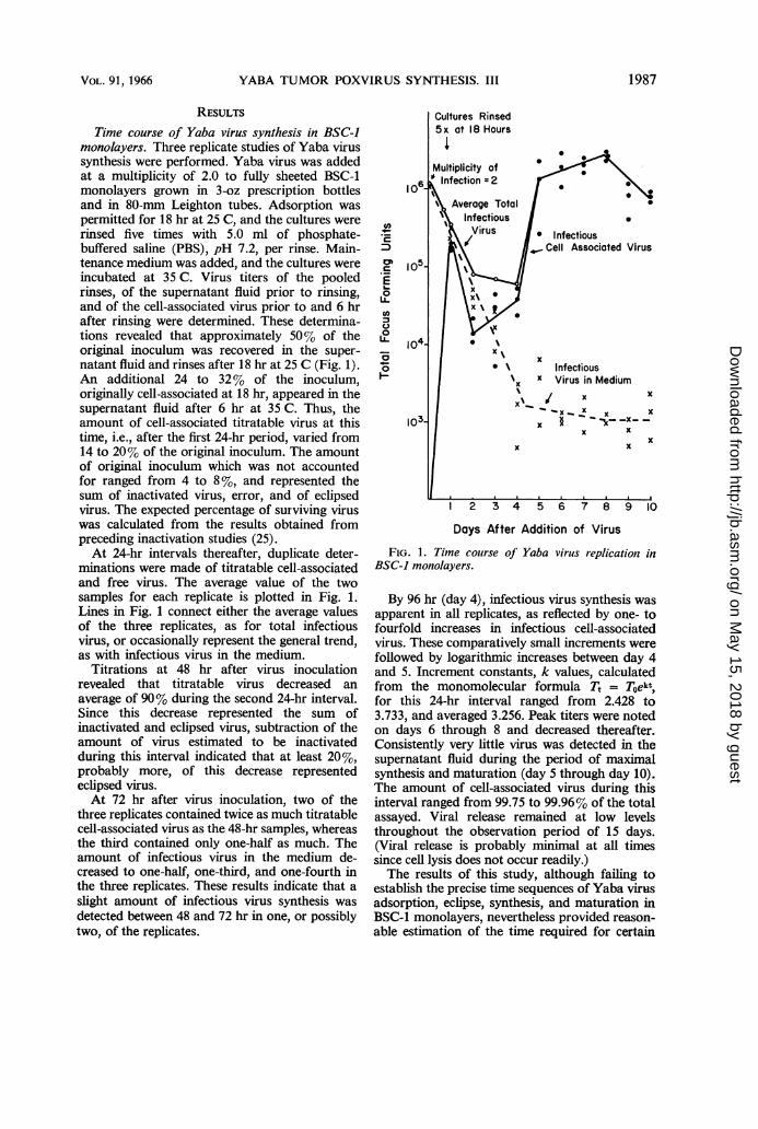
Time course of Yaba virus synthesis in BSC-Jmonolayers. Three replicate studies of Yaba virussynthesis were performed. Yaba virus was addedat a multiplicity of 2.0 to fully sheeted BSC-1monolayers grown in 3-oz prescription bottlesand in 80-mm Leighton tubes. Adsorption waspermitted for 18 hr at 25 C, and the cultures wererinsed five times with 5.0 ml of phosphate-buffered saline (PBS), pH 7.2, per rinse. Main-tenance medium was added, and the cultures wereincubated at 35 C. Virus titers of the pooledrinses, of the supernatant fluid prior to rinsing,and of the cell-associated virus prior to and 6 hrafter rinsing were determined. These determina-tions revealed that approximately 50% of theoriginal inoculum was recovered in the super-natant fluid and rinses after 18 hr at 25 C (Fig. 1).An additional 24 to 32% of the inoculum,originally cell-associated at 18 hr, appeared in thesupernatant fluid after 6 hr at 35 C. Thus, theamount of cell-associated titratable virus at thistime, i.e., after the first 24-hr period, varied from14 to 20% of the original inoculum. The amountof original inoculum which was not accountedfor ranged from 4 to 8 %, and represented thesum of inactivated virus, error, and of eclipsedvirus. The expected percentage of surviving viruswas calculated from the results obtained frompreceding inactivation studies (25).At 24-hr intervals thereafter, duplicate deter-
minations were made of titratable cell-associatedand free virus. The average value of the twosamples for each replicate is plotted in Fig. 1.Lines in Fig. 1 connect either the average valuesof the three replicates, as for total infectiousvirus, or occasionally represent the general trend,as with infectious virus in the medium.
Titrations at 48 hr after virus inoculationrevealed that titratable virus decreased anaverage of 90% during the second 24-hr interval.Since this decrease represented the sum ofinactivated and eclipsed virus, subtraction of theamount of virus estimated to be inactivatedduring this interval indicated that at least 20%,probably more, of this decrease representedeclipsed virus.At 72 hr after virus inoculation, two of the
three replicates contained twice as much titratablecell-associated virus as the 48-hr samples, whereasthe third contained only one-half as much. Theamount of infectious virus in the medium de-creased to one-half, one-third, and one-fourth inthe three replicates. These results indicate that aslight amount of infectious virus synthesis wasdetected between 48 and 72 hr in one, or possiblytwo, of the replicates.
Cultures Rinsed
Virus / Infectious,F 105. Cell Associated Virus
L2 A
x
?L 104-L . X_ I x~~~Xx
o Infectious1 l ~ ~~~~x x Virus in Medium
I \\/~% x x
103- x §-k___x- xI x ~ ~~~~~xx
0~~~~~~~~~~~~~~~
La... 0 .|. x
2' 3 4' s 6' 7' 8 9 lo
Days After Addition of Virus
FIG. 1. Time course of Yaba virus replication inBSC-1 monolayers.
By 96 hr (day 4), infectious virus synthesis wasapparent in all replicates, as reflected by one- tofourfold increases in infectious cell-associatedvirus. These comparatively small increments werefollowed by logarithmic increases between day 4and 5. Increment constants, k values, calculatedfrom the monomolecular formula Tt = Toekt,for this 24-hr interval ranged from 2.428 to3.733, and averaged 3.256. Peak titers were notedon days 6 through 8 and decreased thereafter.Consistently very little virus was detected in thesupernatant fluid during the period of maximalsynthesis and maturation (day 5 through day 10).The amount of cell-associated virus during thisinterval ranged from 99.75 to 99.96% of the totalassayed. Viral release remained at low levelsthroughout the observation period of 15 days.(Viral release is probably minimal at all timessince cell lysis does not occur readily.)The results of this study, although failing to
establish the precise time sequences of Yaba virusadsorption, eclipse, synthesis, and maturation inBSC-1 monolayers, nevertheless provided reason-able estimation of the time required for certain
1987VOL. 91, 1966
on May 15, 2018 by guest
http://jb.asm.org/
Dow
nloaded from
YOHN, HAENDIGES, AND DE HARVEN
steps. Viral eclipse did not occur during the 18-hr period at 25 C. A small proportion of virusmay have been eclipsed during the first 6 hr at35 C; however, the maximal rate of viral eclipseoccurred during the next 24 hr at 35 C. At leastanother 24 hr at 35 C was required before anyevidence of infectious viral synthesis was ob-tained. Maximal viral maturation occurred afterthe 4th day, i.e., 18 hr at 25 C plus 78 hr at 35 C.
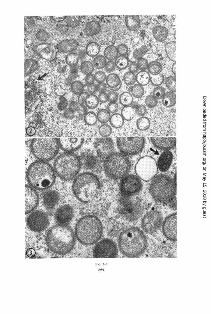
Electron microscopic observations on the de-velopment of Yaba virus in BSC-J. Detaileddescriptions of the fine structure of Yaba poxvirusas determined from these studies and otherscarried out on thin sections of Yaba tumors havebeen reported elsewhere (8). A summary of thein vitro results is included here. Yaba virus wasadded to T-30 flask cultures of BSC-1 at a multi-plicity of 5, and the cultures were placed at 35 Cwithout preadsorption at 25 C. Cultures wereremoved for examination at 20, 44, 68, 92, and116 hr after infection. No evidence of virusassembly was detected until the 68th hr sample.At this time, immature virus particles wereobserved. At 92 hr, the number of immatureparticles had increased markedly. Although afew particles of increased density (Fig. 2 and 3),indicative of increased maturation and designatedas transitional forms (8), were present at 92 hr,fully mature particles with typical biconcave discshape and dumbbell-shaped core were not seenuntil 116 hr (Fig. 3).
DIscussioNThese studies were undertaken to establish the
sequential time relationships of Yaba tumorpoxvirus synthesis in BSC-1 monolayers at 35 C.Certain other information obtained by histo-chemical and immunofluorescence studies de-scribed previously (24) will be discussed in con-junction with the present results.The initiating event in virus infection, irrever-
sible adsorption and penetration, presumablyrequires active cellular phagocytosis as with otherpoxviruses (6), and was, for reasons as yet notclear, relatively inefficient in monolayers ofBSC-1 at temperatures normally employed forvirus adsorption, 25 to 37 C (25). Because of thecomparatively long interval required for Yaba
virus adsorption by BSC-1 monolayers, theadsorption portion of the growth curve studywas carried out at 25 C for 18 hr. Analysis of theamounts of extracellular and cell-associated virusrecoverable at the end of this period indicatedthat viral eclipse (uncoating) had not occurredduring this period. During the next 6 hr, now at35 C, a maximum of 4 to 8% of the originalinoculum may have entered the eclipse phase.During the next 24-hr period, day 2, eclipse
was completed and synthetic processes wereinitiated. Nucleoli became hypertrophic, andpinpoint sites of DNA synthesis were detected inthe cytoplasm; however, viral antigen (protein?)synthesis was not demonstrated until the end ofthis interval, between the 36th and 48th hr (24).At this point, the growth curve analysis indicatedthat no infectious virus had been produced. Inaddition, no evidence of viral assembly was seenby electron microscopy.During the third 24-hr interval, day 3, analysis
of growth curve results indicated that infectiousvirus had been synthesized in one, or possiblytwo, of the three replicates. By electron micros-copy, only immature particles were discernibleat 68 hr. The synthesis of Yaba virus has beendescribed to consist of six stages (8), the first twoof which are immature forms; the next two aretransitional forms, and the final two stages aremorphologically mature with differences only inthe number of exterior coats. Whether a veryfew transitional or even mature forms werepresent at this time and were not located byelectron microscopy is not known. Additionalevidence that infectious virus is synthesized by 72hr was obtained from immunofluorescencestudies (24). After addition of virus to BSC-1monolayers, approximately 48 hr elapses beforeviral antigen appears. Contiguous cells containviral antigen 3 days later. Thus, the infection ofthe contiguous cells must have occurred at least48 hr earlier, i.e., during the 3rd day after in-fection of the primary cell. The physical nature ofthe infectious material which passes from cell tocell is not known at this time. It is tempting tospeculate that contiguous spread may not requiremorphologically mature particles.The onset of logarithmic maturation occurred
FIG. 2. Portionz ofthe cytoplasm ofa BSC-1 cell 5 days after infection with Yaba virus. A group of immatureparticles is seen corresponding to the stages 1, 2, and 3 described elsewhere (8). A group offree ribosomes (arrow)is seen in the vicinity of this "factory area." X 32,500.
FIG. 3. Higher magnification ofanother cellfrom the same sample as Fig. 2. The viral membrane has a typicalunit membrane structure and is covered by short subunits. The arrow points to a more mature particle correspondingto stage 5 in the previously quoted reference. This form is characterized by the appearance within the dense matrixof additional membranes which are going to constitute the inner dumbbell-shaped core of the fully mature virions.X 67,500.
1988 J. BACTERIOL.
on May 15, 2018 by guest
http://jb.asm.org/
Dow
nloaded from
44
I .
Z.
"!
FIG. 2-3.
1989
a.:;e,
.: wi..e
1.,
ME qpl_A
.... ....Tft,MNIWO
AI
4 PA A
,1lt-'. Iit;,
IWA -f A. -UMV.... Ovor . "
i. .1 41MLI,
,. '41IN
Li, on.
.1": AU.
All
Al
on May 15, 2018 by guest
http://jb.asm.org/
Dow
nloaded from
YOHN, HAENDIGES, AND DE HARVEN
during the 4th day and reached its maximal rateduring the 5th day. With electron microscopy,these intervals were characterized by increasednumbers of immature and transitional forms at 4days and the appearance of large numbers ofmature particles at 5 days. Cytopathology wasnow discernible with routine bright-field examina-tion. In all stained preparations, cytoplasmicinclusion bodies were well formed and approachedthe size of the nucleus. Thereafter, the inclusionsenlarged, equaling or exceeding the size of thenucleus. This latter process coincided with theinterval when maximal virus yields were ex-tracted from the cultures.The minimal latent period, defined as the
interval between irreversible adsorption andinitial appearance of infectious progeny, appearsto be of the order of 50 hr. In the growth curveanalysis, this interval encompassed 54 hr. Thepreviously discussed immunofluorescence resultssuggested a minimal period of 48 to 60 hr. Theelectron microscopic study placed the interval atmore than 44 hr but less than 68 hr includingadsorption time. The minimal latent periodrequired for vaccinia virus synthesis in suspendedL cells (5, 13) or in HeLa cell suspensions (12,19, 20) or monolayers (15) is 6 to 10 hr. The timerequired for maximal yields of the two virusesunder the above different conditions is 18 to 24hr for vaccinia virus and 100 to 120 hr for Yabavirus. These differences can be attributed to thecombination of many factors: (i) the 2 C tem-perature differential; (ii) the use of cell culture-adapted vaccinia virus as opposed to our use ofvirus extracted from tumor; (iii) the use of highmultiplicity of infection with vaccinia virusunder optimal adsorption conditions; (iv) theinherent biochemical differences between the cellhosts; and (v) the inherent differences betweenthe synthetic rate directing capacities of the twoviruses. At present, it appears that a direct com-parison can only be made at 35 C in BSC-1,since some process of Yaba virus synthesis inBSC-1 at 37 C appears thermal-sensitive andvirus yields are markedly lower than at 35 C(Marmol and Yohn, unpublished data). Further-more, Yaba virus is not infectious for HeLa (1)or L cells (Marmol and Yohn, unpublished data).Initial studies of vaccinia virus replication inBSC-1 cells at 35 C indicate that the difference inkinetics between Yaba and vaccinia virus syn-thesis is largely viral genome-controlled. Thevaccinia infectious cycle is four to six times morerapid.Means to obtain maximal adsorption of Yaba
virus in short-term suspension cultures of BSC-1cells in a minimal time interval, i.e., less than 1hr, are presently being developed. Once this
technique is available, it should be possible tocompare under identical in vitro conditions thebiochemistry of an oncogenic poxvirus with thatof a normally nononcogenic poxvirus.The protracted cycle of Yaba poxvirus syn-
thesis in BSC-1, although unlike vaccinia, rabbitpox, and shope fibroma virus in this respect,appears to be similar in length to the syntheticcycles of fowlpox in chick chorioallantoic mem-brane (4) and of molluscum contagiosum (9) inHeLa cells. The antigenic relationship of Yabavirus to these and other poxviruses has beenexamined somewhat limitedly. Niven et al. (17)found no cross-protection in monkeys betweenYaba, vaccinia, and orf viruses. Similar results(1) have been obtained with the monkey poxvirusstrains of von Magnus et al. (21) and of Prierand Sauer (18). However, Woodroofe andFenner (22) obtained a weak precipitin reactionwith vaccinia and myxoma nucleoprotein antigensreacted with Yaba antisera. Cross-reactions didnot occur in complement-fixation tests, byimmunofluorescence tests, or in precipitin testswith myxoma and vaccinia soluble antigens Thus,it appears that Yaba tumor poxvirus may differfrom the well-known primate poxviruses bothbiochemically and antigenically. Future studiesalong both these lines should define these differ-ences more precisely.
ACKNOWLEDGMENTS
This investigation was supported by Public HealthService grant CA-07998 from the National CancerInstitute. Etienne de Harven was recipient of aCareer Scientist Award of the Health ResearchCouncil of the City of New York, Contract 1-325.
LITERATURE CITED1. AMBRUS, J. L., E. T. FELTZ, J. T. GRACE, JR.,
AND G. OWENS. 1963. A virus-induced tumorin primates. Natl. Cancer Inst. Monograph 10,p. 447-458.
2. ANDREWES, C. H., A. C. ALLISON, J. A. ARM-STRONG, G. BEARCROFT, J. S. F. NIVEN, ANDH. G. PEREIRA. 1959. A virus disease of mon-keys causing large superficial growths. ActaUnio Intern. Contra Cancrum 15:760-763.
3. APPLEYARD, G., AND J. C. N. WESTWOOD. 1964.The growth of rabbitpox virus in tissue culture.J. Gen. Microbiol. 37:391-401.
4. ARHELGER, R. B., AND C. C. RANDALL. 1964.Electron microscopic observations on the de-velopment of fowlpox virus in chorioallantoicmembrane. Virology 22:59-66.
5. DALES, S. 1963. The uptake and development ofvaccinia virus in strain L cells followed withlabeled viral deoxyribonucleic acid. J. CellBiol. 18:51-72.
6. DALES, S., AND L. SIMINOVITCH. 1961. The de-velopment of vaccinia virus in Earle's L strain
1990 J. BACTERIOL.
on May 15, 2018 by guest
http://jb.asm.org/
Dow
nloaded from

YABA TUMOR POXVIRUS SYNTHESIS. III
cells as examined by electron microscopy.J. Biophys. Biochem. Cytol. 10:475-503.
7. DE HARVEN, E., AND D. S. YOHN. 1964. Electronmicroscopy of the Yaba tumor virus. J. Appl.Phys. 35:3082.
8. DE HARVEN, E., AND D. S. YOHN. 1966. The finestructure of the Yaba monkey tumor poxvirus.Cancer Res., in press.
9. DOURMASHKIN, R., AND B. DUPERRAT. 1958.Observation au microscope electronique duvirus du "molluscum contagiosum." Compt.Rend. 246:3133-3136.
10. FEBVRE, H., 1962. The Shope fibroma virus ofrabbits, p. 79-112. In A. J. Dalton and F.Haguenau [ed.], Tumors induced by viruses:ultrastructural studies. Academic Press, Inc.,New York.
11. ISRAELI, E., AND L. SACHS. 1964. Cell-virus inter-actions with the Shope fibroma virus on cul-tures of rabbit and rat cells. Virology 23:473-485.
12. JOKLIK, W. K., AND Y. BECKER. 1964. The repli-cation and coating of vaccinia DNA. J. Mol.Biol. 10:452-474.
13. KIT, S., AND D. R. DUBBS. 1962. Biochemistry ofvaccinia infected mouse fibroblasts (strainL-M). I. Effects on nucleic acid and proteinsynthesis. Virology 18:274-285.
14. LEVINTHAL, J. M., AND H. M. SHEIN. 1964. Propa-gation of a simian tumor agent (Yaba virus) incultures of human and simian renal cells asdetected by immunofluorescence. Virology 23:268-270.
15. LOH, P. C., AND J. L. RIGGS. 1961. Demonstra-tion of the sequential development of vaccinialantigens and virus in infected cells: observationswith cytochemical and differential fluorescentprocedures. J. Exptl. Med. 114:149-160.
16. LuFT, J. H., 1961. Improvements in epoxy resinembedding methods. J. Biophys. Biochem.Cytol. 9:409-414.
17. NIVEN, J. S. F., J. A. ARMSTRONG, C. H.ANDREWES, H. G. PEREIRA, AND R. C. VALEN-TINE. 1961. Subcutaneous "growths" inmonkeys produced by a pox virus. J. Pathol.Bacteriol. 81:1-14.
18. PRIER, J. E., AND R. M. SAUER. 1960. A pox dis-ease of monkeys. Ann. N.Y. Acad. Sci. 85:951-959.
19. SALZMAN, N. P. 1960. The rate of formation ofvaccinia deoxyribonucleic acid and vacciniavirus. Virology 10:150-152.
20. SHATKIN, A. J., 1963. The formation of vacciniavirus protein in the presence of 5-fluorodeoxy-uridine. Virology 20:292-301.
21. VON MAGNUS, P., E. K. ANDERSEN, K. B. PETER-SEN, AND A. BIRCH-ANDERSEN. 1959. A pox-likedisease in cynomologous monkeys. ActaPathol. Microbiol. Scand. 46:156-176.
22. WOODROOFE, G. M., AND F. FENNER. 1962.Serological relationships within the poxvirusgroup: an antigen common to all members ofthe group. Virology 16:334-341.
23. YOHN, D. S., J. T. GRACE, JR., AND V. A.HAENDIGES. 1964. A quantitative cell cultureassay for Yaba tumour virus. Nature 202:881-883.
24. YOHN, D. S., V. A. HAENDIGES, AND J. T. GRACE,JR. 1966. Yaba tumor poxvirus synthesis invitro. I. Cytopathological, histochemical, andimmunofluorescent studies. J. Bacteriol. 91:1977-1985.
25. YOHN, D. S., F. R. MARMOL, V. A. HAENDIGES,AND J. T. GRACE, JR. 1966. Yaba tumor pox-virus synthesis in vitro. II. Adsorption, inacti-vation, and assay studies. J. Bacteriol. 91:1953-1958.
1991VOL. 91, 1966
on May 15, 2018 by guest
http://jb.asm.org/
Dow
nloaded from