ocular impression-taking -1 which material is best?
TRANSCRIPT
1
Ocular impression-taking - which material is best? 1
Jennifer M Turner PhD1, Christine Purslow PhD1 and Paul J Murphy PhD1,2 2
3
1Cardiff University, School of Optometry and Vision Sciences, Cardiff, UK 4
2University of Waterloo, School of Optometry and Vision Science, Waterloo, Canada 5
6
Address for correspondence: 7
Prof Paul J Murphy 8
University of Waterloo 9
School of Optometry and Vision Science 10
200 Columbia Avenue West 11
Waterloo, N2L 3G1 12
Ontario, Canada 13
Email: [email protected] 14
Tel: (1) 519 888 4567, ext 32020 15
Fax: (1) 519 725 0784 16
17
Tables: 2, Figures: 2 18
19
Running title: Which ocular impression material is best? 20
21
Proprietary Interests and Research Funding: This study was supported by a collaborative partnership between 22
Cardiff University and Menicon (Japan) Ltd. The authors have no proprietary or commercial interests in any of the 23
materials discussed in this article. 24
25
Paper submitted: 3rd April 2017 26

2
Abstract 27
Objectives: To assess the efficacy and effect on clinical signs of a polyvinylsiloxane (TresidentÔ (Shütz Dental 28
Group GmbH, Germany) compared to an irreversible hydrocolloid (OrthoprintÔ, Zhermack SpA, Italy) for ocular 29
impression taking. 30
Methods: Twenty subjects were recruited (13 female and 7 male), mean age 31.1±4.6 years [SD] (range 25.8 to 31
39.7). Subjects attended for 2 sessions, each of 1 hr duration, on 2 separate days. Each session was scheduled at 32
the same time on each day. At each visit the subject underwent an ocular impression procedure, using either 33
Tresident or Orthoprint, in random order and to one eye only. Investigator 2 was blind to this assignment. Two 34
experienced practitioners carried out the study, Investigator 1 performed the ocular impression procedures and 35
Investigator 2 observed and assessed the clinical signs: logMAR visual acuity (VA), ocular surface staining, tear 36
break-up time (TBUT), and ocular hyperaemia. 37
Results: VA was unaffected by either material; TBUT was marginally disrupted by both materials, but was not 38
clinically significant according to published criteria; ocular redness increased with both materials; corneal staining 39
was significantly greater after Orthoprint impression. Less redness and clinically insignificant staining following 40
impression-taking, with fewer clinical complications, was found following use of Tresident. 41
Conclusions: Tresident offers a quicker, more effective and clinically viable method of obtaining ocular impression 42
topography compared to the traditional Orthoprint; and Orthoprint causes significantly more superficial 43
punctuate staining of the corneal epithelium than Tresident. 44
45
Keywords: ocular impression, ocular surface, ocular prosthetics, materials 46
47

3
The purpose of taking an impression of any surface is to mould the negative dimensions of the structure and 48
make a model of the 'positive' physical properties, which then provides an accurate representation of the shape, 49
parameters and spatial relationships. Ocular impression taking is used in scleral contact lens fitting and ocular 50
prosthesis manufacturing. In both situations, an accurate representation of the existing ocular surface is critical 51
for success1. For example, in scleral contact lens fitting, the eye impression produced enables the manufacture of 52
the lens to match with the patient’s ocular surface topography. Alternative optical methods are now available, 53
and used, for scleral lens fitting, but ocular impression-taking remains a vital component of the clinician’s toolkit, 54
and will also provide information over a larger scleral area. Ocular impression taking is also relatively inexpensive, 55
can be used outside of the clinical office room, and are reproducible1,2. To our knowledge, no studies have been 56
published that report on the effect of impression taking on the ocular surface, in a comparison between two 57
established impression materials. 58
59
The characteristics of the 'ideal' ocular impression material include: minimal deleterious effects on the anterior 60
ocular surface (AOS) or exposed ocular adnexa by the material; no lasting discomfort after the procedure (topical 61
anaesthetic blocks the sensory corneal nerves during the procedure); high accuracy - the acceptable magnitude of 62
error in impression taking is determined by its desired application, e.g. gas permeable contact lens manufacturing 63
requires high accuracy (±0.05mm) to match the manufacturing tolerances of BS/EN/ISO/18369-2:20123; excellent 64
dimensional stability to ensure the material is not deformed by plaster pouring, or degraded by environmental 65
conditions or physical manipulation; good flow characteristics and reasonable in-eye working time to allow 66
sufficient time for the material to be applied to the impression tray and inserted without setting; rapid curing or 67
setting time to reduce the amount of time required to maintain the material against the eye, thereby reducing 68
artefacts incurred by random eye movements; and, excellent compatibility with gypsum dental stone (some 69
impression materials are known to cause chemical degradation of the gypsum cast surface). 70
71
Cold, irreversible hydrocolloids or alginates (e.g. OrthoprintÔ, Zhermack SpA, Italy), which have been used for 72
ocular impressions since the introduction of Ophthalmic Moldite4, exhibit poor dimensional stability and poor tear 73

4
strengths, leading to inaccurate casts and the need for multiple impression-taking procedures5,6. The impressions 74
formed are affected by: (1) the level of airflow around the impression, which causes evaporation of water from 75
the gel, resulting in shrinkage; (2) by water, which causes the gel to expand by imbibition and absorption; (3) by 76
high relative humidity, which induces syneresis and shrinkage; and (4) by in-organic salts, which affect the gel and 77
cause physical changes that are dependent on their osmotic potential7. 78
79
Orthoprint (Zhermack SpA, Italy) is a yellow, dust-free, alginate, irreversible, hydrocolloid impression material, 80
which conforms to BS/EN/ISO/21563:20138, with origins in dental practice (Table 1). It provides good surface 81
detail9, is easy to use and mix, is cheap and has a long shelf-life, numbered in years7. The setting time can be 82
controlled with water temperature and, as a gel, it is non-toxic and non-irritant10. However, it has relatively poor 83
dimensional stability, compared with elastomers, and a low tear energy11. It is incompatible with Type 1 or 2 84
gypsum plaster12,13, reacts to humidity, and has a very short on-eye setting time (45 secs). The mixing process is 85
messy and dependent on operator handling. Automated mechanical mixing has been shown to increase speed 86
and quality of alginate sol, eliminating casting imperfections14. For these reasons, the use of alginate for ocular 87
impression-taking has been superseded by silicone rubber-based materials. 88
89
Polyvinylsiloxane polymers appear to allow reproduction of the greatest detail of all dental impression 90
materials15. Indeed, the material provides sufficient detail to identify individuals by fingerprint analysis16. This 91
level of accuracy is defined by BS/EN/ISO/4823:201517, which requires that all Type 3, light-bodied, elastomeric 92
materials be able to reproduce a line 0.02mm in width. In addition, these materials have been found to have very 93
low shrinkage (0.05-0.1%), during the polymerising process18, and are well-matched to the setting expansion of 94
Type 4 gypsum plaster, which is used to cast the impression19. 95
96
TresidentÔ (Shütz Dental Group GmbH, Germany) is a low viscosity, addition-polymerising, polyvinylsiloxane 97
precision impression material with hydrophilic properties, which conforms to BS/EN/ISO/4823:201517 (Table 1). It 98
is supplied in an auto-mix dual-cartridge, which requires a dispensing gun to automatically mix and advance equal 99

5
quantities of each siloxane-based component through a purpose-designed mixing cannula (Injector DS 50, Dreve 100
Otoplastik GmbH, Germany). Tresident provides a working time of 1 min 15 secs, with a setting time of 2 mins 45 101
secs, giving a total setting time of 4 mins. During the setting time, the impression tray and material must be held 102
against the ocular surface under gentle pressure. Plaster casts can be produced from the moulds, and can be 103
poured from 1 hr to 14 days after the procedure. Further casts can be produced from each Impression, which are 104
as accurate as the original, for up to 7 days20, but to do so the impression material must be kept in a dry place at 105
18-25°C. Re-heating the impression to 37°C before pouring the plaster has been shown to improve accuracy of 106
casting. However, it is doubtful if this is clinically significant21. 107
108
The two components of the material are a polymethyl-hydrogen-siloxane copolymer of moderately low molecular 109
mass, which contains silane terminal groups, and an accelerator material of a similar molecular weight, which 110
contains vinyl-terminated polydimethyl siloxane. When mixed, the silane and vinyl groups react, catalysed by 111
chloroplatinic acid (a homogenous, metal complex catalyst). The cross-linking that occurs during the 112
polymerisation process causes minimal dimensional change and there are no by-products22. Both components 113
contain fillers, amorphous silica and a low molecular weight retarder to delay the onset of polymerisation. 114
Additionally, the base component has an emulsifying surfactant that improves the wettability of the impression. 115
Colouring agents are added to distinguish between the two pastes and aid the evaluation of mixing process. 116
117
Polyvinylsiloxane materials have been found to have good long-term dimensional stability (up to 2 weeks), are not 118
susceptible to changes in humidity, and do not undergo further chemical reactions or release by-products15. Tests 119
carried out on intact rabbit skin concluded that the primary skin irritation of polyvinylsiloxane can be considered 120
negligible23. For these reasons, it is considered a superior alternative to the irreversible hydrocolloids. Sydiskis and 121
Gerhardt (1993)24 also showed that while both polyvinylsiloxane and irreversible hydrocolloid materials have a 122
cytotoxic effect on cell culture, the risk of producing an adverse reaction is low. However, the effects of the 123
material on the tear film and adnexa, although considered clinically acceptable, have not previously been 124
reported. 125

6
This study used a single-blind, randomised control trial to assess the efficacy and effect on clinical signs of a 126
polyvinylsiloxane (Tresident) compared to an irreversible hydrocolloid (Orthoprint) for ocular impression taking. 127
The hypotheses proposed are that: (1) Tresident offers a quicker, more effective and clinically viable method of 128
obtaining ocular impression topography compared to the traditional Orthoprint; and (2) Orthoprint causes 129
significantly more superficial punctuate staining of the corneal epithelium than Tresident. 130
131
Materials and Methods 132
Twenty subjects were included in the study, (13 female and 7 male), mean age was 31.1±4.6 years [SD] (range 133
25.8-39.7). Volunteers were recruited from staff and students of Cardiff University, and subjects were excluded if 134
they were pregnant or breastfeeding; had any ocular or systemic condition known to affect the structure or 135
characteristics of the AOS; were taking any medication known to affect the ocular surface; had worn rigid contact 136
lenses in the preceding 6 weeks or soft contact lenses in the preceding 2 weeks. Ethical approval was sought and 137
granted in accordance with the Tenets of the Declaration of Helsinki (2004) from the Cardiff School of Optometry 138
and Vision Sciences Human Research Ethics Committee. 139
140
Subjects attended for 2 sessions, each of 1 hr duration, on 2 separate days. Each session was scheduled at the 141
same time on each day. Two experienced practitioners carried out the study: one performed the ocular 142
impression procedures (Investigator 1), and the other observed and assessed the clinical signs (Investigator 2). 143
The practitioners carried out their investigations in separate rooms without any knowledge of the other’s results. 144
Each subject was randomly assigned to receive an ocular impression in one eye, using one of the two impression 145
material, by a study administrator. Investigator 2 was blind to this assignment. 146
147
Session 1 148
The subject arrived and was assessed by Investigator 2 for suitability and baseline clinical assessment 149
measurements. Both eyes were assessed, but the data analysed only for the eye assigned for treatment. 150
7
1. Best-corrected LogMAR distance acuity (Sussex Vision International Ltd, West Sussex, UK) at 3m direct viewing. 151
Visual acuity was obtained by assigning 0.02 LogMAR units to each letter. 152
2. Instillation of fluorescein, using Fluoret strips (Chauvin, France) (each strip impregnated with approximately 153
1mg of fluorescein sodium BP) moistened with 0.9% physiological saline, to assess invasive tear break-up time. 154
The subject was asked to blink and then hold their eye open as long as possible. The measurement was taken in 155
seconds between the blink and the first appearance of a discontinuity in tear film coverage. Three values were 156
recorded for each eye and the median used for comparison. 157
3. Tear break-up time using Tearscope Plus™ (Keeler Ltd, Windsor, UK), with fine grid insert. The measurement 158
was taken in seconds between the blink and the first appearance of a discontinuity in tear film coverage. Three 159
values were recorded for each eye and the median used for comparison. 160
4. Assessment of ocular integrity using CCLRU grading scales (Brian Holden Vision Institute (BHVI)), interpolated to 161
0.1 unit increments25: bulbar redness; limbal redness; lid redness; lid roughness; type, extent and depth of corneal 162
staining with fluorescein. 163
164
Ocular impression was then performed in a separate room, where Investigator 1 carried out an impression 165
procedure to one ocular surface (randomly-assigned) using one of the two materials. After impression taking, and 166
saline wash-out to remove excess material, the subject returned to the room of Investigator 2 who repeated the 167
clinical tests as above. 168
169
Session 2 170
When the subject arrived, Investigator 2 repeated the clinical tests carried out the day before, followed by 171
Investigator 1 taking an ocular impression with the alternative material to a randomly-assigned eye. Investigator 2 172
repeated the clinical tests following a saline wash-out, post-impression procedure. 173
174
Ocular impression procedure 175

8
Each subject was positioned sitting upright and facing forward. A distant target was provided to align the visual 176
axes, using the contralateral eye for fixation. The ocular surfaces of both eyes were anaesthetised with 0.5% 177
Proxymetacaine HCL Minims eye drops (Chauvin, Kingston-upon-Thames, UK), and the procedure carefully 178
explained to the subject. An impression tray was chosen from the set of 3 sizes, of maximum internal shell 179
diameter 23, 24 or 25mm (Cantor and Nissel Ltd, Brackley, UK). These trays are moulded from acrylic with hollow 180
stems, 32mm in length, marked with red circular indentations providing an anatomical registration at the 12 181
o'clock position (in relation to the cornea). The tray was selected by presenting the 3 sizes to the closed eye and 182
choosing the largest in relation to the aperture and the global contour. Impression material was dispensed onto 183
the internal surface of the shell covering the entire surface with 1.5-2.5mm26 of either Tresident or Orthoprint 184
(Figure 1). 185
186
The subject was instructed to 'look down' whilst remaining in the head upright position. The tray was inserted 187
quickly under the top eyelid, and the subject was asked to 'look up' in order for the lower lid to be freed and the 188
shell held between both eyelids. The tray was carefully positioned to locate the cornea at the centre of the shell; 189
the investigator supported the stem and ensured that the subject maintained composure and optimal fixation 190
(Figure 2). 191
192
After setting of the material, the tray and impression was removed by freeing the lashes of the upper lid and 193
removing the material from the eye surface in one piece. Any material remnants were collected and the fornices 194
irrigated with 0.9% buffered saline. 195
196
Statistical analysis 197
All data was collated with Excel 2007 (Microsoft, WA, US), and analysed within SPSS v13 (IBM, NY, US). The data 198
distribution was evaluated for normality using the Kolmogorov-Smirnov statistical test. Comparisons were made 199
of clinical signs assessed before and after each impression procedure, and paired t-tests used to determine 200
statistical significance at the 95% level. 201

9
202
Results 203
A summary of the results is shown in Table 2. LogMAR acuity was found to be slightly reduced by 0.5 letters, on 204
average, following impression using either material, but this was not statistically significant. 205
206
TBUT was found to be reduced following impression-taking with both materials, but was not clinically significant. 207
Mean TBUT was 7.16±1.40 secs pre- and 6.68±1.27 secs post-Tresident impression, with a difference of -208
0.87±2.61 secs, which was not statistically significant (p=0.383). Mean TBUT was 7.42±1.55 secs pre- and 209
6.61±1.33 secs post-Orthoprint impression, giving a larger difference of -1.28±2.03 secs, but which did not reach 210
statistical significance (p=0.094). 211
212
Ocular redness was found to increase following impression taking, with both impression materials. Bulbar redness 213
increased following impression-taking with Tresident by +0.64±0.58 units (p<0.001), with a substantially greater 214
change in redness observed following the use of Orthoprint +1.12±0.42 units (p<0.001). A statistical difference 215
was found between the numerical values assigned to bulbar redness after Orthoprint compared to Tresident 216
(p=0.0231). Similarly, limbal redness increased following impression-taking, +0.70±0.31 units (p<0.005) after 217
Tresident use and +1.05±0.28 units (p<0.005) after Orthoprint. However, the mean difference between changes in 218
limbal redness when comparing the two materials was not statistically significant (p=0.072). There was a small 219
change in lid redness recorded after impression-taking. For Tresident this was 0.17 ±0.32units, which was 220
statistically significant (p<0.05). However, the change was smaller after Orthoprint use (0.07 ±0.35 units) and was 221
not statistically significant (p=0.487). 222
223
The clinical grading of lid roughness was found to increase following impression-taking with Tresident (0.03 ±0.25 224
CCLRU units), but this was not statistically significant (p=0.459). Orthoprint had no detectable effect on lid 225
roughness. 226
227

10
Corneal staining with fluorescein was recorded following impression-taking with both materials. Staining type was 228
micro-punctate and superficial after Orthoprint; 0.13 ±0.34 grade units (p=0.341), but tended to be macro-229
punctate after Tresident; 0.53 ±0.41 grade units (p=0.167). However, these changes were not statistically 230
significant. 231
232
The extent of staining was found to increase substantially after Orthoprint impression to 2.33 ±0.46 grade units 233
(p<0.001). These measurements indicate that, on average, 22% of the corneal surface (range 15-45%) was 234
covered by staining. The recorded increase in extent of staining after Tresident was small (0.49 ±0.65 grade units), 235
was not statistically significant (p=0.209) and the surface area stained was, on average, only 10% (range 1-22%). 236
Changes in the depth of staining were found to be small (0.31±0.40 grade units after Orthoprint, 0.37±0.47 grade 237
units after Tresident), which were statistically significant for Orthoprint, p<0.05, but not for Tresident (p=0.219). 238
239
Clinical summary 240
Visual acuity was unaffected by either material (clinically significant criterion Test-retest ±>2.4 letters)27. TBUT 241
was marginally disrupted by both materials, but was not clinically significant according to published criteria)28. 242
Bulbar redness increased with both materials. Orthoprint induced a clinically significant hyperaemic response in 243
over half of the cohort, i.e. >2.6 CCLRU grade units29,30, while Tresident was associated with increased bulbar 244
redness within clinically acceptable limits. Both materials increased limbal redness, but this was within clinically 245
acceptable limits (<2.4 CCLRU grade units30). Corneal staining was significantly greater after Orthoprint impression 246
(clinically significant criterion >0.5 grade units)31. Orthoprint produced micro-punctate staining (type) over 15-247
45% (extent) of the cornea, fluorescein penetrated the superficial epithelium. Tresident produced macro-248
punctate staining (type) over 1-22% (extent) of the cornea, fluorescein penetrated the superficial epithelium. 249
250
Discussion 251
For the first time using clinical grading scales, the effects of Orthoprint and Tresident have been evaluated to 252
determine the ocular surface disruption following ocular impression procedures, providing evidence to allow 253

11
practitioners to make an informed choice when deciding which material to use. The results from this study 254
support the use of Tresident as the clinically safer impression material. It also requires less preparation time than 255
Orthoprint. The use of Orthoprint was found to be associated with clinically significant (although superficial), 256
micro-punctate staining of the corneal epithelium, leading to an increased bulbar hyperaemia response. In 257
contrast, following the use of Tresident, ocular signs were within normal limits, with minimal corneal staining. 258
259
After the use of Orthoprint, 7 subjects reported a foreign body sensation accompanied by a slightly red eye, which 260
persisted for up to 24 hrs after the procedure. These subjects were monitored carefully, provided with ocular 261
lubricants and all symptoms resolved spontaneously. Inflammatory signs were not observed on the tarsal 262
conjunctiva or the dermis of the lids, although both areas were also in contact with Orthoprint during the 263
procedure. This, coupled with the hyperaemia response, suggests that there may be some toxicity response from 264
the AOS. This could be due to: poor mixing of the alginate resulting in one or a combination of the chemical 265
constituents causing damage to epithelial cell integrity. In particular, potassium fluorotitanate (chemical modifier) 266
is listed as a hazardous component on the Orthoprint data safety sheet. Between 1-3% of the impression mix is 267
made up of this chemical, and, if in contact with eyes, it advises to wash immediately with water for at least 10 268
mins. This effect might be prolonged if a chemical residue was left on the AOS after the gel was formed, which 269
was not removed by irrigation. 270
271
It is commonly accepted that fluorescein staining of the cornea represents compromised epithelial integrity32. A 272
red eye, accompanied by corneal staining, is intuitively taken as an unhealthy ocular situation and good practice 273
advocates monitoring for signs of deterioration and treatment if necessary. In this study, a clinically significant 274
increase in staining was observed after Orthoprint, but not Tresident (Dundas et al.,200131 suggests that a score 275
of >0.5 should be considered unusual). Damage to corneal integrity caused by one, or a multitude of factors, 276
during ocular impression-taking requires careful monitoring and consideration given that any denudation of 277
epithelium increases the risk of infection33. A number of factors may have contributed to the superficial staining 278
observed on the cornea. 279

12
280
The use of anaesthetic prior to ocular impression-taking may have contributed to the corneal staining – 0.5% 281
Proxymetacaine HCl has been associated with increased corneal permeability to fluorescein31. However, this was 282
applied equally for both impression methods, so it may be assumed that the differences in staining observed 283
between the methods is a true difference. 284
285
The mechanism for observing corneal staining is typically to use surface fluorescein pooling or ingress around 286
epithelial cells32. However, surface toxicity cannot be adequately explained in this manner. If Orthoprint does 287
indeed cause a chemical interaction with tear mucins or membranes of the corneal and conjunctival epithelial 288
cells, then fluorescein may be staining the affected cell complexes. Thus, the increased sensitivity reported by 289
subjects after Orthoprint may be a result of the 'toxic' interaction that remains until the surface cells are sloughed 290
off. The initial increased cell permeability by proxymetacaine anaesthesia may encourage the acute inflammatory 291
response and increase subsequent corneal staining observed. 292
293
This effect may be emphasised by increased permeability of the cornea. Physical contact between the ocular 294
surface and the setting alginate medium may cause the removal of multiple epithelial cells, allowing chemical 295
contamination of the deeper layers of the epithelium. In addition, anaesthetic instillation can cause reduced 296
corneal sensitivity, reduced blink frequency and can precipitate abnormal drying of the AOS35, encouraging 297
adherence of the impression material to the epithelium. Toxic interactions between anaesthetic and corneal 298
epithelial cells have been found to cause loss or damage to surface microvilli and deposition onto the cell 299
membranes36. 300
301
Average bulbar redness using CCLRU scales in the normal population is reported to be 1.93 units, with scores of 302
2.6 units considered abnormal29. Eleven subjects had scores greater than this following the use of Orthoprint, 303
with an average score of 3.14±0.37 grade units (range 2.7-3.8). This constitutes an abnormal level of hyperaemia 304

13
in over half the study cohort. In contrast, after using Tresident, only six subjects had scores above normal, with an 305
average of 3.31±0.41 units (range 2.8 - 3.8). 306
307
The irritation of ocular tissues by irreversible hydrocolloids has been studied on white, adult, New Zealand rabbit 308
eyes37 and clinical observations in human eyes found a range of responses to the material, ranging from slight 309
dehydration and irritation of the tissues to transient corneal abrasions6. Ocularists described capillary dilation, 310
tissue oedema and prolific tearing. The study concluded that the impression material, similar in formulation to 311
Orthoprint, elicited a significant, acute, inflammatory response in the rabbit conjunctiva on histological 312
examination. The authors attributed the tissue insult to the granular alginate material rubbing against the corneal 313
and conjunctival tissue interface, concurrent with blinking and eye movement. Additionally, they speculated, as in 314
this study, that the chemical setting aids (bimetallic fluorides) may have had a toxic effect on the ocular tissues. 315
The effects of the inflammatory response lasted 24-72 hours, leaving no permanent tissue damage37. 316
317
Conclusion 318
The use of Orthoprint during ocular impression-taking caused an abnormal hyperaemic response to the bulbar 319
conjunctiva, accompanied by significant superficial corneal staining. This may be attributed to a toxic reaction 320
between the material and the eye surface, exacerbated by mechanical abrasion caused by eyelid movement and 321
granular material apposition. However, further investigation would be necessary to establish the exact nature of 322
this interaction. 323
324
Tresident was found to be the impression material of choice. This study observed less redness and clinically 325
insignificant staining following impression-taking with fewer clinical complications. To manage any clinical 326
complications from using Tresident, the following advice is given: provide lubricating drops post-impression; 327
review the ocular surface integrity 24 hrs later; exclude dry eye patients and those with a comprised ocular 328
surface, where possible; and, consider prophylactic treatment for patients with damaged or impaired ocular 329
surface function. 330

14
331
These findings, combined with favourable handling and excellent physical properties, makes Tresident a superior 332
material for taking ocular impressions. 333
334
Acknowledgements 335
Thank-you to Dr Ditipriya Mukhopadhyay for her assistance with data collection. This study was supported by a 336
collaborative partnership between Cardiff University and Menicon (Japan) Ltd. 337
338
References 339
1. Pullum K W (2007) Eye Impressions. In: Phillips A J, Speedwell L, and Morris J [eds.] Contact Lenses. (5 edn.) 340
Elsevier Butterworth-Heineman, p. 343. 341
2. Cho SH, Schaefer O, Thompson GA, Guentsch A. Comparison of accuracy and reproducibility of casts made by 342
digital and conventional methods. J Prosthet Dent. 2015;113:310-315. 343
3. BS EN ISO 18369-2:2012 Ophthalmic Optics. Contact Lenses. Tolerances. London: British Standards Institution. 344
4. Obrig TE. A new ophthalmic impression material. Arch Ophthalmol. 1943;30:626-630. 345
5. Imbery TA, Nehring J, Janus C, et al. Accuracy and dimensional stability of extended-pour and conventional 346
alginate impression materials. J Am Dent Assoc. 2010;141:32-39. 347
6. Storey JK. The use of Panasil-C silicone rubber impression material in contact lens work. Optom Today. 348
1987;27:711-714. 349
7. O'Brien WJ. Dental materials and their selection. In: Impression Materials. 3rd edn. MI: Quintessential 350
Publishing, 2002:163-202. 351
8. BS EN ISO 21563:2013 Dentistry. Hydrocolloid impression materials. London: British Standards Institution. 352
9. Hansson O, Eklund J. A historical review of hydrocolloids and an investigation of the dimensional accuracy of 353
the new alginates for crown and bridge impressions when using stock trays. Swed Dent J. 1984;8:81-95. 354
10. Powers JM, Sakaguchi RL. Impression materials. In: Craig's Restorative Dental Materials. 12th edn. St Louis, 355
MS: Mosby, 2006:269-312. 356

15
11. Nallamuthu N, Branden M, Patel MP. Dimensional changes of alginate dental materials. J Mater Sci-Mater 357
Med. 2006;17:1205-1210. 358
12. BS EN ISO 6873:2013 Dentistry. Gypsum products. London: British Standards Institution. 359
13. Morrow RM, Brown JCE, Stansbury BE, et al. Compatibility of alginate impression materials and dental stones. 360
J Prosth Dent. 1971;25:556-566. 361
14. Craig RG. Review of dental impression materials. Ad Dent Res. 1988;2:51-64. 362
15. Chee WWL, Donovan TE. Polyvinyl siloxane impression materials - a review of properties and techniques. J 363
Prosthet Dent. 1992;68:728-732. 364
16. Parthasaradhi STV, Derakhshani R, Hornak LA, et al. Time-series detection of perspiration as a liveness test in 365
fingerprint devices. IEEE T Syst Man Cy C. 2005;35:335-343. 366
17. BS EN ISO 4823:2015 Dentistry. Elastomeric impression materials. London: British Standards Institution. 367
18. Williams JR, Craig RG. Physical properties of addition silicones as a function of composition. J Oral Rehabil. 368
1988;15:639-650. 369
19. Ragain JC, Grosko ML, Raj M, et al. Detail reproduction, contact angles, and die hardness of elastomeric 370
impression and gypsum die material combinations. Int J Prosthodont. 2000;13:214-220. 371
20. Lewinstein I, Craig RG. Accuracy of impression materials measured with a vertical height gauge. J Oral Rehabil. 372
1990;17:303-310. 373
21. Chew C, Chee WWL, Donovan TE. The influence of temperature on dimensional stability of poly (vinyl 374
siloxane) impression materials. Int J Prosthodont. 1993;6:528-532. 375
22. Mandikos MN. Polyvinyl siloxane impression materials: an update on clinical use. Aust Dent J. 1998;43:428-376
434. 377
23. Mazzanti G, Daniele C, Tita B, et al. Biological evaluation of a polyvinyl siloxane impression material. Dent 378
Mater. 2005;21:371-374. 379
24. Sydiskis RJ, Gerhardt DE. Cytotoxicity of impression materials. J Prosthet Dent. 1993;69:431-435. 380
25. Bailey IL, Bullimore MA, Raasch TW, et al. Clinical grading and the effects of scaling. Invest Ophth Vis Sci. 381
1991;32:422-432. 382

16
26. Eames WB, Sieweke C, Wallace GW, et al. Elastomeric impression materials: effect of bulk on accuracy. J 383
Prosthet Dent. 1979;41:304-307. 384
27. Raasch TW, Bailey IL, Bullimore MA. Repeatability of visual acuity measurement. Optom Vis Sci. 1998;75:342-385
348. 386
28. Johnson ME, Murphy PJ. The effect of instilled fluorescein solution volume on the values and repeatability of 387
TBUT measurements. Cornea. 2005;24:811-817. 388
29. Murphy PJ, Lau JSC, Sim MML, et al.. How red is a white eye? Clinical grading of normal conjunctival 389
hyperaemia. Eye. 2007;21:633-638. 390
30. Pult H. The predictive ability of clinical tests for dry eye in contact lens wear. PhD Thesis, Cardiff University, 391
2008. 392
31. Dundas M, Walker A, Woods RL. Clinical grading of corneal staining of non-contact lens wearers. Ophthal 393
Physiol Opt. 2001;21:30-35. 394
32. Morgan PB, Maldonado-Codina C. Corneal staining: Do we really understand what we are seeing? Cont Lens 395
Anterior Eye. 2009;32:48-54. 396
33. Keay L, Edwards K, Naduvilath T, et al. Microbial keratitis: pre-disposing factors and morbidity. 397
Ophthalmology. 2006;113:109-116. 398
34. Green K, Tonjum A. Influence of various agents on corneal permeability. Am J Ophthalmol. 1971;72:897-902. 399
35. Lyle WM, Page C. Possible adverse effects from local anaesthetics and the treatment of these reactions. Am J 400
Optom Physiol Opt. 1975;52:736-744. 401
36. Boljika M, Kolar G, Vidensek J. Toxic side effects of local anaesthetics on the human cornea. Br J Ophthalmol. 402
1994;78:386-389. 403
37. Moergeli J R, Fraleigh EM, Ostrowski JS, et al. Irritation of ocular tissue by irreversible hydrocolloids. J Prosthet 404
Dent. 1985;54:286-290. 405
38. McCaffery S, Simon EM, Fischbein NJ, et al. Three-dimensional high-resolution magnetic resonance imaging of 406
ocular and orbital malignancies. Arch Ophthalmol. 2002;120:747-754. 407

17
39. Anastassiadou V, Dolopoulou V, Kaloyannides A. The relation between thermal and pH changes in alginate 408
impression materials. Dent Mater J. 1995;11:182-185. 409
40. Lamb J, Sabell A. The History of Contact Lenses. In: Phillips AJ, Speedwell L, eds. Contact Lenses. 5th edn., 410
Oxford, UK, Butterworth Heinemann Elsevier; 2007:1-20. 411
41. Miller MW. Syneresis in alginate impression materials. Br Dent J. 1975;139:425-430. 412
413
414
415
18
Titles and legends to figures 416
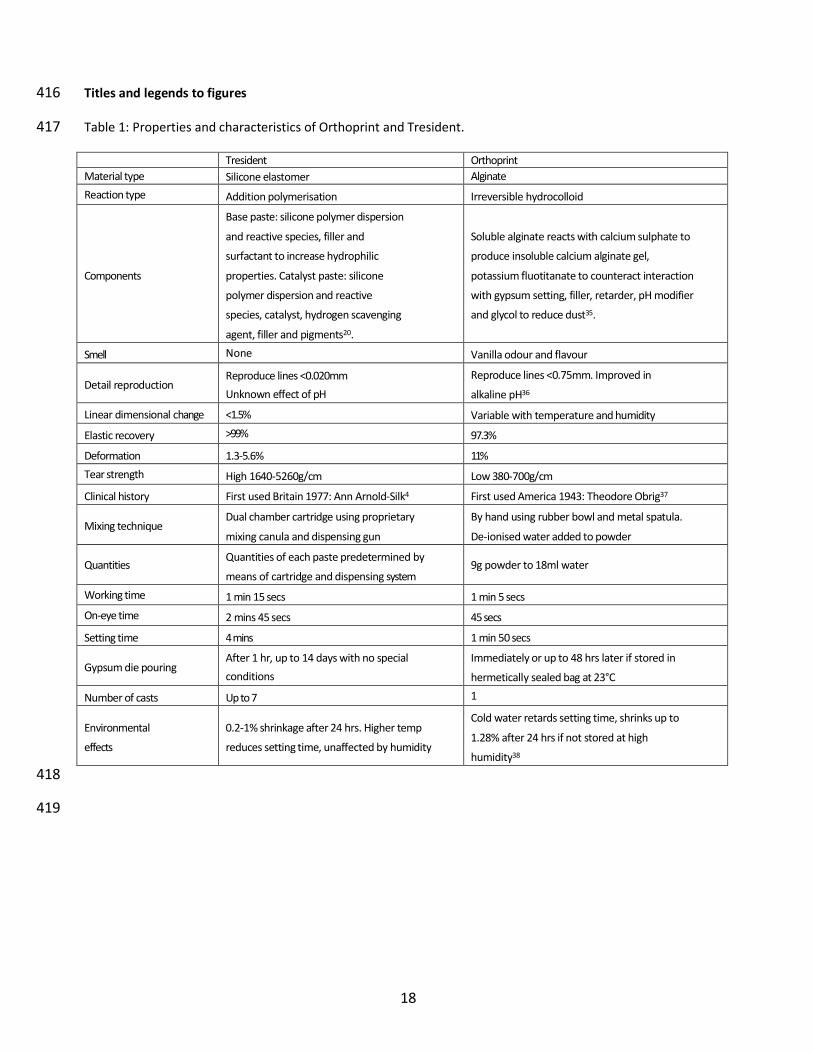
Table 1: Properties and characteristics of Orthoprint and Tresident. 417
Tresident Orthoprint Material type Silicone elastomer Alginate Reaction type Addition polymerisation Irreversible hydrocolloid
Components
Base paste: silicone polymer dispersion
and reactive species, filler and
surfactant to increase hydrophilic
properties. Catalyst paste: silicone
polymer dispersion and reactive
species, catalyst, hydrogen scavenging
agent, filler and pigments20.
Soluble alginate reacts with calcium sulphate to
produce insoluble calcium alginate gel,
potassium fluotitanate to counteract interaction
with gypsum setting, filler, retarder, pH modifier
and glycol to reduce dust35.
Smell None Vanilla odour and flavour
Detail reproduction Reproduce lines <0.020mm Unknown effect of pH
Reproduce lines <0.75mm. Improved in
alkaline pH36
Linear dimensional change <1.5% Variable with temperature and humidity
Elastic recovery >99% 97.3%
Deformation 1.3-5.6% 11% Tear strength High 1640-5260g/cm Low 380-700g/cm
Clinical history First used Britain 1977: Ann Arnold-Silk4 First used America 1943: Theodore Obrig37
Mixing technique Dual chamber cartridge using proprietary
mixing canula and dispensing gun
By hand using rubber bowl and metal spatula.
De-ionised water added to powder
Quantities Quantities of each paste predetermined by
means of cartridge and dispensing system 9g powder to 18ml water
Working time 1 min 15 secs 1 min 5 secs On-eye time 2 mins 45 secs 45 secs
Setting time 4 mins 1 min 50 secs
Gypsum die pouring After 1 hr, up to 14 days with no special conditions
Immediately or up to 48 hrs later if stored in
hermetically sealed bag at 23°C
Number of casts Up to 7 1
Environmental
effects
0.2-1% shrinkage after 24 hrs. Higher temp
reduces setting time, unaffected by humidity
Cold water retards setting time, shrinks up to
1.28% after 24 hrs if not stored at high
humidity38 418
419

19
Table 2: Statistical comparisons of clinical outcomes between Tresident and Orthoprint. 420
Clinical Outcome Mean differences in measurements pre- and post-impression procedure (mean±SD)
Statistical significance
Tresident Orthoprint Tresident pre- vs post-
Orthoprint pre- vs post-
Tresident vs Orthoprint
LogMAR acuity (Log Units) -0.01±0.13 -0.01±0.21 p=0.414 p=0.082 p=0.593
Phenol red test (mm) +5.06±6.22 +5.76±5.81 p=0.308 p<0.05 p=0.829
TBUT (secs) -0.87±2.61 -1.28±2.03 p=0.383 p=0.094 p=0.265 Bulbar Redness (CCLRU units) +0.64±0.58 +1.12±0.42 p<0.001 p<0.001 p<0.05
Limbal Redness (CCLRU units) +0.70±0.31 +1.05±0.28 p<0.005 p<0.005 p=0.072
Lid Redness (CCLRU units) +0.17±0.32 +0.07±0.35 p<0.05 p=0.157 p=0.487
Lid Roughness (CCLRU units) +0.03±0.25 +0.00±0.40 p=0.459 p=1.00 p=0.506
Type of corneal staining (CCLRU units)
+0.53±0.41 +0.13±0.34 p=0.167 p=0.341 p=0.176
Extent of corneal staining (CCLRU units)
+0.49±0.65 +2.33±0.46 p=0.209 p<0.001 p<0.005
Depth of corneal staining (CCLRU units)
+0.37±0.47 +0.31±0.40 p=0.219 p<0.05 p=0.566
421
422

20
Figure 1: 25mm diameter impression tray holding Tresident material prior to insertion. 423
424
425
426
Figure 2: Position of tray and Tresident during impression procedure. 427
428
429