october-december 2014pai-india.org/data/uploads/nijp/nijp-2014/october...ramya v., sarla k., radhika...
TRANSCRIPT
OCTOBER-DECEMBER 2014VOLUME - 3 NUMBER - 4

New Indian Journal of PediatricsNIJP: ISSN no. 2277-9507
Editorial Team
Editor- in- Chief: Dr Satish Tiwari
Editor:Dr Nimain C. Mohanty
Managing Editors: Dr Nitin N KadamDr Amar Varma Dr Jayant Vagha,
Associate Editors: Dr Utpal Kanta Singh, Dr Ashok Kr Datta, Dr Prasanth Saboth
Ethical Issues: Dr Vishesh Kumar
Legal Issues: Dr Balraj Yadav,
Executive Members: Dr Sukanto Chattarjee, Dr Arun Thakur, Dr Jyanindranath Behera, Dr Satish Agrawal, Dr Ranbir Laishram
Treasurer: Dr Vijay Kamale
International Members:Dr Pushpa Chaturvedi
(UAE)Dr Sudipta Misra (USA)Dr Salma S. Syed (USA)
NIJP
Volume 3-4OCTOBER - DECEMBER 2014 159
Contents Page No
Editorial :Medical Ethics : How it is relevant today? 163
Original Research :
Breastfeeding practices among healthcare personnel. 167Monika, Saroj K., Choudhary SR.
Diagnostic value of Gastric lavage and induced sputum in childhood Pulmonary tuberculosis. 172Rahman SR, Chatterjee K, Mandal, Chaudhuri N, Chaudhury N
Review Articles :
The unfolding of growth monitoring and growth charts 178Elizabeth KE
Latent Tuberculosis 182Sawant S. Kamale V. N. Mohanty NC
Case reports :
Klebseilla endocarditis leading to brain abscess in a child with tetrology of Fallot. 187Ojng T, Shaad A, Kamran A., Shaukat A.
Congenital giant melanocytic nevus. 190 Ramya V., Sarla K., Radhika N.
Systemic peripheral of gangrene - A rare presentation of falciparum malaria. 193 Gupta S., Reddy K. Choudhary I.
Congenital leukemia in a 23 day old neonate 196Kataria SP, Gupta M, Singh G, Sanjay K, Sen R, Yadav B*Unusual Presentation of uncommon
Alveolar Proteinosis: An Uncommon Presentation of A Not So Common Disease 199Mahfooz A, Mohanty NC, Joseph JJ, Verma S, Kulkarni P, Patel J
Research Media Watch : 204
Lohar A, Agrawal S.
Manuscript Preparation: Guidelines for Authors: 206
Bibliography 208

Published by:Pediatrics Association of India
Editorial office:Dr Satish Tiwari Dr Nimain MohantyEditor-in-chief, Editor,Professor of Pediatrics Prof. of Pediatrics,Medical College Amravati MGM Hospital, At : Yashodanagar no 2 S/4, E - Kalamboli,Amravati, 444606 Navi Mumbai - 410218Maharashtra, India Ph.: 022-2742 3405, 2742 7104Ph: 0721-2541252, 9422857204 [email protected]: [email protected]@hotmail.com
Disclaimer: Views expressed in the articles or letters are of individual writers.
Publisher or editor may not necessarily agree with the same. All efforts have been taken not to include any unethical or unlawful
advertisements in this journal. Publisher or editor may not necessarily agree with the advertisements or exaggerated claims in any particular advertisements.
Important information: All possible care has been taken to ensure accuracy of the material, but,
the editor, printer or publisher shall not be responsible for any inadvertent error(s) or consequences arising out of this publication.
All rights reserved: Whole or any part of these publications are prohibited to be reproduced or
transmitted in any form, by any means or otherwise, without prior written permission of the editor, printer or publisher.
NIJP: ISSN No. 2277-9507
NIJP
Volume 3-4 OCTOBER - DECEMBER 2014160

NIJP
Volume 3-4 161
ANNUAL NATIONAL CONFERENCE OF PEDIATRICS ASSOCIATION OF INDIA
PAICON 201525-27 September 2015
PLEASE BLOCK YOURDATESVenue
MGM Institute of Health SciencesPlot - 182, Sector - 18 Kamothe, Navi-Mumbai.
Conf. Office : MGM Hospital, 4/E - Kalamboli,Navi Mumbai - 41021891-922 4475712, 91-99 20410522, 91-9987424128
OCTOBER - DECEMBER 2014
Please see Page Nos. 210-212 for registration & other details.

Rs 20000/-
Rs 15000/-
Rs 15000/-
Rs 10000/-
Rs 5000/-
Subscription Rates of NIJP:
Annual
INR 600/- for individuals
INR 1000/- for Institutes
INR 2000/- for over-seas
INR 200/- for Single Copy
Directions for sending advertisements :
Please Send High resolution ad, approx of 2000 x 1800 or more pixels, DPI 300, in Corel Draw format or Jpg image in a CD to Editor, NIJP, MGM Hospital, Sector 4E, Kalamboli, Navi Mumbai, 410218 (Maharashtra) INDIA. Phone : 022 2742 3405, 2742 7104, Mobile 9920410522 or by email to [email protected] along with crossed Drafts / Multi-city cheques be drawn in favour of "New Indian Journal of Pediatrics"
Volume 3-4 OCTOBER - DECEMBER 2014162
NIJP

Editorial: Medical ethics: How relevant it is today?
Introduction:
The practice of medicine has been an art since ancient days. The technical and scientific discoveries made it a science and now ultimately it has become the commerce of human life. There are unprecedented explosions in science and technology influencing the concepts of medical practice (1). There has to be control over the use of technology by the profession. Legislation may not always work effectively. It can only work if the medical profession is willing to be ethical in its use and any misuse is dealt with severely by professional bodies. To maintain purity and sanctity of the art of healing, the Fathers of Medicine had laid down a code of conduct for physicians. These guidelines were framed keeping in mind that the art of healing is a mission for service to humanity. Medicine is a service that can't be governed by “Cold” science alone. It must have a human touch. All these changes in and around third millennium have forced us to think as to whether the ethical issues are relevant in present scenario.
Background:
In the ancient days the physicians were supposed to maintain highest standard of professional conduct uninfluenced by motives of profit. The doctors were considered almost as God or as an angel by society. (2) The Hippocratic Oath, which is now almost 25 centuries old, was based on these basic principles of service to humanity. Doctors need to assess the merits of various competing courses of action, and be able to justify their decisions in ethical terms. The physicians must work with nobility and dignity for the welfare of human beings. The aim was “Never do harm to anyone”.
The present scenario:
In today's world the circumstances have totally
changed. In this age of commercialization, the trust and faith among individual human beings are totally lost. We are looking at each other with disbelief and suspicion. The nobility, dignity and respect of medical profession have been blown into pieces. The scenario is continuously deteriorating. The medico-legal cases are gradually increasing. The overall impact is that the doctor-patient relationship is in doldrums.
Possible reasons for unethical pracice:
The increasing unethical practices in medical profession are multi-factorial in origin. Some of the important reasons could be:
1) Changing pattern of medical education
2) Increasing costs of hospital establishments
3) Lack of political willingness to improve health care system
4) Failure of regulatory bodies
5) Apathy amongst medical associations / organizations
6) Influence of drug industry and media
7) Decreasing morality, organ trading
8) Increasing corruption in public life/ society
Who is to blame? It seems that the whole system is responsible. There is no accountability on the part of anyone.
Ethics of Current Medical Education:
We are worried about increasing incidences of unethical acts amongst physicians. One of the reasons for it is the deteriorating system in medical education. The mushrooming of private medical colleges without due adequate control has acted as a catalyst in this process. These private medical schools are run by managing committees, which are often under the influence of political heavyweights
Volume 3-4 163
NIJP
OCTOBER - DECEMBER 2014

and personalities. The doctors who pass out after paying huge fees, will practice with the aim of recovering these expenses along with the interest.
The teaching faculties in such institutions are often part-time, fixed salaried and 'transient teachers' who are not serious in maintaining desired excellence in teaching. Many teachers in clinical specialties, who have well-established private practices to supplement their income, hardly find time to teach. The few who are enthusiastic are disheartened by uncongenial atmosphere (3). Very few medical institutions are serious in producing medical graduates with due regard to professional competency and knowledge empowerment. There may be exceptions. By and large, the students, their parents, the government, judiciary and regulatory bodies are deceived by money power in this trade. So how do we expect cost effective, ethical health services for the community by these professionals? Not a single medical graduate from these institutions is willing to join government sector or serve in rural areas.
Ethics and Pharmaceutical Industries:
Globalization of economy and world trade marketing has increased the competition amongst the pharmaceutical industries. The drug companies are increasing at an alarming rate. For past many years, activists have been expressing concern over the nexus between doctors and pharmaceutical companies. To promote and sell their products the companies are choosing various methods, which may be unethical as per the standard code of conduct. Various methods include donations, gifts, sponsoring for CME, seminars and conferences etc. About 326 doctors from across the country were under the scanner of the Medical Council of India for allegedly accepting inducements in the form cash, f l a t s , junke t s , o r ca r s f rom Smal l t ime pharmaceutical companies to promote their products by hook or crook. Gifts from modest product samples to exotic cruises turn out to be a good investment for the companies, which spend huge amount of money because they are assured of returns. The “Norm of reciprocity” i.e. the obligation to help those who have helped you has
been one of the fundamental guiding principle of human interactions (4). "Accepting kickbacks from pharmaceutical companies is a serious crime. These firms recover the money spent on doctors by hiking prices of their products. It means patients are the ultimate sufferers. Many irrational drugs or drug combinations are pumped into pharmaceutical market and doctors are influenced to prescribe such products. By the time it is realized that these products are useless, a huge sum of money is already deposited in the wallet of these companies. The MMC will direct all doctors practising allopath in the state to keep away from malpractices and expose drug marketing firms resorting to unethical means for “Business gains” It will ask the Indian Medical Association to issue a similar missive to its doctors. The health body also plans to write to the union ministries of chemical and fertilizers, health and family welfare, demanding a code of ethics for pharmaceutical companies on the lines of guidelines outlined for medical practitioners.
Role of the society:
The technical advances are so phenomenal that community or society is often left bewildered. The role of people in the community is also responsible for increasing unethical practices. The patients or relatives don't value the competence or ethics of doctors. They are more interested in buying health services by paying money. A large building well furnished with many physical comforts,TV, Fridge, water coolers etc., have become trademarks for a good hospital. Many patients, relatives feel that the health care or doctor's service is a commodity, which can be purchased with money. If they are not satisfied with one doctor, they go on doing “Doctor shopping”. In the era of advertisement and globalization most of the commodities are of “Use and throw” variety. Can we dispose off a doctor and select another one if we are not satisfied with the services of the earlier one? What about the damage already caused to the patient? Can doctors and health care system become commodities, which can be hired or purchased with money? Who will tell the society about its responsibilities (5)?
Volume 3-4 OCTOBER - DECEMBER 2014164
NIJP

Role of Regulatory bodies:
Regulatory bodies like medical and dental councils have been formed with the aim of regulating the practice of medicine in the respective systems. The Medical Council of India (MCI), formed under the Indian Medical Council Act 1956, slowly and surely proved to be a toothless tiger. There are many organizations/ institutions taking advantages of corruptions in such councils. Many are conferring unrecognized degrees, diplomas, certificates etc. and thus cheating the highest regulatory body of the country. What about the resulting credibility?
Role of Policy Planners:
The overall situation is disheartening as far as professional ethics are concerned. Policy makers have badly failed in controlling the corruption in medical education, hospital administration and growing malpractice in private establishments. The government has so far been not able to control the admission policy and the fees structure in the private colleges. The judiciary is trying to control the situation but there are no persons who can implement their decisions. The supreme powerful lobby has their way to scuttle any attempt to correct this system. Most judicial decisions are executed only in words but not in spirit. Individualized multiple entrance tests by different institutions with different rules create confusions in minds of students and parents alike. Now is the beginning of globalization and free economy. On the contrary, we are producing “Indebted doctors” and “Indebted medical students” too.
Role of Legislation:
The aim of the professional ethics was to frame a code of conduct for their members, mostly through self-regulation. But as doctors started deviating from these self-regulatory measures, the government was forced to enact legislation. Can ethics be implemented through state laws? What about it's misuse? What about practical difficulties? Who is going to enforce these laws? All these questions need definitive answers, which unfortunately are missing. The Acts have been blatantly violated by large section of society. It
proves the futi l i ty of such laws and the ineffectiveness of the legal processes. One of the codes of conduct for doctors is not to use touts or agents for procuring patients. In today's commercialized practice, we come across not only the agents of doctors but also the increasing trends of “Cut practice”. The decline started when specialist began treating patient according to the dictates of the referring general practitioners. There are many general practitioners who survive only on the “Referral fee” and accumulate a huge amount of money by way of commission. The very fact that the doctors who don't give or take 'Cuts' are a minority nowadays speaks badly enough of our noble profession.In addition ,several unscrupulous elements are thriving solely on their “Ghost hospitals and patients “ with the sole aim of bleeding the insurance companies in collusion with policy holders,TPAs and insurance agents.
Role of Professional Associations:
The professional associations must reaffirm their beliefs in medical ethics to get rid of these ills. However, ethics cannot be practiced by few individuals .It has to become both a norm and a standard. There is need to launch a major drive to educate doctor fraternity to begin with. The scientific bodies should evolve means to reduce their dependence on drug industry for holding their conferences and seminars. Doctors should begin to think in terms of paying for their own learning.(6) The ethics may be included in medical educational curriculum. There is need to standardize treatment protocols and a system of regular monitoring of medical practice through self-regulation. It is time the profession learns to uphold its honor. Someone has to become the “Whistle blower” and professional bodies as as “watch dog “whenever need arises.
Effects of Unethical Practice:
Influence of the pharmaceutical industry on the medical profession can't be denied. It often leads to the unscientific use of expensive and hazardous medicines. As a result, the profession is discredited. While technologies may have enhanced doctors' medical skills and capabilities, they lose out on
Volume 3-4 165
NIJP
OCTOBER - DECEMBER 2014

clinical skills by becoming dependant on technology. Technology has become the means to make quick buck , through its misuse. Sometimes unethical practice also gets involved in criminal activities as has happened in organ transplantations, NRHM scam and so on.(7)
Conclusion:
The ethical practice is not an obsessive, puritanical exercise. It becomes a meaningful, fulfilling challenge.(8) Over the years it has become difficult for the doctors to follow the ethical norms. The medical profession is fast loosing its credibility for reasons cited above. The professionals have to do a lot of soul searching and introspection. They should not behave in a manner that gives an impression that they are in hands and glove with the industry. The concept of global economy has resulted in “Five star health care” facilities, which are out of reach of a common man. Such hospitals have everything except the vital factor- the human touch and the ethics. We should remember that the principle of “Survival of fittest” is true in most of the situations. Is it possible for a sincere, dedicated and ethical doctor to survive in today's world? The apathy and anarchy in the political system is very obvious as the politicians are interested in protecting their own position at any cost. Today, in most of the countries, ethics and economics are seen as adversaries. The common belief is that if you are ethical you can't make money- or if you want to make money then you have to forsake ethics. As is always the case, the truth lies somewhere in the middle. Not only charity, but also cleaning and mopping up should begin at home. Our future doctors must be sensitized to human rights, ethical considerations, communication skills while dealing with patients and gender issues. The money matters
have changed the entire scenario. The question is; are we going in the right direction or on the right path? Finally, honesty to one-self is important. The question is, are we going to improve it through self-regulation? Should we wait for the God's incarnation to check our religious, moral and ethical downfall?
Dr. Satish Tewari, Editior-inchief
References:
1) Tiwari SK, Baldwa M; Medical negligence. In: Gupte S editor Recent Advances in Pediatrics (vol.14) New Delhi Jaypee brothers 2004, 311 -329.
2) Tiwari SK, Legal aspects in medical practice. Indian Pediatr 2000; 37: 961-966
3) Nair VM, Medical college teachers and some ethical issues in Kerala. Indian Journal of Medical ethics 2003; XI: 116-117
4) Verma S K, Physician-Pharmaceutical industry interaction: Changing dimensions and ethics. Indian Pediatr 2004; 41: 29-36
5) Taskar J, Doctors under siege. Indian Journal of Medical ethics 2003; XI: 109-110
6) Agrawal S, Medical profession and the pharmaceuticals: Indian Pediatr 1998; 35: 641-645
7) Duggal R, The political economy of medical ethics. Ind Jl of Med Ethics 2004; XII: 81-82
8) Shetty H, A challenge for medical profession. Ind Jl of Medical Ethics 2003; XI: 111-112.
Volume 3-4 OCTOBER - DECEMBER 2014166
NIJP

Original Research :
Breastfeeding practices among health care professionalsSaroj K, Chaudhary SR
Abstact:
Breastfeeding is an unequalled way of providing ideal food for the adequate growth and development of infants. The objective of this study was to assess the breastfeeding practices followed by the health workers. Material and methods: a cross- sectional study was done on the female health workers and ANM's of Patna Medical College and Hospital. The study is based on a questionnaire related to breastfeeding practices, complimentary feeding and weight gain at one year of age. The significance in the differences was evaluated by using the Chi square test. Results: The prevalence of early initiation of breastfeeding was 16.6% and prevalence of exclusive breastfeeding was 66.4%. While the prevalence of exclusive breastfeeding for up to six months was only 18.5%. The association between exclusive breastfeeding for up to six months and weight gain at one year of age is considered to be very statistically significant. Conclusion: It came out by the study that majority of the health workers were not practicing the appropriate breastfeeding practices. So, we need to do awareness programme repeatedly and train the health care workers and other medical professionals regarding importance of counselling for breast feeding starting from pregnancy and optimum breastfeeding practices.
Keywords:
Exclusive breastfeeding, expressed breast milk, complimentary feeding
Source of support : Nil
Conflict of Interest : None
Introduction:
As a global public health recommendation, infants should be exclusively breastfed for the first six months of life to achieve optimal growth, development and health.(1) It fulfils all the
nutritional requirements of the baby till six months of age. Along with this, it continues to provide up to half of the nutritional needs of the baby from six months to one year and beyond that for up to two years, it fulfils about one-third of the needs. Except this, it also helps in promoting emotional bonding between mother and baby and also stimulates all the five special senses of the body i.e. touch, sight, smell, hearing and taste.
The importance of breastfeeding increases even more in developing countries due to lack of resources and hygienic practices.(2) Breast milk is the best food as it has right proportion of nutrients, at right temperature and is readily available.
Breastfeeding is very common practice in almost all parts of India. But, the knowledge regarding b r e a s t f e e d i n g p r a c t i c e s a n d c o m p l i -mentary feeding are still lacking. The problem increases even more for working ladies if they don't know the concept of expressed breast milk. Most people come to know regarding these facts through their family and health workers. So, we tried to assess the breastfeeding practices among female health workers.
Aims and Objectives
1. To assess the knowledge of female health workers and ANM's regarding the importance of exclusive breastfeeding, time of initiation of weaning.
2. To study practices of feeding followed by them.
3. Assessment of weight at one year of age.
The results would emphasize upon the need to train health personnel about appropriate practices regarding breastfeeding.
Material and Methods:
This cross-sectional study was conducted among
Department of Paediatrics, Patna Medical college, Patna, Bihar. [email protected]
Volume 3-4 167
NIJP
OCTOBER - DECEMBER 2014
Monika, Saroj K., Choudhary SR. Monika, Saroj K., Choudhary SR.
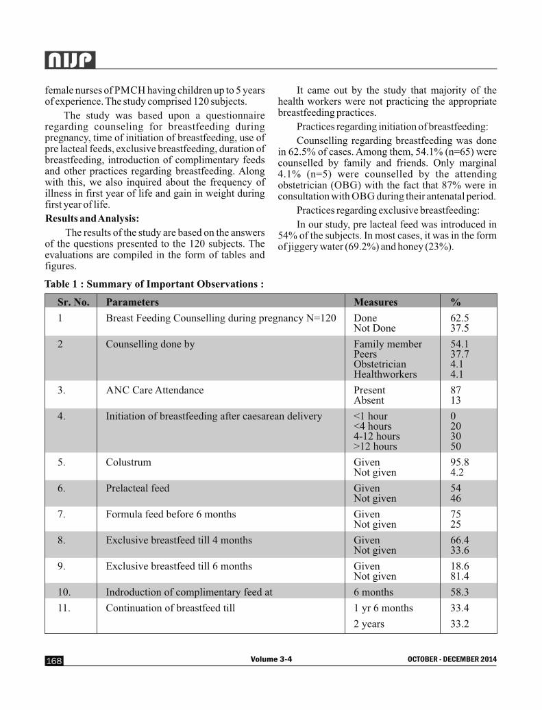
female nurses of PMCH having children up to 5 years of experience. The study comprised 120 subjects.
The study was based upon a questionnaire regarding counseling for breastfeeding during pregnancy, time of initiation of breastfeeding, use of pre lacteal feeds, exclusive breastfeeding, duration of breastfeeding, introduction of complimentary feeds and other practices regarding breastfeeding. Along with this, we also inquired about the frequency of illness in first year of life and gain in weight during first year of life.
Results and Analysis:
The results of the study are based on the answers of the questions presented to the 120 subjects. The evaluations are compiled in the form of tables and figures.
It came out by the study that majority of the health workers were not practicing the appropriate breastfeeding practices.
Practices regarding initiation of breastfeeding:
Counselling regarding breastfeeding was done in 62.5% of cases. Among them, 54.1% (n=65) were counselled by family and friends. Only marginal 4.1% (n=5) were counselled by the attending obstetrician (OBG) with the fact that 87% were in consultation with OBG during their antenatal period.
Practices regarding exclusive breastfeeding:
In our study, pre lacteal feed was introduced in 54% of the subjects. In most cases, it was in the form of jiggery water (69.2%) and honey (23%).
Volume 3-4 OCTOBER - DECEMBER 2014168
NIJP
Table 1 : Summary of Important Observations :
Sr. No. Parameters Measures %
1 Breast Feeding Counselling during pregnancy N=120 Done 62.5 Not Done 37.5
2 Counselling done by Family member 54.1 Peers 37.7 Obstetrician 4.1 Healthworkers 4.1
3. ANC Care Attendance Present 87 Absent 13
4. Initiation of breastfeeding after caesarean delivery <1 hour 0 <4 hours 20 4-12 hours 30 >12 hours 50
5. Colustrum Given 95.8 Not given 4.2
6. Prelacteal feed Given 54 Not given 46
7. Formula feed before 6 months Given 75 Not given 25
8. Exclusive breastfeed till 4 months Given 66.4 Not given 33.6
9. Exclusive breastfeed till 6 months Given 18.6 Not given 81.4
10. Indroduction of complimentary feed at 6 months 58.3
11. Continuation of breastfeed till 1 yr 6 months 33.4
2 years 33.2
Volume 3-4 169
NIJP
OCTOBER - DECEMBER 2014
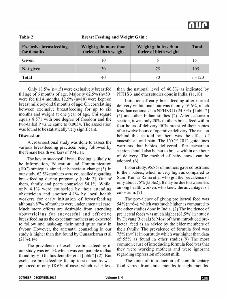
Only 18.5% (n=15) were exclusively breastfed till age of 6 months of age. Majority 62.5% (n=50) were fed till 4 months. 12.5% (n=10) were kept on breast milk beyond 6 months of age. On correlatiing between exclusive breastfeeding for up to six months and weight at one year of age, Chi square equals 8.571 with one degree of freedom and the two-tailed P value came to 0.0034. The association was found to be statistically very significant.
Discussion:
A cross sectional study was done to assess the various breastfeeding practices being followed by the female health workers of PMCH.
The key to successful breastfeeding is likely to be Information, Education and Communication (IEC) strategies aimed at behaviour change.(3) In our study, 62.5% mothers were counselled regarding breastfeeding during pregnancy [table 2]. Out of them, family and peers counseled 54.1%. While, only 4.1% were counseled by their attending obstetrician and another 4.1% by local health workers for early initiation of breastfeeding although 87% of mothers were under antenatal care. Much more efforts are desirable from attending obstetricians for successful and effective breastfeeding as the expectant mothers are expected to follow and make-up their mind quite early in favour. However, the antenatal counseling in our study is higher than that found by Gunasekaran et al (21%). (4)
The prevalence of exclusive breastfeeding in our study was 66.4% which was comparable to that found by H. Gladius Jennifer et al [table2] (2). But exclusive breastfeeding for up to six months was practiced in only 18.6% of cases which is far less
Breast Feeding and Weight Gain :
Exclusive breastfeeding Weight gain more than Weight gain less than T otal for 6 moths thrice of birth weight thrice of birth weight
Given 10 5 1 5
Not given 30 75 1 05
Total 40 80 n =120
Table 2
than the national level of 46.3% as indicated by NFHS 3 and other studies done in India. (11,10)
Initiation of early breastfeeding after normal delivery within one hour was in only 16.6%, much less than national data NFHS311 (24.3%) [Table 2] (5) and other Indian studies (2). After caesarean section, it was only 20% mothers breastfeed within four hours of delivery. 50% breastfed their babies after twelve hours of operative delivery. The reason behind this as told by them was the effect of anaesthesia and pain. The IYCF 2012 guidelines warrants that babies delivered after caesarean section should also be put to breast within one hour of delivery. The method of baby crawl can be adopted. (6)
In our study, 95.8% of mothers gave colostrums to their babies, which is very high as compared to Sunil Kumar Raina et al who got the prevalence of only about 75% [table2]. It may be due to awareness among health workers who knew the advantages of colostrum. (7)
The prevalence of giving pre lacteal feed was 54% (n=64), which was much higher as compared to the other studies done in India. (2) The incidence of pre lacteal feeds was much higher (61.9%) in a study by Devang R et al.(8) Most of them introduced pre-lacteal feed as an advice by the elder members of their family. The prevalence of formula feed was 75% (n=91) in our study which was higher than data of 55% as found in other studies.(9) The most common cause of introducing formula feed was that they were working mothers and were ignorant regarding expression of breast milk.
The time of introduction of complimentary food varied from three months to eight months.

Volume 3-4 OCTOBER - DECEMBER 2014170
NIJP58.3% (n=70) mothers introduced complimentary feed at the age of six months which is well comparable to 56.7% as given by NFHS3 and is much higher as compared to other studies by Radhakrishnan and Balamuruga [table2].(10) Most of the mothers 33 .4% (n=41) cont inued breastfeeding till only one and half years of age. While, only 33.2% (n=39) continued breastfeeding till two years of age which is much less as compared to 87.2% as reported by Banapurmath et al. (12)
The association between exclusive breast feeding for up to six months and weight gain at one year of age is considered to be very statistically significant as the P value equals 0.0034 [table1]. This correlates well with study results of Rosa F. S. V. Marques et al who also found that Children who were exclusively fed at the breast for 6 months exhibited satisfactory weight gain when compared with existing standards. (13)
Conclusion:
Breastfeeding is the gold standard feeding option for the babies. Breastfeeding must be initiated as early as possible after birth for all normal newborns (including those born by caesarean section) avoiding delay beyond an hour. Exclusive breastfeeding should be practiced from birth till six months of age. After completion of six months of age, with introduction of optimal complementary feeding, breastfeeding should be continued for a minimum for 2 years. Mothers who work outside should be encouraged to continue exclusive breastfeeding for 6 months by expressing milk for feeding the baby while they are out at work. These are the optimum breastfeeding practices and lead to the adequate weight gain, as per the existing standard, in the child at one year of age. But, most of the health workers are not aware of the facts and are not following these practices themselves. Since, most of the peoples come to know about these facts through health workers and other medical personnel. It is of utmost importance that initiative should be taken by the health department to vigorously train health care workers and other medical professionals regarding importance of counseling, particularly one to one counseling of
pregnant ladies, start awareness programme on importance of breastfeeding through mass media. Reward should be given at the end of 6 months of exclusive breastfeeding to mothers. Besids, it should be included in the school curriculum so that girls (Future mothers) may know regarding this in orde to bring down the prevalence of malnutrition and decrease loss of lives, without putting undue pressure on the financial status of the family and of the country.
References:
1. UNICEF and WHO Global strategy on infant and young child feeding WHA55 A55/15, paragraph 10)
2. H. Gladius jennifer, k. Muthukumar, .a cross-sectional descriptive study was to estimate the prevalence of the early initiation of and exclusive breast feeding in the rural health training centre of a medical college in Tamilnadu, south India. Journal of clinical and diagnostic research [serial online]2012 11[cited:2014 sep 8]9 1514 1517
3. IYCF Policy and Programme : Information suppor t [h t tp : / /www.bpni .o rg / I Y C F -information-support.html]
4. Antenatal counseling on breastfeeding – is it adequa te? A desc r ip t ive s tudy f rom Pondicherry, India Gunasekaran Dhandapany, A d h i s i v a B e t h o u , A r u l k u m a r a n Arunagirinathan andShanthi Ananthakrishnan. International Breastfeeding Journal 2008, 3:5 doi:10.1186/1746-4358-3-5
5. Issued by Breastfeeding Promotion Network of India on World Breastfeeding Week 2012. www.bpni.org
6. Operational guide for promoting infant and young child feeding practices through the health system. February 2012.
7. Differentials in colostrum feeding among lactating women of block RS Pura of J and K: A lesson for nursing practice Sunil Kumar Raina, Vijay Mengi, and Gurdeep Singh Iran J Nurs Midwifery Res. 2012 Jul-Aug; 17(5): 386–389.

Volume 3-4 171
NIJP
OCTOBER - DECEMBER 2014
8. A study of breast feeding practices among infants living in slums of Bhavnagar city, Gujarat, India Devang Raval, D. V. Jankar, M. P. Singh. H e a l t h l i n e ISSN 2229-337X Volume 2 Issue 2 July-December 2011: 78-83
9. Breastfeeding Practices of Urban and Rural Mothers A oommen, m vatsa, vk paul and r aggarwal. Indian Pediatrics;Oct2009, Vol. 46 Issue 10, p891
10. Radhakrishnan S, Balamuruga S S. Prevalence of exclusive breastfeeding practices among rural women in Tamil Nadu. Int J Health Allied Sci 2012;1:64-7
11. Ministry of Health and Family Welfare. National Family Health Survey (NFHS-3) National fact sheet India. Available at: http:/ /www.nfhsindia.org/ pdf/ I N.pdf. Accessed March 12, 2008
12. Breastfeeding practices in villages of centralkarnataka c.r.banapurmath, M.C. Nagaraj, Shobha Banapurmath and Nirmala Kesaree. Indian Pediatrics - June 1996, Vol. 33, Number 6: 477-480
13. Growth of exclusively breastfed infants in the first 6 months of life Rosa F. S. V. Marques1, Fábio A. Lopez2, Josefina A. P. Braga3 Jornal de Pediatria - Vol. 80, Nº2, 2004: 99-105
With Best Compliments from -

Original Research :
Diagnostic value of gastric lavage and induced sputum in childhood pulmonary tuberculosisRahman SR*, Chatterjee K*, Mandal P*, Chaudhuri N* Chaudhury N**
Department of Pediatrics, IIMSAR & Dr. B. C. Roy Hospital, Haldia, ** Deptt of Microbiology, Dr. Dy. Patil Medical College, Pune.
Abstract:
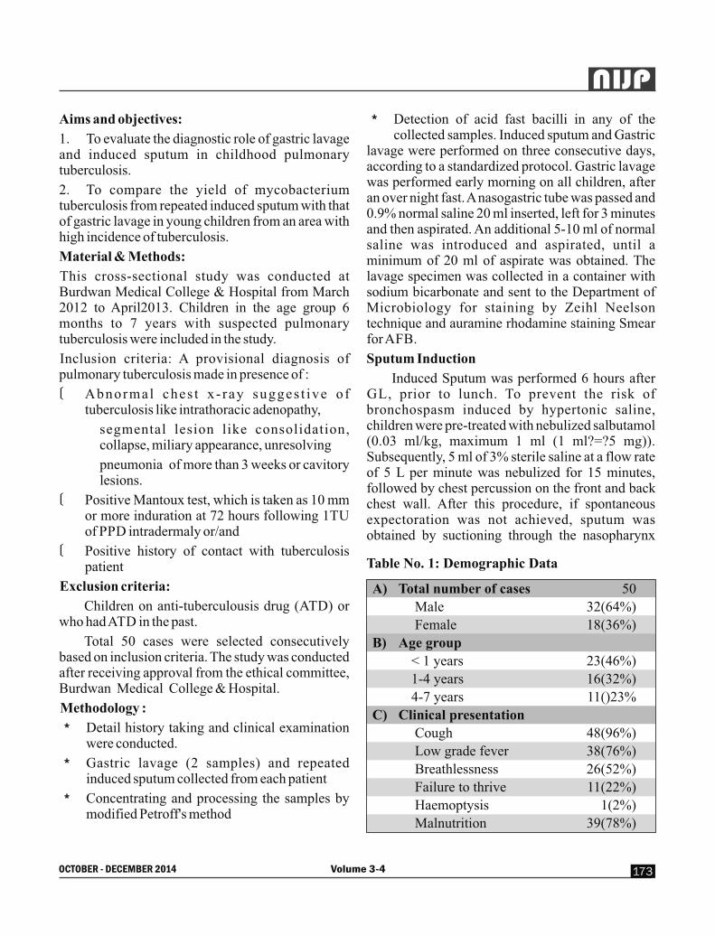
50 children up-to 7 years of age, who were suspicious of having tuberculosis, were subjected to collection of gastric aspiration following over-night fast as well as induced sputum. 96% had cough, 76% low-grade fever, 52% breathlessness, 22% failure to thriv and 2% haemoptysis. Mantoux test positivity was found to be in 25 cases (50%). Among the study population, 2 children were found to be suffering from infection with HIV. 33 cases (66%)ngave positive contact history. The major radiological abnormalities were observed on chest x ray were consolidation in 20 cases (40%), Bronchopne-umonia in 13 cases (26%), Consolidation with collapse in 7cases (14%), Cavitory lesion in 3 cases(6%), Intrathoracic lymphadenopathy in 2cases (4%), miscellaneous findings included 1 case of milary TB and 3 cases of pleural effusion. While 8 cases (16%) of GA showed AFB positivity, 6 (12%) were positive in induced sputum. On statistical analysis it was found that gastric lavage had no added advantage over induced sputum method for AFB detection. Rather both were complementary to each other.
Key words:
Diagnosis of TB, gastric aspirate, induced sputum, Mantoux test
Source of support : Nil
Conflict of Interest : None
Introduction:
Tuberculosis is a chronic bacterial infection caused by mycobacterium tuberculosis that is characterized by the formation of granulomas in infected tissues by cell mediated hypersensitivity. Childhood pulmonary tuberculosis is very much
prevalent with almost 1.3 million cases and 450,000 deaths occurring in children each year worldwide1. In developing countries the annual risk of tuberculosis infection in children is 2-5%, nearly 8-20% of deaths caused by tuberculosis in children.2 The diagnosis of childhood tuberculosis is complicated by the absence of a practical gold standard test due to the difficulty in collecting bacteriological specimens and reportedly low bacteriological yield. Most children can be diagnosed using a combination of clinical and epidemiological features, tuberculin skin testing and chest radiography, as represented in different scoring systems. However, bacteriological confirmation may have particular value in highly endemic zone, where epidemiological indicators such as known exposure to proven infection with mycobacterium tuberculosis contribute little diagnostic value3. However, accurate microbiolo-gical diagnosis has become increasingly important for timely use of effective treatment. Mycobacterial culture confirms the diagnosis of TB and provides drug susceptibility data but is not available in most areas with a high TB prevalence. Moreover, culture has poor sensitivity in children who usually have paucibacillary disease4. For microbiological confirmat ion of diagnosis of pulmonary tuberculosis in young children, sequential gastric lavages are recommended; though the technique of sputum induction with hypertonic saline (3% normal saline) was introduced more than 40 years ago, sputum induction has not been regarded as feasible or useful. Gastric lavage collects the respiratory secretions which are swallowed at night and induced sputum / mucus from lower respiratory tract.
Volume 3-4 OCTOBER - DECEMBER 2014172
NIJP
Aims and objectives:
1. To evaluate the diagnostic role of gastric lavage and induced sputum in childhood pulmonary tuberculosis.
2. To compare the yield of mycobacterium tuberculosis from repeated induced sputum with that of gastric lavage in young children from an area with high incidence of tuberculosis.
Material & Methods:
This cross-sectional study was conducted at Burdwan Medical College & Hospital from March 2012 to April2013. Children in the age group 6 months to 7 years with suspected pulmonary tuberculosis were included in the study.
Inclusion criteria: A provisional diagnosis of pulmonary tuberculosis made in presence of :
v Abnormal ches t x - ray sugges t ive o f tuberculosis like intrathoracic adenopathy,
segmental lesion like consolidation, collapse, miliary appearance, unresolving
pneumonia of more than 3 weeks or cavitory lesions.
v Positive Mantoux test, which is taken as 10 mm or more induration at 72 hours following 1TU of PPD intradermaly or/and
v Positive history of contact with tuberculosis patient
Exclusion criteria:
Children on anti-tuberculousis drug (ATD) or who had ATD in the past.
Total 50 cases were selected consecutively based on inclusion criteria. The study was conducted after receiving approval from the ethical committee, Burdwan Medical College & Hospital.
Methodology :
o Detail history taking and clinical examination were conducted.
o Gastric lavage (2 samples) and repeated induced sputum collected from each patient
o Concentrating and processing the samples by modified Petroff's method
o Detection of acid fast bacilli in any of the collected samples. Induced sputum and Gastric lavage were performed on three consecutive days, according to a standardized protocol. Gastric lavage was performed early morning on all children, after an over night fast. A nasogastric tube was passed and 0.9% normal saline 20 ml inserted, left for 3 minutes and then aspirated. An additional 5-10 ml of normal saline was introduced and aspirated, until a minimum of 20 ml of aspirate was obtained. The lavage specimen was collected in a container with sodium bicarbonate and sent to the Department of Microbiology for staining by Zeihl Neelson technique and auramine rhodamine staining Smear for AFB.
Sputum Induction
Induced Sputum was performed 6 hours after GL, prior to lunch. To prevent the risk of bronchospasm induced by hypertonic saline, children were pre-treated with nebulized salbutamol (0.03 ml/kg, maximum 1 ml (1 ml?=?5 mg)). Subsequently, 5 ml of 3% sterile saline at a flow rate of 5 L per minute was nebulized for 15 minutes, followed by chest percussion on the front and back chest wall. After this procedure, if spontaneous expectoration was not achieved, sputum was obtained by suctioning through the nasopharynx
A) Total number of cases 50
Male 32(64%)
Female 18(36%)
B) Age group
< 1 years 23(46%)
1-4 years 16(32%)
4-7 years 11()23%
C) Clinical presentation
Cough 48(96%)
Low grade fever 38(76%)
Breathlessness 26(52%)
Failure to thrive 11(22%)
Haemoptysis 1(2%)
Malnutrition 39(78%)
Table No. 1: Demographic Data
Volume 3-4 173
NIJP
OCTOBER - DECEMBER 2014

with a sterile mucus extractor of catheter size 6 or 7. Oxygen saturation was monitored throughout. After the procedure, we performed a physical examination to rule out the presence of bronchospasm.
Results and Analysis:
The study was carried out on 50 children in the department of pediatrics who presented with features of tuberculosis.
The age group of the patients ranged from being a post neonatal infant to upto 7 years. The mean age of the children was 3 years and 5 months. 23 children were below the age of 1 year, 16 children between age group (1-4) years and 11 between age group (4-
7) years.Of the 50 children, 32 were males and 18 were females, so a ratio of 1.78 males per females.
The major clinical presentations among the 50 children were cough (48cases), low-grade fever in 38 cases, breathlessness in 26 cases, failure to thrive in 11 children. Haemoptysis was the presenting complaint in 1 case only. So it is clear from above chart that cough either productive or non productive as the most common presentation. Among the study population 2 children were found to be suffering from infection with HIV. It was also observed that 2 children had past history of measles. One of the most important observation was that 78% children (39 cases) were found to be malnourished.
Table No. 2: Characteristics of Children with Suspected Pulmonary Tuberculosis.
Pos. Neg. Pos. Neg. Cons. Cons. Cavity Broncho Intra Mil. Pl. Pos. Pos. With pneumonia Thoracic TB Eff. collapse adenopathy
<1 9 14 18 5 11 2 - 8 - 1 1 5 1
1-4 11 5 9 7 7 3 1 3 1 - 1 2 2
4-7 5 6 6 5 2 2 2 2 1 - 1 1 3
Age Mantoux Contact Radiology(CXR) GL IS(yrs) test History
The mantoux test positivity was found to be in 25 cases (50%). Among them, 9 children were below the age of 1 year, 11 children between 1 to 4 years and the remaining 5 were above 4 years but below 7 years of age.
History of contact is very important pertaining to tuberculosis. In our study we could find 33cases where positive contact history is present, which means that the children were exposed to open cases of tuberculosis in their home or neighborhood.
The major radiological features observed on chest x ray were consolidation in 20 cases (40%), Bronchopneumonia in 13 cases (26%) ,Consolidation with collapse in 7cases (14%), Cavitory lesion in 3 cases(6%), Intrathoracic lymphadenopathy in 2cases (4%),miscellaneous findings included 1 case of milary TB and 3 cases of pleural effusion. There was only one case where the x ray showed no abnormality. With the suspicion of tuberculosis based on mantoux test, positive contact history and characteristics chest x ray findings gastric lavage and induced sputum was collected as sample for microbiological diagnosis of mycobacterium tuberculosis.
Volume 3-4 OCTOBER - DECEMBER 2014174
NIJP

Of the 50 children of our study 8 cases (16%)were found to be positive for the bacteria in gastric lavage although 6 cases(12%) were found to be positive in induced sputum. Most of the cases which showed positivity on gastric lavage were below the age group of 1 year as collection of induced sputum was not easy in those cases. Based on the above, statistical analysis was done to compare whether gastric lavage and induced sputum groups as to which method would be regarded as a better for bacteriological diagnosis of mycobact-erium tuberculosis. On Chi square test (x2) test, the value came out to be 0.39 and was found to be less than the value for null hypothesis. Hence the null hypothesis could not be rejected (p=0.99). Hence gastric lavage was not proved to be better than induced sputum for AFB detection. Rather both are complementary to each other.
Discussion:
A total of 50 children were included in the study. There were 32(64%) boys and 18 (36%) girls. The observed aged distribution was 78% <4years and 22% between 4-7 years. B J Marais et al 5 in his study showed age distribution of 74.9% < 5years and 25 % between 5-12 years of age.
Positive history of contact in our study was identified in 66% and a positive mantoux was observed in 50% of patients. Mantoux reactors were
equally distributed between smear positive and smear negative patients. This is comparable to the study done in Peru by Guillermo E et al6 . 51% of patients had a positive mantoux test. Meenu singh7 et al have showed positive mantoux reaction in 40 (68.9%) of patients and a history of contact was found in32 (55%) of patients.
Major clinical presentation were cough in 48 cases (96%), low grade fever in 38 cases (76%), breathlessness in 26 cases (52%), failure to thrive in 11 children(22%). Hemoptysis was the presenting complaint in 1 case (2%). 39 children(78%) were found to be malnourished in our study. In a similar study by Meenu singh7 et al had cough 96% as a p r e d o m i n a n t s y m p t o m a n d 7 6 % w e r e malnourished.
Major radiological features observed on chest x ray were consolidation in 20 cases (40%), B r o n c h o p n e u m o n i a i n 1 3 c a s e s ( 2 6 % ) ,Consolidation with collapse in 7cases (14%), Cavitory lesion in 3 cases(6%), Intrathoracic lymphadenopathy in 3 cases (4%),miscellaneous findings included 1 case of milary TB and 3 cases of pleural effusion. There was only one case where the x ray showed no abnormality. This is comparable to the study done by Meenu Singh et al7 where 32 (55%) patients had segmental lesions in the form of consolidation or collapse.
Of the 50 children of our study 8 cases (16%)were found to be positive for the bacteria in gastric lavage although 6 cases (12%) were found to be positive in induced sputum. Isolation rates for M tuberculosis from GL ranged from 28% to 40% in children with suspected PTB, although rates can rise to 75% in other series 2,8,9,10. In our study, the diagnostic yield from GL was 16% and induced sputum was12%.this is mostly because we only used smear microscopy of both samples. Culture and other methods of confirmation could not be done in our set up. The amount of time that elapses between sample collection and the start of a microbiological study is a determining factor for obtaining positive results.
In a similar study by Marta Ruiz Jiménez et al11, of the total induced sputum samples (51) from
Frequency Distribution Of StudyPopulation Showing M.TB
Positivity In Different Specimens9
8
7
6
5
4
3
2
1
0Gastric lavage Induced sputum
Frequency Distributionof study Populationshowing M.TB Positivity in different specimens
Volume 3-4 175
NIJP
Figure 1
OCTOBER - DECEMBER 2014

17 patients diagnosed with TB, the culture was positive in 13 (25.5%) and single smear in 2 (3.9%). Of the 51 samples from gastric aspirate, the culture was positive in 17 (33.3%) and single smear in 3 (5.9%).The diagnostic yield from IS was 12%. In previous studies, where IS was used in the diagnosis of TB in children, the results were variable.
Shata et al. published a prospective study on 30 children between 3 and 15 years of age, in which the microbiological isolation from IS (staining or culture) was 28%. However, GL was not measured 12. A study from a national tuberculosis centre in Yemen by Al aghbari N et al13. compared induced sputum to gastric aspirate.the study reported that 3 sets of induced sputum 13/82(16%) had a higher isolated rate than 3 sets of gastric aspirates, 19/203(9%).
Zar et al., in two different studies, recorded 10% and 22% respectively 14,15. The first prospective study included 149 children in South Africa, with ages ranging from 3–20 months, hospitalized for pneumonia with a high risk of PTB 15. The second was a prospective study on 250 children admitted for suspected PTB. All patients underwent three ISs and three GLs (on consecutive days) 14. In this study one induced sputum was equivalent to 3 gastric lavage samples.
Iriso et al. carried out a study on a total of 126 children aged 2–60 months, recruited as probable TB cases. The diagnostic yield from IS was 30% 12. In the study carried out by Hatherill et al16, the diagnostic yield from IS was (5.8%) and GL (6.8%) and the researcher found no statistical difference between the yield of a single sample of induced sputum versus single sample of gastric aspirate.
These differences may be due to the fact that not all studies performed three measurements of IS and to the characteristics of the populations included in the studies, with different degrees of risk of tuberculosis.
In our study, the higher isolation rate achieved by GL (16%) than IS (12%) may be due to the fact that 3 samples each were obtained. The majority of GL positive cases were infants, in which age group
results of IS positivity is less due to difficult sputum induction.
A study conducted at a Madrid hospital was recently carried out to compare IS and GL in the diagnosis of PTB in children. Both techniques were performed on 26% patients diagnosed with PTB. Microbiological diagnosis was performed by GL in 8 patients (30.8%) and IS in 2 (7.7%). Of the total of 77 GL samples, the culture was positive in 19 (24.77%), whereas from the 75 IS samples, the culture was positive only in 3 (4%), with a statistical significance
(p = 0.03, odds ratio 1.33 (95% CI 0.89-1.98). The diagnostic GL yield GL exceeded that of IS by more than 20%. This difference from the literature may be due to the variability of the sampling process and the greater difficulty in obtaining a valid IS 17. The rate of smear-positive cases was low in both GL and IS, being lower in IS. This supports our study in which we also found better yield in GL than from IS.
Limitations of study: The small sample size does not allow for conclusive results. The presence of polymorphonuclear cells in the specimen is indicative of sputum, in contrast to epithelial cells, which originate in saliva. The sputum was not examined to determine whether polymorphonuclear or epithelial cells predominated. The validity of our results may not be extrapolated to other settings. The prevalence of TB and HIV-infection may be major determinants of diagnostic yield.
References :
1. Flor M.Mumoz, Jeffrey R Starke, Nelson Textbook of Pediatrics,19th eds; 2011: 996-1010
2. Marais BJ. Hesseling Gie AC, Schaaf KP, HS Enarson DA Beyers, Bacteriologic confirm-ation may be achieved in majority of children with intrathorasic tuberculosis in highly endemic settings,Clin Infect Dis2006(in press)
3. Kabra SK, Lodha, SethV. Some current concepts on childhood tuberculosis. Indian J Med Res2004;120[4]387-397
4. Star J R, Correa A G. Management of
Volume 3-4 OCTOBER - DECEMBER 2014176
NIJP

Mycobacterial infection and diseases in children. Pediatric infect Dis J.1995;14:455-470
5. Marais BJ, Pai M. “Recent advances in the diagnosis of childhood tuberculosis,” Archives of Disease in Childhood, vol. 92, no. 5, pp. 446–452, 2007.
6. Guillermo E, Salazar, Tracy L, Schimt Z. Pulmonary tuberculosis in children in a developing country. Pediatrics 2001;108:448-453.
7. Lobato MN, Loeffler AM, Furst K, Cole B, Hpewell PC: Detection of mycobacterium tuberculosis in gastric aspirates collected from children: hospitalization is not necessary. Pediatrics 1998, 102:E40.
8. Vallejo JG, Ong LT, Starke JR: Clinical features , d iagnosis and t reatment of tuberculosis in infants. Pediatrics 1994, 84:1-7.
9. Burgos R, Heise S, Rios R, Neumann I: Tuberculosis in infancy. Sixteen year experience. Rev Chil Infect 2002, 19(4):237-244.
10. Shata AMA, Coulder JBS, Parry CM, Ching'ani G, Broadhead RL, Hart CA: Sputum induction for the diagnosis of tuberculosis. Arch Dis Child 1996, 74:535-537.
11. Marta Ruiz Jiménez, Sara Guillén Martín, Luis M Prieto Tato, Juana B Cacho Calvo, Ana Álvarez García, Beatriz Soto Sánchez and Jose
T Ramos Amador. Induced sputum versus gastric lavage for the diagnosis of pulmonary tuberculosis in children. BMC Infectious Diseases 2013, 13:222 doi:10.1186/1471-2334-13-222
12. Iriso R, Mudido PM, Karamagi C, Whalen C: The diagnosis of childhood tuberculosis in an HIV- endemic setting and the use of induced sputum.
13. Al aghbari N,Alsonbali N,Yassin MA Multiple sampling in one day to optimise smear microscopy in children with tuberculosis in Yemen.Plos-one2009;4e:5140
14. Zar HG,Hanslo D,Apolles P. Induced sputum Vs gastric lavage for microbiological confirmation of pulmonary TB in infants and young children. Lancet 2005;365:130-134
15. Zar HG,Tannenbaum E,Apolles P,Roux P,Hanslo D,Hussey G. Sputum induction for diag of pulm TB in infants young children in urban south Africa. Arch Dis Child 2000; 83:276
16. Hatherill M,Hawkridge T,Zar HG et al Induced sputum or gastric lavage for community based d i a g n o s i s o f c h i l d h o o d p u l m o n a r y tuberculosis.Arch Dis Child2009;94:195-201
17. López J, Penín M, Retamosa M, Casado J: Jugo gástrico vs. Esputo inducido para el diagnóstico de tuberculosis pulmonar en niños.Enferm Inf Microbiol Clin 2012, 30(3):163-166.
Volume 3-4 177
NIJP
OCTOBER - DECEMBER 2014

Review Article :
The Unfolding of Growth Monitoring and Growth ChartsElizabeth K E
Introduction:
Anthropometry, also termed auxology, is the gold standard in assessment of growth and nutritional status. Use of growth charts provides documentation of growth and reveals normalcy or any deviation from the normal. Plotting of growth without appropriate action is undesirable and should be discouraged, but that is the truth in most situations. Often growth is not monitored and even when deviation is noted no action is taken. Growth and development denotes dynamic processes going on in an orderly and systematic manner. These are to be showcased as video recordings in the form of growth and developmental milestones. Single recoding of weight, height etc are like still photographs and cannot be interpreted, as what happened before and after are not available. A steadily progressing weight recording within the 'road to health' is desirable. A flat curve and a declining curve are undesirable as growth in a child is an ongoing process.
Growth charts were first used in 1890s in France in the ' De nourrison's and Gouttes de lait – post natal welfare clinics to monitor growth and health of babies discharged from hospital (1). By the middle of twentieth century, growth charts became the panacea in the battle against morbidity and mortality due to malnutrition and growth failure (2). Growth charts transformed into the 'Road to health' offering a simple tool to follow the trajectory of normal growth and document any deviation to plan appropriate intervention (3). David Morley popularized growth charts and also introduced the concept of 'weighing without numbers'. The tricolor or the traffic signal approach with the green representing normal, the yellow representing mild to moderate malnutrition and the red showing severe
malnutrition is now integral part of growth monitoring at the grass root level. In the last century, the growth rate and pattern have changed significantly in United States and Europe. The secular trend seems to be the best in Europe, especially in Norway. The secular increase in physical stature is a culmination of improved nutrition, hygiene and purchasing power. The plasticity of the growth potential permits the 'window of opportunity' and catch up growth during infancy and early childhood, also during puberty (4).
Regional growth charts were popularized based on measurements made cross sectionally on the 'so called normal children' in various countries and settings. Growth charts like the Harvard, the Boston, the National Center for Health Statistics (NCHS) & the Center for Disease Control (CDC) 1977 & 2000 and Euro Growth 2000 were popularized as reference standards and WHO accepted CDC charts for use across the globe. These are 'description type' of charts that describe how children grow in a given setting; for e.g., this is how children in US currently grow (5).
India also had the ICMR growth charts (NNMB 1990) for the Indian setting. Dr Agarwal charts for affluent Indians 1994 were approved by IAP. Dr Shanti Ghosh, Dr Vaman Khadilkar and many other illustrious Indian workers have made region specific growth charts (6, 7). But, India being a diverse population, doubts was raised as to differences in north versus south, east versus west and the Aryan versus Dravidian origin of population of the different states of India. There are ample criticisms as to how to adopt growth charts that were prepared on different ethnic groups and regions to others. Hence, WHO took the challenge and prepared the 'state of the art' WHO growth standards
Prof and Head, Department of Pediatrics, SAT Hospital and Govt. Medical College, Thiruvananthapuram, Kerala 695011 [email protected]
Volume 3-4 OCTOBER - DECEMBER 2014178
NIJP
based on the Multicentric Growth Reference Study – MGRS (8).
Highlights of WHO Growth Charts
In 2006, WHO Growth Charts were released and accepted as the universal growth charts. These are 'prescription type' growth charts compared to the available 'description type', prepared from children belonging to various countries and ethnic groups and ensuring certain prescriptions of optimum IYCF (Infant and Young Child Feeding) practices. Following optimum IYCF practices, avoiding smoking etc. were ensured, but factors like maternal height, birth weight, lactational capacity, cultural influences on complementary feeding, hygiene and child rearing skills remained variable. Another limitation was that the number of children who competed the study was far less than who were recruited.
In MGRS, six countries from different continents participated namely; United States (North America), Brazil (South America), Ghana (Africa), Oman (Middle East), India (Asia) and Norway (Europe). The data were collected by trained personnel using the same protocol. Healthy term infants who were exclusively breastfed as a biological norm and continued as per IYCF recommendations were longitudinally followed up and weighed from birth to 24 months. These children did not have any biologic or environmental constraints to growth. Weight for Age, Height for Age, Weight for Height, Head Circumference and BMI charts were prepared. Thus, BMI charts for babies starting from birth became available for the first time. Mid upper arm circumference, triceps and subscapularis Skin fold thickness and motor mile stones were also recorded. Z scores were depicted for easy reference and understanding; -2 to +2 Z score representing normal, between -2 to + 2 representing low and < 3 Z score representing very low measurements.
Beyond two years, normal children were cross sectionally weighed up to 5 years. The NCHS- CDC data were adopted beyond these age groups. Weight charts are available only up tol 10 years of age. Beyond 10 years, only height and BMI charts are available, as weight will vary depending up on the
stage of puberty and the height of the child beyond 10 years of age.
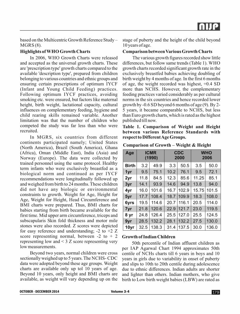
Comparison between Various Growth Charts
The various growth figures recorded show little differences, but follow same trends (Table 1). WHO growth charts recorded significant growth rate in the exclusively breastfed babies achieving doubling of birth weight by 4 months of age. In the first 6 months of age, the weight recorded was highest, +0.4 SD more than NCHS. However, the complementary feeding practices varied considerably as per cultural norms in the six countries and hence recorded lower growth by -0.6 SD beyond 6 months of age (9). By 2-5 years, it became comparable to NCHS, but less than Euro growth charts, which is rated as the highest published till now.
Table 1. Comparison of Weight and Height between various Reference Standards with respect to Different Age Groups.
Comparison of Growth – Weight & Height
Age ICMR CDC WHO (1990) 2000 2006
Birth 3.2 49.9 3.3 50.5 3.5 50.0
1yr 9.5 75.1 10.2 76.1 9.5 72.1
2yr 11.8 84.5 12.3 85.6 11.25 85.1
3yr 14.1 93.9 14.6 94.9 13.8 94.0
4yr 16.0 101.6 16.7 102.9 15.75 101.5
5yr 17.7 108.4 18.7 109.9 18.3 108.0
6yrs 19.5 114.6 20.7 116.1 20.5 114.0
7yr 21.8 120.6 22.9 121.7 23.0 119.5
8 yr 24.8 126.4 25.5 127.0 25.5 124.5
9yr 28.5 132.2 28.1 132.2 27.5 130.0
10yr 32.5 138.3 31.4 137.5 30.0 136.0
Growth of Indian Children
50th percentile of Indian affluent children as per IAP Agarwal Chart 1994 approximates 50th centile of NCHs charts till 6 years in boys and 10 years in girls due to variability in onset of puberty and slips to 10th to 20th centile during adolescence due to ethnic differences. Indian adults are shorter and lighter than others. Indian mothers, who give birth to Low birth weight babies (LBW) are rated as
Volume 3-4 179
NIJP
OCTOBER - DECEMBER 2014

shorter and lighter and hence a weight of 45 Kg and 145 cm is the goal for the prospective mother. This is expected to reduce incidence of LBW in India. As genetic and ethnic factors are important in growth beyond 5 years of age and as WHO charts are equated to NCHS- CDC charts beyond 5 years of age, any region specific charts may be recommended beyond this age group.
Clinical Applications of Growth Charts
It is a record of the child's growth and nutritional status. It provides a platform for analyzing growth, whether maintaining normal growth pattern and growth velocity, whether going static with arrest of growth or taking a downward trend with loss of weight etc. The 50th centile of the growth reference standard is accepted as 100% of the expected and is presumed that majority of the measurements crowd in and around this target maintaining the Gaussian distribution or the bell shaped curve. The age is plotted in the Y axis and the measurement of weight, height etc on the X axis. Those below the 3rd centile are considered too small and those above the 97th centile are considered too big. Making the observed measurement as a percentage of the expected allows grading into normal, mild, moderate or severely affected. The W H O Z scores p rov ide easy and qu ick understanding of the same, especially to the paramedical team members, who struggle with the percentile curves and the percentage calculations.
When a child's growth is found to be trailing, especially with respect to height, the allowances that can be given to the child are the following:
1). Comparison with the corrected age, if the baby is born preterm till attains 2 years of age.
2). Comparison with special syndrome specific charts as in the case of Down, Turner etc.
3). Accepting as 'small but healthy', if weight for height is normal especially in term LBW babies or children from socio-culturally deprived commu-nities with trans-generational malnutrition.
4). Comparison with the Mid Parental Target Height Centile: When a child is having short stature, calculate the Mid parental height by adding father's & mother's height and dividing by 2 and establish the target height also by adding 6.5 cm in case of boys
and by subtracting 6.5 cm in case of girls. Plot this value at 18 years in growth chart in red and read the centile of this marking. It is often plotted at a lower centile as the parents may also be having short stature. If the child is found to grow currently at the same lower centile, the physician and the parents can be reassured. But, if the child is currently growing at a lower centile than the MPH target centile, the child should be subjected to appropriate diagnostic evaluation like bone age, endocrine work up etc..
Tracking of BMI as a Breakthrough Intervention
As overweight and obesity are on the increase and are predictive of obesity related adulthood diseases, tracking of BMI since birth is recomm-ended. BMI is 13 at birth, increases to 17 by 1 year and then dips to 15 during preschool age and then slowly increase to more than 18.5 by adulthood. Human babies are born with abundant subcutaneous fat as protective strategy for the growing brain. The fat is expected to be depleted leading to skinny stature during preschool age and then gaining further fat by the phenomenon of adiposity rebound. It is noted that earlier the dip of BMI, there is more chance of obesity and later the dip, there is more chance of chronic energy deficiency.
The International Obesity Task Force (IOTF) had given overweight and obesity references from 2 year onwards, but WHO growth charts elucidated it with both percentile and Z scores from birth onwards. Conventionally, BMI between 85th to 95th percentile was considered as risk for obesity, > 95th as overweight and > 97th as obesity. WHO charts depicts > 85th as overweight and > 97th as obesity; there is no demarcation at the 95th centile.
There is no doubt as to the usefulness of BMI charts, but the effort in calculating BMI and applying it to different age appropriate charts reduces the ease of its use. Hence, three -in -one screening charts depicting weight on Y axis, height on X axis and BMI on the right margin in single chart has been designed, the ELIZ Health Paths (EHP); EHPUC- for underfive children, EHPOC-for older children, EHPAC- for adolescent children and EHPA- for adults (10). As the differences in BMI among the males and females in the various age groups are only to the tune of decimals, same
Volume 3-4 OCTOBER - DECEMBER 2014180
NIJP

screening charts are recommended for both sexes. The traffic signal tricolor; green, yellow and red are used for easy demonstration. Any deviation noted should be evaluated in depth.
The MCP Cards
The mother child protection (MCP) cards are now widely available to track events during each pregnancy and subsequently in the child. It is started on registration of early pregnancy and is given to the mother and the counterfoil is kept by the public health nurse/ Anganwadi worker for follow up. It can record all events and interventions during pregnancy and also the weight, developmental mile stones, immunization and any significant event during infancy and early childhood. It is a valuable record if the documentation is properly made. It is a colorful document with a tricolor growth record of weight. The health workers can easily identify the children in the red danger zone and make early referral. However, even those who were blaming that growth charts were not made available are found to be hesitant to open the MCP card and make necessary documentation. It is also of concern to note that even when recordings are made in the red zone, no appropriate action is initiated. So, it is recommended that the pediatrician shall supervise and ensure proper documentation.
Conclusion
It has to be remembered that growth and health are expressions of changing and changeable regulatory processes like nutritional, endocrino-logical, genetic and epigenetic factors. The WHO working group has also acknowledged that 'the WHO standards allow for future revision when further biological information on growth of infants and young children become available. The variability in time and place and the plasticity of fetal, infantile and childhood and pubertal growth itself reveals that the current growth charts are not the only growth trajectory for all times. Judicious use of weight, height, BMI charts help to understand the deviation if any in a given child and plan intervention or corrective measures. For under five children, WHO growth charts seems to be most appropriate, but for older children, regional reference standards are recommended. Three-in one
weight, height and BMI charts offer promising convenience for screening.
References
1. Weaver L T. In the balance: Weighing babies and the birth of the Infant Welfare Clinic. Bull Hist Med. 2010, 84: 30-57
2. Weaver L T. How did babies grow 100 years ago? Europ J Clin Nutr. 2011, 65:3-9
3. Morley, D; Woodland, Margaret. See How They Grow: Monitoring Child Growth for Appropriate Health Care in Developing Countr ies . London and Basingstoke: Macmillan, 1979. ISBN 978-0195201604
4. Gluckman PD, Hanson MA, Bateson P, Beedle A, Law CM, Bhutta z etal. Towards a new d e v e l o p m e n t a l s y n t h e s i s : a d a p t i v e developmental plasticity and human disease. Lancet. 2009, 373: 1654-57
5. El izabe th K E. Nut r i t ion and Chi ld Development. 5th ed. Hyderabad: Paras publications; 2015, pp: 91-101
6. Agarwal DK, Agarwal KN. Physical growth in Indian affluent children (Birth – 6 years). Indian Pediatr 1994; 31: 377-413.
7. Khadilkar V V, Khadilkar AV, Cole TJ and Sayyad MG. Cross-sectional Growth Curves for Height, Weight and Body Mass Index for Affluent Indian Children2007, Indian Pediatrics, 2009, 46:477-89
8. WHO Multicentre Growth Reference Study Group. Assessment of differences in linear growth among populations in the WHO Multicentre Growth Reference Study. Acta Paediatr Suppl 2006; 450: 56-65.
9. De Onis M, Garcia c, Onyango AW, Borghi E. Comparison of the WHO Growth Standards with CDC 2000 Growth Charts. J Nutr. 2007, 137: 144-48
10. Elizabeth K E. Three- in – one weight, height and BMI charts for children and adults. J Tropical Pediatr., Oxford University Press, London, 2003, 49: 224-27 Table 1. Comparison of Weight and Height between various Reference Standards with respect to Different Age Groups
Volume 3-4 181
NIJP
OCTOBER - DECEMBER 2014
Review Article :
Latent TuberculosisSawant Sandeep, Kamale VN, Mohanty NC.
Introduction:
Tuberculosis (TB) remains a major global health problem. There were an estimated 550 000 TB cases among children (under 15 years of age) and 80000 TB deaths among children who were HIV-negative in 2013 (6% and 8% of the global totals). In 2013, an estimated 9 million people developed TB and 1.3 million died from the disease (including 320 000 deaths among HIV-positive people). The number of TB deaths is unacceptably large given that most are preventable. The majority of cases worldwide in 2012 were in the South-East Asia (29%), African (27%) and Western Pacific (19%) regions. India and China alone accounted for 26% and 12% of total cases, respectively. Childhood TB accounts for 10% of all TB cases. In developing countries as much as 40% of children are affected. (1)
Keywords :
LTBI, Tuberculosis, latent tuberculosis, IGRA,
Source of support : Nil
Conflict of Interest : None
Infection, Disease and latency in children :
Childhood TB is sentinel event in community suggesting recent transmission from contagious adult. After exposure, it usually takes 8 to 10 weeks before the TB test would show if someone had become infected.(2)Depending on ventilation and other factors, these tiny droplets (from the person who has active tuberculosis) can remain suspended in the air for several hours. Should another person inhale them, he or she may become infected with TB. (3)The probability of transmission will be related to the infectiousness of the person with TB, the environment where the exposure occurred, the duration of the exposure, and the susceptibility of the host. In fact, "it isn't easy to catch TB. You need consistent exposure to the contagious person for a long time. For that reason, you're more likely to catch TB from a relative than a
stranger. Once exposed, people very often have latent tuberculosis. To convert to active tuberculosis, the bacteria must become active. (4)
Some children are prone for disease and death. And some stay with latent infection become reservoir of infection with reactivation in adulthood. Inhaled TB bacilli penetrate into lungs and settle. Infection is contained in a small area without spread or replication (latent TB infection or LTBI).The TB bacilli are well-contained and cannot be released (dormant). But sometimes, Infection spreads to nearby lymph nodes and the lung tissue itself causing TB pneumonia which may lead to primary active TB. Risk of progression in children depends mainly on age and immunity of child.(5) Latent stage may continue for the person's entire lifetime or ,in a matter of few weeks to few years , may progress to a stage of active disease called tuberculosis. It is hypothesized that latent TB is a reservoir of organisms that are encased in caseous lesions under hypoxic condition. (7)One third of world's population is estimated to have latent infection and lifetime risk of reactivation for aperson with documented LTBI is estimated to be 5-10%.Majority of them develop tuberculosis disease within the first five years after initial infection. However , the risk is considerably higher in the presence of predisposing factors.
Pathophysiology: Alveolar macrophage is the first line of defense & plays critical role in amplifying the response to infection. Antigen presentation by dendritic cells(DC),the major antigen presenting cell(APC) in lung & efficiency with which naïve T cells respond to antigen ,also appears less effective in children. While there is growing adult literature on role of candidate gene from this pathway, data from chi ldren is scarce . (7) During infect ion, mycobacterium accumulates intracellular lipid loaded inclusion bodies. Mycobacterial lipids and lipolytic enzymes are thought to play important role during dormancy and reactivation.(6,7) Lipolytic enzymes
Senior Resident-Dept.of Pediatrics,Terna medical college, Navi Mumbai.,Professors -Dept. of Pediatrics, MGM Medical college, Navi Mumbai. [email protected]
Volume 3-4 OCTOBER - DECEMBER 2014182
NIJP

have recently emerged as key factors in lipid metabolism during dormancy and/or exit of the non-replicating growth phase, a prerequisite step of TB reactivation. (8,9,10).Specific antibodies against lipolytic enzymes may be induced at different stages of the infection process. Mycobacterium possesses at least seven genes related to lipase and cutinase family. (11,12,13)Mycobacterium tuberculosis differentially expresses genes of various lipolytic enzymes which could be used for differentiation of activity and progression of tuberculosis.(13,14)
What is Latent Tuberculosis Infection (LTBI) :
Clemens Van Pirquet(1964)described first.
Singh et.al.(IJP ,2011) A Child Who Has a positive skin reaction to tuberculin but had no symptoms of pulmonary or extrapulmonary TB.
CDC has defined it as : A person with latent TB infection Usually has a skin test or blood test result indicating TB infection
l Has a normal chest x-ray and a negative sputum test
l Has TB bacteria in his/her body that are alive, but inactive
l Does not feel sick
l Cannot spread TB bacteria to others
l Needs treatment for latent TB infection to prevent TB disease; however, if exposed and infected by a person with multidrug-resistant TB (MDR TB) or extensively drug-resistant TB (XDR TB), preventive treatment may not be an option 40% likelihood of developing ACTIVE DISEASE.
WHO : Latent tuberculosis infection (LTBI), defined as a state of persistent immune response to stimulation by Mycobactarium tuberculosis antigens without evidence of clinically manifested active TB. A direct measurement tool for M. tuberculosis infection in humans is currently unavailable.
Efforts to diagnose LTBI :
Global efforts are being done for early diagnosis and prediction of progress of disease. These are aimed to 1) Detect presence of active antigenic material from mycobacterium. 2) Develop cost effective test to find out presence of active mycobacterium in person and progression.3) prevent unnecessary use of anti tubercular drugs.(1)
People have medical privacy or “confiden-tiality” and do not have to reveal their active tuberculosis case to family, friends, or co-workers; therefore, the person who gets latent tuberculosis may never know who had the active case of tuberculosis that caused the latent tuberculosis diagnosis for them. Only by required testing (required in some jobs) or developing symptoms of active tuberculosis and visiting a medical doctor who does testing will a person know they have been exposed. Because of taboo of word tuberculosis, it is common in many places to hide identity of index adult case; therefore, they may not test. If a person has symptoms of tuberculosis, it is wise to be tested.(5)
A positive PPD only indicates that someone is infected , It does not give you any information on the time of acquisition, latency, or activity of TB disease . The diagnosis of tuberculosis in children is traditionally based on symptoms, history of contact, chest radiography, tuberculin skin testing, and mycobacterium staining/ culture, although these inquisitions may not always be positive in children with tuberculosis. (3, 4) PCR and immune-based methods are other diagnostic methods which increasingly being used although they are not widely available and have a limited role in routine clinical practice. (6)
TST(Tuberculin skin test) and IGRAs (Interferron – Gamma Release Assays ) are the main tests currently available for diagnosis of LTBI. However these tests have limitation as they cannot distinguish between latent infection with viable microorganisms and healed /treated infection; they also poorly predict who will progress to active TB.
Interferon-Gamma Release Assays (IGRAs) are whole-blood tests that can aid in diagnosing Mycob-acterium tuberculosis infection. They do not help differentiate latent tuberculosis infection (LTBI) from tuberculosis disease. Two IGRAs that have been approved by the U.S. Food and Drug Administ-ration (FDA) are commercially available in the U.S:
l QuantiFERON®-TB Gold In-Tube test (QFT-GIT);
l T-SPOT®.TB test (T-Spot) IGRAs measure a person's immune reactivity to M. tuberculosis. White blood cells from most persons that have been infected with M. tuberculosis will release interferon-gamma
Volume 3-4 183
NIJP
OCTOBER - DECEMBER 2014

(IFN-g) when mixed with antigens (substances that can produce an immune response) derived from M. tuberculosis.
To conduct the tests, fresh blood samples are mixed with antigens and controls. The antigens, testing methods, and interpretation criteria for IGRAs differ Recommendations on when to use IGRA tests
l IGRAs can be used in place of (but not in addition to) TST in all situations in which CDC recommends TST as an aid in diagnosing M. tuberculosis infection, with preferences and special considerations noted below. This includes contact investigations, testing during pregnancy, and screening of health care workers and others undergoing serial evaluation for M. tuberculosis infection. Despite the indication of a preference, use of the alternative test (FDA-approved IGRA or TST) is acceptable medical and public health practice. Caution in interpretation should be used when testing certain populations because of limited data on the use of IGRAs .
l Populations in which IGRAs are preferred for testing:
l Persons who have received BCG (either as a vaccine or for cancer therapy); and
l Persons from groups that historically have poor rates of return for TST reading.
l TST is preferred over IGRAs for testing children less than 5 years of age.
l As with TST, IGRAs generally should not be used for testing persons who have a low risk of infection and a low risk of disease due to M. tuberculosis.
l Each institution and TB control program should evaluate the availability and benefits of IGRAs in prioritizing their use. Either TST or IGRAs can be used to identify candidates to LTBI treatment in high and upper-middle-income countries with estimated TB incidence less than 100,000.IGRAs should not replace TST in low and other middle income countries.
Who should be tested and treated for LTBI?
The risk for progression to active disease is considerably higher in infected individuals who belong to specific high risk populations. Major risk
factors for TB activation are HIV infection ,recent contact with infectious patient, initiation of an anti-tumour necrosis factor (TNF) treatment, receiving dialysis, receiving an organ or hematologic transplantation, silicosis, being in prison,being an immigrant from high TB burden countries, being a homeless person and being an illicit drug user.
Search for novel biomarker in blood or urine that can reliably distinguish active from latent TB in children with or without other co infections remains an important global goal. In recent years number of methods has been developed to address the issue of rapid and sensitive method for detection of MTB, still bigger aspects is not addressed that is reservoir of Bacilli LTB reference to differentiation and progression of disease.. In recent years number of genes and their proteins has been designated for differentiation of LTB from TB. (5, 6)
Management of LTBI :
The management of LT B I requires a comprehensive package of interventions that includes: identifying and testing those individuals who should be tested, delivering effective and safe treatment in a way that the majority of those starting a treatment regimen will complete it with no or minimal risk of adverse events, and ensuring monitoring and evaluation of the process.
WHO guidelines for the management of LTBI are currently only available for people living with HIV and for children below 5 years of age who are household contacts of TB cases .(6)Several WHO Member States had requested WHO for clear policy guidance on the management of LTBI, with due consideration to testing and treatment options. In addition, guidelines on the management of LTBI would be one of the necessary tools for facilitating the implementation of the Global TB Strategy after 2015 to achieve its ambitious targets of 90% reduction in TB incidence and 95% reduction in TB deaths that was endorsed by the World Health Assembly in May 2014. RNTCP has achieved very high cure rates and case fatality has dropped to 4.2 to 4.7% during 2000 to 2003. Without treatment the case fatality is believed to be about 80%, suggesting 90% reduction in case fatality. Thus the first objective of RNTCP is being achieved. Still TB continues as India's public health emergency. (18, 19, 20).
Volume 3-4 OCTOBER - DECEMBER 2014184
NIJP

Reactivation TB can be averted by preventive treatment. Currently available treatments have an efficacy ranging from 60% to 90% (4). The potential benefit of treatment needs to be carefully balanced against the risk of drug-related adverse events. Population-wide mass LTBI testing and treatment are not feasible due to imperfect tests, risk of serious and fatal side-effects and the high cost. The benefits are greater than the harms for infected individuals in population groups in which the risk of progression to active disease significantly exceeds that for the general population.
IAP Guidelines 2012 :
In India, TB accounts for 17.6% of all deaths from communicable disease, and for 3.5% of all cause-mortality .There are about 400 million children who constitute about 34% of the total population. The extent of childhood TB in India is unknown due to diagnostic difficulties; it is estimated to be 10.2% of the total adult incidence. The maximum risk of a child getting TB is between 1-4 years. Globally, about 1 million cases of pediatric TB are estimated to occur every year accounting for 10-15% of all TB ; with more than 100,000 estimated deaths every year. It is one of the top 10 causes of childhood mortality. (17)
The currently recommended dose of INH for chemoprophylaxis is 10 mg/kg (instead of previously recommended dosage of 5 mg/kg) administered daily for 6 months. TB preventive therapy should be provided to:
(a) All asymptomatic contacts (under 6 years of age) of a smear positive case, after ruling out active disease and irrespective of their BCG, TST or nutritional status.
(b) Chemoprophylaxis is also recommended for all HIV infected children who either had a known exposure to an infectious TB case or are Tuberculin skin test (TST) positive (>=5 mm induration) but have no active TB disease.
© All TST positive children who are receiving immunosuppressive therapy (e.g. Children with nephrotic syndrome, acute leukemia, etc.).
(d) A child born to mother who was diagnosed to have TB in pregnancy should receive prophylaxis for 6 months, provided congenital TB has been ruled out. BCG vaccination can be given at birth even if INH
chemoprophylaxis is planned. (21)
The following regimens are recommended by WHO for treatment of LTBI(6)
6–month or 9 month i son iaz id da i ly 3–month rifapentine plus isoniazid weekly 3 –or 4-month isoniazid plus rifampicin daily 3 –or 4-month rifampicin alone daily
Stop TB Strategy (WHO) :
Two approaches can be taken to reduce the burden of TB over the long term. One approach requires early and rapid diagnosis of all persons with active TB with institution of effective therapy to render them non contagious. Another approach is to detect persons while they still have LTBI and treat them to prevent the later development of disease. (18)
Household source contact is commonly implicated in spread of disease in children. The estimated prevalence of TB infection in South Africa among pediatric contacts of adult household cases was high using a follow-up test six months later and a strategy for contact investigations that incorporates follow-up testing may be advisable. Serial testing is feasible and useful in this setting, and IGRAs may represent an alternative testing option to TST. IGRA conversions and reversions occur frequently on serial-testing and further studies are warranted to determine the long-term prognosis of children who exhibit these test patterns.
The main focus of the Stop TB Strategy is on making the best use of currently available tools for diagnosis, treatment and prevention of TB (i.e. programme-based operational research) and the improved tools that are likely to become available in future (through research to develop new diagnostics, drugs and vaccines). The goal of eliminating TB by 2050 depends on the development of new diagnostics, drugs and vaccines. WHO is working in close collaboration with the Stop TB Partnership to enable and promote programme-based operational research and research to develop new diagnostics, drugs and vaccines.(1,19,20) There is urgent need for research into the epidemiology, immune mechanisms, diagnosis, treatment and prevention of childhood TB. Advances in understanding of TB in children would provide a wider insight and chances to facilitate efforts to control this ancient disease. Indeed, diagnosis of mycobacterial infections certainly requires rapid
Volume 3-4 185
NIJP
OCTOBER - DECEMBER 2014

assays for immediate therapy or treatments for disease prevention. (19,20)
References:
1. Global Tuberculosis Control. WHO report 2014
4. Jereb J, Etkind SC, Joglar OT, Moore M, Taylor Z (2003) Tuberculosis contact investigations: outcomes in selected areas of the United States, 1999. Int J Tuberc Lung Dis:S -384-390.
5. Zhang S, Shao L, Mo L, Chen J, Wang F, et al. (2010) Evaluation of Gamma Interfero Release Assays Using Mycobacterium tuberculosis Antigens for Diagnosis of Latent and Active Tuberculosis in Mycobacterium bovis BCG-Vaccinated Populations. Clinical and Vaccine Immunology 17: 1985. 22.
6. WHO:guidelines for latent management tuberculosis 2014,p.10
7. Dheda K, Schwander SK, Zhu B, van Zyl-Smit RN, Zhang Y (2010) The immunology of tuberculosis: from bench to bedside. Respirology 15: 433–450.
8. Dedieu L, Serveau –Avesare C, Kremmer L,Cannan S.(2013)Mycobacterial lipolytic enzyme;a go ld mine fo r tubercu los i s research.Biochemie,95(1)Jan 2013 .p66-73.
9. Peyron P, Vaubourgeix J, Poquet Y, Levillain F, Botanch C, et al. (2008) Foamy macrophages from tuberculous patients' granulomas constitute a nutrient-rich reservoir for M. tuberculosis persistence. PLoS Pathog 4: e1000204.
10. Dhouib R, Laval F, Carriere F, Daffe M, Canaan S (2010) A monoacylglycerol lipase from Mycobacterium smegmatis Involved in bacterial cell interaction. J Bacteriol 192: 4776–4785.
11. Parker SK, Curtin KM, Vasil ML (2007) Purification and characterization of mycobac-terial phospholipase A: an activity associated with mycobacterial cutinase. J Bacteriol 189: 4153–4160.
12. Schue M, Maurin D, Dhouib R, Bakala N'Goma JC, Delorme V, et al. (2010) Two cutinase-like proteins secreted by Mycobacterium tubercu-losis show very different lipolytic activities reflecting their physiological function. Faseb J 24: 1893–1903.
13. Deb C, Daniel J, Sirakova TD, Abomoelak B, Dubey VS, et al. (2006) A novel lipase Belonging to the hormone-sensitive lipase family induced under starvation to utilize stored triacylglycerol in Mycobacterium tuberculosis. J Biol Chem 281: 3866–3875.
14. Mishra KC, de Chastellier C, Narayana Y, Bifani P, Brown AK, et al. (2008) Functional role of the PE domain and immunogenicity of the Mycobacterium tuberculosis triacylglycerol hydrolase LipY. Infect Immun 76: 127–140.
15. Brust B, Lecoufle M, Tuaillon E, Dedieu L, Canaan S, et al. (2011) Mycobacterium tuberculosis Lipolytic Enzymes as Potential Biomarkers for the Diagnosis of Active Tuberculosis. PLoS ONE 6(9): e25078.
16. Mannesse ML, Cox RC, Koops BC, Verheij HM, de Haas GH, et al. (1995) Cutinase from Fusarium solani pisi hydrolyzing triglyceride analogues. Effect of acyl chain Lu C, Wu J Wang H, Wang S, Diao N, et al. (2011) Novel Biomarkers Distinguishing Active Tuberculosis from Latent Infection Identified by Gene Expression Profile of Peripheral Blood Mononuclear Cells. PLoS ONE 6(8): e242
17. The Registrar General & Census Commissioner, India, New Delhi, Ministry of Home Affairs, Government of India; Available from: http://www.censusindia.gov.in/Census_Data_2001/India_at_Glance/broad.aspx
18. Sandhu G, Battaglia F, Ely BK, Athanasakis D, Montoya R, et al. (2012) Discriminating Active from Latent Tuberculosis in Patients Presenting to Community Clinics. PLoS ONE 7(5).
19. Nelson LJ, Wells CD. Global epidemiology of childhood tuberculosis. Internat J Tubercul Lung Dis. 2004; 8:636-47.
20. Newton SM, Brent AJ ,Anderson S ,Whittaker E ,Kampmann B. Pediatric Tuberculosis. Lancet Infect. Dis 2008 August :8 (8) 498-510.
21. Ashok kumar,Gupta D,Nagaraja SB,Singh V,Sethi GR,Prasad J.Updated national guidelines for pediatric tuberculosis in India,2012.Indian Pediatrics 2013,March 2013 vol.50 301-306.
Volume 3-4 OCTOBER - DECEMBER 2014186
NIJP

Case report :
Klebsiella Endocarditis leading to brain abscess in a child with Tetralogy of Fallot.Ojing T, Shaad A, Kamran A, Shaukat A
Abstract:
Though congenital heart disease (CHD) predisposes to Infective endocarditis and brain abscess, endocarditis due to gram negative bacteria is uncommon. Among gram negative bacteria, Klebsiella species are very rare cause of bacterial endocarditis. Here we describe a case of a two and half years old male child with Tetralogy Of Fallot who developed Klebsiella Endocarditis and brain abscess.
Key words: Klebsiella endocarditis, Congenital heart disease (CHD), Brain abscess, Imipenem and Vancomycin
Source of support : Nil
Conflict of Interest : None
Introduction
Infective endocarditis is often a complication of congenital or rheumatic heart disease but can also occur in children without any abnormal valves or cardiac malformations (1). In the western world, the epidemiology of heart disease has changed over the past few decades. There has been a decline in population with rheumatic heart disease and a gradual increase in survivors of children with congenital heart defects (CHDs).CHDs now constitute the predominant underlying cause of infective endocarditis in developed countries (2).Viridians-type streptococci (α-hemolytic streptococci) and Staphylococcus aureus remain the leading causative agents for endocarditis in paediatric patients (1). Endocarditis due to gram negative bacteria is uncommon, although the incidence may be increasing (3). Among gram negative bacteria, Klebsiella species are very rare cause of bacterial endocarditis. A review of 348 cases of gram-negative endocarditis published in literature between 1945 and 1977 revealed only two cases of
Department of Paediatrics, JNMCH, AMU, Aligarh UP. E-mail: [email protected]
endocarditis due to Klebsiella species (4). Infective endocarditis (IE) is associated with a broad array of complications. The likelihood of complications depends on several factors including the infecting pathogen, duration of illness prior to therapy, and underlying comorbidities (5). Brain abscess is considered to be a rare complication of infective endocarditis but was observed in 14 (13%) of 108 critically ill patients with IE (6).
Case report :
A two and half years male child presented to our emergency ward with fever since seven days, bluish discolouration of body and difficulty in breathing since one day. There was no history of seizure, rashes over body, weakness of body or limbs. He had multiple episodes of bluish discolouration of body since last 1 year. On examination, He had a heart rate of 150 beats/min, respiratory rate 64 breaths/min, Temperature 101.7 ̊ F, blood pressure 108/60mm Hg and SpO2 75% on room air and 80% on oxygen supplementation and there was no difference in pre and post ductal saturation of oxygen. The child had both central and peripheral cyanosis and grade III clubbing. There was no pedal or sacral oedema .There was no conjunctival haemorrhage, Janeway lesion and Osler's node. On cardiovascular examination; there was no precordial bulge, apex was in fourth intercostal space without any thrill, P2 was not palpable. Auscultation revealed tachycardia but S1 was normal and S2 was soft and there was no any murmur. He was tachypneic but chest was clear. CNS and Per abdomen examination was within normal limit. ABG shows severe metabolic acidosis i.e pH;6.9,pCO2; 15mmHg, pO2; 45mmHg, HCO3-; 4.8mmol/L. He was managed in the line of cyanotic spell to which he responded. Initial investigation showed Hb=12.5 g/dl, TLC=11.5 x 10³/dl with polymorph predominant, Platelets=184 x 103/dl.
Volume 3-4 187
NIJP
OCTOBER - DECEMBER 2014

Urine microscopic examination showed numerous RBCs and fundus examination was normal. Three Blood samples for culture was sent from three different sites. Chest X-ray was done which showed coer en sabot appearance. Echocardiography revealed Tetralogy of fallot with no evidence of vegetation or any intracardiac mass though aortic valve was mildly thickened and there was no evidence of valve regurgitation.
The child was initially started on ceftriaxone and amikacin. The child was s running fever for seven days. Blood culture reports came after 7 days and two of which showed growth of Klebsiella species resistant to third generation cephalosporin, amikacin and gentamycin but sensitive to imipenem and vancomycin. Imipenem and vancomycin were started based on culture and sensitivity test and continued for 6 weeks. Fever subsided but child had persistent irritability. A CECT head was done after one week, which revealed a ring enhancing lesion in left parieto occipital region suggesting a brain abscess. Neurosurgery reference was done and they advised conservative management. A diagnosis of Klebsiella Endocarditis with septic emboli to brain was made. Patient became afebrile and irritability improved after two weeks of starting these antibiotics. Ultimately the patient was referred for surgery.
Discussions
Klebsiella species are common pathogens causing community as well as hospital-acquired pneumonia, intra-abdominal infections, and bloodstream infections. Infective endocarditis due to Klebsiella species is rare, accounting for less than 1% of cases but frequently accompanied by complications and a high mortality rate (7).
Moreover, with the emergence of extended-spectrum β-lactamase (E S B L) producing Enterobacteriaceae, endocarditis due to ESBL-producing Klebsiella pneumonia (ESBL-KP) is of concern due to the limited treatment options and notoriously high morbidity and mortality. Hospitalization is a significant risk factor for bloodstream infections due to ESBL-producing Enterobacteriaceae.
The treatment for infective endocarditis caused by klebsiella pneumoniae has not been determined.
Usually these patients require surgery combined with prolonged courses of antibiotics. At present, carbapenems are the treatment of choice. In a report by Zimhony (8), despite the low MIC of ciprofloxacin and piperacillin/tazobactam, therapy with ciprofloxacin followed by piperacillin/ tazobactam failed because of the inoculum effect and induction of resistance. Though a case of carbapenem resistant klebsiella endocarditis responsive to colistin and gentamyicin has been reported(9), our patient responded to Imipenem and Vancomycin.
Figure 1: X-ray chest showing typical coer en sabot appearance.
Figure 2: Parasternal long axis view showing non restrictiveVSD with aortic override.
Figure 3: CECT head showing (arrow) ring enhancing lesion in left parieto-occipital region
Volume 3-4 OCTOBER - DECEMBER 2014188
NIJP

Referrences:
1. Daniel Bernstein. In: Nelson Textbook of PEDIATRICS; Saunders; 19 edition (14 July 2011);chapter 431,pg.1632
2. Kothari SS, Ramakrishna S, Bahl VK. Infective endocarditis: an Indian perspective. Indian Heart J. 2005;57(4):289-94.
3. Hessen MT, Abrutyn E. Gram-negative bacterial endocarditis. In: Kate D,ed. Infective endocarditis. 2nd ed. New York: Raven Press, 1992.
4. Cohen PS, Maguire JH, Weinstein L. Infective endocarditis caused by Gram-negative bacteria : a review of the literature, 1945–1977. Prog Cardiovasc Dis 1980;22:205–42
5. Hoen B, Duval X. Infective endocarditis. N Engl J Med 2013; 369:785.
6. Sonneville R, Mirabel M, Hagège D, Tubach F, Vignon P, Perez P, Lavoué S, Kouatchet A, Pajot O, Mekontso-Dessap A, Tonnelier JM, Bollaert PE, Frat JP, Navellou JC, Hyvernat H, Ait Hssain A, Tabah A, Trouilet JL, Wolff M. Neurologic complications and outcomes of
infective endocarditis in critically-ill patients: the ENDOREA prospective multicenter study. Crit Care Med. 2011. in press .
7. David R. Clinical presentation, etiology, and outcome of infective endocarditis in the 21st century: the international collaboration on endocarditis—prospective cohort study. Arch Intern Med 2009;169:463-473.
8. Zimhony O. Endocarditis caused by extended-spectrum-beta-lactamase-producing Klebsiella pneumoniae: emergence of resistance to ciprof loxacin and piperacillin-tazobactam during treatment despite initial susceptibility. Antimicrob Agents Chemother 2006;50:3179-3182.
9. Shmuel Benenson , Shiri Navon-Venezia, Yehuda Carmel i , Amos Adler, Jacob Strahilevitz , Allon E. Moses, Colin Block. Carbapenem-resistant Klebsiella pneumonia endocarditis in a young adult Successful treatment with gentamicin and colistin. International Journal of Infectious Diseases (2009) 13, e295—e298
With Best Compliments from -
Volume 3-4 189
NIJP
OCTOBER - DECEMBER 2014

Case report :
Congenital Giant melanocytic nevusRamya V ,Sarala K, Radhika N*
Abstract
Congenital nevi are hyperpigmented macular lesions that are derivatives of the melanoblasts. They occur in less than 1% of the neonates in any site of the body. The giant congenital nevus is greater than 20 cm in size, pigmented and often hairy. Between 4% and 6% of these lesions develop into a maligant melanoma, early removal is recommended. We report a unique case of giant nevi along with a review of the literature .
Key words: Giant, melanocytic, congenital , nevus
Source of support : Nil
Conflict of Interest : None
Department of Pediatrics & Neonatology& Department of Pathology, Tata Main Hospital, Jamshedpur. Jharkhand, India E mail: [email protected]
Introduction:
Congenital gaint naevus is a congenital melanocytic nevus, also known as garment nevus or giant hairy nevus, bathing trunk nevus ,cape, vest ,coat- sleevs or stocking naevus based on area of involvement. The incidence is rare around 1 in 500,000 newborns.(1) It is obvious at birth with diameters more than 20 cm. presenting in the lower back and thigh areas with associated satellite nevi The surface of the nevus is usually rugose or warty and nodular with large terminal hairs, which become more prominent as the infant grows toward puberty. These have an increased malignant potential. Various associated abnormalities are reported like
Giant Melanocytic nevus covering entire back & front
Histopathology
PIGMENTED SKIN LESION.: Partly skin covered tissue bits measuring 0.5x0.4x0.3 cm(ts). Section shows hyperkeratotic squamous epithelial lining. Subepithelialy nests of uniform round to oval nevus cells arranged in concentric manner seen in papillary dermis, surrounding brostromal tissue with fair number of congested blood vessels seen. There is no evidence of junctional activity, granulomatous inammation or malignancy. Imp - suggestive of intradermal nevus.
Volume 3-4 OCTOBER - DECEMBER 2014190
NIJP
Fig.1 Fig.2
Fig.3

leptomeningeal melanocytosis, spina bifida, meningocoele, hamartomas, club foot and hypertrophy or atrophy of a limb involved by the nevus. (2) The present paper emphasizes the importance of recognizing the worrisome deep extension of pigmentation seen in the gaint congenital melanocytic naevus and raises considerations towards other treatment options.
Case report:
A male new-born out of non-consanguinous marriage at Tata Main Hospital , Jamshedpur, on 19/01/2014by caesarean section at term to a primigravida with H/O premature rupture of membranes, presented with a large black pigmented patch over the trunk and multiple small similar pigmented areas distributed over legs,thighs and face since birth.On examination,there was a large blackish pigmented area with rugosities covering the entire posterior aspect of the trunk,extending anteriorly to involve 40% of anterior aspect.There was a breach in the skin over the back which measured 4-5cms. Multiple satellite lesions of 2-4 cms sizes, widely distributed over thighs,legs and face were seen.No other congenital anomalies were seen. Fundus examination, X ray spine MRI head and spine and ultrasound abdomen were all normal. The biopsy of the patient was taken and the histo-pathological findings were consistent with congenital melanocytic naevus. Baby was treated for probable sepsis with IV antibiotics. Baby improved on antibiotics and was discharged .
Discussion:
The congenital melanocytic nevi are pigmented cutaneous lesions with it's color from the melanin pigment of nevomelanocytes. Congenital melanocytic nevi(CMN) are classified as small (<1.5 cm in diameter), medium (1.5–19.9 cm in diameter), and large (20 cm in diameter or ≥5% of body surface area).(3,4). Large CMN also are referred to as giant congenital melanocytic nevi (GCMN), and if found on the trunk and buttocks, they are commonly called bathing trunks nevi.
Large or giant CMN are rare, occurring in 1 of every 20,000 to 500,000 appearing in all races with
equal prevalence in males and females. Incidence of congenital nevomelanocytic ( CNN) nevus is slightly higher in blacks who are at lower risk of developing melanoma than whites(5). Autosomal dominant inheritance with incomplete penetrance or multifactorial determination occurs in families with small CNN (6). GCMN are thought to be caused by spontaneous mutationsinvolving 1p,12q, and 19pchromosomes.Also a body protein called HGF/SF (hepatocyte growth factor/scatter factor) seems to be responsible for encouraging these neuroectodermal cells to develop, migrate, and “scatter.”
Infants with CMN are at a higher risk (20% to 30%) for developing melanoma compared to acquired nevi. Small and medium CMN have an approximately 1% lifetime risk for progression to melanoma.(7)and should be carefully observed for any change using the ABCD (asymmetry, border irregularity, color, diameter) screening principles. The risk of malignancy is also increased by the presence of larger nevi (greater than 50 cm), axial location such as trunk, head and neck, the presence of multiple satellite lesions, and the existence of nodules, dark patches, junctional activity, deep dermal neurogenic element or a blue nevus component (5,8,9,10,11,12,13).Patients with CMN have a poor prognosis, as they usually die within the first year of life.(14)
Treatment
Treatment of large or giant CMN without neurologic involvement primarily is removal of the lesion, when the patient is 6 months or older, but before puberty when the risk for malignancy increases.(15) Some surgical techniques , like serial excision with advanced skin grafting procedures can be done ..Alternative treatments include phenol chemical peels, dermabrasion, and laser therapies for lesions that cannot be surgically treated but cosmesis is desired. This case report is being presented due to its rarity.
Declaration of Conflict of Interest: The authors have no conflict of interest in this article .
Volume 3-4 191
NIJP
OCTOBER - DECEMBER 2014

References:
1. Grichnik JM, Rhodes AR, Sober AJ. Benign Neoplasias and Hyperplasias of Melanocytes. In: Wolff K, Goldsmith LA, Katz SI, Gilchrest BA, Paller AS, Lefell DJ, editors. Fitzpatrick's Dermatology in General Medicine. 7 th ed. McGraw-Hill; 2008. p. 1099-103.
2. Mackie RM. Disorders of Cutaneous Melanocyte. In: Burns T, Breathnach S, Cox N, Griffiths C, editors. Rook's Textbook of Dermatology. 7 ed. Oxford: Blackwell Science; 2004. p. 38-19.
3. Hoffman D, Ratner D. Diagnosis and management of a changing congenital melanocytic nevus. Skinmed. 2006;5:242-245.
4. Pielop JA. Vascular lesions and congenital nevi in the newborn.http://www.uptodate.com/ contents/benign-skin-and-scalp-lesions-in-the-newborn-and-young-infant Updated October 3, 2007.
5. Kaplan EW. Risk of malignancy in large congenital nevi. Plast Recons Surg1974; 53:421-428.
6. Weston WL, Lane AT, eds. Color Textbook of Pediatric Dermatology. St Louis, Mosby Year Book Inc; 1991:204-207,239.
7. Watt AJ, Kostis S, Chung K. Risk of melanoma arising in large congenital melanocytic nevi: a systematic review. Plast Reconstr Surg. 2004;113:1968-1974.
8. Foster RD, Williams ML, Barkovich AJ, Hoffman WY, Mathes SJ, Frieden IJ. Giant
congenital melanocytic nevi: The significance of neurocutaneous melanosis in neurologically asymptomatic children. Plast Reconstr Surg. 2001;107:933–41.
9. Mark GJ, Mihm MC, Liteplo MG, Reed RJ, Clark W H. Clinical , his tologic and u l t ras t ruc tura l s tudies . Hum Pathol . 1973;4:395–418.
10. DeDavid M, Orlow SJ, Provost N, et al. A study of large congenital melanocytic nevi and associated malignant melanoma: Review of cases in the New York University registry and the world literature. J Am Acad Dermatol. 1997;36:409–16.
11. Ruiz-Maldonado R, Tamayo L, Laterza AM, Duran C. Giant melanocytic nevi: Clinical, h i s t o p a t h o l o g i c , a n d t h e r a p e u t i c considerations. J Pediatr. 1992;120:906–11.
12. Rhodes AR, Wood WC, Sober AJ, Mihm MC., Jr . Nonepidermal origin of malignant melanoma assoc with a giant congenital n e v o c e l l u l a r n e v u s . P l a s t R e c o n s t r Surg1981;67:782–90.
13. Bauer BS, Vicari FA. An approach to excision of congenital giant pigmented nevi in infancy and early childhood. Plast Reconstr Surg. 1988;82:1012–21.
14. OakleyA.Congenital melanocytic naevi. DermNet NZ Web site.
15. Bhatnagar V, Mukherjee MK, Bhargava P. A case of giant hairy pigmented nevus of face. Med J Armed Forces India. 2005;61:200-202.
Volume 3-4 OCTOBER - DECEMBER 2014192
NIJP

Case Report :
Systemic peripheral gangrene :A rare presentation of Falciparum malaria Gupta S, Reddy K, Choudhury I
Abstract :
We report a ten year old girl ,suffering from falciparum malaria ,had symmetrical peripheral gangrene resulting in auto amputation.Once it is set in ,there is no known therapy to reverse the process or inhibiting the progress.
Keywords : falciparum malaria, systemic peripheral gangrene, rossetting, sequestration
Introduction
Symmetric peripheral gangrene was initially described in 1981 by Hutchinson.1 Symmetrical peripheral gangrene (SPG) is a rare clinical condition which manifests as symmetrical distal ischemic damage in 2 or more sites without any evidence of obstruction and vasculitis of the relevant artery.2 P. falciparum is transmitted by the bite of female Anopheles mosquito and causes the most dangerous form of malaria with highest mortality and morbidity due to sequestration.(3)
Plasmodium falciparum malaria compromise the microcirculation due to adhesion of infected erythrocytes to vascular endothelium.4 Heavy malarial parasitemia causes sluggish micro-
circulation due to increased viscosity caused by parasitized red cell along with uninfected erythrocytes to form micro aggregates (rosettes) which attach to different endothelial receptors.3 The fall in the intraluminal pressure below certain critical value (3660mmHg) due to occlusion of small blood vessels are seen mostly in cases of shock and hypovolemia.3 This occlusion of the small capillaries supplying end organs may lead to ischemia followed by gangrene.
SPG is a very rare entity of malarial fever in children with only 5 cases reported till date.3 Here we report a case of rare presentation of P. falciparum malaria with symmetrical peripheral gangrene.
Case Study
A 10 year old female hailing from Bokaro steel city, Jharkhand came with complains of high grade fever with chills since 5 days, altered sensorium since 3 days and abnormal movements since 2 days. She was diagnosed as Plasmodium Falciparum malaria, was started on Intravenous Artesunate and referred to our hospital. On arrival, the patient was febrile (103°C) with severe pallor and altered
Department of Pediatrics, Bokaro General Hospital, Bokaro Steel City, Jharkhand. Email: [email protected]
Volume 3-4 193
NIJP
OCTOBER - DECEMBER 2014
Fig.: 1 Fig.: 2 Fig.: 3
Toes and Wrist showing gangrenous changes.

sensorium with Glasgow coma scale of 8. She also had firm hepatosplenomegaly (liver 4cm, span 9cm and Spleen 4cm).
The investigations revealed Haemoglobin of 4.4gm%, Total leukocyte count of
13500cumm (65% polymorphs , 35% lymphocytes), platelets 90,000/cu mm, indirect bilirubin (1.4 mg %) and serum alkaline phosphatase was 102IU/l. Optimal test for malaria was positive for Plasmodium Falciparum. The peripheral smear showed ring forms of Plasmodium Falciparum and the parasitic index was 80%. Renal profile showed blood urea nitrogen 56 mg%, serum creatinine was 1.8 mg%. The patient was continued on intravenous Artesunate. The patient had abnormal movement thus was loaded with intravenous Phenytoin. Patient was given packed cell transfusion with 10ml/kg. The patient improved over the next 2 days, became conscious and well oriented. Thus intravenous Artesunate was stopped and was put on oral combination therapy. On the 3rd day of admission the patient developed bilateral symmetrical blackish-blue discoloration of the 2nd, 3rd and 4th toes (figure: 1,2) and two similar-appearing patches one on the left wrist and other on the right forearm (figure:3). She was unable to move the toes and the dorsalis pedis artery was not palpable on both feet. Doppler study of peripheral arteries showed low-amplitude signals, with decreased flow suggestive of a vasoconstriction. 2-D echocardiography did not reveal evidence of an intracardiac thrombus. Three consecutive blood cultures were sterile. Coagulation profile was normal. D-dimer assay was strongly positive and serum fibrinogen levels were elevated (702 mg/dl, normal 200-400 mg/dl). Anti-nuclear and anti-dsDNA antibodies were negative. The gangrene became well demarcated over the next month and got auto amputated. The patient was treated with pentoxiphylline drip, intravenous Piperacillin Tazobactum and Amikacin . Limb elevation, early physiotherapy for the toes and fingers , inter-digital packing and protection from trauma were instituted.
Discussion:
Symmetrical peripheral gangrene(SPG) was reported in many medical conditions including
malaria. Our patient had no clinical or laboratory evidence of other common causes of SPG such as sepsis, use of drugs like ergots, vasopressin, noradrenaline or any vasospastic condition like Raynaud's phenomenon .Our patient also did not have any evidence of polycythaemia, thrombos-thaenia or vasculitis. The most common cause of is reported to be disseminated intravascular coagulation (DIC). Rosetting of uninfected erythrocytes around the parasitized red cells occur and further exacerbates vascular obstruction and sequestration.(5 )
Endothelial receptors such as ICAM-1 (Intercellular adhesion molecule-1), ELAM-1 (endothelial leukocyte adhesion molecule-1), VCAM-1 (Vascular cell adhesion molecule-1) interacts with infected erythrocytes in the microcirculation causing sequestration. Changes in membrane that occur in Plasmodium falciparum infected erythrocytes causes activation of the blood coagulation cascade to cause thrombosis. Plasmodium affects the membrane of infected erythrocytes. RBCs infected with parasites develop knob like structures on their surface membranes. These knobs have unique capability of inducing adhesions of ICAM-1 of micro capillaries.
A similar process of receptor induction also transpires on the non-parasitized RBCs induced adhesion molecules facilitates clumping between parasitized and non-infected RBCs, this process of clumping is termed as rosseting. These twin processes of sequestration and resetting result into micro vascular blockade causing decreased organ blood flow and consequent organ tissue damage which manifests as cerebral malaria, renal malaria etc.(6) All these mechanisms may have contributed to the thrombosis, and consequent vascular occlusion, that resulted in the gangrene of her. The gangrene formed was irreversible causing auto amputation. Even though SPG is very rare presentation in P. Falciparum infected children, the treating physician should be very vigilant in anticipating the ischemic changes to prevent the unwanted complication.
Volume 3-4 OCTOBER - DECEMBER 2014194
NIJP

References:
1. Kakati, S, Doley B, Barman B, Devi A. Symmetric peripheral gangrene and falciparum malaria. JAPI. 2004 (52).
2. Kotokey RK, Kaushik BPN. Symmetrical peripheral gangrene and neurological manifestations in Plasmodium falciparum malaria. J Assoc Physicians India. 2011 Jul;59:449-51.
3. Tamhankar P, Tullu MS, Lahiri KR, Deshmukh CT. An unusual complication of Plasmodium falciparum malaria. Indian J Med Sci (serial online) 2008 (cited 2015 Jan 7);62:70-3. Availablfrom:http://www.indianjmedsci.org/te
xt.asp?2008/62/2/70/39371.
4. Thanachartwet V, Krudsood S, Wilairatana P, Phumratanaprapin W, Silachamroon U, Looareesuwan S. Peripheral gangrene in patients with severe falciparum malaria: report of 3 cases. Korean J Parasitol . 2006 Jun;44(2):139-43.
5. Sharma BD, Gupta B. Peripheral gangrene in a case of complicated malaria. JIACM 2002; 3(3); 297-9.
6. Alkizim FO, Matheka D, Mwanda OW. Malaria complicated by gangrene: a case presentation and review. Pan Afr Med J. 2011;10:46. Epub 2011 Nov 25.
Volume 3-4 195
NIJP
OCTOBER - DECEMBER 2014

Case Report :
Congenital leukemia in a 23 day old neonateSant Prakash Kataria, Monika Gupta, Gajender Singh, Sanjay Kumar, Rajeev Sen, Balraj Yadav*
Abstract
Congenital leukemia constitutes a rare and poorly defined entity. It is usually associated with numbers of congenital anomalies. Clinically they present with non specific signs and symptoms. It is known to progress rapidly and paradoxically, may also remit spontaneously. We report this case of congenital leukemia, who presented with non specific symptoms and hyperleukocytosis with no congenital anomalies, to make the clinician aware and keep this possibility in neonates.
Key words: Congenital Leukemia, Infant, Acute Myeloblastic Leukemia, Acute Lymphoblastic Leukemia
Introduction
Congenital leukemia is very rare disorder that develops in utero, and manifests in the newborn to 4 weeks of age1,2. It occurs at the rate of 1 per 5 million births. Only 200 cases of congenital leukemia are reported in the literature. The majority of congenital leukemias are acute non-lymphocytic leukemia (80%), while acute lymphoblastic leukemia (ALL) comprises only < 20%.2
It is more common among patients with chromosomal abnormalities.3 Patients usually presents with non specific signs and symptoms and differentiation from leukamoid reaction is difficult. Congenital leukemia is known to progress rapidly and paradoxically, may also remit spontaneously4. Here, we report a case of congenital acute myeloid leukemia in 23 days old male infant.
Case report :
A 23 days old male infant admitted to PGIMS, Rohtak with history of difficulty in breathing, poor acceptance, swelling over scrotum and fever for two days. He was born full term, to non- consanguineous parents by elective caesarian section due to history of primary infertility in mother. Mother was on
antitubercular treatment six months prior to conception and during first trimester. Baby cried immediately after birth and his birth weight was 2.8 kg.
Department of Pathology, Pt. BD Sharma, PGIMS, Rohtak, Consultant Paediatrician*, Shri Krishna Medicare, Gurgaon, Haryana
Volume 3-4 OCTOBER - DECEMBER 2014196
NIJP
Figure 1 : Bone Aspirate showed blasts (Leishman satinx20x)
On examination, baby was pale, febrile and lethargic with generalized purpuric spots but no dysmorphic features. He had tachypnea (respiratory rate 80/ min) and pulse rate was 128/ min. Abdomen was slightly distended with liver 3 cm and spleen 2 cm below costal margin. Heart sounds were normal and there was no focal neurological deficit.
The baby was evaluated for early neonatal sepsis, which revealed hemoglobin 5.8gm%, total leucocyte count 200 × 103/µl with blasts 95%, neutrophils 4% and lymphocyte 1% and platelet count 32× 103/µl. Peripheral smear examination showed dimorphic red cells. TORCH titers (against toxoplasmosis, cytomegalovirus, herpes simplex virus, rubella and syphilis) were all negative. Blood culture was sterile after 48 hours of incubation. CRP was positive (45 mg/l) while CSF, Blood urea

(20mg %), blood sugar (56mg%), serum Na (137 meq/l), serum K (5.0 meq/l), and fundus were normal,.
Bone marrow aspiration was done which revealed hypercellular marrow replacing all normal hematopoietic elements by blast comprising > 90%, revealing high N:C ratio, slightly condensed nuclear chromatin with prominent 2-3 nucleoli and basophilic cytoplasm containing fine azurophilic granules. Blasts were sudan black positive (>3%) and negative for PAS and ANAE. Immunoph-enotyping revealed blasts to be positive for CD45, CD13, CD33, CD34, HLA-DR and aMPO. Cytogenetic analysis revealed normal 46 XY results. The final diagnosis of acute myeloid leukemia (AML-M0) was confirmed.
Patient was given the supportive treatment in form of IV fluids, antibiotics and platelets transfusion. After confirmation of diagnosis of AML, parents were counseled for starting the chemotherapy but they did not agree due to fear of the toxic effects of the drugs. Neonate expired on 27th day of life, probably because of infiltration into other organs like spleen and liver.
Discussion :
Congenital leukemia constitutes a rare and poorly defined entity. The criteria for diagnosis of congenital leukemia are (i) disease presentation at or shortly after birth (<30 days) (ii) proliferation of immature white cells (iii) infiltration of these cells into hematopoietic tissues (iv) absence of any other condition that causes leukemoid reaction, mimicking leukemia 4,5,6.
It is associated with numbers of congenital anomalies like mental retardation, heart defects, developmental delay and with chromosomal disorders such as Down syndrome, Edward's and Patau syndrome.2 Children with Down syndrome have 15-20 fold increased risk of acute leukemia 8.
Clinically they present with non specific signs like lethargy, poor feeding, purpura, fever and hepatosplenomegaly. Respiratory distress is frequently seen in these patients. It is important to differentiate congenital leukemia from other leucoerythroblastic conditions which are seen in response to bacterial infection, hypoxemia, severe
hemolys i s in the neona te and t r ans ien t myeloproliferative disease (TMD)2,5. TMD is an uncommon syndrome strongly associated with abnormalities of chromosome 21 occurring in 10% of infants with Down syndrome. It is associated with transient polycythemia, blast proliferation and or thrombocytopenia. Majority of patients undergoes spontaneous remission within 3 months of onset.2,8,9
Present case does not show any evidence of secondary disorders which could mimic leukemia. Blasts in the peripheral smear and bone marrow resemble each other and were definite of leukemic blasts. Hepatosplenomegaly may be indicative of infiltration of leukemia. Cytogenetics study was normal in our case. Other neonatal conditions which present with similar peripheral blood picture were excluded by sterile blood culture and negative results for TORCH infection. Both mother and baby had O positive blood group excluding the possibility of hemolytic disease of a new born. Mother was on antitubercular treatment but no known risk factor for leukemia is associated with these drugs.
Prognosis of congenital leukemia is dismal and has a progressive downhill course, even with chemotherapy. However, there are reports of spontaneous remission1. Another problem in leukemia treatment of newborns is the high toxicity of chemotherapeutic standard regimens in first months of life and its influence on the growth and nutrition of newborns in this critical period of life. 10
In our case also, hyperleucocytosis, purpura, bone marrow involvement and hepatosplenomegaly was the clear indication of starting up the chemotherapy but with the fear of unknown toxic effects of the drugs the parents refused to treat. He died on the 5th day of admission due to intra cranial hemorrhage.
In conclusion, we report this case of congenital leukemia to make the clinician aware to keep this possibility in neonates who present with non specific symptoms and hyperleukocytosis. Although it remains the rare disorder with poor prognosis, still further studies are needed to clarify the nature of this disease.
Volume 3-4 197
NIJP
OCTOBER - DECEMBER 2014

Acknowledgement
No financial support was received. The authors declare that there is no conflict of interest.
Every author was active participant in the present case report .
References
1. Narayan S, Sharma S, Chandra J, Kulshreshtha R. Clinico-hematological profile of congenital leukemia. Indian J Pediatr 2004;71: 927-8
2. Mora J, Dobreins AM, Bussel JB, Aledo A. spontaneous remission of congenital acute nonlymphoblastic leukemia with normal karyotype in twins. Medical and Pediatric Oncology 2000;35:110-13.
3. Jafroodi M, Zarkesh M, Kariminejad R. Congenital Leukemia: A case report. Iranian Journal of blood and cancer 2010; 2(3):135-7.
4. Bresters D, Reus AC, Veerman AJ, Van Wering ER, Vander Does-vanden Berg A, Kaspers CJ. Congenital leukemia: The Dutch experience and review of the literature. Br J Haematol 2002;117(3):513-24.
5. Sung TJ, Lee DH, Kim SK, Jun YH.
Congenital acute myeloid leukemia with t(8;16) and t(17;19) double translocation: case presentation and literature review. J Korean Med Sci 2010;25:945-9.
6. Pierce Milla I. Leukemia pattern in the newborn infant. J Pediatr 1959;54:691-6.
7. Verma M, Rawat P, Das S, Kakkar N, Sohi I. Congenital acute myeloid leukemia in a case of Down's syndrome- A case report. Indian J Hemat and Blood Transf 2001;19:110-11.
8. Prakash KP, Rau ATK, Bhat ST, Rau AR. Congenital leukemia- A diagnostic dilemma. Indian J Medical and Paediatric oncology 2008;29:41-3.
9. Zubizarreta P, Muriel FS, Fernandez Barbieri MA. Transient myeloproliferative disorder associated with trisomy 21, a wide range syndrome: report of two cases, with trisomy 21 mosaicism. Med Ped Oncol 1995;25:60-64.
10. Prasad H.V. Raghavendra, Kumar G. Naveen: Congenital leukemia in a 2-month old boy. The Internet Journal of Pediatrics and Neonatology 2 0 0 7 ; Vo l u m e 6 N u m b e r 2 . D O I http://dx.doi.org/10.5580/idf0
WITHBEST
COMPLIMENTSFROM
Volume 3-4 OCTOBER - DECEMBER 2014198
NIJP

Case Report :
Alveolar Proteinosis: An Uncommon Presentation of A Not So Common DiseaseMahfooz A* , Mohanty NC***, Joseph JJ**, Verma S**, Kulkarni P*, Patel J*
Abstract :
We report a 14 month old child presenting with history of recurrent interstitial pneumonia, finally turned out to be Pulmonary Alveolar Proteinosis (PAP) secondary to Niemann Pick Disease, diagnosed on high index of suspicion and appropriate investigations. This is the first case of Secondary Alveolar Proteinosis due to niemann pick disease type C with NPC2 mutation to be reported from India, to the best of our knowledge.
Keywords : Pulmonary Alveolar proteinosis, Niemann Pick Disease, Pneumonia, Interstitial lung disease, NPC2 mutation
Introduction
Pulmonary alveolar proteinosis is caused by alveolar accumulation of surfactant due to defective clearance by alveolar macrophages. Incidence of PAP in children is 1 in 3,00,000 with only a few dozen cases reported in the literature. Clinically it presents as progressive exertional dysponea, non-productive cough, weight loss, hemoptysis or fever. Clinical presentation varies with type and mutations. Rarely it presents as diffuse interstitial lung disease or as secondary alveolar proteinosis (1).
Case
A 14 month old female child presented with history of recurrent episode of respiratory tract infections from 9 months of age. Also hadmarked failure to thrive and gross motor developmental delay. Seven similar episodes had occurred in last 5 months for which she was hospitalized four times; treated with antibiotics, nebulization, anti-fungal agents, systemic corticosteroids and moist oxygen inhalation.
Still, the child remained tachypnoeic and
continued to be oxygen dependent. On examination, her oxygen saturation was 80 % on room air. She had clubbing, was Tachypnoeic with intercostal, sub-costal retractions. There were diffuse bilateral bronchial breathing and coarse crepitations. Liver was enlarged 4 cm BCM with a span of 9cm, smooth surface, firm in consistency and rounded margin. Spleen was 5cm palpable BCM. CVS and CNS essentially normal. Fundus examintion normal. Basic investigations showed Hb 11.2, TLC 13500, D/C: N71 , L24, E4, M1; Platelets 2.75 lakhs, ESR-28, fasting blood sugar 87 mg/dL, total Bilirubin 0.8mg, conjugated 0.2 mg, SGOT 136, SGPT 141.5, Alkaline phosphatase 113, total protein 6.5, Albumin 3.5 and Globulin 3 gm/dL. Mauntoux Test: 11 mm. Broncheoalveolar lavage culture came positive for klebsiella sp and TB PCR was negative. U l t r a s o n o g r a p h y o f a b d o m e n s h o w e d h e p a t o s p l e n o m e g a l y w i t h n o r m a l l i v e r architecthure. 2-D echo was normal. X'Ray chest AP view (Fig-1)and HRCT (Fig-2 & 3):
Volume 3-4 199
NIJP
*Resident, **Asstt Prof, ***Professor, Department of Pediatrics, MGM Medical College, Navi Mumbai. Email: [email protected]
Fig – 1: CxR (AP) showing bilateral diffuse alveolar infiltrates
OCTOBER - DECEMBER 2014

Fig – 2 and 3: HRCT B/L diffuse ground glass opacification with underlying interstitial thickening involving both lower and right upper lobe, suggestive of Alveolar Proteinosis.
Liver Biopsy: Lobular architecture was maintained. Few lymphocytes were seen in portal tracts, fairly well preserved, maintaining typical cord pattern. Fine vacuolated, ballooned hepatocytes and Kupffer cells were scattered all over, suggestive of Niemann Pick disease.
Lung Biopsy : showed Dilated alveoli lined by type II pneumocytes containing foamy macrophages and PAS positive amorphous material proteinaceous nature. The alveolar septa were moderately thickened due to infiltration of lymphocytes and plasma cells in a mosaic fashion, suggestive of Alveolar Proteinosis.
Fig–4: Lung Biopsy (HE, PAS) suggestive of Alveolar Proteinosis
Fig – 5: Liver Biopsy (H&E) showing features of Niemann Pick disease
Volume 3-4 OCTOBER - DECEMBER 2014200
NIJP

Enzyme Studies:
Genetic studies:
G e n o m i c D N A f r o m b l o o d s h o w e d homozygous status for c.141C-A (p.C47Ter) mutation in exon 2 of NPC2 gene
Final diagnosis:
On the basis of history of chronic interstitial lung disease, failure to thrive, Lung and Liver biopsy reports, the diagnosis was concluded as “NIEMANN PICK DISEASE Type C with NPC2 mutation and SECONDARY PULMONARY ALVEOLAR PROTEINOSIS.
Course in hospital:
The child was discharged after control of
infection and after a broncho- pulmonary lavage with advice for regular follow up. She is still oxygen dependent. With such a progressive lung disease, a therapeutic trial miglustat was planned to improve morbidity, although mortality is high in mutation phenotype.
Discussion:
Neimann pick disease is a rare autosomal recessive disorder with the incidence of 1 in 250000. They are of three types- Neuropathic -A, Non-neuropathic type –B and type C Niemann pick d i s o r d e r s c a n h a v e v a r i o u s r e s p i r a t o r y manifestations, depending on phenotype and mutation (2) as given in table 1 below.
Enzymes Observed values Ref. range
Chitotriosidase (DSB) 80.73 umol/hr/ml 0-44 umol/hr/ml
Chitotriosidase (Serum) 265 umol/hr/ml 9-46 umol/hr/ml
Sphingomyelinase 3.6 nmol/hr/mg proteins 8-14.5nmol/hr/mg proteins
Table: 1 Types of Niemann pick disease
Sub-type Basic defect Gene Clinical features Respiratory manifestations
Type A Sphingomyelinase SMPD1 Onset in infancy, Recurrent aspiration deficiency retinopathy hepato- pneumonia in splenomegaly and rapidly life-threatening or fatal progressive neurological events deterioration
Type B Sphingomyelinase SMPD1 Onset in childhood, Mild dyspnoea on deficiency hepatosplenomegaly, exertion, interstitial growth retardation and lung disease, and mild clinical course respiratory insufficiency
Type C Abnormal NPC1, Variable age at onset, Interstitial pulmonary trafficking of NPC2 hepatosplenomegaly, alveolar proteinosis, sphingolipids progressive neurological respiratory insufficiency involvement and subacute (NPC2 genotype) clinical course.
Volume 3-4 201
NIJP
OCTOBER - DECEMBER 2014

Clinical presentation:
Varies with type and mutations. Rarely presents as diffuse interstitial lung disease or secondary alveolar proteinosis (1).
Type A and B characterized by deficiency of sphingomyelinase enzyme with SMPD1 gene defect.
Type A Onset in infancy and present as retinopathy hepatosplenomegaly and rapidly progressive neurological deterioration. Can have respiratory manifestations as recurrent bronchitis or aspiration pneumonia, resulting in life-threatening or fatal events.
Type B Onset in childhood and present as hepatosplenomegaly, growth retardation and mild clinical course .Can have respiratory manifestations as Mild dyspnoea on exertion, interstitial lung disease, and severe and fatal respiratory insufficiency.
Type C is characterised by Abnormal trafficking of Sphingolipids with NPC1 and NPC2 gene defect.
Age of onset is Variable and present as hepatosplenomegaly, progressive neurological involvement and subacute clinical course and rarely with respiratory manifestations as Interstitial lung disease, aspiration pneumonia and pulmonary
alveolar proteinosis with respiratory insufficiency (NPC2 genotype)
Phenotypic severity depends on the type of mutation. NPC1 mutation has less severe phenotypes. NPC-2 gene mutation results in rapid onset of phenotypes and death at 6 month to 4 years. Millat et al identified five mutation of NPC2 gene. All were associated with a severe phenotype, characterized by pulmonary infiltrates, respiratory failure, and death by four years of age (3).
A study in two infants having NPC2 gene mutation presenting with a severe respiratory insufficiency and severe liver disease, showed signs of pulmonary alveolar proteinosis and died by two years of age (4).
NPC1 or NPC2 gene mutations severely impair intracellular lipid transport, cause accumulation of unesterified cholesterol and several glycosphingolipids in some tissues and organs (5).
Pulmonary Alveolar Prteinosis (PAP) can be:
– Primary (congenital-due to mutations /deficiencies in surfactant proteins or the granulocyte macrophage-colony stimulating factor receptor) and
– Secondary due to many causes as shown in table 2 below:
Table – 2: SECONDARY CAUSES OF PAP
Autoimmune Disorders
Psoriasis
Amyloidosis
Immunodeficiecy Disorders
Thymic alymphoplasia
Ig A deficiency
SCID
Infections
Nocardia
HMV
CMV
P.jivoreciiTB
Dusts
SilicaCotton
Cement
Titanium
No 2
Almunium
Hematopoetic Disorders
Fanconi's anemia
MDS
AML
CLL
CML
Genetic Disorders
Lysinuric protein intolerance
Niemann pick
type C2
Volume 3-4 OCTOBER - DECEMBER 2014202
NIJP

Observed with a reassessment of symptoms every 3-6 months
Observed with a reassessment of symptoms with shortened follow-up intervals
Pulmonary function tests [ow volume curves, lung volumes and gas transfer) Arterial blood gas analyses 6 min walking test Chest X-ray Yearly HRCT.
WLL
Inhaled GM-CSF
Progression
IV Rituximab
Progression
Current recommendations for Treatment
For decades, the standard treatment of PAP has been whole lung lavage (WLL), where large quantities of saline are instilled into the lungs to remove the proteinaceous material. However, not all patients respond to this treatment. Hence newer modalities, such as subcutaneous or inhaled granulocyte macrophage colony-stimulating factor (GM-CSF), and the CD20 antibody rituximab and plasmapheresis, have been tried (6) in combinations.
A suggested algorithm is given in table-3.
Conclusion:
Awareness about the disease and availability of diagnostic facilities are bound to facilitate early diagnosis. Facility for WLL and other advanced modalities of therapeutic management are wanting. Appropriate genetic counseling can help lesser morbidity and prevention of such pregnancies in future.
Table – 3: Algorithm for step-wise management of PAP (6)
Stage 1.AsymptomaticDSS1Minimal desaturation during physical activity or a slightreduction in diffusion
Progression/
Regression
Stage 2.Mild to moderate symptoms. DSS 2Require oxygen on minimal physical activity.
Stage 3.Moderate to severe symptoms. DSS3+4+5 P(A-a)02 > 40 mm Hg Shunt fraction > 10-12% Progression of GGO on HRCT
Progression/
Regression
Volume 3-4 203
NIJP
OCTOBER - DECEMBER 2014
References
1) Garver WS, Francis GA, Jelinek D, et al. The National Niemann-Pick C1 disease database: report of clinical features and health problems. Am J Med Genet 2007; 143: 1204-1211.
2) Schuchman EH: The pathogenesis and treatment of acid sphingomyelinase-deficient Niemann-Pick disease. J Inherit Metab Dis 2007, 30:654-663.
3) Bjurulf B, Spetalen S, Erichsen A, Vanier MT, Strom EH, Stromme P: Niemann-Pick disease type C2 presenting as fatal pulmonary alveolar lipoproteinosis: morphological findings in lung and nervous tissue. Med Sci Monit 2008, 14:CS71-CS75.44
4) Vanier MT. Lipid changes in Niemann-Pick disease type C brain: personal experience and review of the literature. Neurochem Res 1999; 24: 481-489.
5) Millat G, Chikh K, Sleat DE, Fensom AH, Higaki K, Elleder M, et al. NPD type C: Spectrum of HE1 mutations and genotype/ phenotype correlations in the NPC-2 group. Am J Hum Genet 2001; 69: 1013- 1021.
6) Leth S. New treatment of Pulmonary Alveolar Proteinosis. Respirology 2013; 18: 82-91
7) R. Borie et al. Pulmonary alveolar proteinosis. Eur Resp Rev 2011; 20:120, 98-107
Autoimmune Pulmonary Alveolar Proteinosis

Research Media Watch :
Effects of Whole Body Therapeutic Hypothermia on Gastrointestinal Morbidity and Feeding Tolerance in Infants with Hypoxic Ischemic Encephalopathy
Perinatal HIE is associated with high morbidity and mortality in the neonatal period as well as long-term neurocognitive deficits. The effects of a perinatal hypoxic ischemic event extend beyond the brain and neurodevelopment. Decreased perfusion to the GI tract and decreased motility leading to feeding intolerance may follow perinatal asphyxia. Whole body therapeutic hypothermia ( WBTH) acts to prevent secondary damage to the brain from ischemia and reperfusion injury that occurs following periods of perinatal anoxia, it is plausible to believe that WBTH could have similar preventative effects on the ischemic damage to the GI system.
Infants ≥ 35 weeks gestational age and ≥1800 grams birth weight with moderate-to-severe HIE treated from 2000 to 2012 were compared. 68 patients had documented strictly defined criteria for WBTH: 32 historical control patients did not receive WBTH (non-WBTH) and 36 cohort patients received WBTH. Kaplan-Meier survival curves were generated for WBTH and non-WBTH patients. Then performed the log-rank test to compare survival curves between the two groups. All analyses were performed using SAS 9.2 (Cary, NC) and SPSS 20. Statistical significance was claimed at 95% confidence level (P < 0.05).
More of the non-WBTH group infants never initiated enteral feeds (28% versus 6%; P = 0.02), never reached full enteral feeds (38% versus 6%, P = 0.002), and never reached full oral feeds (56% versus 19%, P = 0.002). Survival analyses demonstrated that the WBTH group reached full enteral feeds (median time: 11 versus 9 days; P = 0.02) and full oral feeds (median time: 19 versus 10
Anil lohar, Satish Agrawal
days; P = 0.01) sooner. The non-WBTH group had higher combined outcomes of death and gastric tube placement (47% versus 11%; P = 0.001) and death and gavage feeds at discharge (44% versus 11%; = 0.005). So WBTH may have beneficial effects on GI morbidity and feeding tolerance for infants with moderate to-severe HIE.
Source : International Journal of Pediatrics Volume 2014, Article ID 643689, 7 pages
Comments: WBTH used for moderate-to-severe HIE may have beneficial effects to the newborn that extend beyond neurocognitive and neurodevelopmental outcomes. WBTH improves GI morbidity and feeding tolerance for infants with moderate-to-severe HIE.
Effects of maternal epidural analgesia on the neonate - a prospective cohort study
Epidural analgesia is one of the most popular modes of analgesia for child birth. There are controversies regarding adverse effects and safety of epidural analgesia. The study was conducted to know the immediate effects of the maternal epidural analgesia on the neonate during early neonatal phase.
A prospective cohort study of 100 neonates born to mothers administered epidural analgesia were compared with 100 neonates born to mothers not administered epidural analgesia in terms of passage of urine, initiation of breast feeding, birth asphyxia and incidence of instrumentation. The mothers who were regularly followed up in antenatal clinic were included in the study after taking the informed consent. Caesarean sections, preterms (Less than 37 weeks of completed gestation), low birth weights (Less than 2.5 kg), antenatally detected major congenital anomalies, multiple gestations, high risk antenatal factors like gestational diabetes, pregnancy induced hypertension, recurrent abortions, elderly primigravida (Above 40 years)
Volume 3-4 OCTOBER - DECEMBER 2014204
NIJP

and those who did not provide consent were excluded from the study.
The results were compared among the two groups and statistical analysis was done using the software Epi Info 3.5.1 and p value was calculated by Chi square test and Fisher's exact test. P value of <0.05 was considered as statistically significant.
There was significant difference among the two groups in the passage of urine (p value 0.002) and incidence of instrumentation (p value 0.010) but there was no significant difference in regards to initiation of breast feeding and birth asphyxia. Epidural analgesia does not have any effect on the newborns in regards to breast feeding and birth asphyxia but did have effects like delayed passage of urine and increased incidence of instrumentation
Source : http://www.ijponline.net/content/40/1/99
Comments : The effect of epidural analgesia on the neonate is of immense significance and should be further explored in the future with more elaborate randomized controlled multi-centre studies.
Infants Born Late/Moderately Preterm Are at Increased Risk for a Positive Autism Screen at 2 Years of Age
To assess the prevalence of positive screens using the Modified Checklist for Autism in Toddlers (M-CHAT) questionnaire and follow-up interview in late and moderately preterm (LMPT; 32-36 weeks) infants and term-born controls this study conducted.
Population-based prospective cohort study of 1130 LMPT and 1255 term-born infants. Parents
completed the M-CHAT questionnaire at 2-years corrected age. Parents of infants with positive questionnaire screens were followed up with a telephone interview to clarify failed items. The M-CHAT questionnaire was rescored, and infants were classified as true or false positives. Neurosensory, cognitive, and behavioral outcomes were assessed using parent report .
Parents of 634 (57%) LMPT and 761 (62%) term-born infants completed the M-CHAT questionnaire. LMPT infants had significantly higher risk of a positive questionnaire screen compared with controls (14.5% vs 9.2%; relative risk (RR) 1.58; 95% CI 1.18, 2.11). After follow-up, significantly more LMPT infants than controls had a true positive screen (2.4% vs 0.5%; RR 4.52; 1.51, 13.56). This remained significant after excluding infants with neurosensory impairments (2.0% vs 0.5%; RR 3.67; 1.19, 11.3).
So LMPT infants are at significantly increased risk for positive autistic screen. An M-CHAT follow-up interview is essential as screening for autism spectrum disorders is especially confounded in preterm populations. Infants with false positive screens are at risk for cognitive and behavioral problems.
Source: J Pediatr 2014;dec; 1-7.
Comments : LMPT(late and moderately preterm) infants are at significantly increased risk for positive autistic screen. So these infants need regular follow up assessment of Autism spectrum disease along with BERA and ROP follow up.
Volume 3-4 205
NIJP
OCTOBER - DECEMBER 2014

Manuscript Preparation: Guidelines for Authors
All Manuscripts sent to the NIJP should not be sent in any other journal or publication. These become the sole property of the publishers. The manuscripts submitted should be accompanied by a certificate signed by all the authors that “The undersigned author(s) certifies (fy) that the submitted article is original and is not under consideration with other publisher and has not been published anywhere in the past. All copyright ownership of the manuscript is transferred to the publisher of NIJP”. The manuscript should include the ethical clearance certificate from appropriate authorities.
Articles are edited for grammar and style. The spelling pattern in the journal is of American style. The statement and opinions expressed in the manuscripts are essentially of the author(s) and the editors / publishers don't owe any responsibility for these.
The product, service or the claim made by the manufacturer as advertised in the journal are those of the manufacturer. The publishers don't endorse these claims.
Preparation of Manuscripts:
Manuscript should be submitted in MS Office word, Use Font type Times New Roman12 point for text.
Title Page: This contains the title of the manuscripts along with the name of the author(s) with their affiliations. The name of the institute where the study was carried out, the address for correspondence with e-mail of the author should be mentioned at the bottom of the page.
Abstract: The objective, method of the study, results and conclusion should be sent in a paragraph as abstract of the manuscript. It should contain the purpose of the study, selection of study subjects, specific data denoting the main findings and the chief conclusions. The new and important aspects of the study should be revealed by the abstract.
Text: Every article must follow the following format:
Introduction, Material and method, Results, Discussion and Conclusions. The text should preferably be in a simple language which is easy to understand, pertaining to the topic concerned and should avoid vocabulary jargon.
Tables: Each table should have title and be numbered in the order of appearance in the text. Use superscript letters to indicate foot-notes typed on top of the table. Quote all tables in the text.
Images: All image files must be of high quality(300DPI) in TIF or BMP format. Do not send JPG or GIF files. Line diagrams should be in editable format. All images should be submitted separately and not as apart of main manuscript. Quote all images in the text as figure numbers in Arabic numerals printed at the bottom of figures.
Volume 3-4 OCTOBER - DECEMBER 2014206
NIJP

Contributors: At the end of the article, the contribution of all the authors should be mentioned which includes:
(1) Each author's contribution
(2) Conflict of interest
(3) Funding source and its role in the study
The last element of the text is acknowledgment. Abbreviations if used should be in accordance with the international guidelines e.g. cm, ml etc. Abbreviation use should be kept to the minimum and if they are used, they should be preceded by the full form.
References: Only those references which were consulted in original should be cited. They should be arranged sequentially. The references should be visible on the line after punctuation. The author(s) shall be responsible for the accuracy and completeness of the references. Vancouver system is followed in the journal. References should be listed and numbered in the order in which they appear in the text. The list of references at the end of the text includes:
Names of author(s): If more authors are involved in the study, the first 6 names should appear ending with et al, the title of the paper, Name of the journal as appears in Index Medicus, publication year, volume number and first and last page numbers.
References from the book includes, name of the book, editors, place of publication, publisher, edition, the year of publication and first and last page numbers.
If multiple authors are involved then the chapter name, first and last page numbers and names and initials of the editors should be give. For example,
From journals:
1. Mahesh Baldawa, Satish Tiwari: Legal issues and preventable medical errors, New Indian Journal of Pediatrics 2012, 1: 8-12.
From Books:
2. Bhattacharya A, Mishra S.DNR, Euthanasia and Life support systems; In: Tiwari S, Baldwa M, Tiwari M, Kuthe A editors; Text Book on medico-legal issues related to various medical specialties: 1st edition, Jaypee brothers medical Publishers, New Delhi 2012; 179-185
From Newspapers:
Aamir ko ho raha pachhatava. Dainik Bhaskar (Hindi), 2011; 4th December. Mumbai p.5 (Col.3)
From internet:
Harms Roger W. Breas t - feed ing whi le p regnan t : I s i t sa fe? http://www.mayoclinic.com/health/breast-feeding-while-pregnant/AN01840 Accessed on 16th Oct 2011.
Those papers which have been accepted but not published yet should be mentioned as “In press”. Those which are in preparation, personal communication or unpublished observation should be mentioned as such.
Volume 3-4 207
NIJP
OCTOBER - DECEMBER 2014

Bibliography
Volume 3-4 OCTOBER - DECEMBER 2014208
NIJP

Bibliographyn Pgae 2
Volume 3-4 209
NIJP
OCTOBER - DECEMBER 2014
IIIrd National Conference of Pediatric Association of India(PAICON-III)
Organized in Collaboration with :Dept. of Pediatrics, MGM University of Health Sciences,
Sector-1, Kamothe, Navi Mumbai-410218, Maharashtra
Dear Colleagues,
It gives us immense pleasure to announce PAICON III from 25 to 27 September 2015 at MGM University of Health Sciences, Kamothe, Navi Mumbai. The Pediatric Association of India (PAI) is committed for improvement of physical, mental, social and spiritual health of children of this country. The PAI has dedicated itself to the improvement of health of children from conception to adulthood, irrespective of diversities and their background. We will try to do more for the under privileged children and under deveioped areas reaching out to the last child. The association will dedicate itself in achieving the goals of achieving normal newborns with adequate growth and development potential.
The first and second national conferences of PAI were successfully held at Bhubaneshwar and Bardwan respectively. This year, the we feel honoured at MGM MEDICAL COLLEGE, Navi Mumbai to have been given the responsibility to hold the 3rd PAICON. Our MGM University of Health Sciences, a centre of excellence in Medical and Bio-Medical Research (Accredited as A grade by NAAC, UGC) will leave no stone unturned to make the scientific extravaganza to your utmost academic sumptuousity with path breaking wok-shops and talks by eminent authorities in Paediarics.
Located in a periurban RAIGAD district, we cater for the health needs of hundreds and thousands of unprivileged members of the society with a philanthropic zeal as is PAI. The theme of the 3rd national PAICON has therefore been kept as "Sharing knowledge for caring " .
Please register in large numbers and be a part of us in this endeavor. We categorically appeal our young Paediatrician friends to arrive in large numbers and enjoy the scientific meet in the serene back-drop of the up-coming new international airport, the whispering beech of Alibag and the towering hills of Matheran! We will be happy to welcome the professionals from support services like Paediatric / neonatal nursing, paediatric phusio- and occupational therapists and the likes for knowledge sharing and hands on experience.
The Highlights of Conference:
Day-1: Workshops on disaster management, communication skills and medical education in pediatrics, Research methodology, Paediatric Radiology and Imaging, Liver Diseases and endoscopy, Facility based IMNCI for Nurses, Neonatal nursing, Paediatric Physiotherapy.
Day-2: Theme based and problem oriented case discussions by experts.
Day-3: Scientific fiesta for pediatricians on care of children in the developing world, free papers, award papers and so on.
Volume 3-4 OCTOBER - DECEMBER 2014210
NIJP

Organising Chairperson:
Dr. Nimain Mohanty
Co organizing Chairpersons
Dr. Bageshree Seth Dr. Mahesh Mohite Dr. Shekhar Dhabhadkar
Organizing Secretary: Dr. Vijay Kamale Dr. Jeetendra Gavhane
Joint organizing secretary: Dr. Subhash Rao Dr. V. Kotrashetty Dr. N. Revathi
Treasurer: Dr. Rakesh Thamke Dr. Vikram Patra
Subcommittes:
Scientific: Dr. Prisca Colaco, Dr. D. Daruwalla, Dr. P. S. Moralmar Dr. Anjali Kale
Auditorium: Dr. Revathi N. Dr. Pallavi Sharma
Accommodation: Dr. Rakesh Thamke Dr. Shushrut Kumar Dr. Vikram Bedi
Transport: Dr. Jeetendra Gavhane Mr. Arun Khot
Catering: Dr. Vikram Patra Mr. T. P. Mohanty Mr. Santosh Riraksale
Publicity: Dr. Vijay Kamale Dr. Jay Bhandarkar Dr. Patkar Mr. Jeevan Urankar
Mementos: Dr. Piyush Jain Miss. Supriya Kadam
Reception: Dr.Vikas Gupta Dr. Annie George Dr. Nikhil Bharat Dr. Rishabh Dutt Dr. Ankita Pandey
Organising Team
Central PAI office bearers
Dr. H.Parmesh Dr. G.D. Sarangi National President Secretary General
Patrons
Dr. Sudhir Kadam Hon. Vice Chancellor, MGMIHS
Lt. Gen. (Retd) Dr. S. K. Kaul Hon. Pro VC
Dr. Chander puri Hon. Pro VC (research)
Advisors
Dr. N. N. Kadam, HOD, Pediatrics, MGM Medical College, Navi Mumbai
Dr. G.S. Narshetty Dean, MGM Medical college, Navi Mumbai
Dr. A. G. Shroff, Dean, MGM Medical college, Aurangabad
Dr. Z. G. Badade Registrar, MGMIHS, Navi Mumbai
Volume 3-4 211
NIJP
OCTOBER - DECEMBER 2014
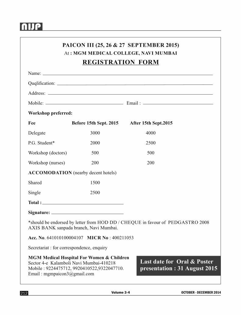
PAICON III (25, 26 & 27 SEPTEMBER 2015)
At : MGM MEDICAL COLLEGE, NAVI MUMBAI
REGISTRATION FORM
Name:
Quqlification:
Address:
Mobile: Email :
Workshop preferred:
Fee Before 15th Sept. 2015 After 15th Sept.2015
Delegate 3000 4000
P.G. Student* 2000 2500
Workshop (doctors) 500 500
Workshop (nurses) 200 200
ACCOMODATION (nearby decent hotels)
Shared 1500
Single 2500
Total :
Signature:
*should be endorsed by letter from HOD DD / CHEQUE in favour of PEDGASTRO 2008 AXIS BANK sanpada branch, Navi Mumbai.
Acc. No. 641010100004107 MICR No : 400211053
Secretariat : for correspondence, enquiry
MGM Medical Hospital For Women & ChildrenSector 4-e Kalamboli Navi Mumbai-410218Mobile : 9224475712, 9920410522,9322047710.Email : [email protected]
Last date for Oral & Poster presentation : 31 August 2015
Volume 3-4 OCTOBER - DECEMBER 2014212
NIJP

Please enclose self attested xerox copies of Graduation, PG, Professional Registration Certificates, along with your identity proof and DD/Multicity cheque of INR 2000/- as life membership fees and send with the form, complete in all respects to the Secretary general, PAI in the above address.
Volume 3-4 OCTOBER - DECEMBER 2014214
NIJP

Giant Melanocytic nevus covering entire back & front - Histopathology ( Page No. 190 )
Toes & Wrist Showing Gangranous Changes ( Page No. 193 )

Bone Aspirate showed blasts (Leishman satinx20x) (Page No. 196)
Lung Biopsy (HE, PAS) suggestive of Alveolar Proteinosis
Liver Biopsy (H&E) showing features of Niemann Pick disease
( Page No.:200 )
