occurrence of house sparrow passer domesticus indicus l ...tnsroindia.org.in/journal/full text issue...
TRANSCRIPT
Indian Journal Of Natural Sciences ISSN: 0976 - 0997
Vol 1 / Issue 3 / December 2010 © IJONS
155
Occurrence of House Sparrow Passer domesticus indicus L. in
Sivakasi, Virudhunagar District, Tamil Nadu, India.
Baskaran S*, Rajesh P, Pavaraj M, and Bakavathiappan Ga
Post-graduate and Research Department of Zoology, Ayya Nadar Janaki Ammal College (Autonomous), Sivakasi – 626 124, Tamil Nadu, India. Received: 15 Oct 2010 Revised: 10 Nov 2010 Accepted: 28 Nov 2010
*Address for Correspondence: Baskaran S Ayya Nadar Janaki Ammal College (Autonomous), Sivakasi – 626 124, Tamil Nadu, India
E mail ID: [email protected]
In India there are reports that house sparrow populations are under threat. The survey was carried out in market and residential areas in Sivakasi Taluk, Virudhunagar District, Tamil Nadu, India. The study revealed that the mean number of house sparrow in residential areas ranged from 9 individual to 18 individuals. Kanthapuram colony showed highest number of house sparrows. The mean number of house sparrows in market areas ranged from 8.6 to 12.8. The vegetable shop area and Parasakthi colony supported high number of sparrows. Only one nesting site was observed in each study site of market area. In residential area the highest number i.e. 3 nesting sites were observed in Kanthapuram colony. House sparrow number over the last decade has fallen. The decline in bird population over the year has been inferred by survey conducted locally. Many theories put forward to explain such as a decline include pollution, increase in predators and loss of a reliable food source. The disappearance of house sparrow seems to be connected to the way we build our houses and the ease with birds can find food. Many new house designs and home improvements have restricted the number of suitable nooks and crannies for the house sparrows to nest in. Key words: House sparrow- Passer domesticus indicus L, decline, population, food sources, home improvements, residential and market areas
INTRODUCTION
Among the various species of birds, the house sparrow Passer domesticus indicus L. is one of the familiar species that has followed man everywhere and is inseparable from human habitations. The non-migratory sparrows are widely distributed in the Indian subcontinent and occur worldwide [1]. The house sparrow is one of the most common bird in the urban environment worldwide, and is ecologically tightly linked to this
RESEARCH ARTICLE
ABSTRACT
Indian Journal Of Natural Sciences ISSN: 0976 - 0997
Vol 1 / Issue 3 / December 2010 © IJONS
156
special habitat. It occupies a variety of environments along the urban gradient [2]. House sparrows are well known for their frequent cheeping and chirruping from roof and gutters [3]. Although the effects of urbanization on avian community composition are well known, there is much less information about how individual birds are affected by these human-generated habitat differences [4]. The reasons for the declining of house sparrow population includes loss of invertebrates, reduction in winter seed food, change in agricultural practices, loss of grass and lawns from gardens, loss of nest sites, predation, pesticide use, changing architecture of human habitation, lack of traditional granaries3. In India also there are reports that house sparrow populations are under threat. Hence, the present study “Occurrence of House Sparrow Passer domesticus indicus L. (PASSERIFORMES: PLOCEIDAE) in Sivakasi” was undertaken
METHOD OF STUDY
Study Area
The study was undertaken in Sivakasi, a town in Virudhunagar District of Tamil Nadu, India. The survey was carried out from December 2008 to March 2009. In Sivakasi 14 places were selected for survey. They are categorized into market area and residential area. Method - Walkover Survey
The number of House Sparrow was counted by following walkover survey method. The house sparrows are active from 8.00 a.m. to 11.00 a.m. In each study site, the number of birds was counted by 30 minutes observation. The nesting sites of house sparrow were also observed.
RESULTS AND DISCUSSION
The study revealed that the house sparrow population was higher in residential area. The house sparrow is an omnivorous bird. It feeds on grains, fruits, insect and insect larva. The house sparrow is present only where the man inhabited. Mostly the house sparrows build their nests in residential area. Among the residential area more number of nests was observed in Kanthapuram colony. This is due to availability of nesting sites. In Kanthapuram colony lot of old buildings are available. It facilitates the house sparrow to build the nest. The high number of house sparrows in Kanthapuram colony may also be due to the availability of food sources which support a good population of insects (Table 1, 3). Rajashekar [1] reported that in Bangalore the house sparrows built their nest on crevices of hatched roofs of old houses, electric pipelines and ventilation holes.
The lowest number of house sparrow was observed in residential area found along the N.R.K.R. Street. The mean number is 9.0. This road is always busy with heavy traffic and human activity. This may be the reason for the less population. Leveau [5] reported that in Argentina significantly lower house sparrow population was observed during weekend days when the pedestrian and vehicle traffic increased when compared to working days. Car traffic noise appears to diminish habitat quality, due to an increase of the stress and cause distortion in vocal communication. On the other hand, vehicle movement also can disturb the house sparrow foraging activity. In market area the house sparrow population was high in vegetable shop area. The mean number is 12.8. The food sources are high in that area but the availability of nesting site is poor. Only one nest has been identified in that area (Table 2). In Bangalore also market areas supported only a moderate number of house sparrows [1]. The house sparrow number declined in Luv mainly due to changes in urban habitats resulting from urbanization process such as, contracting of the residential areas, development of new micro-habitats with very little greenery and architecture unsuitable for nest construction building houses in rural areas covered with weeds [6]. To conserve the house sparrow
Baskaran et al
Indian Journal Of Natural Sciences ISSN: 0976 - 0997
Vol 1 / Issue 3 / December 2010 © IJONS
157
future conservation plans and status assessment, either at the national or international level should be carried out for house sparrows these taxa should be regarded as designable units [7].Many reasons are given for disappearance of house sparrows. The introduction of unleaded petrol is one, as the combustion of which produces compounds methyl nitrite, which is highly toxic for small insects that forms a part of a sparrow chick’s diet. As supermarkets mushroom in urban areas, the old fashioned grain shops are disappearing. It was once common sight to see flocks of sparrows feasting on the grain in the gunny sacks displayed in front of these shops or on the spilt grain. Urbanization has done away with home gardens, which had worms and insects for the young sparrows. But pesticides have proved lethal for their survival. The most recent reason for their disappearance is the mobile phone towers. The waves from the tower are capable of destroying the life in the eggs. Thereby they are incapable of hatching. Reduction of nest-site availability due to improvements in quality and insulation of rooftops and a reduction of food availability due to loss of weedy corners from urban areas have both been suggested as likely explanations, as was in increased transmission of epidemic diseases.
ACKNOWLEDGEMENT
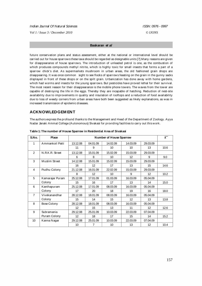
The authors express the profound thanks to the Management and Head of the Department of Zoology. Ayya Nadar Janaki Ammal College (Autonomous) Sivakasi for providing facilities to carry out this work. Table 1: The number of House Sparrow in Residential Area of Sivakasi
S.No. Place Number of House Sparrow x
13.12.08 04.01.09 14.02.09 14.03.09 29.03.09 1 Ammankoil Patti 11 9 10 10 13 10.6
13.12.08 15.01.09 15.02.09 15.03.09 29.03.09 2 N.R.K.R. Street 6 8 10 12 9 9.0
14.12.08 15.01.09 15.02.09 15.03.09 29.03.09 3 Muslim Street 16 12 17 13 15 14.6
21.12.08 16.01.09 22.02.09 15.03.09 29.03.09 4 Pudhu Colony 8 12 10 9 12 10.2
25.12.08 17.01.09 01.03.09 16.03.09 05.04.09 5 Kamarajar Puram Colony 15 16 17 13 14 15.0
25.12.08 17.01.09 08.03.09 16.03.09 05.04.09 6 Kanthapuram Colony 17 20 18 19 16 18.0
28.12.08 18.01.09 08.03.09 16.03.09 05.04.09 7 Vivekanandhar Colony 15 14 15 12 13 13.8
28.12.08 18.01.09 08.03.09 16.03.09 05.04.09 8 Bose Colony 12 15 13 11 12 12.6
29.12.08 25.01.09 10.03.09 22.03.09 07.04.09 9 Subramania Puram Colony 12 18 17 15 14 15.2
29.12.08 25.01.09 10.03.09 22.03.09 07.04.09 10 Kanna Nagar 10 7 10 13 12 10.4
Baskaran et al
Indian Journal Of Natural Sciences ISSN: 0976 - 0997
Vol 1 / Issue 3 / December 2010 © IJONS
158
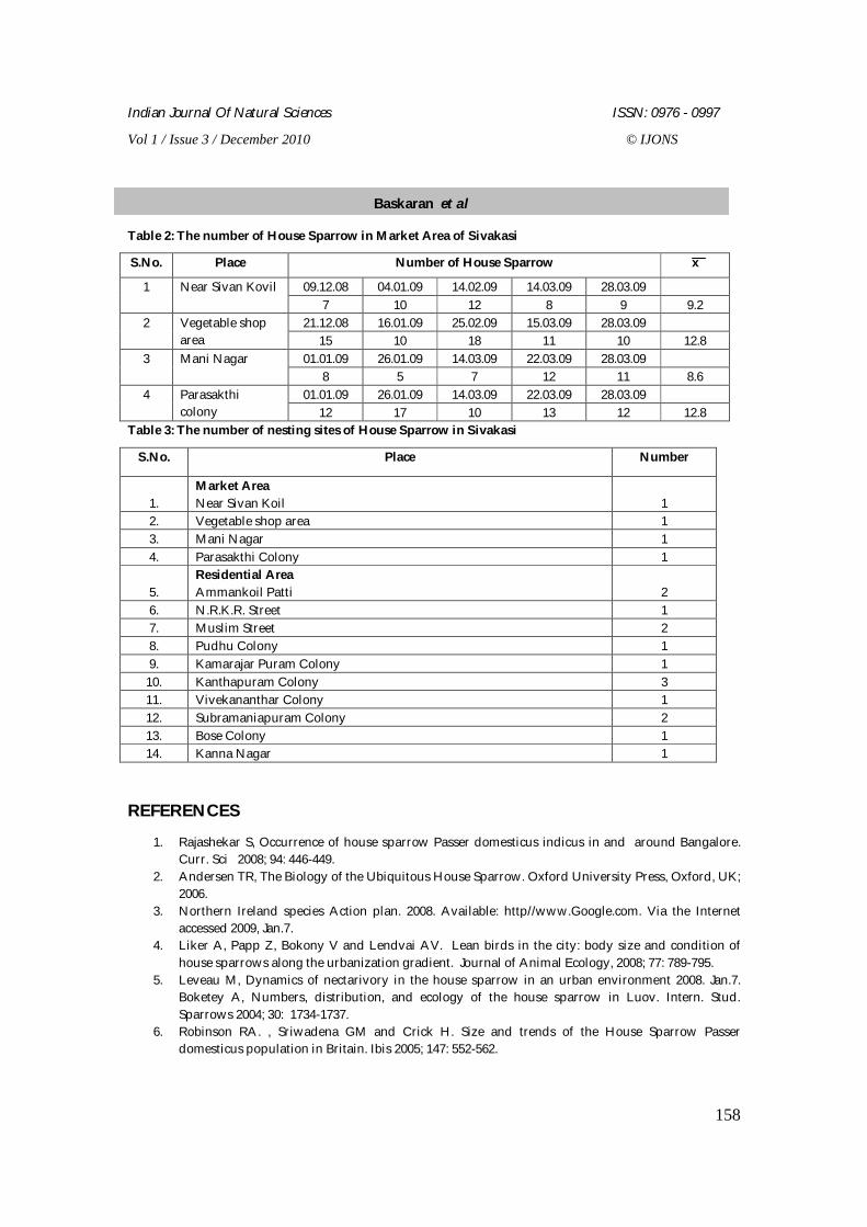
Table 2: The number of House Sparrow in Market Area of Sivakasi
S.No. Place Number of House Sparrow x
09.12.08 04.01.09 14.02.09 14.03.09 28.03.09 1 Near Sivan Kovil 7 10 12 8 9 9.2
21.12.08 16.01.09 25.02.09 15.03.09 28.03.09 2 Vegetable shop area 15 10 18 11 10 12.8
01.01.09 26.01.09 14.03.09 22.03.09 28.03.09 3 Mani Nagar 8 5 7 12 11 8.6
01.01.09 26.01.09 14.03.09 22.03.09 28.03.09 4 Parasakthi colony 12 17 10 13 12 12.8
Table 3: The number of nesting sites of House Sparrow in Sivakasi
S.No. Place Number
1.
Market Area Near Sivan Koil
1
2. Vegetable shop area 1 3. Mani Nagar 1 4. Parasakthi Colony 1
5. Residential Area Ammankoil Patti
2
6. N.R.K.R. Street 1 7. Muslim Street 2 8. Pudhu Colony 1 9. Kamarajar Puram Colony 1
10. Kanthapuram Colony 3 11. Vivekananthar Colony 1 12. Subramaniapuram Colony 2 13. Bose Colony 1 14. Kanna Nagar 1
REFERENCES
1. Rajashekar S, Occurrence of house sparrow Passer domesticus indicus in and around Bangalore. Curr. Sci 2008; 94: 446-449.
2. Andersen TR, The Biology of the Ubiquitous House Sparrow. Oxford University Press, Oxford, UK; 2006.
3. Northern Ireland species Action plan. 2008. Available: http//www.Google.com. Via the Internet accessed 2009, Jan.7.
4. Liker A, Papp Z, Bokony V and Lendvai AV. Lean birds in the city: body size and condition of house sparrows along the urbanization gradient. Journal of Animal Ecology, 2008; 77: 789-795.
5. Leveau M, Dynamics of nectarivory in the house sparrow in an urban environment 2008. Jan.7. Boketey A, Numbers, distribution, and ecology of the house sparrow in Luov. Intern. Stud. Sparrows 2004; 30: 1734-1737.
6. Robinson RA. , Sriwadena GM and Crick H. Size and trends of the House Sparrow Passer domesticus population in Britain. Ibis 2005; 147: 552-562.
Baskaran et al
Indian Journal Of Natural Sciences ISSN: 0976 - 0997
Vol 1 / Issue 3 / December 2010 © IJONS
159
Phytochemical and Anti Bacterial Activity of Tridax procumbens L. Ravikumar VR, *1 Saranya V,1 Sudha T,1 Parimala Devi B,2 and Ganesan V1
1Department of Pharmacognosy, The Erode College of Pharmacy and Research, Erode, Tamil Nadu, India.
2Department of Pharmacy, SASTRA University, Thanjavur, Tamil Nadu, India.
Received: 16 Aug 2010 Revised: 25 Nov 2010 Accepted: 1 Dec 2010
Address for correspondence
*V.R. Ravikumar,
Prof & Head, Department of Pharmacognosy,
The Erode College of Pharmacy & Research,
Perundurai Main Road, Veppampalayam, Erode-638 112, Tamil Nadu, India.
E-mail ID: [email protected].
The whole plant of Tridax procumbens L. family Asteraceae were extracted with three different solvents i.e, pet-ether, aqueous and ethanol. The crude extract fractions pet-ether, aqueous and ethanol of the plant Tridax procumbens L. found to contain Alkaloids, Steroids, Protein and Amino acids. Tridax procumbens L. investigated for its preliminary phytochemical and anti bacterial activity. The extracts showed significant anti bacterial activity against all the micro organisms tested and the effect so produced was compared with standard drug Gentamycin. However pet-ether shows the moderate anti bacterial activity against the entire micro organism. KEY WORDS; Phytochemical, Anti bacterial, Tridax procumbens L., Gentamycin INTRODUCTION Tridax procumbens L. (Asteraceae) is a weed found abundantly in agriculture fields and waste lands. It grows to about 45 cm tall and it has attractive flowers and leaves. It occurs in southern India states like Tamilnadu, Andra Pradesh, Kerala and Karnataka. Extensive literature survey reveals that studies on Hypoglycemic activity [1], Hemostatic activity [2], Anti oxidant activity [3], Hepatoprotective effect [4], Wound healing activity [5], Hypotensive activity [6] and Hepato toxicity[7], GC-MS analysis [8] of volatile compounds of Tridax procumbens L. resulted in the identification of sterols like Camphosterol, Stigmasterol, Beta-Ceto sterol. It is used by the local peoples to control Haemorrhage in wounds. In the present study, different fractions of the extracts of the plant have been investigated for Anti bacterial activity of Tridax procumbens L.
RESEARCH ARTICLE
ABSTRACT
Indian Journal Of Natural Sciences ISSN: 0976 - 0997
Vol 1 / Issue 3 / December 2010 © IJONS
160
MATERIALS AND METHODS Plant material The Tridax procumbens L. were collected from the lands of Anaikatti area in Coimbatore District of Tamil Nadu by uprooting the whole plant. It was identified by G.V.S.Moorthy, Joint director, Botanical survey of India, Tamil Nadu Agricultural University Campus (TNAU) Coimbatore. (Specimen no.BSI/SC/5/23/09-10/Tech-1143). All parts of the plant were removed carefully and shaken to remove unwanted particles like sand and soil. Drying was done under the shade. Shade drying was preferred to avoid denaturation of phytochemical constituents.
Preparation of extracts Dried plant were reduced in size to coarse powder with the aid of suitable means and three portions, each 150 gm was reserved for extraction with three different solvents like Petroleum Ether, Ethanol and aqueous extract. Size reduction provides more contact area for solvent penetration and facilities effective for extraction of phytochemicals. The powdered plant material was subjected to extraction in a soxhlet apparatus for 72 hours each using pet-ether (60-800C), ethanol and aqueous respectively [9]. Preliminary phytochemical studies All the fractions were subjected to preliminary phytochemical investigation for the presence of secondary metabolites such as alkaloids, carbohydrates, proteins and amino acids, saponins and flavonoids utilizing
standard methods of analysis. Anti bacterial screening [10] Anti bacterial activity of the plant was conducted by using Disc diffusion method. Nutrient agar media constituents were mixed with sufficient quantity of distilled water. Transfer into a boiling tube a quantity of 30ml. The tubes were autoclaved for 15min at a pressure of 15 ps/sq inch pressure. The selected micro organisms were Staphylococcus aureus, Micrococcus luteus, Proteus mirabilis and Shigella boydii . They were inoculated in the boiling tube. Later this was transferred into Petri dishes and kept on a flat surface. The test solution and standard solution of the concentration 1200µg/ml and control solution were taken in the Petri dishes and marked. The disc of similar size were made and soaked in the standard (Gentamycin),test and control solution and placed in the petri dishes at a distance. The Petri dishes were incubated at 370C for 24 hours. Inhibition of microbial growth was determined by absorbing the zone of inhibition both in test and standard.
Thin layer chromatography [11] Thin layer chromatography of pet-ether, ethanol and aqueous extract of Tridax procumbens L was carried out in different solvent system and best resolving system was choosen for running the plates. The plates were then exposed to various detecting systems.
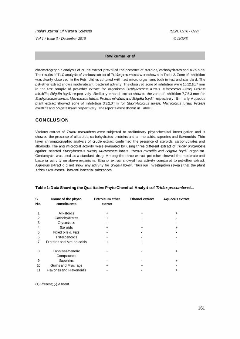
RESULTS The pet-ether and ethanol extract revealed the presence of alkaloids, carbohydrates, steroids, proteins and amino acids, gums and mucilages while the aqueous extract has shown the presence of alkaloids, steroids, tannins, phenolic compounds, saponins and flavonoids. The results were shown in table 1. Thin layer
Ravikumar et al
Indian Journal Of Natural Sciences ISSN: 0976 - 0997
Vol 1 / Issue 3 / December 2010 © IJONS
161
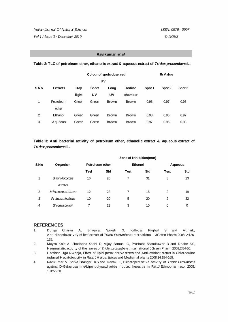
chromatographic analysis of crude extract prevailed the presence of steroids, carbohydrates and alkaloids. The results of TLC analysis of various extract of Tridax procumbens were shown in Table 2. Zone of inhibition was clearly observed in the Petri dishes cultured with test micro organisms both in test and standard. The pet-ether extract shows moderate anti bacterial activity. The observed zone of inhibition were 16,12,10,7 mm in the test sample of pet-ether extract for organisms Staphylococcus aureus, Micrococcus luteus, Proteus mirabilis, Shigella boydii respectively. Similarly ethanol extract showed the zone of inhibition 7,7,5,3 mm for Staphylococcus aureus, Micrococcus luteus, Proteus mirabilis and Shigella boydii respectively. Similarly Aqueous plant extract showed zone of inhibition 3,3,2,0mm for Staphylococcus aureus, Micrococcus luteus, Proteus mirabilis and Shigella boydii respectively. The reports were shown in Table 3.
CONCLUSION Various extract of Tridax procumbens were subjected to preliminary phytochemical investigation and it showed the presence of alkaloids, carbohydrates, proteins and amino acids, saponins and flavonoids. Thin layer chromatographic analysis of crude extract confirmed the presence of steroids, carbohydrates and alkaloids. The anti microbial activity were evaluated by using three different extract of Tridax procumbens against selected Staphylococcus aureus, Micrococcus luteus, Proteus mirabilis and Shigella boydii organism. Gentamycin was used as a standard drug. Among the three extract pet-ether showed the moderate anti bacterial activity on above organisms. Ethanol extract showed less activity compared to pet-ether extract. Aqueous extract did not show any activity for Shigella boydii. Thus our investigation reveals that the plant Tridax Procumbens L has anti bacterial substances.
Table 1: Data Showing the Qualitative Phyto Chemical Analysis of Tridax procumbens L. S. No.
Name of the phyto constituents
Petroleum ether extract
Ethanol extract Aqueous extract
1 Alkaloids + + + 2 Carbohydrates + + - 3 Glycosides - - - 4 Steroids + + + 5 Fixed oils & Fats - - - 6 Triterpenoids - - - 7 Proteins and Amino acids + + -
8 Tannins Phenolic Compounds
- - +
9 Saponins - - + 10 Gums and Mucilage + + - 11 Flavones and Flavonoids - - +
(+) Present; (-) Absent.
Ravikumar et al
Indian Journal Of Natural Sciences ISSN: 0976 - 0997
Vol 1 / Issue 3 / December 2010 © IJONS
162
Table 2: TLC of petroleum ether, ethanolic extract & aqueous extract of Tridax procumbens L.
Colour of spots observed Rf Value
UV
S.No
Extracts
Day
light
Short
UV
Long
UV
Iodine
chamber
Spot 1
Spot 2
Spot 3
1 Petroleum
ether
Green Green Brown Brown 0.98 0.97 0.96
2 Ethanol Green Green Brown Brown 0.98 0.96 0.97
3 Aqueous Green Green brown Brown 0.97 0.96 0.98
Table 3: Anti bacterial activity of petroleum ether, ethanolic extract & aqueous extract of Tridax procumbens L.
Zone of Inhibition(mm)
Petroleum ether Ethanol Aqueous
S.No
Organism
Test Std Test Std Test Std
1 Staphylococcus
aureus
16 20 7 31 3 23
2 Micrococcus luteus 12 28 7 15 3 19
3 Proteus mirabilis 10 20 5 20 2 32
4 Shigella boydii 7 23 3 10 0 0
REFERENCES 1. Durga Charan A, Bhagwat Suresh G, Killedar Raghul S and Adhaik,
Anti-diabetic activity of leaf extract of Tridax Procumbens: International J Green Pharm 2008; 2:126-128.
2. Mayra Kale A, Shadhana Shahi R, Vijay Somani G, Prashant Shamkuwar B and Dhake AS, Heamostatic activity of the leaves of Tridax procumbens :International J Green Pharm 2008;2:54-55.
3. Harrison Ugo Nwanjo, Effect of lipid peroxidative stress and Anti-oxidant status in Chloroquine induced Hepatotoxicity in Rats: J Herbs, Spices and Medicinal plants 2008;14:154-165.
4. Ravikumar V, Shiva Shangari KS and Devaki T, Hepatoprotective activity of Tridax Procumbens against D-Galactosamine/Lipo polysaccharide induced hepatitis in Rat:.J Ethnopharmacol 2005; 101:55-60.
Ravikumar et al
Indian Journal Of Natural Sciences ISSN: 0976 - 0997
Vol 1 / Issue 3 / December 2010 © IJONS
163
5. Udupa AL, Kulkarmi DR and Udupa SL, Effects of Tridax Procumbens Extracts on wound healing:
Pharm biology 1995; 33:37-40. 6. Salhahdeen HM, Yemitan O.K and Alada A.R, Effects of Aqueous leaf extract of Tridax Procumbens
on blood: African J Biomedical Research 2004; 7: 27-29. 7. Kumar S, Asokar D, Kumar P, Kalavathy S and Manoharan N, Anti-hepatotoxic activity and Anti-
oxidant defence potential of Tridax Procumbens: Indian J pharm Sci Jan- Feb-2001; 3:64. 8. Gadre A and Gabhe SY, Identification of some sterols of Tridax Procumbens by GC-MS: Indian J
pharm sci S1993; 55: 191-192. 9. Kokate DRCK, Practical pharmacognosy, IV Edition, Vallabh Prakashan Publication, 1993: 107. 10. Michael. J. Pelzar, J.R E.C.S. Chan, Noel. R. Krieg, Microbiology, Edition-V, Tata Mc Graw-Hill
publishing company Ltd, New Delhi, 1993. 15, 30: 344. 11. Ravisankar S, Text book of Pharmaceutical analysis, Edition-III, Rx Publicatons, 1993: 107.
Ravikumar et al
Indian Journal Of Natural Sciences ISSN: 0976 - 0997
Vol 1 / Issue 3 / December 2010 © IJONS
164
Analysis of Hydrophobic Activity of Drugs for Diabetes Mellitus
Ramanathan K1* and Karthick H2
1Department of Bioinformatics, Thanthai Hans Roever College, Perambalur, Tamil Nadu, India
2Department of Bioinformatics, PRIST University, Thanjavur, Tamil Nadu, India
Received: 10 Oct 2010 Revised: 25 Nov 2010 Accepted: 2 Dec 2010 *Address for correspondence: Ramanathan. K
Department of Bioinformatics, Thanthai Hans Roever College, Perambalur, Tamil Nadu, India
Email ID: [email protected]
Nowadays, Diabetes is a challengeable disease in India. A large work has been focussed on analysis of hydrophobic activity of drugs for diabetes mellitus. In this work we address a specific suitable ligand for diabetes mellitus. We have been retrieved that the Gene and Protein which is responsible for diabetes mellitus and target binding site has been identified. The list of drugs is retrieved from drug bank which are used to treat diabetes mellitus and the best ligand has been identified based on molecular docking. We are highly concentrated in the hydrophobic activity of the ligand. Finally we have observed that the ligand acetohexamide has the highest hydrophobic activity when compared with other ligands. Hydrophobic activity is the most essential parameter in all the drug molecules. Keywords: Diabetes mellitus, Drug designing, Ligands, hydrophobic activity, Docking INTRODUCTION The risk of diabetes is markedly higher (two- to three-fold) in patients with schizophrenia compared with the general population and evidence suggests a similar increased incidence of diabetes in patients with bipolar disorder and schizoaffective disorder The main potential metabolic concerns in addition to the risk of developing type II diabetes are the risk for cardiovascular disease (CVD) and shorter life-expectancy. Mortality among patients with schizophrenia is higher than among the general population, and CVD accounts for a significant proportion of this excess mortality. [1]
RESEARCH ARTICLE
ABSTRACT
Indian Journal Of Natural Sciences ISSN: 0976 - 0997
Vol 1 / Issue 3 / December 2010 © IJONS
165
Diabetes mellitus Type 1 diabetes is an autoimmune disease. An autoimmune disease results when the body’s system for fighting infection turns against a part of the body .Type 2 diabetes is most often associated with older age, obesity, physical activity and certain ethnicities. In Pre-diabetes, blood glucose levels are higher than normal but not high enough to be characterized as diabetes. Pre-diabetes also increases the risk of heart disease and stroke with weight loss and physical activity. Diabetes mellitus is a complex, multifactor and polygenic disease likely to be caused by one or more gene alterations action in combination with non-genetic factors.
Insulin is a hypoglycemic hormone and it is composed of two peptide chains referred to as chain A and chain B. These chains are linked together by two disulfide bonds. Insulin is a small protein with a molecular weight of about 6000 Daltons. It is synthesized in significant quantities only in beta cells of the Pancreas. [2]
Hydrophobic activity
In contrast to many traditional pharmaceutical agents that exhibit a high degree of aqueous solubility, new drug candidates are frequently highly lipophilic compounds. The aqueous environment of the blood provides a thermodynamically unfavourable environment for the disposition of such hydrophobic drugs. However, this limitation can be overcome by association with circulating lipoproteins. [3]
Drug Designing Drug designing is the approach of finding drugs based on their targets and typically a drug target is a key molecule involved in a particular metabolism or signaling Pathway that is specific to a disease condition or Pathology or to be infectivity or survival of a microbial Pathogen. Some approaches attempt to stop the functioning of the pathway in the diseased state by causing a key molecule to stop functioning. However these drugs would also have to be designed in such a way as not to affect any other important molecules that may be similar in appearance to the key molecules. [4]
Docking Docking is a method which predicts the preferred orientation of one molecule to a record when bound to each other to form stable complex knowledge of the preferred orientations in turn may be used to predict the binding strength of association or binding affinity between two molecules. Docking is frequently used to predict the binding orientations of small molecules drug candidates to protein targets in order to in turn predict the affinity and activity of the small molecule. The receiving molecule that primarily binds to a small molecule or another protein or a nucleic acid called receptor. A molecule that forms the complementary partner in the docking process called ligand. [5] Ligand Acetohexamide is the first generation sulfonylurea medication used to treat diabetes mellitus type 2, particularly in people whole diabetes cannot be controlled by diet alone. Acetohexamide lowers blood sugar
Ramanathan and Karthick
Indian Journal Of Natural Sciences ISSN: 0976 - 0997
Vol 1 / Issue 3 / December 2010 © IJONS
166
by stimulating pancreas to secrete insulin and helps the body use insulin efficiently. The pancreas must produce insulin for this medication to work. It is an oral anti diabetic agent and is metabolized by the reductive conversion of the acetoxy group to a secondary alcohol metabolite. We tested whether reductase activity for acetohexamide can be found in human erythrocytes. Acetohexamide interact with other drugs such as alcohol, Beta blockers, cisapride, clofibrate, rifampin etc. If we missed to take dose, skip the missed dose and take only the next regularly scheduled dose. If acetoheaxamide will be a overdose it cause symptoms include hunger, nausea, anxiety, cold sweats, weakness, drowsiness and coma. [6] MATERIALS AND METHODS The Protein sequence has been retrieved from GenBank which is responsible for diabetes mellitus. The no. of amino acids present in IRAK protein has been identified. Then the lists of drugs for diabetes mellitus are retrieved and analyzed the hydrophobic activity for each drugs. Hydrophobic activity is calculated by ALOGPS tool. The Hydrophobic features are more responsible for all types of drugs. The distribution of the Log P and Log S values for each drug show the highest hydrophobic activity of the drug. The structure of the protein retrieved from Protein data bank and binding sites of the receptor was calculated by PROSITE tool.
RESULTS AND DISCUSSION Hydrophobic Activity Of Various Drugs ACETOHEXAMIDE mol_N logP logS SMILES mol_1 1.72 -3.83 CC(=O)c1ccc(cc1)S(=O)(=O)NC(=O)NC2CCCCC2 METFORMIN mol_N logP logS SMILES mol_1 -1.41 -1.76 CN(C)C(=N)N=C(N)N PHENFORMIN mol_N logP logS SMILES mol_1 0.30 -3.02 c1ccc(cc1)CCN=C(N)N=C(N)N MIGLITOL mol_N logP logS SMILES mol_1 -2.29 0.47 C1C(C(C(C(N1CCO)CO)O)O)O
Ramanathan and Karthick
Indian Journal Of Natural Sciences ISSN: 0976 - 0997
Vol 1 / Issue 3 / December 2010 © IJONS
167
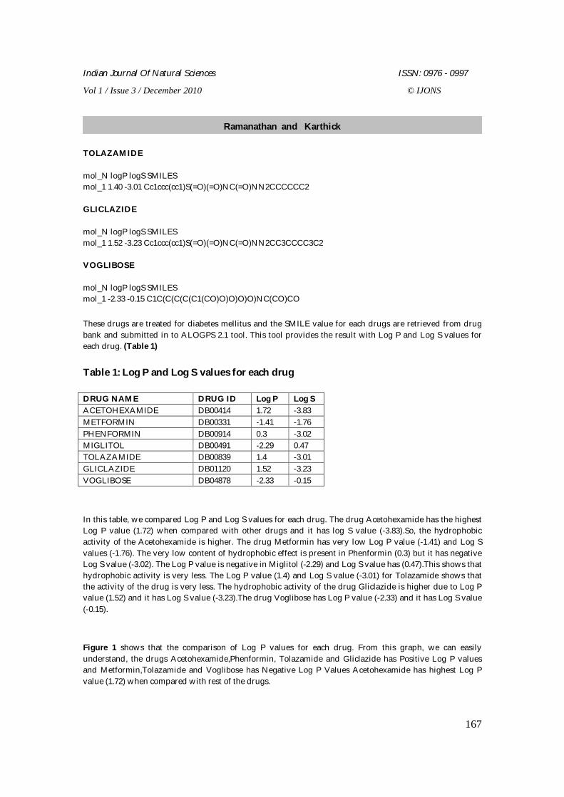
TOLAZAMIDE mol_N logP logS SMILES mol_1 1.40 -3.01 Cc1ccc(cc1)S(=O)(=O)NC(=O)NN2CCCCCC2 GLICLAZIDE mol_N logP logS SMILES mol_1 1.52 -3.23 Cc1ccc(cc1)S(=O)(=O)NC(=O)NN2CC3CCCC3C2 VOGLIBOSE mol_N logP logS SMILES mol_1 -2.33 -0.15 C1C(C(C(C(C1(CO)O)O)O)O)NC(CO)CO These drugs are treated for diabetes mellitus and the SMILE value for each drugs are retrieved from drug bank and submitted in to ALOGPS 2.1 tool. This tool provides the result with Log P and Log S values for each drug. (Table 1) Table 1: Log P and Log S values for each drug DRUG NAME DRUG ID Log P Log S ACETOHEXAMIDE DB00414 1.72 -3.83 METFORMIN DB00331 -1.41 -1.76 PHENFORMIN DB00914 0.3 -3.02 MIGLITOL DB00491 -2.29 0.47 TOLAZAMIDE DB00839 1.4 -3.01 GLICLAZIDE DB01120 1.52 -3.23 VOGLIBOSE DB04878 -2.33 -0.15 In this table, we compared Log P and Log S values for each drug. The drug Acetohexamide has the highest Log P value (1.72) when compared with other drugs and it has log S value (-3.83).So, the hydrophobic activity of the Acetohexamide is higher. The drug Metformin has very low Log P value (-1.41) and Log S values (-1.76). The very low content of hydrophobic effect is present in Phenformin (0.3) but it has negative Log S value (-3.02). The Log P value is negative in Miglitol (-2.29) and Log S value has (0.47).This shows that hydrophobic activity is very less. The Log P value (1.4) and Log S value (-3.01) for Tolazamide shows that the activity of the drug is very less. The hydrophobic activity of the drug Gliclazide is higher due to Log P value (1.52) and it has Log S value (-3.23).The drug Voglibose has Log P value (-2.33) and it has Log S value (-0.15). Figure 1 shows that the comparison of Log P values for each drug. From this graph, we can easily understand, the drugs Acetohexamide,Phenformin, Tolazamide and Gliclazide has Positive Log P values and Metformin,Tolazamide and Voglibose has Negative Log P Values Acetohexamide has highest Log P value (1.72) when compared with rest of the drugs.
Ramanathan and Karthick
Indian Journal Of Natural Sciences ISSN: 0976 - 0997
Vol 1 / Issue 3 / December 2010 © IJONS
168
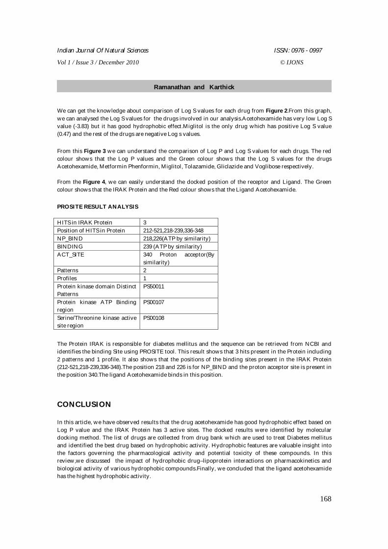
We can get the knowledge about comparison of Log S values for each drug from Figure 2.From this graph, we can analysed the Log S values for the drugs involved in our analysis.Acetohexamide has very low Log S value (-3.83) but it has good hydrophobic effect.Miglitol is the only drug which has positive Log S value (0.47) and the rest of the drugs are negative Log s values.
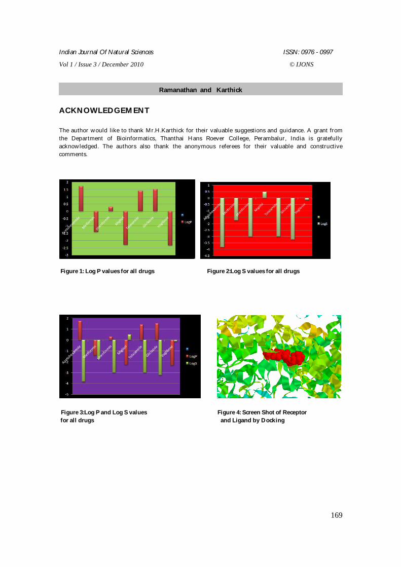
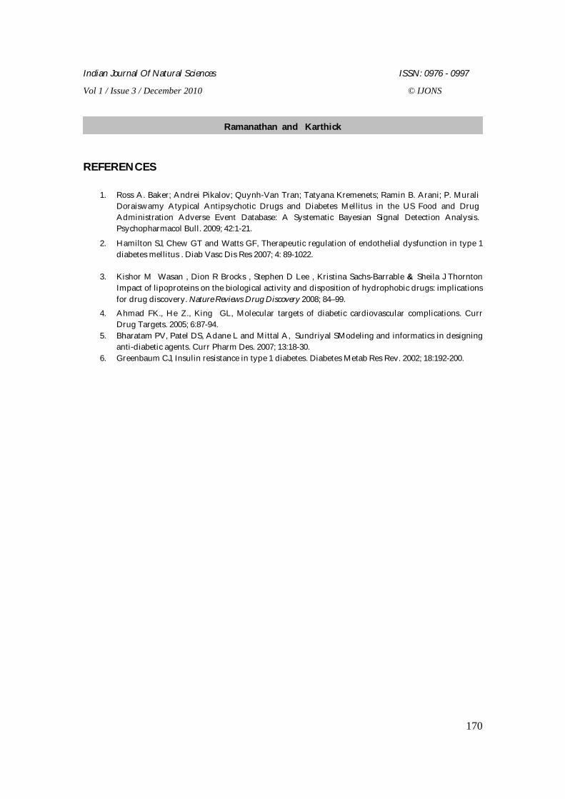
From this Figure 3 we can understand the comparison of Log P and Log S values for each drugs. The red colour shows that the Log P values and the Green colour shows that the Log S values for the drugs Acetohexamide, Metformin Phenformin, Miglitol, Tolazamide, Gliclazide and Voglibose respectively. From the Figure 4, we can easily understand the docked position of the receptor and Ligand. The Green colour shows that the IRAK Protein and the Red colour shows that the Ligand Acetohexamide. PROSITE RESULT ANALYSIS HITS in IRAK Protein 3 Position of HITS in Protein 212-521,218-239,336-348 NP_BIND 218,226(ATP by similarity) BINDING 239 (ATP by similarity) ACT_SITE 340 Proton acceptor(By
similarity) Patterns 2 Profiles 1 Protein kinase domain Distinct Patterns
PS50011
Protein kinase ATP Binding region
PS00107
Serine/Threonine kinase active site region
PS00108
The Protein IRAK is responsible for diabetes mellitus and the sequence can be retrieved from NCBI and identifies the binding Site using PROSITE tool. This result shows that 3 hits present in the Protein including 2 patterns and 1 profile. It also shows that the positions of the binding sites present in the IRAK Protein (212-521,218-239,336-348).The position 218 and 226 is for NP_BIND and the proton acceptor site is present in the position 340.The ligand Acetohexamide binds in this position.
CONCLUSION
In this article, we have observed results that the drug acetohexamide has good hydrophobic effect based on Log P value and the IRAK Protein has 3 active sites. The docked results were identified by molecular docking method. The list of drugs are collected from drug bank which are used to treat Diabetes mellitus and identified the best drug based on hydrophobic activity. Hydrophobic features are valuable insight into the factors governing the pharmacological activity and potential toxicity of these compounds. In this review,we discussed the impact of hydrophobic drug–lipoprotein interactions on pharmacokinetics and biological activity of various hydrophobic compounds.Finally, we concluded that the ligand acetohexamide has the highest hydrophobic activity.
Ramanathan and Karthick
Indian Journal Of Natural Sciences ISSN: 0976 - 0997
Vol 1 / Issue 3 / December 2010 © IJONS
169
ACKNOWLEDGEMENT The author would like to thank Mr.H.Karthick for their valuable suggestions and guidance. A grant from the Department of Bioinformatics, Thanthai Hans Roever College, Perambalur, India is gratefully acknowledged. The authors also thank the anonymous referees for their valuable and constructive comments.
Figure 1: Log P values for all drugs Figure 2:Log S values for all drugs
Figure 3:Log P and Log S values Figure 4: Screen Shot of Receptor for all drugs and Ligand by Docking
Ramanathan and Karthick
Indian Journal Of Natural Sciences ISSN: 0976 - 0997
Vol 1 / Issue 3 / December 2010 © IJONS
170
REFERENCES
1. Ross A. Baker; Andrei Pikalov; Quynh-Van Tran; Tatyana Kremenets; Ramin B. Arani; P. Murali Doraiswamy Atypical Antipsychotic Drugs and Diabetes Mellitus in the US Food and Drug Administration Adverse Event Database: A Systematic Bayesian Signal Detection Analysis. Psychopharmacol Bull. 2009; 42:1-21.
2. Hamilton SJ, Chew GT and Watts GF, Therapeutic regulation of endothelial dysfunction in type 1 diabetes mellitus . Diab Vasc Dis Res 2007; 4: 89-1022.
3. Kishor M Wasan , Dion R Brocks , Stephen D Lee , Kristina Sachs-Barrable & Sheila J Thornton Impact of lipoproteins on the biological activity and disposition of hydrophobic drugs: implications for drug discovery. Nature Reviews Drug Discovery 2008; 84–99.
4. Ahmad FK., He Z., King GL, Molecular targets of diabetic cardiovascular complications. Curr Drug Targets. 2005; 6:87-94.
5. Bharatam PV, Patel DS, Adane L and Mittal A, Sundriyal SModeling and informatics in designing anti-diabetic agents. Curr Pharm Des. 2007; 13:18-30.
6. Greenbaum CJ, Insulin resistance in type 1 diabetes. Diabetes Metab Res Rev. 2002; 18:192-200.
Ramanathan and Karthick
Indian Journal Of Natural Sciences ISSN: 0976 - 0997
Vol 1 / Issue 3 / December 2010 © IJONS
171
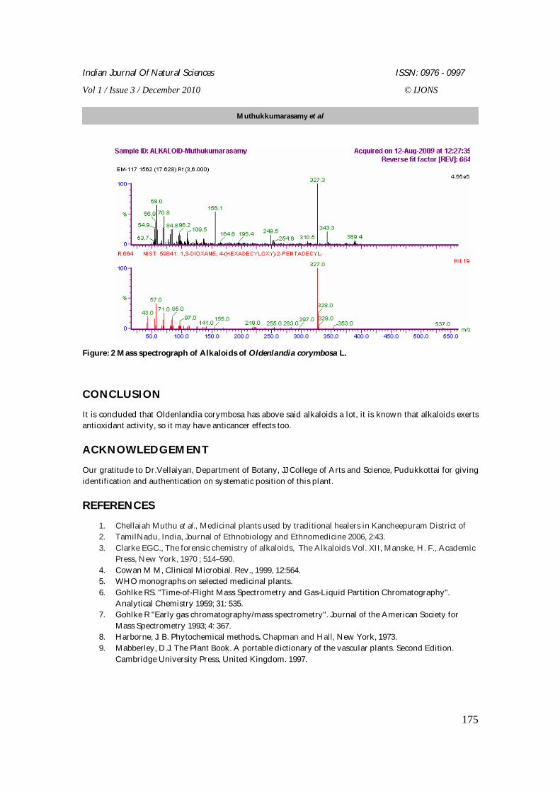
Analysis of Phytochemicals by GC-MS on Oldenlandia corymbosa L. Muthukkumarasamy S1*, Veerappan RM2, and Balasundaram M3 1Department of Nanoscience and Technology, Alagappa University, Karaikudi, Tamil Nadu, India.
2Department of Biochemistry, JJ College of Arts and Science, Pudukkottai Tamil Nadu, India.
3Faculty of Medicine, AIMST University, Malaysia.
Received: 5 Aug 2010 Revised: 28 Nov 2010 Accepted: 1 Dec 2010
*Address for correspondence: Muthukkumarasamy S
Department of Nanoscience and Technology, Alagappa University, Karaikudi 630 003, Tamil Nadu, India
E mail ID: [email protected], [email protected].
Oldenlandia corymbosa L. (Rubiacea) is used against snake bite since ancient period and during labour also it used for strong uterine contraction. But its chemistry was not been explored still hence it has been take for phytochemical analysis by using Gas Chromatography coupled with Mass Spectrometry (GC-MS). There are 15 alkaloids were found out by using ethanolic extract of O. corymbosa, notably morpholin, quinazoline, erucamide alverine and dihydro pyrrole were found to present. Those alkaloids were already tested for its biological functions; the results suggest that plenty of those alkaloids available in this plant. Key words: O. corymbosa, morpholin, quinazoline, erucamide alverine, dihydro pyrrole and GC-MS INTRODUCTION Plants have an almost limitless ability to synthesize aromatic substances mainly secondary metabolites, of which at least 12,000 have been isolated, a number estimated to be less than 10% of the total. In many cases, these substances serve as the molecules of plant defense against predation by microorganisms, insects, and herbivores. Further, some of which may involve in plant odor (terpenoids), pigmentation (tannins and quinines), and flavor (capsacin). However, several of these molecules possess medicinal properties [1, 3, 14]. Manufacturing of Herbal and Ayurvedic products is simple and also good market demand for these products. According to the WHO (World Health Organization) as much as 80% of the world's population relies on traditional medicine. With increased concerns about rising health care costs, some governments are encouraging the use of indigenous form as of medicines rather than expensive drugs. This has been a strong driver for resuscitation of herbal and Ayurvedic medicine in the country. Traditional treatment with
RESEARCH ARTICLE
ABSTRACT
Indian Journal Of Natural Sciences ISSN: 0976 - 0997
Vol 1 / Issue 3 / December 2010 © IJONS
172
Ayurvedic and other herbal medicines is well established and widely acknowledged to be safe effective [2,8,9]. The investigation revealed that, the traditional healers used 85 species (including Corymbosa) of plants distributed in 76 genera belonging to 41 families to treat various diseases. Oldenlandia corymbosa L. is used for arresting blood bleeding [1]. Oldenlandia corymbosa L. of Thai origin, a member of the family Rubiaceae, is widely distributed in tropical regions of Asia. The decoction of whole plants used in tradition Thai medicine for antipyretic purpose to decrease body temperature. Pharmaceutically, the anti-inflammatory, antioxidant, and hepatoprotective properties of plant extracts have been reported. This plant is well known to contain mainly iridoid glucosides. However, there are a few reports on the presence of other types of compounds, particularly anthra quinones and triterpenoids [3, 4]. γ-Sitosterol and the triterpene acids, oleanolic acid and ursolic acid, have been shown to be present in the Indian medicinal plant Oldenlandia corymbosa L. Evidence is presented to show that the plant does not contain any alkaloid [5].
Gas chromatography-mass spectrometry (GC-MS) is a method that combines the features of gas-liquid chromatography and mass spectrometry to identify different substances within a test sample. Applications of GC-MS include drug detection, fire investigation, environmental analysis, explosives investigation, and identification of unknown samples. GC/MS can also be used in airport security to detect substances in luggage or on human beings. Additionally, it can identify trace elements in materials that were previously thought to have disintegrated beyond identification.
MATERIALS AND METHODS Alkaloids cannot be identified or quantified by a single method because these are highly heterogeneous chemically and there are so many of them. In general, it is difficult to identify an alkaloid from a new plant source without knowing approximately what type of alkaloid is likely to be found there. Also, because of the wide range of solubility and other properties of alkaloids, any general screening procedure for alkaloids in plants may fail to detect particular compounds [6].
Preparation of plant extract for phytochemical analysis
About 250g of dry powder of Oldenlandia corymbosa L. was extracted with diethyl ether, acetone and ethanol at 60oc to 80oc by continuous hot percolation using Soxhlet apparatus. The extraction was filtered and kept in over at 50oc for 24 hours to evaporate the extracts from them. A greenish black waxy residue was obtained. These extracts were used for phytochemical analysis qualitatively.
Preparation of plant extract for alkaloid analysis by GC-MS
Plants (whole plant) were collected, cleaned with tap water and dried under shade. The dried parts of medicinal plants were ground well to find powder. The ground sample was made alkaline with 39% ammonia and extracted with chloroform at room temperature for a total period of 24 hrs and then the extract was partitioned between 5% HCl and chloroform. The aqueous phase was made alkaline again with ammonia and partitioned between water and chloroform. Finally chloroform was totally evaporated from the organic phase to form the alkaloid powder [6].
Muthukkumarasamy et al
Indian Journal Of Natural Sciences ISSN: 0976 - 0997
Vol 1 / Issue 3 / December 2010 © IJONS
173
Phytochemical analysis Phytochemical analysis for major phytoconstituents of the plant extract was undertaken using standard qualitative methods as described by various authors [7, 18, 19, 20]. The plant extracts were screened for the presence of biologically active compounds like alkaloids, flavonoids, glycosides, carbohydrates, phytosteroids and fatty acids, proteins, phenolics, tannins and saponins.
RESULTS AND DISCUSSION
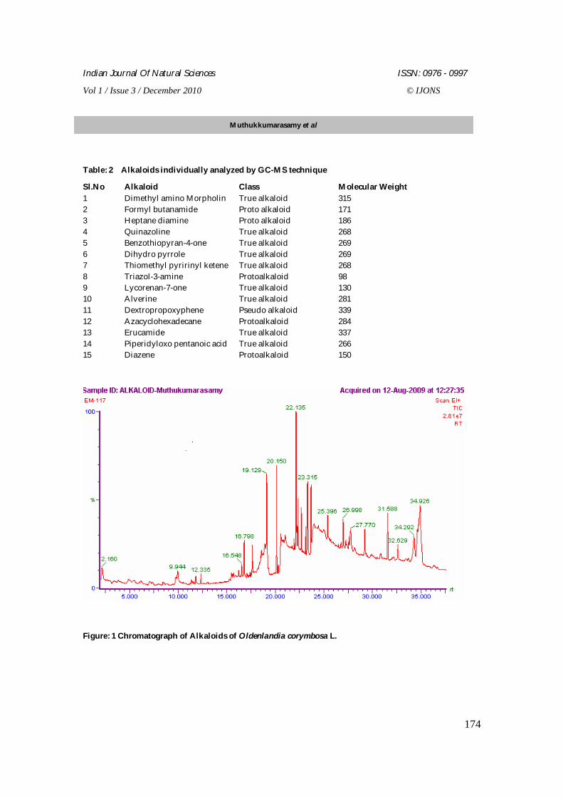
Phytochemical constituents like alkaloids, flavonoids, carbohydrates, glycosides, phytosterols, fixed oil and fats, proteins, phenolic compounds, and saponins of Oldenlandia corymbosa were analyzed by qualitatively and reported in Table - 1. Gas chromatograph of Alkaloids of Oldenlandia corymbosa L. was recorded, each peak in chromatograph implies the presence of group of Alkaloids. Mass spectrograph of Alkaloids of Oldenlandia corymbosa L. was recorded, each peak in spectrograph implies the presence of individual Alkaloids according to their charge to mass ratio (e/m) which are indicated in figures 1and 2 [10,11,12,13].
The following alkaloids were individually analyzed by GC-MS technique
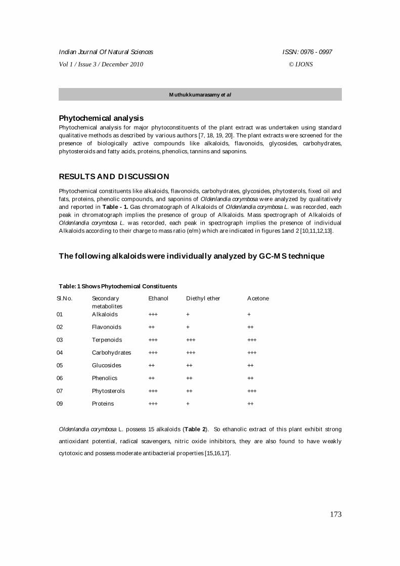
Table: 1 Shows Phytochemical Constituents
Sl.No. Secondary metabolites
Ethanol Diethyl ether Acetone
01 Alkaloids +++ + +
02 Flavonoids ++ + ++
03 Terpenoids +++ +++ +++
04 Carbohydrates +++ +++ +++
05 Glucosides ++ ++ ++
06 Phenolics ++ ++ ++
07 Phytosterols +++ ++ +++
09 Proteins +++ + ++
Oldenlandia corymbosa L. possess 15 alkaloids (Table 2). So ethanolic extract of this plant exhibit strong
antioxidant potential, radical scavengers, nitric oxide inhibitors, they are also found to have weakly
cytotoxic and possess moderate antibacterial properties [15,16,17].
Muthukkumarasamy et al
Indian Journal Of Natural Sciences ISSN: 0976 - 0997
Vol 1 / Issue 3 / December 2010 © IJONS
174
Table: 2 Alkaloids individually analyzed by GC-MS technique
Sl.No Alkaloid Class Molecular Weight 1 Dimethyl amino Morpholin True alkaloid 315 2 Formyl butanamide Proto alkaloid 171 3 Heptane diamine Proto alkaloid 186 4 Quinazoline True alkaloid 268 5 Benzothiopyran-4-one True alkaloid 269 6 Dihydro pyrrole True alkaloid 269 7 Thiomethyl pyririnyl ketene True alkaloid 268 8 Triazol-3-amine Protoalkaloid 98 9 Lycorenan-7-one True alkaloid 130 10 Alverine True alkaloid 281 11 Dextropropoxyphene Pseudo alkaloid 339 12 Azacyclohexadecane Protoalkaloid 284 13 Erucamide True alkaloid 337 14 Piperidyloxo pentanoic acid True alkaloid 266 15 Diazene Protoalkaloid 150
Figure: 1 Chromatograph of Alkaloids of Oldenlandia corymbosa L.
Muthukkumarasamy et al
Indian Journal Of Natural Sciences ISSN: 0976 - 0997
Vol 1 / Issue 3 / December 2010 © IJONS
175
Figure: 2 Mass spectrograph of Alkaloids of Oldenlandia corymbosa L.
CONCLUSION
It is concluded that Oldenlandia corymbosa has above said alkaloids a lot, it is known that alkaloids exerts antioxidant activity, so it may have anticancer effects too.
ACKNOWLEDGEMENT
Our gratitude to Dr.Vellaiyan, Department of Botany, JJ College of Arts and Science, Pudukkottai for giving identification and authentication on systematic position of this plant.
REFERENCES
1. Chellaiah Muthu et al., Medicinal plants used by traditional healers in Kancheepuram District of 2. TamilNadu, India, Journal of Ethnobiology and Ethnomedicine 2006, 2:43. 3. Clarke EGC., The forensic chemistry of alkaloids, The Alkaloids Vol. XII, Manske, H. F., Academic
Press, New York, 1970 ; 514–590. 4. Cowan M M, Clinical Microbial. Rev., 1999, 12:564. 5. WHO monographs on selected medicinal plants. 6. Gohlke RS. "Time-of-Flight Mass Spectrometry and Gas-Liquid Partition Chromatography".
Analytical Chemistry 1959; 31: 535. 7. Gohlke R "Early gas chromatography/mass spectrometry". Journal of the American Society for
Mass Spectrometry 1993; 4: 367. 8. Harborne, J. B. Phytochemical methods. Chapman and Hall, New York, 1973. 9. Mabberley, D.J. The Plant Book. A portable dictionary of the vascular plants. Second Edition.
Cambridge University Press, United Kingdom. 1997.
Muthukkumarasamy et al
Indian Journal Of Natural Sciences ISSN: 0976 - 0997
Vol 1 / Issue 3 / December 2010 © IJONS
176
10. Subramanyam Ragupathy, Consensus of the 'Malasars' traditional aboriginal knowledge of medicinal plants in the Velliangiri holy hills, India, Journal of Ethnobiology and Ethnomedicine 2008, 4:8 doi:10.1186/1746-4269-4-8.
11. V. P. Kamboj ,Herbal medicine ,Current science, 2000, 78- 1:35-39 12. Rudolf Fritz Weiss Lehrbuch der Phytotherapie, Beaconsfield Publishers Ltd, Beaconsfield,
England. 2000, 78: 1. 13. Zobel, A. M. Localization of phenolic compounds in tannin-secreting cells from Sambucus racemosa
shoots. Annals of Botany. 1986, 57: 801-810. 14. Witte L., Muller K. and Alfermann H-AInvestigation of alkaloid pattern of Datura innoxia plants by
capillary gas-liquid-chromatography/mass spectrometry. Planta Medica ,1987, 52:192-197. 15. Parr A., Payne J., Eagles J., Champan B., Robins R. and Rhodes M. Variation of tropane alkaloid
accumulation within the Solanaceae and strategies for itsexploitation. Phytochemistry, 1990,29 : 2545-2550.
16. Pawadee et al, Chemical constituents from Oldenlandia corymbosa L. of Thai origin, Natural medicine 2008 62:249-250.
17. Gill LS , Ehtnomedical use of plants, 1992. 18. Stein, S.E. Scott, D.R ,Optimization and Testing of Mass Spectral Library Search Algorithms for
Compound Identification”. J. Am. Soc. Mass Spectrum. 1994, 5:859-866. 19. Davies, T.,The New Automated Mass Spectrometry Deconvolution and Identification System
(AMDIS)”, Spectroscopy, Europe, 1998, 3: 24-27. 20. Gaithersburg, MD.,Standard Reference Data Program, National Institute of Standards and
Technology, Standard Reference Database IA. Internet address: http://www.nist.gov/srd/nist1a.htm.
Muthukkumarasamy et al
Indian Journal Of Natural Sciences ISSN: 0976 - 0997
Vol 1 / Issue 3 / December 2010 © IJONS
177
Bacopa monnieri (L.) Pennell. - an Overview
Parimala Devi B*1 and Ramasubramaniaraja R2
1Professor, Department of pharmacy SCBT, SASTRA University, Thanjavur, Tamil Nadu, India
2Lecturer, Department of Pharmacognosy, Seven Hills College of Pharmacy, Tirupati, Andhra Pradesh, India Received: 18 Oct 2010 Revised: 6 Nov 2010 Accepted: 28 Nov 2010
*Address for correspondence:
Parimala Devi B,
Prof & Head, Department of Pharmacy, SCBT, SASTRA University, Thanjavur. TamilNadu, India.
Email ID: [email protected], [email protected].
Developing countries like India is having a rich heritage of medicinal plants. Based on the natural resources available, many of the medicinal herbs being used effectively for their therapeutic potentials by the tribal and most of the rural and some of urban societies too. Recently the diseases like cancer and Diabetes found to be much higher ratio among Indian population, based on known and unknown factors too. Among the medicinal plants used in the management of cancer, the plant Bacopa monnieri (L.) Pennell also one reputed plant used in the cancer studies. Bacopa monnieri (L.) Pennell is famous for its antistress, immunomodulatory; cognition-facilitating, anti-inflammatory and anti-aging effects produced by it in experimental animals and in clinical situations and may justify further investigation of its other beneficial properties. Moreover, this experimental evidence suggests that, Bacopa monneri may be useful in the treatment of human pathologies in which free radical production plays a key role. This review highlights the details about the plant and its therapeutic usefulness in the management of cancer and related complications.
Keywords: Bacopa monnieri (L.) Pennell; antioxidant; cancer, medicinal uses.
INTRODUCTION
Bacopa monnera, also called as Bacopa monneri, it has been used in the Indian system of medicine (Ayurvedic) for centuries. Traditionally, it was used as a brain tonic to enhance memory development, learning, and concentration [1], and to provide relief to patients with anxiety or Convulsive disorders [2]. The plant has also been used in India and Pakistan as a cardio tonic, digestive aid, and to improve respiratory function in cases of bronchoconstriction [3]. Recent research has focused primarily on Bacopa’s cognitive-enhancing effects, specifically memory, learning, and concentration and results support the
REVIEW ARTICLE
ABSTRACT
Indian Journal Of Natural Sciences ISSN: 0976 - 0997
Vol 1 / Issue 3 / December 2010 © IJONS
178
traditional Ayurvedic claims. Research on anxiety, epilepsy, bronchitis and asthma, irritable bowel syndrome, and gastric ulcers also supports the Ayurvedic uses of Bacopa. Bacopa’s antioxidant properties may offer protection from free radical damage in cardiovascular disease and certain types of cancer.
Description: Bacopa monnieri(L.) Pennell, a member of the Scrophulariaceae family, is a small, creeping plant with numerous branches, small oblong leaves, and light purple flowers. In India and the tropics it grows naturally in wet soil in condition, shallow water, and marshes. The herb can be found at elevations from sea level the altitudes of 4,000 feet, and is easily cultivated if adequate water is available. Flowers and fruit appear in summer and the entire plant is used medicinally [2, 4].
Bacopa monnieri (L.) Pennell.
Scientific Classification
Kingdom : Plantae
Order : Lamiales
Family : Scrophulariaceae
Genus : Bacopa
Species : monnieri(L.) Pennell.
Phyto constituents: Compounds responsible for the pharmacological effects of Bacopa monnieri (L.) Pennell include alkaloids : (Hydrocotyline, Brahmine, Herpestine) , saponins (d-mannitol , Monnierin Hersaponin , Bacoside A and Bacoside B ) And sterols. Other active constituents have since been identified, including betulic acid, stigmastarol, beta-sitosterol, as well as numerous bacosides and bacopasaponins. The constituents responsible for Bacopa’s cognitive effects are bacosides A and B [5-9].
Mechanism of action in Bacopa monnieri (L.) Pennell: The triterpenoid saponins and their bacosides are responsible for Bacopa’s ability to enhance nerve impulse transmission. The bacosides aid in repair of damaged neurons by enhancing kinase activity, neuronal synthesis, and restoration of synaptic activity, and ultimately nerve impulse transmission [9]. Loss of cholinergic neuronal activity in the hippocampus is the primary feature of Alzheimer’s disease [10]. Based on animal study results, bacosides appear to have antioxidant activity in the hippocampus, frontal cortex, and striatum [11]. Animal research has shown Bacopa extracts modulate the expression of certain enzymes involved in generation and scavenging of reactive oxygen species in the brain [12]. In vitro research has shown Bacopa exerts a protective effect against DNA damage in astrocytes [14] and human fibroblasts [13]. In animals Bacopa has a relaxant effect on pulmonary arteries, aorta, trachea, and ileal and bronchial tissue, possibly mediated by inhibition of calcium- ion influx into cell membranes [14]. Bacopa appears to stabilize mast cells in vitro [15], and possesses anti-inflammatory activity via inhibition of prostaglandin synthesis and lysosomal membrane stabilization [16]. In vitro research suggests an anticancer effect for Bacopa extracts, possibly due to inhibition of DNA replication in cancer cell lines [17].
Anticancer activity in Bacopa monnieri (L.) Pennell: Bhakuni, D et al [18] was evaluated the alcoholic extract of Bacopa has been shown to possess anticancer activity against Walker carcinosarcoma 256 in rats, growth-inhibitory effects on Sarcoma 180 cultures10, activity affecting avoidance response in rats. Elangovan V et al [19] was evaluated In vitro research demonstrated Bacopa saponin fractions have cytotoxic activity for sarcoma-180 cells. It is thought this might be due to Bacopa’s inhibition of DNA replication in the cancerous cell line. Research in humans may be indicated. Prashanth D'Souza et al [20] was screened
Parimala Devi and Ramasubramaniaraja
Indian Journal Of Natural Sciences ISSN: 0976 - 0997
Vol 1 / Issue 3 / December 2010 © IJONS
179
about the Successive petroleum ether, chloroform, ethanol and water extracts, a saponin rich fraction (SRF) and bacoside A isolated from Bacopa monnieri (L.) Pennell were tested for brine shrimp lethality. Successive ethanol extracts and SRF showed potent activity. Bacoside A showed the maximum activity with a LC50 of 38.3 µg/mL. The results confirmed the previous reports of an anticancer effect of Bacopa monnieri (L.) Pennell and suggest bacoside A as the active constituent. M.Mastan et al [21] was screened Cytosine arabinoside (1-β -arabinofuranosylcytosine; Ara-C) is the most important anti metabolite chemotherapeutic drug used for acute leukemia. Panneerselvam Janani et al were evaluated strong anti-oxidant and hepatoprotective effects of bacoside A (BA) against carcinogen. Nevertheless the effect of BA on the activities and expression of MMP-2 and MMP-9 during hepatocellular carcinoma is not yet recognized. Results of gelatin zymography study showed that BA co-treatment significantly decreased the activities of MMP-2 and MMP-9, which is increased during hepatocellular carcinoma. Further immunoblot analysis showed decreased expression of MMP-2 and MMP-9 in rats co-treated with BA compared to DEN-induced hepatocellular carcinoma.
Drug/Botanical Interactions: Bacopa has been noted in animal models to decrease the toxicity of morphine and phenytoin. It has also been shown, albeit inconsistently, to have a slight sedative effect, so caution is advised in combination with other known sedatives. Also, since it appears to stimulate T4 activity in animals at high doses, it is theorized it may potentiate the activity of thyroid-stimulating drugs or inhibit the effect of thyroid-suppressant drugs.
Side Effects and Toxicity: Therapeutic doses of Bacopa are not associated with any side effects, and Bacopa has been used safely in Indian system of medicine for century of years. A double-blind, placebo controlled clinical trial of healthy male volunteers investigated the safety of pharmacological doses of isolated bacosides over a four-week period. Concentrated bacosides given in single (20-30 mg) and multiple (100-200 mg) daily doses were well tolerated and without adverse effects. The LD50 of Bacopa extracts administered orally to rats was 5 g/kg for aqueous extracts and 17 g/kg of the alcohol extract. Neither extract resulted in gross behavioral changes at this concentration.
CONCULSION
Bacopa monnieri (L.) Pennell or Brahmi is an Indian herb that has been in use for a very long time for its wonderful benefits. It is known for its ability to improve brain function, boost memory, and aid in the management of several diseases. It is considered to be a neurotonic due to its value in increasing memory and concentration. It can also be used as an alternative treatment to several disorders like epilepsy, depression, anxiety, and ADHD. It has long been a part of the Ayurveda, the Indian folk medicine system. Bacopa monnieri contains apigenin and luteolin, two flavonoids that make good antioxidants. It also contains two saponins called bacopaside I and II.
Saponins are known to lower blood cholesterol levels, decrease the risk of cancer, and also strengthen the immune system. Flavonoids and saponins are essential to deliver the health benefits associated with Bacopa monnieri (L.) Pennell and helps give the body nutrients.When it comes to the memory enhancing property of bacopa monnieri, it can actually be attributable to the two active molecules called bacopasides. Bacopasides contain properties known to improve memory, enhance focus and concentration, and aids with learning new tasks.
In fact, regular intake of bacopa increases protein kinase activity as well as protein production in the brain cells, which play a role in learning and memory. Bacopa also helps with the repair of damaged neurons in the brain, helping enhance brain function and boosting memory. Bacopa is also of benefit to those with Alzheimer's. It inhibits breakdown of cholinesterase, a neurotransmitter that is primarily affected in the disorder. For those with asthma and bronchitis, bacopa is also valuable because of its relaxing effect on constricted bronchioles. It also has a potential to control allergies and asthma because of its ability to stabilize mast cells. Some studies also suggest that bacopa may have anti-cancer properties due to its ability
Parimala Devi and Ramasubramaniaraja
Indian Journal Of Natural Sciences ISSN: 0976 - 0997
Vol 1 / Issue 3 / December 2010 © IJONS
180
to hinder DNA replication of cancer cells. This herb may also be helpful for sufferers of irritable bowel syndrome or other conditions with intestinal spasms. It may also be of benefit for those with thyroid problems like hypothyroidism due to its stimulating effect on thyroid function. You can take Bacopa monnieri (L.) Pennell alone as an herbal extract but a better way is to take multivitamins that has bacopa as part of the ingredients. That way, you can be sure that you get health benefits from other vitamins and minerals as well. Just make sure that the brand of vitamins you will buy is made from all natural ingredients like green tea extracts and CoQ10 to help give you a healthy body.
ACKNOWLEDGEMENT
The authors are thankful to the authorities of SASTRA University.
REFERENCES
1. Mukherjee DG, Dey CD. Clinical trial on Brahmi.I. J Exper Med Sci 1966; 10:5-11. 2. Chopra RN. Indigenous Drugs of India. 2nd ed.Calcutta, India: U.N. Dhur and Sons 1958:341. 3. Nadkarni KM. The Indian Materia Medica.Columbia, MO: South Asia Books 1988:624-625. 4. Bone K. Clinical Applications of Ayurvedic and Chinese Herbs: Monographs for the Western 5. Herbal Practitioner. Warwick, Queensland: Phytotherapy Press; 1996. 6. Kapoor LD. CRC Handbook of Ayurvedic MedicinalPlants. Boca Raton, FL: CRC Press Inc 1990; 61. 7. Chakravarty AK, Garai S, Masuda K, et al. Bacopasides III-V: three new triterpenoid glycosides
from Bacopa monniera. Chem Pharm Bull (Tokyo) 2003; 51:215-217. 8. Chakravarty AK, Sarkar T, Masuda K, et al.Bacopaside I and II: two pseudojujubogenin glycosides
from Bacopa monniera. Phytochemistry 2001; 58: 553-556. 9. Singh HK, Dhawan BN. Neuropsychopharmacological effects of the Ayurvedic nootropic Bacopa
monniera Linn. (Brahmi). Indian J Pharmacol 1997; 29:S359-S365. 10. Enz A, Amstutz R, Boddeke H, et al. Brain selective inhibition of acetylcholinesterase: a novel
approach to therapy for Alzheimer’s disease. Prog Brain Res1993; 98:431-438. 11. Bhattacharya SK, Bhattacharya A, Kumar A, Ghosal S. Antioxidant activity of Bacopa monniera in
rat frontal cortex, striatum, and hippocampus. Phytother Res 2000; 14:174-179. 12. Chowdhuri DK, Parmar D, Kakkar P, et al. Antistress effects of bacosides of Bacopa monnieri:
modulation of Hsp70 expression, superoxide dismutase and cytochrome P450 activity in rat brain. Phytother Res 2002; 16:639-645.
13. Russo A, Borrelli F, Campisi A, et al. Nitric oxiderelated toxicity in cultured astrocytes: effect of Bacopa monniera. Life Sci 2003; 73:1517-1526.
14. Russo A, Izzo A, Borrelli F, et al. Free radical scavenging capacity and protective effect of Bacopa monniera L. on DNA damage. Phytotherapy Res2003; 17:870-875.
15. Channa S, Dar A, Yaqoob M, et al. Bronchovasodilatory activity of fractions and pure constituents isolated from Bacopa monniera. JEthnopharmacol 2003; 86:27-35.
16. Samiulla DS, Prashanth D, Amit A. Mast cell stabilizing activity of Bacopa monnieri. Fitoterapia 2001; 72:284-285.
17. Jain P, Khanna NK, Trehan TN, et al. Antiinflammatory effects of an Ayurvedic preparation, Brahmi Rasayan, in rodents. Indian J Exp Biol 1994; 32:633-636.
18. Bhakuni, D. S., Dhar, M. L., Dhar, M. M., Dhawan, B. N. and Mehrotra, B. N., Indian J. Exp. Biol. 1969; 7: 250–262.
19. Elangovan V, Govindasamy S, Ramamoorthy N,Balasubramaanian K . In vitro studies on the anticancer activity of Bacopa monnieri. Fitoterapia 1995; 66:211-215
Parimala Devi and Ramasubramaniaraja
Indian Journal Of Natural Sciences ISSN: 0976 - 0997
Vol 1 / Issue 3 / December 2010 © IJONS
181
20. Prashanth D'Souza, Mundkinajeddu Deepak, Padmaja Rani, Sandhya Kadamboor, Anjana Mathew, Arun P. Chandrashekar, Amit Agarwal Phytotherapy Research 2002 ;16:197–198.
21. M. Mastan, U.V. Prasad and P.R. Parthasarathy Protective Effect of Bacopa monniera L. Cyarabine induced Biochemical changes in chick embryo Indian Journal of Clinical Biochemistry 2007;22 :122-127
Indian Journal Of Natural Sciences ISSN: 0976 - 0997
Vol 1 / Issue 3 / December 2010 © IJONS
182
Phytochemical Investigations and Screening of Antihyperlipidemic of Achyranthes aspera Linn. and Achyranthes bidentata Blume. Niranjan Babu M1* and Elango K2 1Seven Hills College of Pharmacy, Tirupati, Andhra Pradesh, India
2JSS College of Pharmacy, Ooty, Tamil Nadu, India
Received: 20 Oct 2010 Revised: 6 Nov 2010 Accepted: 30 Nov 2010
* Address for Correspondence:
Niranjan Babu M,
Seven Hills College of Pharmacy, Tirupati, Andra Pradesh, India.
Achyranthes aspera Linn. and Achyranthes bidentata Blume. two plant materials were collected from different parts of Nilgiri district of Tamilnadu and authenticated. Physicochemical parameters were determined. Fluorescence analysis was carried out for the plant powder and their extracts. The dried and powdered leaves and seeds of plants were extracted with water and 50% ethanol through cold maceration. Alcohol soluble extractive and water soluble extractive values were determined. The phytochemical studies of the plant extracts showed the presence of alkaloids, glycosides, triterpenoids, saponins, flavonoids and mucilage. These results gave clues regarding the presence of some particular phytoconstituents in the respective plant extracts. The in vivo screening for antihyperlipidemic activity with triton induced hyperlipidemia and high fat diet induced hyperlipidemia model showed that both aqueous and 50% ethanol extracts of plants at a dose of 200 mg/kg exhibited significant activity against hyperlipidemia. Antihyperlipidemic activity of the various extracts may be attributed to the presence of triterpenoids, saponins, and flavonoids. From these studies, it can be concluded that both the plants are endowed with significant antihyperlipidemic activity.
Keywords: Achyranthes aspera Linn., Achyranthes bidentata Blume., hyperlipidemia, triterpenoids, saponins, and flavonoids
INTRODUCTION Hyperlipidemia, the elevation of lipid concentration in plasma, is the manifestation of a disorder in the synthesis and degradation of plasma lipoproteins. Primary type hyperlipidemia can be treated with drugs but the secondary type originating from diabetes, renal lipid necrosis or hypothyroidism demands the treatment of original disease rather than hyperlipidemia [1]. Levels between 200 and 240 mg/dL indicate moderate risk, and levels surpassing 240 mg/dL indicate high risk. While their role in heart disease is not entirely clear, it appears that as triglyceride levels rise, levels of good cholesterol fall. It is the complex
RESEARCH ARTICLE
ABSTRACT
Indian Journal Of Natural Sciences ISSN: 0976 - 0997
Vol 1 / Issue 3 / December 2010 © IJONS
183
interaction of these three types of lipids that is thrown off when a person has hyperlipidemia. High cholesterol is characterized by elevated levels of LDL cholesterol, normal or low levels of HDL cholesterol, and normal or elevated levels of triglycerides. According to World Health organization (WHO) 2002, almost one fifth (18 %) of global stroke events (mostly nonfatal events) and about 56 % of global heart disease are attributable to total cholesterol levels above 3.2 mmol/l. This amounts to about 4.4 million deaths (7.9 % of the total) and 2.8 % of the global disease burden.
MATERIALS AND METHODS Collection and authentication of plant materials The plants Achyranthes aspera Linn and Achyranthes bidentata Blume [2] (family: Amaranthaceae) are widely found throughout India up to a height of 1200m. In TamilNadu, these are found in Nilgiri district and in Erode district. Were identified by Prof. Dr. P. Jayaraman, Director, Plant Anatomy Research Center, Chennai, a botanist who authenticated the plant with available literature
Preparation of the extracts The plant material which was powdered and stored was used for extraction. A weighed quantity of each of the plant powdered material was extracted by cold maceration with 50% ethanol for 72 hr with intermediate heating at 40oC one time in a day. The extract was filtered using Whatmann filter paper and then the filtrate was concentrated under reduced pressure and controlled temperature (40o-50oC). The marc was dried and weighted. The marc was again extracted with water by cold maceration for 72 hrs to yield aqueous extract. Phytochemical Screening Phytochemical analyses for the above the plant extracts were performed and the phytoconstituents reported. Antihyperlipidemic screening Healthy adult male albino rats of Wistar strain weighing between 180-220g were obtained from the animal house, J.S.S. College of Pharmacy, Ootacamund, India for the screening of antihyperlipidemic activity of the plant extracts. The animal were housed in polypropylene cages in adequately, well ventilated room and maintained under standard environment conditions (22-280C, 60-70% relative humidity, 12h dark/light cycle). The animals were fed with standard rat feed pellets (Amurth Rat Feed, Nav Maharashtra Chakan Oil Mills Ltd., Pune) and water ad libitum (Aquaguard filter water). The study was approved by the institutional animal ethics committee (approval no: JSSCP/IAEC/Ph.D/Ph.Cology/ 02/2008-09). Acute toxicity studies Acute oral toxicity study was performed as per OECD-423 guidelines (acute toxic class method). Wistar rats (n=3) of either sex selected by random sampling technique were used for the study. The animals were kept fasting for a overnight providing only water, after which the extracts were administered orally at the dose level of 5 mg/kg body weight by intragastric tube and observed for 14 days. If mortality was observed in 2-3 animals, then the dose administered was assigned as toxic dose. If mortality was observed in 1 animal, then the same dose was repeated again to confirm the toxic dose. It mortality was not observed, the procedure was repeated for further higher doses such as 50, 300 and 2000 mg/kg body weight.
Niranjan Babu and Elango
Indian Journal Of Natural Sciences ISSN: 0976 - 0997
Vol 1 / Issue 3 / December 2010 © IJONS
184
Preparation of test solution The aqueous and ethanolic extracts of Achyranthes aspera Linn and Achyranthes bidentata Bl leaf and seeds were suspended in 0.3% CMC separately, and administered orally at 100 mg/kg and 200 mg/kg body weight. Standard drug Atorvastatin (2mg/kg) was suspended in 0.3% CMC and administered orally to rats.
Triton induced hyperlipidemia [3] Grouping of animals The experimental design of the investigation was carried out in 19 groups with six animals in each group in the following regimen; Group I Received 0.3% w/v carboxy methyl cellulose (CMC) orally for one week. Group II Received triton (250 mg/kg b.w) i.p route Group III Received atorvastatin (2 mg/kg) for 7 days in 0.3% CMC, Orally Group IV Received aqueous leaf extract of Achyranthes aspera Linn (100 mg/kg b.w) for 7
days, orally Group V Received aqueous leaf extract of Achyranthes aspera Linn (200 mg/kg b.w) for 7
days, orally Group VI Received ethanolic leaf extract of Achyranthes aspera Linn (100 mg/kg b.w) for 7
days, orally Group VII Received ethanolic leaf extract of Achyranthes aspera Linn (200 mg/kg b.w) for 7
days, orally Group VIII Received aqueous seed extract of Achyranthes aspera Linn (100 mg/kg b.w) for 7
days, orally Group IX Received aqueous seed extract of Achyranthes aspera Linn (200 mg/kg b.w) for 7
days, orally Group X Received ethanolic seed extract of Achyranthes aspera Linn (100 mg/kg b.w) for 7
days, orally Group XI Received ethanolic seed extract of Achyranthes aspera Linn (200 mg/kg b.w) for 7
days, orally Group XII Received aqueous leaf extract of Achyranthes bidentata Bl (100 mg/kg b.w) for 7
days, orally Group XIII Received aqueous leaf extract of Achyranthes bidentata Bl (200 mg/kg b.w) for 7
days, orally Group XIV Received ethanolic leaf extract of Achyranthes bidentata Bl (100 mg/kg b.w) for 7
days, orally Group XV Received ethanolic leaf extract of Achyranthes bidentata Bl (200 mg/kg b.w) for 7
days, orally Group XVI Received aqueous seed extract of Achyranthes bidentata Bl (100 mg/kg b.w) for 7
days, orally Group XVII Received aqueous seed extract of Achyranthes bidentata Bl (200 mg/kg b.w) for 7
days, orally Group XVII Received ethanolic seed extract of Achyranthes bidentata Bl (100 mg/kg b.w) for 7
days, orally Group XIX Received ethanolic seed extract of Achyranthes bidentata Bl (200 mg/kg b.w) for 7
days, orally
Niranjan Babu and Elango
Indian Journal Of Natural Sciences ISSN: 0976 - 0997
Vol 1 / Issue 3 / December 2010 © IJONS
185
On 8th day for the overnight fasted rats, triton was administered at 250 mg/ kg, by intraperitoneal route. The blood samples were collected after triton administration at 0h and 24h. These were centrifuged for 15 minutes at 3000 rpm and plasma was separated. Plasma samples were used for the estimation of cholesterol, triglyceride and HDL cholesterol, using Merck kits in auto analyzer (Microlab 200, Merck). High fat diet induced hyperlipidemia [4] Grouping of animals The experimental design of the investigation was carried out in 18 groups with six animals in each group and carried out in the following regimen; Group I Received normal diet Group II Received cholesterol rich diet for 28 days Group III Received aqueous leaf extract of Achyranthes aspera Linn (100 mg/kg b.w) for 7
days, orally Group IV Received aqueous leaf extract of Achyranthes aspera Linn (200 mg/kg b.w) for 7
days, orally Group V Received ethanolic leaf extract of Achyranthes aspera Linn (100 mg/kg b.w) for 7
days, orally Group VI Received ethanolic leaf extract of Achyranthes aspera Linn (200 mg/kg b.w) for 7
days, orally Group VII Received aqueous seed extract of Achyranthes aspera Linn (100 mg/kg b.w) for 7
days, orally Group VIII Received aqueous seed extract of Achyranthes aspera Linn (200 mg/kg b.w) for 7
days, orally Group IX Received ethanolic seed extract of Achyranthes aspera Linn (100 mg/kg b.w) for 7
days, orally Group X Received ethanolic seed extract of Achyranthes aspera Linn (200 mg/kg b.w) for 7
days, orally Group XI Received aqueous leaf extract of Achyranthes bidentata Bl (100 mg/kg b.w) for 7
days, orally Group XII Received aqueous leaf extract of Achyranthes bidentata Bl (200 mg/kg b.w) for 7
days, orally Group XIII Received ethanolic leaf extract of Achyranthes bidentata Bl (100 mg/kg b.w) for 7
days, orally Group XIV Received ethanolic leaf extract of Achyranthes bidentata Bl (200 mg/kg b.w) for 7
days, orally Group XV Received aqueous seed extract of Achyranthes bidentata Bl (100 mg/kg b.w) for 7
days, orally Group XVI Received aqueous seed extract of Achyranthes bidentata Bl (200 mg/kg b.w) for 7
days, orally Group XVII Received ethanolic seed extract of Achyranthes bidentata Bl (100 mg/kg b.w) for 7
days, orally Group XVIII Received ethanolic seed extract of Achyranthes bidentata Bl (200 mg/kg b.w) for 7
days, orally The animals in group II to XVIII were fed with atherogenic diet (high fat diet) consisting of rat chow (67g), cholesterol (1.5g), milk powder (8g), salt (2g), coconut oil (5ml) and multiple vitamin (0.5g). The control animals (group I) was fed with normal diet for 28 days. Blood sample were collected and plasma was separated. This was used for estimation of cholesterol, triglyceride and HDL cholesterol, LDL and VLDL cholesterol. The rats were sacrificed after the collection of blood samples and the aorta and liver were excised immediately for histopathological examination.
Niranjan Babu and Elango

Indian Journal Of Natural Sciences ISSN: 0976 - 0997
Vol 1 / Issue 3 / December 2010 © IJONS
186
RESULTS AND DISCUSSIONS Table 1: Yield and nature of the extracts
Phytochemical studies Organoleptic characters of Achyranthes aspera and Achyranthes bidentata leaf The leaf powder of Achyranthes aspera and Achyranthes bidentata is pale green in colour with characteristic odour and no characteristic taste. The powder was coarse in appearance and when triturated with water it was non sticky in nature. The powder on shaking with water gave foam like froth and no oil stain was found when the powder was pressed between filter papers for 24 hours. Table 2: Organoleptic character of Achyranthes aspera leaf raw powdered material
Powder character Colour Pale green Appearance Coarse powder Odour Characteristics Taste No Characteristics
Yield S.No Plant material Quantity used for extraction in grams
Type of the extract
Colour of the extract
G %
1 Achyranthes aspera Linn leaf
900 50% ethanol
Dark green 122 13.63
2 Achyranthes aspera Linn leaf
760 Aqueous Dark brown green
147 19.34
3 Achyranthes aspera Linn seed
800 50% ethanol
Dark brown green
130 16.25
4 Achyranthes aspera Linn seed
620 Aqueous Dark brown green
126 20.28
5 Achyranthes bidentata Blume leaf
900 50% ethanol
Dark green 127 14.11
6 Achyranthes bidentata Blume leaf
730 Aqueous Dark brown green
147 20.13
7 Achyranthes bidentata Blume seed
800 50% ethanol
Dark brown green
136 17
8 Achyranthesbidentata Blume seed
610 Aqueous Dark brown green
129 21.14
Treatment Observation Powder triturate with water
Non sticky
Powder shaken with water Foam like froth Powder pressed between filter paper for 24 hrs
No oil stain
Niranjan Babu and Elango
Indian Journal Of Natural Sciences ISSN: 0976 - 0997
Vol 1 / Issue 3 / December 2010 © IJONS
187
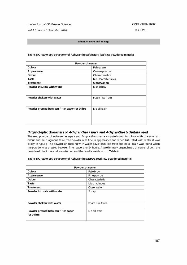
Table 3: Organoleptic character of Achyranthes bidentata leaf raw powdered material.
Powder character
Colour Pale green Appearance Coarse powder Odour Characteristics Taste No Characteristics Treatment Observation Powder triturate with water Non sticky
Powder shaken with water Foam like froth
Powder pressed between filter paper for 24 hrs No oil stain
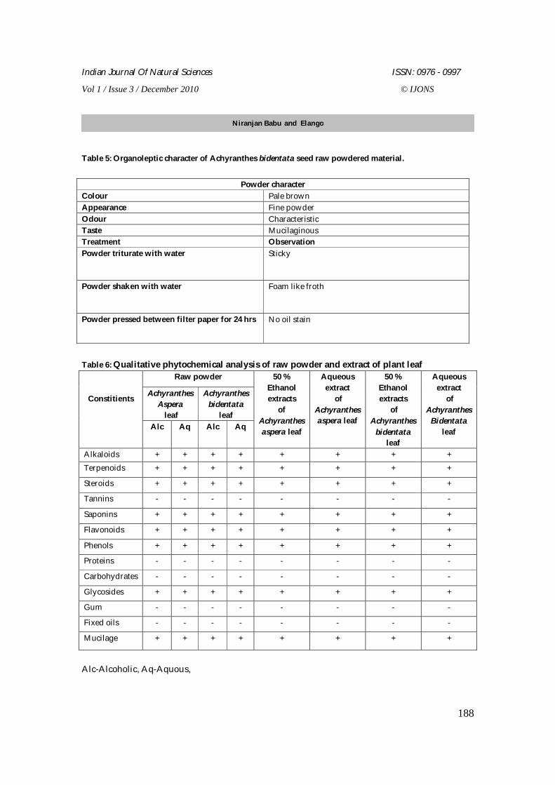
Organoleptic characters of Achyranthes aspera and Achyranthes bidentata seed The seed powder of Achyranthes aspera and Achyranthes bidentata is pale brown in colour with characteristic odour and mucilaginous taste. The powder was fine in appearance and when triturated with water it was sticky in nature. The powder on shaking with water gave foam like froth and no oil stain was found when the powder was pressed between filter papers for 24 hours. A preliminary organoleptic character of both the powdered plant material was studied and the results are shown in Table 4. Table 4: Organoleptic character of Achyranthes aspera seed raw powdered material
Powder character
Colour Pale brown Appearance Fine powder Odour Characteristic Taste Mucilaginous Treatment Observation Powder triturate with water Sticky
Powder shaken with water Foam like froth
Powder pressed between filter paper for 24 hrs
No oil stain
Niranjan Babu and Elango
Indian Journal Of Natural Sciences ISSN: 0976 - 0997
Vol 1 / Issue 3 / December 2010 © IJONS
188
Table 5: Organoleptic character of Achyranthes bidentata seed raw powdered material.
Powder character
Colour Pale brown Appearance Fine powder Odour Characteristic Taste Mucilaginous Treatment Observation Powder triturate with water Sticky
Powder shaken with water Foam like froth
Powder pressed between filter paper for 24 hrs No oil stain
Table 6: Qualitative phytochemical analysis of raw powder and extract of plant leaf
Raw powder
Achyranthes Aspera
leaf
Achyranthes bidentata
leaf
Constitients
Alc Aq Alc Aq
50 % Ethanol extracts
of Achyranthes aspera leaf
Aqueous extract
of Achyranthes aspera leaf
50 % Ethanol extracts
of Achyranthes
bidentata leaf
Aqueous extract
of Achyranthes
Bidentata leaf
Alkaloids + + + + + + + + Terpenoids + + + + + + + +
Steroids + + + + + + + +
Tannins - - - - - - - -
Saponins + + + + + + + +
Flavonoids + + + + + + + +
Phenols + + + + + + + +
Proteins - - - - - - - -
Carbohydrates - - - - - - - -
Glycosides + + + + + + + +
Gum - - - - - - - -
Fixed oils - - - - - - - -
Mucilage + + + + + + + +
Alc-Alcoholic, Aq-Aquous,
Niranjan Babu and Elango
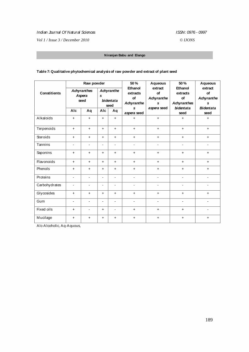
Indian Journal Of Natural Sciences ISSN: 0976 - 0997
Vol 1 / Issue 3 / December 2010 © IJONS
189
Table 7: Qualitative phytochemical analysis of raw powder and extract of plant seed
Raw powder
Achyranthes Aspera seed
Achyranthes
bidentata seed
Constitients
Alc Aq Alc Aq
50 % Ethanol extracts
of Achyranthe
s aspera seed
Aqueous extract
of Achyranthe
s aspera seed
50 % Ethanol extracts
of Achyranthes
bidentata seed
Aqueous extract
of Achyranthe
s Bidentata
seed Alkaloids + + + + + + + +
Terpenoids + + + + + + + +
Steroids + + + + + + + +
Tannins - - - - - - - -
Saponins + + + + + + + +
Flavonoids + + + + + + + +
Phenols + + + + + + + +
Proteins - - - - - - - -
Carbohydrates - - - - - - - -
Glycosides + + + + + + + +
Gum - - - - - - - -
Fixed oils + - + - + + + -
Mucilage + + + + + + + +
Alc-Alcoholic, Aq-Aquous,
Niranjan Babu and Elango
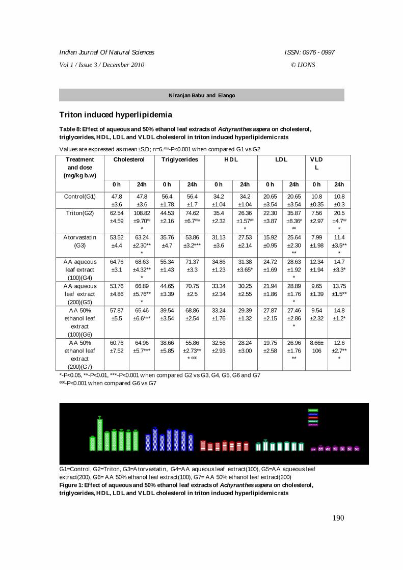
Indian Journal Of Natural Sciences ISSN: 0976 - 0997
Vol 1 / Issue 3 / December 2010 © IJONS
190
Triton induced hyperlipidemia
Table 8: Effect of aqueous and 50% ethanol leaf extracts of Achyranthes aspera on cholesterol, triglycerides, HDL, LDL and VLDL cholesterol in triton induced hyperlipidemic rats
Values are expressed as mean±S.D; n=6.###-P<0.001 when compared G1 vs G2
*-P<0.05, **-P<0.01, ***-P<0.001 when compared G2 vs G3, G4, G5, G6 and G7 €€€-P<0.001 when compared G6 vs G7
G1 G2 G3 G4 G5 G6 G7 G1 G2 G3 G4 G5 G6 G7 G1 G2 G3 G4 G5 G6 G7 G1 G2 G3 G4 G5 G6 G7 G1 G2 G3 G4 G5 G6 G7
0
50
100
150CholesterolTriglyceridesHDLLDLVLDL
Treatment
mg/dl
G1=Control, G2=Triton, G3=Atorvastatin, G4=AA aqueous leaf extract(100), G5=AA aqueous leaf extract(200), G6= AA 50% ethanol leaf extract(100), G7= AA 50% ethanol leaf extract(200) Figure 1: Effect of aqueous and 50% ethanol leaf extracts of Achyranthes aspera on cholesterol, triglycerides, HDL, LDL and VLDL cholesterol in triton induced hyperlipidemic rats
Cholesterol Triglycerides HDL LDL VLDL
Treatment and dose
(mg/kg b.w) 0 h 24h 0 h 24h 0 h 24h 0 h 24h 0 h 24h
Control(G1) 47.8 ±3.6
47.8 ±3.6
56.4 ±1.78
56.4 ±1.7
34.2 ±1.04
34.2 ±1.04
20.65 ±3.54
20.65 ±3.54
10.8 ±0.35
10.8 ±0.3
Triton(G2) 62.54 ±4.59
108.82 ±9.70##
#
44.53 ±2.16
74.62 ±6.7###
35.4 ±2.32
26.36 ±1.57##
#
22.30 ±3.87
35.87 ±8.36#
##
7.56 ±2.97
20.5 ±4.7##
# Atorvastatin
(G3) 53.52 ±4.4
63.24 ±2.30**
*
35.76 ±4.7
53.86 ±3.2***
31.13 ±3.6
27.53 ±2.14
15.92 ±0.95
25.64 ±2.30
**
7.99 ±1.98
11.4 ±3.5**
* AA aqueous leaf extract (100)(G4)
64.76 ±3.1
68.63 ±4.32**
*
55.34 ±1.43
71.37 ±3.3
34.86 ±1.23
31.38 ±3.65*
24.72 ±1.69
28.63 ±1.92
*
12.34 ±1.94
14.7 ±3.3*
AA aqueous leaf extract
(200)(G5)
53.76 ±4.86
66.89 ±5.76**
*
44.65 ±3.39
70.75 ±2.5
33.34 ±2.34
30.25 ±2.55
21.94 ±1.86
28.89 ±1.76
*
9.65 ±1.39
13.75 ±1.5**
AA 50% ethanol leaf
extract (100)(G6)
57.87 ±5.5
65.46 ±6.6***
39.54 ±3.54
68.86 ±2.54
33.24 ±1.76
29.39 ±1.32
27.87 ±2.15
27.46 ±2.86
*
9.54 ±2.32
14.8 ±1.2*
AA 50% ethanol leaf
extract (200)(G7)
60.76 ±7.52
64.96 ±5.7***
38.66 ±5.85
55.86 ±2.73**
* €€€
32.56 ±2.93
28.24 ±3.00
19.75 ±2.58
26.96 ±1.76
**
8.66± 106
12.6 ±2.7**
*
Niranjan Babu and Elango
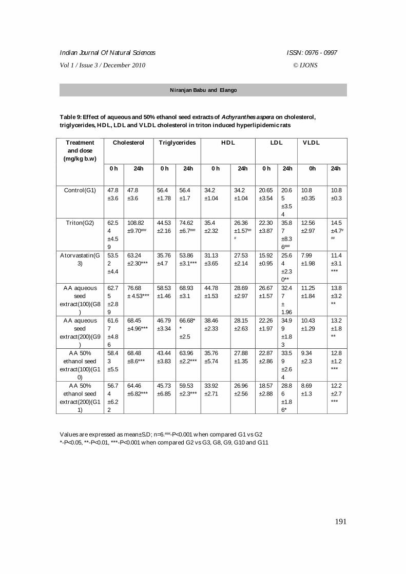
Indian Journal Of Natural Sciences ISSN: 0976 - 0997
Vol 1 / Issue 3 / December 2010 © IJONS
191
Table 9: Effect of aqueous and 50% ethanol seed extracts of Achyranthes aspera on cholesterol, triglycerides, HDL, LDL and VLDL cholesterol in triton induced hyperlipidemic rats
Values are expressed as mean±S.D; n=6.###-P<0.001 when compared G1 vs G2 *-P<0.05, **-P<0.01, ***-P<0.001 when compared G2 vs G3, G8, G9, G10 and G11
Cholesterol Triglycerides HDL LDL VLDL Treatment and dose
(mg/kg b.w) 0 h 24h 0 h 24h 0 h 24h 0 h 24h 0h 24h
Control(G1) 47.8 ±3.6
47.8 ±3.6
56.4 ±1.78
56.4 ±1.7
34.2 ±1.04
34.2 ±1.04
20.65 ±3.54
20.65 ±3.54
10.8 ±0.35
10.8 ±0.3
Triton(G2) 62.54 ±4.59
108.82 ±9.70###
44.53 ±2.16
74.62 ±6.7###
35.4 ±2.32
26.36 ±1.57##
#
22.30 ±3.87
35.87 ±8.36###
12.56 ±2.97
14.5 ±4.7#
##
Atorvastatin(G3)
53.52 ±4.4
63.24 ±2.30***
35.76 ±4.7
53.86 ±3.1***
31.13 ±3.65
27.53 ±2.14
15.92 ±0.95
25.64 ±2.30**
7.99 ±1.98
11.4 ±3.1***
AA aqueous seed
extract(100)(G8)
62.75 ±2.89
76.68 ± 4.53***
58.53 ±1.46
68.93 ±3.1
44.78 ±1.53
28.69 ±2.97
26.67 ±1.57
32.47 ± 1.96
11.25 ±1.84
13.8 ±3.2**
AA aqueous seed
extract(200)(G9)
61.67 ±4.86
68.45 ±4.96***
46.79 ±3.34
66.68** ±2.5
38.46 ±2.33
28.15 ±2.63
22.26 ±1.97
34.99 ±1.83
10.43 ±1.29
13.2 ±1.8**
AA 50% ethanol seed
extract(100)(G10)
58.43 ±5.5
68.48 ±8.6***
43.44 ±3.83
63.96 ±2.2***
35.76 ±5.74
27.88 ±1.35
22.87 ±2.86
33.59 ±2.64
9.34 ±2.3
12.8 ±1.2***
AA 50% ethanol seed
extract(200)(G11)
56.74 ±6.22
64.46 ±6.82***
45.73 ±6.85
59.53 ±2.3***
33.92 ±2.71
26.96 ±2.56
18.57 ±2.88
28.86 ±1.86*
8.69 ±1.3
12.2 ±2.7***
Niranjan Babu and Elango
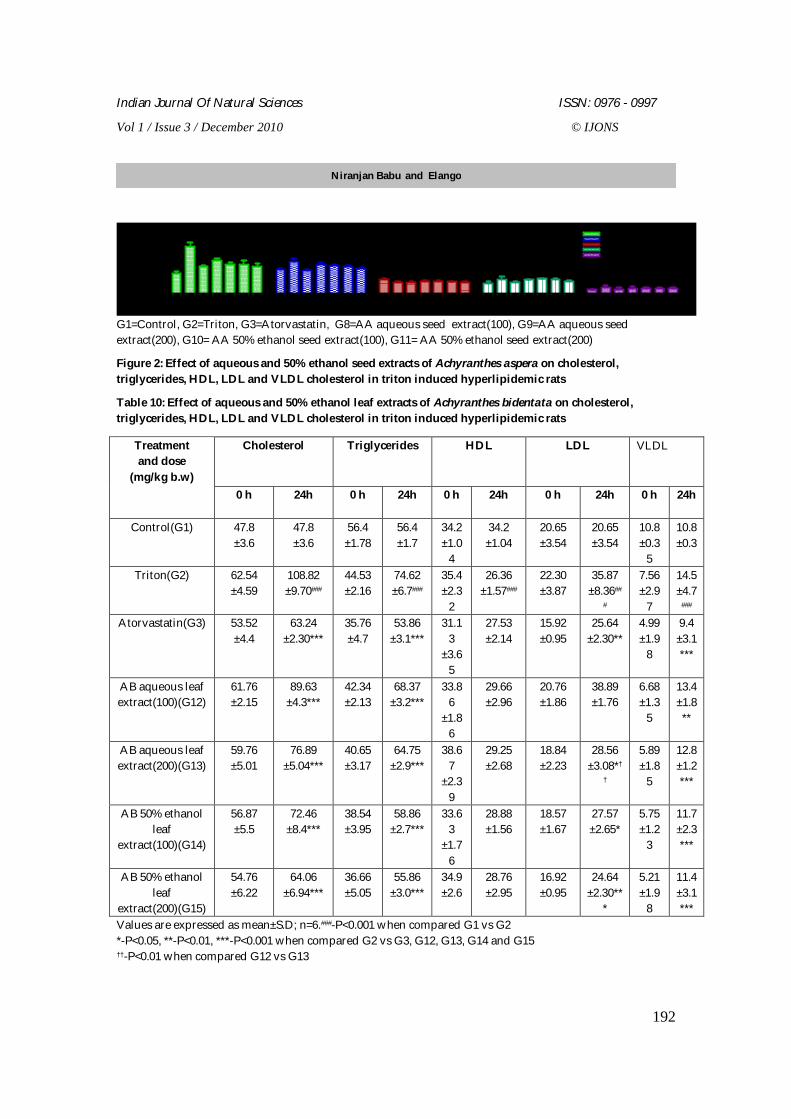
Indian Journal Of Natural Sciences ISSN: 0976 - 0997
Vol 1 / Issue 3 / December 2010 © IJONS
192
G1 G2 G3 G8 G9G10G11 G1 G2 G3 G8 G9 G10G11 G1 G2 G3 G8 G9 G10G11 G1 G2 G3 G8 G9G10G11 G1 G2 G3 G8 G9G10G11
0
50
100
150CholesterolTriglyceridesHDLLDLVLDL
Treatment
mg / dl
G1=Control, G2=Triton, G3=Atorvastatin, G8=AA aqueous seed extract(100), G9=AA aqueous seed extract(200), G10= AA 50% ethanol seed extract(100), G11= AA 50% ethanol seed extract(200)
Figure 2: Effect of aqueous and 50% ethanol seed extracts of Achyranthes aspera on cholesterol, triglycerides, HDL, LDL and VLDL cholesterol in triton induced hyperlipidemic rats
Table 10: Effect of aqueous and 50% ethanol leaf extracts of Achyranthes bidentata on cholesterol, triglycerides, HDL, LDL and VLDL cholesterol in triton induced hyperlipidemic rats
Values are expressed as mean±S.D; n=6.###-P<0.001 when compared G1 vs G2 *-P<0.05, **-P<0.01, ***-P<0.001 when compared G2 vs G3, G12, G13, G14 and G15 ††-P<0.01 when compared G12 vs G13
Cholesterol Triglycerides HDL LDL VLDL Treatment and dose
(mg/kg b.w) 0 h 24h 0 h 24h 0 h 24h 0 h 24h 0 h 24h
Control(G1) 47.8 ±3.6
47.8 ±3.6
56.4 ±1.78
56.4 ±1.7
34.2 ±1.0
4
34.2 ±1.04
20.65 ±3.54
20.65 ±3.54
10.8 ±0.3
5
10.8 ±0.3
Triton(G2) 62.54 ±4.59
108.82 ±9.70###
44.53 ±2.16
74.62 ±6.7###
35.4 ±2.3
2
26.36 ±1.57###
22.30 ±3.87
35.87 ±8.36##
#
7.56 ±2.9
7
14.5 ±4.7
### Atorvastatin(G3) 53.52
±4.4 63.24
±2.30*** 35.76 ±4.7
53.86 ±3.1***
31.13
±3.65
27.53 ±2.14
15.92 ±0.95
25.64 ±2.30**
4.99 ±1.9
8
9.4 ±3.1***
AB aqueous leaf extract(100)(G12)
61.76 ±2.15
89.63 ±4.3***
42.34 ±2.13
68.37 ±3.2***
33.86
±1.86
29.66 ±2.96
20.76 ±1.86
38.89 ±1.76
6.68 ±1.3
5
13.4 ±1.8**
AB aqueous leaf extract(200)(G13)
59.76 ±5.01
76.89 ±5.04***
40.65 ±3.17
64.75 ±2.9***
38.67
±2.39
29.25 ±2.68
18.84 ±2.23
28.56 ±3.08*†
†
5.89 ±1.8
5
12.8 ±1.2***
AB 50% ethanol leaf
extract(100)(G14)
56.87 ±5.5
72.46 ±8.4***
38.54 ±3.95
58.86 ±2.7***
33.63
±1.76
28.88 ±1.56
18.57 ±1.67
27.57 ±2.65*
5.75 ±1.2
3
11.7 ±2.3***
AB 50% ethanol leaf
extract(200)(G15)
54.76 ±6.22
64.06 ±6.94***
36.66 ±5.05
55.86 ±3.0***
34.9 ±2.6
28.76 ±2.95
16.92 ±0.95
24.64 ±2.30**
*
5.21 ±1.9
8
11.4 ±3.1***
Niranjan Babu and Elango
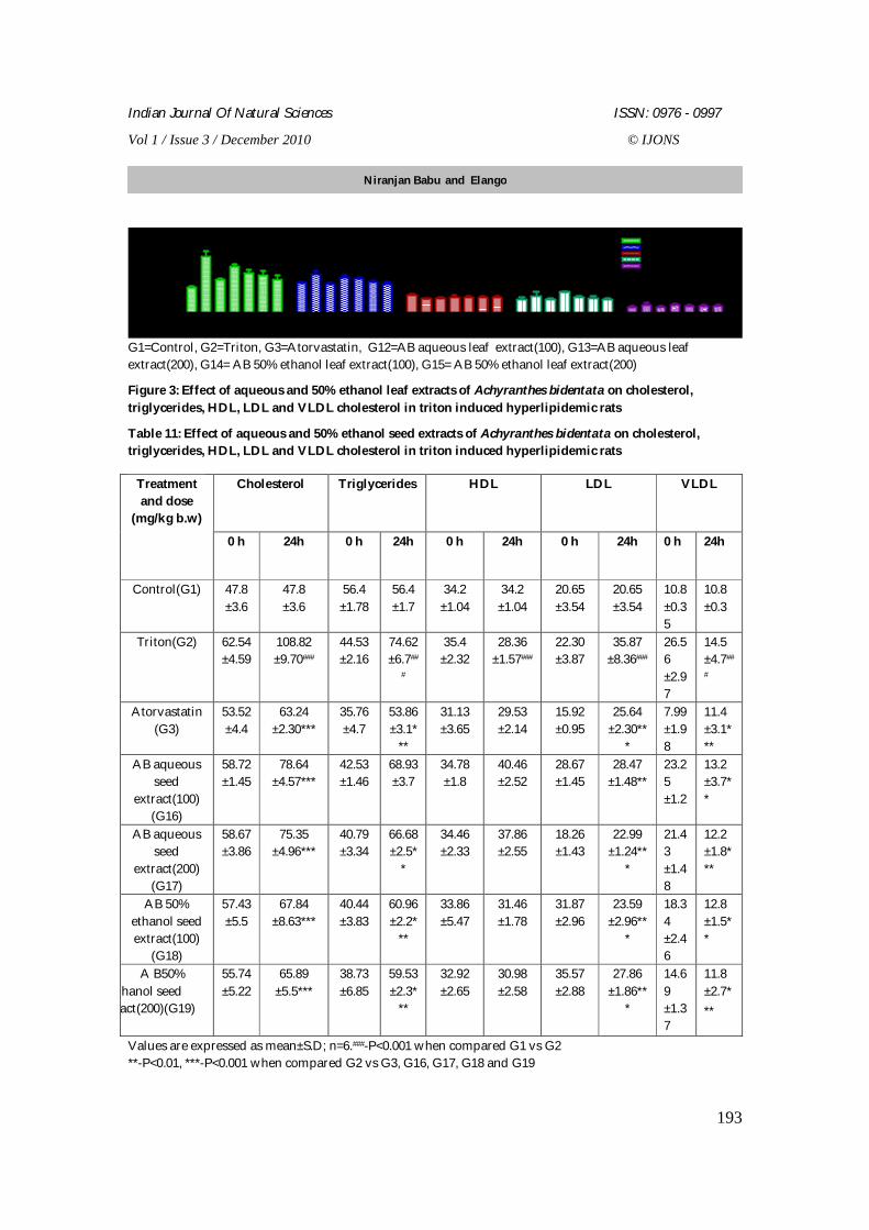
Indian Journal Of Natural Sciences ISSN: 0976 - 0997
Vol 1 / Issue 3 / December 2010 © IJONS
193
G1 G2 G3G12G13G14G15 G1 G2 G3G12G13G14G15 G1 G2 G3G12G13G14G15 G1 G2 G3G12G13G14G15 G1 G2 G3G12G13G14G15
0
50
100
150CholesterolTriglyceridesHDLLDLVLDL
Treatment
mg / dl
G1=Control, G2=Triton, G3=Atorvastatin, G12=AB aqueous leaf extract(100), G13=AB aqueous leaf extract(200), G14= AB 50% ethanol leaf extract(100), G15= AB 50% ethanol leaf extract(200)
Figure 3: Effect of aqueous and 50% ethanol leaf extracts of Achyranthes bidentata on cholesterol, triglycerides, HDL, LDL and VLDL cholesterol in triton induced hyperlipidemic rats
Table 11: Effect of aqueous and 50% ethanol seed extracts of Achyranthes bidentata on cholesterol, triglycerides, HDL, LDL and VLDL cholesterol in triton induced hyperlipidemic rats
Values are expressed as mean±S.D; n=6.###-P<0.001 when compared G1 vs G2 **-P<0.01, ***-P<0.001 when compared G2 vs G3, G16, G17, G18 and G19
Cholesterol Triglycerides HDL LDL VLDL Treatment and dose
(mg/kg b.w)
0 h 24h 0 h 24h 0 h 24h 0 h 24h 0 h 24h
Control(G1) 47.8 ±3.6
47.8 ±3.6
56.4 ±1.78
56.4 ±1.7
34.2 ±1.04
34.2 ±1.04
20.65 ±3.54
20.65 ±3.54
10.8 ±0.35
10.8 ±0.3
Triton(G2) 62.54 ±4.59
108.82 ±9.70###
44.53 ±2.16
74.62 ±6.7##
#
35.4 ±2.32
28.36 ±1.57###
22.30 ±3.87
35.87 ±8.36###
26.56 ±2.97
14.5 ±4.7##
#
Atorvastatin (G3)
53.52 ±4.4
63.24 ±2.30***
35.76 ±4.7
53.86 ±3.1*
**
31.13 ±3.65
29.53 ±2.14
15.92 ±0.95
25.64 ±2.30**
*
7.99 ±1.98
11.4 ±3.1***
AB aqueous seed
extract(100) (G16)
58.72 ±1.45
78.64 ±4.57***
42.53 ±1.46
68.93 ±3.7
34.78 ±1.8
40.46 ±2.52
28.67 ±1.45
28.47 ±1.48**
23.25 ±1.2
13.2 ±3.7**
AB aqueous seed
extract(200) (G17)
58.67 ±3.86
75.35 ±4.96***
40.79 ±3.34
66.68 ±2.5*
*
34.46 ±2.33
37.86 ±2.55
18.26 ±1.43
22.99 ±1.24**
*
21.43 ±1.48
12.2 ±1.8***
AB 50% ethanol seed extract(100)
(G18)
57.43 ±5.5
67.84 ±8.63***
40.44 ±3.83
60.96 ±2.2*
**
33.86 ±5.47
31.46 ±1.78
31.87 ±2.96
23.59 ±2.96**
*
18.34 ±2.46
12.8 ±1.5**
A B50% ethanol seed
extract(200)(G19)
55.74 ±5.22
65.89 ±5.5***
38.73 ±6.85
59.53 ±2.3*
**
32.92 ±2.65
30.98 ±2.58
35.57 ±2.88
27.86 ±1.86**
*
14.69 ±1.37
11.8 ±2.7***
Niranjan Babu and Elango
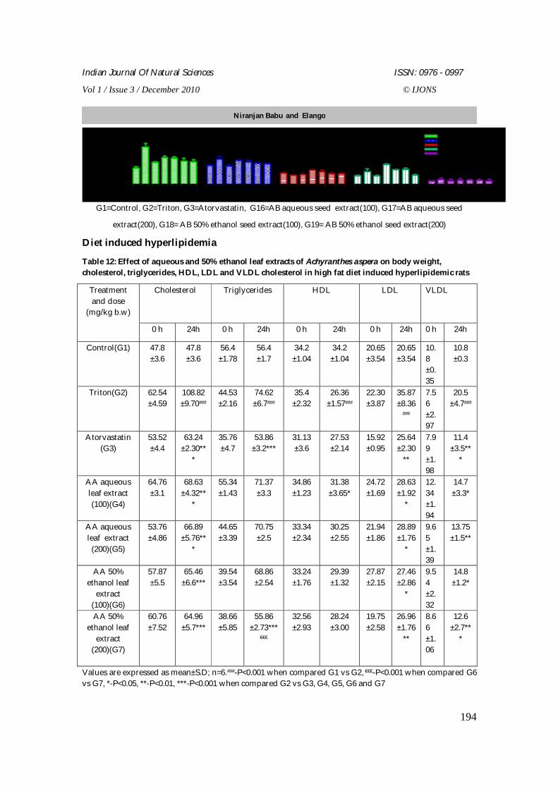
Indian Journal Of Natural Sciences ISSN: 0976 - 0997
Vol 1 / Issue 3 / December 2010 © IJONS
194
G1 G2 G3G16G17G18G19 G1 G2 G3G16G17G18G19 G1 G2 G3G16G17G18G19 G1 G2 G3G16G17G18G19 G1 G2 G3G16G17G18G19
0
50
100
150CholesterolTriglyceridesHDLLDLVLDL
Treatment
mg / dl
G1=Control, G2=Triton, G3=Atorvastatin, G16=AB aqueous seed extract(100), G17=AB aqueous seed
extract(200), G18= AB 50% ethanol seed extract(100), G19= AB 50% ethanol seed extract(200) Diet induced hyperlipidemia
Table 12: Effect of aqueous and 50% ethanol leaf extracts of Achyranthes aspera on body weight, cholesterol, triglycerides, HDL, LDL and VLDL cholesterol in high fat diet induced hyperlipidemic rats
Values are expressed as mean±S.D; n=6.###-P<0.001 when compared G1 vs G2, €€€-P<0.001 when compared G6 vs G7, *-P<0.05, **-P<0.01, ***-P<0.001 when compared G2 vs G3, G4, G5, G6 and G7
Cholesterol Triglycerides HDL LDL VLDL Treatment and dose
(mg/kg b.w)
0 h 24h 0 h 24h 0 h 24h 0 h 24h 0 h 24h
Control(G1) 47.8 ±3.6
47.8 ±3.6
56.4 ±1.78
56.4 ±1.7
34.2 ±1.04
34.2 ±1.04
20.65 ±3.54
20.65 ±3.54
10.8 ±0.35
10.8 ±0.3
Triton(G2) 62.54 ±4.59
108.82 ±9.70###
44.53 ±2.16
74.62 ±6.7###
35.4 ±2.32
26.36 ±1.57###
22.30 ±3.87
35.87 ±8.36
###
7.56 ±2.97
20.5 ±4.7###
Atorvastatin (G3)
53.52 ±4.4
63.24 ±2.30**
*
35.76 ±4.7
53.86 ±3.2***
31.13 ±3.6
27.53 ±2.14
15.92 ±0.95
25.64 ±2.30
**
7.99 ±1.98
11.4 ±3.5**
*
AA aqueous leaf extract (100)(G4)
64.76 ±3.1
68.63 ±4.32**
*
55.34 ±1.43
71.37 ±3.3
34.86 ±1.23
31.38 ±3.65*
24.72 ±1.69
28.63 ±1.92
*
12.34 ±1.94
14.7 ±3.3*
AA aqueous leaf extract
(200)(G5)
53.76 ±4.86
66.89 ±5.76**
*
44.65 ±3.39
70.75 ±2.5
33.34 ±2.34
30.25 ±2.55
21.94 ±1.86
28.89 ±1.76
*
9.65 ±1.39
13.75 ±1.5**
AA 50% ethanol leaf
extract (100)(G6)
57.87 ±5.5
65.46 ±6.6***
39.54 ±3.54
68.86 ±2.54
33.24 ±1.76
29.39 ±1.32
27.87 ±2.15
27.46 ±2.86
*
9.54 ±2.32
14.8 ±1.2*
AA 50% ethanol leaf
extract (200)(G7)
60.76 ±7.52
64.96 ±5.7***
38.66 ±5.85
55.86 ±2.73***
€€€
32.56 ±2.93
28.24 ±3.00
19.75 ±2.58
26.96 ±1.76
**
8.66 ±1.06
12.6 ±2.7**
*
Niranjan Babu and Elango
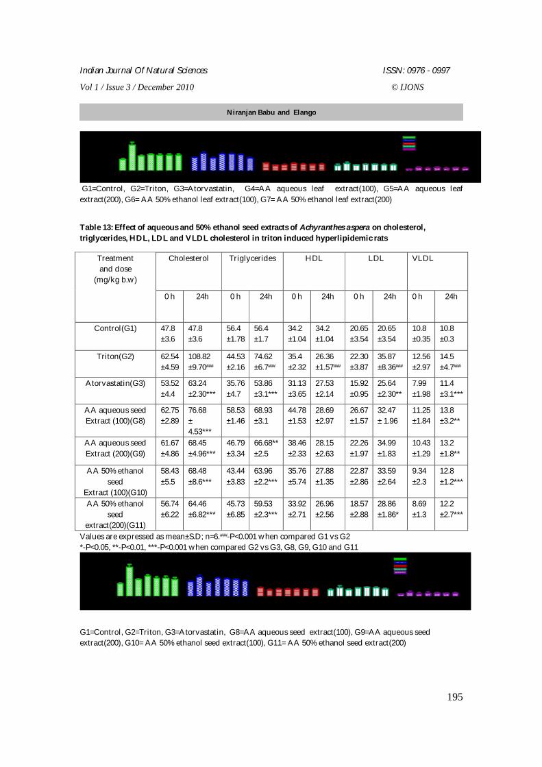
Indian Journal Of Natural Sciences ISSN: 0976 - 0997
Vol 1 / Issue 3 / December 2010 © IJONS
195
G1 G2 G3 G4 G5 G6 G7 G1 G2 G3 G4 G5 G6 G7 G1 G2 G3 G4 G5 G6 G7 G1 G2 G3 G4 G5 G6 G7 G1 G2 G3 G4 G5 G6 G7
0
50
100
150CholesterolTriglyceridesHDLLDLVLDL
Treatment
mg/dl
G1=Control, G2=Triton, G3=Atorvastatin, G4=AA aqueous leaf extract(100), G5=AA aqueous leaf extract(200), G6= AA 50% ethanol leaf extract(100), G7= AA 50% ethanol leaf extract(200)
Table 13: Effect of aqueous and 50% ethanol seed extracts of Achyranthes aspera on cholesterol, triglycerides, HDL, LDL and VLDL cholesterol in triton induced hyperlipidemic rats
Values are expressed as mean±S.D; n=6.###-P<0.001 when compared G1 vs G2 *-P<0.05, **-P<0.01, ***-P<0.001 when compared G2 vs G3, G8, G9, G10 and G11
G1 G2 G3 G8 G9 G10G11 G1 G2 G3 G8 G9 G10G11 G1 G2 G3 G8 G9 G10G11 G1 G2 G3 G8 G9 G10G11 G1 G2 G3 G8 G9 G10G11
0
50
100
150CholesterolTriglyceridesHDLLDLVLDL
Treatment
mg / dl
G1=Control, G2=Triton, G3=Atorvastatin, G8=AA aqueous seed extract(100), G9=AA aqueous seed extract(200), G10= AA 50% ethanol seed extract(100), G11= AA 50% ethanol seed extract(200)
Cholesterol Triglycerides HDL LDL VLDL Treatment and dose
(mg/kg b.w)
0 h 24h 0 h 24h 0 h 24h 0 h 24h 0 h 24h
Control(G1) 47.8 ±3.6
47.8 ±3.6
56.4 ±1.78
56.4 ±1.7
34.2 ±1.04
34.2 ±1.04
20.65 ±3.54
20.65 ±3.54
10.8 ±0.35
10.8 ±0.3
Triton(G2) 62.54 ±4.59
108.82 ±9.70###
44.53 ±2.16
74.62 ±6.7###
35.4 ±2.32
26.36 ±1.57###
22.30 ±3.87
35.87 ±8.36###
12.56 ±2.97
14.5 ±4.7###
Atorvastatin(G3) 53.52 ±4.4
63.24 ±2.30***
35.76 ±4.7
53.86 ±3.1***
31.13 ±3.65
27.53 ±2.14
15.92 ±0.95
25.64 ±2.30**
7.99 ±1.98
11.4 ±3.1***
AA aqueous seed Extract (100)(G8)
62.75 ±2.89
76.68 ± 4.53***
58.53 ±1.46
68.93 ±3.1
44.78 ±1.53
28.69 ±2.97
26.67 ±1.57
32.47 ± 1.96
11.25 ±1.84
13.8 ±3.2**
AA aqueous seed Extract (200)(G9)
61.67 ±4.86
68.45 ±4.96***
46.79 ±3.34
66.68** ±2.5
38.46 ±2.33
28.15 ±2.63
22.26 ±1.97
34.99 ±1.83
10.43 ±1.29
13.2 ±1.8**
AA 50% ethanol seed
Extract (100)(G10)
58.43 ±5.5
68.48 ±8.6***
43.44 ±3.83
63.96 ±2.2***
35.76 ±5.74
27.88 ±1.35
22.87 ±2.86
33.59 ±2.64
9.34 ±2.3
12.8 ±1.2***
AA 50% ethanol seed
extract(200)(G11)
56.74 ±6.22
64.46 ±6.82***
45.73 ±6.85
59.53 ±2.3***
33.92 ±2.71
26.96 ±2.56
18.57 ±2.88
28.86 ±1.86*
8.69 ±1.3
12.2 ±2.7***
Niranjan Babu and Elango
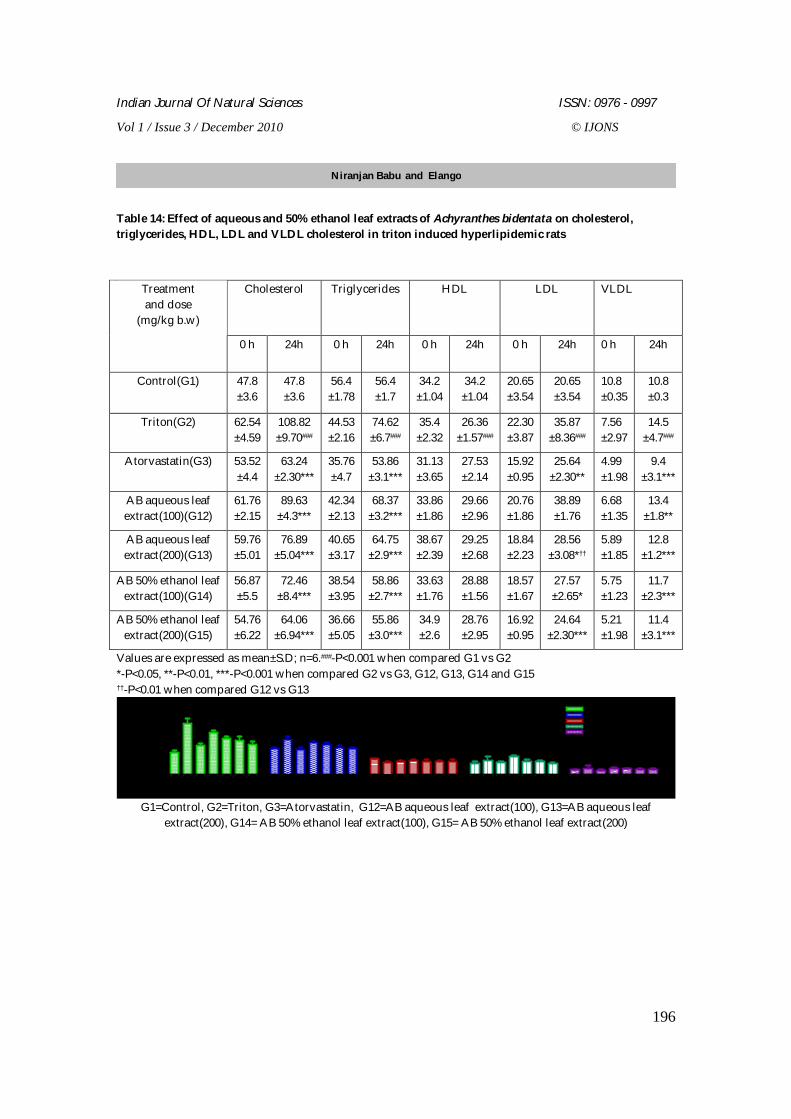
Indian Journal Of Natural Sciences ISSN: 0976 - 0997
Vol 1 / Issue 3 / December 2010 © IJONS
196
Table 14: Effect of aqueous and 50% ethanol leaf extracts of Achyranthes bidentata on cholesterol, triglycerides, HDL, LDL and VLDL cholesterol in triton induced hyperlipidemic rats
Values are expressed as mean±S.D; n=6.###-P<0.001 when compared G1 vs G2 *-P<0.05, **-P<0.01, ***-P<0.001 when compared G2 vs G3, G12, G13, G14 and G15 ††-P<0.01 when compared G12 vs G13
G1 G2 G3G12G13G14G15 G1 G2 G3G12G13G14G15 G1 G2 G3G12G13G14G15 G1 G2 G3G12G13G14G15 G1 G2 G3G12G13G14G15
0
50
100
150CholesterolTriglyceridesHDLLDLVLDL
Treatment
mg / dl
G1=Control, G2=Triton, G3=Atorvastatin, G12=AB aqueous leaf extract(100), G13=AB aqueous leaf extract(200), G14= AB 50% ethanol leaf extract(100), G15= AB 50% ethanol leaf extract(200)
Cholesterol Triglycerides HDL LDL VLDL Treatment and dose
(mg/kg b.w)
0 h 24h 0 h 24h 0 h 24h 0 h 24h 0 h 24h
Control(G1) 47.8 ±3.6
47.8 ±3.6
56.4 ±1.78
56.4 ±1.7
34.2 ±1.04
34.2 ±1.04
20.65 ±3.54
20.65 ±3.54
10.8 ±0.35
10.8 ±0.3
Triton(G2) 62.54 ±4.59
108.82 ±9.70###
44.53 ±2.16
74.62 ±6.7###
35.4 ±2.32
26.36 ±1.57###
22.30 ±3.87
35.87 ±8.36###
7.56 ±2.97
14.5 ±4.7###
Atorvastatin(G3) 53.52 ±4.4
63.24 ±2.30***
35.76 ±4.7
53.86 ±3.1***
31.13 ±3.65
27.53 ±2.14
15.92 ±0.95
25.64 ±2.30**
4.99 ±1.98
9.4 ±3.1***
AB aqueous leaf extract(100)(G12)
61.76 ±2.15
89.63 ±4.3***
42.34 ±2.13
68.37 ±3.2***
33.86 ±1.86
29.66 ±2.96
20.76 ±1.86
38.89 ±1.76
6.68 ±1.35
13.4 ±1.8**
AB aqueous leaf extract(200)(G13)
59.76 ±5.01
76.89 ±5.04***
40.65 ±3.17
64.75 ±2.9***
38.67 ±2.39
29.25 ±2.68
18.84 ±2.23
28.56 ±3.08*††
5.89 ±1.85
12.8 ±1.2***
AB 50% ethanol leaf extract(100)(G14)
56.87 ±5.5
72.46 ±8.4***
38.54 ±3.95
58.86 ±2.7***
33.63 ±1.76
28.88 ±1.56
18.57 ±1.67
27.57 ±2.65*
5.75 ±1.23
11.7 ±2.3***
AB 50% ethanol leaf extract(200)(G15)
54.76 ±6.22
64.06 ±6.94***
36.66 ±5.05
55.86 ±3.0***
34.9 ±2.6
28.76 ±2.95
16.92 ±0.95
24.64 ±2.30***
5.21 ±1.98
11.4 ±3.1***
Niranjan Babu and Elango
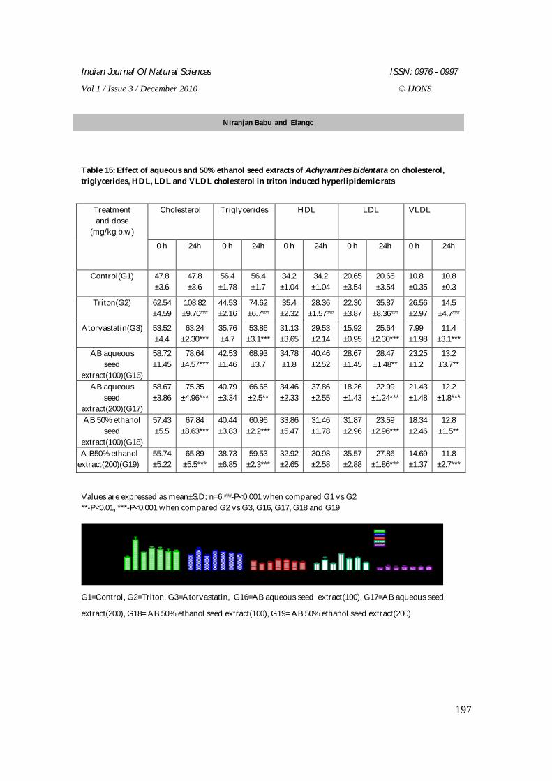
Indian Journal Of Natural Sciences ISSN: 0976 - 0997
Vol 1 / Issue 3 / December 2010 © IJONS
197
Table 15: Effect of aqueous and 50% ethanol seed extracts of Achyranthes bidentata on cholesterol, triglycerides, HDL, LDL and VLDL cholesterol in triton induced hyperlipidemic rats
Values are expressed as mean±S.D; n=6.###-P<0.001 when compared G1 vs G2 **-P<0.01, ***-P<0.001 when compared G2 vs G3, G16, G17, G18 and G19
G1 G2 G3G16G17G18G19 G1 G2 G3G16G17G18G19 G1 G2 G3G16G17G18G19 G1 G2 G3G16G17G18G19 G1 G2 G3G16G17G18G19
0
50
100
150CholesterolTriglyceridesHDLLDLVLDL
Treatment
mg / dl
G1=Control, G2=Triton, G3=Atorvastatin, G16=AB aqueous seed extract(100), G17=AB aqueous seed
extract(200), G18= AB 50% ethanol seed extract(100), G19= AB 50% ethanol seed extract(200)
Cholesterol Triglycerides HDL LDL VLDL Treatment and dose
(mg/kg b.w)
0 h 24h 0 h 24h 0 h 24h 0 h 24h 0 h 24h
Control(G1) 47.8 ±3.6
47.8 ±3.6
56.4 ±1.78
56.4 ±1.7
34.2 ±1.04
34.2 ±1.04
20.65 ±3.54
20.65 ±3.54
10.8 ±0.35
10.8 ±0.3
Triton(G2) 62.54 ±4.59
108.82 ±9.70###
44.53 ±2.16
74.62 ±6.7###
35.4 ±2.32
28.36 ±1.57###
22.30 ±3.87
35.87 ±8.36###
26.56 ±2.97
14.5 ±4.7###
Atorvastatin(G3) 53.52 ±4.4
63.24 ±2.30***
35.76 ±4.7
53.86 ±3.1***
31.13 ±3.65
29.53 ±2.14
15.92 ±0.95
25.64 ±2.30***
7.99 ±1.98
11.4 ±3.1***
AB aqueous seed
extract(100)(G16)
58.72 ±1.45
78.64 ±4.57***
42.53 ±1.46
68.93 ±3.7
34.78 ±1.8
40.46 ±2.52
28.67 ±1.45
28.47 ±1.48**
23.25 ±1.2
13.2 ±3.7**
AB aqueous seed
extract(200)(G17)
58.67 ±3.86
75.35 ±4.96***
40.79 ±3.34
66.68 ±2.5**
34.46 ±2.33
37.86 ±2.55
18.26 ±1.43
22.99 ±1.24***
21.43 ±1.48
12.2 ±1.8***
AB 50% ethanol seed
extract(100)(G18)
57.43 ±5.5
67.84 ±8.63***
40.44 ±3.83
60.96 ±2.2***
33.86 ±5.47
31.46 ±1.78
31.87 ±2.96
23.59 ±2.96***
18.34 ±2.46
12.8 ±1.5**
A B50% ethanol seed extract(200)(G19)
55.74 ±5.22
65.89 ±5.5***
38.73 ±6.85
59.53 ±2.3***
32.92 ±2.65
30.98 ±2.58
35.57 ±2.88
27.86 ±1.86***
14.69 ±1.37
11.8 ±2.7***
Niranjan Babu and Elango
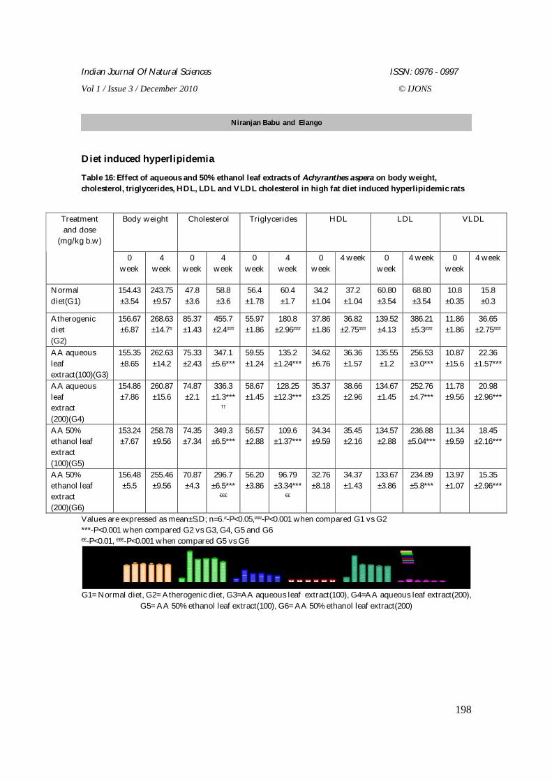
Indian Journal Of Natural Sciences ISSN: 0976 - 0997
Vol 1 / Issue 3 / December 2010 © IJONS
198
Diet induced hyperlipidemia
Table 16: Effect of aqueous and 50% ethanol leaf extracts of Achyranthes aspera on body weight, cholesterol, triglycerides, HDL, LDL and VLDL cholesterol in high fat diet induced hyperlipidemic rats
Values are expressed as mean±S.D; n=6.#-P<0.05,###-P<0.001 when compared G1 vs G2 ***-P<0.001 when compared G2 vs G3, G4, G5 and G6 €€-P<0.01, €€€-P<0.001 when compared G5 vs G6
G1 G2 G3 G4 G5 G6 G1 G2 G3 G4 G5 G6 G1 G2 G3 G4 G5 G6 G1 G2 G3 G4 G5 G6 G1 G2 G3 G4 G5 G6 G1 G2 G3 G4 G5 G6
0
100
200
300
400
500
Body weightCholesterolTriglyceridesHDLLDLVLDL
Treatment
mg/dl
G1= Normal diet, G2= Atherogenic diet, G3=AA aqueous leaf extract(100), G4=AA aqueous leaf extract(200), G5= AA 50% ethanol leaf extract(100), G6= AA 50% ethanol leaf extract(200)
Body weight Cholesterol Triglycerides HDL LDL VLDL Treatment and dose
(mg/kg b.w)
0 week
4 week
0 week
4 week
0 week
4 week
0 week
4 week 0 week
4 week 0 week
4 week
Normal diet(G1)
154.43 ±3.54
243.75 ±9.57
47.8 ±3.6
58.8 ±3.6
56.4 ±1.78
60.4 ±1.7
34.2 ±1.04
37.2 ±1.04
60.80 ±3.54
68.80 ±3.54
10.8 ±0.35
15.8 ±0.3
Atherogenic diet (G2)
156.67 ±6.87
268.63 ±14.7#
85.37 ±1.43
455.7 ±2.4###
55.97 ±1.86
180.8 ±2.96###
37.86 ±1.86
36.82 ±2.75###
139.52 ±4.13
386.21 ±5.3###
11.86 ±1.86
36.65 ±2.75###
AA aqueous leaf extract(100)(G3)
155.35 ±8.65
262.63 ±14.2
75.33 ±2.43
347.1 ±5.6***
59.55 ±1.24
135.2 ±1.24***
34.62 ±6.76
36.36 ±1.57
135.55 ±1.2
256.53 ±3.0***
10.87 ±15.6
22.36 ±1.57***
AA aqueous leaf extract (200)(G4)
154.86 ±7.86
260.87 ±15.6
74.87 ±2.1
336.3 ±1.3***
††
58.67 ±1.45
128.25 ±12.3***
35.37 ±3.25
38.66 ±2.96
134.67 ±1.45
252.76 ±4.7***
11.78 ±9.56
20.98 ±2.96***
AA 50% ethanol leaf extract (100)(G5)
153.24 ±7.67
258.78 ±9.56
74.35 ±7.34
349.3 ±6.5***
56.57 ±2.88
109.6 ±1.37***
34.34 ±9.59
35.45 ±2.16
134.57 ±2.88
236.88 ±5.04***
11.34 ±9.59
18.45 ±2.16***
AA 50% ethanol leaf extract (200)(G6)
156.48 ±5.5
255.46 ±9.56
70.87 ±4.3
296.7 ±6.5***
€€€
56.20 ±3.86
96.79 ±3.34***
€€
32.76 ±8.18
34.37 ±1.43
133.67 ±3.86
234.89 ±5.8***
13.97 ±1.07
15.35 ±2.96***
Niranjan Babu and Elango
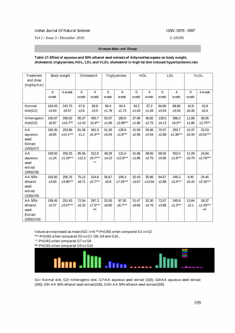
Indian Journal Of Natural Sciences ISSN: 0976 - 0997
Vol 1 / Issue 3 / December 2010 © IJONS
199
Table 17: Effect of aqueous and 50% ethanol seed extracts of Achyranthes aspera on body weight, cholesterol, triglycerides, HDL, LDL and VLDL cholesterol in high fat diet induced hyperlipidemic rats
Values are expressed as mean±S.D; n=6.###-P<0.001 when compared G1 vs G2 ***-P<0.001 when compared G2 vs G7, G8, G9 and G10 , †††-P<0.001 when compared G7 vs G8 €€€-P<0.001 when compared G9 vs G10
G1 G2 G7 G8 G9 G10 G1 G2 G7 G8 G9 G10 G1 G2 G7 G8 G9 G10 G1 G2 G7 G8 G9 G10 G1 G2 G7 G8 G9 G10 G1 G2 G7 G8 G9 G10
0
100
200
300
400
500 Body weightCholesterolTriglyceridesHDLLDLVLDL
Treatment
mg/dl
G1= Normal diet, G2= Atherogenic diet, G7=AA aqueous seed extract (100), G8=AA aqueous seed extract (200), G9= AA 50% ethanol seed extract(100), G10= AA 50% ethanol seed extract(200)
Body weight Cholesterol Triglycerides HDL LDL VLDL Treatment and dose
(mg/kg b.w)
0 week
4 week 0 week
4 week
0 week
4 week
0 week
4 week
0 week
4 week
0 week
4 week
Normal diet(G1)
154.43 ±3.54
243.75 ±9.57
47.8 ±3.6
58.8 ±3.5
56.4 ±1.78
60.4 ±1.73
34.2 ±1.04
37.2 ±1.04
60.80 ±3.54
68.80 ±3.54
10.8 ±0.35
15.8 ±0.3
Atherogenic diet(G2)
156.67 ±6.87
268.63 ±14.7###
85.37 ±1.43
455.7 ±2.4###
55.97 ±1.86
180.8 ±2.96###
37.86 ±1.86
36.82 ±2.75
139.5 ±4.13
386.2 ±5.3###
11.86 ±1.86
36.65 ±2.75###
AA aqueous seed Extract (100)(G7)
156.35 ±8.65
253.86 ±14.1***
81.06 ±1.2
352.3 ±5.3***
51.30 ±5.54
135.8 ±1.8***
31.59 ±2.96
35.86 ±4.54
70.57 ±2.88
293.7 ±1.36***
10.37 ±5.34
25.53 ±0.51***
AA aqueous seed extract (200)(G8)
159.55 ±1.24
255.25 ±1.24***
85.56 ±12.3
312.0 ±9.7***
†††
48.28 ±4.22
131.6 ±12.8***
31.86 ±1.86
38.65 ±2.75
68.92 ±0.95
252.5 ±1.9***
11.09 ±9.79
24.84 ±2.76***
AA 50% ethanol seed extract (100)(G9)
155.65 ±3.65
256.76 ±3.85***
75.12 ±8.71
314.8 ±2.7***
56.67 ±6.8
106.3 ±7.34***
32.43 ±3.67
35.86 ±14.84
64.57 ±2.88
248.3 ±2.4***
9.45 ±5.43
24.45 ±2.16***
AA 50% ethanol seed Extract (200)(G10)
158.45 ±5.37
252.43 ±3.67***
72.54 ±5.32
287.3 ±7.3***
€€€
53.35 ±8.65
97.30 ±6.7***
31.47 ±8.65
32.30 ±6.76
72.67 ±3.86
245.9 ±1.3***
13.84 ±2.1
18.37 ±1.43***
€€€
Niranjan Babu and Elango
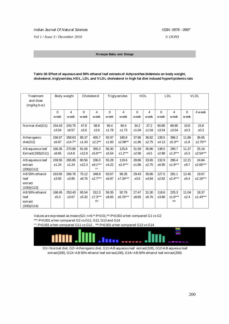
Indian Journal Of Natural Sciences ISSN: 0976 - 0997
Vol 1 / Issue 3 / December 2010 © IJONS
200
Table 18: Effect of aqueous and 50% ethanol leaf extracts of Achyranthes bidentata on body weight, cholesterol, triglycerides, HDL, LDL and VLDL cholesterol in high fat diet induced hyperlipidemic rats
Values are expressed as mean±S.D; n=6.##-P<0.01,###-P<0.001 when compared G1 vs G2 ***-P<0.001 when compared G2 vs G11, G12, G13 and G14 †††-P<0.001 when compared G11 vs G12 , €€€-P<0.001 when compared G13 vs G14
G1 G2 G11 G12 G13 G14 G1 G2 G11 G12 G13 G14 G1 G2 G11 G12 G13 G14 G1 G2 G11 G12 G13 G14 G1 G2 G11 G12 G13 G14 G1 G2 G11 G12 G13 G14
0
100
200
300
400
500 Body weightCholesterol
TriglyceridesHDLLDLVLDL
Treatment
mg/dl
G1= Normal diet, G2= Atherogenic diet, G11=AB aqueous leaf extract(100), G12=AB aqueous leaf extract(200), G13= AB 50% ethanol leaf extract(100), G14= AB 50% ethanol leaf extract(200)
Body weight Cholesterol Triglycerides HDL LDL VLDL Treatment and dose
(mg/kg b.w)
0 week
4 week
0 week
4 week
0 week
4 week
0 week
4 week
0 week
4 week
0 week
4 week
Normal diet(G1) 154.43 ±3.54
243.75 ±9.57
47.8 ±3.6
58.8 ±3.6
56.4 ±1.78
60.4 ±1.73
34.2 ±1.04
37.2 ±1.04
60.80 ±3.54
68.80 ±3.54
10.8 ±0.3
15.8 ±0.3
Atherogenic diet(G2)
156.67 ±6.87
268.63 ±14.7##
85.37 ±1.43
455.7 ±2.2###
55.97 ±1.83
180.8 ±2.96###
37.86 ±1.86
36.82 ±2.75
139.5 ±4.13
386.2 ±5.3###
11.86 ±1.8
36.65 ±2.75###
AB aqueous leaf Extract(100)(G11)
166.35 ±8.65
270.86 ±14.8
81.06 ±12.9
355.3 ±5.6***
56.30 ±5.54
125.8 ±1.2***
31.59 ±2.96
35.86 ±4.5
138.5 ±2.88
290.7 ±1.3***
11.37 ±5.3
25.16 ±2.54***
AB aqueous leaf extract (200)(G12)
159.55 ±1.24
265.85 ±1.24
80.56 ±12.3
336.0 ±9.1***
†††
55.28 ±4.22
119.6 ±2.4***
28.86 ±1.86
33.65 ±2.75
132.9 ±0.95
286.4 ±1.9***
12.21 ±9.7
24.84 ±2.65***
AB 50% ethanol leaf extract (100)(G13)
163.65 ±3.65
266.76 ±3.85
75.12 ±8.76
348.8 ±2.7***
63.67 ±6.87
96.35 ±7.34***
29.43 ±3.6
35.86 ±4.84
127.5 ±2.82
281.1 ±2.4***
12.45 ±5.4
19.67 ±2.16***
AB 50% ethanol leaf extract (200)(G14)
168.45 ±5.3
253.43 ±3.67
65.54 ±5.32
312.3 ±7.3***
€€€
59.35 ±8.65
92.76 ±6.76***
27.47 ±8.65
31.30 ±6.76
118.6 ±3.86
225.3 ±1.6***
€€€
11.04 ±2.4
18.37 ±1.43***
Niranjan Babu and Elango
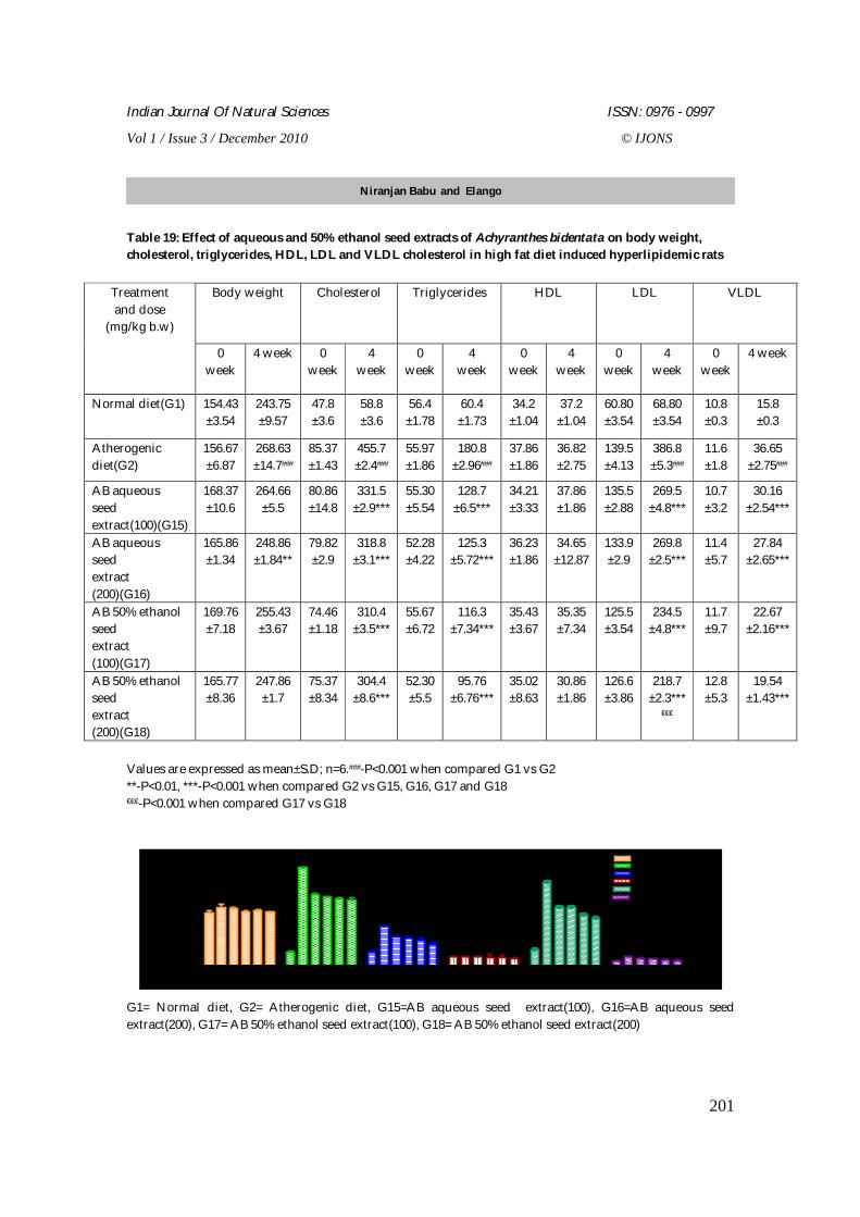
Indian Journal Of Natural Sciences ISSN: 0976 - 0997
Vol 1 / Issue 3 / December 2010 © IJONS
201
Table 19: Effect of aqueous and 50% ethanol seed extracts of Achyranthes bidentata on body weight, cholesterol, triglycerides, HDL, LDL and VLDL cholesterol in high fat diet induced hyperlipidemic rats
Values are expressed as mean±S.D; n=6.###-P<0.001 when compared G1 vs G2 **-P<0.01, ***-P<0.001 when compared G2 vs G15, G16, G17 and G18 €€€-P<0.001 when compared G17 vs G18
G1G2G15G16G17G18 G1G2G15G16G17G18 G1 G2G15G16G17G18 G1G2G15G16G17G18 G1G2G15G16G17G18 G1 G2G15G16G17G18
0
100
200
300
400
500 Body weightCholesterolTriglyceridesHDLLDLVLDL
Treatment
mg/dl
G1= Normal diet, G2= Atherogenic diet, G15=AB aqueous seed extract(100), G16=AB aqueous seed extract(200), G17= AB 50% ethanol seed extract(100), G18= AB 50% ethanol seed extract(200)
Body weight Cholesterol Triglycerides HDL LDL VLDL Treatment and dose
(mg/kg b.w)
0 week
4 week 0 week
4 week
0 week
4 week
0 week
4 week
0 week
4 week
0 week
4 week
Normal diet(G1) 154.43 ±3.54
243.75 ±9.57
47.8 ±3.6
58.8 ±3.6
56.4 ±1.78
60.4 ±1.73
34.2 ±1.04
37.2 ±1.04
60.80 ±3.54
68.80 ±3.54
10.8 ±0.3
15.8 ±0.3
Atherogenic diet(G2)
156.67 ±6.87
268.63 ±14.7###
85.37 ±1.43
455.7 ±2.4###
55.97 ±1.86
180.8 ±2.96###
37.86 ±1.86
36.82 ±2.75
139.5 ±4.13
386.8 ±5.3###
11.6 ±1.8
36.65 ±2.75###
AB aqueous seed extract(100)(G15)
168.37 ±10.6
264.66 ±5.5
80.86 ±14.8
331.5 ±2.9***
55.30 ±5.54
128.7 ±6.5***
34.21 ±3.33
37.86 ±1.86
135.5 ±2.88
269.5 ±4.8***
10.7 ±3.2
30.16 ±2.54***
AB aqueous seed extract (200)(G16)
165.86 ±1.34
248.86 ±1.84**
79.82 ±2.9
318.8 ±3.1***
52.28 ±4.22
125.3 ±5.72***
36.23 ±1.86
34.65 ±12.87
133.9 ±2.9
269.8 ±2.5***
11.4 ±5.7
27.84 ±2.65***
AB 50% ethanol seed extract (100)(G17)
169.76 ±7.18
255.43 ±3.67
74.46 ±1.18
310.4 ±3.5***
55.67 ±6.72
116.3 ±7.34***
35.43 ±3.67
35.35 ±7.34
125.5 ±3.54
234.5 ±4.8***
11.7 ±9.7
22.67 ±2.16***
AB 50% ethanol seed extract (200)(G18)
165.77 ±8.36
247.86 ±1.7
75.37 ±8.34
304.4 ±8.6***
52.30 ±5.5
95.76 ±6.76***
35.02 ±8.63
30.86 ±1.86
126.6 ±3.86
218.7 ±2.3***
€€€
12.8 ±5.3
19.54 ±1.43***
Niranjan Babu and Elango
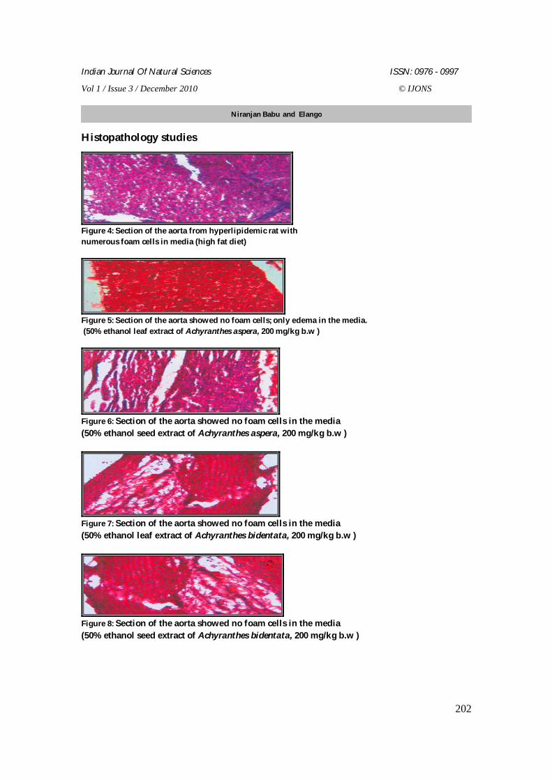
Indian Journal Of Natural Sciences ISSN: 0976 - 0997
Vol 1 / Issue 3 / December 2010 © IJONS
202
Histopathology studies
Figure 4: Section of the aorta from hyperlipidemic rat with numerous foam cells in media (high fat diet)
Figure 5: Section of the aorta showed no foam cells; only edema in the media. (50% ethanol leaf extract of Achyranthes aspera, 200 mg/kg b.w )
Figure 6: Section of the aorta showed no foam cells in the media (50% ethanol seed extract of Achyranthes aspera, 200 mg/kg b.w )
Figure 7: Section of the aorta showed no foam cells in the media (50% ethanol leaf extract of Achyranthes bidentata, 200 mg/kg b.w )
Figure 8: Section of the aorta showed no foam cells in the media (50% ethanol seed extract of Achyranthes bidentata, 200 mg/kg b.w )
Niranjan Babu and Elango
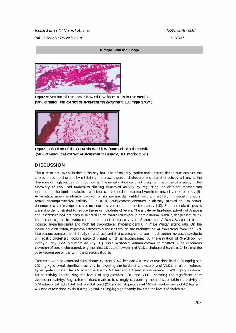
Indian Journal Of Natural Sciences ISSN: 0976 - 0997
Vol 1 / Issue 3 / December 2010 © IJONS
203
Figure 9: Section of the aorta showed few foam cells in the media (50% ethanol leaf extract of Achyranthes bidentata, 100 mg/kg b.w )
Figure 10: Section of the aorta showed few foam cells in the media (50% ethanol leaf extract of Achyranthes aspera, 100 mg/kg b.w ) DISCUSSION The current anti-hyperlipidemic therapy includes principally statins and fibrates; the former corrects the altered blood lipid profile by inhibiting the biosynthesis of cholesterol and the latter acts by enhancing the clearance of triglyceride rich lipoproteins. The investigation on plant drugs will be a useful strategy in the discovery of new lead molecules eliciting improved activity by regulating the different mechanisms maintaining the lipid metabolism and thus can be used in treating hyperlipidemia of varied etiology [5]. Achyranthus aspera is already proved for its spermicidal, antilithiatic, antifertility, immunostimulatory, cancer chemopreventive activity [6, 7, 8, 9,]. Achyranthus bidentata is already proved for its cancer chemoprotective, osteoprotective, neuroprotective, and immunostimulatory [10]. But these plant species were also demonstrated to reduce the serum cholesterol levels. The anti-hyperlipidemic activity of A.aspera and A.bidentata had not been elucidated in an controlled hyperlipidemic animal models, the present study has been designed to evaluate the lipid – controlling activity of A.aspera and A.bidentata against triton-induced hyperlipidemia and high fat diet-induced hyperlipidemia, in male Wistar albino rats. On the induction with triton, hypercholesterolemia occurs through the mobilization of cholesterol from the liver into plasma compartment initially (first phase) and that subsequent to such mobilization increased synthesis of hepatic cholesterol occurs (second phase) which is accompanied by the elevation of 3-hydrocy -3-methylglutatyl-CoA reductase activity [11]. Intra peritoneal administration of resulted in an enormous elevation of serum cholesterol, triglycerides, LDL, and lowering of VLDL cholesterol levels at 24 hrs and the observations are on par with the previous studies .
Treatment with aqueous and 50% ethanol extracts of AA leaf and AA seed at two dose levels 100 mg/kg and 200 mg/kg showed significant activity in lowering the levels of cholesterol and VLDL in triton induced hyperlipidemic rats. The 50% ethanol extract of AA leaf and AA seed at a dose level of 200 mg/kg produced better activity in reducing the levels of triglycerides, LDL and VLDL showing the significant dose dependent activity. Regression of these markers is strongly supporting the antihyperlipidemic activity of 50% ethanol extract of AA leaf and AA seed (200 mg/kg).Aqueous and 50% ethanol extracts of AB leaf and AB seed at two dose levels 100 mg/kg and 200 mg/kg significantly lowered the levels of cholesterol,
Niranjan Babu and Elango
Indian Journal Of Natural Sciences ISSN: 0976 - 0997
Vol 1 / Issue 3 / December 2010 © IJONS
204
triglycerides, LDL, and VLDL. Further, 200 mg/kg of both aqueous and 50% ethanol extract of AB leaf and AB seed produced a better activity when compared to lower dose showing the significant dose dependent activity. The results revealing that the AB leaf and seed extract possessed the better activity than AA leaf and seed extracts showing that the A.bidentata is having high potential in lowering the hypercholesterolemia. These results may indicate that the leaf and seed extracts of A.aspera and A.bidentata may interfere with cholesterol-biosynthesis as Triton accelerated the hepatic synthesis of cholesterol [12]. A high-cholesterol diet regarded as an important factor in the development of cardiac diseases, since it leads to the development of hyperlipidemia, atherosclerosis, and ischemic heart disease. A variety of mechanisms, i.e. inhibition of mevalonate pathway, decrease in NO bioavailability and cGMP metabolism, increase in free radical and peroxynitrite formation, inhibition of heat shock response, and expression of oxidized low density lipoprotein receptors which induces apoptosis, have been shown to play a role in cardiac effects of hyperlipidemia [13].
Triton induced hypercholesterolemia, though simple and rapid for evaluating hypolipidemic compounds, is rather artificial. Hence the lipid controlling potential of A.aspera and A.bidentata leaves and seeds were further evaluated in high-fat diet induced hyperlipidemic rat model. When male Wistar albino rats were kept on cholesterol-rich diet supplemented for 4 weeks and body weight, serum cholesterol, trigylcerides, LDL, VLDL levels were elevated where as HDL levels were significantly reduced. Elevated circulating lipid levels may be outcome of inhibitory effect of high fat dietary intake on lipogenesis. The treatment of high-fat diet induced hyperlipidemic rats with aqueous and 50% ethanol extracts of AA leaf and AA seed at two dose levels 100 mg/kg and 200 mg/kg showed significant activity in lowering the levels of cholesterol and VLDL. Though the levels of HDL were not increased, they are maintained as that of control rats. The extracts also possessed the significant activity in lowering the body weights. Further, the higher dose of aqueous and 50% ethanol extract of AA leaf and AA seed produced better activity in reducing the levels of cholesterol, triglycerides, and VLDL showing the significant dose dependent activity. Aqueous and 50% ethanol extracts of AB leaf and AB seed at two dose levels 100 mg/kg and 200 mg/kg significantly lowered the levels of cholesterol, triglycerides, LDL, and VLDL. The levels of HDL were not reduced and body weights are not increased, they are maintained as that of control rats. Further, the higher dose of aqueous and 50% ethanol extract of AB leaf and AB seed produced better activity in reducing the levels of cholesterol and LDL showing the significant dose dependent activity. The presence of foam cells resulted due to the proliferation of stromal cells is a characteristic feature of lipid deposition and atherosclerosis. Histopathology of aorta of rats treated with atherogenic diet shows presence of numerous foam cells which confirmed the induction of hyperlipidemia. Groups treated with 50% ethanol extract of leaf and seed of A. aspera and A. bidentata at a dose of 200 mg/kg showed absence of foam cells. Whereas few foam cells were found in case of groups treated with 100 mg/kg of 50% ethanol extract of leaf and seed of A. aspera and A. bidentata, supporting the dose dependent activity.
Presence of saponins, flavonoids, and triterpenoids in A. aspera and A. bidentata was already reported. Saponins derived from the Acorus calamus were reported to reduce the serum cholesterol levels by preventing the cholesterol absorption, interfering with entero-hepatic circulation and increasing its fecal excretion [14]. Flavonoids present in the Pterocapus marsupium were observed to be responsible for lowering the serum cholesterol, LDL and VLDL cholesterol levels . Lupeol, a triterpenoid was reported for its cardioprotective activity against experimental hypercholesterolemia . The lipid lowering activity of A. aspera and A. bidentata leaf and seed extract may be attributed to the presence of phytoconstituents such as saponins, flavonoids, and triterpenoids as reported for other plant extracts. Recent studies have demonstrated that increased formation of free radicals or reactive oxygen species (ROS) contribute to cardiovascular disease progression [15]. The generation of large amounts of reactive oxygen species can overwhelm the intracellular antioxidant defense, causing activation of lipid peroxidation, protein modification and DNA breaks. ROS induced depletion of antioxidants is a key factor for initiation of atherosclerosis . The anti hyperlipidemic activity may be attributed to the inhibition of ROS by the
Niranjan Babu and Elango
Indian Journal Of Natural Sciences ISSN: 0976 - 0997
Vol 1 / Issue 3 / December 2010 © IJONS
205
flavonoids and triterpenoids present in the extracts. All the beneficial effects of the extracts may be due to their antihyperlipidemic effects carried out by saponins, flavonoids, and triterpenoids present in them.
CONCLUSION
Achyranthes aspera Linn and Achyranthes bidentata Blume are two perennial herbs with slender and rambling branches. The two plant materials were collected from different parts of Nilgiri district of Tamilnadu and authenticated. Physicochemical parameters like total ash, acid insoluble ash, water soluble ash, and sulphated ash values were determined. Fluorescence analysis was carried out for the plant powder and their extracts.The dried and powdered leaves and seeds of plants were extracted with water and 50% ethanol through cold maceration. Alcohol soluble extractive and water soluble extractive values were determined. The phytochemical studies of the plant extracts showed the presence of alkaloids, glycosides, triterpenoids, saponins, flavonoids and mucilage. These results gave clues regarding the presence of some particular phytoconstituents in the respective plant extracts. The in vivo screening for antihyperlipidemic activity with triton induced hyperlipidemia and high fat diet induced hyperlipidemia model showed that both aqueous and 50% ethanol extracts of plants at a dose of 200 mg/kg exhibited significant activity against hyperlipidemia. Antihyperlipidemic activity activity of the various extracts may be attributed to the presence of triterpenoids, saponins, and flavonoids. From these studies, it can be concluded that both the plants are endowed with significant antihyperlipidemic activity. However, the future studies need to be carried out to isolate and screen the phytoprinciples responsible for the anti-hyperlipidemic activity.
REFERENCES
1. Petri T, Nikkila E, Meittinen T. Regulation of cholesterol synthesis and storage in fat cells. J.Lipid. Res. 1975; 16: 211-223.
2. Sharma SB, Dwivedi S. Medicinal Plants with hypolipidemic activities. Indian drugs. 1997; 34: 242-255.
3. Frantz ID, Hinkelman BT. Acceleration of hepatic cholesterol synthesis bt Triton WR 1339, J Exp Med.1955; 110:225.
4. Filliois LC, Andrus SB, Mann GV, Stare FJ. Experimental production of grass atherosclerosis in rat. J Exp Med.1956;104:539
5. Robert WM, Thomas B. Antihyperlipidemic agents. In: Brunton LL, Lazo SJ, eds. Goodman and Gilman’s the pharmacological basis of therapeutics. 11 th Ed, New York: McGraw-hill, 2006; 933.
6. Daniel Paul, Saradindu Bera, Debasis Jana, Rajkumar Maiti, Debidas Ghosh. In vitro determination of the contraceptive spermicidal activity of a composite extract Achyranthes aspera and Stephania hernandifolia on human semen. 2006; 73:284-288.
7. Deorao M. Awari, Vaishali Mute, Santi Prasad Babhale, Shradha P.chaudhari. Antilithiatic effect of Achyranthes aspera Linn. leaves extract on ethylene glycol induced nephrolithiasis. 2009; 2:994-997.
8. Neeru Vasudeva, Sharma SK. Post-coital antifretility of Achyranthes aspera Linn Root. 2006; 107:179-181.
9. Vasudeva Y, Romesh M, Singh A, Chakrabarti R. Potentiation of antibody production in Indian major carp Labeo rohita rohu, by Achyranthes aspera as a herbal feed ingredient. 2004; 238:67-73.
10. Cui-Cui He, Rong-Rong Hui, Yasuhiro Tezuka, Shigetoshi Kadota, Jain-Xin Li. Osteoprotective effect of extract from Achyranthes bidentata in ovariectomized rats. 2009;32:54-63
11. Kuroda M, Tanzawa K, Tujita Y, Endo A. Mechamism for hepatic cholesterol synthesis and serum cholesterol levels in Triton WR-1339- induced hyperlipidemia. 1977; 489:119-125.
12. Vogel GA, Vogel WH. Drug discovery and evaluation – Pharmacological assays. Berlin-Heidelberg. Springerlink: 1997: 606.
Niranjan Babu and Elango
Indian Journal Of Natural Sciences ISSN: 0976 - 0997
Vol 1 / Issue 3 / December 2010 © IJONS
206
13. Puskas LG, Nagy ZB, Giricz Z, Onody A, Csonka C, Kitajka K, et al. Cholesterol diet-induced hyperlipidemia influences gene expression pattern of rat hearts: a DNS microarray study. FEBS letters. 2004; 562: 99-104.
14. Parab RS, Mengi SA. Hypolipidemic activity of Acorus calamus L. in rats. Fitoterapia. 2002; 73: 451-455.
15. Kaliora AC, Dedoussis GVZ, Schmidt H. Dietary antioxidants in preventing atherogenesis. Atherosclerosis. 2006; 18:1.
Niranjan Babu and Elango
Indian Journal Of Natural Sciences ISSN: 0976 - 0997
Vol 1 / Issue 3 / December 2010 © IJONS
207
Phytochemical Screening and Antibacterial Activity of Plant Extract of Eclipta prostata L. Vinoth Kumar P1*, Amala pricy A2, Veera thamaraie selvi V1, Shanhthi V1, Vimal V3,
Sudheer kumar Ch4 and Kiran Kumar Goud G5. 1* Department of Biotechnology J.J college of Arts and science, Pudukkottai, Tamil Nadu, India. 2 Department of Biotechnology, Bharathidasan University, Trichirappalli, Tamil Nadu India. 1 Department of Biochemistry, J.J College of Arts and Science, Pudukkottai, Tamil Nadu India. 1 Department of Biochemistry, J.J College of Arts and Science, Pudukkottai, Tamil Nadu India. 3 Department of Biotechnology, Bishop Heber College, Trichirappalli, Tamil Nadu India. 4 School of Life Sciences, Department of Biotechnology, University College of engineering, JNTU, Kakinada, Andhra Pradesh, India. 5 Department of Microbiology, J.J college of Arts and science, Pudukkottai, Tamil Nadu, India. Received: 20 Oct 2010 Revised: 15 Nov 2010 Accepted: 1 Dec 2010 *Address for correspondence: Vinoth Kumar P,
Department of Biotechnology, J.J College of Arts and Science, Pudukottai, Tamil Nadu, India.
E mail ID: [email protected].
Eclipta prostata.L (Asteraceae) is an important medicinal plant with lot of phytochemical constituents selected form the available literature was taken for the present study. The aqueous and ethanol extracts of Eclipta prostata.L were used to determine the antibacterial efficiency against Gram positive and Gram negative organisms such as E.coli, Staphylococcus edudemis, Serratia marcenses and Salmonella typhimurium. The ethanol extract of Eclipta prostata.L showed a strong inhibitory activity against all the four species of Gram positive and Gram negative organisms, while the aqueous extract of the plant had not produce inhibitory against any of the species used in the present study. From the result it could be concluded that the ethanol extract of Eclipta prostata.L as a strong antibacterial agent against both Gram positive and Gram negative organisms namely E.coli, Salmonella typhimurium, Serratia marcenses and Staphylococcus edudemis. Phytochemical screening revealed that plant extracts of Eclipta prostata.L contained flavonoids, saponins, phytosterols, carbohydrates, phenols, tannins and terpenoids. Key words: Eclipta prostate L., Antibacterial activity, Phytochemical Analysis. INTRODUCTION Eclipta prostata.L (Asteracae) commonly known with the name of “Vellai karisinakanni” in Tamil nadu, India. Leaf juice is used as tonic for jaundice and leaf paste is applied on the affected area for tooth ache [1]. The roots are very rich of thiophene acetylenes. Four compounds were isolated from E. prostrata, of which
RESEARCH ARTICLE
ABSTRACT
Indian Journal Of Natural Sciences ISSN: 0976 - 0997
Vol 1 / Issue 3 / December 2010 © IJONS
208
two were identified as stigmasterol and alpha-terthenyl. Alpha-terthienyl is isolated from plant and these constituents are significant for hepatoprotective activity [2, 3]. Eclipta prostata.L an aromatic plant is known in Chinese herbal medicine for the treatment of various kidney diseases. Eclipta prostata.L can play an important role in osteoblastic bone formation, and may possibly lead to the development of bone-forming drugs [4]. Leaf extract of E. Prostrata are shown Hypolipidemic activity [5].Phytochemical is a natural bioactive compound found in plants, such as vegetables, fruits, medicinal plants, flowers, leaves and roots that work with nutrients and fibers to act as an defense system against disease or more accurately, to protect against disease. Phytochemicals are divided into two groups, which are primary and secondary constituents, according to their functions in plant metabolism. Primary constituents comprise common sugars, amino acids, proteins and chlorophyll while secondary constituents consists of alkaloids, terpenoids and phenolic compounds and many more such as flavonoids, tannins and so on. Plants do not produce enough secondary compounds to protect all their tissues at all times, thus concentration and types of compounds vary through out the plant. Some compounds are present at all times in cellular tissue and some are activated after attack. The secondary metabolite production of desirable medical compounds from plants, bio-technological approach, especially plants tissue cultures are found to have potential as found to have potential as a supplement to traditional agriculture in the industrial production of bioactive plant metabolites [6]. The main aim of the work is to develop the appropriate extraction methods in order to obtain plant extracts with as many phytochemical compounds as possible is important.
MATERIAL AND METHODS
Plant extraction
Eclipta prostata.L were shade-dried and pulverized to a coarse powder. Equal quantities of the powder was passed through 40-mesh sieve and exhaustively extracted with 90% (v/v) ethanol in soxhlet apparatus at 60oC [7]. The extract was evaporated under pressure until all the solvent had been removed and further removal of the water was carried out by freeze drying to give an extract sample with the yield of 19.7% (w/w.) The extract was stored in refrigerator, weighed amount was dissolved in 2% (v/v) aqueous Tween–80 (2 ml of Tween 80 dissolved in 98 ml of water) and used for present investigation. Preliminary phytochemical analysis The Phytochemical screening procedures were carried out according to the following methods. By using the standard procedures to identify the constituents as described by Evans [8] and Harborne [9]. Test organisms Both Gram positive and Gram negative organisms were used for the test. The Gram positive bacteria include Staphylococcus edudemi and Serratia marcenses. The Gram negative bacteria include E.coli and Salmonella typhimurium. All the bacterial strains were maintained on freshly prepared Muller and Hinton Agar (Hi-Media) slant and stored at 0°C. Screening of antibacterial activity Antibacterial activity was determined by agar well diffusion method. Muller Hinton agar plates were prepared after that the inoculums were swab with the help of cotton swab. The sterile well cutter was to cut 3 wells in each plate. The three crude solvent extract were poured on to the separate well. The plant extract was slowly diffused thorough media in the plates. The inoculated plates were incubated at 37°C for 24 hours.
Vinoth Kumar et al
Indian Journal Of Natural Sciences ISSN: 0976 - 0997
Vol 1 / Issue 3 / December 2010 © IJONS
209
RESULT AND DISCUSSION Preliminary phytochemical Analysis A Phytochemical are important component which was the agents causes the antibacterial activity. The amount of phytochemical may vary in different plants. The determination study of the phytochemical on Eclipta prostata.L gives these results (Table-1). Thin layer chromatography was used for determination of important components of the plant extracts. The Rf value helps to find out the quantity of the detected objects and the colour helps to find out the object. The Rf Value is distance moved by the sample in Cm/Distance the moved by solvent in cm (Table-2). Phytochemically, Eclipta prostata.L is rich in wadeoloctone, eclalbasaponin, ß-amyrin, stigmasterol and luteolin-7- glucoside [10]. We have found that most of the biologically active phytochemicals were present in the ethanol extract of Eclipta prostata.L. The medicinal properties of extracts may be due to the presence of above mentioned phytochemicals. The antibacterial activity of ethanol extracts against four microbial species was shown in Table-3. Serratia marcenses, Salmonella typhimurium, Escheria coli, and Staphylococcus edidemis are the micro organisms used. No inhibition was observed by the aqueous extract of Eclipta prostata.L against any of the microorganisms used in the present study. On contrast, the ethanol extract of Eclipta prostata.L was found to produce inhibition against the growth of all the four microorganisms used in the present study. The ethanol extracts of Eclipta prostata.L were found to be most effective inhibitor of bacterial growth of Escheria coli, Salmonella typhimurium, Serratia marcenses and Staphylococcus ebidemis. In general S.marcescens is not pathogenic to insects when present in the digestive tract in small numbers, but once it enters the hemocoel it multiples rapidly and cause death in one to three days. The inhibitory zone size of gram positive bacteria indicates their less sensitive when compared to the Gram negative bacteria. It may be due to the presence of large amount of peptidoglygon layer which protect against various compounds [11]. The result obtained from this study is compliment to earlier observations by Rotimi [12]. The result of the present study confirms the antimicrobial potential of the ethanolic extracts of Eclipta prostata.L.
CONCLUSION The results of the present in vitro study indicated that the ethanol fraction of Eclipta prostata.L leaf extract possess effective antibacterial properties which is as potent as standard antibacterial properties against certain microorganisms. ACKNOWLEDGEMENT
Authors are thankful to J.J College of arts and science for providing the facilities for me. I would thank to Mr.Joshva, Miss. Jebitha, Miss. Latha, for encouraging me to do my project. I would like to thank I.Raja, R.Palathasar and A.Jude for providing fund to me carrying out my project.
Vinoth Kumar et al

Indian Journal Of Natural Sciences ISSN: 0976 - 0997
Vol 1 / Issue 3 / December 2010 © IJONS
210
Table: 1. Preliminary phytochemicals analysis of Eclipta prostata.L
Phytochemicals Inference % Flavonoids + 0.045 Saponins + 0.029 Phytosterols + 0.026 Carbohydrates + 0.016
Phenols + 0.019 Tannins + 0.009 Terpenoids + 0.004 Alkaloids - - Phlobatannis - -
+ = Presence; - = Absence
Table: 2. Thin Layer chromatographic determinations in Eclipta prostata.L
Colour Rf Value Pink(Amino acids) 0.1 Green(Neutral lipids) 0.13 Brown(Mono-Disaccharides) 0.36
Table: 3. Antibacterial activities of the various extracts of Eclipta prostata.L
Diameter of inhibition of Zone (mm) Name of the microorganism
Plant Extract Solvent Control Test
Water - - E.coli Eclipta prostata.L Ethanol - 11mm Water - - S.typhimurium Eclipta prostata.L Ethanol - 8mm Water - - S.marcenses Eclipta prostata.L Ethanol - 7mm Water - - S.ebidemis Eclipta prostata.L Ethanol - 5mm
REFERENCES
1. Sandhya B, Thomas S, Isabel W, Shenbagarathai R. Ethnomedicinal plants used by the Valaiyan community of Piranmalai hills (reserved forest), Tamil Nadu, India - A pilot study. Afr J Trad Comp Alt Med 2006; 3: 101–114.
2. Han Y, Xia C, Cheng X, Xiang R, Liu H, Yan Q, Xu D. Preliminary studies on chemical constituents
and pharmacological action of Eclipta prostata.L. China J of Chinese materia medica.1998; 23 :680-682.
Vinoth Kumar et al
Indian Journal Of Natural Sciences ISSN: 0976 - 0997
Vol 1 / Issue 3 / December 2010 © IJONS
211
3. Song-chow Lin, Chih-jung Yao, Chun-ching Lin, Yun-ho Lin. Hepatoprotective Activity of Taiwan
Folk Medicine: Eclipta prostata.L Against Various Hepatotoxins Induced Acute Hepatotoxicity. Phytotherapy research.1998; 10: 483-490.
4. Lin XH, Wu YB, Lin S, Zeng JW, Zeng PY, Wu JZ. Effects of volatile components and ethanolic
extract from Eclipta prostata.L on proliferation and differentiation of primary osteoblasts. Molecules (Basel, Switzerland). 2010; 15:241-250.
5. Dhandapani R. Hypolipidemic activity of Eclipta prostata.L leaf extract in atherogenic diet induced
hyperlipidemic rats. Indian J of experimental biology. 2007; 4 5:617-619.
6. Ramachandra Rao S, Ravishankar GA. Plant cell cultures; chemical factories for secondary metabolites. Biotechnol Adv. 2002; 20:101-153.
7. Chattopadhyay RR. Possible mechanism of hepatoprotective activity of Azadirachta indica leaf extract. J Ethnopharmacol. 2003; 89: 217–219.
8. Evans WC. An index of medicinal plants. A Text book of Pharmacognosy. 14 ed. 1997; 7: 12-14.
9. Harborne JB. Phytochemical methods, London. Chapman and Hall, Ltd. 1973; 49-188.
10. Asolkar AV, Kakkar KK, Chakre OJ. Glossary of Indian Medicinal plants with active principles.
Publication and information directorate (CSIR), New Delhi. 1992; 1: 287.
11. Kandler O. Cell wall structure and their phylogentic implications system. Appl.Microbiol. 1982; 3:149-160.
12. Rotimi VO, Laughon BE, Bartlett JG, Mosadomi HA. Activities of Nigerian chewing sticks against bacteriodes-gingivalis and baceriodes-melaninogenicus. Antimicrobiological Agents and Chemotherapy. 1988; 32: 598-600.
Vinoth Kumar et al
Indian Journal Of Natural Sciences ISSN: 0976 - 0997
Vol 1 / Issue 3 / December 2010 © IJONS
212
The Indian Journal of Natural Sciences is a bi-monthly International Journal, which publishes original research work that contributes significantly to further the scientific knowledge in Natural Sciences (All science disciplines). The Journal publishes original research work either as a Full Research Paper or as a Short Communication. Review Articles in Natural Sciences are also considered for publication by the Journal. Type-written manuscripts prepared using MS Word should be submitted in triplicate to The Editor, Indian Journal of Natural Sciences C/oTNSRO,46/141,Meenakshipuram Road,Arimalam-622201,Pudukkottai Dist, Tamilnadu, India. Alternatively, the manuscript can also be sent as a MS word document to [email protected]. Manuscripts will be subjected to peer review process to determine their suitability for publication provided they fulfil the requirements of the journal as laid out in the instructions to authors. After the review, manuscripts will be returned for revision along with reviewer’s and/or editor’s comments. One original copy of the final revised manuscript should be submitted for publication with in two months of receiving the reviewer’s comments. It is also desirable to submit the final revised manuscript on a CD prepared in MS Word Version 97-2002 or a higher version. Prepare the manuscript in Times New Roman font using a font size of 12. Title shall be in a font size 14. All section titles in the manuscript shall be in font size 12, bold face capitals. Subtitles in each section shall be in font size 12, bold face lower case followed by a colon. There shall not be any decorative borders anywhere in the text including the title page. The entire MS Word document with graphs and illustrations pasted in it shall not exceed 1.2 MB. Submission of a manuscript to Indian Journal of Natural Sciences for publication implies that the same work has not been either published or under consideration for publication in another Journal. Authors, in their cover note to the Editor, have to clearly mention whether the manuscript shall be considered as a Research Paper, Short Communication or Review Article and also confirm that the manuscript has not been submitted to any other Journal for publication. Authors publishing results from in vivo experiments involving animals or humans should state whether due permission for conduction of these experiments was obtained, from the relevant ethics committees, in the Materials and Methods section. In addition, authors wishing to publish research work involving human studies should also send a notary verified letter of approval from the Ethics Committee or the Institutional Review Board. PREPARATION OF MANUSCRIPTS: RESAERCH PAPERS Manuscripts must be prepared in accordance with “Uniform requirements for Manuscripts submitted to Biomedical Journal” developed by International Committee of Medical Journal Editors (October 2001). Authors desirous of submitting manuscripts for publication to the Indian Journal of Natural Sciences are advised to refer to the current issue of the Journal to understand the format and style of papers that get published. Authors are also advised to go through the current set of Instructions to Authors and prepare their manuscripts accordingly. Information should be conveyed in simple language with the correct syntax. Care should be taken to avoid common errors like having abbreviations in the Title or Abstract, beginning a sentence with a numeral, adding “etc.” after a few examples, dropping articles and using “&” instead of ‘and’ in the text. The usage of standard abbreviations and symbols is encouraged. Manuscripts that fail to
INSTRUCTION TO AUTHOR
Indian Journal Of Natural Sciences ISSN: 0976 - 0997
Vol 1 / Issue 3 / December 2010 © IJONS
213
conform to the requirements of the Journal, as specified under Instructions to Authors, will be rejected outright. Manuscripts should be concisely written and conform to the following general requirements: Manuscripts should be typewritten in double-space in A4 sized sheets, only on one side, with a 2 cm margin on both sides. Research Papers should have more than 15 pages, Review Articles in the range of 15-30 pages and Short Communications up to 15 pages, inclusive of illustrations. Pages should be numbered consecutively, starting with the title page and the matter arranged in the following order: Title page, Abstract, Introduction, Materials and Methods, Results, Discussion or Results and Discussion, Acknowledgements, References, Illustrations (Tables and figures including chemistry schemes along with titles and legends) and figure and Table titles and legends. Abstract should start on a separate page and each table or figure should be on separate sheets. The titles “Abstract” and “Introduction” need not be mentioned. All other section titles should be in capital letters while subtitles in each section shall be in bold face lower case followed by a colon. Title Page - Title page should contain title of the paper in bold face, title case (font size 14), names of the authors in normal face, upper case (font size 12) followed by the address(es) in normal face lower case. The author to whom all correspondence be addressed should be denoted by an asterisk mark. The title should be as short as possible and precisely indicate the nature of the work in the communication. Names of the authors should appear as initials followed by surnames for men and one given-name followed by surname for women. Full names may be given in some instances to avoid confusion. Names should not be prefixed or suffixed by titles or degrees. Names should be followed by the complete postal address or addresses with pin code numbers of the place(s), where the research work has been carried out. At the bottom left corner of the title page, please mention “*Address For correspondence” and provide a functional e-mail address. Address of the corresponding author to whom all correspondence may be sent should be given only if it is different from the address already given under authors’ names. Trivial sub-titles such as ‘Title’, ‘Author’, ‘Address’ or ‘Place of Investigation’ shall not be included in the title page. Title page should be aligned centre except for “* Address For correspondence”. Provide a running title or short title of not more than 50 characters.
Abstract - Should start on a new page after the title page and should be typed in single-space to distinguish it from the Introduction. Abstracts should briefly reflect all aspects of the study, as most databases list mainly abstracts. Short Communications as well as Review Articles should have an Abstract.
Key-words - Provide four to ten appropriate key words after abstract.
Introduction - Shall start immediately after the Abstract, as the next paragraph, but should be typed in double-space. The Introduction should lead the reader to the importance of the study; tie-up published literature with the aims of the study and clearly states the rationale behind the investigation.
Materials and Methods - Shall start as a continuation to introduction on the same page. All important materials used along with their source shall be mentioned. The main methods used shall be briefly described, citing references. Trivial details may be avoided. New methods or substantially modified methods may be described in sufficient detail. The statistical method and the level of significance chosen shall be clearly stated.
INSTRUCTION TO AUTHOR
Indian Journal Of Natural Sciences ISSN: 0976 - 0997
Vol 1 / Issue 3 / December 2010 © IJONS
214
Results - All findings presented in tabular or graphical form shall be described in this section. The data should be statistically analyzed and the level of significance stated. Data that is not statistically significant need only to be mentioned in the text - no illustration is necessary. All Tables and figures must have a title or caption and a legend to make them self-explanatory. Results section shall start after materials and methods section on the same page. Discussion - This section should follow results, deal with the interpretation of results, convey how they help increase current understanding of the problem and should be logical. Unsupported hypothesis should be avoided. The Discussion should state the possibilities the results uncover, that need to be further explored. There is no need to include another title such as “Conclusions” at the end of Discussion. Results and discussion of results can also be combined under one section, Results and Discussion.
Acknowledgements - Should be given after the text and not in the form of foot-notes.
References - Should be numbered consecutively in the order in which they are first mentioned in the text (not in alphabetic order). Identify references in text, tables, and legends by Arabic numerals in superscript in square brackets. References cited only in tables or figure legends should be numbered in accordance with the sequence established by the first identification in the text of the particular table or figure. Use the style of the examples below, which are based on the formats used by the international journals. The titles of journals should be abbreviated according to the style used in international journals. Use complete name of the journal for non-indexed journals. Avoid using abstracts as references. Information from manuscripts submitted but not accepted should be cited in the text as “unpublished observations” with written permission from the source. Avoid citing a “personal communication” unless it provides essential information not available from a public source, in which case the name of the person and date of communication should be cited in parentheses in the text. For scientific articles, contributors should obtain written permission and confirmation of accuracy from the source of a personal communication. The commonly cited types of references are shown here, for other types of references such as electronic media, newspaper items, etc. please refer to ICMJE Guidelines (http://www.icmje.org). Articles in Journals 1. Devi KV, Pai RS. Antiretrovirals: Need for an Effective Drug Delivery. Indian J Pharm Sci 2006;68:1-6. List the first six contributors followed by et al. 2. Volume with supplement: Shen HM, Zhang QF. Risk assessment of nickel carcinogenicity and
occupational lung cancer. Environ Health Perspect 1994; 102 Suppl 1:275-82. 3. Issue with supplement: Payne DK, Sullivan MD, Massie MJ. Women’s psychological reactions to breast
cancer. Semin Oncol 1996;23(1, Suppl 2):89-97.
Books and other Monographs 4. Personal author(s): Ringsven MK, Bond D. Gerontology and leadership skills for nurses. 2nd ed. Albany
(NY): Delmar Publishers; 1996. 5. Editor(s), compiler(s) as author: Norman IJ, Redfern SJ, editors. Mental health care for elderly people.
New York: Churchill Livingstone; 1996.
INSTRUCTION TO AUTHOR
Indian Journal Of Natural Sciences ISSN: 0976 - 0997
Vol 1 / Issue 3 / December 2010 © IJONS
215
6. Chapter in a book: Phillips SJ, Whisnant JP. Hypertension and stroke. In: Laragh JH, Brenner BM,
editors. Hypertension: pathophysiology, diagnosis, and management. 2nd ed. New York: Raven Press; 1995. p. 465-78.
Illustrations: Tables - Should be typed on separate sheets of paper and should not preferably contain any molecular structures. Only MS word table format should be used for preparing tables. Tables should show lines separating columns but not those separating rows except for the top row that shows column captions. Tables should be numbered consecutively in Arabic numerals and bear a brief title in capital letters normal face. Units of measurement should be abbreviated and placed below the column headings. Column headings or captions shall be in bold face. It is essential that all tables have legends, which explain the contents of the table. Tables should not be very large that they run more than one A4 sized page. Tables should not be prepared in the landscape format, i. e. tables that are prepared width wise on the paper.
Figures - Should be on separate pages but not inserted with in the text. Figures should be numbered consecutively in Arabic numerals and bear a brief title in lower case bold face letters below the figure. Graphs and bar graphs should preferably be prepared using Microsoft Excel and submitted as Excel graph pasted in Word. These graphs and illustrations should be drawn to approximately twice the printed size to obtain satisfactory reproduction. As far as possible, please avoid diagrams made with India ink on white drawing paper, cellophane sheet or tracing paper with hand written captions or titles. Photographs should be on glossy paper. Photographs should bear the names of the authors and the title of the paper on the back, lightly in pencil. Alternatively photographs and photomicrographs can be submitted as jpeg images. Figure and Table titles and legends should be typed on a separate page with numerals corresponding to the illustrations. Keys to symbols, abbreviations, arrows, numbers or letters used in the illustrations should not be written on the illustration itself but should be clearly explained in the legend. Avoid inserting a box with key to symbols, in the figure or below the figure. In case of photomicrographs, magnification should be mentioned either directly on them or in the legend. Symbols, arrows or letters used in photomicrographs should contrast with the background. Method of staining should also be mentioned in the legend.
Chemical terminology - The chemical nomenclature used must be in accordance with that used in the Chemical Abstracts.
Symbols and abbreviations - Unless specified otherwise, all temperatures are understood to be in degrees centigrade and need not be followed by the letter ‘C’. Abbreviations should be those well known in scientific literature. In vitro, in vivo, in situ, ex vivo, ad libitum, et al. and so on are two words each and should be written in italics. None of the above is a hyphenated word. All foreign language (other than English) names and words shall be in italics as a general rule. Words such as carrageenan-induced inflammation, paracetamol-induced hepatotoxicity, isoproterenol-induced myocardial necrosis, dose-dependent manner are all hyphenated.
Biological nomenclature - Names of plants, animals and bacteria should be in italics.
Enzyme nomenclature - The trivial names recommended by the IUPAC-IUB Commission should be used. When the enzyme is the main subject of a paper, its code number and systematic name should be stated at its first citation in the paper.
Spelling - These should be as in the Concise Oxford Dictionary of Current English.
INSTRUCTION TO AUTHOR
Indian Journal Of Natural Sciences ISSN: 0976 - 0997
Vol 1 / Issue 3 / December 2010 © IJONS
216
SHORT COMMUNICATIONS
The journal publishes exciting findings, preliminary data or studies that did not yield enough information to make a full paper as short communications. These have the same format requirements as full papers but are only up to 15 pages in length in total. Short Communications should not have subtitles such as Introduction, Materials and Methods, Results and Discussion - all these have to be merged into the running text. Short Communications preferably should have only 3-4 illustrations.
REVIEW ARTICLES
Should be about 15-30 pages long, contain up-to-date information, comprehensively cover relevant literature and preferably be written by scientists who have in-depth knowledge on the topic. All format requirements are same as those applicable to full papers. Review articles need not be divided into sections such as materials and Methods and Results and Discussion, but should definitely have an Abstract and Introduction, if necessary.
PUBLICATION FEE To publish the paper in our journal author should be paid Rs. 1000 (INDIA) and 70$ (FOREIGN COUNTRIES) in advance by DD drawn in favour of Indian Journal Of Natural Sciences, payable at Arimalam. *All correspondence relating to subscription and manuscript submission may be address to The Editor in Chief,
Indian Journal Of Natural Sciences
C/o Tamil Nadu Scientific Research Organization (TNSRO),
#46/141,Meenakshipuram Road
Arimalam-622 201,
Pudukkottai Dist,
Tamilnadu,
India
Contact Nos: 04333-271989, 9952886637 ,
E.mail:[email protected].
INSTRUCTION TO AUTHOR