obstructive sleep apnea -...
TRANSCRIPT

1050
Obstructive sleep apnea (OSA) is characterized by inter-mittent hypoxemia and associated with increased preva-
lence of vascular dysfunction and arterial hypertension, but the underlying mechanisms are incompletely understood.1 In OSA, apneas are related to excessive throat muscle relaxation with consequent inspiratory efforts against the closed glottis, finally resulting in the patient’s arousal associated with sym-pathetic activation and hyperventilation. The hyperventilation, in turn, contributes to the next apnea by reducing the arterial partial pressure of CO
2 toward the apnea threshold. In this sce-
nario, arterial oxygen desaturation plays a pivotal role by nar-rowing the gap between the eupneic and apneic CO
2 pressure
level, thereby rendering the CO2-controlled breathing regu-
lation more unstable and, thus, prone to periodic breathing.2 In patients with OSA, the presence of patent foramen ovale (PFO) is associated with a marked increase in the frequency and severity of nocturnal oxygen desaturations.3 This could be related, at least in part, to right to left shunting across a
PFO, as evidenced by invasive hemodynamic measurements in healthy humans, demonstrating that during the onset of sim-ulated OSA, the right atrial pressure exceeds left atrial pres-sure in response to the steep decline in intrathoracic pressure.4 Improvement of arterial oxygen saturation by PFO closure may prevent this problem.
We recently reported preliminary findings showing that PFO closure in patients with newly diagnosed OSA was asso-ciated with a reduction of nocturnal apnea–hypopnea events and oxygen desaturations and decreased pulmonary artery pressure.5 Moreover, circumstantial evidence shows that intermittent hypoxia is associated with systemic endothelial dysfunction and arterial hypertension.6,7 In line with these observations, systemic endothelial function is impaired in OSA patients without any traditional cardiovascular risk fac-tors.8 Systemic endothelial dysfunction precedes the develop-ment of arteriosclerosis and contributes to the pathogenesis of arterial hypertension.9 Accordingly, the prevalence of arterial
Abstract—Obstructive sleep apnea (OSA) is a frequent syndrome characterized by intermittent hypoxemia and increased prevalence of arterial hypertension and cardiovascular morbidity. In OSA, the presence of patent foramen ovale (PFO) is associated with increased number of apneas and more severe oxygen desaturation. We hypothesized that PFO closure improves sleep-disordered breathing and, in turn, has favorable effects on vascular function and arterial blood pressure. In 40 consecutive patients with newly diagnosed OSA, we searched for PFO. After initial cardiovascular assessment, the 14 patients with PFO underwent initial device closure and the 26 without PFO served as control group. Conventional treatment for OSA was postponed for 3 months in both groups, and polysomnographic and cardiovascular examinations were repeated at the end of the follow-up period. PFO closure significantly improved the apnea–hypopnea index (ΔAHI −7.9±10.4 versus +4.7±13.1 events/h, P=0.0009, PFO closure versus control), the oxygen desaturation index (ΔODI −7.6±16.6 versus +7.6±17.0 events/h, P=0.01), and the number of patients with severe OSA decreased significantly after PFO closure (79% versus 21%, P=0.007). The following cardiovascular parameters improved significantly in the PFO closure group, although remained unchanged in controls: brachial artery flow–mediated vasodilation, carotid artery stiffness, nocturnal systolic and diastolic blood pressure (−7 mm Hg, P=0.009 and −3 mm Hg, P=0.04, respectively), blood pressure dipping, and left ventricular diastolic function. In conclusion, PFO closure in OSA patients improves sleep-disordered breathing and nocturnal oxygenation. This translates into an improvement of endothelial function and vascular stiffening, a decrease of nighttime blood pressure, restoration of the dipping pattern, and improvement of left ventricular diastolic function.
Clinical Trial Registration—URL: http://www.clinicaltrials.gov. Unique identifier: NCT01780207. (Hypertension. 2015;66:1050-1057. DOI: 10.1161/HYPERTENSIONAHA.115.06303.) • Online Data Supplement
Key Words: arterial hypertension ■ endothelial dysfunction ■ hypoxemia ■ left ventricular diastolic function ■ obstructive sleep apnea ■ patent foramen ovale
Received August 11, 2015; first decision August 19, 2015; revision accepted August 24, 2015.From the Department of Cardiology and Clinical Research (S.F.R., E.R., S.F.d.M., Y.A., U.S., C.S.) and Department of Pneumology (S.O., M.G.),
Inselspital, University Hospital, Bern, Switzerland; and Facultad de Ciencias, Departamento de Biología, Universidad de Tarapacá, Arica, Chile (U.S.).*These authors contributed equally to this work.The online-only Data Supplement is available with this article at http://hyper.ahajournals.org/lookup/suppl/doi:10.1161/HYPERTENSIONAHA.
115.06303/-/DC1.Correspondence to Christian Seiler or Stefano F. Rimoldi, Department of Cardiology and Clinical Research, University Hospital Bern, CH-3010 Bern,
Switzerland. E-mail [email protected] or [email protected]
Patent Foramen Ovale Closure in Obstructive Sleep Apnea Improves Blood Pressure and Cardiovascular FunctionStefano F. Rimoldi,* Sebastian Ott,* Emrush Rexhaj, Stefano F. de Marchi, Yves Allemann,
Matthias Gugger, Urs Scherrer, Christian Seiler
© 2015 American Heart Association, Inc.
Hypertension is available at http://hyper.ahajournals.org DOI: 10.1161/HYPERTENSIONAHA.115.06303
Obstructive Sleep Apnea
by guest on May 29, 2018
http://hyper.ahajournals.org/D
ownloaded from
by guest on M
ay 29, 2018http://hyper.ahajournals.org/
Dow
nloaded from
by guest on May 29, 2018
http://hyper.ahajournals.org/D
ownloaded from
by guest on M
ay 29, 2018http://hyper.ahajournals.org/
Dow
nloaded from
by guest on May 29, 2018
http://hyper.ahajournals.org/D
ownloaded from
Rimoldi et al PFO Closure in Obstructive Sleep Apnea 1051
hypertension is increased in OSA, and resistant hyperten-sion is often found in these patients.10,11 We hypothesized that PFO closure in newly diagnosed OSA patients improves endothelial function, blood pressure (BP), and cardiac func-tion by reducing the number of apnea–hypopnea events and the severity of oxygen desaturations. To test this hypothesis, we searched for PFO in patients with newly diagnosed OSA and performed cardiovascular assessment, including systemic vascular function, 24-h ambulatory BP monitoring, and trans-thoracic echocardiography. Patients with PFO underwent initial device closure, and the others served as control group. Conventional treatment for OSA was postponed for 3 months in both groups, and polysomnographic and cardiovascular examinations were repeated at the end of the follow-up period.
Methods
Study Design and ParticipantsThis was a prospective, open-label interventional clinical trial in 51 consecutive patients with newly diagnosed OSA in whom we searched for PFO between September 2010 and November 2013. After baseline sleep and cardiovascular evaluation, the 17 patients with PFO underwent initial device closure, and the 34 patients with-out PFO served as control group. Conventional treatment for OSA was postponed for 3 months in both groups, and polysomnographic and cardiovascular examinations were repeated at the end of the fol-low-up period. Inclusion criteria for the study were newly diagnosed mild to severe OSA, age >18 years, and written informed consent for study participation. Exclusion criteria were pulmonary disease other than OSA associated with oxygen desaturation, central sleep apnea syndrome, pulmonary hypertension, intracardiac shunt other than via PFO, severe valvular heart disease, body mass index >40 kg/m2, and contraindication to transesophageal echocardiography (TEE).
The study protocol was approved by the ethics committee of the University of Bern, Switzerland. All included patients gave written in-formed consent to participate.
Study ProtocolSee online-only Data Supplement.
Assessment of Obstructive Sleep Apnea by Overnight PolysomnographySix electroencephalograms, 2 electrooculograms, electromyograms, thoraco-abdominal movements (respiratory inductive plethysmogra-phy), electrocardiography, and the patient’s posture were recorded. Nasal airflow was monitored using a nasal prong device, and arterial oxygen saturation was measured using finger pulse oximetry. Sleep state was recorded using the standard placements of electroencephalogram (C4/A1, F4/A1, and O1/A2), left and right electrooculograms, and sub-mental electromyogram according to the classification of the American Academy of Sleep Medicine.12 All measurements were recorded with a computerized recording system (N7000, Embla, Broomfield, CO). Apnea was defined as cessation of oro-nasal airflow ≥10 s, and hypop-nea as a ≥30% reduction from baseline of oro-nasal airflow for ≥10 s that was associated with ≥4% oxygen desaturation from pre-event base-line.12 Apnea–hypopnea index (AHI) was calculated as the mean number of apneas and hypopneas per hour, and apnea index was calculated as the mean number of apneas per hour between lights off and on. Severity of OSA was classified based on the AHI (number of apneas plus hypop-neas per hour of sleep): mild, AHI 5 to 15; moderate, AHI 16 to 30; and severe, AHI >30. Analysis of polysomnography was performed auto-matically and corrected manually by investigators blinded to the PFO closure being present or not, allowing differentiation between obstruc-tive and central apnea according to standard criteria. Central apnea was defined as 50% to 80% of observed apneas being of central origin (ie, cessation of airflow during sleep without respiratory effort).
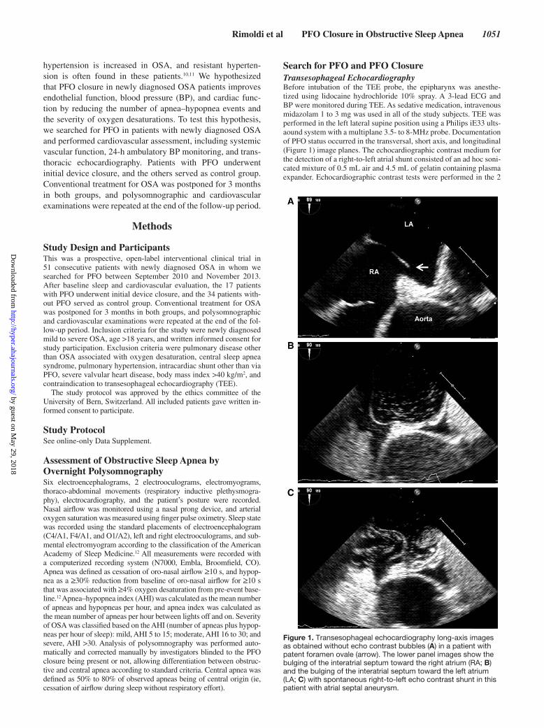
Search for PFO and PFO ClosureTransesophageal EchocardiographyBefore intubation of the TEE probe, the epipharynx was anesthe-tized using lidocaine hydrochloride 10% spray. A 3-lead ECG and BP were monitored during TEE. As sedative medication, intravenous midazolam 1 to 3 mg was used in all of the study subjects. TEE was performed in the left lateral supine position using a Philips iE33 ults-aound system with a multiplane 3.5- to 8-MHz probe. Documentation of PFO status occurred in the transversal, short axis, and longitudinal (Figure 1) image planes. The echocardiographic contrast medium for the detection of a right-to-left atrial shunt consisted of an ad hoc soni-cated mixture of 0.5 mL air and 4.5 mL of gelatin containing plasma expander. Echocardiographic contrast tests were performed in the 2
Figure 1. Transesophageal echocardiography long-axis images as obtained without echo contrast bubbles (A) in a patient with patent foramen ovale (arrow). The lower panel images show the bulging of the interatrial septum toward the right atrium (RA; B) and the bulging of the interatrial septum toward the left atrium (LA; C) with spontaneous right-to-left echo contrast shunt in this patient with atrial septal aneurysm.
by guest on May 29, 2018
http://hyper.ahajournals.org/D
ownloaded from
1052 Hypertension November 2015
mentioned image planes by injection of 5 mL contrast into the right antecubital vein. Using a Valsalva maneuver (strain phase starting simultaneously with the contrast bolus injection), a leftward devia-tion of the interatrial septum in the fossa ovalis region was observed immediately after release of the Valsalva strain phase (lasting 5 to 10 s).13 The diagnosis of PFO required the crossing of bubbles from the right to the left atrium (Figure 1) within 4 heartbeats after the release of the Valsalva strain phase. The degree of PFO was qualitatively characterized by a score of 0 to 3, with a score of 1 representing the crossover of a few single bubbles and a score of 3 representing the shunt of an entire cloud of bubbles (Figure 1).
PFO Device ClosureAfter the application of 5000 U of intravenous heparin, a 6F cath-eter with a guidewire introduced from the right femoral vein was used to probe the PFO. Then, a 9F long sheath was inserted. The PFO occluder device (Amplatzer PFO Occluder; St Jude Medical, Plymouth, MN; 25–35 mm in diameter depending on the size of the PFO) was delivered through the sheath and placed in the PFO under fluoroscopic guidance according to device-specific implantation rec-ommendations. Before release of the PFO occluder, device position was checked by right atrial contrast angiography to delineate the atrial septum. All patients were treated with acetylsalicylic acid 100 mg and clopidogrel 75 mg once daily for 1 month followed by acetyl-salicylic acid 100 mg/d until 6 months after PFO closure. Exactly 3 months following PFO device occlusion, TEE was repeated to search for the presence of residual right-to-left shunts.
Cardiovascular EvaluationAll cardiovascular studies were interpreted by investigators who were blinded to the PFO closure being present or not.
Assessment of Systemic Endothelial-Dependent and -Independent FunctionSystemic conduit artery endothelial function was assessed using the flow-mediated diameter increase of the brachial artery in response to reactive hyperemia by high-resolution ultrasound and wall tracking as previously described.6,14,15 For details see online-only Data Supplement.
Assessment of Aortic and Carotid StiffnessAortic stiffness was assessed noninvasively by measuring carotid-femoral pulse wave velocity using the Complior device (Artech Medical, Pantin, France) as described previously.14,15 For details, see online-only Data Supplement.
Assessment of Carotid Intima-Media ThicknessCarotid intima-media thickness (IMT) was measured as previously described.6,14 For details see online-only Data Supplement.
Ambulatory Blood Pressure MonitoringTwenty-four-hour ambulatory BP monitoring was performed using validated recorders (Spacelabs model 90217, USA) during usual daily activities as previously described.14 The arm cuff was pro-grammed to inflate every 20 minutes at daytime and every 30 minutes during night. Patients completed a diary for the identification of activ-ity, sleep and wake periods.
Assessment of Sympathetic ActivityHeart rate (HR) derived from the ambulatory BP monitoring (average 24-h HR, daytime and nighttime HR) and overnight HR variability derived from polysomnography were assessed as a proxy of sympa-thetic nervous system activity.
Estimation of Pulmonary Artery Pressure by Doppler EchocardiographyAll the study subjects underwent conventional transthoracic echocar-diography on a Philips iE33 ultrasound system. Pulmonary pressure was estimated on the basis of the trans-tricuspid right ventricular-to-right atrial systolic peak velocity; from this the respective pressure gradient was calculated using the simplified Bernoulli equation.
Statistical AnalysisIntraindividual comparison of continuous parameters between base-line and follow-up was performed by a paired sign test in case of non-normal data distribution (all polysomnographic data) and by a paired student’s t test in case of normally distributed data. Interindividual comparison of continuous parameters between groups was performed using the Mann–Whitney test in case of non-normal data distribution (all polysomnographic data) and using the unpaired student’s t test in case of normally distributed data. Categorical data were compared by a chi-squared test. Relations between variables were analyzed by calculating the r2 product–moment correlation coefficients. Unless otherwise indicated, data are presented as mean±standard deviation. Statistical significance level was defined at a P level <0.05.
ResultsA total of 51 patients were originally recruited for the study (17 patients with and 34 without PFO). Thus, the prevalence of PFO amounted to 33%. In the PFO group, 3 patients had to be excluded for the following reasons: pulmonary hyper-tension related to pulmonary disease other than OSA causing nocturnal oxygen desaturation (n=1), acute cerebral ischemia in the context of OSA (n=1), and insufficient quality base-line polysomnography data (n=1). In the non-PFO group, 8
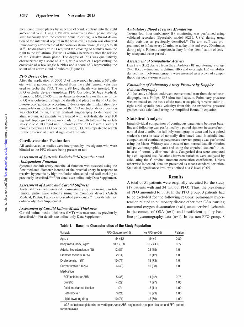
Table 1. Baseline Characteristics of the Study Population
Variable PFO Closure (n=14) No PFO (n=26) P Value
Age, y 54±12 54±9 0.89
Body mass index, kg/m2 31.1±3.8 30.7±4.6 0.77
Arterial hypertension, n (%) 12 (86) 22 (85) 1.0
Diabetes mellitus, n (%) 2 (14) 3 (12) 1.0
Dyslipidemia, n (%) 10 (71) 19 (73) 1.0
Current smoker, n (%) 6 (43) 10 (38) 1.0
Medication
ACE inhibitor or ARB 5 (36) 11 (42) 0.75
Diuretic 4 (29) 7 (27) 1.00
Calcium channel blocker 1 (7) 3 (11) 1.00
Beta-blocker 3 (21) 6 (23) 1.00
Lipid-lowering drug 10 (71) 18 (69) 1.00
ACE indicates angiotensin-converting enzyme; ARB, angiotensin receptor blocker; and PFO, patent foramen ovale.
by guest on May 29, 2018
http://hyper.ahajournals.org/D
ownloaded from
Rimoldi et al PFO Closure in Obstructive Sleep Apnea 1053
patients refused to undergo follow-up examination and were, thus, defined as dropouts. Hence, 40 patients were finally ana-lyzed for the study, 14 in the group with PFO closure and 26 in the control group without PFO. Data on AHI, pulmonary artery pressure, and 24-h nighttime systolic BP have been pre-viously published in form of a research letter.5
Patient CharacteristicsParticipants with and without PFO were comparable regard-ing age, height, body mass index, number of cardiovascular risk factors, and use of cardiovascular drugs (Table 1).
Cardiovascular Parameters at BaselineWith the exception of a significantly higher right ventricular–right atrial systolic pressure gradient in participants with PFO, all other variables were similar in the 2 groups (Table 2).
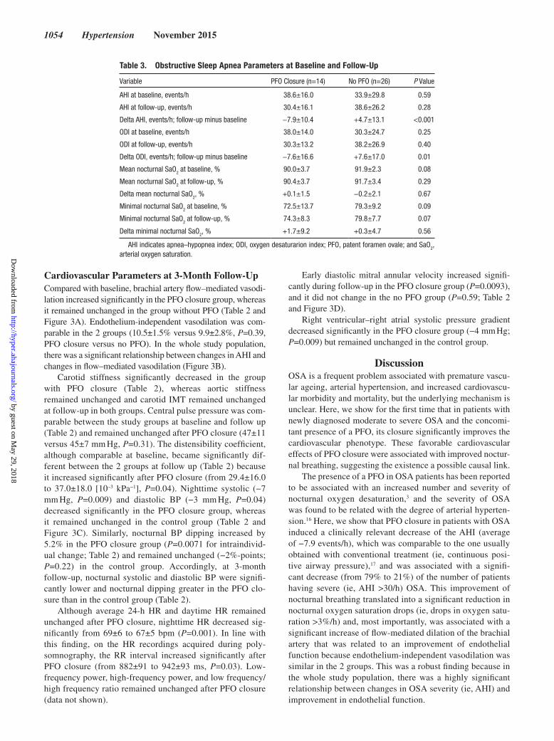
OSA Parameters at 3-Month Follow-UpAHI decreased significantly during the 3-month follow-up period in patients who underwent PFO closure, whereas it remained unchanged in patients without PFO (Figure 2). Accordingly, the interindividual change in AHI differed sig-nificantly between the 2 groups (Table 3). Although AHI was not statistically different between groups at baseline or at fol-low up (P=0.59 and 0.29, respectively), PFO closure was asso-ciated with a clinically and statistically (P=0.007) significant improvement of OSA severity; at baseline, 11 of 14 patients with PFO (79%) had a severe OSA (ie, AHI >30/h), whereas at follow-up after PFO closure, only 3 of 14 (21%) still had severe OSA. Oxygen desaturation index was not statistically differ-ent between the groups at baseline or at follow-up (Table 3); it decreased significantly in the PFO closure group (P=0.0225),
and it did not change statistically in the no PFO group (P=0.29). The change in oxygen desaturation index during follow-up dif-fered significantly between the groups (Table 3).
At baseline, mean and minimal nocturnal oxygen satura-tion tended to be lower in the PFO closure than in the control group. These variables did not change significantly during follow-up (Table 3).
Table 2. Cardiovascular and Doppler Echocardiographic Parameters at Baseline and Follow-Up
Variable
Baseline Follow-Up
PFO Closure (n=14) No PFO (n=26) P Value PFO Closure (n=14) No PFO (n=26) P Value
Heart rate, bpm 69±10 69±9 0.92 69±11 68±9 0.65
Brachial artery FMD, % 5.1±1.2 5.2±1.5 0.77 6.3±1.4 5.1±1.6 0.03
Carotid-femoral PWV, m/s 10.5±1.4 10.7±2.1 0.83 10.4±1.7 10.8±2.1 0.54
Carotid pulse pressure, mm Hg 47±11 44±12 0.38 45±7 45±11 0.88
Distensibility coefficient (10–3 kPa−1) 29.4±16.0 24.9±10.6 0.39 37.0±18.0 25.6±9.6 0.04
Carotid stiffness, m/s 6.3±1.5 6.6±1.4 0.58 5.4±1.1 6.4±1.1 0.02
Carotid IMT, μm 720±160 700±110 0.65 670±140 660±80 0.76
Average systolic BP during ABPM, mm Hg 125±8 131±14 0.17 124±7 129±14 0.13
Average diastolic BP during ABPM, mm Hg 78±6 84±12 0.12 77±5 82±10 0.07
Nocturnal systolic BP, mm Hg 115±7 118±17 0.51 108±8 119±18 0.01
Nocturnal diastolic BP, mm Hg 70±5 73±14 0.38 67±5 74±13 0.02
Nocturnal BP dipping, % 9.8±4.4 12.3±7.3 0.24 15.0±4.0 10.5±8.2 0.03
Left ventricular mass index, g/m2 96±21 101±19 0.53 99±16 100±19 0.87
Left ventricular ejection fraction, % 63±3 63±5 0.68 63±5 64±9 0.42
Early diastolic septal mitral annular velocity, e′, cm/s 7.0±2.0 6.9±1.8 0.90 7.4±1.3 7.0±1.6 0.58
Late diastolic septal mitral annular velocity, a′, cm/s 10.8±1.8 10.6±2.2 0.78 11.0±2.1 10.6±2.4 0.61
Systolic tricuspid annular free wall velocity, S′, cm/s 14.2±2.1 15.2±1.7 0.13 15.7±2.7 15.1±2.4 0.53
RV-RA systolic pressure gradient, mm Hg 24±4 19±4 0.0069 20±4 20±6 0.74
ABPM indicates ambulatory (24-hour) blood pressure monitoring; BP, blood pressure; FMD, flow-mediated dilation; IMT, intima-media thickness; PFO, patent foramen ovale; PWV, pulse wave velocity; RA, right atrial; and RV, right ventricular.
Figure 2. Difference between apnea–hypopnea index (AHI) at 3-months follow-up examination minus AHI at baseline (delta AHI, vertical axis) in patients with patent foramen ovale (PFO) at baseline and in patients without PFO. Box plots and error bars indicate the median and interquartile range and 10% and 90% confidence intervals, respectively.
by guest on May 29, 2018
http://hyper.ahajournals.org/D
ownloaded from
1054 Hypertension November 2015
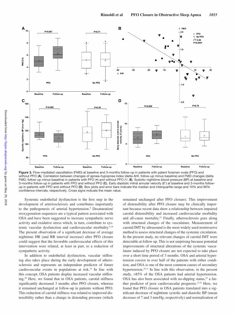
Cardiovascular Parameters at 3-Month Follow-UpCompared with baseline, brachial artery flow–mediated vasodi-lation increased significantly in the PFO closure group, whereas it remained unchanged in the group without PFO (Table 2 and Figure 3A). Endothelium-independent vasodilation was com-parable in the 2 groups (10.5±1.5% versus 9.9±2.8%, P=0.39, PFO closure versus no PFO). In the whole study population, there was a significant relationship between changes in AHI and changes in flow–mediated vasodilation (Figure 3B).
Carotid stiffness significantly decreased in the group with PFO closure (Table 2), whereas aortic stiffness remained unchanged and carotid IMT remained unchanged at follow-up in both groups. Central pulse pressure was com-parable between the study groups at baseline and follow up (Table 2) and remained unchanged after PFO closure (47±11 versus 45±7 mm Hg, P=0.31). The distensibility coefficient, although comparable at baseline, became significantly dif-ferent between the 2 groups at follow up (Table 2) because it increased significantly after PFO closure (from 29.4±16.0 to 37.0±18.0 [10–3 kPa−1], P=0.04). Nighttime systolic (−7 mm Hg, P=0.009) and diastolic BP (−3 mm Hg, P=0.04) decreased significantly in the PFO closure group, whereas it remained unchanged in the control group (Table 2 and Figure 3C). Similarly, nocturnal BP dipping increased by 5.2% in the PFO closure group (P=0.0071 for intraindivid-ual change; Table 2) and remained unchanged (−2%-points; P=0.22) in the control group. Accordingly, at 3-month follow-up, nocturnal systolic and diastolic BP were signifi-cantly lower and nocturnal dipping greater in the PFO clo-sure than in the control group (Table 2).
Although average 24-h HR and daytime HR remained unchanged after PFO closure, nighttime HR decreased sig-nificantly from 69±6 to 67±5 bpm (P=0.001). In line with this finding, on the HR recordings acquired during poly-somnography, the RR interval increased significantly after PFO closure (from 882±91 to 942±93 ms, P=0.03). Low-frequency power, high-frequency power, and low frequency/ high frequency ratio remained unchanged after PFO closure (data not shown).
Early diastolic mitral annular velocity increased signifi-cantly during follow-up in the PFO closure group (P=0.0093), and it did not change in the no PFO group (P=0.59; Table 2 and Figure 3D).
Right ventricular–right atrial systolic pressure gradient decreased significantly in the PFO closure group (−4 mm Hg; P=0.009) but remained unchanged in the control group.
DiscussionOSA is a frequent problem associated with premature vascu-lar ageing, arterial hypertension, and increased cardiovascu-lar morbidity and mortality, but the underlying mechanism is unclear. Here, we show for the first time that in patients with newly diagnosed moderate to severe OSA and the concomi-tant presence of a PFO, its closure significantly improves the cardiovascular phenotype. These favorable cardiovascular effects of PFO closure were associated with improved noctur-nal breathing, suggesting the existence a possible causal link.
The presence of a PFO in OSA patients has been reported to be associated with an increased number and severity of nocturnal oxygen desaturation,3 and the severity of OSA was found to be related with the degree of arterial hyperten-sion.16 Here, we show that PFO closure in patients with OSA induced a clinically relevant decrease of the AHI (average of −7.9 events/h), which was comparable to the one usually obtained with conventional treatment (ie, continuous posi-tive airway pressure),17 and was associated with a signifi-cant decrease (from 79% to 21%) of the number of patients having severe (ie, AHI >30/h) OSA. This improvement of nocturnal breathing translated into a significant reduction in nocturnal oxygen saturation drops (ie, drops in oxygen satu-ration >3%/h) and, most importantly, was associated with a significant increase of flow-mediated dilation of the brachial artery that was related to an improvement of endothelial function because endothelium-independent vasodilation was similar in the 2 groups. This was a robust finding because in the whole study population, there was a highly significant relationship between changes in OSA severity (ie, AHI) and improvement in endothelial function.
Table 3. Obstructive Sleep Apnea Parameters at Baseline and Follow-Up
Variable PFO Closure (n=14) No PFO (n=26) P Value
AHI at baseline, events/h 38.6±16.0 33.9±29.8 0.59
AHI at follow-up, events/h 30.4±16.1 38.6±26.2 0.28
Delta AHI, events/h; follow-up minus baseline −7.9±10.4 +4.7±13.1 <0.001
ODI at baseline, events/h 38.0±14.0 30.3±24.7 0.25
ODI at follow-up, events/h 30.3±13.2 38.2±26.9 0.40
Delta ODI, events/h; follow-up minus baseline −7.6±16.6 +7.6±17.0 0.01
Mean nocturnal SaO2 at baseline, % 90.0±3.7 91.9±2.3 0.08
Mean nocturnal SaO2 at follow-up, % 90.4±3.7 91.7±3.4 0.29
Delta mean nocturnal SaO2, % +0.1±1.5 −0.2±2.1 0.67
Minimal nocturnal SaO2 at baseline, % 72.5±13.7 79.3±9.2 0.09
Minimal nocturnal SaO2 at follow-up, % 74.3±8.3 79.8±7.7 0.07
Delta minimal nocturnal SaO2, % +1.7±9.2 +0.3±4.7 0.56
AHI indicates apnea–hypopnea index; ODI, oxygen desaturarion index; PFO, patent foramen ovale; and SaO2,
arterial oxygen saturation.
by guest on May 29, 2018
http://hyper.ahajournals.org/D
ownloaded from
Rimoldi et al PFO Closure in Obstructive Sleep Apnea 1055
Systemic endothelial dysfunction is the first step in the development of arteriosclerosis and contributes importantly to the pathogenesis of arterial hypertension.9 Desaturation/reoxygenation sequences are a typical pattern associated with OSA and have been suggested to increase sympathetic nerve activity and oxidative stress which, in turn, contribute to sys-temic vascular dysfunction and cardiovascular morbidity.6,18 The present observation of a significant decrease of average nighttime HR (and RR interval increase) after PFO closure could suggest that the favorable cardiovascular effects of this intervention were related, at least in part, to a reduction of sympathetic activity.
In addition to endothelial dysfunction, vascular stiffen-ing also takes place during the early development of athero-sclerosis and represents an independent predictor of future cardiovascular events in populations at risk.19 In line with this concept, OSA patients display increased vascular stiffen-ing.20 Here, we found that in OSA patients, carotid stiffness significantly decreased 3 months after PFO closure, whereas it remained unchanged at follow-up in patients without PFO. This reduction of carotid stiffness was related to improved dis-tensibility rather than a change in distending pressure (which
remained unchanged after PFO closure). This improvement of distensibility after PFO closure may be clinically impor-tant because recent data show a relationship between impaired carotid distensibility and increased cardiovascular morbidity and all-cause mortality.21 Finally, atherosclerosis goes along with structural changes of the vasculature. Measurement of carotid IMT by ultrasound is the most widely used noninvasive method to assess structural changes of the systemic circulation. In the present study, no relevant changes of carotid IMT were detectable at follow up. This is not surprising because potential improvements of structural alterations of the systemic vascu-lature induced by PFO closure are not expected to take place over a short time period of 3 months. OSA and arterial hyper-tension coexist in over half of the patients with either condi-tion, and OSA is one of the most common causes of secondary hypertension.10,11 In line with this observation, in the present study, >85% of the OSA patients had arterial hypertension. OSA has also been associated with no-dipping status,22 a fur-ther predictor of poor cardiovascular prognosis.23,24 Here, we found that PFO closure in OSA patients translated into a sig-nificant decrease of nighttime systolic and diastolic BP (mean decrease of 7 and 3 mm Hg, respectively) and normalization of
Figure 3. Flow-mediated vasodilation (FMD) at baseline and 3-months follow-up in patients with patent foramen ovale (PFO) and without PFO (A). Correlation between changes of apnea–hypopnea index (delta AHI, follow-up minus baseline) and FMD changes (delta FMD, follow-up minus baseline) in patients with PFO (•) and without PFO (▽; B). Systolic nighttime blood pressure (BP) at baseline and 3-months follow-up in patients with FPO and without PFO (C). Early diastolic mitral annular velocity (E′) at baseline and 3-months follow-up in patients with FPO and without PFO (D). Box plots and error bars indicate the median and interquartile range and 10% and 90% confidence intervals, respectively. Cross signs indicate the mean value.
by guest on May 29, 2018
http://hyper.ahajournals.org/D
ownloaded from
1056 Hypertension November 2015
the dipping pattern. This is clinically relevant because epide-miological data show that a drop of 2 mm Hg of systolic and diastolic BP result in a reduction of cardiovascular mortality of ≈7% to 10% and 20%, respectively.25 Interestingly, the antihy-pertensive effect of continuous positive airway pressure treat-ment in OSA patients is ≈2 to 3 mm Hg.26,27 Long-term arterial hypertension is associated with target organ damage, and left ventricular diastolic dysfunction is an early marker for target organ damage in hypertensive patients.28 In line with this con-cept, in our study population, left ventricular diastolic function was impaired. Most importantly, PFO closure in OSA patients improved diastolic left ventricular function, suggesting that the favorable effects of this intervention on nocturnal BP regula-tion and oxygenation may translate into cardioprotection.
Previous anecdotal evidence of improvement of dyspnea and desaturation in a case report29 and improvement of symp-toms and nocturnal breathing in 2 of 3 patients with OSA after PFO closure30 are consistent with the present findings. The present study was the first to search for PFO in consecutive patients with OSA and to examine the effects of its closure on nocturnal breathing, vascular function, and arterial BP while following in parallel OSA patients in whom no PFO was detected as a control group to account for the natural variation of the study end points.
Sleep-disordered breathing is a frequent syndrome in patients with cardiovascular diseases in general, and particu-larly, in patients with congestive heart failure, obstructive and central sleep apnea are often present simultaneously.31 In our study, the presence of central sleep apnea was an exclusion criterion. It would be interesting to investigate the effects of PFO closure on central sleep apnea in the future. Moreover, we were unable to assess whether the degree of PFO was related with the change of AHI or nocturnal oxygenation after closure because all but one of the patients studied had a PFO grade III.
PFO closure is in general safe with a low complication rate. The most relevant complications are peri-procedural events, vascular access site problems, cardiac tamponade, ero-sion, and embolization. In a series of 825 consecutive patients undergoing PFO closure at our institution,32 complications were present in 18 (2.2%) and included device embolization (n=5), air embolism with transient symptoms (n=5), cardiac tamponade requiring pericardiocentesis (n=1), and vascular access site problems (n=7).
PerspectivesOSA is a frequent syndrome associated with increased car-diovascular morbidity and mortality. The presence of PFO is associated with more severe OSA. Here, we found that PFO closure in OSA patients improved sleep-disordered breath-ing and nocturnal arterial oxygenation. These improvements of nocturnal breathing were associated, 3 months after PFO closure, with a significant attenuation of endothelial dysfunc-tion and vascular stiffening, a decrease of nighttime BP and restoration of its dipping pattern, and an improvement of left ventricular diastolic function. We speculate that PFO closure in OSA patients is a valid alternative to conventional therapy, particularly in those who are intolerant to continuous positive airway pressure therapy and dental device treatment. Larger randomized studies should confirm our results and assess the
impact of PFO closure on cardiovascular morbidity and mor-tality in this growing population at high cardiovascular risk.
Sources of FundingThe patent foramen ovale (PFO) closure devices were paid for by the Administration of Teaching and Research of our Institution.
DisclosuresNone.
References 1. Yaggi HK, Concato J, Kernan WN, Lichtman JH, Brass LM, Mohsenin
V. Obstructive sleep apnea as a risk factor for stroke and death. N Engl J Med. 2005;353:2034–2041. doi: 10.1056/NEJMoa043104.
2. Xie A, Skatrud JB, Dempsey JA. Effect of hypoxia on the hypopnoeic and apnoeic threshold for CO(2) in sleeping humans. J Physiol. 2001;535(pt 1):269–278.
3. Johansson MC, Eriksson P, Peker Y, Hedner J, Råstam L, Lindblad U. The influence of patent foramen ovale on oxygen desaturation in obstructive sleep apnoea. Eur Respir J. 2007;29:149–155. doi: 10.1183/09031936. 00035906.
4. Konecny T, Khanna AD, Novak J, Jama AA, Zawadowski GM, Orban M, Pressman G, Bukartyk J, Kara T, Cetta F, Jr, Borlaug BA, Somers VK, Reeder GS. Interatrial pressure gradients during simulated obstruc-tive sleep apnea: a catheter-based study. Catheter Cardiovasc Interv. 2014;84:1138–1145. doi: 10.1002/ccd.25433.
5. Rimoldi SF, Ott SR, Rexhaj E, von Arx R, de Marchi SF, Brenner R, Scherrer U, Meier B, Gugger M, Allemann Y, Seiler C. Effect of patent foramen ovale closure on obstructive sleep apnea. J Am Coll Cardiol. 2015;65:2257–2258. doi: 10.1016/j.jacc.2015.01.062.
6. Bailey DM, Rimoldi SF, Rexhaj E, Pratali L, Salinas Salmòn C, Villena M, McEneny J, Young IS, Nicod P, Allemann Y, Scherrer U, Sartori C. Oxidative-nitrosative stress and systemic vascular function in highland-ers with and without exaggerated hypoxemia. Chest. 2013;143:444–451. doi: 10.1378/chest.12-0728.
7. Kato M, Roberts-Thomson P, Phillips BG, Haynes WG, Winnicki M, Accurso V, Somers VK. Impairment of endothelium-dependent vaso-dilation of resistance vessels in patients with obstructive sleep apnea. Circulation. 2000;102:2607–2610.
8. Bruno RM, Rossi L, Fabbrini M, Duranti E, Di Coscio E, Maestri M, Guidi P, Frenzilli G, Salvetti A, Taddei S, Bonanni E, Ghiadoni L. Renal vasodilating capacity and endothelial function are impaired in patients with obstructive sleep apnea syndrome and no traditional cardiovascular risk factors. J Hypertens. 2013;31:1456–1464; discussion 1464. doi: 10.1097/HJH.0b013e328360f773.
9. Deanfield JE, Halcox JP, Rabelink TJ. Endothelial function and dysfunc-tion: testing and clinical relevance. Circulation. 2007;115:1285–1295. doi: 10.1161/CIRCULATIONAHA.106.652859.
10. Pedrosa RP, Drager LF, Gonzaga CC, Sousa MG, de Paula LK, Amaro AC, Amodeo C, Bortolotto LA, Krieger EM, Bradley TD, Lorenzi-Filho G. Obstructive sleep apnea: the most common secondary cause of hypertension associated with resistant hypertension. Hypertension. 2011;58:811–817. doi: 10.1161/HYPERTENSIONAHA.111.179788.
11. Rimoldi SF, Scherrer U, Messerli FH. Secondary arterial hypertension: when, who, and how to screen? Eur Heart J. 2014;35:1245–1254. doi: 10.1093/ eurheartj/eht534.
12. Iber C, Ancoli-Israel S, Chesson A, Quan S. The AASM Manual for the Scoring of Sleep and Associated Events: Rules, Terminology and Technical Specifications. Westchester, IL: The American Academy of Sleep Medicine; 2007.
13. Seiler C. How should we assess patent foramen ovale? Heart. 2004;90:1245–1247. doi: 10.1136/hrt.2003.031500.
14. Rimoldi SF, Rexhaj E, Pratali L, Bailey DM, Hutter D, Faita F, Salmòn CS, Villena M, Nicod P, Allemann Y, Scherrer U, Sartori C. Systemic vascular dysfunction in patients with chronic mountain sickness. Chest. 2012;141:139–146. doi: 10.1378/chest.11-0342.
15. Scherrer U, Rimoldi SF, Rexhaj E, Stuber T, Duplain H, Garcin S, de Marchi SF, Nicod P, Germond M, Allemann Y, Sartori C. Systemic and pulmonary vascular dysfunction in children conceived by assisted repro-ductive technologies. Circulation. 2012;125:1890–1896. doi: 10.1161/CIRCULATIONAHA.111.071183.
16. Marin JM, Agusti A, Villar I, Forner M, Nieto D, Carrizo SJ, Barbé F, Vicente E, Wei Y, Nieto FJ, Jelic S. Association between treated and
by guest on May 29, 2018
http://hyper.ahajournals.org/D
ownloaded from
Rimoldi et al PFO Closure in Obstructive Sleep Apnea 1057
untreated obstructive sleep apnea and risk of hypertension. JAMA. 2012;307:2169–2176. doi: 10.1001/jama.2012.3418.
17. Giles TL, Lasserson TJ, Smith BJ, White J, Wright J, Cates CJ. Continuous positive airways pressure for obstructive sleep apnoea in adults. Cochrane Database Syst Rev. 2006:1–80.
18. Konecny T, Kara T, Somers VK. Obstructive sleep apnea and hyper-tension: an update. Hypertension. 2014;63:203–209. doi: 10.1161/HYPERTENSIONAHA.113.00613.
19. Laurent S, Cockcroft J, Van Bortel L, Boutouyrie P, Giannattasio C, Hayoz D, Pannier B, Vlachopoulos C, Wilkinson I, Struijker-Boudier H; European Network for Non-invasive Investigation of Large Arteries. Expert consensus document on arterial stiffness: methodological issues and clinical applica-tions. Eur Heart J. 2006;27:2588–2605. doi: 10.1093/eurheartj/ehl254.
20. Drager LF, Bortolotto LA, Figueiredo AC, Silva BC, Krieger EM, Lorenzi-Filho G. Obstructive sleep apnea, hypertension, and their interaction on arterial stiffness and heart remodeling. Chest. 2007;131:1379–1386. doi: 10.1378/chest.06-2703.
21. van Sloten TT, Schram MT, van den Hurk K, Dekker JM, Nijpels G, Henry RM, Stehouwer CD. Local stiffness of the carotid and femoral artery is associated with incident cardiovascular events and all-cause mortality: the Hoorn study. J Am Coll Cardiol. 2014;63:1739–1747. doi: 10.1016/j.jacc.2013.12.041.
22. Hla KM, Young T, Finn L, Peppard PE, Szklo-Coxe M, Stubbs M. Longitudinal association of sleep-disordered breathing and nondipping of nocturnal blood pressure in the Wisconsin Sleep Cohort Study. Sleep. 2008;31:795–800.
23. Dolan E, Stanton A, Thijs L, Hinedi K, Atkins N, McClory S, Den Hond E, McCormack P, Staessen JA, O’Brien E. Superiority of ambula-tory over clinic blood pressure measurement in predicting mortality: the Dublin outcome study. Hypertension. 2005;46:156–161. doi: 10.1161/01.HYP.0000170138.56903.7a.
24. Hansen TW, Li Y, Boggia J, Thijs L, Richart T, Staessen JA. Predictive role of the nighttime blood pressure. Hypertension. 2011;57:3–10. doi: 10.1161/HYPERTENSIONAHA.109.133900.
25. Lewington S, Clarke R, Qizilbash N, Peto R, Collins R; Prospective Studies Collaboration. Age-specific relevance of usual blood pressure to vascular mortality: a meta-analysis of individual data for one million adults in 61 prospective studies. Lancet. 2002;360:1903–1913.
26. Fava C, Dorigoni S, Dalle Vedove F, Danese E, Montagnana M, Guidi GC, Narkiewicz K, Minuz P. Effect of CPAP on blood pressure in patients with OSA/hypopnea a systematic review and meta-analysis. Chest. 2014;145:762–771. doi: 10.1378/chest.13-1115.
27. Gottlieb DJ, Punjabi NM, Mehra R, Patel SR, Quan SF, Babineau DC, Tracy RP, Rueschman M, Blumenthal RS, Lewis EF, Bhatt DL, Redline S. CPAP versus oxygen in obstructive sleep apnea. N Engl J Med. 2014;370:2276–2285. doi: 10.1056/NEJMoa1306766.
28. Rovner A, de las Fuentes L, Waggoner AD, Memon N, Chohan R, Dávila-Román VG. Characterization of left ventricular diastolic func-tion in hypertension by use of Doppler tissue imaging and color M-mode techniques. J Am Soc Echocardiogr. 2006;19:872–879. doi: 10.1016/ j.echo.2006.01.023.
29. Agnoletti G, Iserin L, Lafont A, Sidi D, Desnos M. Obstructive sleep apnoea and patent foramen ovale: successful treatment of symptoms by percutaneous foramen ovale closure. J Interv Cardiol. 2005;18:393–395. doi: 10.1111/j.1540-8183.2005.00072.x.
30. White JM, Veale AG, Ruygrok PN. Patent foramen ovale clo-sure in the treatment of obstructive sleep apnea. J Invasive Cardiol. 2013;25:E169–E171.
31. Sin DD, Fitzgerald F, Parker JD, Newton G, Floras JS, Bradley TD. Risk factors for central and obstructive sleep apnea in 450 men and women with congestive heart failure. Am J Respir Crit Care Med. 1999;160:1101–1106. doi: 10.1164/ajrccm.160.4.9903020.
32. Wahl A, Praz F, Stinimann J, Windecker S, Seiler C, Nedeltchev K, Mattle HP, Meier B. Safety and feasibility of percutaneous closure of patent fora-men ovale without intra-procedural echocardiography in 825 patients. Swiss Med Wkly. 2008;138:567–572. doi: 2008/39/smw-12255.
What Is New?•Obstructive sleep apnea (OSA) is a frequent syndrome associated with
increased cardiovascular morbidity and mortality. The presence of patent foramen ovale (PFO) is associated with more severe OSA. PFO closure improves sleep-disordered breathing and nocturnal oxygenation. This translates into a decrease of nighttime blood pressure, restoration of the dipping pattern, and improvement of cardiovascular function.
What Is Relevant?•These findings provide proof-of-principle that in OSA patients, PFO clo-
sure has an important positive impact on sleep-disordered breathing that translate into improvement of blood pressure control and cardiovascular function. This suggests the existence of a possible causal link.
Summary
This study highlights that in patients with newly diagnosed mod-erate to severe OSA and the concomitant presence of a PFO, its closure significantly improves the cardiovascular phenotype. We speculate that PFO closure in OSA patients is a valid alternative to conventional therapy, particularly in those who are intolerant to continuous positive airway pressure therapy or dental device treatment.
Novelty and Significance
by guest on May 29, 2018
http://hyper.ahajournals.org/D
ownloaded from
Matthias Gugger, Urs Scherrer and Christian SeilerStefano F. Rimoldi, Sebastian Ott, Emrush Rexhaj, Stefano F. de Marchi, Yves Allemann,
Cardiovascular FunctionPatent Foramen Ovale Closure in Obstructive Sleep Apnea Improves Blood Pressure and
Print ISSN: 0194-911X. Online ISSN: 1524-4563 Copyright © 2015 American Heart Association, Inc. All rights reserved.
is published by the American Heart Association, 7272 Greenville Avenue, Dallas, TX 75231Hypertension doi: 10.1161/HYPERTENSIONAHA.115.06303
2015;66:1050-1057; originally published online September 21, 2015;Hypertension.
http://hyper.ahajournals.org/content/66/5/1050World Wide Web at:
The online version of this article, along with updated information and services, is located on the
http://hyper.ahajournals.org/content/suppl/2015/09/22/HYPERTENSIONAHA.115.06303.DC1Data Supplement (unedited) at:
http://hyper.ahajournals.org//subscriptions/
is online at: Hypertension Information about subscribing to Subscriptions:
http://www.lww.com/reprints Information about reprints can be found online at: Reprints:
document. Permissions and Rights Question and Answer this process is available in the
click Request Permissions in the middle column of the Web page under Services. Further information aboutOffice. Once the online version of the published article for which permission is being requested is located,
can be obtained via RightsLink, a service of the Copyright Clearance Center, not the EditorialHypertensionin Requests for permissions to reproduce figures, tables, or portions of articles originally publishedPermissions:
by guest on May 29, 2018
http://hyper.ahajournals.org/D
ownloaded from
1
Online Supplement
PATENT FORAMEN OVALE CLOSURE IN OBSTRUCTIVE SLEEP APNEA
IMPROVES BLOOD PRESSURE AND CARDIOVASCULAR FUNCTION
Stefano F. Rimoldi1*, MD, Sebastian Ott
2*, MD, Emrush Rexhaj
1, MD,
Stefano F. de Marchi1, MD, Yves Allemann
1, MD, Matthias Gugger
2, MD,
Urs Scherrer1,3
, MD, Christian Seiler1, MD
1Department of Cardiology and Clinical Research, Inselspital,
University Hospital, Bern, Switzerland
2Department of Pneumology, Inselspital, University Hospital, Bern, Switzerland
3Facultad de Ciencias, Departamento de Biología, Universidad de Tarapacá, Arica, Chile
Short title: PFO closure in obstructive sleep apnea
*Contributed equally to this work
Correspondence to:
Christian Seiler, MD, FACC, FESC, Professor of Medicine and Co-Chairman of Cardiology
Department of Cardiology, University Hospital Bern, CH-3010 Bern, Switzerland,
Phone: + 41 31 632 36 93; Fax: +41 31 632 42 99; e-mail: [email protected] or
Stefano F Rimoldi, MD, Department of Cardiology and Clinical Research, University
Hospital Bern, 3010 Bern, Switzerland, Phone: +41 31 632 41 50; Fax: +41 31 632 42 11
e-mail: [email protected]
2
1) Expanded materials and methods
1.1. Study protocol
Once the diagnosis of OSA was established by polysomnography, patients willing to
participate in the study underwent transthoracic echocardiography before performing TEE.
During the first visit after study inclusion, assessment of systemic vascular function was
carried out. At the end of this visit, 24h ABPM was performed. Three to 10 days after this
visit, patients with diagnosed PFO underwent percutaneous PFO closure. After PFO closure, a
control TTE to confirm the correct position of the device was done during the same day in all
patients. After 3 months, in all patients the following procedures were repeated:
polysomnography, 24-hour ABPM, systemic vascular function and TTE. In patients with PFO
closure, TTE was performed followed by TEE. During the entire follow-up period, there was
no change in cardiovascular drug therapy and non-invasive ventilatory therapy was deferred.
1.2.Methods for assessment of vascular function
Assessment of systemic endothelial-dependent and -independent function
The brachial artery was identified approximately 5cm above the antecubital fossa with a high-
resolution ultrasound device (Esaote MyLab30 Gold, Esaote SpA, Italy). Images of the artery
were analyzed using an edge detection software with a system for real-time measurement of
the brachial artery diameter in B-mode ultrasound images (Cardiovascular Suite, Quipu, Pisa,
Italy) for determination of the baseline diameter of the vessel. Then, a forearm pressure cuff
was inflated to 250mmHg for 5 minutes. After deflation of the cuff, the hyperemia-induced
change of the brachial artery diameter was recorded continuously during the entire
examination of 9 minutes. Flow-mediated dilatation was expressed as the maximal percent
change in vessel diameter from baseline.
Endothelium-independent dilation change of the brachial artery was assessed at follow-up by
measuring the increase of the brachial artery diameter evoked by oral glyceryl-trinitrate (GTN
50 μg, UCB-Pharma, Bulle, Switzerland).
Assessment of aortic and carotid stiffness
Carotid and femoral artery waveforms were simultaneously recorded with mechano-
transducers directly applied to the skin over the arteries and the mean wave transit time for 10
heart beats was calculated by the system software using the foot-to-foot method. To determine
the pulse wave velocity, the surface distance between the recording sites was measured.
Carotid stiffness was assessed non-invasively immediately after carotid tonometry with the
SphygmoCor system (AtCor Medical, Sydney, Australia) by measuring parameters of local
elasticity with a validated edge detection system (Carotid Studio, Cardiovascular Suite;
Quipu srl, Pisa, Italy) applied to B-mode ultrasound sequences of longitudinal section of the
carotid artery obtained using an Esaote 30 MyLab Gold (Esaote SpA, Genova, Italy). To
determine carotid pulse pressure (PP), carotid distension waveforms were obtained and
rescaled using brachial distension waveforms. The distensibility coefficient (DC) was
calculated using the following equation: DC= (ΔA/A) /cPP, where ΔA=stroke change (i.e.
distension) of the carotid artery cross sectional area, A=total diastolic carotid artery cross
sectional area, cPP=carotid PP. Carotid PWV was calculated using the Bramwell-Hill
equation (carotid PWV=
( where =density of blood, assumed to be 1050 kg/m
3).

3
Assessment of carotid intima-media thickness.
Briefly, after identification of the carotid bulb, the segments of the right and left common
carotid artery 1 to 2 cm proximal to the bulb were scanned to identify the optimal angle of
incidence in accordance with current guidelines. Carotid intima-media thickness (IMT) was
measured using radiofrequency signals with a 21 µm resolution (RF QIMT, Esaote, Genova,
Italy).