obstructive sleep apnea: a pediatric epidemic
TRANSCRIPT
O
J
FRC
(tMadacScie
mB
0d
Seminars in Anesthesia, Perioperative Medicine and Pain (2006) 25, 109-116
bstructive sleep apnea: a pediatric epidemic
errold Lerman, MD, FRCPC, FANZCA
rom the Women and Children’s Hospital of Buffalo, SUNY, Buffalo and Strong Memorial Hospital, University ofochester, Rochester, New York; and University of Rochester, Rochester, New York, and St. Christopher’s Hospital for
hildren, Philadelphia, PennsylvaniaSleep disordered breathing is a continuum of breathing abnormalities that affects children and adults.In children, the diagnosis of the most severe form, obstructive sleep apnea (OSA), is difficult.Polysomnography is not widely used, rather the diagnosis is based on clinical history and physicalexam. These children may suffer from cardiorespiratory and neurobehavioral effects from the OSA.Treatment for children is primarily tonsillectomy and adenoidectomy surgery. Children who desaturateintermittently during sleep appear to be more sensitive to the respiratory depressant effects of opioids.Careful postoperative care for those at risk for complications is warranted.© 2006 Elsevier Inc. All rights reserved.
KEYWORDS:Pediatric;Respiratory;Apnea;Otolaryngology
booirmc
S
WbttficstdbrA
The stupid-lazy child who frequently suffers fromheadaches at school, breathes through his mouthinstead of his nose, snores and is restless at night,and wakes up with a dry mouth in the morning, iswell-worthy of the solicitous attention of theschool medical officer.W. Hill. British Medical Journal, 1899
In the past several decades, obstructive sleep apneaOSA) has emerged as a widely diagnosed disorder, al-hough for the most part, limited to morbidly obese adults.
ore recently, OSA has become a familiar term in pediatricnesthesia as the primary diagnosis for children with sleepisordered breathing (SDB), who require tonsillectomy anddenoidectomy (T&A). Ever since the first cases of OSA inhildren were reported 30 years ago by Guilleminault at thetanford Sleep Disorder Clinic, the implications of OSA inhildren has become the focus of research and clinicalnvestigation. We now understand that OSA is but onextreme of a continuum of SDB in children. This continuum
Address reprint requests and correspondence: Dr. J. Lerman, Depart-ent of Anesthesia, Women and Children’s Hospital of Buffalo, 219ryant St., Buffalo, NY 14222.
sE-mail: [email protected].
277-0326/$ -see front matter © 2006 Elsevier Inc. All rights reserved.oi:10.1053/j.sane.2006.05.011
egins with simple snoring and ends with the most seriousf the conditions, OSA (Figure 1).1 With attention focusedn the continuum of SDB, clinician-scientists have begun tonvestigate this disorder with a renewed perspective. In thiseview, we explore SDB in children in terms of its epide-iology, its pathophysiology, and treatment, and briefly
onclude with the anesthetic considerations.
leep disordered breathing (SDB)
hat is SDB? This is a continuum of sleep-related distur-ances in breathing that is characterized by the severity ofhe upper airway obstruction. In its mildest form, SDB isermed primary snoring (Figure 1). Primary snoring is de-ned as noisy breathing through the upper airway withoutlinical signs or symptoms referable to the snoring. Primarynoring appears to be benign. However, as the resistance inhe upper airway increases, clinical signs and symptomsuring the daytime or nighttime, begin to appear. The com-ination of snoring and clinical signs or symptoms is noweferred to as upper airway resistance syndrome (UARS).s the upper airway resistance increases further, progres-
ive hypercapnia and hemoglobin oxygen desaturation oc-

cfsaieatittOdcdds
3cmi1gfhcafimepAytdnfmfac
cpmaci
dwrtriwwm(t
1otmiotm
3atocdoauapff
Ffa
110 Seminars in Anesthesia, Perioperative Medicine and Pain, Vol 25, No 3, September 2006
ur. This is termed obstructive hypopnea. In the most severeorm of SDB, intermittent or complete upper airway ob-truction that is complicated by daytime or nighttime signsnd/or symptoms of SDB is termed OSA. Nighttime man-festations of OSA include snoring, increased respiratoryffort, intermittent periods of apnea from airway obstructionnd hemoglobin oxygen desaturation, hypercapnia, and dis-urbances in sleep patterns. Daytime manifestations of SDBn children are variable and include any or all of failure tohrive, poor school performance, inattentiveness, hyperac-ivity, and behavior disturbances. In contrast to adults withSA, somnolence is an uncommon daytime manifestation,isturbed sleep pattern is uncommon, and obesity rarelyomplicates OSA in children. Unfortunately, there are fewiagnostic criteria for SDB in children at this time; theiagnosis of SDB is based on a spectrum of signs andymptoms and the judgment of the otolaryngologist.
Snoring occurs in children with a prevalence between% and 20%.1 Primary snoring occurs in isolation, withoutardiorespiratory or neurobehavioral manifestations. Pri-ary snoring is regarded as a benign condition. It resolves
n 50% of children over time, although in a minority, about0%, it progresses to OSA. Guillenminault et al. investi-ated 25 children who snored heavily and who also sufferedrom daytime somnolence and lethargy.2 Neither nighttimeemoglobin desaturation nor OSA was present. After thesehildren underwent T&A, their daytime sleepiness and leth-rgy either disappeared or were attenuated. Gozal noted thatrst grade students who performed poorly in school wereore likely to snore at night and exhibit episodic hypox-
mia at night.3 He also noted that T&A significantly im-roved their performance in school. In a longitudinal study,li et al. noted that half of the children who snored at 4-5ears of age stopped snoring by 7 years without interven-ion.4 Children who snored exhibited more hyperactivity,aytime somnolence, and restless sleep than those who didot. The prevalence of snoring appears to increase steadilyrom pre-adolescence to adults, with a preponderance inales in this age group. We now know that snoring occurs
ar more commonly in children than previously appreciated,lthough if it occurs in isolation, it is not likely to be of
Normal Primary Upper airwSnoring resistance synd
(UARS)
Normal UAR, Snoring not Increased UANo snoring not assoc’d sufficient to c
with daytime symptomssymptoms
igure 1 Continuum of sleep disordered breathing. As upper airwrom primary snoring to upper airway resistance syndrome (UARSpnea (OSA). Adapted with permission.1
linical significance. On the other hand, snoring that is T
omplicated by clinical manifestations, such as poor schoolerformance, cognitive impairment, and behavior problems,ay signify a forme fruste of OSA that may continue to
dulthood. In both cases, T&A appears to be salutory forhildren who snore and exhibit clinical manifestations ofncreased airway resistance.
The prevalence of UARS in children is unknown. Theemography of UARS in adults points to a preponderance inomen versus men, snoring at night, and frequent and
epeated microarousals from sleep. These patients have nei-her obstructive breathing nor nighttime hemoglobin desatu-ation. In the past, children who snored and exhibited man-festations of cardiovascular or neurobehavioral changesere diagnosed with OSA, and very few were diagnosedith UARS. This trend appears to be changing. The treat-ent of UARS is continuous positive airway pressure
CPAP), although most children are referred for T&A ratherhan nasal CPAP.
The prevalence of OSA in children is approximately-3% in all age groups.5,6 Of the three types of sleep apnea,bstructive is the most common type, comprising 85% ofhe apneas, with the remainder being central, 10%, andixed, 5%, in origin. The prevalence of OSA is said to be
ncreasing, perhaps as a result of a decrease in the numberf T&A surgeries performed in the past decade as well ashe increased survival of children with congenital malfor-ations that are associated with OSA.The peak prevalence of OSA in children occurs in those
-6 years of age. At this age, children have the greatestmount of lymphoid tissue in their upper airways relative tohe dimensions of the airways. The degree of collapsibilityf their upper airway may also be increased. Children withraniofacial anomalies (associated with maxillary and man-ibular hypoplasia, macroglossia), hypotonia, and a varietyf other disorders (cerebral palsy and post-pharyngeal flap)re at risk for upper airway collapse because of narrowpper airway passages or poor muscle tone in the upperirway.7 These findings decrease the dimensions of theatent upper airway in children and increase the propensityor OSA as shown radiographically below. The clinicaleatures of OSA in children and adults are presented in
OH OSAe (obstructive (obstructive
hypopnea) sleep apnea)
Increased UAR Intermittentsufficient to cause upper airwayPaCO2 or SaO2 obstruction
istance increases, the severity of the signs and symptoms increasesstructive hypopnea and finally, the most severe, obstructive sleep
ayrom
Rause
ay res) to ob
able 1. Several salient differences between the features in

ceadAl(fhcsHabbO(mDelaw
ntcuppcTtthtbrrCtsi
awpsfeOtndilltsc
spicoiwcesfatndrO
oatcmhi
111Lerman Obstructive Sleep Apnea
hildren and adults are noteworthy: gender prevalence isqual, failure to thrive is the most common body habitus,nd excessive daytime somnolence is uncommon in chil-ren with OSA. Other features of OSA in children includefrican-American descent (with a 3.5-fold greater preva-
ence than Caucasian children),8 respiratory disease, obesityPrader-Willi syndrome) (OR of 4.6),9 prematurity,8 cranio-acial disorders, chromosomal abnormalities, and a familyistory of OSA. Obesity, which is a growing epidemic inhildhood afflicting 16% of children today, may be respon-ible for the increasing prevalence of children with OSA.owever, this is a reversible complication as weight loss
ttenuates or eliminates the symptoms of OSA in the mor-idly obese. In a study of 35 adolescents who underwentariatric surgery, 25 had evidence of OSA before surgery.10
f the 10 with post-surgery follow-up polysomnogramsPSGs), 9 showed no evidence of OSA. OSA is also com-on in children with neuromuscular disorders, includingown syndrome, achondroplasia, Arnold-Chiari, and my-
lomeningocoele.1 The combination of lymphoid accumu-ation in a small hypopharynx, defects in the upper airwaynatomy, loss of airway muscle tone, and control combinedith sleep disturbances place the child at high risk for OSA.The complications associated with OSA include but are
ot limited to failure to thrive, cardiovascular complica-ions, and central nervous system, neurobehavioral, andognitive manifestations. In terms of growth, these childrensed to suffer from failure to thrive, appearing in the �5thercentile on the growth charts. Several reasons have beenostulated to explain the findings of failure to thrive inhildren with OSA, although none has been substantiated.hese reasons include reduced caloric intake due to adeno-
onsillar hypertrophy, increased caloric expenditure duringo increased work of breathing at night, reduced growthormone due to sleep disturbances, and peripheral resis-ance to growth factors.11 More recently, however, OSA haseen associated with obesity. Insulin resistance has beeneported in adults with OSA.12 Interestingly, the insulinesistance resolves when these adults were treated withPAP.13 Cardiovascular complications include right ven-
ricular hypertrophy, cor pulmonale, and systemic hyperten-ion. If uncorrected, the natural progression of this disorder
Table 1 Clinical features of obstructive sleep apnea in childre
Clinical Features Children
Peak Age PreschoolGender ratio Male � Female
Causes T&A hypertrophy, craniobesity, chromosoma
Body habitus FTT, normal, obeseExcessive daytime somnolence UncommonNeurobehavioral Hyperactivity, developm
cognitive impairment
ncludes death from right heart failure or arrhythmias. Tal et W
l. noted that 37% of children less than 7 years of age whoere diagnosed with OSA had right ventricular hypertro-hy, and yet 80% had no evidence of pulmonary hyperten-ion. They also noted a decrease in right ventricular ejectionraction that was reversed after T&A surgery.14 The geom-try of the LV is also abnormal in 40% of children withSA, a three-fold greater prevalence than in the 15% of
hose with primary snoring. Central nervous system andeurobehavioral changes have also been reported in chil-ren with OSA. These changes include neurocognitive def-cits (memory problems, attention deficits, dysphasia),earning disabilities or deficiencies, and behavioural prob-em.15 Recognition of the potential implications of OSA andhe spectrum of SDB may bring these children to the phy-ician earlier, thereby permitting earlier intervention thatould obviate serious sequelae.
The pathophysiology of OSA begins with the onset ofleep and the onset of mouth breathing. As sleep deepens,haryngeal muscle tone decreases, which leads to a decreasen airway dimensions and disco-ordinated pharyngeal mus-le tone. As the airway begins to collapse, it becomesbstructed intermittently, with zero airflow, despite increas-ng respiratory effort. After several bouts of prolonged air-ay obstruction, the hemoglobin oxygen saturation de-
reases and carbon dioxide tension increases. Respiratoryffort steadily increases as the negative intra-thoracic pres-ure increases. This cycle of airway obstruction in OSA isueled by the presence of sedatives, such as alcohol andnxiolytics, which themselves decrease pharyngeal muscleone and blunt the responsiveness to hypoxia and hypercap-ia.16 The severity and frequency of nighttime hemoglobinesaturation, duration of apneas and hypopneas, and respi-atory efforts are all important measures of the severity ofSA.Most, but not all, of the episodes of OSA in children
ccur during REM sleep. During this period, upper airwaynd intercostal muscle tone is reduced. With this loss ofone, airway collapse is exaggerated and airway obstructionan occur. During these episodes of airway obstruction,icroarousals from sleep occur frequently. Despite this,
owever, the proportion of REM sleep in children with OSAs unchanged compared with that in children without OSA.
adults (adapted from Ref. 6, Table 1)
Adults
Middle ageMale �� Female; postmenopausal females
increase their riskdisorder, Obese
ObeseVery common
delay, Cognitive impairment, impaired vigilance
n and
ofaciall abn
ental
hether these microarousals terminate the obstruction in
cttm
saawalpwnapsapcasvegartpnpdrttMntr
rltbd
OtuMpicioisha�etrcpvcrc
itiPnitaabsiwivcaett
112 Seminars in Anesthesia, Perioperative Medicine and Pain, Vol 25, No 3, September 2006
hildren as they do in adults remains controversial.11 Fur-hermore, whether these microarousals are responsible forhe altered neurobehavioral or cognitive activity also re-ains controversial.Recent radiographic studies have shed new light on the
ites of airway obstruction in SDB. The sites of increasedirway resistance and airway obstruction in OSA includentero-posterior opposition of the tongue to the pharyngealall, posterior displacement of the soft palate by the tongue
gainst the posterior pharyngeal wall, and opposition of theateral pharyngeal walls.7 Together, these changes close theharynx in a circular pattern, thereby increasing upper air-ay resistance. Cross-sectional CT scans of the head andeck of children with and without OSA show thickeningnd elongation of the soft palate, hypertrophy of the retro-haryngeal wall, glossoptosis, and encroachment of the in-ertion of the tongue onto the epiglottis.17 Much of theirway obstruction described above is compounded by lym-hoid hypertrophy, which often occurs in preschool agehildren who have repeat throat infections. Tonsillar anddenoidal hypertrophy may become so severe that the ton-ils appear as a single mass behind the soft palate.18 In aery elegant MRI study using subtraction imaging, Schwabt al. compared and contrasted the retropalatal and retro-lottic dimensions in obese adults (BMI 32.5 kg/m2) withnd without OSA.19 The findings corroborate many of thoseeported previously with CT scanning and extend some ofhe previous observations with additional detail. For exam-le, the retropalatal dimensions in the adult with OSA arearrowed by approximately 80% and lengthened when com-ared with the non-OSA adult. Similarly, the retroglotticimensions with OSA are narrowed superiorly and extendedostrally compared with the non-OSA adult. The sizes of theongue, soft palate, and lateral pharyngeal walls are larger inhe OSA adult compared with the non-OSA adult. These
RI findings validate the severity of the upper airwayarrowing that occurs in patients with OSA and that leads tohe pathological changes in the cardiorespiratory and neu-obehavioral systems.
The role of PSG in the diagnosis of SDB in childrenemains controversial. In part, the controversy relates to theack of standardized criteria for interpreting and diagnosinghe severity of the SDB in children. In contrast, PSG haseen validated in adults with clearly defined criteria for
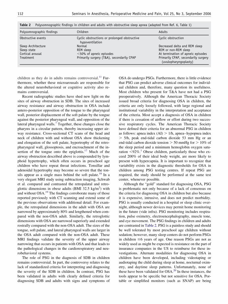
Table 2 Polysomnographic findings in children and adults wit
Polysomnographic findings Children
Obstructive events Cyclic obstructions or phypoventilation
Sleep Architecture NormalSleep state REM sleepCortical arousal �50% of apneic episodTreatment Primarily surgery (T&A)
iagnosing SDB and adults with signs and symptoms of t
SA do undergo PSGs. Furthermore, there is little evidencehat PSG can predict adverse clinical outcomes for individ-al children and, therefore, many question its usefulness.ost children who present for T&A have not had a PSG
reoperatively. Although the American Thoracic Societyssued broad criteria for diagnosing OSA in children, theriteria are only loosely followed, with large regional andnstitutional variability in the interpretation and acceptancef the criteria. Most accept a diagnosis of OSA in childrenf there is cessation of airflow or effort during two succes-ive respiratory cycles. The American Thoracic Societyave defined their criteria for an abnormal PSG in childrens follows: apnea index (AI) � 1/h, apnea–hypopnea index
5/h, peak end-tidal carbon dioxide 53 mm Hg or annd-tidal carbon dioxide tension � 50 mmHg for � 10% ofhe sleep period and a minimum hemoglobin oxygen satu-ation �92%.1 Obese children, particularly those who ex-eed 200% of their ideal body weight, are more likely toresent with hypercapnia. It is important to recognize thatariability exists in the diagnostic thresholds for OSA inhildren among PSG testing centres. If repeat PSG areequired, the study should be performed at the same testenter, whenever possible.
Although the “gold” standard for diagnosing OSA, PSGs problematic not only because of a lack of consensus onhe criteria for diagnosing OSA in children but also becauset is expensive, intrusive, and does not predict morbidity.SG is usually conducted in a hospital or sleep clinic over-ight, although newer devices may permit home monitoringn the future (vide infra). PSG monitoring includes respira-ion, pulse oximetry, electroencephalography, muscle tone,nd eye movement. The PSG findings in children and adultsre contrasted in Table 2. PSG is a painless study and shoulde well tolerated by most preschool age children withoutedation; however, many sleep centers do not perform PSGsn children �6 years of age. One reason PSGs are not asidely used as might be expected is resistance on the part of
nsurance companies in the US to reimburse for these in-estigations. Alternate modalities for diagnosing OSA inhildren have been developed, including videotaping orudiotaping the child during sleep at home, nocturnal oxim-try, and daytime sleep patterns. Unfortunately, none ofhese have been validated for OSA.20 In these instances, theools appear to be specific but not sensitive for OSA. Por-
ructive sleep apnea (adapted from Ref. 6, Table 1)
Adults
ed obstructive Cyclic obstruction
Decreased delta and REM sleepREM or non-REM sleepAt termination of apneic episodes
darily CPAP Primarily CPAP, secondarily surgery(uvulopharyngoplasty)
h obst
rolong
es, secon
able or simplified monitors (such as SNAP) are being

dtrPdtc
ctsehTvihtnqrachnsOcmshlcasnrca
tsislicamtpapoaa
satuO
r(h((omf
msighapewcaibs
ihcuObptmwpi
113Lerman Obstructive Sleep Apnea
eveloped to clinically diagnose SDB because they are easyo use, easy to use in the home setting (where the child iselaxed and comfortable), and inexpensive compared withSG.21 However, these monitors too have not been vali-ated for diagnosing OSA in children. Where the future willake us in terms of newer modalities for diagnosing SDB inhildren remains unclear at this time.
If PSGs are not widely used for children, then how dolinicians identify those at risk for SDB? For the most part,he diagnosis is based on clinical signs and symptoms thatuggest SDB and the otolaryngologist’s judgment. Clinicalvaluation of a child with a history of SDB requires aistory including a sleep history and physical examination.he history must include questions regarding the sleep en-ironment, sleep history, snoring frequency, daytime man-festations of SDB, medication, past medical and surgicalistories, and a family history of snoring, OSA, obesity, andreatments for OSA. Snoring itself is insufficient to diag-ose OSA as mentioned above; however, its presence re-uires that we enquire about episodes of apnea and desatu-ation (cyanosis) during sleep and restless sleep. Other signsnd symptoms referable to SDB and OSA in children in-lude enuresis, nightmares, hyperactivity disorder, and be-avior disorders. Again, none of these signs alone is diag-ostic of OSA. A history of poor academic performance inchool and decreased activity level may be suggestive ofSA. Finally, a full review of systems that focuses on
raniofacial, chromosomal, and neuromuscular disordersay also suggest OSA. A recent systematic review demon-
trated that, in spite of the high quality of the evidence,istory, and/or physical examination (in the absence ofaboratory tests) were inadequate to diagnose OSA whenompared with PSG.22 To improve the diagnostic predict-bility of clinical findings for OSA, a questionnaire of theigns and symptoms was developed. However, the question-aire was subsequently invalidated. Clinicians must nowely on clinical judgment and objective laboratory tests toomplement the history and/or physical examination if theyre to make presumptive diagnoses of OSA without PSGs.
A select number of interventions are available for pa-ients with SDB. These may be categorized into medical,urgical, and mechanical equipment. Medical interventionsnclude non-sedating nasal decongestants, nasal steroidprays such as fluticasone,23 and weight loss,10,24 particu-arly for morbidly obese patients. Surgical interventionsnclude T&A, which is the most common intervention inhildren, uvulopalatopharyngoplasty, lingual tonsillectomy,nd maxillo/mandibular surgery. The latter are more com-only performed in adults with OSA. Mechanical interven-
ions that are effective include continuous positive airwayressure (CPAP) or bilateral positive airway pressure (BiPAP)nd oxygen.25,26 CPAP delivers approximately 5-7 cm H2Oositive pressure to the upper airway. CPAP is believed to stentpen the upper airways. BiPAP, on the other hand, deliverspproximately 2 cm H2O positive pressure during inspiration
nd 5-7 cm H2O during expiration. Thus, the airways are tupported during inspiration, reducing the extent of snoringnd, during expiration, reducing the extent of airway obstruc-ion. Preliminary evidence suggests that BiPAP may also besed successfully in infants and young children to attenuateSA.27,28
Preoperative investigations for the child with OSA whoequires surgery include a PSG, hemoglobin/hematocritwhich may be increased if chronic intermittent nocturnalemoglobin desaturation is present), electrocardiogramnote evidence of right heart strain) or an echocardiogramdecreased ejection fraction), and pulse oximetry (episodesf hemoglobin desaturation, particularly at night). However,ost children who present for T&A surgery have completed
ew of these investigations.Before anesthesia commences, a postoperative plan to
anage the child with OSA should be established. Thetakeholders who should participate in such discussionsnclude the surgeon, anesthesiologist, and the parents oruardians. During the past decade, several investigatorsave attempted to identify those children with OSA who aret high-risk for developing postoperative respiratory com-lications. After a consensus meeting, the American Acad-my of Pediatrics issued guidelines for high-risk childrenith OSA in 2002 (Table 3).29 These guidelines have been
ontroversial and inconsistently applied. Nonetheless, thesere the best practice data available to date and, unless theres compelling evidence to the contrary, they should form theasis for overnight admission of children with OSA who arecheduled for T&A surgery.
Complications after T&A surgery in children with OSAnclude laryngospasm, apnea, pulmonary edema, pulmonaryypertension, and pneumonia.30 Postoperative respiratoryomplications occur in 20% of the children with OSA whondergo T&A compared with 1% in children withoutSA.20,31 Wilson et al. reviewed PSGs from 349 childrenetween 1992 and 1998 to determine whether they couldredict which children are at risk for postoperative respira-ory complications after T&A.20 Complications occurred
ore frequently in children �2 years of age, and in thoseith concomitant medical conditions. Those whose PSGreoperatively exhibited OAH of �5 events/hour had anncreased probability of postoperative respiratory complica-
Table 3 Risk factors for postoperative respiratorycomplications in children with OSA undergoingadenotonsillectomy (Reproduced with permission29)
Age less than 3 yearsSevere OSA on PSGCardiac complications from OSA (ie, right ventricular
hypertrophy)Failure to thriveObesityPrematureRecent respiratory infectionCraniofacial abnormalities
ions (OR 7.2). Furthermore, those children with a preop-
ei2ctpabcuimbaeswaSpdtvarspwnplaomar
tascmcc
cserRsGsrdw
atcssatwsbc
wrbcwatsegipdptypiurs
inlpadetcucasjstOebts
114 Seminars in Anesthesia, Perioperative Medicine and Pain, Vol 25, No 3, September 2006
rative nocturnal SaO2 � 80% had a 3.1 likelihood ratio ofncreasing the postoperative respiratory complications from0% to 50%. Recent interest in the etiology of respiratoryomplications in children after T&A surgery has focused onwo observations. First, Strauss et al. demonstrated thatreschool and school age children with OSA who werenesthetized with 0.5 MAC halothane during T&A hadlunted CO2 response curves compared with age-matchedhildren without OSA undergoing T&A as well as childrenndergoing non-airway ambulatory surgery.32 Second, stud-es in young swine demonstrated that intermittent hypoxiaay increase the membrane density of � receptors in the
rain, thus increasing their sensitivity to opioids.33 Brown etl. then posited that this may also hold true for children whoxperienced intermittent nocturnal hemoglobin oxygen de-aturation. In a retrospective study of children with OSAho experienced intermittent nocturnal hypoxia, Brown et
l. showed that younger children and those with lower theaO2 nadir during nocturnal oximetry required less opioidsostoperatively than older children and those who did notesaturate.34 More recently, the same group demonstratedhat opioids decreased the respiratory effort and minuteentilation in rats that were made intermittently hypoxic togreater extent than in normoxic rats.35 Others have also
eported that children with OSA were sensitive to the re-piratory effects of small doses of fentanyl.16 Then, in arospective study, Brown et al. demonstrated that childrenho were intermittently hypoxic (nocturnal desaturationadir �85%) required less opioid after T&A for the sameain scores as those whose nadir was �85%.36 This pre-iminary evidence suggests that children with OSA may bet greater risk for the respiratory depressant effects of opi-ids. Children who undergo T&A surgery in the afternoonay also be at greater risk for perioperative complications,
lthough the evidence for this caution was based on a singleetrospective study.37
Cardiac-related complications resulting from chronic in-ermittent airway obstruction relate to intermittent hypoxiand hypercapnia. These may lead to pulmonary hyperten-ion, pulmonary edema, and cor pulmonale. These cardiacomplications do not resolve immediately after T&A anday complicate recovery. It is strongly recommended that
hildren with OSA who undergo T&A surgery should belosely monitored for complications postoperatively.
Long-term studies have suggested that T&A surgery inhildren with OSA has salutory effects. Neurobehavioral,chool performance, and cognitive difficulties as well asmotional symptoms, enuresis, and daytime somnolenceesolve are markedly diminished after T&A surgery.38,39
ecent evidence suggests that growth hormone may beuppressed in children with OSA and failure to thrive.40
rowth hormone secretion is often normalized after T&Aurgery and weight then increases. It is equally important toecognize that the majority of children who undergo T&Ao so with only a clinical diagnosis of OSA. Obese children
ith OSA show a reduction in respiratory distress index and tn improvement in the quality of life while maintainingheir BMI after T&A.41 That even children without PSGonfirmation of OSA have a dramatic improvement in theigns and symptoms related to OSA questions the need toubject all children who are scheduled for T&A to undergoPSG study. Moreover, recent evidence has demonstrated
hat even children with negative PSG studies for OSA, butho have a clinical diagnosis of OSA, improve after T&A
urgery.42 There is little doubt that T&A surgery appears toe a very beneficial intervention for a select population ofhildren with SDB.
The anesthetic management for the preschool age childith OSA who is scheduled for T&A needs to address the
isks and benefits of each intervention. Although there haveeen no reports of sequelae after oral midazolam premedi-ation in children with OSA, it behooves us to be vigilanthen administering a sedative agent to a child with OSA in
n unmonitored setting. If the choice is to premedicate, thenhe dose of oral midazolam and the choice of monitorshould be evaluated based on the severity of the OSA andxisting concomitant disorders, ie, morbid obesity and con-enital heart disease. Morbidly obese children with congen-tal heart disease (at risk for right heart strain/failure, su-raventricular tachyarrhythmia) may be at risk for suddenecompensation after a dose of oral midazolam. Althougharental presence at induction of anesthesia is not as effec-ive as oral midazolam in attenuating anxiety and stress inoung children, parental presence may be a reasonable com-romise in those in whom midazolam may place the child atncreased risk for cardiorespiratory sequelae. Careful eval-ation of the child, the cardiorespiratory status, and otherisk factors for OSA should all be considered before con-idering the use of any premedication.
The choice of induction techniques may be limited to annhalational induction if the child does not have an intrave-ous cannula in place. Inhalational inductions may be chal-enging, particularly if a craniofacial abnormality is alsoresent. Upper airway obstruction, a characteristic of UARSnd OSA, is likely to occur once oropharyngeal muscle toneiminishes during the inhalational induction. Positive end-xpiratory pressure, 100% oxygen, and manipulating theempero-mandibular joint (via pressure on the coronoid pro-ess of the mandible)43 will all assist in maintaining a patentpper airway. Manipulating the TM joint at the level of theoronoid process reduces the need for an oropharyngealirway. The most commonly used inhaled agent today isevoflurane, although halothane remains in use in someurisdictions. Sevoflurane in an 8% inspired concentrationhould be administered in a step-wise increase in concen-ration after administering 70% nitrous oxide for 1 minute.nce the eyelash reflex is lost and intravenous access is
stablished, a single dose of propofol, 1.5-2 mg/kg (leanody weight), may be administered intravenously to facili-ate tracheal intubation.44 After the trachea is intubated,pontaneous ventilation is maintained with isoflurane/ni-
rous oxide and oxygen. Anti-emetics [ondansetron (0.05
mdpbs
iOcsiwem�liadDtaraep
seatimsPss
ceToOtcmNaTngtrmas
ao
dcrdwbttpatt
R
1
1
1
1
1
1
1
115Lerman Obstructive Sleep Apnea
g/kg) and dexamethasone (0.15-0.4 mg/kg, maximumose of 8 mg)] should be administered intravenously torevent postoperative nausea and vomiting from swallowedlood. Balanced salt solution in a volume of 15-25 ml/kghould be infused during the surgery.
Opioids have been the cornerstone of pain managementn T&A, although their indiscriminate use in children withSA may be hazardous as outlined above. Accordingly, in
hildren who are suspected of having OSA, I maintainpontaneous respiration during T&A surgery titrating smallncremental aliquots of intravenous morphine, 0-50 �g/kg,hile monitoring the respiratory rate and capnometry. For
xample, in children with severe OSA, my initial dose oforphine intra-operatively may be exquisitely small (10-20g/kg), particularly if the child became apneic for a pro-
onged period after the induction dose of propofol. Depend-ng on the response to the initial dose of morphine, I maydminister incremental supplemental doses of morphineuring anesthesia or in the PACU (postanesthesia care unit).uring report to the nurses in PACU, it is vitally important
o convey concerns regarding the child’s airway (from OSAnd other existing conditions), sensitivity to opioids, and theisk of respiratory obstruction. Downsizing the frequencynd size of the doses of postoperative opioids must bexplained and then documented in the PACU orders torevent respiratory complications.
At the conclusion of surgery, children with OSAhould be extubated awake and transferred to the recov-ry room with a pulse oximeter and supplemental oxygens required. I do not recommend extubating the trachea ofhese children in a deep plane of anesthesia, particularlyn the presence of craniofacial and other disorders thatay compromise cardiorespiratory homeostasis. They
hould be transferred with oxygen and a patent airway toACU. These children remain susceptible to airway ob-truction and all of the problems arising from OSA foreveral weeks postoperatively.
Perioperative complications occur commonly in thesehildren. Laryngospasm and negative pressure pulmonarydema may complicate the early post-extubation period.herefore, awake extubation is an important approach tobviate these complications. Children with documentedSA should then be closely monitored overnight in hospi-
al. Whether this involves a monitored bed in the intensiveare unit or in a step-down unit depends on the level ofonitoring and medical/nursing supervision available.ixon et al. studied children with OSA during the first night
fter T&A surgery using PSG, oximetry, and air flow.45
hey found that children with severe OSA experiencedumerous episodes of obstructive sleep apnea and hemo-lobin oxygen desaturation. Swelling, blood, and redundantissue combined with the respiratory depressant effects ofesidual anesthesia and perioperative analgesics may pro-ote airway obstruction in the early postoperative period
fter T&A surgery. Nasal CPAP or bi-pap with oxygen
hould be reinstituted after surgery if it was used preoper-tively or if needed in the post-extubation period because ofn-going airway obstruction.
OSA is a rapidly evolving and changing disease in chil-ren. Children who snore, experience neurobehavioral, psy-hological, and educational changes, or who are growthetarded may benefit from T&A. Although children withocumented OSA benefit from T&A surgery, even thoseith clinical criteria for OSA appear to achieve equivalentenefits. Recognizing that children with isolated intermit-ent nighttime desaturation or OSA may be more sensitiveo the respiratory depressant effects of intravenous opioids,rone to an obstructed upper airway during anesthesia, andt risk for postoperative airway obstruction may help anes-hesiologists to design anesthetic regimens that minimizehe risks of perioperative complications in these children.
eferences
1. Carroll JL: Obstructive sleep-disordered breathing in children: newcontroversies, new directions. Clin Chest Med 24:261-282, 2003
2. Guilleminault C, Winkle R, Korobkin R, et al: Children and nocturnalsnoring: evaluation of the effects of sleep related respiratory resistiveload and daytime functioning. Eur J Pediatr 139:165-171, 1982
3. Gozal D: Sleep-disordered breathing and school performance in chil-dren. Pediatrics 102:616-620, 1998
4. Ali NJ, Pitson DJ, Stradling JR: Snoring, sleep disturbance, and be-haviour in 4-5 year olds. Arch Dis Child 68:360-366, 1993
5. Rosen CL: Obstructive sleep apnea syndrome in children: controver-sies in diagnosis and treatment. Pediatr Clin North Am 51:153-167,2004
6. Bandla P, Brooks LJ, Trimarchi T, et al: Obstructive sleep apneasyndrome in children. Anesthesiol Clin North Am 23:535-549, 2005
7. Warwick JP, Mason DG: Obstructive sleep apnea in children. Anaes-thesia 53:571-579, 1998
8. Rosen C, Larkin EK, Kirchner HL, et al: Prevalence and risk factorsfor sleep-disordered breathing in 8 to 11 year-old children: associationwith race and prematurity. J Pediatr 142:383-389, 2003
9. Redline S, Tishler PV, Schluchter M, et al: Risk factors for sleep-disordered breathing in children: associations with obesity, race, andrespiratory problems. Am J Respir Crit Care Med 159:1527-1532,1999
0. Kalra M, Inge T, Garcia V, et al: Obstructive sleep apnea in extremelyoverweight adolescents undergoing bariatric surgery. Obes Res 13:1175-1179, 2005
1. Nixon GM, Brouillette RT: Pediatric obstructive sleep apnea. Thorax60:511-516, 2005
2. Punjabi NM, Shahar E, Redline S, et al: Sleep-disordered breathing,glucose intolerance, and insulin resistance: the Sleep heart healthstudy. Am J Epidemiol 160:521-530, 2004
3. Harsch IA, Schahin SP, Radespiel-Troger M, et al: Continuous posi-tive airway pressure treatment rapidly improves insulin sensitivity inpatients with obstructive sleep apnea syndrome. Am J Respir Crit CareMed 169:156-162, 2004
4. Tal A, Leiberman A, Margulis G, et al: Ventricular dysfunction inchildren with obstructive sleep apnea: radionuclide assessment. PediatrPulmonol 4:139-143, 1988
5. Kurnatowski P, Putynski L, Lapienis M, et al: Neurocognitive abilitiesin children with adenotonsillar hypertrophy. Int J Pediatr Otorhinolar-yngol 70:419-424, 2006
6. Waters KA, McBrien F, Stewart P, et al: Effects of OSA, inhalationalanesthesia, and fentanyl on the airway and ventilation of children.
J Appl Physiol 92:1987-1994, 2002
1
1
1
2
2
2
2
2
2
2
2
2
2
3
3
3
3
3
3
3
3
3
3
4
4
4
4
4
4
116 Seminars in Anesthesia, Perioperative Medicine and Pain, Vol 25, No 3, September 2006
7. Arens R, McDonough JM, Costarino AT, et al: Magnetic resonanceimaging of the upper airway structure of young children with obstruc-tive sleep apnea syndrome. Am Rev Resp Crit Care Med 164:698-703,2001
8. Shine NP, Coates HL, Lannigan FJ: Obstructive sleep apnea, morbidobesity, and adenotonsillar surgery: a review of the literature. IntJ Pediatr Otorhinolaryngol 69:475-482, 2005
9. Schwab RJ, Pasirstein M, Pierson R, et al: Identification of upperairway anatomic risk factors for obstructive sleep apnea with volu-metric magnetic resonance imaging. Am J Resp Crit Care Med 168:522-530, 2003
0. Wilson K, Lakheeram I, Morielli A, et al: Can assessment of obstruc-tive sleep apnea help predict postadenotonsillectomy respiratory com-plications? Anesthesiology 96:313-322, 2002
1. Su S, Baroody FM, Kohrman M, et al: A comparison of polysomnog-raphy and a portable home sleep study in the diagnosis of obstructivesleep apnea syndrome. Otolaryngol Head Neck 131:849-850, 2004
2. Brietzke SE, Katz ES, Roberson DW: Can history and physical ex-amination reliably diagnose pediatric obstructive sleep apnea/hypop-nea syndrome? A systematic review of the literature. OtolaryngolHead Neck Surg 131:827-832, 2004
3. Brouillette RT, Manoukian JJ, Ducharme FM, et al: Efficacy of fluti-casone nasal spray for pediatric obstructive sleep apnea. J Pediatr138:838-844, 2001
4. Kudoh F, Sanai A: Effect of tonsillectomy and adenoidectomy onobese children with sleep-associated breathing disorders. Acta Otolar-yngol Suppl 523:216-218, 1996
5. Van de Graaf WB: Thoracic influence on upper airway patency.J Apply Physiol 65:2124-2131, 1988
6. Gordon P, Sanders MH: Positive airway pressure therapy for obstruc-tive sleep apnoea/hypopnoea syndrome. Thorax 60:68-75, 2005
7. Guilleminault C, Pelayo R, Clerk A, et al: Home nasal continuouspositive airway pressure in infants with sleep-disordered breathing.J Pediatr 127:905-912, 1995
8. Downey R III, Perkin RM, MacQuarrie J: Nasal continuous positiveairway pressure use in children with obstructive sleep apnea youngerthan 2 years of age. Chest 117:1608-1612, 2000
9. Section on Pediatric Pulmonology, Subcommittee on Obstructive sleepapnea syndrome of the American Academy of Pediatrics: Clinicalpractice guideline: diagnosis and management of childhood obstruc-tive sleep apnea syndrome. Pediatrics 109:704-712, 2002
0. Blum RH, McGowan FX: Chronic airway obstruction and cardiacdysfunction: anatomy, pathophysiology and anesthetic implications.
Ped Anesth 14:75-84, 20041. Richmond KH, Wetmore RT, Baranak CC: Postoperative complica-tions following tonsillectomy and adenoidectomy: who is at risk? IntJ Pediatr Otorhinolaryngol 13:117-124, 1987
2. Strauss SG, Lynn AM, Bratton SL, et al: Ventilatory response to CO2
in children with obstructive sleep apnea from adenotonsillar hypertro-phy. Anesth Analg 89:328-332, 1999
3. Moss IR, Laferriere A: Central neuropeptide systems and respiratorycontrol during development. Respir Physiol Neurobiol 131:15-27,2002
4. Brown KA, Laferriere A, Moss IR: Recurrent hypoxemia in youngchildren with obstructive sleep apnea is associated with reduced opioidrequirement for analgesia. Anesthesiology 100:806-810, 2004
5. Moss IR, Brown KA, Laferriere A. Recurrent hypoxia in a rat duringdevelopment increases subsequent sensitivity to fentanyl. Anesthesi-ology (in press)
6. Brown KA, Laferriere A, Lakheeram I, et al: Recurrent hypoxemia inchildren is associated with increased analgesic sensitivity to opiates.Anesthesiology (in press)
7. Koomson A, Morin I, Brouillette R, et al: Children with severe OSAS,who have adenotonsillectomy in the morning, are less likely to havepostoperative desaturation than those operated on in the afternoon. CanJ Anesth 51:62-67, 2004
8. Goldstein NA, Fatima M, Campbell TF, et al: Child behavior andquality of life before and after tonsillectomy and adenoidectomy. ArchOtolaryngol Head Neck Surg 128:770-775, 2002
9. Mitchell RB, Kelly J: Long-term changes in behavior after adenoton-sillectomy for obstructive sleep apnea syndrome in children. Otolar-yngol Head Neck Surg 134:374-378, 2006
0. Nieminen P, Lopponen T, Tolonen U, et al: Growth and biochemicalmarkers of growth in children with snoring and obstructive sleepapnea. Pediatrics 109:e55, 2002
1. Mitchell RB, Kelly J: Adenotonsillectomy for obstructive sleep apneain obese children. Otolaryngol Head Neck Surg 131:104-108, 2004
2. Goldstein NA, Pugazhendhi V, Rao SM, et al: Clinical assessment ofpediatric obstructive sleep apnea. Pediatrics 114:33-43, 2004
3. Larson CP Jr: Laryngospasm-the best treatment. Anesthesiology 89:1293-1294, 1998
4. Mathews BT, Lerman J, Burrows FA: Optimal dose of propofol tofacilitate tracheal intubation during induction of anesthesia withsevoflurane in healthy children. Anesthesiology (abstract) 101:A1418,2004
5. Nixon GM, Kermack AS, McGregor CD, et al: Sleep and breathing onthe first night after adenotonsillectomy for obstructive sleep apnea.
Pediatr Pulmonol 39:332-338, 2005