observations upon the bacterial spore nucleus - europe pubmed
TRANSCRIPT
[ 201 ]
OBSERVATIONS UPON THE BACTERIAL SPORE NUCLEUS
BY K. A. BISSET AND C. M. F. HALE
From the Department of Bacteriology, University of Birmingham
(With Plate 11 and 3 Figures in the Text)
INTRODUCTION
Discrete, Feulgen-positive granules, attached to the periphery of bacterial spores,were described by Pietschmann & Rippel (1931), who considered them to beresidual cytoplasmic structures; by Stille (1937), who was apparently in doubt asto their nature; and by others, including Robinow (1945), who at first describedthem as the natural appearance of the spore nucleus. Delaporte (1950) figured thespore nucleus as a less discrete, crescentic body lying against one side of the spore,and in a later study Robinow (1951) accepted this view, and presented evidence toprove that the appearance of the discrete body was an artefact, caused by thehydrolysis with HC1 which was employed in staining. Robinow, in this later study,employed nitric acid instead of hydrochloric acid, and claimed that it gave a truerpicture. Both Delaporte and Robinow observed a central, stainable body, whichthey considered to be cytoplasmic.One of us has included, in a general study of the bacterial resting nucleus, a
criticism of the original concept of a discrete, extra-cytoplasmic nucleus, and hassuggested that the spore nucleus appears to be similar to that of other bacterialresting cells, a central, vesicular structure (Bisset, 1950a). The partially revisedsuggestion of Delaporte and of Robinow, of a crescentic, external nucleus, and aninternal cytoplasmic body, appears equally unusual, with respect to the arrange-ment in cells of other types, and does not accord with our own observations uponthe nuclei of bacterial spores.
In this paper, we will attempt to show that the bacterial spore, like the eubacterialand myxobacterial microcyst (Bisset, 1950a), possesses an internal, vesicularnucleus, which can be observed in the living spore, under suitable conditions, byphase-contrast microscopy, and that the discrete and the crescentic externalnuclei are alike artefacts.
TECHNIQUE
Spores were stained after treatment with normal HC1 at 60° C. or with normalnitric or perchloric acid, at room temperature. These methods were not significantlydifferent in respect of their capacity to induce the various artefacts described inthis paper. After acid treatment, preparations were stained with Giemsa, crystalviolet, or tannic acid and crystal violet, for the simultaneous demonstration of thecell wall. Preparations were mounted in water for examination.
Living spores were examined by phase-contrast microscopy. Gold-shadowedelectron micrographs were also made.
K. A. BISSET AND C. M. F. HALE
OBSERVATIONS
Spores of several species of Bacillus and Clostridium were examined. Differentstrains varied markedly in their reaction to acid hydrolysis. In extreme cases,after approximately 5 min., large protuberances appeared upon their surfaces.These protuberances stained deeply with Giemsa or crystal violet. If these pre-parations were first hydrolysed and then stained by the tannic-acid-violet techniqueit was apparent that they lay outside the spore coat (P1. 1 1, fig. 1). At the other endof the scale, spores of certain strains showed the stained appearance of an un-disturbed central nucleus (P1. 1 1, fig. 2). In the spores which exhibited protuberancesthe position of the central nucleus was occupied by a less deeply stained centralbody (P1. 11, figs. 1, 3 and 4).
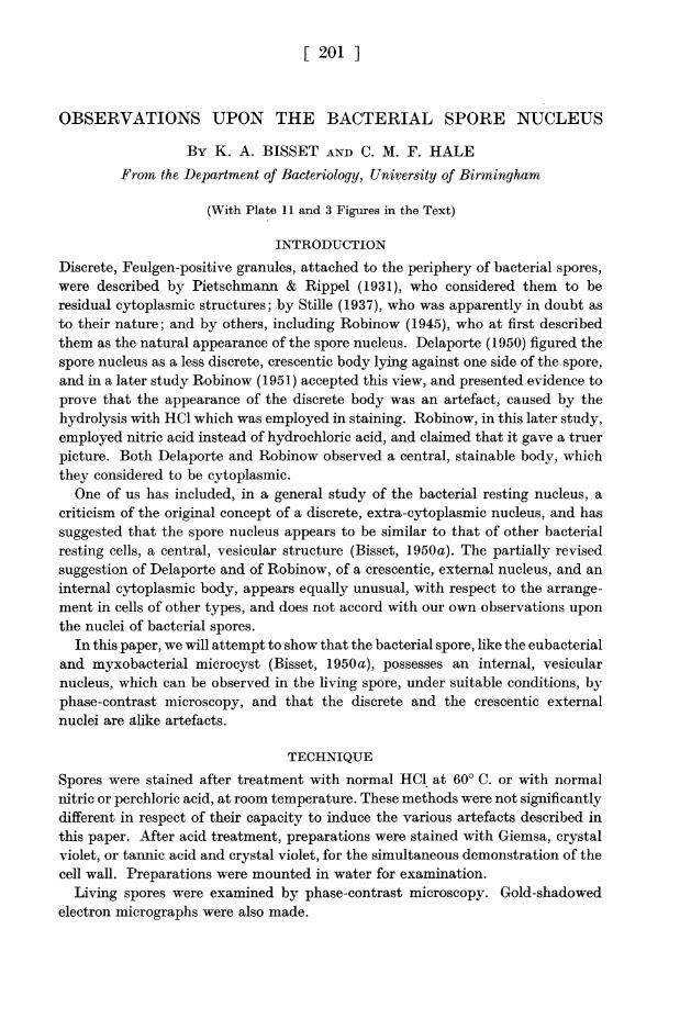
Cl. welchii, which was examined with great thoroughness, showed all stagesbetween these extremes, sometime sin a single preparation (P1. 11, figs. 3, 4; Text-
Text-fig. 1. Text-fig. 2.
Text-figs. 1 and 2. Spores of Clostridium welchii. Text-fig. 1. As seen by phase-contrastmicroscopy; central or slightly eccentric, vesicular nucleus. Text-fig. 2. Spores from the sameculture, treated with N/I nitric acid for 20 min., stained crystal violet. All stages seen, fromnormal condition of the nucleus to complete ejection.
fig. 2). Spores of strains which were very markedly affected by a brief exposure toacid sometimes disintegrated completely if the treatment was prolonged for morethan a few minutes, but in these cases the stainable body retained its form (P1. 11,fig. 6).
Electron micrographs were made of spores subjected to normal nitric acid.These confirmed that the protuberances were beyond the spore coat which appearedrelatively undistorted (P1. 11, fig. 7).
Suspensions of spores were mounted in normal nitric acid and examined by thephase-contrast microscope. After a few minutes the oval outline of the majorityof spores was abruptly broken by the appearance of a bulge. In a short time, manyhad burst and disintegrated completely.
Slide cultures were made of spores of Cl. welchii upon nutrient agar containing
202
Observations upon the bacterial spore nucleus
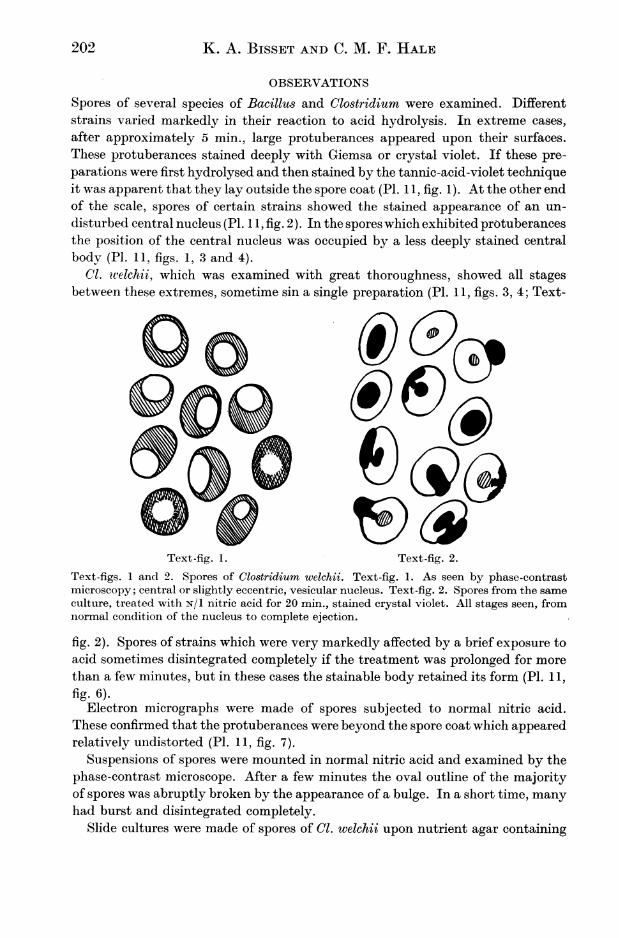
0.05 % sodium thioglycollate to initiate anaerobic conditions, and sealed under acover-glass with paraffin wax. These were incubated at 370 C. and examined atintervals by the phase-contrast microscope. These spores took a considerable timeto germinate, seldom less than 12 hr., often much more. Before germinating mostof them became much less opaque, and in them a spherical or oval eccentricnucleus could readily be discerned, occupying the same position as the stainednuclei seen in the undisrupted spores (Text-figs. 1, 2). No signs were seen of anystructure resembling the 'external nucleus'.
Stained preparations of the spores of Cl. welchii in process of maturation showedthe nucleus in a central position, from which no amount of acid treatment wascapable of moving it (P1. 11, fig. 5).
b16a c d e
Text-fig. 3. Artefacts produced by acid treatment of bacterial spores. a, natural appearance ofthe nucleus; b, 'crescentic nucleus' produced by nuclear material forced into a pool betweenthe spore coat and the cytoplasm; c, d, 'discrete peripheral nucleus', when the spore coat isbulged outward; e, nuclear material ejected.
DISCUSSION
It appears from the foregoing observations that the nucleus of the bacterial sporeis a central body resembling the nuclei of the resting cells of other types of bacteria.It can be demonstrated in stained preparations and can be observed by phase-contrast microscopy in the living spore. A short exposure to normal acid producesa whole series of artefacts in some strains but not in others. In the mature (butnot in the immature) spore, the nuclear contents are apparently in a condition ofgreat turgidity. This is an observation which accords well with what is known ofthe concentration of the spore proteins (Bisset, 1950b). When the spore coat issoftened by acid attack, it may be bulged outwards to form a small discreteblister of nuclear material (Text-fig. 3, a, c, d), or if the spore coat holds relativelyfirm, or stretches more evenly, the nuclear material may form a pool between thespore coat and the body of the cytoplasm, giving the appearance of the crescenticnucleus of Delaporte (1950) (Text-fig. 3 b). If the spore coat is ruptured, the nuclearmaterial may be ejected (Text-fig. 3e). This fluidity of the nuclear material wasremarked upon by Delaporte. The central body, less strongly stained, whichappears in preparations showing one or other of these artefacts, is presumably aremnant of nuclear material, occupying the natural site.
203
204 K. A. BISSET AND C. M. F. HALE
REFERENCES
BISSET, K. A. (1950a). The Cytology and Life-History of Bacteria. Edinburgh: E. and S.Livingstone Ltd.
BISSET, K. A. (1950b). Nature, Lond., 166, 431.DELAPORTE, B. (1950). Advanc. Genet. 3, 1.PIETSCHMANN, K. & RIPPEL, A. (1931). Arch. Mikrobiol. 3, 422.ROBINOW, C. F. (1945). Addendum to: Dubos, R. J., The Bacterial Cell. Harvard University
Press.ROBINOW, C. F. (1951). Symposium upon Bacterial Cytology. Microbiological Panel, Society
of Chemical Industry, London.STILLE, B. (1937). Arch. Mikrobiol. 8, 125.
EXPLANATION OF PLATE 11
Fig. 1. Spores of Bacillus sp., treated with N/l nitric acid for 5 min., stained Giemsa andrestained tannic-acid-violet, to demonstrate that the ejected nuclear material is outside thespore coat. One spore has retained it within the spore coat, and is exhibiting the 'peripheralnucleus'. x 5000.Fig. 2. Spores of B. subtilis, showing the spore nucleus in its natural condition. Acid-Giemsa.x 3000.Figs. 3, 4. Spores of Cl. welchii. Nitric acid for 10 min., stained Crystal violet. All conditionsseen: a, nucleus in natural condition; b, 'crescentic nucleus'; c, 'peripheral nucleus'. Otherspores are in intermediate stages of change. x 3000.Fig. 5. Maturing spore of Cl. welchii, as in Figs. 3 and 4, showing no displacement of nuclearmaterial. x 3000.Fig. 6. Spores of the same Bacillus as Fig. 1. Several hours after subjection to N/l nitric acidfor 5 min. The spore coat is disrupted, but the nuclear material retains its form. StainedGiemsa. x 3000.Fig. 7. Spores of the same Bacillus as Fig. 1. Electron micrograph, gold-shadowed, madeafter treatment of the mount with N/I nitric acid for 5 min. Showing ejected nuclear material.x 16,000.
(MS. received for publication 1. iII. 51.)

JOURNAL OF HYGIENE, VOL. 49, NOS. 2 & 3 PLATE 11