observations on the nature of the relationship between...
TRANSCRIPT

Observations on the Nature of the Relationshipbetween Cell Division and Carbohydrate Metabolism
by R. j . O'CONNOR1
John Burford Carlill Pathological Laboratories, Westminster School of Medicine
INTRODUCTION
A L T H O U G H there is considerable evidence that carbohydrate metabolism playsan important part in the process of cell division (see Bullough, 1952), conclu-sions have differed regarding the relative importance of glycolysis and cata-bolism involving respiration. In the adult mouse epidermis Bullough & Johnson(1951) found that cell division was dependent upon the respiratory oxidation ofpyruvate by the tricarboxylic acid cycle. On the other hand, Pomerat & Willmer(1939) showed that, in tissue culture, agents that inhibited respiration had littleimmediate effect on cell division, while Laser (1933) found that growth of cul-tured fibroblasts could continue when respiration was greatly diminished bylow oxygen tensions. Further, Warburg (1930) concluded that cell growth isassociated with glycolysis.
A similar difference in the relationship of mitotic activity to the respiratoryand glycolytic forms of carbohydrate metabolism was indicated by changesoccurring in the midbrain and the red-blood cells of the chicken embryo duringembryonic development. In the midbrain (O'Connor, 1950a) there was a corre-sponding decrease in the number of dividing cells in a unit volume of tissue andin the rate of aerobic glycolysis as measured by the production of acid by mid-brain tissue isolated in a glucose containing medium x(acid formation did notoccur when glucose was omitted from the medium; O'Connor, 1949). Underthese conditions the rate of respiration remained constant. Since the respiratoryquotient was unity, this constant rate of respiration was taken to indicate a con-stant rate of glucose utilization by respiratory metabolism. Further investigations(O'Connor, 1950ft) showed that, when the rate of aerobic glycolysis of the iso-lated midbrain was decreased by fluoride or iodoacetate, cell division wasinhibited by concentrations which did not diminish the rate of respiration noralter the respiratory quotient. Thus it was concluded that, in the midbrain, celldivision was dependent on aerobic glycolysis, as measured by the acid produc-tion of the isolated tissue in a glucose-containing medium.
When the red-blood cells were investigated in a similar medium (O'Connor,1 Author's address: Westminster School of Medicine, Horseferry Road, London, S.W.I, U.K.[J. Embryol. exp. Morph. Vol. 2, Part 1, pp. 26-37, March 1954]

CARBOHYDRATE METABOLISM 27
1951) it was found that acid formation did not occur but the rate of respirationdecreased during development in a manner corresponding to the decrease in theproportion of dividing cells. The respiratory quotient of the red-blood cells wasunity, so that these findings suggested that there was a corresponding decreasein mitotic activity and the catabolism of glucose by processes involving respira-tion. This suggestion was supported by subsequent investigations which showedthat the inhibition of respiration by fluoride and by iodoacetate resulted in in-hibition of cell division (O'Connor, 1952). Mitotic activity in the red-blood cellsand in the midbrain thus differs in its relationship to carbohydrate catabolism,since in the former it is associated with the respiratory utilization of glucose, andin the latter with aerobic glycolysis, as measured by the acid production of theisolated tissue in a glucose-containing medium.
In order better to assess the significance of this difference, comparisons havebeen made of the changes undergone by mitotic activity and carbohydrate meta-bolism in the developing liver of the chicken embryo, the metabolism of whichdiffers in certain relevant respects from that of the midbrain and red-blood cells.As shown previously (O'Connor, 1953a), in isolated liver tissue, the respiratoryquotient is less than unity, indicating that oxygen is utilized in the metabolismof substrates other than carbohydrate. Further, the carbohydrate metabolismof the liver is complicated by the deposition of considerable amounts of glyco-gen during the course of its development.
MATERIAL AND METHODS
All observations were made on the liver of chicken embryos incubated at38° C. The eggs were taken from the batches used previously (O'Connor, 1953a),and the observations were scattered at random among the batches. As pre-viously, the embryos were grouped in arbitrarily chosen stages of development.Each stage represents an increase in the eye diameter of 0 7 mm. and is referredto below by its median eye diameter (M.E.D.). The mean time of incubation foreach of these stages has already been recorded (O'Connor, 1953a), and, since theeggs used in these experiments are from the same batches, these times areapplicable to the present observations and are repeated in Table 1.
The estimation of glucose utilization by isolated liver tissue
In the accepted scheme of carbohydrate catabolism in animal tissues the sub-strate, whether glucose or glycogen, is first transformed into pyruvic acid. In thepresence of oxygen, part or all of the pyruvic acid is oxidized by way of thetricarboxylic acid cycle with the consumption of oxygen. On the other hand,even in the presence of oxygen, part of the pyruvic acid may be reduced to lacticacid. These two processes, respiratory catabolism and aerobic glycolysis, havebeen separately estimated from observations on the gaseous exchange of iso-lated liver tissue.
(a) Acid production by isolated liver tissue. In order to determine the rate of

28 R. J. O ' C O N N O R — C E L L D I V I S I O N A N D
aerobic glycolysis of the isolated liver, the rate of total acid production wasmeasured by a Cartesian diver micromanometer. Although the technique usedhas already been recorded (O'Connor, 1950a), it possesses certain limitationsrequiring present consideration and is, therefore, recapitulated in part.
Livers were removed from the embryos and placed in the following medium:NaCl 0-9 g., KC1 002 g., MgCl2 002 g., CaCl2 002 g., glucose 020 g., water100 ml., to which was added 10 ml. M/15 phosphate buffer (S0rensen) to pro-duce pH 7 4; this is the medium previously used (O'Connor, 1953a) to determinethe rate of respiration and the respiratory quotient. As in those determinations,fragments of liver were cut from the anterior surface of the right lobe of the liverand the volume measured; this volume varied from 0 3 to 0 8 c.mm. The frag-ments were then washed twice in the following medium: NaCl 0 9 g., KC1002 g.,MgCl2 002 g., CaCl2 002 g., glucose 020 g., NaHCO3 020 g., water 100 c.c.The liver fragments were then introduced into divers containing this medium.The gaseous phase of the divers was 95 per cent. O2:5 per cent. CO2 and thearrangement of fluids within the divers, as well as their dimensions, were asdescribed previously (O'Connor, 1950a). In such divers it should be noted thatthere is no alkali to absorb carbon dioxide so that the manometric reading isaffected not only by carbon dioxide released from the bicarbonate in the mediumas the result of acid formation, but also by the respiration of the liver tissue andby the carbon dioxide formed as a consequence. In order to take account of thelast two factors the assumption was made that they occurred at the same rateunder the conditions of the present observations as they did under the conditionsin which respiration and respiratory quotient were investigated previously(O'Connor, 1953a), when the medium contained phosphate and not bicarbonate(see above) and the gaseous phase of the divers was oxygen. Using these results,a figure was calculated which represented the amount of carbon dioxide pre-sumed to result from acid formation, and this will be referred to as 'presumedacid formation'. In arriving at this figure the factor was calculated, both foroxygen and carbon dioxide, which related manometric change to the change ingaseous content of the diver. The formula given by Boell, Needham, & Rogers(1939) was used. Comparable observations and calculations were made whenthe isolated liver was suspended in the bicarbonate-containing medium fromwhich glucose was omitted. All observations were made over a period of 2 hours,during which time the rate of change in the manometric reading did not varysignificantly in any one particular observation.
(b) Respiratory utilization of glucose by isolated liver tissue. Since, as men-tioned above, the oxygen consumed by isolated liver tissue is used in the cata-bolism of substrates other than carbohydrate, the rate of respiration cannot beused directly as a measure of the respiratory catabolism of glucose as it was inthe case of the midbrain and red-blood cells. It was considered, however, thatthis rate could be estimated from the decrease in the rate of respiration whichresulted when glucose was omitted from the medium in which respiration was
CARBOHYDRATE METABOLISM 29
measured. Rates of respiration of the isolated liver in the presence and absenceof glucose have been recorded previously (O'Connor, 1953a), and these wereused in the present investigations to calculate the rate of the respiratory utiliza-tion of glucose at different stages of development.
The estimation of mitotic activity of the liver cellsThis was carried out by determining, from histological sections, the percent-
age of hepatic cells in mitosis. Cells other than hepatic cells were not considered.Sections cut at a thickness of 6/x from livers fixed in Bouin's fluid were stainedwith iron haematoxylin. No counter-stain was used.
RESULTS
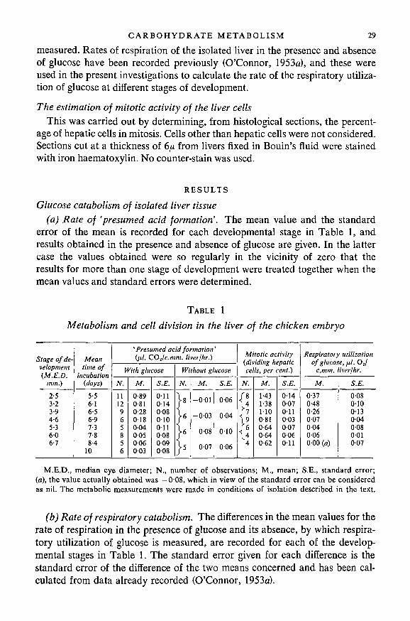
Glucose catabolism of isolated liver tissue(a) Rate of 'presumed acid formation'. The mean value and the standard
error of the mean is recorded for each developmental stage in Table 1, andresults obtained in the presence and absence of glucose are given. In the lattercase the values obtained were so regularly in the vicinity of zero that theresults for more than one stage of development were treated together when themean values and standard errors were determined.
TABLE 1
Metabolism and cell division in the liver of the chicken embryo
Stage of de-velopment(M.E.D.
mm.)
2-53-23 94-65-3606-7
Meantime of
incubation(days)
5-5616-56-97-37-88-4
10
'Presumed acid formation'(/*/. COj/c.mm. liver/hr.)
With glucose
N.
1112965856
M.
0-890-810-28018004005006003
S.E.
011014008010011008009008
Without glucose
N.
-8
•6
•6
•5
M.
-001
-003
008
007
S.E.
006
004
010
006
Mitotic activity(dividing hepaticcells, per cent.)
N.
/8\4/ 7\9/6\4
4
M.
1-431-381100-810-640-640-62
S.E.
014007011003007006011
Respiratory utilizationof glucose, fxl. OJ
c.mm. liver/hr.
M.
0-370-480-26007004006000 (a)
S.E.
0080100130-04008001007
M.E.D., median eye diameter; N., number of observations; M., mean; S.E., standard error;(a), the value actually obtained was —008, which in view of the standard error can be consideredas nil. The metabolic measurements were made in conditions of isolation described in the text.
(b) Rate of respiratory catabolism. The differences in the mean values for therate of respiration in the presence of glucose and its absence, by which respira-tory utilization of glucose is measured, are recorded for each of the develop-mental stages in Table 1. The standard error given for each difference is thestandard error of the difference of the two means concerned and has been cal-culated from data already recorded (O'Connor, 1953a).

30 R. J. O 'CONNOR—CELL D I V I S I O N A N D
Mitotic activity in liver cells
To determine the percentage of hepatic cells in mitosis 2,000-3,000 cells wereexamined in each individual case. The mean value for a number of embryos ineach developmental stage and the standard error of the mean are recorded inTable 1. Since the nuclei of dividing and resting cells differ in size an error ispresumably introduced (Abercrombie, 1946). This error, however, would notdiffer greatly at different stages of development and would not, therefore, dimin-ish the significance of the decrease during development of the percentage of cellsseen in mitosis and particularly between the stages of M.E.D. 3 2 mm. andM.E.D. 4 6 mm.
DISCUSSION
Aerobic glycolysis by isolated liver tissue
The rates of 'presumed acid formation' were calculated in order to measurerates of aerobic glycolysis, that is the production of lactic acid from glucose.However, before the results are used for this purpose certain considerations arenecessary. It will be recalled that the 'presumed acid production' was calculatedon the assumption that the rate of respiration and the respiratory quotient werethe same under the conditions of the present observations as in the conditionsunder which they were previously determined (O'Connor, 1953a); Dixon (1951)has pointed out that this assumption may not be justifiable. However, thegaseous exchange concerned, namely the uptake of oxygen and the resultingevolution of carbon dioxide, would affect manometric readings in oppositedirections and so decrease any error introduced. Also, none of the results re-corded in Table 1 for 'presumed acid formation' are significantly below zero.Since negative results can be considered 'absurd', their absence is in favour ofthe calculation being based on correct assumptions. Therefore, even if the pos-sibility of error is not completely eliminated, it is considered that 'presumed acidformation' sufficiently represents true acid formation by isolated liver tissue toconclude that during development this decreases to reach a rate of nearly zeroat stages of M.E.D. 5 3 mm. and later (see Table 1). In a previous publication(O'Connor, 1953a) the evidence was considered which excludes the possibilitythat this decrease is due to the addition, during development, of metabolicallyinert substances to the hepatic cells.
Although uric acid may be formed by isolated liver tissue (O'Connor, 1953a),the amount of carbon dioxide it releases from bicarbonate is unimportant be-cause when glucose is omitted from the medium, acid formation nearly dis-appears (Table 1). Thus the acid formed is nearly, if not completely, derivedfrom glucose, and can therefore be presumed to be lactic acid. For these reasonsit is considered that 'presumed acid formation' is sufficiently a measure of lacticacid production from glucose to permit its use as a measure of aerobic glycolysis.

CARBOHYDRATE METABOLISM 31
The relationship of cell division to carbohydrate metabolism
Using 'presumed acid formation' as a measure of aerobic glycolysis the rateof this process at different developmental stages has been compared, in Text-fig. 1, with corresponding rates of respiratory glucose utilization and withmitotic activity. Line AB is added to the figure to indicate the first appearanceof glycogen in embryos of the stage of M.E.D. 4 6 mm. These embryos have a
O-5
2-5 3-2 3-9 4-6 5-3 6-OStage of development (median eye diameter, mm.)
6-7
TEXT-FIG. 1. Mitotic activity (A A), rate of 'presumed acidformation' ( # '•—•), and the rate of the respiratory utilization ofglucose (O O) of the liver tissue at different stages of develop-ment. The metabolic measurements were made as described in the text,where reasons are given for regarding 'presumed acid formation' as ameasure of aerobic glycolysis. Line AB indicates the first appearance of
glycogen in the liver cells.
mean incubation time of 6 9 days (O'Connor, 1953a). The figure shows that withthe appearance of glycogen there is an alteration in the relationship betweenmitotic activity and the glucose metabolism of isolated liver tissue. Before glyco-gen appears there is a corresponding decrease in the proportion of dividing cells,the rate of 'presumed acid formation', and the rate of the respiratory utilizationof glucose (Text-fig. 1). After the appearance of glycogen these metabolic pro-cesses almost disappear, although dividing cells persist and constitute about0 6 per cent, of hepatic cells. Furthermore, both before and after the appearanceof glycogen the relationship between mitotic activity and glucose metabolismby the isolated tissue differs from that found in the midbrain and in the cir-culating red-blood cells, for in the former decreasing mitotic activity is asso-ciated with decreasing acid production from glucose (O'Connor, 1950a), while

32 R. J. O'CONNOR—CELL DIVISION AND
in the latter the association is with decreasing respiratory metabolism (O'Con-nor, 1951). These differences are summarized in Table 2, and it is possible thatthey represent a significant difference in the metabolic relationships of mitoticactivity in the four tissues. On the other hand, the similarity of the mitoticmechanism in these and other tissues suggests the alternative possibility that
TABLE 2
Changes in the rate of carbohydrate metabolism associated with thedecrease in mitotic activity which occurs during the development of
tissues in the chicken embryo
Tissue
Midbrain (O'Connor, 1950o)
Red-blood cells (O'Connor, 1951)
Liver before glycogen appears(see text)
Liver after glycogen appears (seetext)
Rate of carbohydrate metabolism
Respiratory
Remains constant
Decreases as mitoticdecreases
Decreases as mitoticdecreases
Very low
activity
activity
Producing acid
Decreases as mitoticdecreases
Absent
Decreases as mitoticdecreases
Very low
activity
activity
The rates of carbohydrate metabolism are those of the isolated tissue in a glucose-containing medium. In the references quoted and in the text reasons are given for
regarding acid production as a measure of aerobic glycolysis.
the differences recorded in Table 2 are nevertheless an expression of a commondependence of cell division on carbohydrate catabolism, as has been suggestedby Bullough (1952) and which, in the case of the midbrain and the red-bloodcells, has been indicated by the correspondence between the effect on cell divi-sion and the inhibition of carbohydrate catabolism produced by fluoride andiodoacetate (O'Connor, 19506, 1952).
In the case of the midbrain, red-blood cells, and the liver before the appear-ance of glycogen, the findings of Table 2 could be accounted for if mitoticactivity were dependent on the total amount of pyruvate disposed of by con-version to lactic acid in aerobic glycolysis together with its oxidation by thetricarboxylic acid cycle. Thus in the case of these three tissues it is possible thatthe reactions by which pyruvate is formed from glucose play an essential partin cell division. Such reactions could occur in the liver after the appearance ofglycogen, in spite of the findings in Table 2, if pyruvate or compounds inter-mediate in its formation were incorporated into larger molecules. Such reactionsleading to glycogen formation are well known in the adult liver, and it is likelythat they occur in the embryonic liver after the appearance of glycogen, forDal ton (1937) has concluded that at this stage the liver of the chicken embryobecomes capable of adult functions. Thus the findings in all four tissues recordedin Table 2 could be accounted for on the assumption that mitotic activity was

CARBOHYDRATE METABOLISM 33
dependent on the reactions leading to formation of pyruvate from glucose.Although there is not sufficient evidence to establish such a relationship, it ispossible to consider its implications and compare them with results obtainedby other investigators. Such is the purpose of the following discussion.
There is considerable evidence that in the metabolism of glucose there is apathway alternative to the glycolytic pathway of the Meyerhof scheme. In thishexose monophosphate oxidative route1 glucose is converted into ribose-5-phosphate by reactions which involve the consumption of oxygen (Dickens &Glock, 1951). In the further metabolism of ribose-5-phosphate, a triose is formedwhich is incorporated into glucose-6-phosphate. Evidence for such reactions hasbeen found in a number of tissues which include, in the adult, the liver (Glock,1952), the red-blood cells (Dische, 1951), and brain tissue (Sable, 1952). Pyruvatecould be formed as a result of such reactions since both the triose and theglucose-6-phosphate could be acted upon by enzymes of the glycolytic system.Thus the possibility that mitotic activity depends upon reactions leading topyruvate formation gives rise to the further possibility that the actual reactionsconcerned may be those associated with the formation of ribose-5-phosphate.Since ribose-5-phosphate is similar to the pentose component of ribonucleicacid, this possibility would be in accordance with the well-established associa-tion of cell proliferation and ribonucleic acid (Caspersson, 1950; Brachet, 1947).Further, the association of cell division with the alternative hexose monophos-phate oxidative route of glucose metabolism might constitute a basis for anexplanation for a special form of metabolism which, it has been claimed, isassociated with embryonic development (cf. Moog, 1944).
However, pyruvate formation by the hexose monophosphate oxidative routewith ribose-5-phosphate as an intermediate compound involves the consump-tion of oxygen. Therefore, to suggest that such a process plays a dominant partin cell division makes it necessary to consider objections that might arise fromobservations suggesting that cell division can occur independently of oxygenconsumption. In the case of the observations made on the isolated midbraintissue, and recorded in Table 2, the association of decreasing mitotic activitywith a constant rate of oxygen consumption might be the basis of such an objec-tion. However, modifications of enzyme activity occurring in the course of dif-ferentiation might be adequate to meet this objection, since it is possible thatany decrease of the oxygen utilized in the formation of ribose-5-phosphate maybe balanced by an increase in the proportion of pyruvate oxidized by the tri-carboxylic acid cycle. After the appearance of glycogen in the liver of thechicken embryo it might be questioned whether the oxidative formation ofribose-5-phosphate could occur because of the low values obtained for oxygenconsumption associated with the catabolism of glucose (see Table 1). However,
1 There has been discussion about the most appropriate name for this metabolic pathway(Dickens, 1953). The term 'hexose monophosphate oxidative route', suggested by Dickens, willte used.
5584.2 D

34 R. J. O'CONNOR—CELL DIVISION AND
since measurements were made on isolated liver tissue they would not excludethe presence of the hexose monophosphate oxidative route in the intact em-bryonic liver, particularly since, in the adult liver, the necessary enzymes havebeen demonstrated (Dickens & Glock, 1951), and it is to be expected that suchenzymes would be present in the liver of the chicken embryo after glycogenappears because of the evidence that, at this period of development, the em-bryonic liver is capable of adult function (Dalton, 1937).
Apart from the results recorded in Table 2 the possibility of a dependence ofmitotic activity on an oxygen-consuming process might appear inconsistent withobservations that, in some cells, division can be completed in the absence ofoxygen (e.g. Lettre, 1951). Although, in such circumstances, glycolytic reactionscould continue and be adequate to meet the energy requirements of the cell, thereactions leading to ribose-5-phosphate formation would presumably cease.However, if cell division were dependent on ribose-5-phosphate formation, itsfailure in such conditions might not inhibit mitosis, at least for a time, since thefailure of ribose-5-phosphate formation might be balanced by a decreased rateof destruction. Further, if ribose-5-phosphate is concerned in the synthesis ofribonucleic acid (Cohen, 1951) any failure of its formation might be met byutilization of precursors of nucleic acid in the cell (cf. Walker & Yates, 1952).
Even if it were established that cell division was dependent on the reactionsleading to pyruvate formation, it is unlikely in ordinary circumstances thatpyruvate formation, whether by the glycolytic or by the hexose monophosphateoxidative route, meets the energy requirements of the dividing cell because thegreater part of the energy produced in carbohydrate catabolism comes from theoxidation of pyruvate by the tricarboxylic cycle (Burton & Krebs, 1953). Fur-ther, Bullough & Johnson (1951) have shown by experiment that, in the isolatedadult mouse epidermis, cell division is dependent on the oxidation of pyruvatein the tricarboxylic acid cycle. In most cells this would constitute a dependenceof mitotic activity on carbohydrate catabolism, since pyruvate is derived fromcarbohydrate and in cells with a respiratory quotient of unity carbohydrate maybe its sole source (e.g. the midbrain tissue and red-blood cells of the chickenembryo; O'Connor, 1950a, 1951). However, in the case of the liver after theappearance of glycogen it may be necessary to make an exception, because therespiratory quotient of isolated liver tissue is 069, which suggests that the oxy-gen consumed is utilized in the formation of uric acid from protein (O'Connor,1953a). In this case, therefore, it may be that the energy requirements of dividingcells are met by the catabolism of protein rather than of carbohydrate. It isrelevant to note that after glycogen appears no difference could be detected, byhistochemical methods, in the amount present in dividing and non-dividingcells (O'Connor, 19536).
This possible exception apart, the suggestion that the process of cell divisiondepends on reactions leading to pyruvate formation can be regarded as addi-tional to the dependence of cell division on carbohydrate metabolism for its

C A R B O H Y D R A T E METABOLISM 35
energy requirements. It may be that one or other dependence is limiting to celldivision at different stages of the mitotic cycle, for Bullough & Johnson (1951)concluded that the respiratory oxidation of pyruvate is necessary for the initia-tion of cell division but not for its completion (see also Bullough, 1950). Itmight therefore be suggested that a continuance of cell division, once it hasbegun, depends upon reactions leading to pyruvate formation and in particularupon those associated with ribose-5-phosphate formation. Such a suggestionwould be in accordance with the observations of Jacobson & Webb (1952) thatribonucleoprotein is formed in the nucleus during mitosis and extruded into thecytoplasm at anaphase. As mentioned above, such a formation of ribonucleicacid might, as an alternative, be possible from precursors already in the cell,and thus account for the ability of some cells to complete cell division, onceit has begun, 'in almost any circumstances short of death of the cell itself(Bullough, 1952).
If this suggested double dependence of mitotic activity on carbohydrate cata-bolism exists, cell proliferation might be controlled by a balance between thereactions leading to the formation of pyruvate and those of the tricarboxylicacid cycle. If the latter were limited by anaerobiosis an altered balance mightcause a modification of proliferative capacity and in this way account for theassociation found by Goldblatt & Cameron (1953) between intermittent anaero-biosis and the malignant conversion of cultured fibroblasts. Again, it can be sug-gested that such a disturbed balance might occur if the respiratory mechanismof cells was damaged and replaced by a process involving lactic acid formation—a change that Warburg (1930) associated with the development of malignancyand one that might follow deficiencies in the tricarboxylic acid cycle for whichevidence has been found in malignant tissue (Potter & Busch, 1950). Further, itmight be possible to relate such a disturbed balance to changes in the cell frac-tions that can be separated by ultracentrifugation. It has been found that thereactions by which pyruvate is formed, whether by the glycolytic or the hexosemonophosphate oxidative route, are associated with the soluble fraction (LePage & Schneider, 1948; Glock & McLean, 1952), while the reactions of thetricarboxylic acid cycle are associated with the mitochondria (Harman, 1950).
SUMMARY
1. From micromanometric measurements made on liver tissue of the chickenembryo, isolated in a glucose-containing medium, estimates have been made ofthe rate of glucose catabolism ending in acid formation and that utilizingoxygen.
2. The variations undergone by these metabolic processes during normaldevelopment have been compared with the variations in mitotic activity of thehepatic cells as determined by the proportion of dividing cells seen in histologicalsections.
3. The comparison revealed two different associations in the embryonic liver

36 R. J. O'CONNOR—CELL DIVISION AND
between carbohydrate metabolism and mitotic activity. Before the appearanceof glycogen, mitotic activity, the estimated rate of acid production from glucose,and the rate of respiratory glucose utilization decreased in a correspondingmanner. After the appearance of glycogen, mitotic activity persisted, but theestimated rates of both metabolic processes fell nearly to zero.
4. Both before and after the appearance of glycogen these relationships ofmitotic activity to carbohydrate metabolism differed from those previously re-corded for the midbrain and the red-blood cells, which in turn differ one fromthe other. The suggestion has been made that these differences are consistentwith a common dependence of mitotic activity on metabolic reactions leadingto pyruvate formation, special consideration being given to the reactions of thehexose monophosphate oxidative route (Dickens, 1953), concerned in the forma-tion of ribose-5-phosphate.
5. This possible dependence is considered to be additional to a dependenceof mitotic activity on the energy produced from carbohydrate by the reactionsof the tricarboxylic cycle. The implications of such a double dependence ofmitotic activity on carbohydrate metabolism have been discussed.
These investigations were made while receiving a personal grant from theBritish Empire Cancer Campaign. Grateful acknowledgement is made of MissEileen Blake's skilled technical assistance.
R E F E R E N C E S
ABERCROMBIE, M. (1946). Estimation of nuclear population from microtome sections. Anat. Rec.94, 233-47.
BOELL, E. J., NEEDHAM, J., & ROGERS, V. (1939). Morphogenesis and metabolism: studies withthe Cartesian diver ultra-micromanometer. I. Anaerobic glycolysis of the regions of theamphibian gastrula. Proc. roy. Soc. B, 127, 322-55.
BRACHET, J. (1947). Embryologie Chimique. Paris: Masson & Cie.BULLOUGH, W. S. (1950). The mitogenic actions of starch and oestrone on the epidermis of the
adult mouse. / . Endocrinol. 6, 350-61.(1952). The energy relations of mitotic activity. Biol. Rev. 27, 133-68.& JOHNSON, M. (1951). The energy relations of mitotic activity in the adult mouse epidermis.Proc. roy. Soc. B, 138, 562-75.
BURTON, K., & KREBS, H. A. (1953). The free-energy changes associated with the individual stepsof the tricarboxylic acid cycle, glycolysis and alcoholic fermentation and with the hydrolysisof the pyrophosphate troups of adenosinetriphosphate. Biochem. J. 54, 94-107.
CASPERSSON, T. O. (1950). Cell Growth and Cell Function. New York: Norton.COHEN, S. S. (1951). The role of the oxidative pathway of glucose-6-phosphate in growing and
virus-infected E. Coli. In Phosphorus Metabolism, ed. W. D. McElroy and B. Glass, pp. 148—58. Baltimore: Johns Hopkins Press.
DALTON, A. J. (1937). The functional differentiation of the hepatic cells of the chick embryo.Anat. Rec. 68, 393^05.
DICKENS, F. (1953). Alternative routes of carbohydrate metabolism. Colloquia on Endocrino-logy, Ciba Foundation, 6,1-21. London: J. & A. Churchill, Ltd.& GLOCK, G. E. (1951). Direct oxidation of glucose-6-phosphate, 6-phosphogluconate andpentose-5-phosphates by enzymes of animal origin. Biochem. J. 50, 81-95.
DISCHE, Z. (1951). Synthesis of hexosemono- and diophosphate from adenosine and ribose-5-

C A R B O H Y D R A T E METABOLISM 37
phosphate in human blood. In Phosphorus Metabolism, ed. W. D. McElroy and B. Glass,pp. 171-99. Baltimore: Johns Hopkins Press.
DIXON, M. (1951). Manometric Methods. Cambridge University Press.GLOCK, G. E. (1952). Formation and breakdown of pentose phosphates by liver fractions. Nature,
Lond. 170, 162-3.& MCLEAN, P. (1952). Glucose-6-phosphate dehydroginase activity of rat liver. Nature,Lond. 170, 119-20.
GOLDBLATT, H., & CAMERON, G. (1953). Induced malignancy in cells from rat myocardium sub-jected to intermittent anaerobiosis during long propagation in vitro. J. exp. Med. 97, 525-53.
HARMAN, J. (1950). Studies on mitochondria. I. The association of cyclophorase with mito-chondria. Exp. Cell Res. 1, 382-93.
JACOBSON, W., & WEBB, M. (1952). The two types of nucleo-proteins during mitosis. Exp. CellRes. 3, 163-83.
LASER, H. (1933). Der Stoffwechsel von Gewebekulturen und ihr Verhalten in der Anaerobiose.Biochem. Z. 264, 72-86.
LE PAGE, G. A., & SCHNEIDER, W. C. (1948). Centrifugal fractionisation of glycolytic enzymesin tissue homogenates. / . biol. Chem. 176, 1021-7.
LETTR6, H. (1951). Verhalten von Fibroblasten unter aeroben und anaeroben Bedingungen.Naturwissenschaften, 21, 504-5.
MOOG, F. (1944). The chloretone sensitivity of frogs' eggs in relation to respiration and develop-ment. / . cell. comp. Physiol. 23, 131-55.
O'CONNOR, R. J. (1949). The metabolism of the cell division and differentiation. Rep. Brit. Emp.Cancer Campgn., 27, 131-3.(1950a). The metabolism of cell division. Brit. J. exp. Path. 31, 390-6.
——(19506). The effect on cell division of inhibiting aerobic glycolysis. Brit. J. exp. Path. 31,449-53.(1951). Respiration and cell division in the red-blood cells of the chicken embryo. Brit. J.
exp. Path. 32, 336-40.(1952). Carbohydrate metabolism and cell division in developing red-blood cells. Brit. J.
exp. Path. 33, 462-7.(1953a). Metabolism and glycogen formation in the liver of the chicken embryo. / . Embryo!.exp. Morph. 1, 105-14.(19536). Glycogen in the dividing cells of the liver of the chicken embryo. Nature, Lond.
172, 678-9.POMERAT, C. M., & WILLMER, E. N. (1939). Studies of the growth of tissues in vitro. VII. Carbo-
hydrate metabolism and mitosis. / . exp. Biol. 16, 232-49.POTTER, V. R., & BUSCH, H. (1950). Citric acid content of normal and tumour tissues in vitro fol-
lowing injection of fluoroacetate. Cancer Res. 10, 353-6.SABLE, H. Z. (1952). Pentose metabolism in extracts of yeast and mammalian tissues. Biochim.
Biophys. Ada, 8, 687-97.WALKER, P. M. B., & YATES, H. B. (1952). Nuclear components of living cells. Proc. roy. Soc. B,
140, 274-99.WARBURG, O. (1930). The Metabolism of Tumours. Translated by F. Dickens. London: Con-
stable & Co.