observations on the electrical and mechanical propertie...
TRANSCRIPT

J. Exp. Biol. (1970), 53, 401-417 4OIWith 7 text-figures
f Printed in Great Britain
OBSERVATIONS ON THEELECTRICAL AND MECHANICAL PROPERTIES OF THE
MYOTOMES OF THE LANCELET (BRANCHIOSTOMALANCEOLATUM)
BY D. M. GUTHRIE AND J. R. BANKS
Department of Zoology, Marischal College, Aberdeen
{Received 30 September 1969)
INTRODUCTION
Over the last decade considerable advances have been made in our understandingof the fine structure, electrical properties and neural activation of the trunk muscula-ture of fish (Barets, 1961; Jansen, Andersen, and Loyning, 1962; Bone, 1966;Hudson, 1969; Roberts, 1969). Apart from the work of Geduldig (1965) on the perme-ability to potassium and the resting potential, there is no information on the physiologyof the myotomes of Amphioxus. It seems reasonable to hope that studies on lanceletmyotomes would throw some light on the origin of vertebrate trunk muscles, andin particular on the dual system of slow and fast muscle fibres in fish.
In 1966 Flood showed, by means of electronmicrographs, that in common withnematodes and echinoderms Amphioxus possesses muscle fibres which make contactwith the nerve cord by means of fine cytoplasmic extensions or muscle tails. The'ventral roots' described by earlier authors are composed of these muscle tails.In this and a later communication in 1967 Flood was also able to show that the myo-tomes contained three morphologically distinct types of muscle fibre or lamella. Thedeep and the superficial lamellae corresponded well with the fast and slow fibres offish in terms of position, fine structure and histochemistry. Furthermore Guthrie (1967)showed that slow and fast contractions could be evoked separately from the myo-tomes by local electrical stimulation of the spinal cord. The work described here seeksto extend these findings.
METHODS
Pieces of lancelet trunk musculature were mounted in a Perspex perfusion bathgenerally similar in design to the one figured by Usherwood & Machili (1968) forinsect nerve-muscle preparations. Sea water in which exposed tissues of Amphioxussurvive well was used as the perfusion medium. The water was oxygenated and cooledin an ice chamber to below 18 °C. When microelectrode and tension records wererequired a small three-pronged rake was used to immobilize the chosen myotome,which was also impaled by the hook from a force displacement transducer. Thisarrangement is shown in Fig. 1 c.
Glass microelectrodes were pulled having tip diameters between 0*2 fi and 0*4 fi.

402 D . M . GUTHRIE AND J. R. BANKS
-nc
Fig. i. (a) Diagram of the sagittal half of a transverse section through a lancelet to show:parts of adjacent myotomes, my; the nerve cord, nc; the dorsal muscle strand, dms; the ventralmuscle-tail strand, vms. (6) Diagrammatic representation of part of a myotomal field to show thethree kinds of muscle lamellae: si, luperficial lamellae; dl, deep lamellae; il, intermediatelamellae; nc, nerve cord, (c) lateral view of a piece of trunk musculature arranged for mechanicaland electrical recording; m, microelectrode; se, stimulating electrode in the nerve cord; ta,transducer arm and hook. The stippled area shows the position of the neuromuscularsynapses.

Myotomes of the lancelet 403
^established by electron microscopical examination of a similar batch of electrodes).When filled with electrolyte their resistance did not exceed 40 M£2.
Initial experiments showed that KCl-nlled electrodes were associated with a rapiddecline in the value of the resting potential and therefore sodium chloride, sodiumacetate and the dye Fast Green (FCF) were tested as electrolytes. NaCl was eventuallyused in almost all experiments.
RESULTSThe structure of the myotomes
A transverse section through the pharyngeal region of Amphioxus reveals thelamellar nature of the myotomes, their orientation, and the position of the muscle-tail strands (Fig. 1 a). Under the light miscrocope it is just possible to see individuallamellae.
Flood (1966, 1967) showed that the muscle lamellae were of three main types,(i) Superficial lamellae, approximately 2 fi thick and extending about 10 ji inwardsfrom the surface of the myotomes. These are sarcoplasm-rich fibres with largeamounts of glycogen. (ii) Deep lamellae, approximately 1 ji thick which may extendthe full depth of the myotome, that is to say 300 fi or more. These form the bulk ofthe myotome and contain little sarcoplasm or glycogen. Near the midline some of thedeep lamellae may expand to a thickness of 4-5 /i. (iii) Intermediate lamellae inter-mediate between deep and superficial lamellae in terms of amount glycogen and sarco-plasmic material. They may extend from the surface to near the centre of the myotome,and are very thin (0-5 /i). These different types of lamellae are shown diagrammaticallyin Fig. ib.
A striking feature is that Flood's electronmicrographs show many tight junctionsor zonulae occludentes between adjacent lamellae of the same or of a different type andalso between the muscle tails. Similar junctions were found by deBell, del Castilloand Morales (1967) between muscle-tail terminals in Ascaris.
Mechanical responses
If the myotome is stretched by small increments of length up to 150 % of the relaxedlength of the lamellae, and mechanical responses to a standard stimulus are evoked aftereach increment, the height of the contraction increases progressively. It is not feasibleto stretch the myotome by more than 50 % of its length without tearing its insertionson the myocommata, and measurements of increases in the length of the myotomewhen the animal is severely flexed (1300) indicate that the myotomal length does notincrease by more than 25-30 % of the relaxed length under these conditions. This ispresumably associated with the large number (60) of very narrow myotomes into whichthe trunk musculature of the lancelet is divided. The term 'relaxed length' is usedhere to describe the length of the quiescent myotome measured at the inflexion (seeFig. 1 c), when the body axis is quite straight. For recordings the myotome was normal-ly stretched by 25-30 %. Unless otherwise stated stimulus pulses were always appliedto the nerve cord as indicated in Fig. 1 c.
The maximal response to a single shock is a 'fast' twitch which sends to obscureany slow response, as the amplitude of the slow response is usually less than 10 % of

404 D- M. GUTHRIE AND J. R. BANKS
the fast response in the previously fresh, unstimulated myotome. A myotome from thtjmiddle of the body is about 1 mm long at its inflexion and produces a maximumtension of 150-200 mg. The fast twitch has a rise time of 70-80 msec, and falls tonear zero [tension in about 120 msec., as can be seen from Fig. 2 a.
Fig. 2. Mechanical responses of the myotome to stimulation of the nerve cord, (a) Compoundresponse; largely fast. (6) Slow response revealed by repetitive stimulation (c) Compoundresponse with delayed slow phase; deep insertion of transducer hook, (d) Compound response;middle layers of the myotome. High-intensity stimulation. Time scale 500 ms.
Fig. 3. Mechanical responses, (a) and (6) Responses to a stimulus frequency of 5 Hz. In (a) theslow component appears initially, but then declines. In (6) the slow contraction summates at suc-cessive responses, (c) Onset of summation of the fast response at 15 Hz.(d) Typical high-frequency response to a stimulus frequency of 30 Hz. Rapid decline in response amplitude afterthe first three pulses. Time scales 500 ms.
With repetitive stimulation the fast response fatigues and the relaxation phase canbe seen to include a separate slow contraction. Both contractions are responses to asingle shock and may therefore be described as a slow and a fast twitch respectively.

Myotomes of the lancelet 405
A slow and a fast twitch response, with in each case a similar time course and a similarrelation to fatigue, have been described by Jansen et al. (1962) in the muscles ofMyxine. By altering the position of the stimulating electrodes, the pulse length andthe stimulus intensity it is possible to evoke responses which are largely fast or slow.It is also possible to delay the slow response by lodging the transducer hook in thedeeper layers of the myotome. A variety of slow and fast responses are illustrated inFig. 2. The slow twitch rises to a maximum in about 250 msec, and declines to nearzero tension in 500-700 msec.
A few of the responses of the myotome to repetitive stimulation are shown in Fig. 3.The degree to which the slow system is activated determines to a large extent theproportion of the total tension appearing as a fused or non-repetitive response, butat 25-30 Hz the reduction of the repetitive response to less than 10 % can be accoun-ted for by summation of the fast contractions alone. Between 6 and 7 Hz the muscleno longer responds to the whole of a stimulus train if this is prolonged for more than2 sec, while at a stimulus frequency of 40 Hz the mechanical response is only main-tained for 100-150 msec, when the period of stimulation is 2 sec. This high-frequencyfatigue is illustrated in Fig. 3 d. The increase in tension with increased frequency ofstimulation is not nearly so striking as it is in the notochordal muscles (Guthrie &Banks, 1970), amounting to 1-5-2-0 times the maximal isolated fast twitch at 40 Hz.
Attempts were made to obtain separate fast and slow repetitive responses to a rangeof stimulus frequencies, by starting with either fresh or fatigued muscles, but the rapidrecovery of the fast response makes this difficult. Where the slow component is smallin responses to isolated shocks, repetitive stimulation produces only a gradual increasein the size of the fused response, and this fusion plateau remains level at low frequen-cies as in the response shown in Fig. 3 c. Sometimes a slow component appears andthen suddenly drops away, demonstrating, as in Fig. 3 a, that a stimulus frequencyof 5 Hz does not produced any summation of the fast response. Where on the otherhand the slow component is relatively large at the outset, and has been accentuated bylow-frequency repetitive stimulation (1 Hz), raising the frequency to 5 Hz producesa marked summation of the slow response. This is illustrated in Fig. 3 b. As could bepredicted from their relaxation times, summation begins to be noticeable at frequen-cies above 1 Hz for the slow twitch and at about 7 Hz for the fast twitch response.The slow response when recorded with the transducer hook inserted to the fullestextent in the myotome often shows a marked delay as compared with the fast compo-nent. This can be seen in Fig. 2 c. Compared with the normal compound responserecorded near the surface, the fast component is 5-10 msec, earlier, and the slowcomponent may be delayed by as much as 20 msec.
Under these conditions there is a closer contact between the transducer hook andthe deep lamellae that in the case of the superficial lamellae, and this may be thereason for the delay in the registration of mechanical events in the outer region of themyotome; it would refer the slow contractions to the superficial lamellae. In the fatiguemyotome the height of the slow response compared to the fast phase varied from 60 %to 180% with the depth of the transducer arm in the myotome, the higher valuesderiving from the middle of the myotome, the lowest from the outer layers. This mightoccur if the slow contractions were produced by elements lying at mid-depth in themyotome, but microelectrode recordings (described later) suggest instead that these

406 D. M. GUTHRIE AND J. R. BANKS
unusually large slow contractions involve a fast component, and therefore they do notenter into the question of the origin of the slow response.
The problem of synchrony of contractions seemed interesting in relation to theabsence of a fast pathway in the form of motor nerves and the unusually long ventralarm of the myotome. We were prepared to find nevertheless that the abundance oftight junctions between the lamellae ensured very small differences in latency. Infact contractions commenced in the muscle fibres near the ventral root areas 12-16 m-sec. after the rising phase of a shock applied to the nerve cord opposite the ventralroot, 16-20 msec, later in the dorsal arm of the myotome 0-9 mm. away, and 28-40 msec, later in the extremity of the ventral arm 3.6-4-0 mm. away. The differencesin these conduction times must be those involved in transmission through the muscle-tail strands, and suggests a conduction rate of 0-25 m./sec (12 measurements). Thismay account for the discrepancy between the rise time of a fast twitch involving mostof the fibres in the myotome (70-80 msec.) and a very localized twitch produced bystimulation in the myotome with a micropipette (30 msec). This method incidentallydemonstrated similar latencies to those derived from nerve-cord stimulation. Thedelay in the onset of contraction in the extremities of the myotome may explain theacute inflexion of the myotome which results in the anterior border of the ventralextremity of one myotome lying on the same vertical line as the most anterior boundaryof the fourth myotome posterior to it. Thus flexion of the body in a particular verticalplane during swimming will require contractions in different parts of four adjacentmyotomes. It can be seen that nerve-cord conduction (Guthrie, 1967) delays at theneuromuscular synapse (see later sections) and conduction in the muscle tails intro-duce about the right order of delays to ensure a synchronous contraction at thesame level.
Local thresholds to direct stimulation within the myotome were investigated. Doesthe occurrence of many tight junctions allow the myotome to be activated from theperiphery? Stimulating the superficial lamellae electrically by means of a 10 fi micro-pipette suggests that this is not so. Stimulus intensities just sufficient to producecontractions do not spread far from the electrode, but the same stimulus applied nearthe ventral root area results in large responses which are also widespread. Very smalladjustments of electrode position within this area make a considerable difference tothe size of contractions.
Resting potentials and spontaneous activity
Altogether 563 measurements of resting potential were made from the myotomallamellae, and these provided a mean value of 48-94 mV. The standard deviation ofthe sample was ± 15-5 mV.
This value differs considerably from the mean of Geduldig's (i965)resting potentialsfrom the lancelet myotome, which was 57-5 mV. when the muscle was bathed in anartificial sea water containing 9 mM/1. of potassium.
There are a number of possible reasons for this discrepancy. We found a con-siderably higher mean value when the preparations were examined in uncirculated seawater than when the preparations were pinned out in a bath that was part of a flowsystem. Sixty-six penetrations made under the former conditions yielded a mean of59-2 mV.
Myotomes of the lancelet 407
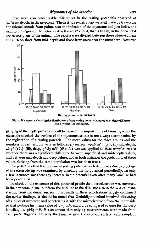
There were also considerable differences in the resting potentials observed atdifferent depths in the myotome. The first 322 penetrations were all made by traversingthe microelectrode from points near the inflexion of the myotome and just below theskin to the region of the notochord or the nerve chord, that is to say, in the horizontaltransverse plane of the animal. The results were divided between those observed nearthe surface, those from mid-depth and those from areas near the notochord. Accurate
50r
40
§ 30
9
6 20
10
o1-10 20 30 40 50 60 70 80
Superficial10 20 30 40 50 60 70 80
Mid-depth
Resting potential In millivolts
10 20 3040 50 60 70 80Deep
Fig. 4. Histogram* showing the distribution of 32a resting potentials recorded at three differentlevels within the myotome.
gauging of the depth proved difficult because of the impossibility of knowing when theelectrode touched the surface of the myotome, as this is not always accompanied bythe registration of a resting potential. The mean values for the three groups and thenumbers in each sample were as follows: (i) surface, 39*46 mV. (93); (ii) mid-depth,46-36 (161); (iii), deep, 56-85 mV. (68). A t test was applied to these samples to seewhether there was a significant difference between superficial and mid-depth values,and between mid-depth and deep values, and in both instances the probability of thesevalues deriving from the same population was less than o-ooi.
The possibility that the increase in resting potential with depth was due to blockageof the electrode tip was examined by checking the tip potential periodically. In onlya few instances was there any increase in tip potential even after many lamellae hadbeen penetrated.
To check on the existence of this potential profile the microelectrode was traversedin the horizontal plane, but from the mid line to the skin, and also in the vertical planestarting from the dorsal surface. The results of these penetrations largely confirmedthe earlier findings. It should be noted that Geduldig's method involved dissectingoff a piece of myotome and penetrating it with the microelectrode from the inner sideso that perhaps his mean value of 57-5 mV. should be compared to ours for the deeplamellae, i.e. 56-85 mV. His statement that only 15 measurements were made fromeach piece suggests that only the lamellae near the exposed surface were sampled.

408 D. M. GUTHRIE AND J. R. BANKS
These observations indicate that there is a potential profile across the myotome,and therefore these three groups mentioned earlier are presented in the form of ahistogram in Fig. 4.
(c)
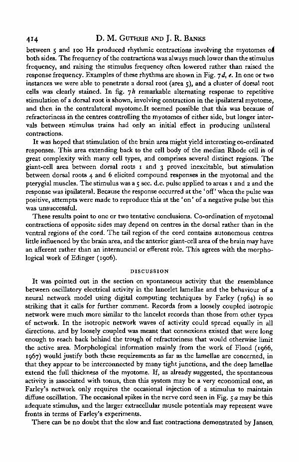
Fig. 5. Recordings of spontaneous potentials (a) extracellular, the rest intracellular. (a)Recordings from the nerve cord (upper trace), and an adjacent myotome (lower trace).Burstsof potentials in the nerve cord are reflected in the myotome, but many of the large myotomalpotentials are independent of activity[in the nerve cord. Mechanical stimulation at filled rectangleon the time trace. (6) Negative spontaneous potentials, (c) A drift from — 50 mv. to about10 mV. in the resting potential is accompanied by a reversal of polarity of the spontaneouspotentials at about —15 mv. The other trace shows tension, maximum downwards, (d)Positive spontaneous potentials. («) Spontaneous potentials at 30 % maximum resting potential.(/) Spontaneous potentials displayed as in (a). Horizontal line, zero potential. Resting level,52 mV. Time scales: (a) and (/) lsec., (b) 100 msec, (c) (d) and (e) 500 msec.
The question of the identification of the three types of lamellae with particularresting potential values must remain an open one because of the great range and varia-bility of these resting potentials. The potential gradient does not correspond in anydirect way with the distribution of the three types of lamella described by Flood.

Myotomes of the lancelet 409
foamage to the skin during dissection and osmotic effects accentuated by circulatingsea water may be associated with low resting potentials at the surface of the myotome,but cannot explain differences between the mid-depth and deep layers. It is clear,however, that the deep lamellae which constitute over 90 % of the muscle by volumeare likely to have a distribution of values similar to those of our sample.
Recordings made with fine wire electrodes from the nerve cord and the myotomesshowed that while muscle twitches were accompanied by bursts of potentials at bothrecording sites, large potentials occurred in the myotomes without any sign of neuralactivity. These spontaneous potentials occurred fairly regularly at rates of one every1-4 sec. The animals were lying in a completely relaxed or quiescent state. Deadlancelets are noticeably more flaccid than quiescent ones, and this may be associatedwith the loss of a maintained tonus. Some myotomes show a very high level ofspontaneous activity, others very little. A recording made with external leads is shownin Fig. 5 a.
Microelectrode penetrations of the lamellae immediately demonstrated trains ofspontaneous potentials at all levels within the myotome. These potentials varied inamplitude from 5 to 30 mV., and occurred in rhythmic bursts of 10 to 20 potentialsrepeated once every 1-5 sec. Within a burst the frequency of potentials was between10 and 20 per sec. The shape and polarity of these potentials varied considerably,and some of them are shown in Fig. 5 c. The rise time of a spontaneous potential wasbetween 10 and 20 msec. If intracellular and extracellular records are compared it canbe seen that the large extracellular potentials correspond to the bursts obtained withthe microelectrode, and if as shown in Fig. 5 / the intracellular recording is displayedin the same way as the external one the bursts appear as single large potentials.
The polarity of the spontaneous potentials varies with the resting potential level,reversing from positive to negative at about 60 % of the maximum resting potentialfor that fibre. Where there is a rapid drift in the resting potential this reversal can beseen clearly, as in Fig. 5 c. The magnitude of the spontaneous potentials does notdiminish appreciably as the potential level drifts from 50 mV. to 10 or 15 mV., butdeclines abruptly beyond this point. Near the reversal point the spontaneous potentialsmay take on a sinusoidal appearance, at least for short periods of time, as shown inFig. 5 c Farley (1964) has produced oscillograms of remarkable similarity to thesespontaneous potentials by a computer simulation technique in which various irregulari-ties are faithfully reproduced. The type of model that was used by Farley was a looselycoupled isotropic network, and its significance will be examined in the discussion.
Very small mechanical changes could sometimes be observed during particularlyintense electrical activity in the myotome, but they were too small for systematicstudy.
The spontaneous potentials in the muscles of Ascaris described by de Bell, delCastillo and Sanchez (1963) are generally similar to those observed in the lancelet.However, they occur at a slightly lower frequency and fall more clearly into smallpotentials (3-5 mV.), and spikes (15-30 mV.) which may have a positive overshoot asthe mean resting potential was only 29-4 mV. These authors were able to show thatthe spontaneous potentials originated in the region of muscle-tail terminals wherethe tight junctions occurred.
37 S X B J 3

410 D . M . GUTHRIE AND J. R. BANKS
Electrical responses
During a reflexly evoked twitch, extracellular recordings made with fine silver wireleads reveal a burst of small potentials in the myotome. Their highest frequencycorresponds roughly with peak tension in the muscle (Guthrie, 1967). Later workhas suggested that this may be an atrial stretch-receptor discharge, as the axons fromthese sense cells run in the adjacent myocommata, but it was not possible to pursuethis further at the time. A record of this kind of burst is shown in Fig. 5 a.
Fig. 6. Electrical and mechanical responses. The upper trace in (d) to (/) marks zero potentialand shows the stimulus pulse, the second trace shows the electrical response, the third tracethe mechanical response, (a) The fast response. (V) and (c) small potentials accompanying theslow component of a compound mechanical response, (d) to (/) The development of the secondfast spike, (g) Extracellular recording made during a compound mechanical response, {h) Ano-malous slow spike. Recording as (a) to (/), but second trace, stimulus trace. All time scales100 msec., except {g) 10 msec. Some spikes retouched.
If the recording electrode is a glass capillary with a tip diameter of 10-20 /i filledwith 3 M-KCl, a different kind of electrical response can be observed, and this is illustra-ted in Fig. 6g. At first sight it appears to be a reflexion of the mechanical changes

Myotomes of the lancelet 411
•during a normal contraction, but the initial rise precedes that of the mechanicalresponse by 10 msec., and the peak of the slow phase is 20-50 msec, in advance of thesimilar part of the slow twitch. An electrode with a diameter of 15 fi would be expectedto record potential changes in 15 or 20 lamellae, and the combined signal might besmoothed by secondary fluctuations through the tight junctions. This type of recordsuggests that during the normal biphasic contraction closely grouped large potentialsappear first in the lamellae followed by temporally dispersed small potentials.
The method employed for making intracellular recordings is indicated in Fig. 1,and has been mentioned in the section on resting potentials. The NaCl-filled micro-electrode was traversed slowly through the myotome, and a shock was applied to thenerve cord when a steady resting potential was obtained. Less than 20 % of satisfactoryimpalements produced a transient depolarization even when accompanied by amechanical response, and it seems probable that only a proportion of the lamellae areelectrically active during most contractions.
The electrical events that accompanied mechanical responses fell into four maincategories, (a) Large potentials with a total amplitude of 30-70 mV., and a durationof 10-15 msec. The largest may have a positive overshoot of 15-20 mv. They followthe stimulus artifact by about 10 msec, and precede the commencement of the fasttwitch by 10-15 msec. Examples are shown in Fig. 6 a, d-f. (b) Small simple potentialswith a value of 1-10 mV., and a duration of 10-15 msec. These potentials mainlyappear 70-90 msec, after the stimulus artifact, and between one and five may occurin response to a single shock. Their relation to the slow twitch is very variable. Theymay precede the commencement of the slow response by 10-40 msec., or may occurat any time during the rising phase of the contraction. These are illustrated in Fig. 6b,c. (c) Compound potentials. These, as can be seen in Fig. 6e are initial stages in theformation of a second spike-like response, but may be confused with class (b) poten-tials when they are small, (d) A rare category of potentials is occasionally observed atlow resting potentials, having the appearance of very slow spikes. An example isgiven in Fig. 6 A. The duration is that of a spontaneous potential—about 30 msec.
The most striking feature of the large class (a) potentials is that two of them mayoccur in response to a single shock separated by an interval of about 15 msec. If thecompound mechanical response which accompanies the double spike-like response isexamined it can be seen to belong to the type in which the slow phase is unusuallylarge, and is characterized by a shorter rise time than normal. This can be seen inFig. id. The slow phase in this case is associated with both class (a) and (b) electricalevents. The second spike-like response does differ from the first in that it is a gradedevent, rather than an all-or-nothing potential. The absence of a fast twitch in responseto the second spike does suggest that the fatigue so characteristic of the fast system isa property of the contractile mechanism rather than of the lamellar membrane orneuromuscular synapse.
Do these records present any information bearing on the function of the tight junc-tions? Those records, like the one in Fig. 6 b which show the small potentials, also showvery small deflexions with the latency characteristic of the fast or large potentials, aswell as other small deflexions in the 0-5-2 mV. range which may derive from adjacentlamellae. It is possible of course that even the class (b) events are taking place in otherlamellae and are merely reflected in an attenuated form in the lamellae from which

412 D . M. GUTHRIE AND J. R. BANKS
these records are taken. It might be suggested that the graded nature of the second(spike owes something to local reinforcement of the type occurring in Farley's (1964)neuromime.
Another point concerns latency and synchrony in the neuromuscular pathways.The earliest electrical responses in the lamellae occur 10 msec, after the stimulusartifact even when the tip of the recording electrode is very close to the nerve cord. Inthe section on mechanical responses it was suggested that conduction in the muscle-tail strand at the rate of 0-25 m./sec., so that a delay of 10 msec, if it was due solely toconduction through the muscle tails would require this pathway to be 2*5 mm. long.This indicates that nerve-cord stimulation is occurring via presynaptic elements, andas the electrodes were close to the neuromuscular synapse this delay can be largelyreferred to delay at this synapse. Incidentally, deBell et al. (1963) showed that conduc-tion in the muscle tails of Ascaris was three times slower (0-06 m./sec.) than in thelancelet. The delay in the appearance of the class (b) small potentials is perhapsassociated with Flood's (1966) finding that the muscle tails of the superficial lamellaeare approximately half the diameter (0-5 /i instead of i-o fi), and several times as long,as those of the deep lamellae. On the other hand the form of the dorsal and ventralneuromotor synapses may also be involved. Flood showed that the presynapticboutons opposite the thick muscle tails consisted of a single layer of large profilescontaining large vesicles, while the synaptic envelopes opposite the fine fibres werearranged in layers, were smaller, and contained smaller vesicles. The single-layersystem of synapses is clearly one that is likely to be associated with a rapid synchronousresponse, rather than with a delayed progressive type of response.
From the evidence presented here it appears likely that the fast twitch response isassociated with large potentials, the slow twitch with delayed small potentials.
Motor localization in the spinal cord
Pieces of lancelet were set up in a vertical position so that micro-electrodes filledwith the dye Fast Green (FCF) could be traversed into the spinal cord along itsmain axis, as shown in the diagram in Fig. ja, following the entry of an electrode intoan area where stimulation produced contractions, a steady outward current at 5-10 fiK.was passed until dye appeared at the stimulus site. The spot dye could be limited toan area 20-30 fi, across, and often stained small groups of cells. Owing to the verticalorientation of the preparation it was difficult to set up the myotomes for the recordingof mechanical responses, but this was done in a number of experiments.
For the purposes of this description the nerve cord was divided into five partscorresponding to the areas shown in the diagrammatic transverse section in Fig. 7 b.
In the pharyngeal region (the atriopore to somite 6) brief shocks applied to areas 1and 2 caused marked contractions in the myotome on the same side. Stimulationin areas 1, 2 or 3 never produced contalateral contractions. Most of these responsescould be described as local fast twitches at intensities just above threshold. At higherintensities the contraction spread to several adjacent myotomes. Stimulation withinarea 3 produced rather weak local myotomal twitches to isolated shocks, but shorttrains of stimuli at 5HZ sometimes provoked powerful complex contractions. D.c.pulses with a duration of 5 sec. often produced strong complex contractions at the 'off'.These complex responses involved the pterygial musculature which is believed to be

Myotomes of the lancelet 413
(connected with the median Rhode cell (Bone, i960), and this lies in area 3. Stimulationof areas 4 and 5 was not effective in this region.
Thepost-atrial region resembled the pharyngeal region as far as most areas were con-cerned, with the following exceptions. Stimulation within areas 1 and 2 at frequencies
t» ms Is
II1111IIIIII
te)
CO
-H- -H-t- - f - H -Fig. 7. Mechanical responses elicited by local stimulation within the nerve cord, (a) Arrange-ment of: the transducer arm, ta; microelectrode for stimulation, ms; indifferent electrode, is.(b) Areas of the nerve cord explored, (c-h). Responses to stimulation in the following areas:(c) Areas i and 2 in the pharyngeal region, (d) and (e) Areas i and 2 in the post-atrial region.(/) and (g) Areas 1 and 2 in the brain region (somites 1 to 6). (k) Area 5 in the post-atrialregion. Stimulus frequencies (d) 50 Hz, (e) 100 Hz, (A) Bursts of 100 Hz. All time markers inseconds.
414 D . M. GUTHRIE AND J. R. BANKS
between 5 and 100 Hz produced rhythmic contractions involving the myotomes o^both sides. The frequency of the contractions was always much lower than the stimulusfrequency, and raising the stimulus frequency often lowered rather than raised theresponse frequency. Examples of these rhythms are shown in Fig. id, e. In one or twoinstances we were able to penetrate a dorsal root (area 5), and a cluster of dorsal rootcells was clearly stained. In fig. 7 A remarkable alternating response to repetitivestimulation of a dorsal root is shown, involving contraction in the ipsilateral myotome,and then in the contralateral myotome.lt seemed possible that this was because ofrefractoriness in the centres controlling the myotomes of either side, but longer inter-vals between stimulus trains had only an initial effect in producing unilateralcontractions.
It was hoped that stimulation of the brain area might yield interesting co-ordinatedresponses. This area extending back to the cell body of the median Rhode cell is ofgreat complexity with many cell types, and comprises several distinct regions. Thegiant-cell area between dorsal roots 1 and 3 proved inexcitable, but stimulationbetween dorsal roots 4 and 6 elicited compound responses in the myotomal and thepterygial muscles. The stimulus was a 5 sec. d.c. pulse applied to areas 1 and 2 and theresponse was ipsilateral. Because the response occurred at the ' off' when the pulse waspositive, attempts were made to reproduce this at the 'on' of a negative pulse but thiswas unsuccessful.
These results point to one or two tentative conclusions. Co-ordination of myotomalcontractions of opposite sides may depend on centres in the dorsal rather than in theventral regions of the cord. The tail region of the cord contains autonomous centreslittle influenced by the brain area, and the anterior giant-cell area of the brain may havean afferent rather than an internuncial or efferent role. This agrees with the morpho-logical work of Edinger (1906).
DISCUSSION
It was pointed out in the section on spontaneous activity that the resemblancebetween oscillatory electrical activity in the lancelet lamellae and the behaviour of aneural network model using digital computing techniques by Farley (1964) is sostriking that it calls for further comment. Records from a loosely coupled isotropicnetwork were much more similar to the lancelet records than those from other typesof network. In the isotropic network waves of activity could spread equally in alldirections, and by loosely coupled was meant that connexions existed that were longenough to reach back behind the trough of refractoriness that would otherwise limitthe active area. Morphological information mainly from the work of Flood (1966,1967) would justify both these requirements as far as the lamellae are concerned, inthat they appear to be interconnected by many tight junctions, and the deep lamellaeextend the full thickness of the myotome. If, as already suggested, the spontaneousactivity is associated with tonus, then this system may be a very economical one, asFarley's network only requires the occasional injection of a stimulus to maintaindiffuse oscillation. The occasional spikes in the nerve cord seen in Fig. 5 a may be thisadequate stimulus, and the larger extracellular muscle potentials may represent wavefronts in terms of Farley's experiments.
There can be no doubt that the slow and fast contractions demonstrated by Jansea

Myotomes of the lancelet 415
et al. (1963) in the muscle fibres of the hagfish Myxme are similar to those we haveobserved in the lancelet. The small and large potentials which accompany these con-tractions are also similar, except that their amplitudes are larger in the hagfish,presumably in association with the higher resting potentials. Furthermore, we havenot observed the polarization plateau that repetitive stimulation produces in the slowfibres of the hagfish. Fast spikes are rather slower in the hagfish (10-15 msec, against7 msec.) and both slow and fast electrical responses had a recovery phase of 50-100 m-sec. which did not occur in Amphioxus. The tench deep musculature provided similarfast spikes to those of the lancelet (Barets, 1961). The graded slow response thatoccurs in teleosts like the catfish (Barets, 1961) presumably arose at a post-cyclostomephase of evolution. Muscle-tail rates of conduction of 0-25 m./sec. in the lanceletcompare with I-I m./sec. in Cottus (Hudson, 1969) and i-6 m./sec. in frog muscle(Eccles, Katz & Kuffler, 1941).
The fact that the muscle lamellae of the lancelet are like those of Ascaris in havinga high level of rhythmic spontaneous activity has been mentioned earlier. Some furtherpoints of similarity are that, as in the lancelet, spikes evoked by nerve-cord stimulationare of much shorter duration than spontaneous potentials, and shocks of high intensitymay produce more than one spike. As many as three spikes may originate in this wayin the nematode, but they arise from a depolarization plateau which is not the case inAmphioxus. Large spikes in Ascaris are followed by a gradual repolarization phase notfound in the lancelet or the tench, but these spikes do decline in amplitude with distancefrom the nerve cord and we found evidence for this in the lancelet. However, we didnot observe the prolonged refractoriness of up to two seconds demonstrated bydeBell et al. (1963) in the nematode muscles.
Attempts to equate the superficial lamellae with the slow twitch by examiningmechanical responses from a myotome from which the outer layer had been removedgave ambiguous results, and this question must be regarded as open despite evidencesupporting this correlation.
Recordings from the spinal cord during slow swimming (unpublished data) dosuggest that the Rhode cell system is involved in slow swimming as well as in largefast contractions (Guthrie, 1967). The stimulus frequencies for the onset of summa-tion in the slow and fast system (2 Hz and 15 Hz) accord very well with the frequencyof contractions in slow-swimming and fast-swimming lancelets examined by cine-photography; at the same time the slow twitch system appears too weak to flex thebody through 900 as occurs during slow swimming.
A final point of comparison lies with the echinoderms. Cobb & Laverack (1966) wereable to produce separate slow and fast contractions from the lantern retractor muscleof Echinus by local stimulation of the nervous system. The slow contraction wasdescribed by these authors as a delayed response. In Astropecten muscle fibres withmuscle tails were found.
The results discussed in this paper help to reinforce the belief that in the lanceletwe see a remarkable mosaic of vertebrate and invertebrate features.

416 D. M. GUTHRIE AND J. R. BANKS
SUMMARY
1. Two basic types of mechanical response can be elicited from the lanceletmyotome by stimulation of the nerve cord by single shocks: a fast twitch with a risetime of 70-80 msec, and a slow twitch/with a rise time of 200-250 msec.
2. The two twitch components can easily be elicited together to produce a biphasiccontraction. A second type of biphasic contraction occurs more rarely in which theslow phase has a faster rise time and a higher peak tension.
3. The mean of all resting potentials was 48-94 mV. ± 15-5 mV. The commonestvalues were near 50 mV. and this must refer to the deep lamellae which form the bulkof the myotome.
4. Spontaneous potentials are a striking feature of the lancelet muscles.5. The fast twitch is accompanied by an all-or-nothing, spike-like potential of
between 30 and 70 mV. The slow twitch is accompanied by one or more potentials ofbetween 1 and 10 mV. The type-2 biphasic mechanical response mentioned above isaccompanied by two large potentials which attain similar amplitude, but differ inthat the second is a graded response.
6. Some of the features of lancelet myotomal muscle have also been described incyclostomes, nematodes and echinoderms.
We should like to extend our warmest gratitude to Dr P. N. R. Usherwood of theUniversity of Glasgow for reading and criticising the MS. of this paper. Any errorsthat remain are solely attributable to us. Dr R. C. L. Hudson of the Gatty Laboratoryhas also been most helpful especially in bringing references to our notice. The MarineBiological Association Laboratory at Plymouth, and in particular Mr R. Tozer, havecontinued their invaluable assistance. We wish, furthermore to record our indebtednessto the Science Research Council for generous financial assistance both for apparatusand for a maintenance grant to one of us.
REFERENCES
BARETS, A. (1961). Contribution a l'itude des systemes moteur lent et rapide du muscle lateral dest&eosteens. Archs. Anat. microsc. Morph. exp. 50, 91-187.
DE BELL, J. T., DEL CASTILLO, J. and SANCHEZ, V. (1963). The electrophysiology of the muscle cellsof Ascaris lumbricoides. J. cell. comp. Phsyiol. 6a, suppl., 159-77.
DE BELL, J. T., DEL CASTILLO, J. & MORALES, T. (1967). The initiation of action potentials in the somaticmusculature of Ascaris lumbricoides J. exp. Biol. 46, 263-80.
BONE, Q. (i960). The central nervous system of Amphioxus. J. comp. Neurol. 115, 27-62.BONE, Q. (1966). On the function of the two types of myotomal muscle fibre in fish. J. mar. biol. Au.
U.K. 46, 321-49.COBB, J. S. & LAVERACK, M. S. (1966). The lantern of Echinus. Proc. Roy. Soc. Lend. B 164, 624-40.ECCLES, J. C, KATZ, B. & KUFFLER, W. S. (1941). The nature of the endplate in curarized muscle.
J. Neuropkysiol. 4, 362—87.EDENGER, A. (1906). Uber das Gehim von Amphioxus. Anat. Anz. 38, 211-16.FARLEY, B. G. (1964). The use of computer technics in neural research. In Neural Theory and Modelling.
Ed. R. F. Reiss. Stanford University Press.FLOOD, P. (1966). A peculiar mode of muscular innervation in Amphioxus. J. Comp. Neurol. 136, 181-
217.FLOOD, P. (1967). The structure of the segmental trunk muscle in Amphioxus. Z. Zellforsch. mikrosk.
Anat. 84, 380-416.GEDULDIG, D. S. (1965). Resting potential dependency on external potassium in Amphioxus muscle. Am.
J. Physiol. 308, 852-4.

Myotomes of the lancelet 417GUTHRIE, D. M. (1967). Control of muscular contractions by spinal neurones in Amphioxus (BranMo-
stoma lanceolatum). Nature, Land. 216, 5121, 1224-5.GUTHRIE, D. M. & BANKS, J. R. (1970). Observations on the function and physiological properties of a
fast paramyosin muscle—the notochord of Amphioxus (BranMostoma lanceolatum). J. exp. Biol.52, 125-38.
HUDSON, R. C. L. (1969). Polyneuronal innervation of the fast muscles of the marine teleost Cottustcorpius L. J. exp. Biol. 50, 47-67.
JANSKN, P., ANDERSEN, J. K. S. & LOYNTNG, Y. (1963). Slow and fast muscles in the atlanric hagfish.Acta pkysiol. tcand. 57, 167-79.
ROBERTS, B. L. (1969). The spinal nerves of the dogfish (Scyliorhima). J. mar. biol. Ass. UJC. 49, 51-75-
USHERWOOD, P. N. R. & MACHILI, P. (1968). Pharmacological properties of excitatory neuromuscularsynapses in the locust. J. exp. Biol. 49, 341-61.