observations on the activation of chronic compensated hydrocephalus · pdf file ·...
TRANSCRIPT

Copyright © 2012 Journal of Korean Neurotraumatology Society 139
CLINICAL ARTICLEKorean J Neurotrauma 2012;8:139-145 ISSN 2234-8999
Introduction
There is no nomenclature consensus in describing pa-tients with chronic form of exceptionally enlarged ventri-cles and no overt sign of increased intracranial pressure (ICP). There is a broad spectrum of compensated hydro-cephalus and the underlying cause of this form of chronic compensated hydrocephalus has been questioned. Little at-tention has given to this entity and not so many studies have
been made so far. There are confusing terms used to de-scribe these conditions such as the syndrome of hydroceph-alus in young and middle-aged adults (SHYMA), late-onset idiopathic aqueductal stenosis (LIAS), long-standing overt ventriculomegaly (LOVA) of the adult, and late-onset aque-ductal stenosis (AS).3,5,13) Some authors proposed that LOVA must be considered as a special form of LIAS.8)
Cerebrospinal fluid (CSF) dynamics may change over time between active and inactive states in these entities but the reason for late decompensation remains obscure. How-ever, a new event in the central nervous system (CNS) may offset the compensatory mechanism in some patients. How to treat these patients is also a very controversial issue.10) Ventriculoperitoneal shunt (VPS) with adjustable valve or endoscopic third ventriculostomy (ETV) is generally se-lected. The authors illustrate their experiences and review
Observations on the Activation of Chronic Compensated Hydrocephalus in Adult Patients
Se-Il Jeon, MD, Dae-Hee Seo, MD, PhD, Young Sub Kwon, MD, Il-Seung Choe, MD, PhD and Sung-Choon Park, MD, PhDDepartment of Neurosurgery, Myongji Hospital, Kwandong University College of Medicine, Goyang, Korea
Objective: There is a broad spectrum of compensated hydrocephalus. Various terms such as long-standing overt ventricu-lomegaly in adult (LOVA) has been coined, however, even such terms leave diverse aspect of this condition out of account. We have experienced compensated hydrocephalus cases which were considered to be activated after a long time period of quiescent state, and tried to compare their clinical characteristics with the relatively well described entity of LOVA.Methods: We conducted a retrospective review of 206 patients who underwent ventriculoperitoneal shunt (VPS) between February 2001 and May 2012. Of these, 6 patients had chronic compensated hydrocephalus. The clinical and radiological characteristics are evaluated.Results: Definite triventriculomegaly was observed in two patients. Macrocephaly was observed in two cases, one with aq-ueductal stenosis (AS), the other with unknown status of aqueduct. All of the cases with triventriculomegaly were normoce-phalic. Spinal causes were thought as aggravating factor in two. Two endoscopic third ventriculostomy and eight VPS were performed in five patients. Four patients responded well but one took a very complicated course.Conclusion: The relationships between macrocephaly, triventriculomegaly, and AS suggested in other studies were incon-sistent. Blockage or narrowing of cerebrospinal fluid pathways were observed at various sites. Disturbances of spinal arach-noid pathways were related to the activation in some cases. Treatment is to be tailored individually considering various reig-niting event. It is suggested that this entity is to be evaluated for better nomenclature reflecting diverse aspects of this condition. Further study is needed to elucidate underlying pathophysiology and effective management.
(Korean J Neurotrauma 2012;8:139-145)
KEY WORDS: Hydrocephalus ㆍLong-standing overt ventriculomegaly in adult ㆍMacrocephaly ㆍAqueductal stenosis.
Received: September 3, 2012 / Revised: September 22, 2012Accepted: September 23, 2012Address for correspondence: Dae-Hee Seo, MD, PhDDepartment of Neurosurgery, Myongji Hospital, Kwandong Uni-versity College of Medicine, 697-24 Hwajeong-dong, Deogyang-gu, Goyang 412-270, KoreaTel: +82-31-810-5443, Fax: +82-31-810-5109E-mail: [email protected]
online © ML Comm

140 J Korean Neurotraumatol Soc 2012;8:139-145
Activation of Compensated Hydrocephalus in Adult
the literature.
Materials and Methods
The authors conducted a retrospective review of 206 pa-tients who underwent VPS for hydrocephalus between Feb-ruary 2001 and May 2012. Of these, 6 patients had chronic compensated hydrocephalus. Chronic compensated hydro-cephalus was defined as a case that has exceptionally en-larged ventricles with relatively well identified sulci and a significantly enlarged or destroyed sella turcica, and/or macrocephaly. The frontal horn size and biparietal diameter (BPD) were checked to figure out the Evan’s ratio (ratio of frontal horn to maximal BPD) in all patients. Dimensional change & contour of sella turcica were examined using skull X-rays or scout film of computed tomography (CT). Macrocephaly was diagnosed when head circumference was measuring more than two standard deviations. The presence of triventriculomegaly and AS were checked on imaging studies. AS was defined as absence of the whole length of the aqueduct on midsagittal T1- and T2-weighted magnetic resonance imaging (MRI) images. Spine imaging was performed for patients who had symptoms or possibili-ty of spine lesions.
Results
There were 91 women and 115 men whose age ranged from 20 to 78 years (58.9±13.1). The common cause of hy-drocephalus was subarachnoid hemorrhage (SAH), trau-ma, intracerebral hemorrhage in order. The gravity assist-ed valve (GAV) valve (Aesculap-Miethke, Tuttlingen/Postdam, Germany) was used in 82 patients (39.8%). Pro-grammable valves were placed using ProGAV (Aesculap-Miethke, Tuttlingen/Postdam, Germany) in 38 (18.4%), Codman Hakim programmable valve (CHPV, Codman, Johnson & Johnson Co., Raynham, MA) in 37 (18%), and Strata valve (Medtronic Neurosurgery, Goleta, CA) in 12 patients (5.8%). Various pressure regulated valves such as Pudenz (Heyer-Schulte Pudenz Flushing valves, IntegraTM, Plainsboro, NJ) or Novus valves (NovusTM valve system, IntegraTM, Plainsboro, NJ) were used in 37 patients (18%).
The frontal horn size was 52.0±9.9 mm, BPD was 131.1± 11.4 mm, and the average of Evan’s ratio was 0.42±0.30 (mean±standard deviation). Six cases of chronic form of marked ventriculomegaly including one nonoperative case were identified (Figure 1). And their mean frontal horn size was 61.6±8.2 mm, and mean BPD was 138.7±10.3 mm. The average Evan’s ratio of these patients was 0.44±0.05.
Mean age was 47.8 years old (range: 23-66) and male to female ratio was 2 : 1. Mean follow-up period was 12 months (range: 2-24). Two ETV and eight VPS operation were performed in five patients. Four patients had good results but one patient took a very complicated clinical course and beyond remedy. The relationships between macrocephaly, triventriculomegaly, and AS were not constant. Definite AS was observed only in 1 case. Other 2 cases had narrow-ing distal to the aqueduct itself. Triventriculomegaly was observed in two patients. Macrocephaly was observed in two cases, one with AS, the other with unknown status of aqueduct. It was unable to evaluate the status of aqueduct because MRI had not been done in this case. All of the cases with triventriculomegaly were normocephalic. Spinal causes were thought as aggravating factor in two. Spinal causality was suspected in another one. Cases were summarized in Table 1.
Illustrative cases
Case 2A 43-year-old male driver presented with chronic head-
ache. After workup, a pressure regulated valve was placed. At the age of 46 and 48, he underwent operations at other hospital for spinal arachnoid cyst. At the age of 52, he col-lapsed during bathing and was brought to the emergency room (ER). CT scan showed ventriculomegaly and Evan’s ratio was increased to 0.42. Spine MRI showed persistent long segmental (T10-L2) arachnoid cyst and severe arach-noid adhesion on magnetic resonance myelography (Fig-ure 2A). ETV was thought to be inadequate to perform in this case. Shunt was revised with GAV. The patient’s con-dition improved rapidly and postoperative Evan’s ratio de-creased to 0.37. Six months later, he lost his consciousness during driving and developed progressive mutism, urinary incontinence. CSF was aspirated by shunt puncture. There was no evidence of shunt infection and the patient’s condi-tion improved after aspiration. It was decided to change GAV to ProGAV with concerns about underdrainage of GAV that have been reported.6,7) The mental status deteriorat-ed unexpectedly first postoperative day and follow-up CT showed severe ventriculomegaly. Emergent external ven-tricular drainage (EVD) was placed on left side and then the patient’s neurological condition recovered. It was planned to check distal catheter in the suspicion of problem in the peritoneal absorptive capability and found there was very se-vere adhesion in the peritoneal cavity. Omental adhesiolysis and repositioning of distal catheter were done. EVD was changed to low pressure regulated valve and the program-

www.neurotrauma.or.kr 141
Se-Il Jeon, et al.
mable valve pressure was down regulated, but it was useless for symptom improvement. MRI taken before ETV showed AS, sellar enlargement, upwardly stretched corpus callosum and periventricular edema (Figure 2B). As a last resort, ETV was performed and the ProGAV was removed (Figure 2C). Unexpectedly, third ventricular floor was very narrow and the membrane was relatively thick. Compliance loss due to repeated operation and repetitive deteriorations was consid-ered to be the cause of incomplete recovery and very compli-cated clinical course. He showed apathy and developed fa-cial palsy and tremor later.
Case 3A 61-year-old woman with normal intelligence, normo-
cephaly visited ER. A few months ago, she was diagnosed having an unruptured aneurysm and scheduled to emboli-zation. At that time, brain MRI showed diffuse ventriculo-megaly. Brain CT taken at ER showed SAH and coil embo-lization was performed. During intensive care, she becomes drowsy and right upper extremity weakness developed. Lumbar puncture (LP) showed elevated pressure (22 cmH2O) and CSF drainage made full neurological recovery. A few days later, right hemiparesis with left lower extremity weak-ness developed. Imaging study showed no ischemic lesion. Postoperative day 16, EVD was placed. After EVD place-ment, upper extremity weakness was improved but para-paresis persisted. Due to septicemia, EVD was removed seven days later and intravenous antibiotics were used. Af-ter general ward transfer, lower extremity weakness per-sisted and cognitive impairment was observed (Mini-men-tal state examination 11/30). Lumbar drainage improved neurological condition temporarily. Postictus day 61, a VPS (ProGAV) was placed. After shunt placement the patient’s neurological condition improved remarkably. She com-plained back pain during rehabilitation. Pyogenic spondylo-discitis due to septicemia was found on further examination. Continuous antibiotic irrigation was done and intravenous antibiotics were used. Diagnosis of spinal infection was somewhat delayed and it might be another aggravating fac-tor of CSF absorption disturbance.
She made a near complete recovery and during follow-up, she developed Parkinsonism such as festinating gait, mag-netic gait, and difficulty in turning around 14 months after shunt placement. Adjustment of shunt valve pressure made a remarkable improvement within two weeks.
Case 4 A 65-year-old woman visited ER complaining parapare-
sis, vomiting and voiding difficulty. She had neck pain for a
TAB
LE 1
. Sum
mar
y of
cas
es
Cas
eN
oSe
x/A
gePH
xC
hron
icIC
P Sx
Mac
ro-
ceph
aly
IQSe
llar
enla
rgem
ent
Triv
entri
-cu
lom
egal
yA
S2n
d h
itO
pera
tion
Follo
w-u
p( m
onth
s)m
RS
1M
/29
Men
ingi
tis
at 3
YO
Non
eY
Subn
orm
alY
±N
AED
HC
rani
otom
y24
0
2M
/43
Non
eHe
adac
heN
N
orm
alY
Y Y
Spin
al o
pera
tion,
omen
tal a
dhe
sion
Pr-v
alve
, spi
nal o
p,
GA
V, P
ro-G
AV+
Pude
nz,
ETV
+pr
o-G
AV
rem
oval
54
3F/
61N
one
Non
eN
N
orm
alY
N
N
SAH,
spin
al In
fect
ion
(?)
Embo
lizat
ion,
pro
-GA
V,
spin
al o
p16
1
4F/
65N
one
Non
eN
N
orm
alY
Y ±
Non
traum
atic
spin
al
hem
orrh
age
GA
V15
0
5M
/23
Non
eHe
adac
he,
naus
eaN
N
orm
al±
N
N
NA
ETV
, GA
V2
0
6M
/66
Non
eN
one
Y N
orm
alY
N
N
NA
Pro-
GA
V10
0
AS:
aqu
educ
tal s
teno
sis, E
DH:
epid
ural
hem
atom
a, E
TV:
end
osco
pic
third
ven
tricu
lost
omy,
F:
fem
ale,
GA
V:
grav
ity a
ssist
ed v
alve
, IC
P: in
tracr
ania
l pre
ssur
e, IQ
: in
tellig
ence
quo
-tie
nt, M
: m
ale,
mRS
: m
odifi
ed R
anki
n sc
ore,
N:
no, N
A:
not a
vaila
ble,
No:
num
ber,
op:
oper
atio
n, P
Hx:
past
hist
ory,
pr-v
alve
: pr
essu
re re
gula
ted
val
ve, P
roG
AV
: pr
ogra
mm
able
G
AV
, Sx:
sym
ptom
, Y:
yes,
YO: y
ears
old
, ±: i
ndefi
nite
, SA
H : s
ubar
achn
oid
hem
orrh
age

142 J Korean Neurotraumatol Soc 2012;8:139-145
Activation of Compensated Hydrocephalus in Adult
Case 1 Case 2 Case 3
Case 4 Case 5 Case 6
FIGURE 1. CT/MRI axial scans demonstrating ventriculomegaly with relatively well- identified sulci. Periventricular edema is ob-served in case 4 which suggest acute aggravation of quiescent long-standing hydrocephlalic process. CT: computed tomography, MRI: magnetic resonance imaging.
FIGURE 2. T2-weighted sagittal MRI showing persistent T10-L2 segmental arachnoid cyst (A), MRI taken before ETV show AS, sellar enlargement, upwardly stretched corpus callosum and periventricular edema (B), ETV was performed and the fenestration was enlarged using a 3-French Fogarty balloon catheter. Unexpectedly, third ventricular floor was very narrow and the membrane was thick (C, arrow). AS: aqueductal stenosis, ETV: endoscopic third ventriculostomy, MRI: magnetic resonance imaging.
C
A B
www.neurotrauma.or.kr 143
Se-Il Jeon, et al.
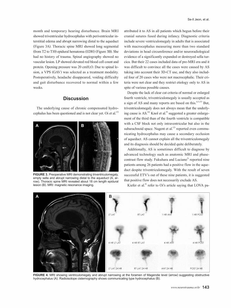
month and temporary hearing disturbance. Brain MRI showed triventricular hydrocephalus with periventricular in-terstitial edema and abrupt narrowing distal to the aqueduct (Figure 3A). Thoracic spine MRI showed long segmental (from T2 to T10) epidural hematoma (EDH) (Figure 3B). She had no history of trauma. Spinal angiography showed no vascular lesion. LP showed elevated red blood cell count and protein. Opening pressure was 20 cmH2O. Due to spinal le-sion, a VPS (GAV) was selected as a treatment modality. Postoperatively, headache disappeared, voiding difficulty and gait disturbance recovered to normal within a few weeks.
Discussion
The underlying cause of chronic compensated hydro-cephalus has been questioned and is not clear yet. Oi et al.13)
attributed it to AS in all patients which begun before their cranial sutures fused during infancy. Diagnostic criteria include severe ventriculomegaly in adults that is associated with macrocephalus measuring more than two standard deviations in head circumference and/or neuroradiological evidence of a significantly expanded or destroyed sella tur-cica. But their 22 cases included data of pre-MRI era and it was difficult to convince all the cases were caused by AS taking into account their 3D-CT use, and they also includ-ed four of 20 cases who were not macrocephalic. Their cri-teria were not clear and they restrict etiology only to AS in spite of various possible causes.
Despite the lack of clear cut criteria of normal or enlarged fourth ventricle, triventriculomegaly is usually accepted as a sign of AS and many reports are based on this.4,5,15) But, triventriculomegaly does not always mean that the underly-ing cause is AS.16) Knol et al.9) suggested a greater enlarge-ment of the third than of the fourth ventricle is compatible with a CSF block not only intraventricular but also in the subarachnoid space. Nugent et al.12) reported even commu-nicating hydrocephalus may cause a secondary occlusion of aqueduct. AS cannot explain all the triventriculomegaly and its diagnosis should be decided quite deliberately.
Additionally, AS is sometimes difficult to diagnose by advanced technology such as anatomic MRI and phase-contrast flow study. Fukuhara and Luciano5) reported nine patients among 26 patients had a positive flow in the aque-duct despite triventriculomegaly. With the result of seven successful ETV’s out of these nine patients, it is suggested that positive flow does not necessarily exclude AS.
Kiefer et al.8) refer to Oi’s article saying that LOVA pa-
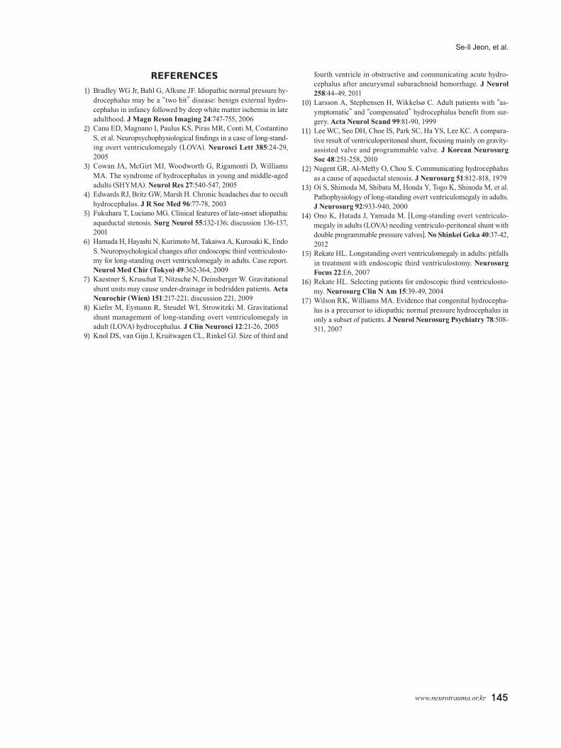
FIGURE 4. MRI showing ventriculomegaly and abrupt narrowing at the foramen of Magendie level (arrow) suggesting obstructive hydrocephalus (A). Radioisotope cisternography shows communicating type hydrocephalus (B).
A B
LT LAT RT LAT 1 HR ANT 1 HR POST
4 HR LT LAT 4 HR RT LAT 4 HR ANT 4 HR POST
LT LAT 24 HR RT LAT 24 HR ANT 24 HR POST 24 HR
FIGURE 3. Preoperative MRI demonstrating triventriculomegaly, empty sella and abrupt narrowing distal to the aqueduct (A, ar-row). Thoracic spine MRI revealed about 18 cm length epidural lesion (B). MRI: magnetic resonance imaging.
A B

144 J Korean Neurotraumatol Soc 2012;8:139-145
Activation of Compensated Hydrocephalus in Adult
tients are usually macrocephlic and diagnosis needs clinical or radiological signs of a long-standing increase in ICP. As Oi’s study include three normocephalic and one unknown status patients as stated above, the authors questioned mac-rocephaly may not an absolutely necessary condition to di-agnose this entity and tried to find the real incidence in this population. In similar context, Wilson and Williams17) re-ported that a significantly larger proportion of patients with idiopathic normal pressure hydrocephalus (iNPH) have a head circumference greater than the 90th or 97th percentile than normal population. But they concluded that compen-sated congenital hydrocephalus is just one etiology of iNPH but not all.
Our cases make it clear that macrocephaly may not be an essential condition for diagnosis of LOVA and diagnostic criteria of this condition should be clarified more. With this finding and the past history of meningitis at three years old also suggest the onset age cannot be limited to infancy be-fore closure of calvarial suture.
The term of LOVA, rather than SHYMA of LIAS, is used in this paper: there were patients in old age, and presence of AS is inconsistent in our series. But when using the term LOVA in a more widened meaning, not in the sense that the Oi et al.13) previously defined but verbatim without men-tion of causality, AS as a sole causality and macrocephly as a diagnostic criteria should be reevaluated. As previously stat-ed, definite AS was observed only in 1 case and other 2 cas-es had narrowings distal to the aqueduct itself in this study (Figure 3A, 4).
Sellar turcica widening means previous hydrocephalic process which was arrested or radiological evidence of long-standing increased ICP. Our cases showed sellar widening in all as expected.
Others reported chronic headache as a symptom of long-standing increased ICP.4,15) But, there were only two symp-tomatic cases of long-standing increased ICP, and other cases were asymptomatic before activation.
CSF dynamics may change over time between active and inactive states but the reason for activation of once-arrested hydrocephalus remains unclear. A two-hit disease theory of Bradley may be applied to this condition.1) In terms of case 1, EDH as a second hit does not cause activation because it might not cause the CSF blockage. The chronic increased ICP symptom was tried to be cured by shunt and effective for a long time in case 2, but further progression was sup-posed to be caused by CSF absorption disturbance due to omental adhesion and spinal problem with loss of compli-ance. SAH caused activation in case 3 and spinal hemor-rhage activated the process in case 4, and shunt operation
restablized the hydrocephalic process. The cause of aggra-vation in case 5 was not obvious. Considering case 2, case 4, and possibly case 3 the spinal lesion should be suspected as a possible disturbing source and evaluated accurately.
It remains to be determined how to monitor and decide when to operate patients with incidental, compensated hy-drocephalus to prevent irreversible neurological deteriora-tion while simultaneously avoiding premature or even un-necessary treatment. Opinions are divided on the matter of treatment. VPS with adjustable valve or ETV is generally selected. Some cases are very difficult to treat because of compliance loss or other reasons.14,15) Some authors recom-mend third ventriculostomy as a treatment of choice.5) But, the result of ETV was not satisfactory in other series.15) If the initiating event causes blockage in the once-reestablished pathways such as subarachnoid space hemorrhage, it may be inappropriate to contemplate ETV in these patients. In these cases, VPS would be an appropriate selection. Inappropriate selection of treatment modality without deliberate consider-ation of relevant factors may lead to surgical failure or even deterioration.
The limitation of this study is small number of patients firstly. Secondly, detailed evaluation of cognitive function sug-gested by some authors was done only in two patients.2,6,15) Thirdly, relevant studies were not sufficiently done such as cine phase-contrast MRI despite there is insufficient evidence to support the value in predicting response to shunting.
Conclusion
The relationships between macrocephaly, triventriculo-megaly, and AS suggested in other studies were not consis-tent in this study. Dimensional and configurational chang-es of sella turcica were observed constantly. In contrast to the attributing AS itself as a sole causality, blockage or narrowing of CSF pathways were observed at various sites. Disturbances of spinal arachnoid pathways were related to the activation in some cases. A new event in the CNS may initiate activation of quiescent hydrocephalic process in some patients, therefore selection of treatment modality is to be tailored individually considering these factors. It is suggested that this entity is to be evaluated for better no-menclature reflecting diverse aspects of this condition. Further study is needed to elucidate underlying pathophys-iology and effective management.
■ The authors have no financial conflicts of interest.
www.neurotrauma.or.kr 145
Se-Il Jeon, et al.
REFERENCES1) Bradley WG Jr, Bahl G, Alksne JF. Idiopathic normal pressure hy-
drocephalus may be a “two hit” disease: benign external hydro-cephalus in infancy followed by deep white matter ischemia in late adulthood. J Magn Reson Imaging 24:747-755, 2006
2) Canu ED, Magnano I, Paulus KS, Piras MR, Conti M, Costantino S, et al. Neuropsychophysiological findings in a case of long-stand-ing overt ventriculomegaly (LOVA). Neurosci Lett 385:24-29, 2005
3) Cowan JA, McGirt MJ, Woodworth G, Rigamonti D, Williams MA. The syndrome of hydrocephalus in young and middle-aged adults (SHYMA). Neurol Res 27:540-547, 2005
4) Edwards RJ, Britz GW, Marsh H. Chronic headaches due to occult hydrocephalus. J R Soc Med 96:77-78, 2003
5) Fukuhara T, Luciano MG. Clinical features of late-onset idiopathic aqueductal stenosis. Surg Neurol 55:132-136; discussion 136-137, 2001
6) Hamada H, Hayashi N, Kurimoto M, Takaiwa A, Kurosaki K, Endo S. Neuropsychological changes after endoscopic third ventriculosto-my for long-standing overt ventriculomegaly in adults. Case report. Neurol Med Chir (Tokyo) 49:362-364, 2009
7) Kaestner S, Kruschat T, Nitzsche N, Deinsberger W. Gravitational shunt units may cause under-drainage in bedridden patients. Acta Neurochir (Wien) 151:217-221; discussion 221, 2009
8) Kiefer M, Eymann R, Steudel WI, Strowitzki M. Gravitational shunt management of long-standing overt ventriculomegaly in adult (LOVA) hydrocephalus. J Clin Neurosci 12:21-26, 2005
9) Knol DS, van Gijn J, Kruitwagen CL, Rinkel GJ. Size of third and
fourth ventricle in obstructive and communicating acute hydro-cephalus after aneurysmal subarachnoid hemorrhage. J Neurol 258:44-49, 2011
10)Larsson A, Stephensen H, Wikkelsø C. Adult patients with “as-ymptomatic” and “compensated” hydrocephalus benefit from sur-gery. Acta Neurol Scand 99:81-90, 1999
11) Lee WC, Seo DH, Choe IS, Park SC, Ha YS, Lee KC. A compara-tive result of ventriculoperitoneal shunt, focusing mainly on gravity-assisted valve and programmable valve. J Korean Neurosurg Soc 48:251-258, 2010
12)Nugent GR, Al-Mefty O, Chou S. Communicating hydrocephalus as a cause of aqueductal stenosis. J Neurosurg 51:812-818, 1979
13)Oi S, Shimoda M, Shibata M, Honda Y, Togo K, Shinoda M, et al. Pathophysiology of long-standing overt ventriculomegaly in adults. J Neurosurg 92:933-940, 2000
14)Ono K, Hatada J, Yamada M. [Long-standing overt ventriculo-megaly in adults (LOVA) needing ventriculo-peritoneal shunt with double programmable pressure valves]. No Shinkei Geka 40:37-42, 2012
15)Rekate HL. Longstanding overt ventriculomegaly in adults: pitfalls in treatment with endoscopic third ventriculostomy. Neurosurg Focus 22:E6, 2007
16)Rekate HL. Selecting patients for endoscopic third ventriculosto-my. Neurosurg Clin N Am 15:39-49, 2004
17)Wilson RK, Williams MA. Evidence that congenital hydrocepha-lus is a precursor to idiopathic normal pressure hydrocephalus in only a subset of patients. J Neurol Neurosurg Psychiatry 78:508-511, 2007