observation of backaction and self-induced trapping in a ... · observation of backaction and...
TRANSCRIPT

Observation of Backaction and Self-Induced Trappingin a Planar Hollow Photonic Crystal Cavity
Nicolas Descharmes, Ulagalandha Perumal Dharanipathy, Zhaolu Diao, Mario Tonin, and Romuald Houdre
Institut de Physique de la Matiere Condensee, Ecole Polytechnique Federale de Lausanne (EPFL), Station 3,CH-1015 Lausanne, Switzerland
(Received 20 December 2012; published 20 March 2013)
The optomechanical coupling between a resonant optical field and a nanoparticle through trapping
forces is demonstrated. Resonant optical trapping, when achieved in a hollow photonic crystal cavity is
accompanied by cavity backaction effects that result from two mechanisms. First, the effect of the particle
on the resonant field is measured as a shift in the cavity eigenfrequency. Second, the effect of the resonant
field on the particle is shown as a wavelength-dependent trapping strength. The existence of two distinct
trapping regimes, intrinsically particle specific, is also revealed. Long optical trapping (> 10 min ) of
500 nm dielectric particles is achieved with very low intracavity powers (< 120 �W).
DOI: 10.1103/PhysRevLett.110.123601 PACS numbers: 42.50.Wk, 42.60.Da, 42.70.Qs, 87.80.Cc
Recent experiments have widely studied the couplingbetween a resonant optical field and single atoms [1,2] ormicromechanical resonators [3,4]. However, the interac-tion between the mechanics of a large isolated nanoscaleparticle and a high finesse optical cavity, though theoreti-cally predicted [5–8], is yet to be reported. Quantitativeinvestigation of this optomechanical interaction requiresthe use of cavities with high quality factors (Q) and largefield gradients. In this context, it has been suggested[6,9,10] that photonic crystal (PhC) cavities tailored withan appropriate mode configuration would qualify as excel-lent candidates. However, the inherent nature of light toconcentrate in the high refractive index medium results inthe fact that the trapped particle interacts only with alimited fraction of the optical field. As a first consequence,input laser powers required are in the order of tens or evenhundreds of milliwatts. Nevertheless it has been predictedas early as 2006 that sub-mW trapping of 100 nm sizedparticles should be achievable [9]. The second conse-quence is that the cavity mode is weakly perturbed bythe presence of a particle in its vicinity making standardPhC cavities unsuitable for noticeable backaction effects.To circumvent this issue, it is possible to design dielectricmicrocavities with a significant field overlap on the lowindex medium, which we will refer to as hollow opticalcavities [11–13] in analogy with the hollow core fiberterminology. In such cavities, it is expected that theenhanced field overlap can simultaneously allow for quan-tification of the particle perturbation on the cavity modeand the observation of backaction [3,14] phenomena.
In this work, the backaction between a resonant field in aplanar photonic crystal cavity and a single dielectric nano-particle through optical gradient forces is clearly evidencedfor the first time. This phenomenon, where the motion ofthe center of mass of the particle and the cavity field aremutually coupled, derives from two classical mechanisms.The first one is the existence of strong optical forces and is
assessed by stably trapping the particle within the cavityover tens of minutes. The converse mechanism is a dy-namic eigenmode perturbation and manifests as a largeshift in the cavity resonance. The resulting couplingbetween the particle and the optical field is revealed inthe wavelength-dependent trapping strength and confirmsthe existence of two distinct trapping regimes for the firsttime. In both the trapping regimes, the particle itselfactively contributes to the evolution of the trapping forcesconstituting the first experimental demonstration of self-induced trapping [9].In this experiment, the hollow structure consists of a
circular defect (� 700 nm in diameter) in a triangular PhClattice on a silicon membrane [cf. Fig. 1(b)]. This cavityallows for a hexapole field distribution as shown inFig. 1(a) displaying experimental quality factors of up to2000 in water (QFEM � 3300), an estimated mode volumeof 0:36 �m3 and field overlap of 15% with the volumeenclosed by the circular defect. The hollow photonic crys-tal cavities are implemented in a 30� 12 mm integratedoptofluidic chip [15,16]. Resonant excitation of the pho-tonic crystal cavities is performed using a tuneable laserdiode coupled to lensed fibers in an end-fire setup. A two-layer, 170 �m-thick, polydimethylsiloxane membrane hasbeen developed to bring the particle in the vicinity of thecavity while maintaining high numerical aperture imagingfrom the top. The microfluidic circuitry includes pressuredriven valves [17] that enable us to control the flow pre-cisely in the channel. The flow in the channel is arrested,placing the particles in nonconstrained Brownian motionnear the cavity region.It can be seen in Fig. 1(c) that the cavity field extends
over a few hundred nanometres above and below thesurface of the slab. This allows for the capturing of aparticle in the vicinity of the circular defect. The fieldgradient along the vertical direction gives rise to a restoringforce pulling the particle towards the central plane of
PRL 110, 123601 (2013) P HY S I CA L R EV I EW LE T T E R Sweek ending
22 MARCH 2013
0031-9007=13=110(12)=123601(4) 123601-1 � 2013 American Physical Society
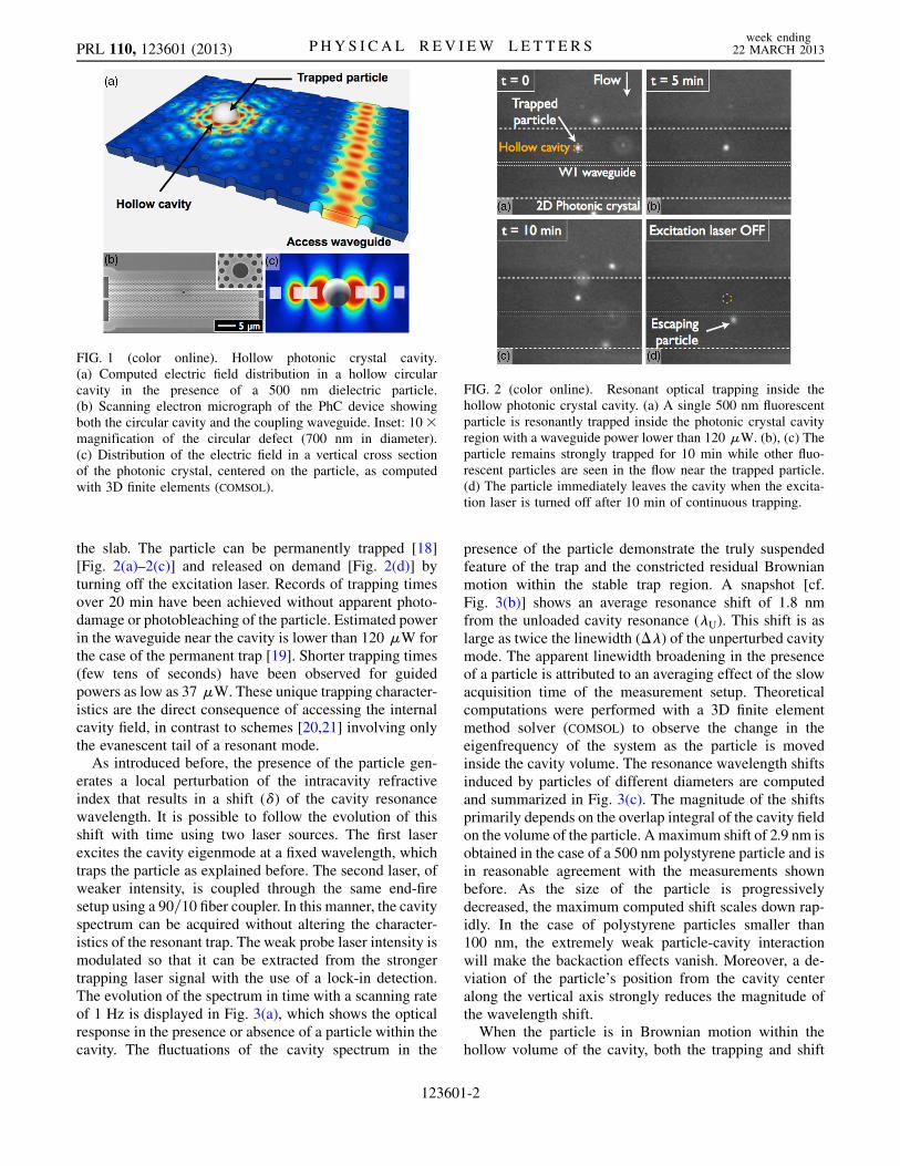
the slab. The particle can be permanently trapped [18][Fig. 2(a)–2(c)] and released on demand [Fig. 2(d)] byturning off the excitation laser. Records of trapping timesover 20 min have been achieved without apparent photo-damage or photobleaching of the particle. Estimated powerin the waveguide near the cavity is lower than 120 �W forthe case of the permanent trap [19]. Shorter trapping times(few tens of seconds) have been observed for guidedpowers as low as 37 �W. These unique trapping character-istics are the direct consequence of accessing the internalcavity field, in contrast to schemes [20,21] involving onlythe evanescent tail of a resonant mode.
As introduced before, the presence of the particle gen-erates a local perturbation of the intracavity refractiveindex that results in a shift (�) of the cavity resonancewavelength. It is possible to follow the evolution of thisshift with time using two laser sources. The first laserexcites the cavity eigenmode at a fixed wavelength, whichtraps the particle as explained before. The second laser, ofweaker intensity, is coupled through the same end-firesetup using a 90=10 fiber coupler. In this manner, the cavityspectrum can be acquired without altering the character-istics of the resonant trap. The weak probe laser intensity ismodulated so that it can be extracted from the strongertrapping laser signal with the use of a lock-in detection.The evolution of the spectrum in time with a scanning rateof 1 Hz is displayed in Fig. 3(a), which shows the opticalresponse in the presence or absence of a particle within thecavity. The fluctuations of the cavity spectrum in the
presence of the particle demonstrate the truly suspendedfeature of the trap and the constricted residual Brownianmotion within the stable trap region. A snapshot [cf.Fig. 3(b)] shows an average resonance shift of 1.8 nmfrom the unloaded cavity resonance (�U). This shift is aslarge as twice the linewidth (��) of the unperturbed cavitymode. The apparent linewidth broadening in the presenceof a particle is attributed to an averaging effect of the slowacquisition time of the measurement setup. Theoreticalcomputations were performed with a 3D finite elementmethod solver (COMSOL) to observe the change in theeigenfrequency of the system as the particle is movedinside the cavity volume. The resonance wavelength shiftsinduced by particles of different diameters are computedand summarized in Fig. 3(c). The magnitude of the shiftsprimarily depends on the overlap integral of the cavity fieldon the volume of the particle. A maximum shift of 2.9 nm isobtained in the case of a 500 nm polystyrene particle and isin reasonable agreement with the measurements shownbefore. As the size of the particle is progressivelydecreased, the maximum computed shift scales down rap-idly. In the case of polystyrene particles smaller than100 nm, the extremely weak particle-cavity interactionwill make the backaction effects vanish. Moreover, a de-viation of the particle’s position from the cavity centeralong the vertical axis strongly reduces the magnitude ofthe wavelength shift.When the particle is in Brownian motion within the
hollow volume of the cavity, both the trapping and shift
FIG. 2 (color online). Resonant optical trapping inside thehollow photonic crystal cavity. (a) A single 500 nm fluorescentparticle is resonantly trapped inside the photonic crystal cavityregion with a waveguide power lower than 120 �W. (b), (c) Theparticle remains strongly trapped for 10 min while other fluo-rescent particles are seen in the flow near the trapped particle.(d) The particle immediately leaves the cavity when the excita-tion laser is turned off after 10 min of continuous trapping.
FIG. 1 (color online). Hollow photonic crystal cavity.(a) Computed electric field distribution in a hollow circularcavity in the presence of a 500 nm dielectric particle.(b) Scanning electron micrograph of the PhC device showingboth the circular cavity and the coupling waveguide. Inset: 10�magnification of the circular defect (700 nm in diameter).(c) Distribution of the electric field in a vertical cross sectionof the photonic crystal, centered on the particle, as computedwith 3D finite elements (COMSOL).
PRL 110, 123601 (2013) P HY S I CA L R EV I EW LE T T E R Sweek ending
22 MARCH 2013
123601-2

phenomena described before occur in a coupled manner.This coupling is at the core of the particle-cavity back-action, wherein the trapping characteristics strongly dependon the excitation wavelength. The Brownian motion indu-ces resonance wavelength shifts, which, for a monochro-matic excitation, renormalizes the amount of energycoupled to the cavity and consequently the trapping forces.Concurrently, the energy required by the particle to escapethe resonant trap depends on the excitation wavelength in anontrivial manner. In other words, the wavelength-dependent escape energy profile does not reproduce theloaded cavity optical response as would be expected froma noncoupled system. On the contrary, a more complexprofile extending over several cavity linewidths is observed.As a direct signature of the backaction effect, the largestescape energy required for the particle to leave the trap isexpected to occur neither at the unloaded (�U) nor at theloaded (�L) resonance wavelengths but in a region inbetween. This is demonstrated by monitoring the trappingthreshold power for a range of wavelengths centred on �U.While a particle is held in the resonant trap for a givenwavelength, the excitation power is progressively decreaseduntil the particle escapes. Figure 4 reports the evolution ofthe escape threshold power for a range of wavelengthsdetuned from �U. As expected, the resonant nature of the
trap not only results in a restricted range of wavelengthsonly for which it is possible to observe trapping but alsotwo distinct minima associated with two trappingregimes can be observed. The first regime (I) has a largetrapping bandwidth and the lowest threshold power of37 �W. The second regime (II) covers a narrower band-width centred near �L and has a larger threshold power of99 �W.The forces acting on the particle were computed by
using the Maxwell’s stress tensor formalism integratedover the surface of the spherical particle for various detun-ing values. The trapping forces for a 500 nm dielectricparticle are presented in Fig. 5(a), taking into account thefield renormalization effect. It can be seen that the magni-tude of the out-of-plane and in-plane force componentsexperienced by the particle vary with detuning. In the caseof the out-of-plane component, the position of the maxi-mum force is also a detuning-dependent quantity. In addi-tion to this, the restoring force profile displays a largedegree of anharmonicity. Finally, one can notice that theout-of-plane and in-plane components get sequentiallypredominant with detuning leading to the existence of thetwo experimentally observed regimes. The first regimecan be conceptualized as a quasi-1D trap as illustrated inFig. 5(b), where the particle is held within the hollowvolume wherein it experiences weak trapping forces. Assoon as the particle deviates from the cavity volume, theintracavity field builds up and strong pulling forces arise inthe vertical direction bringing it back to the center of thetrap. In these detuning values, the in-plane forces areconsiderably weaker compared to their out-of-plane coun-terparts. The second regime appears at the farther detuningvalues, closer to the loaded cavity wavelength. It can beseen as a 3D trap where the stable position is located nearthe inner sidewalls of the cavity as illustrated in Fig. 5(c).This position corresponds to the maximum field amplitudeand hence the magnitudes of the forces are larger. For thesedetuning values, the in-plane force component is largerthan the out-of-plane one. A maximum in-plane force of1.5 nN is computed near the sidewall of the cavity for an
FIG. 4 (color online). Demonstration of particle-cavity cou-pling. Record of trapping threshold powers as a function of theestimated guided power in the access waveguide (left axis) andthe laser diode output power (right axis). Experimental pointscorrespond to escape trapping times of the order of 10 s.
FIG. 3 (color online). Demonstration of particle-induced reso-nance frequency shift. (a) Record of the evolution of the cavityspectrum in timewhile a particle is trapped in the cavity and after itis released. (b) A single snapshot from (a), displaying an averageresonance shift of 1.8 nm from the unloaded cavity resonance.(c) Computed resonancewavelength shifts as a function of particlesize as it is moved along the vertical direction from the center ofthe cavity. The shaded area (in grey) shows the extent of the siliconslab. A maximum shift of 2.9 nm is found for a 500 nm particleplaced in the center of the cavity.
PRL 110, 123601 (2013) P HY S I CA L R EV I EW LE T T E R Sweek ending
22 MARCH 2013
123601-3

input power of 1 mW, which corresponds to a 3 orders ofmagnitude increase compared to previous studies involv-ing evanescent field trapping [20].
In summary, self-induced trapping along with the back-action between a resonant optical field and a singlenanoparticle has been reported. This mechanism opensthe door to new kinds of all-optical schemes where singlebiological entities like cell organelles and viruses can beisolated, analyzed, and sorted depending on their size,shape, and optical properties. The novel trapping mecha-nisms can be extended to a variety of sizes owing to thescalability of photonic crystals. An additional remarkablefact from this scheme is that it addresses both the lack ofexclusivity and specificity of standard optical tweezers[22]. The long trapping times along with very small resid-ual Brownian motion is of major interest for studies thatuse spatially resolved spectroscopy. These results openthe way to the exploration of such coupled systemswhere dynamical effects are expected to play a dramaticrole [5].
We would like to thank Professor Sebastian Maerkl forhis support in the fabrication of the microfluidic circuit.The authors acknowledge the financial support from the
Swiss National Centre of Competence in ResearchQuantum Photonics and the Swiss National ScienceFoundation Projects No. 200021_134541 and200020_140406.
[1] T. Aoki, B. Dayan, E. Wilcut, W. P. Bowen, A. S. Parkins,T. J. Kippenberg, K. J. Vahala, and H. J. Kimble, Nature(London) 443, 671 (2006).
[2] J. McKeever, J. R. Buck, A. D. Boozer, A. Kuzmich, H. C.Nagerl, D.M. Stamper-Kurn, and H. J. Kimble, Phys. Rev.Lett. 90, 133602 (2003).
[3] T. J. Kippenberg and K. J. Vahala, Science 321, 1172(2008).
[4] A. Naik, O. Buu, M.D. LaHaye, A. D. Armour, A. A.Clerk, M. P. Blencowe, and K. C. Schwab, Nature(London) 443, 193 (2006).
[5] P. F. Barker and M.N. Shneider, Phys. Rev. A 81, 023826(2010).
[6] J. Hu, S. Lin, L. C. Kimerling, and K. Crozier, Phys. Rev.A 82, 053819 (2010).
[7] D. E. Chang, C. A. Regal, S. B. Papp, D. J. Wilson, J. Ye,O. Painter, H. J. Kimble, and P. Zoller, Proc. Natl. Acad.Sci. U.S.A. 107, 1005 (2010).
[8] O. Romero-Isart, A. C. Pflanzer, M. L. Juan, R. Quidant,N. Kiesel, M. Aspelmeyer, and J. I. Cirac, Phys. Rev. A 83,013803 (2011).
[9] M. Barth and O. Benson, Appl. Phys. Lett. 89, 253114(2006).
[10] A. Rahmani and P. C. Chaumet, Opt. Express 14, 6353(2006).
[11] M. R. Lee and P.M. Fauchet, Opt. Lett. 32, 3284 (2007).[12] A. Di Falco, L. O’Faolain, and T. F. Krauss, Appl. Phys.
Lett. 94, 063503 (2009).[13] J. Jagerska, H. Zhang, Z. Diao, N. Le Thomas, and
R. Houdre, Opt. Lett. 35, 2523 (2010).[14] M. L. Juan, R. Gordon, Y. Pang, F. Eftekhari, and
R. Quidant, Nat. Phys. 5, 915 (2009).[15] D. Psaltis, S. R. Quake, and C. Yang, Nature (London)
442, 381 (2006).[16] C. Monat, P. Domachuk, and B. J. Eggleton, Nat.
Photonics 1, 106 (2007).[17] M.A. Unger, H.-P. Chou, T. Thorsen, A. Scherer, and S. R.
Quake, Science 288, 113 (2000).[18] See Supplemental Material at http://link.aps.org/
supplemental/10.1103/PhysRevLett.110.123601 for moviewith a detailed demonstration of positioning, trapping, andreleasing of a 500 nm dielectric particle in a 2D photoniccrystal hollow cavity.
[19] See Supplementary Material at http://link.aps.org/supplemental/10.1103/PhysRevLett.110.123601 formethod and details on the estimation of guided power.
[20] S. Mandal, X. Serey, and D. Erickson, Nano Lett. 10, 99(2010).
[21] S. Lin, E. Schonbrun, and K. Crozier, Nano Lett. 10, 2408(2010).
[22] K. C. Neuman and A. Nagy, Nat. Methods 5, 491 (2008).
FIG. 5 (color online). (a) Theoretical computations for a500 nm particle including the particle-cavity renormalizationeffect: Trapping forces as a function of the position of theparticle for various detuning values along the horizontal axisinside the cavity (from�100 nm to 0) and along the vertical axis(0 to 800 nm). The shaded area corresponds to the half-thicknessof the silicon slab. (b) Illustration of the first trapping regime.The eigenmode of the particle located in the cavity is weaklycoupled to the excitation laser (�Ex1). An out-of-plane displace-ment of the particle couples the eigenmode back to the excitationresulting in strong gradient forces. (c) Illustration of the secondtrapping regime. A particle outside the cavity weakly couplesthe eigenmode to the excitation laser (�Ex2). As soon as theparticle enters further into the cavity, the eigenmode is stronglycoupled to the excitation and hence increases the trappingstrength.
PRL 110, 123601 (2013) P HY S I CA L R EV I EW LE T T E R Sweek ending
22 MARCH 2013
123601-4