objectives - continuing ed - 6 slides per page.pdfprinciples of neurodynamics objectives participant...
TRANSCRIPT
Jason Zafereo, PT, OCS, FAAOMPT
With contributions from: Leslie Nelson, PT
Principles of NeurodynamicsObjectives
� Participant will review the relevant
anatomy and function of the peripheral, central, and autonomic nervous systems
� Participant will describe the intraneural and extraneural factors contributing to
nerve injury
Objectives
� Participant will describe the tension
points in the body where pathology is most likely to occur
� Participant will review a system of examination for adverse neural tension
Foundations of Anatomy
� Continuous tissue tract
– Connective tissue interconnected
– Electrically interconnected neurons
– Chemical consistency
� Neurotransmitters
� Cytoplasm
Structure Meets Function
� Accommodation of body movements
– Meninges
– (Endo, peri, epi)-neurium
� Impulse conduction
– Axons
– Myelin
Peripheral Nervous System (PNS)
� Axon
– Run undulatory course in endoneurial tubes
� Myelin

Peripheral Nervous System
� Diagram of Connective tissue coverings
Central Nervous System
� Nerve roots
– Undulations
– Dural and epidural meninges
– Extrathecal attachments
(low cervical, LS)
– Plugging by dorsal root
ganglion
Central Nervous System
� Spinal cord
– Undulations
– Movement in relation to neighboring vertebral segments
– Meninges
� Pia and arachnoid mater
� Dura mater
Autonomic Nervous System
� Sympathetic trunk and ganglia
� Pre-ganglionic neurons
– Head and neck C8-T5
– UE T2-T10
– LE T10-L2
� Post-ganglionic neurons
– Efferent to smooth muscle and glands
Intraneural Causes of ANT
� Decreased blood supply to nervous system
� Disruptions in axonal transport
� Nociceptive stimulation of connective tissue
coverings of nervous system
Vascular considerations
� 2% of body mass consumes 20% of
available Oxygen
� CNS: Critical vascular zone exists from
T4-T9
– Narrow spinal canal
� PNS: Arrest of blood flow at 8%
elongation, complete at 15%
Axonal transport systems
� Two-way movement of materials and
substances in the cytoplasm
� Double crush
Innervation of Connective Tissue
� Meninges– Dura mater highly
innervated ventrally
– Arachnoid/pia mater innervation uncertain
� (Endo, peri, epi)-neurium– Intrinsic innervation
– Free nerve endings
Extraneural Causes of ANT
� Mechanical Interface
– Spinal foramen
– Muscle/fascia
– Joint
– Ligament
– Blood vessel
Intraneural versus Extraneural Symptom & Sign Differentiation
Extraneural Intraneural
(Conducting)
Intraneural
(Connective)
Description of symptoms
Catches, twinges around
vulnerable areas
Burning, tingling, electric in
innervation field
Lines of pain, along trunks,
nondermatomal
Physical signs Comparable signs with
movement of interfacing
structure
Neurological signs and
symptoms; palpation refers
sx elsewhere; increased sx with
tension testing
Palpation elicits local pain;
increased sx with tension testing
Sites of Injury
� Soft tissue, osseus or fibro-osseus tunnels
� Where the nervous system branches
� Where the system is relatively fixed
� Where the nerves pass over unyielding interfaces
� Tension points
Tunnels
� Median nerve in carpal
tunnel
� Spinal nerve in
intervertebral foramen
� Posterior interosseus nerve in arcade of Frohse
Branches
� Union of lateral and medial
plantar nerves to form the common plantar digital
nerve
Fixed System
� Common peroneal nerve at
head of fibula
� Dura mater at L4
� Radial nerve to the head of radius
� Suprascapular nerve in scapular notch
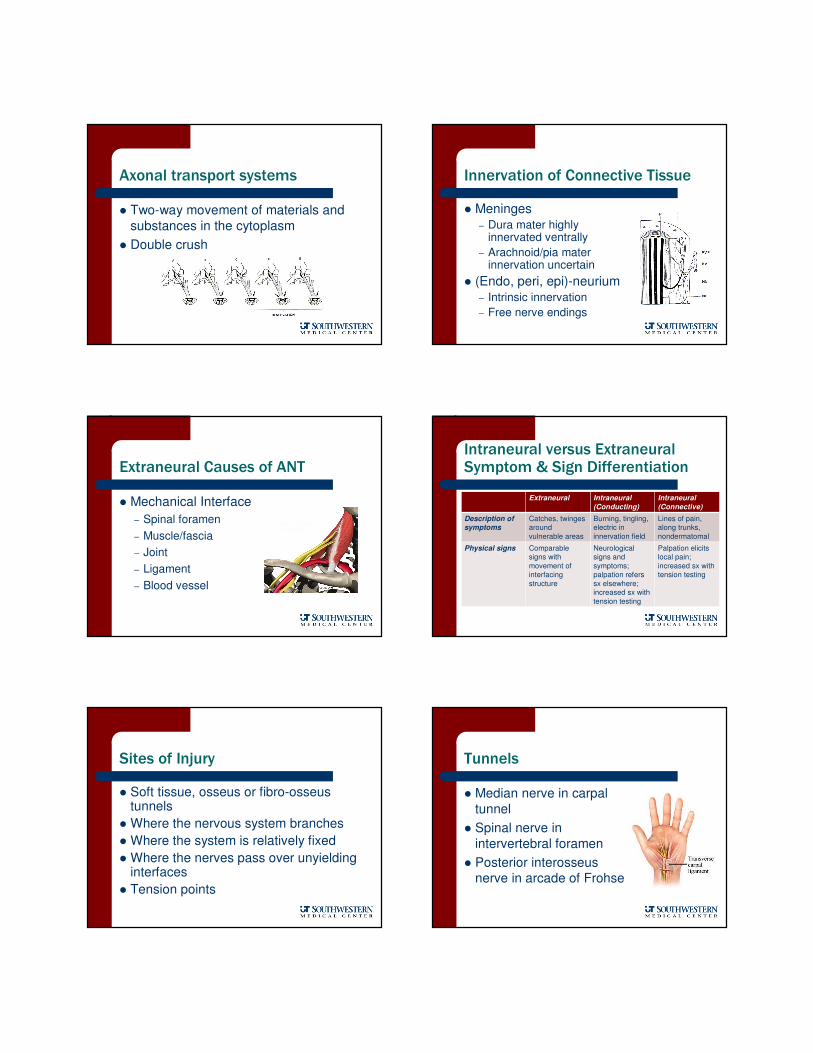
Unyielding Surface
� Cords of plexus over
first rib
� Radial nerve in radial
groove of humerus
� Dural sleeve at pedicles
Unyielding Surfaces (continued)
� Greater occipital nerve
through fascia
� Lateral femoral
cutaneous nerve through fascia
� Nerves in feet through
plantar fascia
“Tension Points”
� Key points in the body where movement
demands (and tension) are increased
– C6
– T6
– L4
– Tibial nerve at posterior knee
– Median nerve at anterior elbow
Neurodynamic Examination
� Tension testing– Longitudinal deformation
– Elasticity intraneural
– Elasticity extraneural
� Palpation– Perpendicular deformation
� Neurological testing
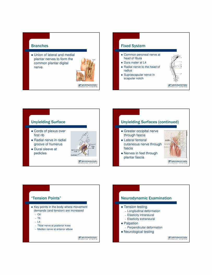
Tension Testing
ULTT1 SLR
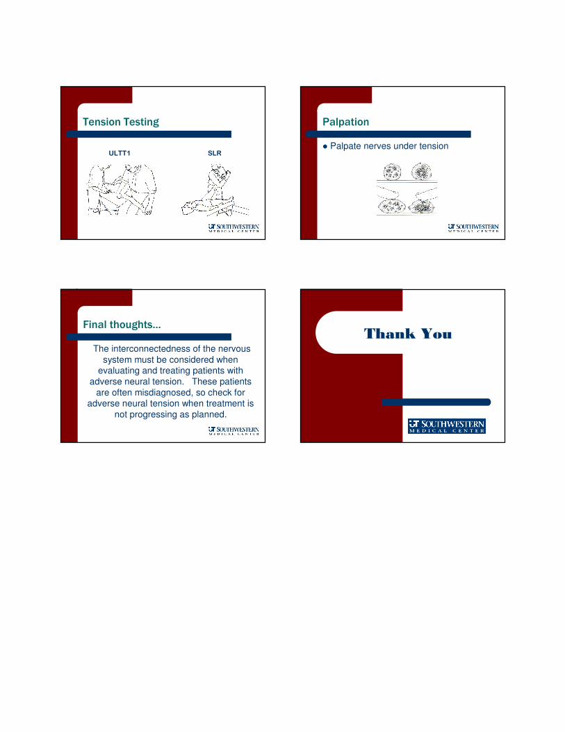
Palpation
� Palpate nerves under tension
Final thoughts…
The interconnectedness of the nervous
system must be considered when evaluating and treating patients with
adverse neural tension. These patients
are often misdiagnosed, so check for adverse neural tension when treatment is
not progressing as planned.
Thank You