o01 - issls.org
TRANSCRIPT

ORAL PRESENTATIONS
1
O01 SURGICAL VERSUS NON‐OPERATIVE TREATMENT FOR LUMBAR DISC HERNI‐ATION: EIGHT‐YEAR RESULTS FOR THE SPINE PATIENT OUTCOMES RESEARCH TRI‐AL (SPORT) Jon D. Lurie; Tor D. Tosteson; Anna N.A. Tos‐teson; Wenyan Zhao; Tamara S. Morgan; William A. Abdu; Harry Herkowitz; James N. Weinstein; Geisel School of Medicine at Dartmouth, Hanover, NH and Dartmouth‐Hitchcock Medical Center, Lebanon, NH (Abdu);William Beaumont Hospital, Royal Oak, MI (Herkowitz)
INTRODUCTION: Eight year outcomes of surgery vs. non‐operative care for lumbar intervertebral disc herniation (IDH). Though randomized trials have demonstrated small short‐term differences in favor of surgery, long‐term outcomes comparing surgical to non‐operative treatment remain contro‐versial. METHODS: Eligible surgical candidates with imaging‐confirmed IDH enrolled into pro‐spective randomized (n=501) and observa‐tional cohorts (n=743) at 13 spine clinics in 11 US states. Interventions were standard open discectomy versus usual non‐opera‐tive care. Primary outcomes were changes from baseline in the SF‐36 Bodily Pain (BP) and Physical Function (PF) scales and the modified Oswestry Disability Index (ODI) assessed at 6 weeks, 3 and 6 months, and annually thereafter. RESULTS: Advantages were seen for surgery in intent‐to‐treat (ITT) analyses for the ran‐domized cohort for all primary and sec‐ondary outcomes other than work status; however, with extensive non‐adherence to treatment assignment (49% patients as‐signed to non‐operative therapy received surgery versus 60% of patients assigned to surgery) these observed effects were rela‐tively small and not statistically significant for BP, PF or ODI. An as‐treated analysis showed significant surgical treatment ef‐
fects for primary outcomes (mean change Surgery vs. Non‐operative; treatment effect; 95% CI): BP (45.3 vs. 34.4; 10.9; 7.7 to 14); PF (42.2 vs. 31.5; 10.6; 7.7 to 13.5) and ODI (‐36.2 vs. ‐24.8; ‐11.2; ‐13.6 to ‐9.1). Im‐portantly, the overall comparison of sec‐ondary outcomes was significantly greater with surgery in the ITT analysis (sciatica bothersomeness [p > 0.005], satisfaction with symptoms [p > 0.013], and self‐rated improvement [p > 0.013]) in long‐term fol‐low‐up. DISCUSSION: Carefully selected patients who underwent surgery for IDH achieved greater improvement than non‐operatively treated patients; there was little to no deg‐radation of outcomes in either group (oper‐ative vs non‐operative) from 4 to 8 years.
O02 BODY MASS INDEX AND ITS ASSOCIATION WITH LUMBAR DISC HERNIATION AND SCIATICA: A LARGE‐SCALE, POPULATION‐BASED STUDY Dino Samartzis, (+)Jaro Karppinen, Keith DK Luk, Kenneth MC Cheung; Department of Orthopaedics and Traumatology, University of Hong Kong, Pokfulam, Hong Kong, SAR, China and the (+) Institute of Clinical Medi‐cine, University of Oulu, Oulu, Finland
INTRODUCTION: Elevated body mass index (BMI) or overweight and obesity are pan‐demics. Lumbar disc herniation and sciatica occur in every population and present se‐vere socioeconomic consequences. How‐ever, little is known regarding the role of BMI with lumbar disc herniation and sciat‐ica. As such, the following large‐scale study addressed the association of BMI, in partic‐ular overweight and obesity, with disc her‐niation, its global lumbar involvement and its implications with the development of sciatica. METHODS: A cross‐sectional study of 2,596 Southern Chinese (mean age: 42 years; 60% females) was conducted assessing T2‐

ORAL PRESENTATIONS
2
weighted MRI, environmental and lifestyle factors, as well as clinical profiles of sciatica. On imaging, the presence of disc bulge/extrusion (DBE) and other spinal phe‐notypes from L1‐S1 were assessed. A total DBE (TDBE) Score of L1‐S1 was calculated. Asian‐modified BMI values and categories were obtained of each subject. Results: DBE was noted in 46.3% of the sub‐jects, mainly occurring at L4‐S1. The mean TDBE score was 0.7. Historical prevalence of sciatica was 44.6%, with 17.9% reporting sciatica at the time of assessment. The mean BMI was 22.9 kg/m2 (7.2% under‐weight, 47.9% normal‐weight, 36.1% over‐weight, 8.9% obese). TDBE Score sig‐nificantly increased with elevated BMI cate‐gories (p<0.001). Multivariate analyses not‐ed that elevated BMI was significantly asso‐ciated with DBE [normal‐weight (Ref); un‐derweight OR: 0.71(0.49‐1.03); overweight OR: 1.26(1.04‐1.52); obese OR: 1.78(1.30‐2.44)]. TDBE score (OR: 1.36; 1.15‐1.60) and obesity (OR: 1.68;1.25‐2.24) were signifi‐cantly related with sciatica. Worse function‐al and disability scores were associated with sciatica (p<0.05). DISCUSSION: Based on the largest popula‐tion‐based study to assess the role of BMI and its association with disc herniation, overweight and obesity significantly in‐creased the likelihood of having lumbar DBE, its global severity and the risk of de‐veloping sciatica.
O03 DEFINING DISC HERNIATION: IS CLINICAL DIAGNOSIS ENOUGH? M.C. Battié, L. Gibbons, M. Bruno‐Brayda, J. Fairbank, C. Heywood, A. Lazary, I. McCall, S. Roberts, P.P. Varga; University of Alberta, Canada; University of Helsinki, Finland; Ox‐ford University, Keele University, & East Midlands Spine Limited, UK; National Center for Spinal Disorders, Hungary; IRCCS Istituto Ortopedico Galeazzi, Italy; University of
Washington, USA
INTRODUCTION: Whether comparing pa‐tient outcomes between clinics, conducting multi‐center trials, or pooling subjects for gene association studies, clearly defined phenotypes are needed. The simplest ap‐proach to defining a clinical phenotype for disc herniation (DH) is to rely on the treat‐ing or consulting physician’s diagnosis, but is clinical diagnosis enough? METHODS: Patients in the Genodisc re‐search consortium project were recruited from spine surgeons’ clinics in 3 European countries. There were 1134 patients dia‐gnosed with lumbar DH who also had an independent assessment of their lumbar MRI by a radiologist blinded to diagnosis. We compared data from the standard dia‐gnostic sheet completed by the spine sur‐geon, the MRI assessments, and patient self‐report between 5 clinical sites, using ANOVA or Fisher's exact test. RESULTS: Spine surgeons noted radicular pain in a dermatomal pattern in 89.0‐95.9% of patients (p=0.139), and associated nerve root compression was identified on imaging in 76.2‐86.3% (p=0.032). Substantial differ‐ences were reported by the spine surgeons for the portion of subjects with extruded or sequestered discs (64.6‐95.8%; p<.001), but these differences were not supported by the independent MRI assessments (56.5‐69.9%; p=0.342). There were notable differ‐ences across sites for spinal comorbidity (spondylolisthesis (4.1‐16.0%; p<0.001) and spinal stenosis (16.4‐33.0%; p< 0.001)), du‐ration of leg pain (4.5‐22.0 months; p<0.001), and interference with work (43.5‐79.8%; p<0.001). DISCUSSION: Is the clinical diagnosis of DH enough to achieve a homogeneous pheno‐type? The answer is mixed. While some findings for the clinical phenotype of dia‐gnosed DH were similar, there were also variations by site for severity, chronicity, and complexity of cases that could affect

ORAL PRESENTATIONS
3
patient outcomes and comparisons. If relat‐ed data are not gathered and differences addressed, mixed phenotypes, mis‐interpretation, and failure to replicate find‐ings could result.
O04 HOW HEALTHY DISCS HERNIATE: A BIO‐MECHANICAL AND MICROSTRUCTURAL STUDY INVESTIGATING THE COMBINED EFFECTS OF COMPRESSION RATE AND FLEXION Kelly R. Wade, Peter A. Robertson, Ashvin Thambyah, Neil D. Broom; Experimental Tissue Mechanics Laboratory, Department of Chemical and Materials Engineering, Uni‐versity of Auckland, New Zealand. Depart‐ment of Orthopaedic Surgery, Auckland City Hospital, New Zealand
INTRODUCTION: At the level of the motion segment failure of the disc in compression has been extensively studied. However, at the microstructural level the exact mecha‐nisms of disc failure are still poorly under‐stood, especially in relation to loading pos‐ture and rate. The purpose of this study was to provide a microstructural analysis of the mechanisms of annular wall failure in healthy discs subjected to flexion and an elevated rate of compression. METHODS: 72 healthy mature ovine lumbar motion segments were compressed to fail‐ure in either a neutral posture or in high physiological flexion (10º) at a displacement rate of either 2mm/min (low) or 40mm/min (high). Testing at the high rate was termi‐nated at stages ranging from initial wall tearing through to facet fracture so as to capture the evolution of failure up to full herniation. The damaged discs were then analysed microstructurally. RESULTS: ~50 % of the motion segments compressed in flexion at the high rate suf‐fered annulus or annulus‐endplate junction failure, the remainder failed via endplate fracture with no detectable wall damage.
The average load to induce disc failure in flexion was 18% lower (p < 0.05) than that required to induce endplate fracture. Mi‐crostructural analysis indicated that wall rupture occurred first in the posterior mid‐then‐outer annulus. DISCUSSION: Disc wall failure in healthy motion segments requires both flexion and an elevated rate of compression. Damage is initiated in the mid‐then‐outer annular fi‐bres, this a likely consequence of the higher strain burden in these same fibres arising from endplate curvature. Given the similari‐ty in geometry between ovine and human endplates it is proposed that comparable mechanisms of damage initiation and herni‐ation occur in human lumbar discs.
O05 THE SIGNIFICANCE OF CARTILAGE END‐PLATE WITHIN A LUMBAR DISC HERNI‐ATION P Lama, U Zehra, C Balkovec, IJ Harding*, P Dolan, MA Adams; Centre for Comparative and Clinical Anatomy, University of Bristol, Bristol, U.K *Department of Orthopaedics, Southmead Hospital, Bristol, UK
INTRODUCTION: Herniated disc tissue re‐moved at surgery contains varying pro‐portions of nucleus pulposus, annulus fibro‐sus, hyaline cartilage and bone. The origins and significance of hyaline cartilage within a herniation are largely unknown. METHODS Lumbar herniated (extruded) disc tissue was removed surgically from 21 patients aged 35‐74 yrs. Frozen sections, 5‐µm thick, were examined histologically, and antibodies were used to label cells for the pro‐inflammatory mediator TNFα, and the matrix‐degrading enzyme MMP1. Propor‐tions of each tissue type were quantified using image analysis software. Cartilage‐bone junctions were examined in 5‐µm fro‐zen sections from 17 cadaveric spines, aged 61‐98 yrs. Strength of the disc‐cartilage and cartilage‐bone interfaces of the endplate

ORAL PRESENTATIONS
4
were compared by stretching to failure 5 mm‐thick cadaveric bone‐disc‐bone speci‐mens. RESULTS: Fragments of hyaline cartilage were found in 10/21 herniations. On aver‐age they were 5.0 mm long, and occupied 26% of the area of the herniated mass. Two had a small quantity of bone attached. Hya‐line cartilage was present in a higher pro‐portion of herniations from patients with sciatica (7/10) compared to those from pa‐tients whose main symptom was back pain (3/11) (P<0.05). Cartilage appeared to have been peeled off the bony endplate along the ‘tide mark’, and this was the mode of failure in the mechanical experiments. Hya‐line cartilage showed little swelling, proteo‐glycan loss or inflammatory cell invasion, although cartilage chondrocytes often formed small clusters which expressed TNFα and MMP1. DISCUSSION: Disc herniations often include hyaline cartilage pulled from the vertebral endplate. The collagen network of cartilage fragments minimises swelling, proteoglycan loss and resorption, so they are likely to cause persisting sciatica. Loss of cartilage endplate will increase endplate permeabil‐ity, increasing the risk of inflammatory ‘Modic’ changes, and of disc infection.
O06 SPONTANEOUS RESORPTION OF LUMBAR DISC HERNIATION IS LESS LIKELY WHEN MODIC CHANGES ARE PRESENT Zhi Shan M.D, Shunwu Fan M.D, Qingbo Xie M.D, Letu Suyou M.D, Junhui Liu M.D, Chongyan Wang M.D, Fengdong Zhao M.D.; Department of Orthopaedics, Sir Run Run Shaw Hospital, School of Medicine, Zhejiang University
INTRODUCTION: Spontaneous resorption of lumbar disc herniation (LDH) has been demonstrated, while the mechanisms are unclear. Modic changes (MCs) are closely associated with disc degeneration, but re‐
search focusing on their association with spontaneous resorption of LDH have not been specifically investigated. METHODS: Eighty‐five consecutive LDH pa‐tients (52 men, 33 women, aged 20‐66yrs) were included. Patient diagnosis was based on clinical presentation, MRI and CT. Pa‐tients were divided into surgical and con‐servative groups, and further divided into MC and non‐MC subgroups. Spontaneous resorption and clinical success in the con‐servative group were assessed by reduction in the herniated volume AND Oswestry dis‐ability index (ODI). Disc tissues collected from the surgical group were examined his‐tologically, and immunohistochemistry was used to identify endothelial cells and mac‐rophages. RESULTS: In total, 35/85 patients showed MC, mostly Type II. Herniated tissue in MC group contained relatively more hyaline cartilage endplate than non‐MC group (on average, 50% vs 8%, P<0.05) but less nucle‐us pulposus (18% vs 55%, P<0.05). Con‐servative treatment reduced ODI scores in non‐MC group from 29.4 to 23.5 on average (P <0.05), but reductions in MC group (30.1 to 29.0) were non‐significant. Herniated volumes reduced following conservative treatment in non‐MC group (0.44 to 0.21 cm3, P<0.05) but not in MC group (0.52 to 0.45 cm3, P>0.05). More neovascularization and macrophage infiltration was observed in herniated tissue from non‐MC group than MC group (P <0.001). CONCLUSIONS: Modic changes in LDH pa‐tients are associated with cartilaginous her‐niations which resorb poorly, so that pa‐tients respond less well to conservative treatments. Loss of cartilaginous endplate may explain the origins of MCs, and their association with disc infection.

ORAL PRESENTATIONS
5
O07 ACCELERATED DDD IN YOUNG ADULTS IS ASSOCIATED WITH POLYMORPHISMS IN MMP 7, CALM 1 AND COX 2 GENES. ANA‐LYSIS OF 58 SINGLE NUCLEOTIDE POLY‐MORPHISMS IN 580 INDIVIDUALS BASED ON TOTAL DEGENERATIVE DISC SCORE 1.Prof.S.Rajasekaran,Ph.D, 2.Dr.Rishi M Kanna, 3.Prof.Kenneth Cheung, 4.Prof.Danny Chan, 5.Dr.Patrick Kao, 6.Dr.Anita Yee, 7.Dr.Raveendran M, 8.Dr.Senthil N; 1,2 Dept. of Spine Surgery, Ganga Hospital, Coimbatore, India 3,4,5,6 ‐ University of Hong Kong, Pokfulam, Hong Kong. 7,8 Tamil Nadu Agricultural Universi‐ty, Coimbatore, India.
INTRODUCTION: Significant genetic influ‐ence for degenerative disc disease (DDD) has been established, but however studies focusing specifically on young adults with DDD are not present. METHODS: Patients < 40 years with lumbar DDD were evaluated with MRI imaging and Genetic association studies for 58 SNPs of 35 candidate genes. Subjects were stratified into three groups based on a Total Degen‐erative Disc Score (TDDS), which was deve‐loped by adding the individual scores of Pfirmann grading of all five lumbar discs. The severity of DDD was classified as mild (TDDS <10), moderate (TDDS of 10 to 15) and severe (TDDS > 15). 58 potential SNPs based on previous genetic studies on DDD and located in candidate genes encoding for vital disc components (collagen, proteo‐glycan, degradative enzymes); in bone re‐modeling (Vitamin D receptor, bone mor‐phogenic protein) and those involved in important intracellular signaling mecha‐nisms were considered. RESULTS: In 580 subjects (308 patients ‐ mild TDDS, 211 ‐ moderate TDDS and 61 ‐ severe TDDS), there was no significant dif‐ference in the gender and the mean age. Association analysis of SNPs was performed between mild and severe TDDS groups. 3 of
the 35 candidate genes showed significant association with severe TDDS. SNPs viz., rs1940044 of MMP (Matrix Metalloprotein‐ase 7), rs2300496 SNP and rs3213718 SNPs of CALM1 (Calmodulin) and rs5277 of COX 2 (Cyclooxygenase) were found to be signifi‐cantly associated with severe TDDS. CONCLUSION: Our study is the first to re‐port SNP analysis specific to DDD in the young adults. The study has identified spe‐cific SNP associations of three genes (MMP 7, CALM 1 and COX2) in young adults with severe DDD. Involvement of Matrix genes MMP 7 and CALM 1 and inflammatory gene COX2 shows that DDD is a complex process with an interplay of multiple genetic poly‐morphisms.
O08 CHARACTERIZATION OF ANNULUS LAYER DEFORMATIONS IN AN INTACT INTERVER‐TEBRAL DISC UNDER MECHANICAL COM‐PRESSION Han Sang Kuy,1 Tang Qinggong,1 Kim Hyun‐chul, 1, Chen Yu,1 Hsieh H. Adam 1,2; 1 Fischell Department of Bioengineering, Uni‐versity of Maryland, College Park, MD, Unit‐ed States; 2 Department of Orthopaedics, University of Maryland, Baltimore, MD, United States
INTRODUCTION: Annulus fibrosus (AF) in the intervertebral disc (IVD) plays a key role for bearing external loads on the IVD. AF in mechanical compression has been exten‐sively studied, but the deformations of an individual annulus lamellar layer in a fully intact IVD are not well understood. In this study, we utilized optical coherence tomo‐graphy (OCT), which is a non‐invasive im‐aging technique for visualizing AF mesostructural features, to characterize AF lamellar layer deformations of fully intact IVDs in mechanical compression. METHODS: Fresh‐frozen ovine lumbar mo‐tion segments were subjected to different compressive loads (300, 500 and 1000 N). A

ORAL PRESENTATIONS
6
swept‐source OCT system was paired with a materials testing system to visualize a 2.1 x 2.1 x 3.6 mm3 volume in AF layers from the anterior region in mechanical compression. Outer annular layer deformations including trans‐lamellar cross bridge were character‐ized. RESULTS: OCT image is able to characterize the most outer AF layers in an intact IVD (Fig. 1a). In mechanical compression of the IVD, AF lamellar layers were compressed in the radial direction and extended in the axial and circumferential direction (Fig. 1b and c). Furthermore, the deformations of trans‐lamellar cross bridge structures indi‐cated that shear loads were occurred within an individual lamellar layer (Fig. 1c). DISCUSSION: To our knowledge, this is the first study to characterize the deformations of an individual annular layer of fully intact IVDs in mesoscale level. Based on our re‐sults, individual annuls layer mechanics is more complicated under mechanical com‐pression because of multiple strains, i.e., compression, tensile and shear, within the annulus layer. Further study will aim to in‐vestigate annular layer mechanics of de‐generated IVDs, and the onset and progress of lamellar herniation using an OCT elasto‐graphy.
Figure 1. (a) OCT images of AF lamellar layers in a fully intact IVD (2.1 x 2.1 x 3.6 mm3). (b) and (c) An‐nular layer deformations.
O09 EARLY PATTERN AND LONGITUDINAL EVO‐LUTION OF DEGENERATIVE CHANGES IN INDIVIDUAL COMPONENTS OF INTERVER‐TEBRAL DISCS IN STRESSED AND NON‐STRESSED SEGMENTS OF LUMBAR SPINE: AN IN‐VIVO MR IMAGING STUDY.
Aseem Sharma, M.D.; Samantha Lancaster, M.D.; Swapnil Bagade, M.D.; Charles Hilde‐bolt, D.D.S., Ph.D.; Washington University School of Medicine, St. Louis, MO, USA
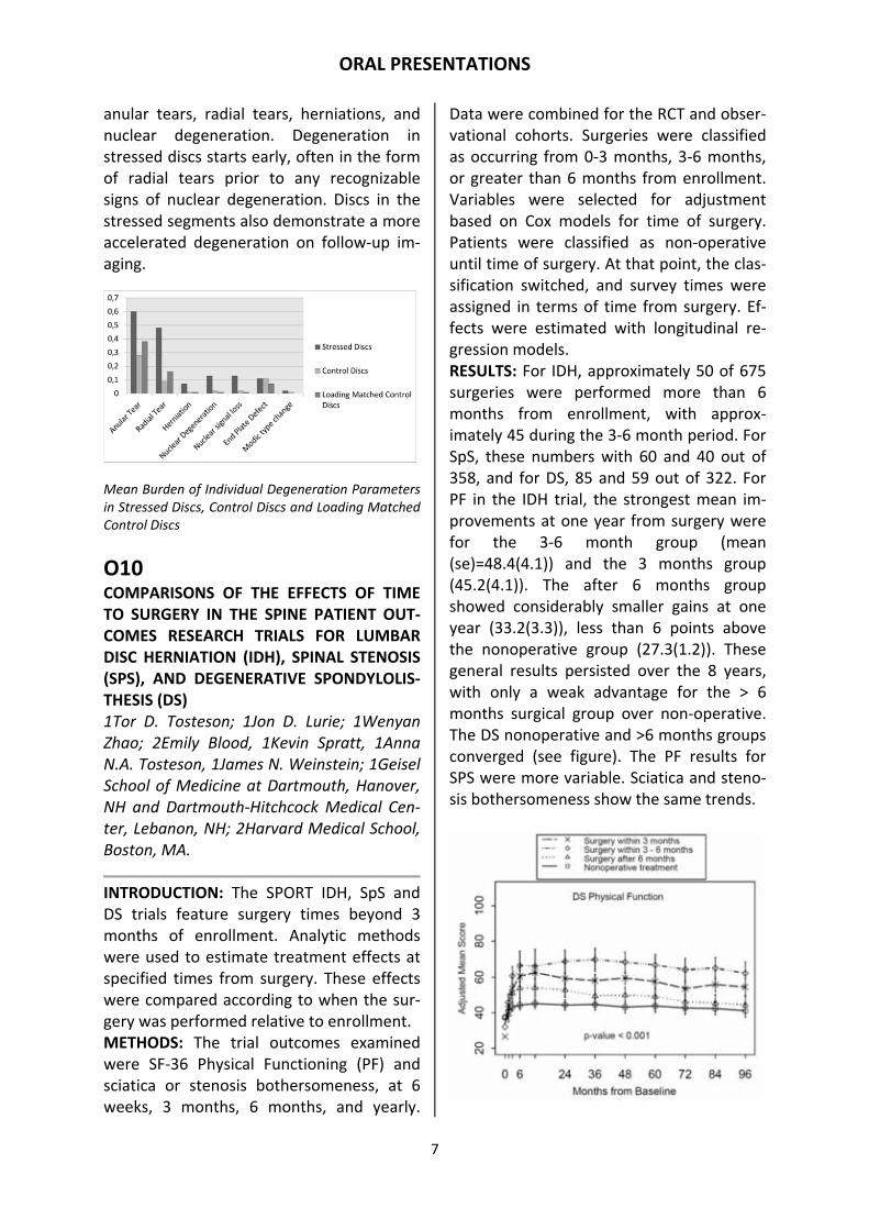
INTRODUCTION: Young patients with im‐aging signs of increased mechanical stress in pedicles or pars‐interarticularis provide an excellent in‐vivo model to study the early effects of mechanical stresses on lumbar intervertebral discs without the confound‐ing effects of genetics or the environmental factors. We aimed to study the burden and longitudinal evolution of disc degeneration in the segments of lumbar spine with and without signs of increased mechanical stresses. METHODS: Using MR imaging, two radiolo‐gists assessed intervertebral discs around 93 stressed lumbar spinal segments in 87 pa‐tients (55 males, 32 females; mean age 15.3 ± 3.3 years, range 5‐25 years) as well as lumbar discs in non‐stressed segments for signs of degeneration. Differences between stressed, control, and loading‐matched con‐trol discs were assessed using Wilcoxon signed rank sum test. Longitudinal evolution of these changes was studied on follow‐up imaging of 15 patients. RESULTS: Burden of anular tears, radial tears, herniations, and nuclear degenera‐tion was significantly higher in stressed discs (0.70 ± 0.34, 0.48 ± 0.39, 0.07 ± 0.19, and 0.17± 0.31 respectively) compared with control (0.29 ± 0.25, 0.09 ± 0.17, 0.01 ± 0.04, and 0.02 ± 0.08 respectively) or load‐ing‐matched control discs (0.44 ± 0.47, 0.16 ± 0.36, 0.01 ± 0.04, and 0.01 ± 0.11 respec‐tively) (p <0.01 for all). Stressed discs did not show increased end plate degeneration. Anular tears were most prevalent of all de‐generative parameters studied. Stressed discs had higher incidence of future wors‐ening. DISCUSSION: Intervertebral discs within the lumbar segments exposed to increased me‐chanical stresses have a higher burden of
ORAL PRESENTATIONS
7
anular tears, radial tears, herniations, and nuclear degeneration. Degeneration in stressed discs starts early, often in the form of radial tears prior to any recognizable signs of nuclear degeneration. Discs in the stressed segments also demonstrate a more accelerated degeneration on follow‐up im‐aging.
Mean Burden of Individual Degeneration Parameters in Stressed Discs, Control Discs and Loading Matched Control Discs
O10 COMPARISONS OF THE EFFECTS OF TIME TO SURGERY IN THE SPINE PATIENT OUT‐COMES RESEARCH TRIALS FOR LUMBAR DISC HERNIATION (IDH), SPINAL STENOSIS (SPS), AND DEGENERATIVE SPONDYLOLIS‐THESIS (DS) 1Tor D. Tosteson; 1Jon D. Lurie; 1Wenyan Zhao; 2Emily Blood, 1Kevin Spratt, 1Anna N.A. Tosteson, 1James N. Weinstein; 1Geisel School of Medicine at Dartmouth, Hanover, NH and Dartmouth‐Hitchcock Medical Cen‐ter, Lebanon, NH; 2Harvard Medical School, Boston, MA.
INTRODUCTION: The SPORT IDH, SpS and DS trials feature surgery times beyond 3 months of enrollment. Analytic methods were used to estimate treatment effects at specified times from surgery. These effects were compared according to when the sur‐gery was performed relative to enrollment. METHODS: The trial outcomes examined were SF‐36 Physical Functioning (PF) and sciatica or stenosis bothersomeness, at 6 weeks, 3 months, 6 months, and yearly.
Data were combined for the RCT and obser‐vational cohorts. Surgeries were classified as occurring from 0‐3 months, 3‐6 months, or greater than 6 months from enrollment. Variables were selected for adjustment based on Cox models for time of surgery. Patients were classified as non‐operative until time of surgery. At that point, the clas‐sification switched, and survey times were assigned in terms of time from surgery. Ef‐fects were estimated with longitudinal re‐gression models. RESULTS: For IDH, approximately 50 of 675 surgeries were performed more than 6 months from enrollment, with approx‐imately 45 during the 3‐6 month period. For SpS, these numbers with 60 and 40 out of 358, and for DS, 85 and 59 out of 322. For PF in the IDH trial, the strongest mean im‐provements at one year from surgery were for the 3‐6 month group (mean (se)=48.4(4.1)) and the 3 months group (45.2(4.1)). The after 6 months group showed considerably smaller gains at one year (33.2(3.3)), less than 6 points above the nonoperative group (27.3(1.2)). These general results persisted over the 8 years, with only a weak advantage for the > 6 months surgical group over non‐operative. The DS nonoperative and >6 months groups converged (see figure). The PF results for SPS were more variable. Sciatica and steno‐sis bothersomeness show the same trends.

ORAL PRESENTATIONS
8
DISCUSSION: Surgeries occurring more than 6 months from enrollment showed dimin‐ished gains for surgery. Surgeries occurring between 3‐6 months appeared to be at least as effective as surgery within the first 3 months.
O11 IMPROVEMENT OF GLOBAL SAGITTAL ALIGNMENT AFTER LUMBAR DECOMPRES‐SION WITHOUT FUSION – ANALYSIS OF 88 CASES. *Kengo Fujii, *Naohiro Kawamura, *Shigeru Masuyama, *Kazuhiro Masuda, *Yujiro Hirao, *Gaku Niitsuma, *Naoki Takahashi, **Masachika Ikegami, *Jun‐ichi Kunogi; *Japanese Red Cross Medical Center, De‐partment of Spine and Orthopedic surgery **Tokyo Metropolitan Cancer and Infectious Diseases Center Komagome Hospital, De‐partment of Musculoskeletal Oncology
INTRODUCTION: In surgical treatment for lumbar canal stenosis with degenerative spondylosis, we consider of correction of deformity if there exists global sagittal im‐balance. However, little is known about the autonomic alignment change after lumbar decompression. The objective of this study is to evaluate the short‐term radiological change after lumbar decompression with‐out fusion. METHODS: Retrospective analysis of 88 pa‐tients (53 males and 36 females, with an average age of 69.6 years) who underwent lumbar decompression without fusion at a single institution between October 2011 and May 2013, minimum 5‐months follow up. Standing radiographs of preop and final follow up were accessed. Radiological pa‐rameters included Sagittal Vertical Axis (SVA), Lumbar Lordosis (LL), and pelvic pa‐rameters. RESULTS: Mean follow up period was 12.8 months and mean decompression level was 2.2 levels. LL (38.4°vs 45.0°, p<0.001) in‐creased and SVA (49mm vs 32mm, p<0.001)
decreased significantly. There was no sig‐nificant correlation between the levels of decompression and LL increment (p=0.47). Furthermore, preop PI‐LL correlated signifi‐cantly with LL increment (r=0.63), and SVA decrement (r=0.45). Preop LL also nega‐tively correlated significantly with LL incre‐ment (r=0.74), and SVA decrement (r=0.51). Interestingly, over 40% of the cases with preoperative sagittal imbalance showed normalization of imbalance postoperatively without corrective procedure. Of 51 cases with preop excessive PI‐LL (>=10°), 21cases (47%) showed improvement to <10°. Also, of 49 cases with preop excessive SVA (>=40mm), 20 cases (41%) showed im‐provement to <40mm. DISCUSSION: This study suggests that lum‐bar decompression can induce autonomic improvement of lumbar and global sagittal alignment, even if there exists sagittal im‐balance (PI‐LL>10°, and SVA>40mm) in some cases. This finding provides new in‐sight into proper indication for corrective procedure in lumbar canal stenosis.
O12 TOEI STUDY ‐ SAGITTAL SPINAL ALIGN‐MENT VERSUS HEALTH‐RELATED QUALITY OF LIFE IN HIGH AGE VOLUNTEERS Togawa D, Yamato Y, Yasuda T, Watanabe Y, Ide K, Yamada T, Kobayashi S, Arima H, Banno T, Hasegawa T, Hoshino H, Matsu‐yama Y.; Department of Orthopaedic Sur‐gery, Hamamatsu University School of Med‐

ORAL PRESENTATIONS
9
icine, Shizuoka, Japan
INTRODUCTION: In adult spinal deformity, sagittal mal‐alignment has been demon‐strated to correlate with health related quality of life (HRQOL), but the frequency of this pathology was still unknown. The pur‐pose of this study was to investigate the relationship between sagittal spinal / pelvic alignment and quality of life in high age vol‐unteers. METHODS: From June to December 2012, 746 people (413 Male, 333 Female) with age over 50 (average 73) underwent mus‐culoskeletal (physical) and radiographic ex‐amination in Toei town of Aichi prefecture in Japan. Total spinal and pelvic X‐rays in standing position were taken. Radiographic parameters (Sacral Slope (SS), Pelvic Tilt (PT), Pelvic Incidence (PI), Lumbar Lordosis (LL), Thoracic Kyphosis (TK), and Sagittal Vertical Axis (SVA)) were measured by im‐age analysis software. HRQOL scores were investigated by EuroQOL and Oswestry Dis‐ability Index (ODI). The correlation among these radiographic parameters and HRQOL was investigated. RESULTS: Poor quality X‐rays and question‐naires in 56 cases were excluded. Radio‐graphic and HRQOL data valuable for analy‐sis was available in 694 people. According to the sagittal modifiers of Schwab SRS classi‐fication, PI ‐ LL less than 10 degrees were seen in 400 (57.6%), 10 to 20 degrees in 161 (23.2%), more than 20 degrees in 133 (19.2%). SVA less than 40mm was seen in 330 (47.6%), 40 to 95mm in 265(38.2%), and more than 95mm in 99 (14.2%). PT less than 20 degrees was seen in 404(58.2%), 20 to 30 degrees in 182(26.2%), more than 30 degrees in 108(15.6%). EuroQOL value was significantly decreased (p<0.0001) in worse sagittal modifier levels in all PI‐LL, SVA, and PT categories. ODI was also significantly increased in worse sagittal modifier levels in all categories (p<0.0001).
DISCUSSION: Even in volunteers, relatively frequent sagittal spinal mal‐alignment was seen in high age population (average 73). Worse sagittal mal‐alignment decreased their HRQOL.
O13 PREDICTABLE FACTORS OF DEEP VEIN THROMBOSIS IN PATIENTS UNDERGOING SPINE SURGERY Ikeda T MD, Miyamoto H, Hashimoto K, Ak‐agi M.; Dept. of Orthopaedic Surg., Kinki University Faculty of Medicine
INTRODUCTION: Postoperative incidence of Deep Venous Thrombosis (DVT) can cause catastrophic complications such as pulmo‐nary embolism, therefore careful examina‐tions and treatments have been carried out for the cases who undergo orthopaedic sur‐geries, especially in the lower extremities. However, little is known about the inci‐dence of DVT in spine surgery. If the pre‐dictable factors of the incidence can be de‐tected, it must be a big advantage for pre‐venting complications. The purpose of the present study, therefore, was to elucidate the possible predictable factors of the post‐operative DVT after spine surgery. METHODS: One hundred ninety‐five pa‐tients who underwent spine surgeries (male 104, female 91, mean age of 65.5 years old, cervical surgeries 58 case, lumbar surger‐ies137 case,) were enrolled. The incidence of postoperative DVT was examined using ultrasonography (US). Preoperative amounts of D‐dimer, age, gender, BMI, op‐eration time, amount of bleeding, preopera‐tive ambulatory status, and usage of in‐strumentation were compared between DVT(+) and DVT(‐) groups for detecting the predictable factors of postoperative DVT. RESULTS: Fifty‐nine cases (59 cases, male 20, female 39) of postoperative DVT were detected by the US after spine surgery. Sig‐nificant difference was found in respect of preoperative higher amounts of D‐dimer,

ORAL PRESENTATIONS
10
older age (>75 years old), female, non‐ambulatory, usage of instrumentation be‐tween DVT(+) and DVT(‐) groups, therefore these factors should be recognized as pre‐dictable factors of the incidence. Cut‐off amount of preoperative D‐dimer was found to be 1.4µg/ml (ROC analysis,AUC0.88). DISCUSSIONS: The present study has shown several predictable factors of the incidence of DVT after spine surgery. When we intend to perform spine surgery on the patients with such clinical factors, US should be ap‐plied postoperatively, and preventive treatment such as administration of antico‐agulant therapy and early take‐off from the bed should be considered.
O14 POST‐SURGICAL REHABILITATION PATI‐ENTS HAVE SIMILAR FEAR AVOIDANCE BE‐HAVIOUR LEVELS AS THOSE IN NON‐OPER‐ATIVE CARE Chris Gregg MHealSc, Greg McIntosh MSc, Hamilton Hall MD, Chris Hoffman MD, Tom Carter BSc PT; The Back Institute, Welling‐ton, New Zealand; CBI Health Group Re‐search Department, Toronto, Canada
INTRODUCTION: The effect of spine surgery on baseline Tampa Scale for Kinesiophobia (TSK) levels is uncertain and limits the de‐velopment of appropriate postoperative care. The purpose of this study was to measure baseline and change in fear avoid‐ance levels for those with previous spine surgery commencing active rehabilitation compared to non‐surgical LBP patients. METHODS: This was a prospective study of LBP cases (n=305) treated at four spine care rehabilitation clinics in New Zealand be‐tween January 2008 and October 2012. In addition to baseline data on pain, function, and sociodemographics, all patients com‐pleted the TSK at assessment and discharge from treatment. All patients had mechanical LBP with no abnormal neurology, as deter‐
mined by the Saskatchewan Spine Pathway triage methodology. RESULTS: Of the 305 cases, 129 (42.2%) stated they had previously had spine sur‐gery and 176 (57.8%) had been managed non‐operatively. There were no baseline statistically significant differences between groups for: medication use, gender, pain classification, SLR testing, numeric pain rat‐ing, perceived function or work status. The median symptom duration for the surgical group was 367 days (7% acute, 93% chronic) and non‐surgical was 118 days (39% acute, 61% chronic). The surgery group had signifi‐cantly better (less fear) baseline TSK scores (39.9 vs 42.0, p<0.008). At the conclusion of rehabilitation, there was no statistically significant difference in the reduction of TSK scores between groups. CONCLUSION: Post‐surgical patients had less fear avoidance than those treated non‐operatively, at baseline. Surgery did not have a negative consequence on fear avoid‐ance changes following rehabilitation. Post‐surgical patients do not require additional rehabilitation input to address kinesio‐phobia than that provided to non‐operative patients.
O15 THE RE‐OPERATION RATE IN A SINGLE DA‐TASET VARIES SIGNIFICANTLY DEPENDING ON THE DEFINITIONS APPLIED Donna D. Ohnmeiss, Dr.Med., Ray Baker, M.D., Richard D. Guyer, M.D., Scott L. Blu‐menthal, M.D., Jack E. Zigler, M.D.; Texas Back Institute Research Foundation, Plano, TX; Washington Interventional Spine Assoc., Kirkland, WA; and Texas Back Institute, Pla‐no, TX
INTRODUCTION: Re‐operation is an im‐portant factor for safety of spine surgery. Various criteria have been applied in differ‐ent studies when defining re‐operation. This creates challenges in meta‐analyses or

ORAL PRESENTATIONS
11
comparing results. The purpose of this study was to determine the impact of varying def‐initions for determining re‐operation when applied to a single large dataset. METHODS: The study population was 1,279 consecutive patients undergoing lumbar total disc replacement or serving as fusion control group in randomized trials compar‐ing artificial disc to fusion. Various defini‐tions of re‐operation, nested such that each progressive definition included the previ‐ous, were applied to determine the rates produced by each. RESULTS: There were statistically significant differences in re‐operation rates based on definitions applied to one dataset (p<0.05), ranging from 1.9% (implant removal or revi‐sion), 3.7% (implant revision, removal, or addition of supplemental fixation), 4.5% (any structural surgery at index level ‐ pre‐vious criteria with addition of decompres‐sion or similar procedure; does not include surgery for infection, hematoma, etc.), 7.2% (any structural surgery at index or any other lumbar level (previous criteria with addition of surgery at non‐index level)), and 10.9% (previous criteria with addition of treatment of infection, hematoma, spinal cord stimula‐tor, etc.)). DISCUSSION: Re‐operation rates varied sig‐nificantly in the same study population based solely on definitions applied. It is hoped that this study increases awareness of the importance of clearly describing cri‐teria used to determine re‐operation in publications as well as exercising caution when combining or comparing re‐operation rates in different studies. These results also highlight the importance of developing and rigorously applying standardized re‐opera‐tion definition for use with registries to pro‐duce valid results when performing bench‐marking comparison across multiple provid‐ers.
O16 PREVALENCE OF NEUROPATHIC PAIN AND ITS SURGICAL OUTCOME IN SURGICALLY INDICATED DEGENERATIVE LUMBAR SPI‐NAL DISEASE: MULTI‐CENTER PROSPECTIVE SURVEY Yong Eun Cho***, Chang‐Joo Whang**, Kyung‐Soo Suk*, Jee‐Hye Kim*, Moon‐Soo Park*, Jae‐Ho Yang*, Sun‐Young Kim*, Hwan‐Mo Lee*, Seong‐Hwan Moon*; De‐partment of Orthopaedic Surgery, Yonsei University College of Medicine **Ulsan Uni‐versity College of Medicine ***Department of Neurosurgery, Yonsei University, Seoul, Korea
INTRODUCTION: Leg pain caused by lumbar disc herniation or spinal stenosis possibly has nociceptive pain and/or neuropathic pain (NP). However there is no epidemio‐logic study regarding prevalence of NP in degenerative lumbar spinal disease (DLSD) with lower back pain and leg pain. Hence current multi‐center prospective survey was performed to examine the prevalence and characteristics of NP and its surgical out‐come in surgically indicated DLSD. METHODS: Forty‐four spinal centers with ortho (22) and neurospinal surgeons (22) were included. Total of 1109 patients (M:F 459:650) were enrolled for prospective sur‐vey. Visual analog pain scale (VAS), Leeds assessment of neuropathic symptoms and signs (LANSS) scale, EuroQol (EQ)‐5D, SF‐36 were measured preoperatively, 1‐2 weeks, and 3 months postoperatively. LANSS scale 12< was defined as having NP. RESULTS: Among 1109 patients, NP was identified in 404 (36%) patients. During postoperative follow up at 1‐2 weeks, and 3 months, NP was found in 95 (8.5%) and 44 (3.9%) patients respectively. In preoperative analysis, patients with NP (Vs. nociceptive pain) showed more pain (VAS 7.5, 7.2) worst EQ‐5D (0.49, 0.55) and all items of SF 36 except general health. (p<0.05) However patients with NP gained more quality of life

ORAL PRESENTATIONS
12
than those without NP, as measured by EQ‐5D (quality of life gain 0.37 with NP, 0.31 without NP) and also in all items of SF 36 except general health. (p<0.05) DISCUSSION: This is first report regarding prevalence of NP (36%) in surgically indi‐cated DLSD. Patients with NP complaint more pain, poor quality of life preopera‐tively, however those with NP underwent significant reduction of NP (36.0% to 3.9%) and higher quality of life gain. In conclusion, although NP was prevalent in DLSD causing poor quality of life, NP was efficiently and rapidly subsided with surgery gaining more quality of life.
O17 PROXIMAL JUNCTIONAL FAILURE IN ADULT DEFORMITY PATIENTS RESULTS IN HIGHER RATE OF REVISION BUT LIMITED IMPACT ON CLINICAL OUTCOME Hart, Robert A.1; Hiratzka, Jayme R.1; Ham‐ilton, D. Kojo1; Bess, Shay 2; Schwab, Frank J.3; Shaffrey, Christopher I.4; Ames, Christo‐pher P.5; Lafage, Virginie3; Smith, Justin S.4; Mummaneni, Praveen V.5; Klineberg, Eric7; McCarthy, Ian8; Burton, Douglas C; 1. Or‐thopaedic Surgery, Oregon Health and Sci‐ence University, Portland, OR, United States. 2. Orthopaedic Surgery, Rocky Mountain Hospital for Children, Denver, CO, United States. 3. Orthopaedic Surgery, NYU Hospi‐tal for Joint Diseases, New York, NY, United States. 4. Neurosurgery, University of Virgin‐ia Medical Center, Charlottesville, VA, Unit‐ed States. 5. Neurosurgery, University of California, San Francisco Medical Center, San Francisco, CA, United States. 6. Ortho‐paedic Surgery, University of California, San Francisco Medical Center, San Francisco, CA, United States. 7. Orthopaedic Surgery, Uni‐versity of California, Davis, Sacramento, CA, United States. 8. Institute for Health Care Research and Improvement, Baylor Health Care System, Plano, TX, United States. 9. Orthopaedic Surgery, University of Kansas Medical Center, Kansas City, KS, United
States. 10. Orthopaedic Surgery, Baylor Sco‐liosis Center, Plano, TX, United States. 11. ISSGF, Littleton, CO, United States.
INTRODUCTION: Proximal Junctional Failure (PJF), a more severe form of Proximal Junc‐tional Kyphosis (PJK) that includes evidence of mechanical failure, has been recognized as an important concern in adult deformity patients. Prospective evaluation of inci‐dence and clinical impact of PJF has not been reported. We performed a prospective evaluation of PJF in patients undergoing adult deformity surgery. METHODS: 172 patients from 10 centers were followed prospectively with minimum 2 year follow‐up. PJF was defined as in‐creased proximal kyphosis of > 10 degrees plus fracture of the upper instrumented vertebrae (UIV) or UIV+1 or instrumentation failure. PJK was defined as increased kypho‐sis of > 10 degrees without evidence of me‐chanical failure. Patients were grouped as PJF, PJK, or neither. One and two year HRQoL scores, rate of revision surgery, and development of neurological deficit were compared among the 3 groups. RESULTS: There were 23 PJF patients, 36 PJK patients, and 113 with neither (NoPJF), for a PJF incidence of 13.3% and a PJK inci‐dence of 20.5%. There was no worsening among PJF or PJK patients in 1‐year, 2‐year, or change from baseline scores for ODI, SF‐36 PCS, or SRS‐22 scores compared to NoPJF patients. There was a significant increase in rate of proximal extension of fusion among PJF versus PJK patients (14.6% vs. 1.9%; p=0.018). No PJF or PFK patients experi‐enced neurological motor deficits due to their junctional compromise in this patient cohort. DISCUSSION: PJF represents a more sub‐stantial complication than PJK, as shown by the increased rate of fusion extension among patients with PJF. However, negative impact on HRQoL measures were not found at 1 or 2 year follow up between patients

ORAL PRESENTATIONS
13
with PJF or PJK compared to NoPJF patients. There were no neurological deficits due to PJF in this cohort. The reported incidence of 13.3% represents the highest level of medi‐cal evidence to date for occurrence of PJF among adult deformity surgical patients.
O18 TLIF SURGERY RESULTS IN SLIGHTLY HIGH‐ER RISK OF NEUROGENIC LEG PAIN 2 YEARS AFTER SURGERY COMPARED TO STAND‐ARD INSTRUMENTED POSTEROLATERAL FUSION. RESULTS FROM A RANDOMIZED CLINICAL TRIAL. Kristian Høy, Blazej Grycel, Thomas Ander‐sen, Bent Niederman, Peter Helmig, Ebbe Stender Hansen, Haisheng Li, Cody Bünger; Spine Section, Department of Orthopedics E, Aarhus University Hospital, Denmark
INTRODUCTION: TLIF has gained increasing popularity as an easy way of obtaining cir‐cumferential fusion using a posterior only procedure. Due to cage insertion close to the exiting nerve root concerns has been raised as to whether the procedure carries an increased risk of subsequent neurogenic pain due to damage to the dorsal nerve root ganglion. METHODS: Pain drawings from 100 patients (40 male, 58 female) included in a RCT com‐paring TLIF to posterolateral instrumented fusion (PLF) was analyzed. 51 patients had TLIF, 47 PLF. Mean age was 49(TLIF)/45(PLF). Pain drawings were com‐pleted preoperatively and at 1 and 2 year follow‐up. The pain drawing consisted of a front and back outline of a person as well as the area under the feet. Six different sym‐bols could be used for marking pain: dull/aching, burning, numbness, pins and needles, stabbing/cutting and muscular cramps. Pain drawing analyses were done assessing presence and type of pain marks in both legs. RESULTS: A slightly higher number of pa‐tients in the TLIF group reported any leg
pain a two year follow‐up: No leg pain 47% (PLF) 37% (TLIF), Unilateral leg pain 31% (PLF) 25% (TLIF), Bilateral leg pain 22% (PLF) 37% (TLIF), p=0.270. Likewise looking at pain radiating below the knee: No leg pain 55% (PLF) 45% (TLIF), Unilateral leg pain 29% (PLF) 25% (TLIF), Bilateral leg pain 16% (PLF) 29% (TLIF), p=0.294. Numbness and pins & needles on the anterior aspect of the lower leg were marked by 10% and 12% of TLIF patients compared to 6% and 4% in PLF patients (p=0.498/0.197). Looking at the posterior aspect of the lower leg numbness and pins & needles were marked by 10% and 10% of TLIF patients compared to 16% and 8% in PLF patients (p=0.332/0.774). DISCUSSION: TLIF patients were more likely to have bilateral leg pain two years after surgery and also used pain symbols com‐monly associated with neurogenic pain to a slightly higher extent than patients who underwent PLF.
O19 LUMBAR FUSION SURGERY FOR DEGENER‐ATIVE DISC DISEASE IS ASSOCIATED WITH SIGNIFICANTLY HIGHER RATES OF FAILED BACK SURGERY SYNDROME DEVELOPMENT WHEN COMPARED TO FUSION FOR SPON‐DYLOLISTHESIS IN WORKER’S COMPEN‐SATION SUBJECTS Joshua T. Anderson, BS (1,2), Ryan J. Duff (3), Uri M. Ahn, MD (4), Nicholas U. Ahn, MD (1); 1 University Hospitals Case Medical Center Department of Orthopaedic Surgery 2 Case Western Reserve University School of Medicine 3 University of Minnesota Twin Cities 4 New Hampshire Spine Institute
INTRODUCTION: Failed back surgery syn‐drome (FBSS) is a feared complication of back surgery that leaves the patient with decreased functional capacity, morale, and productivity. FBSS is also associated with psychosocial problems and addiction to pain medication. Few studies have evaluated predictors of poor lumbar fusion outcomes

ORAL PRESENTATIONS
14
in the worker’s compensation (WC) popula‐tion. METHODS: We used ICD‐9 diagnosis and CPT procedural codes to identify 2321 sub‐jects receiving medical benefits from the Ohio Bureau of Worker’s Compensation that underwent lumbar fusion surgery after injury for the indication of spondylolisthesis or degenerative disc disease (DDD), each with 5 years of follow‐up minimum. We determined which subjects developed FBSS within 5 years of fusion. Subjects with a positive smoking history and pre‐fusion FBSS were not included. We used a logistic regression. RESULTS: Subjects undergoing fusion for spondylolisthesis had significantly lower rates of FBSS development (p=0.01; OR 0.68, CI 0.49‐0.93) than subjects undergoing fusion for DDD. The number of levels fused at index fusion did not significantly impact FBSS rates. 60 of 700 (8.6%) subjects under‐going fusion for spondylolisthesis developed FBSS. 217 of 1404 (15.5%) subjects under‐going fusion for DDD developed FBSS. The number of fusion surgeries within 5 years of index fusion significantly impacted FBSS rates (p=0.05; OR 1.34, CI 1.00‐1.79), but this follows logically. Lower income sig‐nificantly affected FBSS rates (p=0.03). The odds ratio (1.00) suggests that this impact was not considerable. Age, gender, and obesity did not significantly impact FBSS rates. DISCUSSION: We demonstrated that per‐forming lumbar fusion, irrespective of levels fused for the indication of degenerative disc disease in worker’s compensation subjects is associated with significantly higher rates of failed back surgery syndrome within 5 years of surgery compared to subjects un‐dergoing fusion for spondylolisthesis.
O20 LONG‐TERM COST EFFECTIVENESS OF LUMBAR SPINE SURGERY IN THE SPINE PATIENT OUTCOMES RESEARCH TRIAL (SPORT) 1Anna N.A. Tosteson, 1 Tor D. Tosteson; 1Jon D. Lurie; 1Wenyan Zhao; 2Emily Blood, 1Margaret R. Grove, 1William A. Abdu, 1James N. Weinstein; 1Geisel School of Medicine at Dartmouth, Hanover, NH and Dartmouth‐Hitchcock Medical Center, Leba‐non, NH.
INTRODUCTION: Surgery appeared to be cost‐effective over 4 years for SPORT par‐ticipants with intervertebral disc herniation (IDH), stenosis alone (SPS) or with degener‐ative spondylolisthesis (DS). However, sur‐gery’s long‐term value based on patient‐reported health utility is unknown. METHODS: Mean cost per quality‐adjusted life year (QALY) gained was estimated for surgery vs. non‐operative treatment by dis‐ease group using pooled data from the SPORT randomized and observational co‐horts. Costs using Medicare standardized payments were estimated based on medical resource use and impact on usual activi‐ties/work status at 6 weeks, 3, 6, 12, 24, 36, 48, 60, 72, and 84 months. Time‐weighted sums of health utilities obtained with EQ‐5D (US scoring) were used to estimate QALYs. Longitudinal regression analyses according to treatment received controlling for base‐line covariates were used to estimate cost/QALY gained with bootstrapped 95% confidence intervals (CI). We examined the impact of higher fee schedules, SF‐6D, and other factors in sensitivity analyses. RESULTS: Among 1,195 IDH participants, 803(67%) underwent surgery, among 634 SPS participants, 422 (67%) underwent sur‐gery with most involving decompression alone 329/422 (78%). Surgery improved health with QALY differences observed through 8 years (IDH QALY gain 0.42, 95%CI:0.30, 0.55; SPS QALY gain 0.32,

ORAL PRESENTATIONS
15
95%CI: 0.15, 0.48 and DS QALY gain 0.46, 95%CI: 0.32, 0.59). Costs per QALY gained remained stable for SPS at $60,100 (95%CI: 28,700, $146,900) [vs. $64,400 at 4 years] and for DS at $45,700 (95%CI: 14,800, $79,500) [vs. $54,500 at 4 years] and im‐proved for IDH at $8,800 (95%CI: cost‐sav‐ing, $24,800) [vs. $20,600 at 4 years]. While higher fee schedules increased costs per QALY gained, the highest cost per QALY ob‐served in sensitivity analyses was $111,000 for SPS. CONCLUSION: The value of surgery re‐mained fairly stable at 8 years when com‐pared with cost‐effectiveness results at 4 years.
O21 TRANSFORAMINAL LUMBAR INTERBODY FUSION VS. POSTEROLATERAL INSTRU‐MENTED FUSION ‐ COST‐UTILITY EVALUA‐TION ALONGSIDE AN RCT WITH 2‐YEARS OF FOLLOW‐UP K. Høy2, A Christensen1, T Andersen2, C. Bünger2, P. Helmig2, E. S. Hansen2, & R. Søgaard3,4; 1. Centre for Applied Health Services Research, University of Southern Denmark, Odense C, Denmark 2. SpineSec‐tion, Departments of Orthopedics E, Aarhus University Hospital, Aarhus C, Denmark 3. Public Health and Quality Improvement, Central Denmark Region, Aarhus N, Den‐mark 4. Institute of Public Health, Aarhus University, Denmark
INTRODUCTION: Long‐lasting low back pain is an increasing problem and for some pa‐tients surgery is the final option for im‐provement. Several techniques for spinal fusion are available and the optimal tech‐nique remains uncertain. The objective of this study was to assess the cost‐effective‐ness and cost‐utility of transforaminal lum‐bar interbody fusion (TLIF) compared to posterolateral fusion (PLF) from the societal perspective.
METHODS: 100 patients were randomized to TLIF or PLF (51/49) and followed for 2 years. Cost data was acquired from national registers and the Oswestry Disability Index and health utility scores were collected us‐ing questionnaires. A conventional cost ef‐fectiveness methodology was employed to estimate net benefit and to illustrate cost effectiveness acceptability curves. The sta‐tistical analysis is based on means and boot‐strapped confidence intervals. All monetary estimates are in 2012‐€. RESULTS: Results showed no statistically significant difference in either cost or ef‐fects although a tendency for the TLIF regi‐men being more costly on bed days (€2,554) and a higher production loss (€1,915) was observed. The probability that TLIF would be cost effective did not exceed 30% for any threshold of willingness to pay per quality‐adjusted life year (QALY). Sensitivity analysis was conducted and supported the statistical model for handling of missing data. DISCUSSION: TLIF does not seem to be a relevant alternative to PLF from a socio‐economic, societal point of view. Keywords: RCT, economics, cost‐effective‐ness, cost‐utility, transforaminal lumbar interbody fusion, posterolateral fusion
O22 TRENDS IN THE USE OF BONE MORPHO‐GENETIC PROTEIN AMONG PATIENTS UN‐DERGOING FUSION FOR DEGENERATIVE DIAGNOSES IN THE UNITED STATES, 2002‐2011. Brook I. Martin, PhD MPH [1] ; Jon D. Lurie, MD MS [1]; Richard A. Deyo, MD MPH [2]; Anna N.A. Tosteson, ScD [1]; Farrokh Far‐rokhi, MD [3] ; Sohail K. Mirza, MD MPH [1]; [1] Geisel School of Medicine at Dartmouth, The Dartmouth Institute for Health Policy and Clinical Practice, and Dartmouth‐Hitch‐cock Medical Center [2] Oregon Health & Science University [3] Virginia Mason Medi‐cal Center

ORAL PRESENTATIONS
16
INTRODUCTION: Independent reviews of pivotal trials and a US Senate Finance Committee investigation have supported concerns regarding the safety, effective‐ness, and financial conflicts associated with Recombinant Human Bone Morphogenetic Protein‐2 (BMP) as an adjunct to spinal fu‐sion. We determined whether clinical prac‐tice has changed as concerns emerged. METHODS: We examined spinal fusion ad‐missions in the 2002‐2011 Nationwide In‐patient Sample, a nationally representative discharge registry in the United States. We used the International Classification of the Diseases, 9th revision, Clinical Modification to identify the proportion of fusion opera‐tions involving BMP for degenerative dia‐gnoses. A time‐series regression tested the significance of a change in the proportion of BMP use following a 2008 FDA Safety Noti‐fication. RESULTS: The age and sex‐adjusted rate of fusion operations for degenerative diagno‐ses in the U.S. was 133.1 per 100,000 in 2011 (95%CI 132.6, 133.6). Following its FDA approval, BMP use increased rapidly until 2008, involving up to 44.4% of lumbar and 13.4% of cervical fusions. Its use de‐creased following a 2008 FDA Safety Noti‐fication. The coefficient for the monthly difference in the proportion of BMP use following the notification was ‐0.012 (p<0.001) for lumbar and ‐0.003 (p=0.117) for cervical fusion, compared to their pre‐notification rate of 0.006 and 0.001, respec‐tively. The decrease in BMP use continued through 2011, when additional concerns about risks, efficacy, and inadequate scien‐tific reporting arose. By the end of 2011, BMP was used in 26.1% of lumbar and 4.9% of cervical fusion cases. DISCUSSION: The use of BMP in fusion ap‐peared to decline subsequent to published safety concerns and amid revelations of financial conflicts of interest among investi‐gators involved in pivotal trials. Developing ongoing, systematic, population‐based
methods to monitor the use and safety of emerging technologies may improve patient care.
O23 COST‐EFFECTIVENESS OF SPECIALTY CARE VS SPINE STRENGTHENING FOR CHRONIC SPINAL PAIN Paul D. Kim, MD* Ramin Raiszadeh, MD* Conor W. O'Neill, MD** Choll W. Kim, MD, PhD* Eden Keh, MPH*** Paul Durr, CPA*** Robert Jamison, M.T.M*** Glenn Perelson, MD*** John Jenrette, MD*** Kamshad Raiszadeh, MD*; * Spine Institute of San Diego, San Diego, CA, USA **University of California, San Francisco, San Francisco, CA, USA***Sharp Community Medical Group. San Diego, CA, USA
INTRODUCTION: Patients with spinal pain who fail primary care are often referred to surgeons or other specialists and ultimately receive expensive treatments such as sur‐gery. The evidence suggests that intensive multi‐disciplinary rehabilitation, supervised by physical therapists and psychologists and lasting several hours/day for several weeks, is as effective for chronic low back pain as surgery. Due to a lack of perceived value, insurance plans rarely cover these pro‐grams, so an alternative is needed. This study compared the costs and effectiveness of usual specialty care with a moderate‐in‐tensity, low‐cost rehabilitation program that focuses on spinal strengthening. METHODS: The study population was all patients in a managed care group in San Diego, California diagnosed with new onset low back or neck pain between June 2007 and June 2008 who were referred by their primary care physician (PCP) because of persistent symptoms. Patients were allo‐cated into 2 groups‐ those referred for spe‐cialty care and those referred to a spine strengthening program, which uses progres‐sive resistance exercises with pelvic stabili‐zation, consists of two sessions/week over

ORAL PRESENTATIONS
17
ten weeks, and is supervised by exercise physiologists. All medical costs were de‐termined for the 2 years following PCP re‐ferral. Effectiveness was measured by nar‐cotic consumption. RESULTS: Cost per patient in the specialty care group (n=2373) was $3,091 and in the exercise group(n=340) $2,139, for a cost savings of $954/patient. Surgery costs were 3.5x higher in the specialty care group. Narcotic consumption was 57% less in the strengthening group. DISCUSSION: A spine strengthening pro‐gram was less costly and more effective than usual specialty care for chronic spinal pain. Prospective studies comparing spinal strengthening with intensive multi‐discipli‐nary programs and surgery are needed to define the appropriate patient population for each of these treatments.
O24 DEFINING CLINICALLY‐RELEVANT VALUES FOR DEVELOPMENTAL SPINAL STENOSIS: A LARGE SCALE MRI STUDY Jason Pui Yin Cheung(1), Dino Samartzis (1), Hideki Shigematsu (2), Kenneth Man‐Chee Cheung (1); (1)Department of Orthopaedics and Traumatology, Queen Mary Hospital, University of Hong Kong, Pokfulam, Hong Kong SAR, China (2)Department of Ortho‐paedics Surgery, Nara Medical University, Kahihara, Nara, Japan
INTRODUCTION: Developmental spinal ste‐nosis is a precipitating factor in patients presenting with lumbar canal stenosis. Yet due to a lack of agreement on definitions and methods of assessment, as well as eth‐nic‐specific normative values, its prevalence and significance is not known. The aim of this study was to define lumbar spinal ste‐nosis in a cohort of 100 surgical cases and 100 asymptomatic controls. METHODS: This was a case‐control study comparing 100 age and sex‐matched asymptomatic, volunteers to that of 100
patients who underwent surgery for spinal stenosis. All patients were of Chinese eth‐nicity and their details were blinded to two observers. Spinal stenosis parameters were measured based on axial (pedicle level) and sagittal (mid‐sagittal) MRI scans. RESULTS: Anteroposterior (AP) spinal canal diameters changes with levels. At each lev‐el, patients were found to have significantly narrower AP canal diameters compared with controls. By use of receiver operating characteristic (ROC) curve, we defined de‐velopmental spinal stenosis if the AP canal diameter at L1<20mm, L2<19mm, L3<19mm, L4<17mm, L5<16mm and at S1<16mm based on a value including 50% of controls and demonstrated best sensitivity and specificity. Furthermore, for L4, L5 and S1, critical stenosis values could be defined, below which almost all subjects needed surgery, these were 14mm for L4, 14mm for L5 and 12mm for S1. DISCUSSION: This is the largest MRI‐based study with standardized measurements and comparable groups to determine clinically‐relevant radiographic criteria for lumbar spinal stenosis. The findings strongly sug‐gest that developmental stenosis plays an important role in the pathogenesis of symp‐tomatic spinal stenosis. Critical values of stenosis below which symptoms were highly likely were defined. These will need to be validated by longitudinal studies in future. However, they may possess clinical utility in determining the appropriate levels requiring canal‐widening surgery.
O25 PATIENT‐RATED OUTCOME OF SPINAL FU‐SION IN GERIATRIC PATIENTS (> 80 YEARS OF AGE) WITH LUMBAR DEGENERATIVE SPONDYLOLISTHESIS: DOES AGE MATTER? Marbacher S, Mannion AF, Burkhardt JK, Schär R, Porchet F, Kleinstück FS, Jeszenszky D, Fekete TF, Haschtmann D; Spine Center, Schulthess Clinic, Zurich, Switzerland

ORAL PRESENTATIONS
18
INTRODUCTION: Current demographic changes are characterized by population aging and surgical treatment of degener‐ative spine conditions in the elderly is gain‐ing increasing relevance. However, there is a general reluctance of considering spinal fusion procedures in this patient age group due to anticipated potential complications. The aim of this study was to assess compli‐cations of fusion surgery and patient‐rated outcome of lumbar fusion procedures in three different age groups. METHODS: Data from consecutive patients, who underwent one to three level instru‐mented fusion for degenerative spondylo‐listhesis of the lumbar spine between 2004 – 2011 in a single center were obtained from the International Spine Tango Register. Patients completed the multidimensional Core Outcome Measures Index (COMI), the Global Treatment Outcome (GTO) and satis‐faction with care before surgery, at 3month and 12 month. Patients were divided into three groups according to their ages: younger (YG, =50y <65y); older (OG =65y <80y), and geriatric group (GG, = 80y). RESULTS: 707 consecutive patients were included. The comorbidity status were sig‐nificantly different (p<0.0001) with the highest scores in GG (n=40). General com‐plications were lowest in YG (n=317) com‐pared to OG (n= 350; p=0.006). Duration of hospital stay was longer in GG compared to YG (p=0.007). There was no significant dif‐ference among the groups in any of the COMI domains (pain, function, symptom specific well‐being, general QOL, and so‐cial/work disability), GTO and patient‐rated satisfaction. DISCUSSION: With increasing life expec‐tancy spinal fusion procedures in older and geriatric patients is gaining increasing rele‐vance. Frequent comorbidities increase the risk for general intra‐ and perioperative complications. However, the results among the age groups are comparable. This sug‐gests that geriatric age per se is not a con‐
traindication for lumbar fusion surgery and a good outcome can be expected.
O26 THE CORRELATION OF OSTEOPOROTIC VERTEBRAL FRACTURE WITH SPINOPELVIC SAGITTAL ALIGNMENT Yongsoo Choi, Daehee Kim, Minwook Kim; Department of Orthopaedic Surgery, Kwangju Christian Hospital, Gwangju, Korea
INTRODUCTION: The spinopelvic sagittal imbalance causes abnormal axial force transmission to vertebra and causes fatigue of back extensor muscle, which can provoke the insufficiency fracture of vertebra body. We were to examine the correlation of os‐teoporotic vertebral fracture with spinopel‐vic sagittal alignment. METHODS: Thirty‐eight patients with osteo‐porotic vertebral fracture and thirty‐four non‐ fracture patients were enrolled in this study. The spinopevic sagittal parameters (PI; pelvic incidence, PT; pelvic tilt, SS; sacral slope, L4 slope, L5 slope, thoracic kyphosis, lumbar lordosis), age, lumbar bone mineral density and amount of back muscle around lumbar spine were analyzed. RESULTS: Several spinopelvic parameters, such as PI, PT, and L5 slope, in the osteo‐porotic vertebral fracture group were sign‐ificantly greater than those in the non‐frac‐ture group. The mean PI was 51.02 degrees in fracture group and 43.35 degrees in non‐fracture group, respectively (p=0.007). The mean PT was 22.13 degrees in fracture group and 13.70 degrees in non‐fracture group (p=0.002), which meant that the pel‐vis of the fracture group was more retro‐verted than the non‐fracture group. The mean L5 slope was 17.89 degrees in frac‐ture group and 14.20 degrees in non‐frac‐ture group (p=0.044). The mean amount of lower back extensor muscle in the fracture group was 2,170mm2, which was lower than non‐fracture group, 3,040mm2 (p= 0.001).

ORAL PRESENTATIONS
19
DISCUSSION: High PI has more potential to get gradual increasing of pelvic retroversion with age and this serial change can result in stretching of back extensor muscle. The spinopelvic sagittal imbalance and weak‐ness of back extensor muscle can be a risk factor of vertebral fracture in the osteo‐porosis patients. The strengthening exercise of extensor back muscle is recommended in these patients.
O27 DECREASING DISC HEIGHT AFFECTS LUM‐BAR SEGMENTAL LORDOSIS IN THE AGING LUMBAR SPINE Hidetoshi Nojiri, MD, PhD; Alejandro A. Es‐pinoza Orías, PhD; Louis Fogg, PhD; Gunnar B.J. Andersson, MD, PhD; Howard S. An, MD; Nozomu Inoue, MD, PhD; Rush Univer‐sity Medical Center, Chicago, IL 60612
INTRODUCTION: The influence of aging on segmental lumbar lordosis (SLL) to aging is important to assess natural history and low‐back‐pain symptoms, however, precise SLL changes are not well understood. This study investigated correlations of disc height (DH) to SLL in vivo and in 3D. METHODS: IRB‐approved study with twen‐ty‐seven volunteers, (mean age 49.8±16.4, range: 24.3‐ 86.0 years old), were classified into three decade‐based age groups: young (20s/30s: n=10), middle‐aged (40s/50s: n=13) and an older adult group (60s and older: n=10). Discs were graded using Pfirr‐mann’s MRI classification. 3D lumbar verte‐bral surface models were created from CT images. This data was used to determine the vertebral posterior wall eigenvectors from all lumbar levels (L1 to S1), and subse‐quently calculate the SLL, described by the angle subtended between axially oriented eigenvectors in two adjacent vertebrae. Cluster analysis was used to study relation‐ships between DH and alignment. A cutoff value was sought to show at what point
changes in SLL occurred with decreasing disc height. RESULTS: Disc grade increased with each older group (p<0.001). There was significant DH decrease in the older age group (p<0.001). Disc grade and DH were inversely proportional (r =‐0.58, p<0.001). SLL in the older age group was significantly smaller than the other groups (p<0.01), and this decrease was level‐dependent. Particularly at the L5/S1 level, the mean SLL value was significantly smaller than in the other age groups (p<0.05). The disc height cutoff val‐ues were found to be 5mm for L1/2‐L2/3 and 6mm for L3/4 and below. DISCUSSION: This study demonstrates that with advancing age, the lumbar spine straightens up with a maximum decrease in lordosis at L5S1. SLL showed an accelerated kyphotic transition when DH was less than 5mm in upper segments and less than 6mm in lower levels. These changes in the L5‐S1 SLL can affect biomechanics and kinematics of the upper lumbar motion segments.
O28 CAN PRO‐INFLAMMATORY CYTOKINE GENE EXPRESSION EXPLAIN MULTIFIDUS MUSCLE FIBER CHANGES AFTER AN INTERVERTE‐BRAL DISC LESION? 1Paul W. Hodges PhD, 1Gregory James PhD, 1Linda Blomster PhD, 1Leanne Hall PhD, 1Annina Schmid PhD, 2Cindy Shu PhD, 2Chris Little PhD, 2James Melrose PhD; 1The University of Queensland, Centre of Clinical Research Excellence in Spinal Pain, Injury and Health, School of Health and Rehabili‐tation Sciences, Brisbane QLD 4072, Aus‐tralia 1University of Sydney, Raymond Purves Bone and Joint Research Laboratories, Kolling Institute of Medical Research, The Royal North Shore Hospital, St. Leonards, NSW 2065, Australia
INTRODUCTION: Structure and behavior of the multifidus muscle change in acute and

ORAL PRESENTATIONS
20
chronic back pain, but the mechanisms are surprisingly poorly understood and the link between structure and behavior is tenuous. Although changes in muscle fiber types have the potential to unify the observations, the effect of injury on muscle fiber distribution has not been adequately tested and under‐standing of possible mechanisms is limited. This study aimed to investigate the effect of an intervertebral disc (IVD) lesion on the proportion of slow, fast and intermediate muscle fiber types in the multifidus muscle in sheep, and whether muscle fiber changes were paralleled by local gene expression of the pro‐inflammatory cytokine Tumor Ne‐crosis Factor‐alpha (TNF‐a). METHODS. The L1‐2, L3‐4 and L5‐6 IVDs of 11 male whether sheep received antero‐lateral lesions. Six control sheep underwent no surgical procedures. Multifidus muscle tissue was harvested at L4 for muscle fiber analysis using immunohistochemistry, and L2 for cytokine analysis with Polymerase Chain Reaction (PCR) for local gene expres‐sion of TNF‐a. RESULTS. The proportion of slow muscle fibers in multifidus was significantly less in the lesioned animals both ipsilateral and contralateral to the IVD lesion. The greatest reduction in slow fibers was in the deep medial muscle region. A greater prevalence of intermediate fibers on the uninjured side implies a delayed fiber type transformation on that side. TNF‐a gene expression in mul‐tifidus was greater on the side of the lesion than the muscle of control animals. DISCUSSION. These data provide definitive evidence of muscle fiber changes following induction of an IVD lesion and a parallel increase in TNF‐a expression. Pro‐inflamma‐tory cytokine changes provide a novel mechanism to explain behavioral and struc‐tural changes in multifidus.
O29 LEPTIN AND THE INTERVERTEBRAL DISC: A PRO‐INFLAMMATORY ENVIRONMENT MAY POTENTIATE THE BIOCHEMICAL EFFECTS OF OBESITY Anand Segar BHB MBChB (1,2), Keir Ed‐wards (1); Jeremy Fairbank MD FRCS (2); Jill Urban PhD(2); 1 Department of Physiology, Anatomy and Genetics, University of Oxford, Oxford, United Kingdom 2 Nuffield Depart‐ment of Orthopaedics, Rheumatology and Musculoskeletal Sciences, University of Ox‐ford, Oxford, United Kingdom
INTRODUCTION: Obesity is a significant risk factor for development of low back pain and intervertebral disc (IVD) degeneration. The mechanism underlying this link is unclear but is commonly thought to arise from al‐tered loading. However, adipokines such as leptin, produced by adipose tissue, are now known to be involved in inflammation. Hence there may be a biochemical link be‐tween obesity, back pain and disc degenera‐tion. The aim of the study was to identify responses of nucleus pulposus (NP) and outer annulus fibrosus (OA) cells to leptin and to determine if synergistic effects exist in the presence of other pro‐inflammatory cytokines. METHODS: Bovine intervertebral discs were used as a model system. Freshly isolated NP and OA cells embedded in 3D alginate beads, were cultured under varying concen‐trations of leptin alone or together with the pro‐inflammatory cytokines TNF‐a and IL‐1ß. Lactate was used to assess energy me‐tabolism. Active and proMMP‐2 and ‐9 in the culture medium were measured using gelatin zymography. Western blotting was used to assess levels of MMP‐1, ‐3 and ‐13 and quantitative real‐time PCR was used to assess expression levels of anabolic and catabolic genes. RESULTS: Leptin alone significantly in‐creased energy metabolism and production of some proteases at the gene and protein

ORAL PRESENTATIONS
21
level for both NP and OA cells. Addition of leptin to medium containing the pro‐in‐flammatory cytokines, demonstrated a marked synergistic effect with energy me‐tabolism and some proteases, especially MMP‐2. DISCUSSION: OUR results show that leptin can upregulate proteases involved in de‐generative processes in the IVD, and that this effect is potentiated in the presence of pro‐inflammatory cytokines such as TNF‐a and IL‐1ß. Leptin levels are increase mark‐edly in obese patients and hence a bio‐chemical mechanism may be involved in the association between obesity, disc degenera‐tion and back pain particularly in an in‐flammatory environment.
O30 EVALUATION OF CARTILAGE ENDPLATE BIOCHEMICAL COMPOSITION WITH UL‐TRASHORT ECHO TIME (UTE) MRI Aaron J. Fields, Misung Han, Roland Krug, Jeffrey C. Lotz; Department of Orthopaedic Surgery, University of California San Fran‐cisco
INTRODUCTION: Proper cartilage endplate function is important for maintaining inter‐vertebral disc health, yet endplate function is not assessed in the clinic. Since endplate function is governed by its biochemical composition, non‐invasive assessment of endplate composition may be a powerful clinical tool. We determined the sensitivity of ultrashort echo time (UTE) MRI to deple‐tion‐induced changes in endplate biochemi‐cal composition. METHODS: Twenty‐three endplate samples (8 x 10 x 10 mm) comprising of cartilage and subchondral bone were harvested from three cadaveric lumbar spines. Following baseline UTE MR imaging on a 3T GE scan‐ner (0.22 x 0.22 x 0.9 mm3 resolution, TR = 30 ms, TE = 0.075, 2, 5, 12.5, 19, 25 ms), endplate samples were digested in solutions of collagenase P enzyme (1, 2, or 4 mg/ml)
for 5 hr at 37°C. Control samples were maintained in PBS under equivalent condi‐tions. After digestion, all samples were re‐imaged. Changes in endplate T2* relaxation time at site‐matched locations (1.8 mm3) in the baseline and post‐digestion images were correlated with changes in endplate GAG, collagen, and water content. RESULTS: Enzymatic digestion significantly depleted endplate GAG content by 14.4 ± 25.8% (p < 0.01, paired t‐test) and collagen content by 12.3 ± 15.7% (p < 0.001 paired t‐test). These changes induced variations in water content (Panel A), which correlated significantly with UTE T2* (Panel B, negative values indicate decrease relative to base‐line). UTE T2* decreased with depth (Panel C, * p < 0.005 paired t‐test) and did not as‐sociate with changes in GAG or collagen content (p > 0.40). DISCUSSION: We found that UTE MRI is sensitive to changes in cartilage endplate water content. Prior work showed that loss of endplate water content reduces solute transport and associates with disc degen‐eration. Taken together, these findings sug‐gest that T2* mapping may be a useful tool for longitudinal assessment of cartilage endplate transport function.
O31 A PHASE III, MULTICENTER, DOUBLE‐BLIND, RANDOMIZED, PLACEBO‐CONTROLLED, STUDY OF CONDOLIASE FOR THE TREAT‐MENT OF PATIENTS WITH LUMBAR DISC HERNIATION Kazuhiro Chiba, MD, PhD1, Yukihiro Matsu‐yama, MD, PhD2, Yoshiaki Toyama, MD, PhD3, the Japanese SI‐6603 Study Group; 1. Department of Orthopaedic Surgery, Kita‐sato University Kitasato Institute Hospital 2.

ORAL PRESENTATIONS
22
Department of Orthopaedic Surgery, Ha‐mamatsu University, School of Medicine 3. Department of Orthopaedic Surgery, Keio University, School of Medicine
INTRODUCTION: Chemonucleolysis is a method to dissolve a herniated nucleus pul‐posus by injecting an enzyme into the inter‐vertebral disc. In 1980s, chemonucleolysis using chymopapain, a proteolytic enzyme, was widely used and favorable clinical out‐comes were reported. However, its use was discontinued due to occurrence of serious adverse events including anaphylaxis and paraplegia. We have conducted a multicen‐ter, double‐blind, randomized, placebo‐con‐trolled trial to evaluate the efficacy and safety of condoliase, an enzyme that spe‐cifically degrades glycosaminoglycans, main constituents of the nucleus pulposus, in the treatment of patients with lumbar disc her‐niation. METHODS: The subjects were patients 20 to 70 years of age, with a contained disc her‐niation at L4‐L5 or L5‐S1 and positive in the straight leg raising test, who showed no improvement of their symptoms after at least 6 weeks of conservative treatment. The subjects were equally randomized into two groups, condoliase and placebo group. One milliliter of solution containing either condoliase or placebo was administered into the intervertebral disc. The primary endpoint was the changes of mean VAS score of the worst leg pain over the past 24 hours at week 13 from the baseline. Oswestry Disability Index (ODI) and physical component of SF‐36 were evaluated as sec‐ondary endpoints. The efficacy and safety data were collected up to week 52. RESULTS: A total 163 subjects received con‐doliase (n=82) or placebo injection (n=81). The VAS for leg pain, ODI and SF‐36 im‐proved significantly greater in the condo‐liase group than in the placebo group. Ad‐verse events including back pain, disc height decrease and Modic changes occurred, but
without serious consequences and good tolerability of condoliase by the subjects was demonstrated. DISCUSSION: These results suggest that condliase is a novel and potent chemo‐nucleolytic drug for patients with lumbar disc herniation unresponsive to conserva‐tive treatment.
O32 PHYSICAL LIMITATIONS TO TISSUE ENGI‐NEERING OF INTERVERTEBRAL DISC CELLS USING GROWTH FACTORS. EFFECT OF BONE MORPHOGENETIC PROTEIN (BMP)‐7 AND FIBROBLAST GROWTH FACTOR (FGF)‐2 ON GLYCOSAMINOGLYCAN PRODUCTION AND CELL METABOLISM Kobayashi S, Takeno K; Department of Or‐thopaedics and Rehabilitation Medicine, Faculty of Medical Sciences, The University of Fukui
INTRODUCTION: Proteoglycan loss is one of the first signs of disc degeneration; there is increasing interest in developing biological methods for its replacement both by in vivo repair and through tissue engineered con‐structs. Regeneration of disk tissue with sufficient mechanical strength particularly requires the production of glycosamino‐glycan (GAG), which accounts for 7‐10% of healthy disk tissue. In this study, we exam‐ine how growth factors influence the rate at which proteoglycans can be accumulated in a three dimensional cell culture system. METHODS: Cells were isolated from the nucleus pulposus of adult bovine coccygeal discs by enzyme digeston. They were cul‐tured for 5 days in alginate beads in DMEM containing 6% FBS at densities of 4 million cells/ml under 5% oxygen and a normal os‐motic condition (400 mOsm) like that of healthy discs. The medium was changed every day and Bone morphogenetic proten‐7 (BMP‐7[OP‐1]: 100 ng/ml ) or fibroblast growth factor 2 (FGF‐2: 50 µl/ml) was added to both groups every day. GAG accumu‐

ORAL PRESENTATIONS
23
lation (as a measure of proteoglycan) was measured using a DMB assay. RESULT and DISCUSSION: At cell densities found in vivo (standard conditions) in the disc nucleus viz. 4.106 cells/ml and at 5% oxygen the concentration of GAG in the bead reached 0.077 ± 0.005 mg/ml/day. Calculated times to produce a concentration equal to the in vivo concentrations of 7% GAG per wet weight (viz. 70 mgs/ml) assum‐ing initial rates were maintained and there was no loss of GAG, were > 900 days. This concentration could be increased to 0.190 ± 0.005 and 0.159 ± 0.011 mg/ml/day by BMP‐7 and FGF‐2, respectively. Growth fac‐tors support could increase rates of GAG production by up to 2‐3 fold. However the theoretical time necessary to produce a construct with the same concentration as the disc matrix even under ideal conditions would still be >>1 year. Such long culture times are consistent with results seen in articular cartilage tissue engineering.
O33 DIFFUSION PROPERTIES OF HUMAN AN‐NULUS FIBROSUS. ‐ A SERIAL POST‐CON‐TRAST MRI STUDY DOCUMENTING THE “DUAL SOURCE” OF NUTRITION. Naresh‐Babu J (Presenting author) ReshmaBegum SK, Neelima G, Adinara‐yanaRao M, SivaLeela Voleti; Mallika Spine Centre, Guntur, Hyderabad, India
INTRODUCTION: Intervertebral disc being avascular, depends on nutrition either from endplate or annulus fibrosus (AF). Role of endplate on disc diffusion had been exten‐sively studied. However diffusion of human AF remains poorly understood due to lack of reliable techniques to study AF in‐vivo & non‐invasively. Present study for the first time evaluates the 24‐hr diffusion charac‐teristics of AF in radial, axial & circumferen‐tial directions. METHODS: 25 discs from 5 healthy volun‐teers (age < 20 yrs) were studied. Diffusion
over 24‐hours following i.v gadodiamide injection (0.3mmol/kg) was studied at 10min, 2, 4, 6, 12 and 24 hrs. Axial images at cranial, middle and caudal zones of disc were obtained. 39 ROIs (24 in AF, 15 in Nu‐cleus‐pulposus) at each disc were analysed. Peak enhancement percentage (EPmax) and time to attain EPmax (Tmax) were calcu‐lated. Radial (outer vs inner AF), axial (cra‐nial vs caudal vs middle zone) and circum‐ferential diffusion was analysed. RESULTS: AF showed a biphasic pattern of diffusion with a characteristic “double peak”. Early peak was seen at 10mins (coin‐ciding with Tmax of VB) and delayed peak at 6 hrs (coinciding with Tmax of nucleus pul‐posus) and characteristically noted after Tmax of endplate (2hrs). Inner AF showed significant regional differences both at the early and delayed peaks but outer AF had no regional differences in the early peak. In axial direction, both outer and inner AF showed maximum EP at middle zone fol‐lowed by caudal and least at cranial zone. CONCLUSION: Annulus fibrosus characteris‐tically showed a “double peak” pattern of diffusion. Both the peaks had different characteristics confirming two different sources of nutrition. Initial peak was con‐tributed by periannular vascularity and de‐layed one via endplate from vertebral body. The fact that even AF depends on endplate for nutrition, help us to better understand the complex nutritional pathways of inter vertebral discs.
O34 THE EFFECT OF CAPACITIVELY COUPLED (CC) ELECTRICAL STIMULATION ON HUMAN INTERVERTEBRAL DISC CELLS AND THE RE‐LATIONSHIP BETWEEN CC AND BMP Zili Wang, William C Hutton, and S. Tim Yoon; Atlanta VA Medical Center; Emory Spine Center, Emory University School of Medicine, Atlanta, GA, USA

ORAL PRESENTATIONS
24
INTRODUCTION: Capacitively coupled (CC) electrical stimulation is a non‐invasive ad‐junctive treatment (FDA approved) for spine fusion. However any effect that CC stimula‐tion may have on intervertebral disc cells has not been established. We carried out experiments to determine: 1) if CC can stimulate the synthesis of the disc matrix macromolecules; 2) if CC stimulation can be enhanced by BMP‐7 or negated by the ab‐sence of BMPs. METHODS: Human nucleus pulposus (NP) cells were cultured in alginate beads and were stimulated with CC or not (control). The effect of BMP on CC stimulation was evaluated by applying a BMP blocker (nog‐gin) or by applying additional BMP‐7 to the culture. The mRNA levels of aggrecan, col‐lagen II and BMPs were measured by real‐time PCR. The protein levels were deter‐mined by ELISAs and Western blots. sGAG was assayed by the DMMB method. The data presented are the mean ± S.D. of three independent experiments. A 2‐tailed T‐test was used to compare between the results of two groups. p<0.01was considered signifi‐cant. RESULTS: 1) CC stimulation upregulates the production of the disc matrix macromol‐ecules: sGAG (50%), aggrecan (120%), and collagen II (100%) (p<0.01); 2) CC stimula‐tion induces the expression of endogenous BMP‐4 (130%) and BMP‐7 (90%) (p<0.01); 3) Inhibition of BMP activity reduces CC‐medi‐ated upregulation of aggrecan and collagen II (p<0.01); 4) CC plus BMP‐7 applied to‐gether (acting in synergy) upregulates ag‐grecan and collagen II to greater effect than BMP‐7 applied alone plus CC applied alone (see Figure using aggrecan mRNA as an ex‐ample) (p<0.01). CONCLUSION: CC stimulation upregulates the production of the intervertebral disc matrix macromolecules by a mechanism involving BMPs. CC stimulation acts in syn‐ergy with BMP‐7 to amplify the upregula‐tion of the disc matrix macromolecules. CC
stimulation is a noninvasive strategy that could be useful in the biological treatment of disc degeneration and disc disease.
O35 LUMBAR DISC DEGENERATION PROGRES‐SION IN YOUNG ADULTS IN THEIR 20’S Hiroto Makino, Yoshiharu Kawaguchi, Ma‐sato Nakano, Taketoshi Yasuda, Shoji Seki, Tomoatsu Kimura; Toyama University, Ja‐pan
INTRODUCTION: Lumbar disc diseases (LDDs) are the main cause of low back dis‐orders. LDDs sometimes contribute to the development of low back pain. These dis‐eases are based in part on the aging pro‐cess, however LDDs also occur in the young population. The purpose of this study was to clarify the process of lumbar disc degen‐eration in young adults and to evaluate the relationship between progression of disc degeneration and low back pain. PATIENTS AND METHODS: Prospective study was carried out in 86 university stu‐dents in nursing department. All of them were in their 20’s. MRI of the lumbar spine was taken twice: first when they were a university student and second at 6‐12 years (average: 9.8±3.5 years) after the first MRI. The grade of disc degeneration was deter‐mined according to Schneiderman's four‐grade classification. The progression of disc degeneration was evaluated by the change of DDD score (the summation of degener‐

ORAL PRESENTATIONS
25
ated score at each disc level). The subjects were divided into two groups based on MRI findings: those who did not have disc de‐generation at the first MRI (Group A, 58) and those who did (Group B, 28). They completed a questionnaire on the presence of low back pain at the first and second MRI. The relationship between the progres‐sion of the disc degeneration and the pres‐ence of low back pain was also evaluated. RESULTS: There was no significant differ‐ence in sex, age and the observation period between the two groups. DDD score in Group B was more progressed than in Group A. In Group A, 19 subjects (32.8%) had degeneration progression, whereas in Group B, 18 (63.4%) had degeneration pro‐gression (p=0.0057, OR=3.69). The low back pain tended to be frequent in the subjects who had progression of disc degeneration. DISCUSSION: This study revealed that disc degeneration rapidly progressed in the sub‐jects who already had disc degeneration in their early 20’s and this progression might be related to the occurrence of low back pain.
O36 HEALING OF THE BONE‐BONE INTERFACE IN LUMBAR INTERVERTEBRAL DISC ALLO‐GRAFT TRANSPLANTATION IN A GOAT MODEL Yong‐Can Huang1, Jun Xiao2, William W. Lu1, Victor Y.L. Leung1, Keith D.K. Luk1; 1 Department of Orthopaedics and Trauma‐tology, The University of Hong Kong, Pok‐fulam, Hong Kong SAR, China 2 Department of Joint Surgery, Nanfang Hospital, Southern Medical University, Guangzhou, China
INTRODUCTION: A previous human study suggested that intervertebral disc allograft transplantation in the cervical spine can relieve neurological symptoms and restore segmental kinematics. We have demon‐strated that lumbar intervertebral disc transplantation could restore the global and
segmental mobility after 12m using a goat model. However, the healing process of the bone interface and the subsequent remod‐eling of the bony endplate of the disc allo‐graft are still unknown. This study examines this issue. METHODS: 20 male goats were used in this study, with 5 goats as disc allograft donors and the remaining 15 goats as allograft re‐cipients. Disc allograft transplantation with‐out internal fixation was performed at lum‐bar L4/L5. Radiological healing was assessed with lateral radiographs of the lumbar spine at 1, 3, 6, 9 and 12m post‐op. 5 goats were sacrificed at 1.5, 6 and 12m postoperatively respectively. The transplanted segments together with the adjacent levels were then harvested en bloc and fixed for Micro‐CT scanning and 3D reconstructions. Sequen‐tially, the sample was mid‐sagittally cut. One half was decalcified for Masson’s tri‐chrome staining to morphologically observe new bone formation and bone remodeling; the other without decalcification was used for SEM and line‐scan EDX analysis for as‐sessing distribution profile of calcium(Ca) and phosphate (P) as well as the ratio of Ca/P at the newly formed bone of healing sites. RESULTS: Radiographically, bony union was seen after 3 months. Micro‐CT and EDX re‐sults demonstrated that healing and re‐modeling of the host‐graft bony interface was basically completed at 6 months post‐op. Histologically, the bony structure of the disc allograft was replaced by newly formed trabecular bone through creeping substitu‐tion. DISSCUSION: Healing of the bone interface and the subsequent remodeling of the bony endplate of the allograft were determined. A further study on the blood vessels in‐growths of disc allograft is underway.

ORAL PRESENTATIONS
26
O37 HUMAN CARTILAGE ENDPLATE COMPRES‐SIVE AND PERMEABILITY PROPERTIES ARE ALTERED BY DEGENERATION AND DISC LOCATION John F. DeLucca, Daniel H. Cortes, Nathan T. Jacobs, Randall L. Duncan, Dawn M. Elliott; University of Delaware Department of Bio‐medical Engineering University of Delaware Department of Biological Sciences University of Pennsylvania Department of Mechanical Engineering
INTRODUCTION: Degenerative changes of the nucleus pulposus (NP) and the annulus fibrosus (AF) have been well documented, but the effects of degeneration on human cartilage endplate (CEP) mechanics and permeability are unknown. The objective of this study was to measure the human CEP compressive and permeability properties, their changes with degeneration and loca‐tion within the disc (superior vs inferior). METHODS: Human lumbar discs were ac‐quired, imaged with MRI, and graded for degeneration. CEPs were removed via sharp dissection and a 4 mm diameter cylindrical sample was prepared from the central re‐gion. Samples were tested in confined com‐pression using established protocol (Cortes et al, J Ortho Res, 2013). A tare load of 0.1N was applied, the sample swelled to equilib‐rium in 0.15M PBS bath, and then was com‐pressed at a quasi‐static rate to 5%, 10%, and 15% strain, and held until equilibrium after each strain increment. The equilibrium compressive modulus was calculated and the load curves were curvefit to determine permeability. The effect of degeneration and disc location on compressive modulus and permeability was analyzed using a two‐way ANOVA with Bonferonni post‐hoc com‐parisons. RESULTS: The equilibrium compressive modulus in degenerate discs was twice that of healthy discs for inferior endplates (p<0.05), while degeneration did not alter
the modulus of superior endplates (p>0.05) (Figure 1A). Superior endplate modulus was greater than inferior modulus (p<0.005). Permeability significantly degreased with degeneration (p<0.05) and tended to be greater in superior endplates (p=0.09) (Fig‐ure 1B).
DISCUSSION: Healthy and degenerate hu‐man CEP compressive and permeability properties were measured for the first time. Disc degeneration was accompanied by in‐creased inferior modulus and decreased CEP permeability. Future finite element modeling studies will address the impact of altered CEP properties on overall disc me‐chanics.
O38 VERTEBRAL ENDPLATE POROSITY REFLECTS MECHANICAL LOADING IN OLD AND DE‐GENERATED DISCS Uruj Zehra, Kate Robson‐Brown*, Michael A Adams, Patricia Dolan; Centre for Compara‐tive and Clinical Anatomy, University of Bris‐tol, Bristol, UK *School of Archaeology and Anthropology, University of Bristol, Bristol, UK
INTRODUCTION: Decreased endplate poros‐ity has been proposed as a risk factor for intervertebral disc degeneration, because it interferes with disc metabolite transport. However, endplate porosity has recently been shown to increase with age and disc degeneration. We hypothesise that this in‐crease reflects adaptive remodelling in re‐sponse to altered mechanical loading from adjacent discs. METHODS: Nineteen cadaver motion seg‐ments (61‐98 yrs) were compressed at 1kN
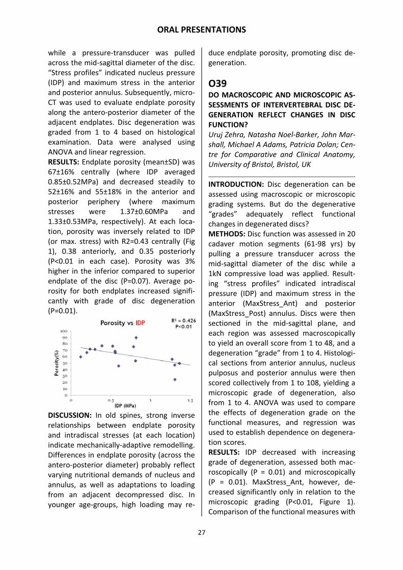
ORAL PRESENTATIONS
27
while a pressure‐transducer was pulled across the mid‐sagittal diameter of the disc. “Stress profiles” indicated nucleus pressure (IDP) and maximum stress in the anterior and posterior annulus. Subsequently, micro‐CT was used to evaluate endplate porosity along the antero‐posterior diameter of the adjacent endplates. Disc degeneration was graded from 1 to 4 based on histological examination. Data were analysed using ANOVA and linear regression. RESULTS: Endplate porosity (mean±SD) was 67±16% centrally (where IDP averaged 0.85±0.52MPa) and decreased steadily to 52±16% and 55±18% in the anterior and posterior periphery (where maximum stresses were 1.37±0.60MPa and 1.33±0.53MPa, respectively). At each loca‐tion, porosity was inversely related to IDP (or max. stress) with R2=0.43 centrally (Fig 1), 0.38 anteriorly, and 0.35 posteriorly (P<0.01 in each case). Porosity was 3% higher in the inferior compared to superior endplate of the disc (P=0.07). Average po‐rosity for both endplates increased signifi‐cantly with grade of disc degeneration (P=0.01).
DISCUSSION: In old spines, strong inverse relationships between endplate porosity and intradiscal stresses (at each location) indicate mechanically‐adaptive remodelling. Differences in endplate porosity (across the antero‐posterior diameter) probably reflect varying nutritional demands of nucleus and annulus, as well as adaptations to loading from an adjacent decompressed disc. In younger age‐groups, high loading may re‐
duce endplate porosity, promoting disc de‐generation.
O39 DO MACROSCOPIC AND MICROSCOPIC AS‐SESSMENTS OF INTERVERTEBRAL DISC DE‐GENERATION REFLECT CHANGES IN DISC FUNCTION? Uruj Zehra, Natasha Noel‐Barker, John Mar‐shall, Michael A Adams, Patricia Dolan; Cen‐tre for Comparative and Clinical Anatomy, University of Bristol, Bristol, UK
INTRODUCTION: Disc degeneration can be assessed using macroscopic or microscopic grading systems. But do the degenerative “grades” adequately reflect functional changes in degenerated discs? METHODS: Disc function was assessed in 20 cadaver motion segments (61‐98 yrs) by pulling a pressure transducer across the mid‐sagittal diameter of the disc while a 1kN compressive load was applied. Result‐ing “stress profiles” indicated intradiscal pressure (IDP) and maximum stress in the anterior (MaxStress_Ant) and posterior (MaxStress_Post) annulus. Discs were then sectioned in the mid‐sagittal plane, and each region was assessed macroscopically to yield an overall score from 1 to 48, and a degeneration “grade” from 1 to 4. Histologi‐cal sections from anterior annulus, nucleus pulposus and posterior annulus were then scored collectively from 1 to 108, yielding a microscopic grade of degeneration, also from 1 to 4. ANOVA was used to compare the effects of degeneration grade on the functional measures, and regression was used to establish dependence on degenera‐tion scores. RESULTS: IDP decreased with increasing grade of degeneration, assessed both mac‐roscopically (P = 0.01) and microscopically (P = 0.01). MaxStress_Ant, however, de‐creased significantly only in relation to the microscopic grading (P<0.01, Figure 1). Comparison of the functional measures with

ORAL PRESENTATIONS
28
overall scores of degeneration showed that both IDP and MaxStress_Ant fell linearly with increasing macroscopic score (RSq = 0.46, P<0.01 and RSq = 0.25, P = 0.02 re‐spectively). Furthermore, IDP, Max‐Stress_Ant and MaxStress_Post all de‐creased with microscopic score (RSq = 0.47, P<0.01; RSq = 0.20, P = 0.02; RSq = 0.38, P = 0.01, respectively).
DISCUSSION: Macroscopic and microscopic “grades” of disc degeneration both reflect changes in disc function. However, altered function was better predicted by detailed “scores” of degeneration, especially when those scores were based on microscopic observations.
O40 LUMBAR DISC AND FACET JOINT DEGEN‐ERATION: A LONGITUDINAL IN VIVO STUDY Hidetoshi Nojiri, MD, PhD; Alejandro A. Es‐pinoza Orías, PhD; Louis Fogg, PhD; Gunnar B.J. Andersson, MD, PhD; Howard S. An, MD; Nozomu Inoue, MD, PhD; Rush Univer‐sity Medical Center, Chicago, IL 60612
INTRODUCTION: Lumbar spine degenera‐tion is a leading source of physical disability. In spite of general consensus that aging re‐sults in disc height (DH) and facet joint space width (FJSW) alterations, their pro‐gression and correlations are not well un‐derstood.
METHODS: Twenty‐three subjects (mean age 40.6±8.7, range 24.3‐54.8 years old) underwent MR for disc grading and CT for facet joint grading and morphologic meas‐urements twice over a mean follow‐up pe‐riod of 5.96 years. DH and FJSW were measured from high‐resolution, subject‐specific 3D models and multivariate statis‐tics analysis was used to probe the data for interactions and level‐effects. RESULTS A total of 110 discs from the L1/2 to L5/S1 levels were evaluated. Of these, 12.8% exhibited disc grade deterioration, and a mean DH loss of 0.51 mm (7.3% of the initial height) when assessed at follow‐up. DH decreases began in Grade 2 discs. DH reduction in middle‐aged adults was larger than in younger subjects. 17.8% exhibited deterioration of the joint status in CT grad‐ing and FJSW decreased at an average of 0.10 mm (6.8% of the initial width) in 214 facet joints. Facet narrowing was found to be non‐uniform across the facet surface, as shown by local zone topographic analyses. Facet joints with low initial grades showed significantly larger decreases in facet gap than those with worse initial scores (p=0.01). Of note, younger subjects experi‐enced significant reductions in FJSW (p = 0.0001), whereas middle‐aged adults did not (p=0.49). There was a weak inverse cor‐relation between changes in the DH and FJSW (r=‐0.339). Multivariate statistics anal‐yses confirmed the multifactorial nature of spine degeneration. DISCUSSION Both DH and FJSW narrowing begin at an early age, but the degenerative processes follow different paths when low back pain symptoms are considered.
O41 EFFECT OF ANTI‐BFGF NEUTRALIZING AN‐TIBODY ON NEUROPATHIC PAIN INDUCED BY SPINAL NERVE LIGATION IN RATS Hisako Fujimaki, Gen Inoue, Kentaro Uchida, Hiroyuki Sekiguchi, Masaki Ueno, Wataru Saito, Naonobu Takahira, Masashi Takaso;

ORAL PRESENTATIONS
29
Department of Orthopaedic Surgery, Kita‐sato University School of Medicine, Kana‐gawa, Japan
INTRODUCTION: Basic fibroblast growth factor (bFGF) is a potent trophic factor for neurons and astrocytes that has recently been reported to be a pain transmitter. We evaluated the expression of bFGF and glial activation in the rat spinal nerve ligation model and investigated the potential of anti‐bFGF neutralizing antibodies as a treatment for neuropathic pain. METHODS: Sprague‐Dawley rats (5 weeks old) underwent L5 spinal nerve ligation. To evaluate the contribution of bFGF in neuro‐pathic pain, we injected neutralizing anti‐bodies against bFGF (40 ul) intrathecally twice a week (experimental group). Addi‐tional rats were injected with PBS (control group). Two weeks after surgery, ligated left L5 spinal nerves, bilateral L5 dorsal root ganglia (DRG), and spinal cords were pro‐cessed for immunohistochemistry, western blotting, and RT‐qPCR for bFGF, glial fibrilla‐ry acidic protein (GFAP, an astrocytic mark‐er), and Iba1 (a marker of microglia or mac‐rophages). Additionally, von Frey tests were conducted post‐surgery. RESULTS: Following injection of the neutral‐izing antibody, bFGF expression was signifi‐cantly suppressed in the experimental group at both the mRNA and protein levels. GFAP and Iba1 expression was also sup‐pressed. In immunohistochemical analyses, bFGF expression in astrocytes in the dorsal horn and in neuronal cells in the left DRG was suppressed. Significantly fewer astro‐cytes and microglia were present in the dor‐sal horn of the experimental group. Finally, post‐injury allodynia was alleviated in the experimental group. DISCUSSION: Neutralizing antibodies sup‐pressed both peripheral and central expres‐sion of bFGF, reduced glial activation, and alleviated allodynia. These results suggest that bFGF may play an essential role in neu‐
ropathic pain, and that anti‐bFGF treatment may facilitate recovery of neurological func‐tion and peripheral axon regeneration. bFGF could represent a novel therapeutic target for the treatment of chronic neuropathic pain.
O42 WHERE DO SMOKERS HURT? A COMPRE‐HENSIVE BODY‐WIDE ANALYSIS OF PAIN REVEALS SMOKING IS MOST ASSOCIATED WITH BACK PAIN. Matthew Smuck, MD; Christy Thomkins‐Lane, PhD; Ming‐Chih Kao, PhD, MD; Stan‐ford School of Medicine, Departments of Orthopaedic Surgery and Anesthesiology; Mount Royal University, Department of Physical Education and Recreation
INTRODUCTION: Smoking has been shown to increase the odds of back pain by a factor of two to three fold. To elucidate its associ‐ation with back pain relative to other mus‐culoskeletal body pain, we perform a body‐wide analysis of pain in a large population‐based cohort. In addition, we study the mit‐igating effects of physical activity, a protec‐tive factor of back and musculoskeletal pain. METHODS: Data on 6,781 subjects from the National Health and Nutrition Examination Survey was obtained from the CDC: com‐prehensive pain reports, smoking history, demographics, anthropometrics, medical history, and 7‐day physical activity meas‐urements. With custom SAS macros (Cary, NC) and Python 2.7, we performed weighted multivariate logistic regression analyses. RESULTS: After adjusting for demographic, anthropometric, and medical variables, sig‐nificant associations of smoking were ob‐served in all body regions except chest and foot pain. When clustered as axial (neck, upper and low back), appendageal, truncal, and head pain, this association is strongest with axial pain (OR 2.89, 95% CI 2.21‐3.77),

ORAL PRESENTATIONS
30
compared to appendageal (OR 1.99), truncal (OR 2.17) and head (OR 2.47). Mitigating effects were seen for sustained light physi‐cal activity (7‐day average light activity bout) with each SD increase association with 22.2% reduction of the risk from smok‐ing on truncal pain (p= 0.070), and 14.6% reduction for appendageal pain (p=0.038). This effect was not seen for head or axial pain. CONCLUSION: This population‐based study examined the association of smoking with musculoskeletal and regional body pain, finding statistical associations between smoking and pain and nearly all body re‐gions. The association between smoking and pain was particularly strong for axial pain. In addition, sustained light physical activity can mitigate substantial portions of this risk for truncal and appendageal pain, but it has no effect on smoking’s link to axial pain.
O43 WORKAHOLISM AS A RISK FACTOR FOR DEPRESSION AND DISABLING BACK PAIN AMONG JAPANESE WORKERS Ko Matsudaira(1), Takayuki Sawada(2), Norimasa Kikuchi(2), Emiko Sato(2), Mari Suzuki(2).; (1) Clinical Research Center for Occupational Musculoskeletal Disorders, Kanto Rosai Hospital, 1‐1 Kizukisumiyoshi‐cho, Nakahara‐ku, Kawasaki, Kanagawa 211‐8510, Japan (2) CLINICAL STUDY SUP‐PORT, INC., Nagoya Life Science Incubator, 2‐22‐8, Chikusa, Chikusa‐ku, Nagoya, Aichi 464‐0858, Japan
INTRODUCTION: Although it is well‐known that work‐related factors, including job de‐mands, job control, and workplace support, are associated with workers’ health and well‐being including low back pain (LBP), the role played by personal characteristics, especially workaholism (WH), has not been fully examined. This study investigated
WH’s associations with depression and LBP with disability among Japanese workers. METHODS: A cross‐sectional Internet survey was conducted using self‐administered questionnaires. Data from 3,899 Japanese workers were analyzed. WH was measured using the Dutch Workaholism Scale. Scores were divided into tertiles, where respond‐ents were classified into three groups (high, middle, and low). Depression was assessed using the SF‐36 mental health subscale. Dis‐abling back pain was defined as LBP that had occurred within one year and caused disruption to a worker’s job. Multiple lo‐gistic regression analyses were conducted to examine the association between worka‐holism and depression, low back pain with disability, adjusting for demographic charac‐teristics, job demand, job control, and workplace support. RESULTS: Compared to the low WH group, the middle and high WH groups had signifi‐cantly higher odds for depression (Odds ratio (OR) = 1.93 and 3.59 for the middle and high groups, respectively), disabling back pain (ORs = 1.38 and 1.90 for the mid‐dle and high groups, respectively). DISCUSSION: WH is significantly associated with not only poor psychological health, but also disabling back pain. To our knowledge, this is the first study to find an association specifically between WH and LBP. The mechanism by which WH might lead to dis‐abling LBP is unclear. However, since psy‐chological factors are known to be associat‐ed with LBP, poor psychological health caused by WH may play a role. WH needs to be studied further as a risk factor for LBP, along with other psychosocial factors.
O44 LIMITS OF HIGH FIELD MRI FOR THE DE‐TECTION OF INTERVERTEBRAL DISC LE‐SIONS – A COMPARISON BETWEEN 11.7 T AND 3 T Nikolaus Berger‐Roscher (1), Fabio Galbuse‐ra (1,2), Volker Rasche (3,4), Hans‐Joachim

ORAL PRESENTATIONS
31
Wilke (1); (1) Institute of Orthopaedic Re‐search and Biomechanics, Ulm University, Ulm, Germany (2) IRCCS Istituto Ortopedico Galeazzi, Milan, Italy (3) Department of In‐ternal Medicine II, University Hospital Ulm, Ulm, Germany (4) Small Animal MRI, Medi‐cal Faculty, Ulm University, Ulm, Germany
INTRODUCTION: It is assumed that fissures in the intervertebral disc lead to instability, which is an indication for surgery. Until now, investigations by discography, aimed to detect fissures, are under criticism and not recommended. Therefore, alternative methods, such as MRI, have to be investi‐gated. We want to evaluate the limits of clinically available systems and the capabili‐ties of state of the art techniques to see how clinical systems may evolve in the near future. METHODS: Three fresh frozen bovine tail discs (Cy2 – Cy3) were used for analysis. Six needles with different diameters (0.3 – 2.2 mm / 30 – 14G) were inserted 13 mm deep into the anulus with a custom made device in anteroposterior direction in the mid‐transverse plane of the disc with a distance of 3 mm to each other. The needles were then removed and the discs were scanned in an 11.7 T MRI (Bruker, USA; Res.: 0.058 x 0.058 x 0.625 mm3, tscan: 31 min.) as well as in a 3 T MRI (Philips, Netherlands) with an enhanced clinical protocol (Res.: 0.44 x 0.44 x 0.50 mm3, tscan: 12 min.). The scanned images were analyzed for lesion volume and lesion length using specialized software. RESULTS: Reconstructed lesions did not have an ideal cylindrical shape. The meas‐ured volumes of the lesions ranged from 1.24 to 11.5 mm3 (11.7 T) and 0.96 to 8.45 mm3 (3 T). The lengths ranged from 5.7 to 10.0 mm (11.7 T) and 1.2 to 5.1 mm (3 T), respectively (Figure).
Even lesions of 0.3 mm were clearly visible in 11.7 T, whereas in 3 T scans smaller le‐sions (= 0.5 mm) were not or almost not visible. DISCUSSION: The reconstructed volumes correlated, but were smaller than the corre‐sponding needle size. These differences may be attributed to the elastic collapse of the soft tissue. Results showed that diagnosis of degenerative disc disease may be improved with more modern MRI and enhanced pro‐tocols.
O45 LONG‐TERM RESULTS OF TOTAL LUMBAR DISC REPLACEMENT: A PROSPECTIVE ANALYSIS WITH 5‐10 YEAR FOLLOW‐UP Siepe CJ, Wiechert K, Heider F, Korge A, Mehren C, Mayer HM; Schön Clinic Munich Harlaching, Spine Center, Harlachinger Str. 51, D‐81547 Munich, Germany
INTRODUCTION: The role of fusion of lum‐bar motion segments for the treatment of intractable LBP from DDD remains contro‐versially debated. Total lumbar disc re‐placement (TDR) has been used as an alter‐native in a highly selected patient cohort. However, the amount of long‐term FU data on TDR is limited. MATERIALS AND METHODS: Outcome Pa‐rameters VAS, ODI and satisfaction rates were acquired within the framework of an ongoing prospective clinical trial. Profes‐sional activity / employment status, compli‐cations and reoperations were recorded.

ORAL PRESENTATIONS
32
RESULTS: 181 out of 201 patients were available for final FU (90.0% FU rate) after a mean FU of 7.4 years (range 5.0–10.8 yrs). VAS and ODI scores revealed a highly signifi‐cant improvement (p<0.0001). VAS scores demonstrated a slight (VAS 2.6 to 3.3) but statistically significant deterioration from 48 months onward (p<0.05). Patient satisfac‐tion rates remained stable throughout the postoperative course, with 63.6% reporting a ´highly satisfactory´ or ´satisfactory out‐come´ (22.7%), whilst 13.7% were not satis‐fied. The overall complication rate was 14.4% (n=26/181). The incidence of revision surgeries for general and/or device related complications was 7.2% (n=13/181). Results for 2‐level TDR were significantly inferior in comparison to 1‐level cases and were associated with higher complication (11.9% vs. 27.6%; p=0.03) and inferior satis‐faction rates (p<0.003). CONCLUSION: Although the current data from one of the first European Centers to employ TDR comprises the early experienc‐es and learning curve associated with a new surgical technique, the results demonstrate satisfactory mid to long‐term outcome after a mean FU of 7.4 years. Patient safety was proven with acceptable complication and reoperation rates. Fear of excessive late complications or reoperations cannot be substantiated with the present data. In carefully selected cases, TDR can be consid‐ered a viable treatment alternative to lum‐bar fusion.
O46 SINGLE LEVEL LUMBAR FUSION IN WORK‐ER’S COMPENSATION SUBJECTS FOR DE‐GENERATIVE DISC DISEASE IS ASSOCIATED WITH LOWER RETURN TO WORK RATES COMPARED TO FUSION FOR SPONDYLO‐LISTHESIS Joshua T. Anderson, BS (1,2), Ryan J. Duff (3), Uri M. Ahn, MD (4), Nicholas U. Ahn, MD (1); 1 University Hospitals Case Medical Center Department of Orthopaedic Surgery
2 Case Western Reserve University School of Medicine 3 University of Minnesota Twin Cities 4 New Hampshire Spine Institute
INTRODUCTION: Clinical outcomes of lum‐bar fusion are difficult to predict, and work‐er’s compensation (WC) subjects undergo‐ing this surgery consistently have worse outcomes when compared to non‐WC sub‐jects. Few studies have evaluated predictors of poor fusion outcomes in WC subjects. METHODS: We used ICD‐9 diagnosis codes and CPT procedural codes to identify 1851 subjects receiving medical benefits from the Ohio Bureau of Worker’s Compensation that underwent single level lumbar fusion surgery as their index fusion after injury for the primary indication of either spondylolis‐thesis or degenerative disc disease (DDD), each with 3 years of follow‐up minimum. We determined which subjects were able to return to work within 2 years after fusion and maintain a returned to work status for 1 year minimum. Subjects with a positive smoking history and a pre‐fusion diagnosis of failed back syndrome were not consid‐ered. We used a logistic regression analysis. RESULTS: Return to work rates were signifi‐cantly higher in patients with a pre‐fusion diagnosis of spondylolisthesis when com‐pared to those with a pre‐fusion diagnosis of degenerative disc disease and discogenic low back pain without spinal deformity or instability (p < 0.001). The odds ratio was 1.42 (CI 1.16‐1.74). Age significantly influ‐enced return to work rates (p = 0.01). The odds ratio (0.99, CI 0.98‐1.00) suggests that this impact was not considerable. Gender, obesity, and income were not significant. 375 of the 714 spondylolisthesis subjects (53%) and 494 of the 1137 DDD subjects (43%) returned to work. DISCUSSION: We demonstrated that per‐forming single level fusion surgery for the indication of degenerative disc disease and discogenic low back pain in worker’s com‐pensation subjects is associated with lower

ORAL PRESENTATIONS
33
return to work rates when compared to WC subjects undergoing fusion for spondylolis‐thesis. Further studies of the WC population are needed to determine which patients are the best candidates for fusion surgery.
O47 UNIVERSAL NO FAULT COMPENSATION IS ASSOCIATED WITH IMPROVED RETURN TO WORK RATES IN SPINE FUSION Manson JF, Montgomery AS, Cunningham JE, Landham PR, Don AS, Robertson PA.; Auckland Hospital Orthopaedic Department, Auckland, New Zealand
INTRODUCTION: Worker’s or injury com‐pensation is an unfavorable prognostic fac‐tor for return to work (RTW) after spinal fusion. An adversarial environment, en‐gagement of lawyers, and a need to display disability to achieve financial gain are likely factors. Universal no fault compensation improves outcomes in whiplash, and im‐proves functional gains from spine fusion to the equivalent of non‐compensation pa‐tients. This study examined RTW rates for spine fusion patients in New Zealand, where a no fault universal compensation system (Accident Compensation‐ACC) exists. METHODS: 108 lumbar spine fusion pa‐tients who received ACC coverage complet‐ed a phone interview. Work related param‐eters were assessed. A meta‐analysis of RTW rates for compensation patients un‐dergoing lumbar fusion surgery was per‐formed.
RESULTS: 42% of patients were injured in the work place. 88% were employed at the time of injury. 57% continued to work until surgery. 77% returned to work at an aver‐age of 8.7 months post surgery. Physical demands of the work were the same in 52%, less demanding in 38% and more de‐manding in 10%. Non‐RTW was due to pain in 10, other medical problems in 2, unavail‐ability of work in 2, retirement in 8, and unknown in 3. Literature meta analysis demonstrates a mean RTW rate in compen‐sation patients that is dramatically inferior to the NZ rates for ACC patients. The RTW rate for NZ ACC patients is comparable with the RTW rates for non‐compensation pa‐tients elsewhere. DISCUSSION: This study suggests that RTW rates are dramatically improved in a univer‐sal no fault compensation environment, matching the RTW rates for non‐compensation patients elsewhere. The gen‐erally accepted poor RTW rates for spine fusion in compensation patients relate to the type of compensation rather than the presence of compensation itself.
O48 THE ASSOCIATION BETWEEN MORPHO‐LOGICAL STENOSIS GRADE AND THE OUT‐COME OF SURGERY FOR LUMBAR SPINAL STENOSIS. Mannion AF, Fekete T, Luca A, Schizas C; Spine Center, Schulthess Klinik, Zürich, Swit‐zerland, Service de d'orthopédie et trauma‐tologie CHUV, Lausanne, Switzerland
INTRODUCTION: A new measure of radio‐logical lumbar spinal stenosis (LSS) was re‐cently introduced, based on grading of the rootlet/cerebrospinal fluid ratio rather than the dural sac cross‐sectional area. However, there has been no external validation of the measure or the rationale for its use as a surgical indication. We sought to validate the new grading system by relating patient outcomes to the stenosis grade.

ORAL PRESENTATIONS
34
METHODS: Blinded T2 axial magnetic reso‐nance images were evaluated from 65 pa‐tients (72±8y) with LSS and 15 “controls” (54±15y) with e.g. disc degeneration. Classi‐fication was based on dural sac morphology: Grades A and B showed cerebrospinal fluid presence while grades C and D showed none. In examining the correlation between outcomes (Core Outcome Measures Index (COMI)) and stenosis grades, the worst grade of all vertebral levels was used. RESULTS: In the control group, 15/15 (100%) patients were (at worst) grade A. In the LSS group, 14/65 (21.5%) were grade A, 8/65 (12.3%) grade B, 29/65 (44.7%) grade C, and 14/65 (21.5%) grade D. Increasing stenosis grade showed a tendency (n.s.) to correlate with higher preoperative pain (worst out of back or leg pain), but not with any other baseline outcome measures. Mul‐tiple regression analysis, accounting for number of affected levels, revealed signifi‐cant (p<0.01) associations between stenosis grade and the change‐score (reduction, preop to 12 mo) for pain and COMI score. In each case, a higher grade resulted in a bet‐ter outcome. The difference between A and D grades in the change‐scores for COMI (2.6 points) and worst pain (3.9 points) were clinically relevant. DISCUSSION: This study suggests that the new grading system, which gives most con‐sideration to the impingement of neural tissue, may represent an appropriate clinical tool and be of prognostic value. It should be evaluated in larger prospective studies.
O49 PREOPERATIVE PAIN PATTERN PREDICTS SURGICAL OUTCOME MORE THAN TYPE OF SURGERY IN PATIENTS WITH CENTRAL SPI‐NAL STENOSIS WITHOUT CONCOMITANT SPONDYLOLISTHESIS A REGISTER STUDY OF 9,051 PATIENTS Sigmundsson FG, Jönsson B, Strömqvist B.; Department of Orthopedics, Clinical Scienc‐es, Lund University, Skåne University Hospi‐
tal, Malmö, Sweden
INTRODUCTION: Predominant back pain in lumbar stenosis (LSS) is associated with in‐ferior outcome of surgical treatment. It is unknown if adding spinal fusion improves outcomes. The aim of our study was to evaluate outcome of surgery for LSS without concomitant degenerative spondylolisthesis in relation to predominance of pain and to analyze the if adding spinal fusion to the decompression improves outcome in pa‐tients with predominant leg or back pain. METHODS: In a register study of 9,051 pa‐tients we studied outcome of surgery in terms of back and leg pain (VAS), function (ODI and self‐estimated walking distance), health‐related quality of life (SF‐36 and EQ‐5D), and satisfaction with outcome. Out‐come was analyzed for 4 groups at 1 and 2 year follow up; preoperative back pain = leg pain and decompression, preoperative back pain = leg pain and decompression and fu‐sion, preoperative back pain < leg pain and decompression, preop back pain < decom‐pression and fusion. RESULTS: Patients with concomitant fusion were younger and had higher level of back pain and ODI scores and lower preoperative EQ‐5D. Predominant back pain was associ‐ated with inferior outcome in terms of pain, HRQoL and function. Patients most often satisfied (69%) were patients with back pain < leg pain treated with decompression and fusion and the least satisfied group was pa‐tients with back pain = leg pain treated with decompression (54%). Fusion was associat‐ed with higher EQ‐5D at 1‐year follow up for patients with predominant back pain up but was also associated with increased leg pain at 2‐ year follow up in patients with pre‐dominant leg pain. Patients with predomi‐nant back pain experienced small gains in the physical component summary by fusion. The benefits of fusion generally decreased when adjustments were made for con‐founders.

ORAL PRESENTATIONS
35
DISCUSSION: Predominance of back pain infers inferior outcome of LSS surgery. Add‐ing spinal fusion improves outcome but the benefit is small and not clinically relevant.
O50 USE OF LUMBAR DISCOGRAPHY IS ASSOCI‐ATED WITH SIGNIFICANTLY LOWER RE‐TURN TO WORK RATES AFTER LUMBAR FUSION IN WORKER’S COMPENSATION SUBJECTS Joshua T. Anderson, BS (1,2), Ryan J. Duff (3), Uri M. Ahn, MD (4), Nicholas U. Ahn, MD (1); 1 University Hospitals Case Medical Center Department of Orthopaedic Surgery 2 Case Western Reserve University School of Medicine 3 University of Minnesota Twin Cities 4 New Hampshire Spine Institute
INTRODUCTION: Lumbar discography (LD) is an injection technique used to evaluate pa‐tients being considered for back surgery, typically lumbar fusion. Recent studies have questioned its use and thrown LD into sig‐nificant controversy. Our study is the first to evaluate the efficacy of LD in worker’s com‐pensation (WC) subjects. METHODS: We used ICD‐9 and CPT codes to identify 2887 subjects from the Ohio Bureau of Worker’s Compensation that underwent lumbar fusion following injury with 3 years of follow‐up minimum. 1003 subjects un‐derwent fusion for the indication of spondy‐lolisthesis, 118 of which had LD performed on them before fusion. 1884 subjects un‐derwent fusion for degenerative disc dis‐ease (DDD), 756 of which had LD performed on them before fusion. We determined which subjects returned to work within 2 years of fusion and maintain a returned to work status for 1 year minimum. Subjects with a smoking history and pre‐fusion diag‐nosis of failed back syndrome were not con‐sidered. We analyzed the spondylolisthesis group and the DDD group separately with logistic regression.
RESULTS: Return to work (RTW) rates were significantly lower in subjects that under‐went LD before fusion in both the spondylo‐listhesis group (p=0.05; OR=0.65) and the DDD group (p<0.00; OR=0.71). Among the spondylolisthesis group, 453 subjects (51.2%) that did not undergo LD before fu‐sion returned to work. 47 LD subjects (39.8%) returned to work. Male gender was associated with higher RTW rates (p=0.05, OR=1.34). Among the DDD group, 515 sub‐jects (45.7%) that did not undergo LD before fusion returned to work. 286 LD subjects (37.8%) returned to work. Older age was associated with lower RTW rates (p=0.01, OR=0.99). In both groups, obesity and levels fused at index fusion did not significantly impact RTW rates. DISCUSSION: Use of lumbar discography as a patient selection tool before lumbar fu‐sion in worker’s compensation subjects is associated with significantly worse return to work rates.
O51 PROSPECTIVE ANALYSIS OF RISK FACTORS FOR PROXIMAL JUNCTIONAL FAILURE IN ADULT DEFORMITY PATIENTS Hart, Robert A.1; Hamilton, D. Kojo1; Hiratzka, Jayme R.1; Bess, Shay 2; Schwab, Frank J.3; Shaffrey, Christopher I.4; Ames, Christopher P.5; Lafage, Virginie3; Mum‐maneni, Praveen V.; Smith, Justin S.4; Kline‐berg, Eric7; McCarthy, Ian8; Burton, Douglas C.; 1. Orthopaedic Surgery, Oregon Health and Science University, Portland, OR, United States. 2. Orthopaedic Surgery, Rocky Mountain Hospital for Children, Denver, CO, United States. 3. Orthopaedic Surgery, NYU Hospital for Joint Diseases, New York, NY, United States. 4. Neurosurgery, University of Virginia Medical Center, Charlottesville, VA, United States. 5. Neurosurgery, University of California, San Francisco Medical Center, San Francisco, CA, United States. 6. Ortho‐paedic Surgery, University of California, San Francisco Medical Center, San Francisco, CA,

ORAL PRESENTATIONS
36
United States. 7. Orthopaedic Surgery, Uni‐versity of California, Davis, Sacramento, CA, United States. 8. Baylor Health Care System, Institute for Health Care Research and Im‐provement, Plano, TX, United States. 9. Or‐thopaedic Surgery, University of Kansas Medical Center, Kansas City, KS, United States. 10. Orthopaedic Surgery, Baylor Sco‐liosis Center, Plano, TX, United States. 11. ISSGF, Littleton, CO, United States.
INTRODUCTION: Proximal Junctional Failure (PJF), a severe form of Proximal Junctional Kyphosis (PJK) with evidence of mechanical failure, is an important concern in adult de‐formity patients. Post‐operative sagittal imbalance and proximal and distal end points for the fusion are possible risk factors for PJF. Prospective evaluation of these fac‐tors has not been reported. METHODS: 352 adult deformity surgical patients from 10 centers were followed prospectively with minimum 1 year follow‐up. PJF was defined as increased proximal kyphosis of >10 degrees plus fracture of the upper instrumented vertebrae (UIV), UIV+1, or instrumentation failure. PJK was defined as increased kyphosis of >10 degrees with‐out evidence of mechanical failure. Patients were grouped as PJF, PJK, or neither (NoPJF). Proximal fusion levels were Upper Thoracic (UT, T2‐T5) or Thoracolumbar (TL, T9‐T12). Age, Sacral Slope (SS), Pelvic Tilt (PT), Pelvic Incidence (PI), Sagittal Vertical Axis (SVA), Lumbar Lordosis (LL), and PI‐LL were compared. RESULTS: There were 41 (11.7%) PJF, 54 (15.3%) PJK, and 257 (73.0%) NoPJF patients found. There were significant differences in age (65.1 vs. 55.7; p =.001) and pre‐op PT (26.8 vs. 22.0; p=.043) between PJF and NoPJF patients. There was a trend toward increased pre‐op SVA and PI‐LL, as well as operative change in SVA, LL, and PI‐LL among PJF patients. Postop SVA and PI‐LL were similar between PJF and no PJF pa‐tients. No patient experienced PJF without
fusion to the pelvis. Among patients fused to the pelvis, there was no difference be‐tween UT vs. LT proximal endpoints for rate of PJF (13.4% vs. 16.8%; p>.05), although the rate of PJK was greater for UT patients (26.8% vs. 13.5%; p=.017). DISCUSSION: Post‐op sagittal alignment and proximal fusion end point did not differ be‐tween PJF and NoPJF patients. Older pa‐tients with worse pre‐op sagittal imbalance, larger sagittal corrections and pelvic fixation were identified as those at greatest risk of PJF.
O52 DISCOGRAPHY AND AXIALLY LOADED MRI IN THE PROVOCATION OF DISCOGENIC PAIN ‐ A COMPARATIVE PILOT STUDY. Hanna Hebelka MD* and Tommy Hansson MD, Ph.D**.; Departement of Radiology*, Sahlgrenska University Hospital, SU/M and Department of Orthopedics**, Sahlgrenska University Hospital, SU/S, University of Gothenburg, Gothenburg, Sweden.
INTRODUCTION: Recent findings of induced pressure increase in adjacent discs during discography strongly question the validity of discography. Alternative diagnostic tools seem necessary. The aim of this study was to investigate if axial loaded MRI (alMRI) can induce discogenic pain and if positive discograms display any specific morphologi‐cal features under increased axial loading. METHODS: 41 patients with clinically as‐sessed discogenic pain were examined with MRI, alMRI and pressure controlled discog‐raphy (PCD) (119 discs) at the same occa‐sion. Provoked pain at discography and at alMRI was classified as concordant or dis‐cordant with the daily experienced pain as reference. PCD was performed until one of following endpoints; 100psi (pressure), 3.5ml (contrast volume) and/or concordant pain. Concordant discogram demanded pain intensity =5/10 at =50psi and a negative control disc. Associations between pain at

ORAL PRESENTATIONS
37
discography and at alMRI were evaluated as were relations between pain and morpho‐logical disc measures (degeneration, height, bulge, angle, area, and circumference) at MRI/alMRI/difference between MRI and alMRI. RESULTS: At discography 98% of the pa‐tients experienced concordant pain and 78% at alMRI with a significant correlation between the two (p=0.01). The PPV of a concordant discogram when concordant pain was evoked at alMRI was 97%. None of the morphological MRI features predicted painful discs in a clinically relevant way. DISCUSSION: Pain provoked at alMRI was highly predictive of corresponding pain at discography. No morphological MRI fea‐tures or dynamic properties predicting pain‐ful discs were found but alMRI may be a valuable tool in future research trying to establish such features. Provoking pain at a similar frequency as discography, being non‐invasive and without risk for injection induced degeneration, makes alMRI a realis‐tic alternative in the diagnosis of painful disc(s) at least when single level discogenic pain is suspected clinically.
O53 ANALYSIS OF VASCULAR PATTERNS FOR THE POSTERIOR ELEMENTS OF THE LOWER LUMBAR SPINE Fumitake Tezuka, Toshinori Sakai, Toshihiko Nishisho, Yoichiro Takata, Yuichiro Goda, Kosaku Higashino, Shoichiro Takao, Masashi Harada, Koichi Sairyo; Department of Or‐thopaedic Surgery, Tokushima University Department of Rehabilitation, Tokushima University Hospital Department of Radiolo‐gy, Tokushima University
INTRODUCTION: The Lower lumbar spine around L5 is the most susceptible to spon‐dylolysis or degenerative disorders such as disc herniation, spinal canal stenosis, spon‐dylolisthesis. In this study, to clarify the ana‐tomical differences affecting their
pathomechanism around L5 compared with the other lumbar spinal levels, we evaluated the vascular patterns in detail, particularly focused on them surrounding the posterior elements. METHODS: We evaluated 300 abdominal contrast‐enhanced multidetector 3D‐CT scans that were taken for surgical plans of colon cancer patients in our hospital. Ex‐traosseous vascular patterns surrounding the lumbar spine were evaluated by two orthopaedic surgeons independently. Three hundreds patients included 187 male and 113 female, whose age ranged from 15 to 89 years (mean: 66.6). RESULTS AND DISCUSSION: Segmental ar‐teries. On the L1‐L4 levels, each segmental artery was observed from the vertebra through the lamina in more than 90% pa‐tients. However, on the L5 level, it was ob‐served in less than 10% patients (Rt: 5.0%, Lt: 8.3%). Extraosseous vascular supply for the posterior element of the L5 Extraosse‐ous vascular supply for the L5 lamina was basically composed of two vessels on each side. One vessel was mostly derived from the L4 segmental artery (Rt: 92.0%, Lt: 91.7%) that was distributed around the su‐perior articular process, the other one was derived from iliolumbar artery (Rt: 63.0%, Lt: 57.3%) that was distributed around the inferior articular process through the lami‐na. There were some variations one those two vessels. Our results showed there were various vascular patterns around lower lumbar spine. Focusing on the posterior elements of the L5, there were only approx‐imately 60% subjects with regular vascular patterns that had been written in the old textbook. Although there are some limita‐tions of this study as the intraosseous vas‐cular was not evaluated, it is true that L5 has more various vascular patterns than the other l

ORAL PRESENTATIONS
38
O54 (ISSLS PRIZE PAPER) LONG‐TERM FOLLOW UP SUGGESTS SPINAL FUSION IS ASSOCIATED WITH INCREASED ADJACENT SEGMENT DISC DEGENERATION BUT WITHOUT INFLUENCE ON CLINICAL OUTCOME. RESULTS OF A COMBINED FOL‐LOW‐UP FROM 4 RCTS Anne F Mannion, Gunnar Leivseth, Jens‐Ivar Brox, Peter Fritzell, Olle Hägg, Jeremy CT Fairbank; Spine Center, Schulthess Klinik, Lengghalde 2, 8008 Zürich, Switzerland De‐partment Of Clinical Medicine, Neuromuscu‐lar Diseases Research Group, University of Tromsø, Norway Department of Orthope‐dics, Oslo University Hospital, Oslo, Norway Neuroortopediskt Centrum, Länssjukhuset Ryhov, 551 85 Jönköping, Sweden Spine Center Göteborg, Gruvgatan 8, SE 421 30, V:a Frölunda, Sweden Nuffield Orthopaedic Centre, Oxford University Hospitals, Oxford, OX3 7HE, UK
INTRODUCTION: There is ongoing debate as to whether adjacent segment disc degener‐ation results from the increased mechanical stress of fusion. We analysed long term fol‐low‐up (LTFU) data from four randomized controlled trials of operative versus non‐operative treatment for chronic low back pain to examine the influence of spinal fu‐sion on adjacent segment disc space height as an indicator of disc degeneration at LTFU. METHODS: Plain standing lateral radio‐graphs were taken at 13±4 years follow‐up in 229/464 (49%) patients randomized to surgery and 140/303 (46%) to non‐operative care. Disc space height and posteroanterior displacement were meas‐ured for each lumbar segment using a vali‐dated computer‐assisted distortion com‐pensated roentgen analysis (DCRA) tech‐nique. Values were reported in units of standard deviations (SDs) above or below age and gender‐adjusted normal values. Patient‐rated outcomes included the Oswestry Disability Index and pain scales.
RESULTS: Radiographs were usable in 355/369 (96%) patients (259 fusion and 96 non‐operative treatment). Both treatment groups showed significantly lower values for disc space height of the adjacent segment compared with norms. There was a signifi‐cant difference between treatment groups for the disc space height of the cranial adja‐cent segment (in both as‐treated and inten‐tion‐to‐treat analyses). The mean treatment effect of fusion on adjacent segment disc space height was ‐0.44 SDs (95% CI, ‐0.77 to ‐0.11; p=0.01; as‐treated analysis); there was no group difference for posteroanterior displacement (0.18 SDs (95% CI, ‐0.28 to 0.64, p=0.45)). Adjacent level disc space height and posteroanterior displacement were not correlated with Oswestry or pain scores at LTFU (r=0.010‐0.05; p>0.33). DISCUSSION: Fusion was associated with lower disc space height at the adjacent segment after an average of 13 years fol‐low‐up. However, the reduced disc space height had no influence on patient self‐rated outcomes (pain or disability).
O55 (ISSLS PRIZE PAPER) MECHANICAL INFLUENCES IN PROGRESSIVE INTERVERTEBRAL DISC DEGENERATION Manos Stefanakis PhD*, Jin Luo PhD**, Phil‐lip Pollintine PhD Patricia Dolan PhD, Mi‐chael A. Adams PhD; Centre for Comparative and Clinical Anatomy, University of Bristol, Bristol, U.K.* School of Science and Engi‐neering, University of Nicosia, Cyprus ** University of Roehampton, London, U.K.
INTRODUCTION: Although mechanical load‐ing can initiate disc degeneration, it may be unimportant in disease progression because degenerative changes cause human lumbar discs to be increasingly “stress‐shielded” by the neural arch. However, the most typical feature of advanced disc degeneration (de‐lamination and collapse of the annulus) may not depend on absolute values of compres‐sive stress, but on gradients of compressive

ORAL PRESENTATIONS
39
stress which act to shear annulus lamellae. We hypothesise that, as grade of disc de‐generation increases, stress gradients also increase, even though nucleus pressure and annulus compressive stresses decrease. METHODS: 191 motion segments (T8‐9 to L5‐S1) were dissected from 42 cadavers aged 19‐92 yrs. Each was subjected to ap‐proximately 1 kN compression, while intra‐discal stresses were measured by pulling a pressure transducer along the disc’s mid‐sagittal diameter. “Stress gradients” in the inner annulus were quantified as the maxi‐mum rate of increase in compressive stress (MPa/mm) between the nucleus and the region of maximum stress in the anterior or posterior annulus. Measurements were re‐peated before and after creep loading, and in simulated flexed and erect postures. Disc degeneration was assessed macroscopically on a scale of 1 to 4. RESULTS: As grade of disc degeneration increased from 2 to 4, nucleus pressure de‐creased by an average 68%, and maximum compressive stress in the annulus decreased by 48‐64%, depending on location and pos‐ture. In contrast, stress gradients in the an‐nulus increased by an average 75% in the anterior annulus (in flexed posture), and by 108% in the posterior annulus (in erect pos‐ture). Spearman rank correlation showed that these increases were statistically signif‐icant (P<0.05). DISCUSSION: Despite stress‐shielding by the neural arch, gradients of compressive stress increase as disc degeneration progresses. Stress gradients act to shear adjacent lamel‐lae, and can explain progressive annulus delamination and collapse
O56 (ISSLS PRIZE PAPER) INCREASED INNERVATION AND SENSORY NERVOUS SYSTEM PLASTICITY IN A MOUSE MODEL OF LOW BACK PAIN DUE TO IN‐TERVERTEBRAL DISC DEGENERATION. Masayuki Miyagi, MD, PhD1‐3,7 Magali Millecamps, PhD1‐3, Alexander T Danco,
MSc1‐3,6, Seiji Ohtori, MD, PhD7, Kazuhisa Takahashi, MD, PhD7, Laura S. Stone, PhD1‐6.; 1 Faculty of Dentistry, McGill University; 2Alan Edwards Centre for Research on Pain, 3McGill Scoliosis & Spine Research Group, McGill University; Departments of 4Anesthesiology, 5Pharmacology & Thera‐peutics, and 6Neurology & Neurosurgery, Faculty of Medicine, McGill University, Mon‐treal, Canada 7Department of Orthopaedic Surgery, Graduate School of Medicine, Chiba University, Chiba, Japan
INTRODUCTION: Intervertebral disc (IVD) pathology is thought to be a significant con‐tributor to chronic low back pain (LBP). In humans and rodents, decreased expression of SPARC (Secreted Protein, Acidic, Rich in Cysteine) is associated with IVD degenera‐tion. We previously reported that SPARC‐null mice exhibit age‐dependent behavioral signs of chronic axial LBP and radiating leg pain. The aim of the current study was to determine if behavioral signs of LBP and radiating pain in SPARC‐null mice are ac‐companied by sensory nervous system plas‐ticity. METHODS: SPARC‐null and age‐matched Wild‐Type control mice in three age cohorts (young, 1.5 months old: SPARC n=21, WT n=18; middle aged, 6 months old: SPARC n=18, WT n=16; and old, 24 months old: SPARC n=25, WT n=23) were evaluated. Cutaneous sensitivity to cold, heat, and me‐chanical stimuli were used as measures of radiating pain. Grip force and tail suspen‐sion tests were performed to evaluate axial LBP. Motor impairment was assessed using an accelerating rotarod. Innervation of IVDs was determined by immunohistochemistry targeting the nerve fiber marker PGP 9.5 and the sensory nerve neuropeptide calci‐tonin gene‐related peptide (CGRP). To as‐sess sensory neuron and spinal cord plastici‐ty, CGRP‐ and neuropeptide‐Y‐immuno‐reactivity (‐ir) were measured in dorsal root ganglia (DRG) neurons and GFAP‐, iba‐1‐

ORAL PRESENTATIONS
40
and CGRP‐ir were measured in the spinal cord. RESULTS: SPARC‐null mice developed (1) hypersensitivity to cold, (2) axial discomfort, (3) age‐dependent motor impairment, (4) age‐dependent increases in sensory nerve fibers in and around the IVDs, (5) age‐dependent upregulation of CGRP and neu‐ropeptide‐Y in DRGs, and (6) age‐dependent upregulation of CGRP, microglia and astro‐cytes in the spinal cord dorsal horn. DIS‐CUSSION: The increased innervation of de‐generating IVDs by sensory nerve fibers and the neuroplasticity in sensory neurons and spinal cord could contribute to the underly‐ing pathobiology of chronic discogenic LBP.
O57 CORRELATION BETWEEN BIOMECHANICAL PROPERTIES OF ANNULUS FIBROSUS AND MAGNETIC RESONANCE FINDINGS Zhi Shan M.D, Fengdong Zhao M.D, Chong‐yan Wang M.D, Junhui Liu M.D, Letu Suyou M.D, Shunwu Fan M.D.; Department of Or‐thopaedics, Sir Run Run Shaw Hospital, School of Medicine, Zhejiang University.
INTRODUCTION: Annulus fibrosus, located in the exterior region of intervertebral disc, is primarily a tensile component providing structural stability against multidirectional loads and deformations, but limited under‐standing associating its tensile properties with MRI findings has been reported in hu‐man. METHODS: 27 human cadaveric lumber spines were harvested (16 male, 11 female, mean age 44.3 years, range 31‐57). All spines underwent MRI examination. Then all intervertebral discs were removed from between adjacent motion segments, divid‐ed into parts as nucleus pulposus, anterior AF, anterolateral AF, lateral AF and poster‐olateral AF, and further divided into three subgroups averagely: inner, middle and outer for tensile test. RESULTS: 1738 speci‐mens from 158 discs, 27 cadavers were
tested. All degeneration and MCs visible by MRI are seen in L4‐L5 and L5‐S1. The mean tensile module of all tested specimens is 4.80±1.96MPa, 8.25±2.73MPa and 13.20±3.19MPa, for inner, middle and outer AF, respectively. The overall UTS are 1.13±0.54MPa, 1.73±0.87MPa and 3.37±1.30MPa, respectively. No significant age‐ or gender‐related change can be found. Tensile module and UTS all increased from the inner to outer annulus, with elon‐gation decreased. Tensile module and UTS all decreased with the severity of degenera‐tion increases, except the UTS of middle annulus. The outer annulus presents the highest UTS decreasing of 40% in severely degenerated disc, and in the middle and inner annulus the decrease was 18% and 28% respectively. When MCs presents, in‐ner annulus showed significantly lower module with MCs, while this difference did not appear in middle and outer annulus. The UTS presented decreased significantly in all inner to outer region when MCs ap‐pears. CONCLUSION: The tensile properties de‐creases with higher Pfirrmann grade of MRI. When MCs presents, AF showed lower ten‐sile module, especially the inner region.
O58 BIOMECHANICAL EVALUATION OF THE TRANSPEDICULAR NUCLEOTOMY WITH INTACT ANULUS FIBROSUS Fabrizio Russo1, Gianluca Vadala1, Robert Allen Hartman2, Kevin M. Bell2, Gwendolyn A. Sowa2, Nam Vo2, James D. Kang2, Vin‐cenzo Denaro, MD1; 1 Department of Or‐thopedic and Traumatology, University Campus Bio‐Medico of Rome, Rome ‐ Italy 2 Ferguson Lab for Orthopedic and Spine Research, University of Pittsburgh, Pitts‐burgh, PA ‐ USA
INTRODUCTION: Mechanical loading repre‐sents a crucial part of intervertebral disc (IVD) homeostasis. However, traditional

ORAL PRESENTATIONS
41
regenerative strategies require violation of the annulus fibrosus (AF) that results in sig‐nificant alteration of joint mechanics. The transpedicular nucleotomy represents a suitable method to create a cavity into the nucleus polposus (NP), as a model to study IVD regeneration with intact AF (Vadala` et al. 2013). The aim is to study how the transpedicular approach (TA) and the novel mechanical nucleotomy (MN) affect the biomechanics of the NP. METHODS: 60 ovine lumbar FSUs (L1‐L6) randomly assigned to 5 groups: control, TA, TA + polymethylmethacrylate (PMMA), TA + MN, TA + MN + PMMA. TA and MN were performed. The robotic spine testing system consists of a robotic manipulator (Staubli RX90) and a six‐axis load cell (JR3 Inc.). We evaluated flex‐extension, lateral bending and axial rotation under adaptive displace‐ment control. Axial compression was ap‐plied for 15 cycles of preconditioning fol‐lowed by 1 h constant compression. Creep properties were determined. RESULTS: TA has minimal effects on IVD biomechanics. MN increases ROM and NZ displacement width while decreasing NZ stiffness. TA + PMMA has small effects in terms of ROM. TA + MN + PMMA brings ROM back to the control, increases NZ stiff‐ness and decreases NZ displacement width. TA and MN primarily altered early creep response. The use of PMMA reduced viscos‐ity (S2) and increased stiffness (.2) in late creep. DISCUSSION: Biomechanical properties of NP are crucial for IVD repair. TA does not affect segmental stability. Mechanical changes that occur with nucleotomy are similar to those characteristics of early de‐generation. In particular, the NP play a cru‐cial role for spine stability and early creep response, while the late creep response may be determined more by the AF and endplate. These results could be crucial for understanding the mechanism of spine in‐stability caused by IVD degeneration.
O59 DOES DAILY TERIPARATIDE ENHANCE THE HEALING PROCESS OF OSTEOPOROTIC VERTEBRAL FRACTURE? EVALUATION US‐ING MRI AND BIOCHEMICAL MARKERS OF BONE TURNOVER Akira Iwata, MD 1; Masahiro Kanayama MD 1; Keiichi Shigenobu, MD 1; Fumihiro Oha, MD 1; Shingo Onda, MD 1; Masaru Tanaka, MD 1; Tomoyuki Hashimoto, MD 1; Nori‐masa Iwasaki, MD 2; 1 Spine Center, Hako‐date Central General Hospital, Hakodate, Japan 2 Dept. of Orthopedic surgery, Hok‐kaido University, Sapporo, Japan
INTRODUCTION: Teriparatide (recombinant human PTH 1‐34) is increasingly used for osteoporosis treatment, which stimulates bone formation and might have a potential advantage to enhance fracture healing. The current randomized controlled trial aims to evaluate the effect of teriparatide (TPD) versus bisphosphonate (BP) on the healing process of vertebral fracture using MRI ex‐amination and biochemical markers of bone turnover. METHODS: Twenty‐two osteoporosis pa‐tients who underwent non‐operative treat‐ment for a single‐level fresh thoracolumbar vertebral fracture were enrolled in this trial. They were randomly assigned to (1) TPD group (daily subcutaneous injection of 20 micrograms of TPD) or (2) BP group (weekly oral administration of 35mg alendro‐nate).The fracture healing process was as‐sessed by temporal changes in MRI T2 signal intensity of fractured vertebra with refer‐ence to the pre‐treatment baseline. BAP (bone formation marker) and TRACP5b (bone resorption marker) were measured periodically for biochemical evaluation of the fracture healing. RESULTS: With reference to the pretreat‐ment baseline, MRI T2 signal intensity of fractured vertebra was decreased by 46% at 3 months and 60% at 6 months in the TPD group; BP group showed 10% increase at 3

ORAL PRESENTATIONS
42
months and 20% decrease at 6 months. Re‐garding bone formation marker, BAP at 1, 2, 3 and 6 months after treatment was 44%, 73%, 132% and 103% in the TPD group, and 25%, 8%, 6%, 10% in the BP group, respec‐tively. TRACP5b as a bone resorption mark‐er was 84%, 78%, 65% and 64% in the TPD group, and 23%, 24%, 8% and 31% in the BP group, respectively. DISCUSSION: The current study demon‐strated that MRI signal intensity of fractured vertebra was returning to normal in the early phase in the TPD group. Bone for‐mation and resorption markers were ele‐vated at any phase of healing process in the TPD group. TPD enhances the healing and remodeling processes in fresh vertebral compression fractures related to osteopo‐rosis.
O60 USEFULNESS OF “ONE STRETCH”, A SIMPLE, DAILY, STANDING BACK EXTENSION EXER‐CISE, FOR THE PREVENTION OF ONSET OR AGGRAVATION OF LOW BACK PAIN IN CARE WORKERS Mari Suzuki (1), Ko Matsudaira (2), Takayuki Sawada (1), Emiko Sato (1), Tatsuya Isomu‐ra (1, 3).; (1) CLINICAL STUDY SUPPORT, INC., Nagoya Life Science Incubator, 2‐22‐8, Chikusa, Chikusa‐ku, Nagoya, Aichi 464‐0858, Japan (2) Department of Medical Re‐search and Management for Musculoskele‐tal Pain, 22nd Century Medical and Re‐search Center, Faculty of Medicine, The Uni‐versity of Tokyo, Japan (3) Institute of Medi‐cal Science, Tokyo Medical University, 6‐1‐1 Shinjuku, Shinjuku‐ku, Tokyo 160‐8402, Ja‐pan
INTRODUCTION: Low back pain (LBP) is a worldwide health problem. Recently, exten‐sion of the back has been recognized as an effective exercise to prevent back problems. We suggested “One Stretch”, a simple, dai‐ly, standing back extension exercise, and
examined its efficacy to improve or prevent LBP in Japanese care workers. METHODS: This study was performed at a single health care facility in Japan. We pro‐vided 166 care workers with an exercise manual, where we described how to do the exercise “One Stretch” based on the McKenzie method, and assigned them to the intervention or control groups according to their working floors. Participants in the intervention group took a 30‐min seminar on LBP, and were encouraged to exercise on working hours. We mainly evaluated the change of severity of LBP and compliance with the exercise by using self‐administrated questionnaires at baseline and one year later. RESULTS: A total of 136 care workers com‐pleted the study (64 in the intervention group and 72 in the control group). 30 care workers were excluded from analysis due to lack of information. Baseline characteristics, including age, sex, mental health score of SF‐36, and severity of LBP, had no statisti‐cally significances between the groups. One year later, a rate of care workers who im‐proved LBP were 42.9% in the intervention group and 15.0% in the control group, and some care workers without LBP at baseline remained healthy condition. About compli‐ance with the exercise, the intervention group indicated higher proportion of care workers who frequently exercised than the control group (82.8% vs. 8.8%, p <0.01; chi‐square test). No care workers in the inter‐vention group had a first medical consulta‐tion or work disability in the year. DISCUSSION: The exercise “One Stretch” could improve or prevent LBP in Japanese care workers. For better compliance with the exercise, support from co‐workers would play an important role.

ORAL PRESENTATIONS
43
O61 PERSISTENT RACIAL AND ECONOMIC DIS‐PARITIES IN PHYSICAL THERAPY REFERRAL FOR LOW BACK PAIN PATIENTS IN PRIMA‐RY CARE Matthew Smuck, MD; Patricia Zheng, MD; Nicholas Karayannis, PhD; Ming‐Chih Kao, PhD, MD; Stanford School of Medicine, De‐partments of Orthopaedic Surgery and An‐esthesiology
INTRODUCTION: Evidence suggests that physiotherapist‐led exercise programs can decrease disability, but the rate of physical therapy referral in clinical practice has been dismal, averaging, about 7% for a primary diagnosis for low back pain (LBP). On the contrary, the rate of opioid prescription has steadily increased. This study examines the low referral rate with respect to socioeco‐nomic factors. METHODS: Nationally representative sur‐veys National Ambulatory and National Hospital Ambulatory Medical Care Surveys (NHAMCS, NAMCS) from the Center for Dis‐ease Control and Prevention (CDC) were investigated for PCP visits between the years 1997 and 2009. Diagnoses, prescrip‐tion medications, insurance source, and demographics were determined. Weighted logistic regression modeling with SAS esti‐mated covariate effects. RESULTS: Referrals have remained stable among PCPs from 1997 to 2006, in sharp contrast to our previous finding of increas‐ing rates of opioid prescription for LBP dur‐ing this time period. Among Medicaid and Medicare insured population and African Americans, referrals have decreased. The data supports evidence that there is a grow‐ing U.S. trend of providing cheaper forms of treatment for chronic LBP (i.e., opioid pre‐scription) which are less effective. Low re‐ferral rates for African‐American patients is also concerning. While the reasons for this disparity is unknown, it has been conjec‐tured that such differences result from
complex patient, health care provider and health care system factors . CONCLUSIONS: With more evidence now that PT is an effective method of treating LBP, there needs to be a push for greater insurance coverage of physiotherapy com‐mensurate with its low‐risk and high effica‐cy profile. In addition, disparities in subpop‐ulations must be further elucidated and addressed. As the Affordable Care Act is being implemented, we must work to as‐sure that PT access is improved, especially for those patients suffering from LBP.
O62 ASSESSMENT OF NEUROFORAMINAL AREA OF THE LUMBAR SPINE DURING FLEXION‐EXTENSION MOTION Havey R, Muriuki M, Carandang G, Voronov L, Patwardhan A; Edward Hines, Jr. VA Hos‐pital, Hines, IL, USA Loyola University Medi‐cal Center, Maywood, IL, USA
INTRODUCTION: Degenerative changes of the lumbar spine affect the area available for the nerve roots. The area is greatly af‐fected by the spine posture. This study as‐sessed foraminal area of lumbar spines un‐dergoing flexion‐extension (FE) range of motion (ROM) testing. METHODS: Six human cadaveric spines (L1‐Sacrum, 40.2±6.9 years) were studied. Each specimen was instrumented with five 3.2mm spherical radiopaque markers per body. A 3‐dimensional (3D) specimen‐specific anatomical model was reconstruct‐ed using fine‐slice (0.63mm) CT scans. 3D vertebral motions were measured using optoelectronic motion measurement sys‐tem in FE (8‐6Nm). During testing a digital link was made between the radiopaque spheres in the CT reconstruction and the motion measurement system. The 3D ver‐tebral motion data was used to drive the CT anatomical model, resulting in the ability to evaluate motion of any anatomical land‐mark moving in response to loads applied

ORAL PRESENTATIONS
44
during testing. The L4‐5 and L2‐3 neural foramen isthmus were traced on the recon‐struction and tracked throughout the arc of motion of the spine. Change in area was expressed as percent of area in the neutral posture. RESULTS: The 3D CT model was used to quantify percent change of foraminal area relative to the neutral posture throughout the ROM. Foraminal area increased in flex‐ion and decreased in extension (Fig. 1).
Regression analysis showed a significant correlation between percent change in fo‐raminal area and angular motion (R2 =0.9, p<0.05). Foraminal area changed by 2.4% for every degree of motion in flexion‐extension at L4‐5, and by 3.2% per degree at L2‐3. DISCUSSION: This is the first work to dy‐namically assess foraminal area of the spine throughout the arc of motion of lumbar motion segments. Increased foraminal area in flexion, demonstrated in this study is consistent with treatment modalities used to relieve radicular symptoms associated with foraminal stenosis.
O63 HRQOL SCORES AND RADIOGRAPHIC PA‐RAMETERS DO NOT DRIVE PATIENT SATIS‐FACTION AFTER ADULT SPINAL DEFORMITY SURGERY D. Kojo Hamilton M.D., Jayme HiratzkaM.D., Shay Bess M.D., Frank Schwab M.D. Ph.D, Christopher I Shaffrey M.D., Chris AmesM.D., Gregory Mundis M.D. , Virginie Lafarge Ph.D, Vedat DevirenM.D. , Justin Smith M.D., Ph.D, Eric KlinebergM.D, OheneBa Boachi; Oregon Health and Science University, International Spine Study Group
INTRODUCTION: Drivers of patient satisfac‐tion with fusion surgery for adult spinal de‐formity are incompletely understood. To date, there is no large prospective evalua‐tions of HRQoL measures and impacts of radiographic parameters on patient satisfac‐tion. This study assessed correlations of HRQoL measures and radiographic parame‐ters with patient satisfaction. METHODS: A prospective multicenter co‐hort of 157 adult patients undergoing fusion for adult thoracolumbar spinal deformity with minimum 2year follow‐up. Analyses were performed between SRS‐22 satisfac‐tion scores and final and change from base‐line for Visual Analogue Scale (VAS) back/leg, Oswestry Disability Index (ODI), SRS‐22, and Short Form‐36 Mental Compo‐nent Score (MCS) and Physical Component Score (PCS). Satisfaction scores were also correlated to radiographic changes in sagit‐tal vertical axis (SVA), coronal C7 plumbline, lumbar lordosis, pelvic tilt (PT) and the dif‐ference between pelvic tilt and lumbar lor‐dosis (LL). We also compared three patient groups: Highly Satisfied (HS; N=123, SRS‐22 > 4.0) and Less Satisfied (LS; N=34, SRS‐22 < 4.0) and Not Satisfied (NS; N=9, SRS<2.5)‐a subset of LS. RESULTS: Overall SRS‐22 satisfaction scores were high (Mean 4.29, range 1‐5). There was a moderate correlation between satis‐faction and final SRS‐22 score (r2= 0.40),

ORAL PRESENTATIONS
45
and a weak correlation, with change in SRS‐22 score from baseline (r2=0.32). All other HRQol data and radiographic parameters showed weaker correlations to SRS satisfac‐tion scores (r2 range 0.00001‐0.27). There were significant differences between HS and NS patients in all HRQols (p<.0001‐.0089). There were no differences in final and change in radiographic parameters be‐tween HS, LS and NS patients. CONCLUSIONS: Radiographic parameters have weak correlations to patient satisfac‐tion. HRQoL scores are also weakly correlat‐ed. Factors driving patient satisfaction ap‐pear to differ from surgical goals and cur‐rent HRQoL measures.
O64 SURGICAL INDICATION IN DEGENERATIVE LUMBAR SPINAL DISEASE BASED ON HEALTH RELATED QUALITY OF LIFE: MULTI‐CENTER PROSPECTIVE OUTCOME BASED ANALYSIS Hwan‐Mo Lee*, Kyung‐Soo Suk*, Jae‐Ho Yang*, Sun‐Young Kim*, Byung‐Ho Lee*, Jee‐Hye Kim* Hak‐Sun Kim*, Moon‐Soo Park*, Sang‐Eun Park**, Seong‐Hwan Moon*; *Department of Orthopaedic Sur‐gery, Yonsei University College of Medicine ** Department of Applied Statistics, School of Business, Yonsei University, Seoul, Korea
INTRODUCTION: Surgical indications for degenerative lumbar spinal disease (DLSD) were largely ambiguous. Most surgical indi‐cations reside on patients’ subjective presentation. Hence, in this study, we are to validate success predictive value of quanti‐tatively described surgical indication in mul‐ticenter prospective surgical outcome based analysis. METHODS: Quality of life was evaluated, using the EuroQol (EQ)‐5D in 393 patients (M:F 153:240, mean age of 67 years) from 17 tertiary hospitals after spinal fusion for DLSD, preoperatively and postoperatively at 3 months. Five items of EQ‐5D: mobility(M),
self‐care(S), usual activities(A), pain/discomfort(P), and anxie‐ty/depression(D) were checked as level 1, 2, 3 and 3 as worst. Minimal significant change in calculated EQ‐5D (cEQ‐5D) was set as 0.05. Logistic regression analysis was per‐formed to predict highest successful out‐come (calculated EQ‐5D change after opera‐tion >0.05) with given sets of 5 items of EQ‐5D. Significant level was <0.05. RESULTS: In calculated EQ‐5D analysis, pa‐tients with formula score as S+A+2*P+D =<8, 50/68 (73%) patients showed reduction or no significant change in cEQ‐5D postop‐eratively. However formula score >9 248/287 (86%) patients demonstrated sig‐nificant improvement in cEQ‐5D postopera‐tively. (p<0.05) In general health score (0‐100, 100 as best) analysis, preoperative M and A positively, but D negatively correlated with improvement in general health score. (p<0.05) Other factors including age (<70 years), multiple perioperative pain man‐agement, and preemptive pain control ren‐der significant improvement in calculated EQ‐5D. (p<0.05) DISCUSSION: Preoperative poor health re‐lated quality of life in terms of S, A, P, D positively predict successful surgical out‐come as expressed as improvement in cEQ‐5D. Also worst M and A, and with less D predict improvement in general health score. In conclusion, S+A+2*P+D =>9 in EQ‐5D dimensions can be quantitatively de‐scribed surgical indication for DLSD.
O65 ULTRASONIC WAVES REMOVE A BACTERI‐AL BIOFILM ON IMPLANT SURFACE AND PREVENT THE IMPLANT‐ASSOCIATED IN‐FECTION 1Yoshioka Kenji, 1Ishii Ken, 2Nagai Shigeno‐ri, 4Kakinuma Yusuke, 3Sasaki Aya, 4Aizawa Mamoru, 3Okada Yasunori,2Koyasu Shigeo, 1Toyama Yoshiaki, 1Matsumoto Morio; 1 Department of Orthopedic Surgery, School of Medicine, Keio University, Shinjuku, To‐

ORAL PRESENTATIONS
46
kyo, JAPAN 2 Department of Microbiology and Immunology, School of Medicine, Keio University, Shinjuku, Tokyo, JAPAN 3 De‐partment of Pathology, School of Medicine, Keio University, Shinjuku, Tokyo, JAPAN 4 Department of Applied Chemistry, School of Science and Technology, Meiji University, Ikuta, Kanagawa, Japan
INTRODUCTION: The purpose of this study was to evaluate the antibacterial effect of irradiation of ultrasonic wave (UW) against the biofilm and implant‐associated infection (IAI) both in vitro and in vivo. METHODS: To prepare for bacterial biofilm, metal plates were incubated in medium containing bioluminescent strain of Staphy‐lococcus aureus. Twelve treatments (no treatment, dry, washing with natural saline (NS), jet irrigation, washing with purified water, washing with NS containing cefazolin sodium, washing with povidone iodine, washing with hydrogen peroxide water, and UW irradiation (800kHz, 1‐3w/cm2, 3 and 10 min)) were applied to the bacterial bio‐film in vitro. The biofilm configuration after each treatment was observed by fluores‐cent microscope and electric microscope (EM). To evaluate whether UW treatment prevents the incidence of IAI in vivo, the UW treated or no treated plates were bur‐ied into the paraspinal muscle of BALB/c adult mice. After the surgery, the bacterial signals were sequentially measured by bio‐luminescence imaging. RESULTS: The thickness of biofilm treated with dry and jet irrigation was significantly reduced compared with that of no treat‐ment group in vitro. The thickness and the density of biofilm in four UW treatments were markedly reduced. Only a few bacteria were observed in UW treatments groups in EM. In vivo study, the mean signal in control increased and reached a peak at day 3 and gradually decreased and stabilized for over 2 weeks. Surprisingly, the signals in UW treatments were significantly weaker than
those in the control at the early stage and disappeared on day 3. Histologically, muscle structure was maintained in only UW treat‐ed groups. DISCUSSION: The assessment of biofilm removal by UW irradiation showed that only UW treatments markedly removed biofilm in vitro. In addition, no developments of IAI were observed in the UW irradiation group in vivo. Intraoperative UW irradiation may be useful to avoid the IAI.
O66 A LARGE‐SCALE STUDY OF MODIC CHANG‐ES IN THE LUMBAR SPINE: MORPHOLOGY AND ASSOCIATION WITH MRI PHENOTYPES Juhani Määttä1, Kenneth MC Cheung2, Jaro Karppinen1,3, Dino Samartzis2; 1Institute of Clinical Medicine, Department of Physical and Rehabilitation Medicine, University of Oulu, and Medical Research Center Oulu, Oulu, Finland 2Department of Orthopaedics and Traumatology, University of Hong Kong, Hong Kong, China 3Finnish Institute of Oc‐cupational Health, Health and Work Ability, and Disability Prevention Center, Oulu, Fin‐land
INTRODUCTION: Modic changes (MC) are associated with low back pain. They repre‐sent vertebral endplate (EP) and adjacent vertebral marrow changes on MRI, tradi‐tionally classified into three types (M1, M2, M3). Due to methodological biases in previ‐ous studies, the morphology, involvement of MC and their association with other spi‐nal phenotypes remains speculative. As such, the aim of this study was to evaluate the relationship of MC with spinal MRI phe‐notypes in a large‐scale population‐based study. METHODS: Based on the Hong Kong Disc Degeneration (DD) Cohort of Southern Chi‐nese, we assessed the T1‐ and T2‐weighted MRIs of 1,604 subjects (62.4% females; mean age 49 years) from L1 to S1. The MC assessment included: the presence, type,
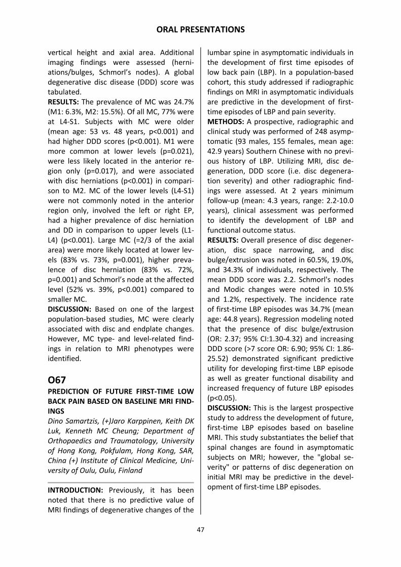
ORAL PRESENTATIONS
47
vertical height and axial area. Additional imaging findings were assessed (herni‐ations/bulges, Schmorl’s nodes). A global degenerative disc disease (DDD) score was tabulated. RESULTS: The prevalence of MC was 24.7% (M1: 6.3%, M2: 15.5%). Of all MC, 77% were at L4‐S1. Subjects with MC were older (mean age: 53 vs. 48 years, p<0.001) and had higher DDD scores (p<0.001). M1 were more common at lower levels (p=0.021), were less likely located in the anterior re‐gion only (p=0.017), and were associated with disc herniations (p<0.001) in compari‐son to M2. MC of the lower levels (L4‐S1) were not commonly noted in the anterior region only, involved the left or right EP, had a higher prevalence of disc herniation and DD in comparison to upper levels (L1‐L4) (p<0.001). Large MC (=2/3 of the axial area) were more likely located at lower lev‐els (83% vs. 73%, p=0.001), higher preva‐lence of disc herniation (83% vs. 72%, p=0.001) and Schmorl’s node at the affected level (52% vs. 39%, p<0.001) compared to smaller MC. DISCUSSION: Based on one of the largest population‐based studies, MC were clearly associated with disc and endplate changes. However, MC type‐ and level‐related find‐ings in relation to MRI phenotypes were identified.
O67 PREDICTION OF FUTURE FIRST‐TIME LOW BACK PAIN BASED ON BASELINE MRI FIND‐INGS Dino Samartzis, (+)Jaro Karppinen, Keith DK Luk, Kenneth MC Cheung; Department of Orthopaedics and Traumatology, University of Hong Kong, Pokfulam, Hong Kong, SAR, China (+) Institute of Clinical Medicine, Uni‐versity of Oulu, Oulu, Finland
INTRODUCTION: Previously, it has been noted that there is no predictive value of MRI findings of degenerative changes of the
lumbar spine in asymptomatic individuals in the development of first time episodes of low back pain (LBP). In a population‐based cohort, this study addressed if radiographic findings on MRI in asymptomatic individuals are predictive in the development of first‐time episodes of LBP and pain severity. METHODS: A prospective, radiographic and clinical study was performed of 248 asymp‐tomatic (93 males, 155 females, mean age: 42.9 years) Southern Chinese with no previ‐ous history of LBP. Utilizing MRI, disc de‐generation, DDD score (i.e. disc degenera‐tion severity) and other radiographic find‐ings were assessed. At 2 years minimum follow‐up (mean: 4.3 years, range: 2.2‐10.0 years), clinical assessment was performed to identify the development of LBP and functional outcome status. RESULTS: Overall presence of disc degener‐ation, disc space narrowing, and disc bulge/extrusion was noted in 60.5%, 19.0%, and 34.3% of individuals, respectively. The mean DDD score was 2.2. Schmorl's nodes and Modic changes were noted in 10.5% and 1.2%, respectively. The incidence rate of first‐time LBP episodes was 34.7% (mean age: 44.8 years). Regression modeling noted that the presence of disc bulge/extrusion (OR: 2.37; 95% CI:1.30‐4.32) and increasing DDD score (>7 score OR: 6.90; 95% CI: 1.86‐25.52) demonstrated significant predictive utility for developing first‐time LBP episode as well as greater functional disability and increased frequency of future LBP episodes (p<0.05). DISCUSSION: This is the largest prospective study to address the development of future, first‐time LBP episodes based on baseline MRI. This study substantiates the belief that spinal changes are found in asymptomatic subjects on MRI; however, the "global se‐verity" or patterns of disc degeneration on initial MRI may be predictive in the devel‐opment of first‐time LBP episodes.

ORAL PRESENTATIONS
48
O68 MRI FEATURES OF THE PSOAS MUSCLES IN PATIENTS WITH PARASPINAL MUSCLE AT‐ROPHY 1Teruo Kita, 1Kazushi Takayama, 2Hiroaki Nakamura, 3Fumiaki Kanematsu, 3Toshiya Yasunami, 1Shigehiro Ikeda, 1Kohei Yama‐moto, 1Hideki Ueyama, 1Yusuke Hori, 1Hideki Sakanaka, 1Yoshiki Yamano; 1Dept. of Orthopaedic Surgery, Seikeikai Hospital, Osaka, Japan; 2Dept. of Orthopaedic Sur‐gery, Osaka City University Graduate School of Medicine, Osaka, Japan; 3Dept. of Ortho‐paedic Surgery, Saiseikai Nakatsu Hospital, Osaka, Japan
INTRODUCTION: We have already reported that the cross‐sectional area (CSA) of the paraspinal muscles tends to decrease with age and that our new index, referred to as the T‐back value (used to quantify the depth of the groove between the paraspinal mus‐cles; equal to the length of the bulge of the muscle to the attachment of the spinous process) was strongly correlated with the CSA of the paraspinal muscles (K. Takayama et al. ISSLS 2012). Additionally, we reported that the CSA of the paraspinal muscles and psoas muscles showed a similar decreasing tendency with age, but the fat infiltration rate of the paraspinal muscles was markedly higher compared to that of the psoas mus‐cles in the elderly (T. Kita et al. ISSLS 2013). Therefore, we consider that the paraspinal muscles are more prone to atrophy with age compared with the psoas muscles. The aim of this study was to investigate the CSA and fat infiltration of the psoas muscles in pa‐tients with paraspinal muscle atrophy by MRI and to compare the findings with those in patients with less degenerative spine. METHODS: We considered patients with T‐back values =0 as those with atrophied paraspinal muscles. Of 704 patients who underwent MRI of the lumbar spine at our hospital during 2010, 45 male (13%) and 80 female (22%) patients with T‐back values =0
were included in this study and were com‐pared with previously reported patients with less degenerative spine. Sagittal T2‐weighted MRI was used to measure lumbar lordosis (L1–S1 angle). Axial T2‐weighted MRI was used to measure the CSA and fat infiltration rate of the psoas muscles at the intervertebral disc levels from L1/2 to L4/5. RESULTS: The mean lumbar lordosis was 19.5°, which was lower than that of patients with less degenerative spine (36.7°). How‐ever, the CSA and fat infiltration rate of the psoas muscles was similar to those in pa‐tients with less degenerative spine. Thus, the psoas muscles are not significantly af‐fected in patients with paraspinal muscle atrophy.
O69 COMPREHENSIVE ANALYSIS OF MRI‐PHENOTYPES PROVIDES NEW INSIGHTS INTO LUMBAR DISC DEGENERATION FOR GENETIC STUDIES Yan Li 1, Dino Samartzis3, Desmond Camp‐bell1, Kenneth M.C. Cheung3, Keith D.K. Luk3, Jaro Karppinen4, Kathryn Cheah2, Danny Chan2, Pak Sham 1*; 1. Department of Psychiatry, The University of Hong Kong, Hong Kong, SAR, China 2. Department of Biochemistry, The University of Hong Kong, Hong Kong, SAR, China 3. Department of Orthopaedics and Traumatology, The Uni‐versity of Hong Kong, Hong Kong, SAR, Chi‐

ORAL PRESENTATIONS
49
na 4. Institute of Clinical Medicine, Universi‐ty of Oulu, Oulu, Finland
INTRODUCTION: Numerous genetic associa‐tion studies addressing lumbar disc degen‐eration (LDD) have been performed, but few of them can be replicated. The possible reasons could be the phenotype definition of LDD was highly variable between studies and level‐specific variations were not ad‐dressed. As such, this study addressed the relationship between MRI features of the discs at different lumbar levels to explore the etiology of LDD; thereby, providing new insights and measurements for genetic studies. METHODS: Sagittal T2‐weighted MRI of the lumbar spine was assessed in a population sample of 2,952 Southern Chinese (mean age, 41.1 years; range, 15.0 to 65.4 years; 40.7% males; 59.3% females). Loss of disc signal intensity, disc bulges/extrusions, Schmorl's nodes, high‐intensity zones, and bone marrow changes were assessed on imaging. Subject demographics, environ‐mental and lifestyle factors were also eval‐uated. Polychoric correlations, heritability estimations and local regression statistical analyses were performed. RESULTS: Analyses suggested distinct genet‐ic etiologies for the upper (L1‐L2) versus lower regions (L3‐S1). Age‐related condition was restricted to lower regions while the upper region suggests congenital. By com‐bining highly‐correlated MRI‐phenotypes in the upper and lower regions separately, two composite scores were generated: a degen‐erative score (DgS) (represents age‐related disc changes) and a developmental score (DvS) (represents congenital variations). DISCUSSION: Based on one of the largest population‐based studies of LDD, compre‐hensive analyses of MRI‐phenotypes pro‐vided new insights into its etiology. Our study proposes a novel phenotype scoring system. This scoring system can be used as the basis to promote a standardization of
phenotype delineation, maximizing the po‐tential of replication and meta‐analysis studies of genetic risk factors for LDD.
O70 SURGICAL INTERSPINOUS IMPLANT VER‐SUS CONVENTIONAL DECOMPRESSION FOR LUMBAR SPINAL STENOSIS ‐ A RANDOM‐IZED CONTROLLED TRIAL Wouter A Moojen, Mark P Arts, Wilco CH Jacobs, Erik W van Zwet, M Elske van den Akker‐van Marle, Bart W Koes, Carmen LAM Vleggeert‐Lankamp, Wilco C Peul; Leiden University Medical Center, Leiden, The Netherlands (author 1, 3, 4, 5, 7 and 9) de‐partment of Neurosurgery (author 1, 3, 7 and 9), Medical Statistics, and Medical Deci‐sion Making (author 5) Erasmus Medical Center, Rotterdam, The Netherlands, de‐partment of General Medicine (author 6). Medical Center Haaglanden, The Hague, The Netherlands, department of Neurosurgery (authors 1 and 2)
INTRODUCTION: Interspinous process de‐vices (IPD) are implanted to treat patients with intermittent neurogenic claudication based on lumbar spinal stenosis. Although widely implemented by the surgical com‐munity, efficacy has not been compared with standard bony decompression. It is hypothesized that implanting an interspi‐nous process device results in faster post‐operative recovery, and a higher success rate according to the Zurich Claudication Questionnaire at eight weeks compared to conventional bony decompression. DESIGN: A randomized design with variable block sizes was used, with allocations strati‐fied according to center. Patients and re‐search nurses were blind throughout the follow‐up. METHODS: 211 participants were referred to the Leiden‐The Hague Spine Prognostic Study Group. 159 participants with intermit‐tent neurogenic claudication based on lum‐bar spinal stenosis at one or two levels with

ORAL PRESENTATIONS
50
an indication for surgery were blindly ran‐domized into two groups. 80 participants received an IPD and 79 participants under‐went spinal bony decompression. The pri‐mary outcome at short‐term (eight weeks) and long‐term (one year) follow‐up was the score for the Zurich Claudication Question‐naire. Repeated‐measurement analyses were applied to compare outcomes over time. RESULTS: At eight weeks, the success rate according to the Zurich Claudication Ques‐tionnaire for the IPD group (63 % CI 51 to 73) was not superior to that for standard bony decompression (72 % CI 60 to 81). No differences in disability (ZCQ p 0.44) or oth‐er outcomes were observed between groups during the first year. The repeat sur‐gery rate in the interspinous implant group was substantially higher, 29 %, in the early post‐surgical period composed to the con‐ventional group, 6% (p‐value <0.001). DISCUSSION: This double blinded study could not confirm the hypothesized short‐term advantage of IPD over conventional ”simple” decompression, and even showed a fairly high re‐operation rate after IPD use.
O71 SURGICAL VERSUS NON‐OPERATIVE TREATMENT FOR LUMBAR SPINAL STENO‐SIS EIGHT‐YEAR RESULTS OF THE SPINE PATIENT OUTCOMES RESEARCH TRIAL (SPORT) 1Jon D. Lurie, MD, MS; 1Tor D. Tosteson, ScD; 1Anna N.A. Tosteson, ScD; Wenyan Zhao, PhD; 1Tamara S. Morgan, MA; 1William A. Abdu, MD, MS; 2Harry Herko‐witz, MD; 1James N. Weinstein, DO, MS; 1Geisel School of Medicine at Dartmouth, Hanover, NH and Dartmouth‐Hitchcock Medical Center, Lebanon, NH; 2William Beaumont Hospital, Royal Oak, MI
INTRODUCTION: Surgery for lumbar spinal stenosis (SpS) has been shown to be more effective compared to non‐operative treat‐
ment over four years in as‐treated analyses. In this study we compare 8‐year SpS out‐comes from SPORT. METHODS: Surgical candidates from 13 cen‐ters in 11 U.S. states with at least 12 weeks of symptoms and confirmatory imaging were enrolled in a randomized cohort (RCT) or observational cohort (OBS). Protocol treatments were decompressive laminec‐tomy or standard non‐operative care. Pri‐mary outcomes were SF‐36 bodily pain (BP) and physical function (PF) scales and the modified Oswestry Disability index (ODI) assessed at 6 weeks, 3 months, 6 months and yearly thereafter. RESULTS: 55% of RCT and 52% of OBS par‐ticipants provided data at the 8‐year follow‐up. Intent‐to‐treat analyses showed no dif‐ferences between randomized cohorts, however 70% of those randomized to sur‐gery and 52% of those randomized to non‐op had undergone surgery by 8 years. As‐treated analyses in the RCT showed an early benefit to surgery out to 4 years (as previ‐ously reported) however the two groups converged overtime with no significant treatment effect of surgery seen in years 6‐8 for any of the primary outcomes. Analysis of the OBS group showed a stable ad‐vantage for the surgical groups in all out‐comes between years 5‐8. Re‐operation rate for the combined cohorts was 18% through 8 years. Patients who were lost to follow‐up were older, less well‐educated, sicker, and had worse outcomes over the first 2 years in both surgery and non‐operative arms. DISCUSSION: Patients with symptomatic spinal stenosis show diminishing benefits of surgery in as‐treated analyses of the RCT between 4‐8 years while outcomes in the OBS group remained stable. Loss to follow‐up of patients with worse early outcomes in both treatment groups suggest that the reported outcomes are likely to be overly‐optimistic but the treatment effect esti‐mates are unlikely to be biased.

ORAL PRESENTATIONS
51
O72 WHAT LEADS TO SURGICAL DECISION IN DEGENERATIVE LUMBAR STENOSIS IN THE PREFERENCE‐BASED, SHARED DECISION? Ho‐Joong Kim, Jae‐Young Park, Kyoung‐Tak Kang, Bong‐Soon Chang, Choon‐Ki Le , Jin S. Yeom; Spine Center and Department of Or‐thopaedic Surgery, Seoul National University College of Medicine and Seoul National Uni‐versity Bundang Hospital
INTRODUCTION: Several subjective and/or objective factors may influence the final surgical decision for degenerative LSS, in‐cluding back/leg pain, symptom duration, neurologic deficit, radiologic severity of ste‐nosis, and amount of disability in the pref‐erence‐based, shared decision making. Nonetheless, the understanding about the significance of each influencing factor on surgical decision making remains primitive. Therefore, the purpose of this study was to clarify the significance of each influencing factor on the surgical decision for the treatment of LSS in a preference‐based shared decision‐making setting. METHODS: A total of 555 patients, aged 45–80 years, who were given conservative or surgical treatment for chronic leg and/or back pain caused by LSS from April to De‐cember 2012 in the preference‐based shared decision‐making system were in‐cluded in this study. Univariate and multi‐variable‐adjusted logistic regression anal‐yses were used to assess the association of surgical decision‐making with age, sex, body mass index, symptom duration, radiologic stenotic grade, Oswestry Disability Index (ODI), Visual Analog Scale (VAS) for back/leg pain, Short Form (SF)‐36 subscales, and mo‐tor weakness. RESULTS: In univariate analysis, male sex, VAS for leg pain, ODI, morphological stenot‐ic grades B, C, and D, motor weakness, and physical function, role of physical, bodily pain, social function, and role of emotion
SF‐36 subscales were associated with higher odds of a surgical decision for LSS. Multivar‐iate analysis revealed that male sex, ODI, grade C and D stenosis, and motor weak‐ness were significantly associated with higher possibility of a surgical decision (Fig‐ure 1).
DISCUSSION: This study suggests that male sex, morphological stenotic grade and the amount of disability are critical factors lead‐ing to surgical decision in the preference‐based shared decision‐making process.
O73 CLINICAL DEPRESSION: A PREDICTOR OF POOR RETURN TO WORK RATES AMONG WORKER’S COMPENSATION SUBJECTS FOL‐LOWING LUMBAR FUSION Joshua T. Anderson, BS (1,2), Ryan J. Duff (3), Uri M. Ahn, MD (4), Nicholas U. Ahn, MD (1); 1 University Hospitals Case Medical Center Department of Orthopaedic Surgery 2 Case Western Reserve University School of Medicine 3 University of Minnesota Twin Cities 4 New Hampshire Spine Institute
INTRODUCTION: Worker’s compensation (WC) subjects undergoing lumbar fusion consistently have worse outcomes than the general population. Few studies have evalu‐ated specific predictors of poor fusion out‐comes in WC subjects.

ORAL PRESENTATIONS
52
METHODS: We used ICD‐9 and CPT codes to identify subjects. We identified 4107 sub‐jects from the Ohio Bureau of Worker’s Compensation that underwent single level lumbar fusion as their index fusion after injury. 720 subjects had fusion for the indi‐cation of spondylolisthesis, 37 of which were had depression before fusion. 1166 subjects underwent fusion for degenerative disc disease, 78 of which had depression before fusion. All subjects had 3 years of follow‐up minimum. We determined who was able to return to work within 2 years of fusion and maintain a returned to work sta‐tus for 1 year minimum. Subjects with a smoking history and pre‐fusion diagnosis of failed back syndrome were not considered. We analyzed the spondylolisthesis group and the DDD group separately with logistic regression. RESULTS: Return to work rates were signifi‐cantly lower in patients with depression in the spondylolisthesis group (p<0.00; OR=0.18, CI=0.07‐0.47) and the DDD group (p<0.00; OR=0.42, CI=0.23‐0.75). Among the spondylolisthesis group, 368 of 683 subjects (53.9%) without depression returned to work. Only 9 of 37 subjects (24.3%) with depression and spondylolisthesis returned to work. Among the DDD group, 483 of 1088 subjects (44.3%) without depression returned to work. 19 of 78 subjects (23.1%) with depression and DDD returned to work. For the DDD group, age significantly influ‐enced return to work rates (p=0.03). The odds ratio (0.98) suggests that this impact was not considerable. For both populations, influences of gender, obesity, and income were not significant. DISCUSSION: Single level fusion surgery in WC subjects with clinical depression for the indication of either spondylolisthesis or de‐generative disc disease is associated with poor return to work rates.
O74 THE ASSESSMENT OF PATIENT‐RATED OUTCOME IN SPINAL STENOSIS: COULD LESS BE MORE? Mannion AF, Fekete T, Kleinstück F, Jeszenszky D, Wertli M*, Porchet F in collab‐oration with the Lumbar Stenosis Outcome Study (LSOS) group; Spine Center, Schulthess Klinik, Zürich, Switzerland; *Horten Centre, Zürich University Hospital, Switzerland
INTRODUCTION: The Core Outcome Measures Index (COMI) is a short, validated questionnaire for assessing the key out‐comes of importance to back patients. With just one question per domain, it enables the efficient assessment of large groups, with minimal respondent burden. However, for a given pathology, intuitively it may be ex‐pected to be less responsive than a disease specific instrument. In patients with lumbar spinal stenosis (LSS), we compared the per‐formance of COMI and the widely accepted Swiss Spinal Stenosis Measure (SSM). METHODS: The following questionnaires were completed before surgery by 91 LSS patients (73±8y; 50 m, 45 f) and after sur‐gery by 47 patients who had reached 12mo post‐op: SSM, COMI, Roland Morris disabil‐ity (RM), EuroQoL‐VAS (EQ‐VAS), complaints “feeling thermometer” (FT), pain numeric rating scale (pain NRS). At 12 mo post‐op, SSM “satisfaction with treatment result” and the Global Treatment Outcome (rated 1‐5) were assessed. RESULTS: The COMI correlated with the SSM and the comparator questionnaires to the expected (r=0.4=0.8) extent, indicating good construct validity. The external criteri‐on of Global Treatment Outcome correlated better with the change score (baseline to 12 mo FU) for COMI (r=‐0.62) than for SSM symptoms (r=‐0.43) or SSM function (r=‐0.42) (each p <0.05). Similarly, "SSM satis‐faction" correlated better with the change score for COMI (r=‐0.63) than for SSM

ORAL PRESENTATIONS
53
symptoms (r=‐0.59) or SSM function (r=‐0.45). DISCUSSION: COMI correlated to the ex‐pected extent with the SSM and its sub‐scales, and with pain, disability and QoL, suggesting construct validity. With either “SSM‐satisfaction with treatment results" or Global Treatment Outcome serving as the external criterion, COMI was more respon‐sive than the SSM subscales. The COMI is well able to detect important change in LSS and has the added benefits of facilitating outcome comparisons with other spinal pathologies and reducing the response bur‐den for the patient.
O75 SURGICAL OUTCOME OF LUMBAR SPINAL STENOSIS: 10 YEARS PROSPECTIVE FOL‐LOW‐UP. Tuomainen I1, Aalto T1,2, Vanhanen S1, Pakarinen M1, , Sinikallio S1Leinonen V3, Herno A1, Kröger H4, Turunen V4, Savo‐lainen S3, Miettinen H, Airaksinen O1; 1Department of Physical and Rehabilitation Medicine, Kuopio University Hospital, P.O.Box 1777, FIN‐70211 Kuopio, Finland 2Kyyhkylä Rehabilitation Hospital, Mikkeli, Finland 3Department of Neurosurgery, Kuo‐pio University Hospital, Kuopio, Finland 4Department of Orthopaedics and Trauma‐tology Kuopio University Hospital, Kuopio, Finland
INTRODUCTION: Previous prospective clini‐cal studies on lumbar spinal stenosis (LSS) have reported clinical outcomes with fol‐low‐up times from one to five years in. Our aim was to evaluate the surgical outcome in LSS up to t 10 years. METHODS: At baseline a total of 102 LSS patients were operated (mean age 62 years, 59 women, 43 men). Inclusion criteria were 1) presence of back, buttock, and/or lower extremity pain, with radiographic evidence (MRI) of compression of neural elements; 2) conservative therapy for several months
with insufficient outcome. Exclusion criteria were 1) emergency or urgent spinal opera‐tion; 2) cognitive impairment prohibiting completing the questionnaire or other fail‐ure in co‐operation. At 10 years follow‐up there were 72 LSS patients (mean age 69 years, 45 women, 27 men) in the study. 18 patients were died and 12 patients were not available for the evaluation (various rea‐sons). Main outcome measures were Oswestry Disability Index (ODI), Visual Ana‐logue Scale (VAS) and Patients Global As‐sessment and Satisfaction to Surgery (PGASS), prior to the intervention at 2 years and the last evaluation at least 10 years postoperatively. RESULTS: The mean ODI before surgery was 43,9 (SD 15.2); at two years follow‐up 26.6 (SD 19.2) (p<0.001) and 29.4 (SD 20.9 ) ( p<0,05 ), respectively. The mean baseline VAS was 33.0 (SD 23.6); at two years 11.6 (SD 16.9) (p<0,001); and at 10 years 32,8 (SD 28.2 ) ( p<0.001): The PGASS for good or excellent outcome was 61 % at two years follow‐up 61and 68 % at 10 years follow‐up, respectively (p<0,05 ). 3 patients had moved from good outcome to totally cured group during the follow‐ up DISCUSSION: In general VAS (pain) and ODI values deteriorated during the 10 years fol‐low‐up . However patients Global Assess‐ment and Satisfaction for the Surgery Out‐come improved. Patients comorbidities may explain the conflicting results. Furthermore the experience of disability and pain may change during aging.
O76 RELATIONSHIP BETWEEN LUMBAR SPINAL STENOSIS AND PSYCHOSOCIAL FACTORS ‐ A MULTICENTER CROSS‐SECTIONAL STUDY (DISTO‐PROJECT) ‐ Miho Sekiguchi 1), Koji Yonemoto 2), Tatsu‐yuki Kakuma 2), Takuya Nikaido 1), Kazuyuki Watanabe 1), Kinshi Kato 1), Koji Otani 1), Shoji Yabuki 1), Shin‐ichi Kikuchi 1), Shin‐ichi Konno 1); 1) Department of Orthopaedic

ORAL PRESENTATIONS
54
Surgery, Fukushima Medical University School of Medicine, Fukushima, Japan 2) Biostatistics Center, Kurume University, Fu‐kuoka, Japan
INTRODUCTION: Lumbar spinal stenosis (LSS) is a lumbar spinal disorder and causes leg symptoms and intermittent claudication. It is reported that the risk factors for low back pain are age, family history, smoking, obesity, work‐related physical load, exer‐cise, stress, depression and etc. In contract, few large‐scale surveys in Japan have been performed to investigate risk factors of LSS. The aim of this study was to investigate the prevalence of LSS with age, and the rela‐tionship between LSS and the psychosocial factors and job satisfaction. METHODS: This study enrolled subjects who were 50 and over from the survey of lumbar spinal ste‐nosis diagnosis support tool (DISTO)‐project in 2,177 hospital and general practices na‐tionwide and 18,642 participants were ana‐lyzed. LSS was diagnosed using the diagnos‐tic support tool (LSS‐DST). The clinical char‐acteristics between LSS and non‐LSS groups were compared using x2 test and multivari‐ate logistic regression analysis were per‐formed to examine the associated factors of LSS. RESULTS: A total 18,642 patients (8,338 male and 10, 267 female) were analyzed and the rate of LSS was 38.3%. The rate of subjects who do regular exercise in the LSS group was lower than that in the non‐LSS group (p<0.001). The odds ratios for having LSS were higher than in subjects with having perceived stress and strenuous use of low back or legs (p<0.001). The satisfaction in all items job related in the LSS group were less than in the non‐LSS group (p<0.001). There was an increase the odds of heart diseases, hypertension, while a decrease the odds of diabetes mellitus, respectively (p<0001). CONCLUSION: This study was investigated the associated factors with LSS. Perceived stress, strenuous use of low back or legs
might be associated with LSS, and job satis‐faction was lower with LSS.
O77 AGE ASSOCIATED RELATIVE RISK INCREASE FOR IN‐HOSPITAL MORTALITY AND COM‐PLICATIONS FOLLOWING LUMBAR SPINE SURGERY Hamid Hassanzadeh MD, Sreeharsha V. Nandyala BA, Alejandro Marquez‐Lara MD, Kern Singh MD; Department of Orthopaedic Surgery, Rush University Medical Center
INTRODUCTION: Lumbar arthrodesis is commonly utilized to treat degenerative conditions of the spine. Despite its wide‐spread utilization, few studies have charac‐terized the age associated relative risk in‐crease for inpatient complications and mor‐tality following a lumbar fusion (LF) proce‐dure. METHODS: Data from the Nationwide Inpa‐tient Sample was queried from 2002‐2011. Patients who underwent an index lumbar fusion were identified and stratified into age groups as follows: 18‐39, 40‐49, 50‐59, 60‐69, 70‐79, and 80+. Postoperative inpa‐tient complications and mortality were as‐sessed. The relative risks in each decade were calculated in reference to the previous respective age group. SPSS v.20 was utilized for statistical analysis with a 95% confidence interval for relative risk. RESULTS: A total of 315,597 LF procedures were recorded from 2002‐2011. The relative risk for inpatient mortality, pulmonary em‐bolism (PE), cardiac events, and renal failure was significant beginning at the 5th decade and compounded for the subsequent age groups. The risk increase for mortality was most dramatic for patients >80 years when compared to the 7th decade. The initial sig‐nificant relative risk increase for deep vein thrombosis (DVT) was demonstrated at the 4th decade and was consistently significant for the subsequent age groups. There was no relative risk increase in each decade in

ORAL PRESENTATIONS
55
regards to inpatient wound infection or ile‐us following LF. DISCUSSION: In this analysis of the NIS da‐tabase, the 4th decade demonstrated an increased relative risk for DVT whereas the 5th decade demonstrated the initial risk increase for in‐hospital mortality, PE, cardi‐ac events, and renal failure after LF. These results suggest that the risk increase for in‐hospital mortality and catastrophic compli‐cations develops early, in the 4th and 5th decades. Further studies are warranted to investigate the etiologies for this early risk increase for complications following lumbar arthrodesis.
O78 LUMBAR DEGENERATIVE SPONDYLOLIS‐THESIS IS NOT ALWAYS UNSTABLE ~ A CLINICO‐BIOMECHANICAL EVIDENCE ~ Kazuhiro Hasegawa, Ko Kitahara, Haruka Shimoda, Keiji Ishii, Takao Homma; Niigata Spine Surgery Center, Niigata, Japan
INTRODUCTION: Is a segment with lumbar degenerative spondylolisthesis (LDS) always unstable? Although radiographic evaluation of the lesion is extensively performed, the diagnosis of segmental instability remains controversial. The purpose of this study is to clarify clinico‐biomechanical characteristics of the segment with LDS using an original intraoperative measurement system (IOM). METHODS: IOM is the first clinically availa‐ble system which performs a cyclic flexion‐extension displacement of the segment with all ligamentous structures intact and can determine stiffness (N/mm), absorption energy, and neutral zone (NZ, [mm/N]). For‐ty‐eight patients with LDS (M/F=19/29, 68.5 years) (Group L) were compared with 48 lumbar spinal stenosis patients without LDS (M/F=33/15, 64.8 years, Group N) in terms of symptoms, radiological, and biomechani‐cal results. Instability was defined as a seg‐ment with NZ>2mm (JBJS2011). Symptoms (SF‐36), radiographic findings (X‐rays, MRI,
CT), stiffness, NZ, and frequency of Instabil‐ity were also compared. Risk factors of in‐stability were analyzed by multivariate lo‐gistic regression with a forward stepwise procedure. RESULTS: There was neither significant dif‐ference in all categories in physical function of SF‐36 and low back pain (VAS) between the groups, nor is in radiological findings. Although NZ in Group L (1.97) was signifi‐cantly greater than that in Group N (1.73) (p<0.05), there was no difference in fre‐quency of Instability. Facet opening (Odd’s 10.4, p<0.01) and sagittally oriented facets (Odd’s 4.4, p<0.05) were risk factors of In‐stability. DISCUSSION: There was no significant dif‐ference in symptoms and the frequency of Instability between the groups. The indica‐tive radiological finding of Instability was not LDS, but facet opening and sagittally oriented facets. Thus, LDS is neither always unstable nor symptomatic, suggesting that the instability of LDS can be stabilized spon‐taneously in the natural course.
O79 SURGICAL VERSUS NON‐OPERATIVE TREATMENT FOR LUMBAR DEGENERATIVE SPONDYLOLISTHESIS: EIGHT‐YEAR RESULTS OF THE SPINE PATIENT OUTCOMES RE‐SEARCH TRIAL (SPORT) 1Jon D. Lurie, MD, MS; 1Tor D. Tosteson, ScD; 1Anna N.A. Tosteson, ScD; Wenyan Zhao, PhD; 1Tamara S. Morgan, MA; 1William A. Abdu, MD, MS; 2Harry Herko‐witz, MD; 1James N. Weinstein, DO, MS; 1Geisel School of Medicine at Dartmouth, Hanover, NH and Dartmouth‐Hitchcock Medical Center, Lebanon, NH; 2William Beaumont Hospital, Royal Oak, MI
INTRODUCTION: Surgery for lumbar spinal stenosis with associated degenerative spondylolisthesis (DS) has been shown to be more effective compared to non‐operative treatment over four years in as‐treated

ORAL PRESENTATIONS
56
analyses from SPORT. In this study, we compare 8‐year DS treatment outcomes from SPORT. METHODS: Surgical candidates from 13 cen‐ters in 11 U.S. states with at least 12 weeks of symptoms and confirmatory imaging were enrolled in a randomized cohort (RCT) or observational cohort (OBS). Protocol treatments were decompressive laminec‐tomy with (93%) or without (7%) fusion compared to standard non‐operative care. Primary outcomes were SF‐36 bodily pain and physical function scales and the modi‐fied Oswestry Disability index assessed at 6 weeks, 3 months, 6 months and yearly thereafter. RESULTS: 69% of RCT and 57% of OBS par‐ticipants provided data at the 8‐year follow‐up; 72% of those randomized to surgery and 55% of those randomized to non‐operative treatment had undergone surgery by 8 years. Intent‐to‐treat analyses showed no differences between randomized cohorts at early time points, however they favored the non‐operative groups at later time points with differences that were statistically sig‐nificant in years 6,7,8 and averaged over all time points. As‐treated analyses in the RCT adjusted for selection bias showed a stable advantage for surgery out to 8 years with similar findings in the OBS group. Re‐operation rate for the combined cohorts was 22% through 8 years. Patients who were lost to follow‐up were older, less well‐educated, sicker, and had worse surgical outcomes but similar non‐operative out‐comes over the first 2 years. DISCUSSION: SPORT DS patients random‐ized to surgery had somewhat worse long‐term outcomes; however, high rates of cross‐over and long‐term loss to follow‐up complicate the interpretation of these find‐ings. Factors differentially affecting loss to follow‐up between comparison groups could affect treatment effects estimates.
O80 LINKING PARITY, HISTORY OF ABDOMINAL SURGERY, AND TRUNK MUSCLE FUNCTION TO DEGENERATIVE SPONDYLOLISTHESIS IN OLDER WOMEN Cholewicki J, Lee A[1], Popovich Jr. JM[1], Mysliwiec LW[1], Winkelpleck MD[1], Flood JN[1], Pathak PK[1,2], Kaaikala, KH[1], Reeves NP[1], Kothe R[3]; [1] MSU Center for Orthopedic Research, College of Osteo‐pathic Medicine, Michigan State University, East Lansing, USA [2] Dept. of Statistics and Probability, College of Natural Sciences, Michigan State University, East Lansing, USA [3] Dept. for Spinal Surgery, Schön Klin‐ik, Hamburg, Germany
INTRODUCTION: Degenerative spondylolis‐thesis (DS) is a major cause of disabling low back pain among the older population, of‐ten requiring surgery to alleviate symptoms. But the question remains as to why women suffer from DS at a rate 3‐9 times higher than men. One study found a relationship between the number of pregnancies and DS [1] and suggested that impaired abdominal muscle function causing poor spine me‐chanics could eventually lead to degenera‐tive spine disease. Therefore, this study ex‐amined the association of parity, abdominal surgery, and trunk muscle function to DS. METHODS: A questionnaire containing in‐formation about parity and past abdominal surgeries was completed by 324 women between ages of 40 and 80 (153 with DS and 171 controls without DS determined radiologically). A sub‐set of 32 women with DS and 63 controls volunteered for a clinical assessment of their trunk muscles (blinded abdominal muscle function and hip exten‐sion tests). RESULTS: Pearson’s Chi‐squared tests re‐vealed a significantly higher presence of past abdominal surgeries (p=0.001) and pregnancies (p=0.012) among women with DS. Logistic regression analysis identified age, body‐mass index, hysterectomy, and

ORAL PRESENTATIONS
57
the number of full‐term pregnancies as spe‐cific factors predictive of DS (p<0.001, con‐cordance 73%). Moreover, women with DS were more likely to exhibit abdominal mus‐cle weakness (p=0.041), whereas the hip extension tests revealed no significant dif‐ferences. DISCUSSION: These results support our hy‐pothesis that DS is associated with a history of abdominal surgeries, especially hysterec‐tomies (nearly twice greater risk), and the number of full‐term pregnancies. DS is likely mediated by impaired abdominal muscle function (Fig. 1). Specific interventions could be designed to target these muscles post‐partum and following hysterectomies.
[1] Sanderson PL. J Bone Joint Surg Br 78:951‐4, 1996.
O81 CLINICAL OUTCOME OF DECOMPRESSIVE SURGERY FOR LUMBAR EPIDURAL LI‐POMATOSIS Haschtmann D, Ferlic P, Kleinstück FS, Por‐chet F, Mannion A, Jeszenszky D, Fekete TF; Spine Center, Schulthess Clinic, Zurich, Swit‐zerland
INTRODUCTION: The treatment of spinal epidural lipomatosis (SEL) is controversial. The limited evidence for the success of sur‐gical decompression is mostly derived from case reports. Moreover, no patient‐reported outcome studies are available. The objective of this study was to evaluate pa‐tient‐reported outcome after lumbar de‐compressive surgery for SEL in the largest series of patients examined to date. METHODS: Consecutive patients with symp‐tomatic SEL documented on MRI undergo‐ing surgical treatment in a single centre (2004 – 2012) were analysed. Data were obtained from the International Spine Tan‐
go Register. Patients with spinal stenosis caused by other pathologies were excluded. Outcome was evaluated using Core Out‐come Measure Index (COMI; scored 0 – 10) including the pain scale for leg and back. Three months was defined as the minimal follow‐up time; for most patients a two‐year follow‐up period was available (mean 20.2 months). RESULTS: In 170 patients undergoing lum‐bar decompressive surgery SEL was docu‐mented. 141 pts. were excluded due to ac‐companying discogenic or arthrogenic ste‐nosis. In 6 pts. MRI scans was unavailable and 1 pt. did not complete the forms. Final‐ly, 22 patients (19 male, 3 female, mean age: 68.2, 50.4 – 88.7 years) were included. At 3 months the outcome scores showed a significant improvement: COMI from 7.5 ± 1.7 (mean ± SD) to 4.9 ± 2.5 (p<0.0001), leg pain from 5.9 ± 2.6 to 3.5 ± 2.8 (p=0.0042) and back pain from 5.4 ± 3.2 to 4 ± 2.8 (p=0.004). The effect was retained for 2 years (COMI: 5.1 ± 3.1, p=0.003 vs. preoper‐ative). CONCLUSION: Based on patient‐reported outcome, we could demonstrate a benefi‐cial effect of decompressive surgery for spi‐nal stenosis due to epidural lipomatosis. As expected, the improvement of leg pain was more pronounced than the effect on back pain and lasted for at least 2 years.

SPECIAL POSTERS
58
SP01 REOPERATION RATE AFTER MICRODISCEC‐TOMY FOR THE TREATMENT OF LUMBAR DISC HERNIATION: LONG‐TERM FOLLOW‐UP AFTER AN AVERAGE OF 10.8 YEARS Alexander Aichmair, MD*; Jerry Y. Du, BSc†; Jennifer Shue, MS*; Gisberto, Evangelisti, MD‡; Andrew A. Sama, MD*; Alexander P. Hughes, MD*; Darren R. Lebl, MD*; Frank P. Cammisa, MD*; Federico P. Girardi, MD*; *Department of Orthopaedic Surgery, Spine and Scoliosis Service, Hospital for Special Surgery, Weill Cornell Medical College, New York, NY †Weill Cornell Medical College, New York, NY ‡1st Orthopaedic Clinic, Cisanello Hospital, University of Pisa, Italy
INTRODUCTION: The aim of the present study was to assess the reoperation rate after microdiscectomy for the treatment of lumbar disc herniation (LDH) in patients with a minimum five‐years follow‐up and identify demographic, perioperative, and outcome‐related differences between pa‐tients with and without a re‐operation. METHODS: The medical records, operative reports, and office notes of patients who had undergone microdiscectomy at a single institution between 03/1994 and 12/2007 were reviewed, and long‐term follow‐up assessed via a telephone questionnaire. RESULTS: Forty patients (M:24, F:16) with an average age at surgery of 39.9±12.5 years (range: 18‐80) underwent micro‐discectomy at the levels L5‐S1 (n=28, 70%), L4‐L5 (n=9, 22.5%), L3‐L4 (n=2, 5.0%), and L1‐L2 (n=1, 2.5%). After an average of 40.4±40.1 months (range: 1‐128), 25% of patients (10/40) required further spine sur‐gery related to the initial microdiscectomy. At an average post‐operative follow‐up of 10.8±4.0 years (range: 5‐19), additional symptoms apart from back and leg pain were reported more frequently by patients who underwent a re‐operation (p=0.005). Patient satisfaction was significantly higher in patients who did not undergo a re‐
operation (p=0.041). For the ODI, pain in‐tensity (p=0.036) and pain‐related sleep disturbances (p=0.006) were reported to be more severe in the re‐operation group. DISCUSSION: Microdiscectomy for the treatment of LDH results in favorable long‐term outcomes in the majority of cases. The re‐operation rate was higher in our series than reported in previous investigations with shorter follow‐up. Although there were no pre‐ or perioperative differences between patients with and without re‐operation, our findings suggest a difference in self‐reported long‐term outcome measures.
SP02 ANALYSIS OF INTERNET INFORMATION ON THE CONTROVERSIAL X‐STOP DEVICE Joshua T. Anderson, BS (1,2), T. Barrett Sul‐livan (1,2), Uri M. Ahn, MD (3), Nicholas U. Ahn, MD (1); 1 University Hospitals Case Medical Center Department of Orthopaedic Surgery 2 Case Western Reserve University School of Medicine 3 New Hampshire Spine Institute
INTRODUCTION: Patients frequently use the internet to aid in medical decision‐making, and this information actively influ‐ences patient choices. The X‐Stop is a con‐troversial interspinous spacer device for the treatment of intermittent neurogenic clau‐dication secondary to lumbar spinal steno‐sis. Our study is the first to examine inter‐net information on the controversial X‐Stop. METHODS: Search engines Google, Yahoo!, and Bing and search terms X‐Stop, interspi‐nous distraction device, and interspinous spacer decompression were used to identify and characterize 105 sites providing infor‐mation on X‐Stop. RESULTS: 43% of websites were authored by private medical groups, 4% by academic medical groups, 16% by insurance compa‐nies, 9% by biomedical industry, 10% by news sources, and 19% by websites not

SPECIAL POSTERS
59
fitting into those categories. 31% of webpages cited peer‐reviewed literature. 56% of all sites provided at least one appro‐priate patient inclusion criterion (in addition to lumbar spinal stenosis). 33% of sites re‐ported at least one patient exclusion crite‐ria. 91% of sites reported at least one pur‐ported benefit of X‐Stop. One or more complications were reported in 23% of sites. One of the most commonly seen complications with all interspinous spacers, spinous process fractures, was discussed in 19% of sites. Specific non‐surgical treat‐ment alternatives were discussed in no higher than 28% of sites. 33% of sites men‐tioned the existence of conservative thera‐py without including specifics. 51% of sites included laminectomy and 22% fusion as surgical treatment options. DISCUSSION: Publicly accessible infor‐mation on the X‐Stop was of poor quality, often incomplete, and potentially mislead‐ing. The high reporting rates of benefits (91%) contrasted with the abysmally low reporting rates of complications (23%) rais‐es the concern that this information lends itself more toward patient recruitment than to education.
SP03 A PROSPECTIVE COMPARATIVE STUDY OF TWO DIFFERENT MINIMALLY‐INVASIVE PROCEDURES FOR LUMBAR SPINAL CANAL STENOSIS ‐ UNILATERAL LAMINOTOMY FOR BILATERAL DECOMPRESSION (ULBD) VERSUS MUSCLE‐PRESERVING INTERLAMI‐NAR DECOMPRESSION (MILD) Yoshiyasu Arai, Takashi Hirai, Toshitaka Yoshii, Kenichi Shinomiya, Makoto Takahashi, Shigenori Kawabata, Mitsuhiro Enomoto, Tsuyoshi Kato, Shoji Tomizawa, Kenichirou Sakai, Atsushi Okawa; Depart‐ment of Orthopedic Surgery, Graduate School, Tokyo Medical and Dental University 1‐5‐45 Yushima, Bunkyo‐ku, Tokyo 113‐8519, JAPAN
INTRODUCTION: Unilateral laminotomy for bilateral decompression (ULBD) and muscle‐preserving interlaminar decompression (MILD), which were two different types of minimally invasive surgery for lumbar canal stenosis, were compared prospectively to reveal whether there are any differences in clinical and radiological outcomes. METHODS: From 2005 to 2009, we prospec‐tively enrolled 50 LSCS patients for the treatment with ULBD, and 50 patients for MILD. The patients’ symptoms were evalu‐ated using Japanese Orthopedic Association (JOA) score, JOA Back Pain Evaluation Ques‐tionnaire (JOABPEQ), and visual analog scale (VAS) before and 2‐year after opera‐tion. For radiological evaluation, changes in disc height, sagittal translation, and lateral wedging at the decompressed segment, as well as lumbar lordosis were investigated using plain X‐rays. RESULTS: Ninety‐nine of 100 patients were followed for a minimum of 2 years. No sig‐nificant differences were found in the re‐covery rate of JOA score, improvement of JOABPEQ, and changes of the VAS between the two groups. Radiologically, no signifi‐cant differences were present in the post‐operative degenerative changes in disc height, sagittal translation, and lateral wedging. In multi‐level surgeries; however, clinical scores in low back pain and lumbar function were significantly greater in the ULBD group than those in the MILD group. The lateral wedging change at L2/3 and L3/4 more frequently occurred in the ULBD group than in the MILD group. On the con‐trary, the number of patients who demon‐strated the postoperative sagittal transla‐tion at L4/5 was significantly greater in the MILD group than in the ULBD group. DISCUSSION: Both MILD and ULBD were efficacious procedures for improving neuro‐logical symptoms in LSCS patients. In multi‐level decompression surgeries, ULBD was superior to MILD in terms of improvement

SPECIAL POSTERS
60
of low back pain and lumbar function at 2‐year time‐point.
SP04 EPIDEMIOLOGICAL STUDY OF LUMBAR SPINAL STENOSIS: SIX‐YEAR FOLLOW‐UP IN THE COMMUNITY Koji Otani, Shinichi Kikuchi, Shoji Yabuki, Takuya Nikaido, Kazuyuki Watanabe, Kinshi Kato, Shinichi Konno; Dept. of Orthopaedic Surgery, Fukushima Medical University School of Medicine, Fukushima, Japan
INTRODUCTION: It is well known that Lum‐bar Spinal Stenosis (LSS) is one of the most serious problems in the elderly because of its high prevalence and negative impact on quality of life (QoL). Natural history of LSS is still not clear. The purpose of this study was to clarify natural history and the associated factors of LSS in the community. PARTICIPANTS AND METHODS: 1578 peo‐ple (age ranged from 40 to 79 yo) agreed to participate and were interviewed about LSS and comorbidities including osteoarthritis of the hip and the knee (hip OA, knee OA) by Altman’s criteria (Altman 1996, 2001) in 2004. The presence of LSS was assessed by a specially designed questionnaire (Konno 2007). In 2010, follow‐up study was per‐formed using mail. 790 people were traced. Follow up rate was 50%. RESULTS: 1. 161 (20.4%) in 2004 and 155 (19.6%) in 2010 of 790 were judged as LSS‐positive. At least for 6‐year, total preva‐lence of LSS did not increase. 2. 69 of 161(42.9%) showed that LSS existed in both 2004 and 2010. On the other hand, in the rest of 92 (57.1%), they were judged as LSS‐negative at the follow‐up. In 2004, 629 were judged as LSS‐negative and 86 of 629 (13.7%) were judged as LSS‐positive in 2010. 3. From a multiple logistic regression analy‐sis, male and the presence of LSS and knee OA in the initial analysis might be risk fac‐tors for 6‐year follow‐up. Depressive symp‐
tom, hypertension, cardiovascular disease, cerebrovascular disease, respiratory dis‐ease, diabetes mellitus and hip OA did not affect the presence of LSS for 6 years at least. DISCUSSION: To maintain and improve QoL through the prevention and treatment of musculoskeletal diseases in the elderly, it is believed that LSS is one of the most serious health problems that should be targeted because of its high prevalence and strong negative influence on QoL. Therefore asso‐ciated and/or risk factors for LSS revealed may provide preventability of LSS and pos‐sible application in the promotion of health in the community.
SP05 NIGHT LEG CRAMP AND LUMBAR SPINAL STENOSIS ‐ AN ANALYSIS OF 304 PEOPLE IN THE COMMUNITY Junichi Handa, Koji Otani, Takuya Nikaido, Shin‐ichi Kikuchi, Shin‐ichi Konno; Depart‐ment of Orthopaedic Surgery, Fukushima Medical University School of Medicine, Fu‐kushima, Japan
INTRODUCTION: Night leg cramp is thought as one of the characteristic symptoms of the lumbar spinal stenosis (LSS). However, details of night leg cramps with LSS are still unknown. The purpose of this study was to determine the characteristic of night leg cramp with LSS in the community. METHODS: 304 voluntary participants (men 93, women 211, most frequently age group; 60s) were enrolled in this study. The pres‐ence of LSS was assessed by a validated and self‐administrated diagnostic support tool (Konno et al: Eur Spine J. 2007). The pres‐ence of night leg cramp and physical exami‐nation such as sensory disturbance and mo‐tor weakness of the lower extremities were evaluated by one experienced spine sur‐geon. Furthermore, Roland‐Morris Disability Questionnaire (RDQ) was also assessed. All participants received lumbar MRI and cross‐

SPECIAL POSTERS
61
sectional area of dural tube at from L1/2 to L5/S1 was calculated by Hamanishi’s meth‐od (Hamanishi et al: J Spinal Disorders. 1994). RESULTS: 1.) 77 participants (25.3%) were judged as LSS positive. In LSS positive group, 41 of 77 participants (53.2%) showed night leg cramp. On the other hand, in LSS nega‐tive group, 65 of 227 participants (28.6%) showed night leg cramp. There was statisti‐cally significant difference of prevalence of leg cramp between two groups (p<0.0001). 2.) Cross‐sectional area of dural tube did not show any influence on the presence of night leg cramp. 3.) Sensory disturbance, motor weakness and lower RDQ score showed statistically significant difference of the presence of night leg cramp (p<0.05). DISCUSSION: From the results of this study, the severity of mechanical compression such as the area of dural tube did not affect the presence of night leg cramp. The severi‐ty of symptom such as sensory disturbance, motor weakness and lower low‐back‐pain related QoL evaluated by RDQ were associ‐ated with the presence of night leg cramp. These results mean that night leg cramp with LSS might show the severity of symp‐tom caused by LSS.
SP06 EVOLUTION OF SPINAL CANAL DIMEN‐SIONS IN SWITZERLAND. A NARROWING EPIDEMIC? Constantin Schizas MD FRCS, Aline Schmit, Alexis Schizas, Fabio Becce MD, Gerit Kulik PhD, Katarzyna Pierzchala PhD; University of Lausanne, Lausanne Switzerland and Spine Unit, Cecil Clinic, Lausanne Switzer‐land
INTRODUCTION: Development of sympto‐matic lumbar spinal stenosis (LSS) is among others related to the dimensions of the bony canal the latter reaching adult size early on in life. Several factors can influence its final dimensions such as protein intake.
We hypothesized that similar to what pre‐vails in human stature, adult bony canal size has grown larger in recent generations giv‐en the improvement of socioeconomic con‐ditions. METHODS: Computer tomographies (CT) from 184 subjects performed for either trauma (n= 81) or abdominal pathologies (n=103) were included in this study. CTs performed on patients born either between 1940‐49 (n=88) or 1970‐79 (n=96) were selected. Bony canal was digitally measured at pedicle level (i.e. at a level not influenced by degenerative changes) for each lumbar vertebra. Intra and inter‐observer reliability was assessed. RESULTS: Intra and inter‐observer meas‐urement reliability was excellent (ICC =0.87) and good (ICC= 0.61) respectively. Contrary to our hypothesis we observed that the younger generation of patients had highly statistically significant differences at all lev‐els compared to the older subjects, the lat‐ter having larger lumbar canals (p< 0.001). This difference was particularly pronounced in the trauma subgroup. DISCUSSION: Given that human stature evolution has stabilized and that adult height is determined by the first two years of long bone growth, it is possible that an‐tenatal factors are responsible for this sur‐prising finding. Maternal smoking and age could be possible explanations. This finding could have significant implications with an increased number of patients developing LSS as degenerative changes develop put‐ting at strain health resources. Further stud‐ies in different population groups and coun‐tries would be important in order to further confirm this trend.
SP07 THE ROLE OF BMI IN MATRIX DELAMINA‐TION DURING INTERVERTEBRAL DISC DE‐GENERATION Hans‐Joerg Meisel1, Andrea Friedmann2,3, Stefan Schwan2,3, Andreas Heilmann3, Felix
SPECIAL POSTERS
62
Goehre1,2; 1 BG Kliniken Bergmannstrost, Department of Neurosurgery, Halle, Germa‐ny 2 Univeristy Leipzig, Translationl Centre of Regenerative Medicine, TRM, Leipzig, Germany 3 Fraunhofer Institute for Mechan‐ics of Materials, IWM, Halle, Germany
INTRODUCTION: Intervertebral disc (ID) degeneration is a natural process during aging, contributed by mechanical‐, genetic and biochemical parameters. Interactions between these parameters cause an in‐stance of matrix degeneration in ID tissue on the macroscopic and microscopic scale. The process of disc degeneration by means of matrix destruction should be promoted by an increased body mass index (BMI) rep‐resenting increased mechanical loadings. The objective is to determine and correlate tissue changes of ID degeneration in differ‐ent states under consideration of patients’ BMI value. METHODS: As part of a clinical study, hu‐man ID tissue samples from 60 patients removed during standard sequestrectomy as neurosurgical treatment after disc herni‐ations are investigated regarding their mi‐crostructure using scanning electron mi‐croscopy. Investigations are focused on the delamination of the tissue matrix as well as cellular changes. With digital image analyz‐ing software, degeneration characteristics are evaluated and correlated with clinical relevant parameters. RESULTS: Matrix delamination is identified by digital image analysis as microstructural destruction in disc degeneration closely related to patients’ BMI in the one hand but on the other hand not related to patients’ age or gender. Cellular variations are proved as decrease in cell density and in‐crease in cell size while creeping reduction of the thickness of the extracellular matrix. DISCUSSION: Due to the future develop‐ment of regenerative therapies for interver‐tebral disc degeneration it is essential to understand the mechanism of degenerative
processes in disc tissue. This study demon‐strates that structural variations in the mi‐cro‐scale are characteristically for disc de‐generation and may be the link or trigger for prolapse of degenerated disc tissue.
SP08 CEREBRAL VASCULAR ACCIDENTS FOLLOW‐ING LUMBAR SPINE FUSION SURGERY Alejandro Marquez‐Lara MD, Sreeharsha V. Nandyala BA, Steven J. Fineberg MD, Kern Singh MD; Department of Orthopaedic Sur‐gery, Rush University Medical Center
INTRODUCTION: Although rare, postopera‐tive cerebral vascular accidents (CVA) can be catastrophic. However, the impact of a postoperative CVA on perioperative out‐comes in lumbar spinal fusion has not been well characterized. METHODS: The Nationwide Inpatient Sam‐ple (NIS) was queried from 2002‐2011. Pa‐tients undergoing elective anterior (ALF), posterior (PLF), or circumferential (APLF) lumbar fusions were selected. Patients with a documented postoperative CVA were identified. Patient demographics, comorbid‐ity burden (CCI), length of stay (LOS), costs, and mortality were assessed. Regression analysis was utilized to identify significant risk factors of a CVA with a 95% confidence interval. SPSS v.20 was utilized for statistical analysis and a p‐value of <0.001 denoted statistical significance. RESULTS: A total of 264,891 LFs were identi‐fied between 2002‐2011 of which 340 (1.3 per 1,000) developed a postoperative CVA. The CVA cohort was significantly older and demonstrated a greater CCI than unaffected patients. In addition, affected patients more often underwent 3+ level fusions and de‐formity corrections. Furthermore, regres‐sion analysis demonstrated that age >65 years, and a history of neurologic disorders, paralysis, liver disease, congestive heart failure (CHF), or electrolyte imbalance were significant risk factors for a postoperative

SPECIAL POSTERS
63
CVA. Lastly, the CVA cohort was associated with a significantly greater LOS, costs, and mortality than unaffected patients. DISCUSSION: A postoperative CVA following a LF procedure is associated with worsened patient outcomes and greater hospital re‐source utilization. Older patients with a his‐tory of CHF, neurologic disorders, or elec‐trolyte imbalance were at a significantly increased risk for developing a CVA. The risk of mortality from a CVA following lumbar fusion surgery is exponential. Further stud‐ies are warranted to characterize and iden‐tify pre‐operative risks in order to mitigate these catastrophic events from occurring
SP09 DE NOVO POST‐OPERATIVE NEUROPATHIC PAIN IN LUMBAR SPINAL SURGERY Yong Eun Cho***, Chang‐Joo Whang**, Kyung‐Soo Suk*, Byung‐Ho Lee*, Jee‐Hye Kim*, Moon‐Soo Park*, Jae‐Ho Yang*, Sun‐Young Kim*, Hwan‐Mo Lee*, Seong‐Hwan Moon*, ; *Department of Orthopaedic Sur‐gery, Yonsei University College of Medi‐cine**Ulsan University College of Medicine ***Department of Neurosurgery, Yonsei University, Seoul, Korea
INTRODUCTION: Neuropathic pain (NP) is caused by damage or disease that affects the somatosensory system, associated with abnormal sensations called dysesthesia and allodynia. Prevalence of NP in failed back surgery syndrome has been reported to be 6‐16%. However there is no report regard‐ing postoperative de novo NP in spinal sur‐gery. Hence prospective multicenter survey was performed to identify iatrogenic NP postoperatively, which was not found pre‐operatively. METHODS: Forty‐four spinal centers with ortho (22) and neurospinal surgeons (22) were included. Total of 1109 patients (M:F 459:650) were enrolled for prospective sur‐vey. Visual analog pain scale (VAS), Leeds assessment of neuropathic symptoms and signs (LANSS) scale, EuroQol (EQ)‐5D, SF‐36 were measured preoperatively, 1‐2 weeks, and 3 months postoperatively. Demograph‐ic characteristics, other surgery and clinical‐ly related data were collected. LANSS scale 12< was defined as having NP. RESULTS: Among 1109 patients, NP was identified in 404 (36%) patients while re‐maining 705 (64%) patients represented as having nociceptive pain. In patients group without NP (n=705) preoperatively, during postoperative follow up at 1‐2 weeks, and 3 months, NP was found in 22 (3.1%) and 16 (2.2%) patients respectively. Patients with de novo postoperative NP showed more pain, poor quality of life as measured by EQ‐5D and SF 36. (p<0.05) In logistic regression analysis, gender, diagnosis, surgical proce‐dures, symptom duration, level of surgery, preoperative pain scale, and quality of life did not provide significant impact on post‐operative NP. DISCUSSION: Surgical procedure including nerve root retraction and posterior lumbar interbody procedure might cause de novo NP postoperatively. The current study showed incidence of de novo postoperative NP in spinal surgery was 3.1% at 2 weeks and 2.2% at 3 months. Surgical procedure
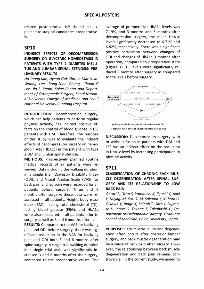
SPECIAL POSTERS
64
related postoperative NP should be ex‐plained to surgical candidates preoperative‐ly.
SP10 INDIRECT EFFECTS OF DECOMPRESSION SURGERY ON GLYCEMIC HOMEOSTASIS IN PATIENTS WITH TYPE 2 DIABETES MELLI‐TUS AND LUMBAR SPINAL STENOSIS: PRE‐LIMINARY RESULTS Ho‐Joong Kim, Hyeon‐Guk Cho, Je‐Min Yi, Ki‐Woong Lee, Bong‐Soon Chang, Choon‐Ki Lee, Jin S. Yeom; Spine Center and Depart‐ment of Orthopaedic Surgery, Seoul Nation‐al University College of Medicine and Seoul National University Bundang Hospital
INTRODUCTION: Decompression surgery, which can help patients to perform regular physical activity, has indirect positive ef‐fects on the control of blood glucose in LSS patients with DM. Therefore, the purpose of this study was to evaluate the indirect effects of decompression surgery on hemo‐globin A1c (HbA1c) in the patient with type 2 DM and lumbar spinal stenosis. METHODS: Prospectively planned routine medical records of 27 patients were re‐viewed. Data including the walking duration in a single trial, Oswestry Disability Index (ODI), and Visual Analog Scale (VAS) for back pain and leg pain were recorded for all patients before surgery. Three and 6 months after surgery, these data were re‐assessed in all patients. Height, body mass index (BMI), fasting total cholesterol (TC), fasting blood glucose (FBG), and HbA1c were also measured in all patients prior to surgery as well as 3 and 6 months after it. RESULTS: Compared to the VAS for back/leg pain and ODI before surgery, there was sig‐nificant reduction in the VAS for back/leg pain and ODI both 3 and 6 months after spine surgery. A single trial walking duration in a single trial walk was significantly in‐creased 3 and 6 months after the surgery, compared to the preoperative values. The
average of preoperative HbA1c levels was 7.19%, and 3 months and 6 months after decompression surgery, the mean HbA1c levels significantly decreased to 6.71% and 6.82%, respectively. There was a significant positive correlation between changes of ODI and changes of HbA1c 3 months after operation, compared to preoperative state (Figure 1). TC levels were significantly re‐duced 6 months after surgery as compared to the levels before surgery.
DISCUSSION: Decompression surgery with or without fusion in patients with DM and LSS has an indirect effect on the reduction in HbA1c level by increasing participation in physical activity.
SP11 CLASSIFICATION OF CHRONIC BACK MUS‐CLE DEGENERATION AFTER SPINAL SUR‐GERY AND ITS RELATIONSHIP TO LOW BACK PAIN Ohtori S, Orita S, Yamauchi K, Eguchi Y, Aoki Y, Miyagi M, Suzuki M, Sakuma Y, Kubota G, Oikawa Y, Inage K, Sainoh T, Sato J, Fujimo‐to K, Inoue G, Toyone T, Takahashi K.; De‐partment of Orthopaedic Surgery, Graduate School of Medicine, Chiba University, Japan
PURPOSE: Back muscle injury and degener‐ation often occurs after posterior lumbar surgery, and back muscle degeneration may be a cause of back pain after surgery. How‐ever, the relationship between back muscle degeneration and back pain remains con‐troversial. In the current study, we aimed to

SPECIAL POSTERS
65
classify back muscle degeneration using MRI and investigate its relationship with back pain after surgery. METHOD: A total of 84 patients (average age: 65.1 years, 38 men, 46 women) with lumbar spinal stenosis underwent only pos‐terior decompression surgery. MRI (1.5 tes‐la) was evaluated before and more than a year after surgery in all patients. Muscle on MRI was classified into 3 categories: low intensity in T1 weighted image, high in T2 weighted image (type 1), high in both T1 and T2 weighted images (type 2), and low in both T1 and T2 weighted images (type 3). The proportion of types and their relation‐ship with back pain (visual analog scale: VAS) was evaluated. RESULTS: MRI revealed muscle degenera‐tion in all patients after surgery (type 1: 6%, type 2: 82%, and type 3: 12%). Type 2 was significantly frequent compared with type 1 and 3 (P < 0.01). Low back pain before sur‐gery (average: VAS 7.5) significantly im‐proved after surgery (average: VAS 2.0) (P < 0.01). Low back pain was not associated with any muscle degeneration MRI types after surgery (P > 0.05). DISCUSSION: The current study showed the pathology of back muscle degeneration was various after posterior lumbar surgery. Type 2 (fatty change) was most frequent, and some patients were classified into type 3 (scar change), and type 1 (inflammation or water‐like change). Modic classification is used for bone marrow change, and type 1 is associated with inflammation and back pain. However, no type of back muscle de‐generation was correlated with back pain after surgery.
SP12 THE TYPE OF METAL, LOCAL ANTIBIOTICS OR PROPHYLACTIC IV ANTIBIOTICS, WHAT INFLUENCES POSTOPERATIVE SPINE INFEC‐TIONS WITH MRSA THE MOST? Sachin Gupta, Sukanta Maitra, Maria Graca, Pumibal Wetpiriyakul, Kavita Gupta, Blythe
Durbin‐Johnson, Munish Gupta; University of California at Davis, Sacramento, Califor‐nia,USA
INTRODUCTION: The purpose of this study was to determine the efficacy of Vancomy‐cin powder and IV prophylactic Vancomycin in treatment of spine infection with MRSA in presence of different metals. METHODS: 42 rabbits underwent a posteri‐or L5‐L6 approach. A 4 cm wire was placed around the L5 and L6 spinous processes. The implant was inoculated with 100 µL MRSA containing 1000000 colonies. 40 mg of Powder Vancomycin was placed in the wound prior to closure. The dose of IV Van‐comycin was 15mg/kg. Proportions of in‐fected rabbits were compared between groups using chi‐square tests, and com‐pared between groups adjusting for con‐founding factors using Cochran‐Mantel‐Haenzsel tests. CFU counts for the tissue and implant were compared between groups using Wilcoxon rank sum tests. RESULTS: The proportions of rabbits with infection were significantly higher in rabbits who received no Vancomycin compared ones that received powdered Vancomycin (P < 0.001 for tissue‐oxy, tissue‐blood, im‐plant‐oxy, and implant‐blood). CoCr had higher rates of residual infection despite the use of Vancomycin powder than Ti and SS (P = 0.040). In rabbits with SS, the pro‐portions of infection differed significantly among Vancomycin types (non, powdered, or IV) (P = 0.015 for tissue‐oxy, P = 0.020 for tissue‐blood, P = 0.025 for implant‐oxy, P = 0.023 for implant‐blood), with a significant‐ly lower proportion infected among those with powdered Vancomycin. Rabbits with SS implants treated with Vancomycin pow‐der had a lower infection rate than those treated with IV Vancomycin, although this difference was not statistically signifi‐cant(p=0.557) DISCUSSION: The Vancomycin powder is very successful in eradicating infection.
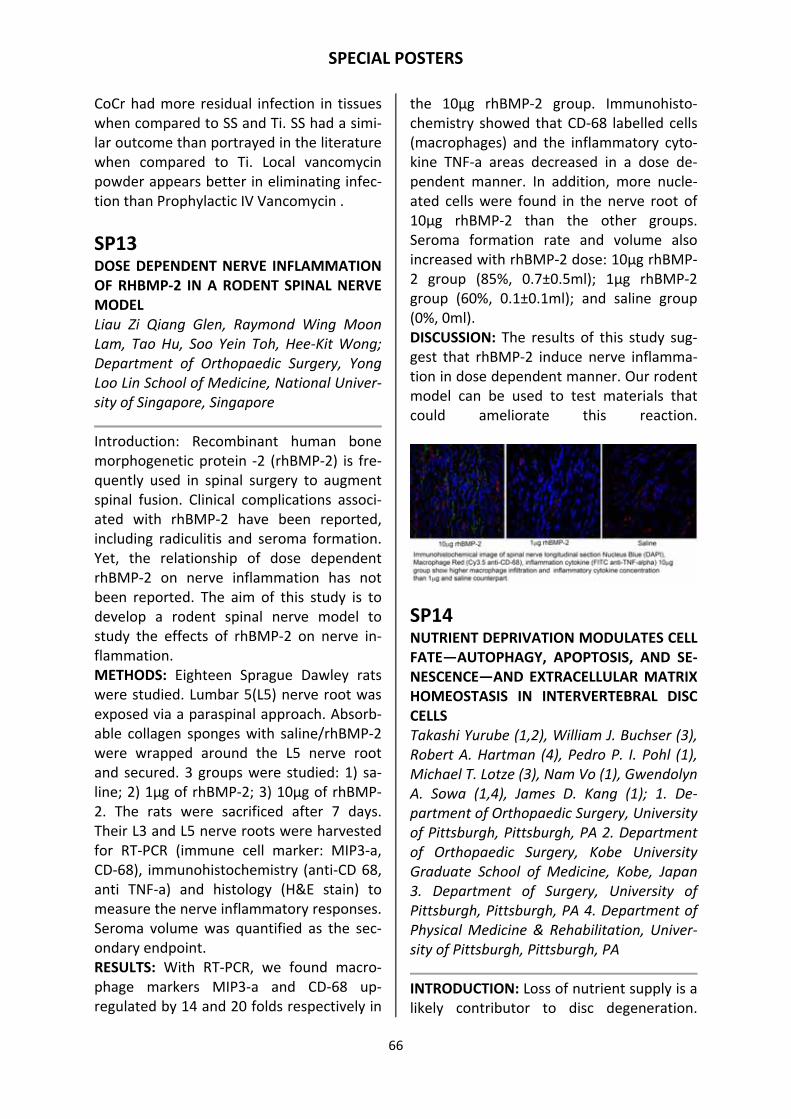
SPECIAL POSTERS
66
CoCr had more residual infection in tissues when compared to SS and Ti. SS had a simi‐lar outcome than portrayed in the literature when compared to Ti. Local vancomycin powder appears better in eliminating infec‐tion than Prophylactic IV Vancomycin .
SP13 DOSE DEPENDENT NERVE INFLAMMATION OF RHBMP‐2 IN A RODENT SPINAL NERVE MODEL Liau Zi Qiang Glen, Raymond Wing Moon Lam, Tao Hu, Soo Yein Toh, Hee‐Kit Wong; Department of Orthopaedic Surgery, Yong Loo Lin School of Medicine, National Univer‐sity of Singapore, Singapore
Introduction: Recombinant human bone morphogenetic protein ‐2 (rhBMP‐2) is fre‐quently used in spinal surgery to augment spinal fusion. Clinical complications associ‐ated with rhBMP‐2 have been reported, including radiculitis and seroma formation. Yet, the relationship of dose dependent rhBMP‐2 on nerve inflammation has not been reported. The aim of this study is to develop a rodent spinal nerve model to study the effects of rhBMP‐2 on nerve in‐flammation. METHODS: Eighteen Sprague Dawley rats were studied. Lumbar 5(L5) nerve root was exposed via a paraspinal approach. Absorb‐able collagen sponges with saline/rhBMP‐2 were wrapped around the L5 nerve root and secured. 3 groups were studied: 1) sa‐line; 2) 1µg of rhBMP‐2; 3) 10µg of rhBMP‐2. The rats were sacrificed after 7 days. Their L3 and L5 nerve roots were harvested for RT‐PCR (immune cell marker: MIP3‐a, CD‐68), immunohistochemistry (anti‐CD 68, anti TNF‐a) and histology (H&E stain) to measure the nerve inflammatory responses. Seroma volume was quantified as the sec‐ondary endpoint. RESULTS: With RT‐PCR, we found macro‐phage markers MIP3‐a and CD‐68 up‐regulated by 14 and 20 folds respectively in
the 10µg rhBMP‐2 group. Immunohisto‐chemistry showed that CD‐68 labelled cells (macrophages) and the inflammatory cyto‐kine TNF‐a areas decreased in a dose de‐pendent manner. In addition, more nucle‐ated cells were found in the nerve root of 10µg rhBMP‐2 than the other groups. Seroma formation rate and volume also increased with rhBMP‐2 dose: 10µg rhBMP‐2 group (85%, 0.7±0.5ml); 1µg rhBMP‐2 group (60%, 0.1±0.1ml); and saline group (0%, 0ml). DISCUSSION: The results of this study sug‐gest that rhBMP‐2 induce nerve inflamma‐tion in dose dependent manner. Our rodent model can be used to test materials that could ameliorate this reaction.
SP14 NUTRIENT DEPRIVATION MODULATES CELL FATE—AUTOPHAGY, APOPTOSIS, AND SE‐NESCENCE—AND EXTRACELLULAR MATRIX HOMEOSTASIS IN INTERVERTEBRAL DISC CELLS Takashi Yurube (1,2), William J. Buchser (3), Robert A. Hartman (4), Pedro P. I. Pohl (1), Michael T. Lotze (3), Nam Vo (1), Gwendolyn A. Sowa (1,4), James D. Kang (1); 1. De‐partment of Orthopaedic Surgery, University of Pittsburgh, Pittsburgh, PA 2. Department of Orthopaedic Surgery, Kobe University Graduate School of Medicine, Kobe, Japan 3. Department of Surgery, University of Pittsburgh, Pittsburgh, PA 4. Department of Physical Medicine & Rehabilitation, Univer‐sity of Pittsburgh, Pittsburgh, PA
INTRODUCTION: Loss of nutrient supply is a likely contributor to disc degeneration.

SPECIAL POSTERS
67
However, the extent to which limited nutri‐tion affects disc cells is unknown. In addi‐tion, the involvement of autophagy, the intracellular process by which cells recycle their damaged components, in disc disease is poorly understood. Our objective was to elucidate the effect of nutrient deprivation on disc cell fate and matrix homeostasis. METHODS: Rabbit annulus fibrosus cells were cultured under a varying supply of nutrition. To characterize cellular respons‐es, cell proliferation and metabolic activity were measured. To assess autophagy, au‐tophagic flux was longitudinally monitored by imaging cytometry and Western blotting. To analyze cell fate, apoptosis and senes‐cence levels were determined. To evaluate matrix homeostasis, catabolic, anti‐catabolic, and anabolic gene expression and matrix protein synthesis and degradation were investigated. RESULTS: With nutrient deprivation, cell proliferation and metabolic activity de‐creased. Then, autophagy markers, LC3 puncta number and LC3‐II expression, in‐creased time‐dependently. Cytoplasmic HMGB1 intensity also increased following elevated total and nuclear expression. A negatively correlated marker, p62/SQSTM1 expression, decreased over time, all indicat‐ing enhanced autophagy. TUNEL, SA‐ß‐gal, cleaved caspase‐3, and p16/INK4A analysis indicated increased apoptosis and senes‐cence. Quantitative PCR showed up‐regulated MMPs and down‐regulated ADAMTSs, TIMPs, aggrecan‐1, and collagens under serum starvation. While newly syn‐thesized proteoglycans and collagens de‐creased, aggrecan degradation increased, notably by MMPs. DISCUSSION: Nutrient deprivation increases apoptosis and senescence along with au‐tophagy in addition to shifting matrix ho‐meostasis toward catabolism in disc cells—changes consistent with initial stages of disc degeneration. This study suggests a new
treatment strategy for degenerative disc disease, autophagy regulation.
SP15 MOLECULAR EFFECTS OF BONE MORPHO‐GENETIC PROTEIN 13 ON INTERVERTEBRAL DISC CELLS Twishi Gulati, Aiqun Wei, Sylvia Chung, Ash‐ish D Diwan; Department of Orthopaedic Surgery, St George Hospital Clinical School, University of New South Wales, Sydney, Australia
INTRODUCTION: BMP‐13 stimulates prote‐oglycan production in an ovine disc injury model, however its presence or absence in degenerate disc and its effect on degener‐ate human IVD cells has not been investi‐gated. METHODS: 22 IVDs were obtained from patients undergoing either scoliosis or de‐generative surgery. Endogenous gene and protein expression of BMP‐13 in human degenerate IVD was determined. The effect of rhBMP‐13 on human derived nucleus pulposus (NP), annulus fibrosus (AF) and end plate (EP) cells cultured in alginate beads was evaluated by changes in cell via‐bility (DNA content Hoechst dye) and pro‐teoglycan accumulation (Alcian blue stain). The effect of BMP‐13 on the expression of chondrogenic genes (Aggrecan, Col1, Col2, COMP, SOX9 using RT‐PCR and REST) and proteins (Col1, Col2 ImmunoHistochemis‐try) by IVD cells was determined. The migra‐tory potential (Boyden Chamber assay) of degenerate disc cells towards BMP‐13 was examined. RESULTS: The endogenous expression of BMP‐13 in degenerate NP was not different to non‐degenerate disc. While BMP‐13 stimulation had a limited effect on the cel‐lular viability of IVD cells, BMP‐13 induced significant proteoglycan accumulation in NP, AF and EP cells cultured in alginate beads at a concentration of 400ng/ml after 7 days. REST results showed considerable

SPECIAL POSTERS
68
variation in the relative change of gene ex‐pression amongst four independent NP, AF and EP samples. In NP and EP cells 400ng/ml of BMP‐13 increased collagen I and II protein expression. NP cells displayed chemotactic properties towards 800ng/ml of BMP‐13 in the absence of fetal calf se‐rum. DISCUSSION: This is the first study to cul‐ture human end plate cells in three‐dimensional culture and to investigate the effect of BMP‐13 on human derived IVD cells. BMP‐13 has the potential to enhance proteoglycan accumulation and induce cell migration in certain IVD cells. This data sup‐ports further exploration of BMP‐13 as a probable therapeutic agent to ameliorate IVD degeneration.
SP16 THE EFFECT OF NERVOUS SYSTEM ACTIVA‐TION BY PAIN STIMULUS OF CAPCISIN IN‐JECTION TO INTERVERTEBRAL DISC Hironobu Nishiori1), Kazuyo Yamauchi2), Yoshihiro Sakuma2), Yasuhiro Oikawa2), Kazuhide Inage2), Go Kubota2), Ken Saino2), Miyako Suzuki2), Yukio Nakata2), Jun Sato2), Sadao Arai2), Kazuhisa Takahashi2), Seiji Ohtori2), Sumihisa Ori‐ta2); 1) School of Medicine, Chiba University 2) Department of Orthopaedic Surgery, Chi‐ba University
BACKGROUND: Previously, it was reported that pain perception is related to not only DRG but also to the reaction of the central nervous system; however, the reaction of the dominant nervous system to pain stimu‐lus in the intervertebral discs is unknown. The purpose of this research was to verify the mechanism of reaction of the nervous system, which controls the intervertebral discs, using chemical pain stimulus. METHODS: We used 6‐week‐old Sprague Dawley rats, and injected 20 µL capcisin solution, which contains 2% Fluoro‐ Gold (FG), into the L5/6 intervertebral disc. FG is
an antidromic neuronal tracer. Seven days after injection, we prepared perfusion fixa‐tion of the rats and extracted DRGs from L1 to L6 along with the lumbar enlargement of spinal cord. At the DRG, we detected CGRP, which is a nociceptive pain‐related protein, by immunostaining, and measured the dis‐tribution of the expression. At the lumbar enlargement of the spinal cord, we detect‐ed Iba1, which is a marker for pain stimulus‐activated micro glia, and compared Iba 1 expression in the capcisin group and vehicle group, in which we injected only 20 µL sa‐line solution (each is n = 5). RESULT: The rate of distribution of CGRP positive cells in the FG positive rats was highest at the L2 DRG, and the rate was higher in the capcisin group (54.9%) than in vehicle group (27.1%), with a statistically significant difference (p < 0.05). The num‐ber of Iba1 positive micro glia cells per 1.4 × 10 mm2 was also larger in the capcisin group (31.7 cells) than in the vehicle group (14.8 cells), with a statically significant dif‐ference (p < 0.05) DISCUSSION: The results of this experiment suggested that the chemical stimulus to the L5/6 intervertebral disc resulted in an ele‐vated CGRP expression that caused pain around the L2 DRG area, and the mecha‐nism of pain perception is strongly related to the activation of micro glia at the poste‐rior horn of the spinal cord.
SP17 UP‐REGULATION OF NAV1.7 IN DORSAL ROOT GANGLIA AFTER INTERVERTEBRAL DISC INJURY IN RATS. Aya Sadamasu, MD, Yoshihiro Sakuma, MD, Miyako Suzuki, MD, PhD, Sumihisa Orita, MD, PhD, Kazuyo Yamauchi, MD, PhD, Gen Inoue, MD, PhD, Yasuchika Aoki, MD, PhD, Tetsuhiro Ishikawa, MD, PhD, Masayuki Miyagi, MD, PhD, Hiroto Kamoda, MD, PhD, Gou Kubota, MD,; Department of Ortho‐paedic Surgery, Graduate School of Medi‐

SPECIAL POSTERS
69
cine Chiba University, 1‐8‐1 Inohana, Chuo‐ku, Chiba 260‐8670, Japan
OBJECTIVE: To investigate pain‐related ex‐pression of NaV1.7 in dorsal root ganglia (DRG) innervating intervertebral discs. SUMMARY OF BACKGROUND DATA: Pur‐pose. The pathophysiology of discogenic low back pain is not fully understood. Pros‐taglandins and cytokines produced by de‐generated discs can cause pain, but non‐steroidal anti‐inflammatory and steroid medications are often ineffective at pain reduction. Tetrodotoxin‐sensitive voltage‐gated sodium (NaV) channels are associated with sensory transmission in primary senso‐ry nerves, and the NaV1.7 channel has emerged as an attractive analgesic target. The purpose of this study was to investigate pain‐related expression of NaV1.7 in DRG innervating intervertebral discs. METHODS: Using a rodent model of disc puncture, we labeled DRG neurons inner‐vating L5/6 discs with FluoroGold neuro‐tracer (n = 20). Half of the rats (n = 10) un‐derwent intervertebral disc puncture using a 23‐gauge needle (puncture group), and the other half underwent non‐puncture sham surgery (non‐puncture group). Seven and fourteen days after surgery, DRGs from the L1 to L6 levels were harvested, sec‐tioned, and immunostained for NaV1.7, and the proportion of NaV1.7‐ immunoreactive DRG neurons was evaluated. RESULTS: NaV1.7 was expressed in DRG neurons innervating intervertebral discs from L1 to L5. The ratio of NaV1.7‐expressing DRG neurons to total FG‐labeled neurons was 7.2% and 7.6% at 1 and 2 weeks after surgery, respectively, in the non‐puncture group and 16.2% and 16.3% at 1 and 2 weeks, respectively, in the punc‐ture group. The up‐regulation of NaV1.7 after puncture was significant at both 1 and 2 weeks after surgery (P < 0.01). Conclusions. We found that disc injury in‐creases NaV1.7 expression in DRG neurons
innervating injured discs. NaV1.7 may be a therapeutic target for pain control in pa‐tients with lumbar disc degeneration.
SP18 ABNORMAL CELL METABOLISM IN DEGEN‐ERATED INTERVERTEBRAL DISCS: A RE‐SPONSE TO FOCAL DAMAGE AND SWELL‐ING? Lama P, H A Claireaux, L Flower, Le Maitre C, Dolan P, Tarlton J, Harding IJ, Adams MA; Centre for Comparative and Clinical Anato‐my, University of Bristol, United King‐dom*Biomedical Research Centre, Sheffield Hallam University, United Kingdom ** De‐partment of Orthopaedics, Southmead Hos‐pital, United Kingdom
INTRODUCTION: Physical disruption of the extracellular matrix influences the mechani‐cal and chemical environment of interver‐tebral disc cells. We hypothesise that this can explain degenerative changes such as focal proteoglycan loss, impaired cell‐matrix binding, cell clustering, and increased activi‐ty of matrix‐degrading enzymes. METHODS: Disc tissue samples were re‐moved surgically from 11 patients (aged 34‐75 yrs) who had a painful but non‐herniated disc. Each sample was divided into a pair of speciemns (approximately 5mm3), which were cultured at 37°C under 5% CO2. One of each pair was allowed to swell, while the other was restrained by a perspex ring. Live‐cell imaging was performed with a wide field microscope for 36 hrs. Specimens were then sectioned at 5 and 30 µm for histology and immunofluorescence using a confocal microscope. Antibodies were used to rec‐ognise free integrin receptor a5ß1, matrix metalloprotease MMP‐1, and denatured collagen types I‐III. Proteoglycan content of the medium, analysed using the colorimet‐ric DMMB assay, was used to assess tissue swelling and GAG loss. Con‐strained/unconstrained results were com‐pared using matched‐pair t‐tests.

SPECIAL POSTERS
70
RESULTS: Time‐lapse cinematography re‐vealed small cell movements in uncon‐strained specimens, for up to 12 hrs. By 36 hrs, unconstrained (free swelling) samples showed greater: loss of GAG’s (p<0.003), loss of integrin binding (p<0.02), synthesis of MMP‐1 (p<0.03), and collagen denatura‐tion (p<0.009). Cell clustering was evident in all tissues after 36 hrs. DISCUSSION: Swelling of disrupted disc tis‐sue allows increased proteoglycan loss, dis‐turbs cell‐matrix binding, increases matrix degradation and thus could act as a stimu‐lus for cell cluster formation. This sequence of events could follow disc injury or herni‐ation in‐vivo.
SP19 STIFFNESS AFTER FUSION FOR ADULT SPI‐NAL DEFORMITY DOES NOT SIGNIFICANTLY IMPACT PATIENTS’ FUNCTIONAL STATUS OR SATISFACTION Hiratzka, Jayme R.1; Hamilton, D. Kojo1; Bess, Shay 2; Schwab, Frank J.3; Shaffrey, Christopher I.4; Ames, Christopher P.5; Mundis, Gregory M.6; Lafage, Virginie3; Deviren, Vedat7; Smith, Justin S.4; Kline‐berg, Eric8; Boachie‐Adjei, Oheneba9; Bur‐ton, Doug; 1. Orthopaedic Surgery, Oregon Health and Sciences University, Portland, OR, United States. 2. Orthopaedic Surgery, Rocky Mountain Hospital for Children, Den‐ver, CO, United States. 3. Orthopaedic Sur‐gery, NYU Hospital for Joint Diseases, New York, NY, United States. 4. Neurosurgery, University of Virginia Medical Center, Char‐lottesville, VA, United States. 5. Neurosur‐gery, University of California, San Francisco Medical Center, San Francisco, CA, United States. 6. San Diego Center for Spinal Disor‐ders, La Jolla, CA, United States. 7. Ortho‐paedic Surgery, University of California, San Francisco Medical Center, San Francisco, CA, United States. 8. Orthopaedic Surgery, Uni‐versity of California, Davis, Sacramento, CA, United States. 9. Orthopaedic Surgery, Hos‐pital for Special Surgery, New York, NY,
United States. 10. Orthopaedic Surgery, University of Kansas Medical Center, Kansas City, KS, United States. 11. ISSGF, Littleton, CO, United States.
INTRODUCTION: The Lumbar Stiffness Dis‐ability Index (LSDI) is a validated measure of the effect of spinal stiffness on function following lumbar fusion surgery. No pro‐spective analyses of stiffness impacts fol‐lowing adult spinal deformity surgery have been reported. METHODS: The LSDI, SRS‐22, SF‐36 and ODI were administered prospectively at baseline and 2‐year minimum followup to 50 adult patients undergoing thoracolumbar fusions to the pelvis for spinal deformity (excluding those with prior history of lumbar fusion). Patients were divided into 2 groups based on upper thoracic (UT, T2‐5) or thoracol‐umbar (TL, T10‐T11) proximal endpoints. Comparisons of pre‐and post‐operative HRQoL and LSDI scores and correlation of LSDI to SRS‐22 satisfaction scores were per‐formed. RESULTS: Significant improvements were seen in both the UT group in ODI (36.4 to 21.8), SRS22 (3.1 to 3.8) and SF‐36 PCS (32.1 to 45.0), and in the TL group in ODI (38.8 to 18.4), SRS22 (2.9 to 4.0), SF‐36 PCS (30.5 to 44.1) (Table 1). In contrast, LSDI scores did not change from baseline in either group. There was a trend toward higher final LSDI scores among UT compared to TL patients which did not reach significance. No corre‐lation was found between 2‐year LSDI and overall SRS‐22 satisfaction scores (R2=0.0193).
DISCUSSION: Adult deformity patients un‐dergoing instrumented thoracolumbar fu‐sion to the pelvis report no significant in‐creased difficulty in performing of ADL’s as

SPECIAL POSTERS
71
a result of increased stiffness at 2 year fol‐low up. While patients fused to the upper thoracic spine trended toward higher LSDI scores than patients with thoracolumbar stopping points, both groups experienced significant improvements in ODI, SRS22 and SF‐36 PCS scores. In addition, stiffness as measured by LSDI did not correlate with overall patient satisfaction scores. These results suggest that adult deformity pa‐tients experience little increased disability due to stiffness, even after fusion of their entire lumbar spine.
SP20 RISK FACTORS FOR FLOATING SEGMENT DEGENERATION IN ADULTS AFTER LONG SEGMENT FUSION UNTO THE LUMBAR SPINE. Woojin Cho, MD, PhD, Abhijit Pawar, MD, Andrew Lee, MD, Mike Faloon, MD, Mat‐thew Cunningham, MD, PhD, Bernard Rawlins MD and Oheneba Boachie‐Adjei, MD; Albert Einstein College of Medicine
INTRODUCTION: Risk factors for adjacent segment degeneration (ASD) of floating non‐fused segments between the long thoracolumbar fusion construct and sacrum after correction of adult spine deformity have been rarely reported. METHODS: In this retrospective study, med‐ical records of 198 patients with adult spinal deformity who underwent long fusion of 6 segments or more in the thoracolumbar spine for the treatment of adult spine de‐formity with minimum 2‐year follow‐up were included. Patients who underwent primary long fusion down to the sacrum, and revision cases were excluded. Out of the 47 patients which matched the criteria, 8 Pts who underwent subsequent surgery extending the fusion to the sacrum were included in the Failed group (F) and 39 Pts were included in the non‐failed group (NF). The radiological data at final follow up in NF
group and final follow up before revision to sacrum in the F group was compared. RESULTS: Patient demographics were simi‐lar in both groups. The average follow up was 2.9 years in NF and 4.8 years in F. The thoracic and lumbar curve correction was better in the NF as compared to failed group but the difference was not signifi‐cant. The upper thoracic cobb was smaller in the NF at final f/u. The saggital balance was significantly better in the NF; however, the coronal balance was similar in both groups at final follow up. The average lum‐bar lordosis and thoracic kyphosis was sig‐nificantly better in NF. The average LIV (lowest instrument vertebra) tilt and disc angulation below LIV was smaller in NF. The LIV translation group and preoperative ra‐diographic disc degeneration were higher in F but the difference was not significant. The pelvic tilt (PT) was larger in F while the pel‐vic incidence was similar. DISCUSSION: Adequate restoration of Lum‐bar lordosis, Thoracic kyphosis, LIV tilt, LIV translation, disc angulation, sagittal bal‐ance, PT is important to prevent ASD after long fusions unto the lumbar spine.
SP21 SAGITTAL SPINAL ALIGNMENT OF DIFFUSE IDIOPATHIC SKELETAL HYPEROSTOSIS IN LUMBAR SPINAL STENOSIS Yamada Kentaro, Toyoda Hiromitsu, Terai Hidetomi, Takahashi Shinji, Nakamura Hi‐

SPECIAL POSTERS
72
roaki; Department of Orthopaedic Surgery, Osaka City University Graduate School of Medicine, Osaka, Japan
INTRODUCTION: DISH has been considered a radiographic entity with less importance given to clinical signs. Some studies report‐ed postural abnormality in DISH. However, there are no detailed reports regarding spinopelvic alignment in DISH. Sagittal spi‐nal alignment is especially an important factor in the management of lumbar spinal stenosis (LSS). The purpose of this study was to evaluate the characteristics of DISH and its spinopelvic alignment in patients with LSS. METHODS: A total of 132 patients over 40 years of age who required surgical proce‐dures for LSS were investigated. DISH was defined by Resnick’s and Mata’s criteria on preoperative, standing radiographs of the whole spine. We investigated the preva‐lence and localization of DISH, and its rela‐tion to preoperative clinical symptoms. Sag‐ittal spinal alignment was measured by sag‐ittal C7 plumb line, lumbar lordosis, thoracic kyphosis, sacral slope, pelvic incidence, pel‐vic tilt, and overhang of S1. The association between DISH and spinopelvic alignment was analyzed using covariance adjusted with age, sex, spondylolisthesis, and degen‐erative lumbar scoliosis. RESULTS: The prevalence of DISH was 39.4% (52/132), and increased with aging. Preoperative symptoms showed no differ‐ences, regardless of the presence of DISH. Lumber and thoracic alignment showed kyphotic change in DISH. DISH patients with lower fused vertebral ends in the lumbar level (46% of DISH) still showed significantly decreased lumbar lordosis (p=0.014) and horizontalization of the sacrum (p=0.001) after adjusting age, sex, spondylolisthesis, and degenerative lumbar scoliosis. DISCUSSION: DISH prevalence increases with age, and spinopelvic alignment is af‐fected by the presence of DISH in LSS pa‐
tients, especially in patients with lower fused vertebral ends at the lumbar lesion. These results may therefore offer an expla‐nation as to why elderly patients frequently show kyphotic changes in their spine.
SP22 RELATIONSHIP BETWEEN SPINAL LOADS AND PHYSIOLOGICAL VARIATIONS OF THE LUMBAR SAGITTAL ALIGNMENT: A NU‐MERICAL STUDY Fabio Galbusera (1), Marco Brayda‐Bruno (1) and Hans‐Joachim Wilke (2); (1) IRCCS Istituto Ortopedico Galeazzi, Milan, Italy (2) Institute of Orthopedic Research and Bio‐mechanics, Ulm University, Ulm, Germany
INTRODUCTION: This study presents a nu‐merical approach to reproduce various pat‐terns of spino‐pelvic organization and is aimed to predict the spinal loads in two static conditions (standing and holding a weight in the hands) in the entire range of normal variability of the shape of the lum‐bar spine. METHODS: First, a simplified finite element model of a healthy, normal thoracolumbar spine including relevant muscles has been developed, based on a published series of papers. The model was then modified to represent the physiological variation of the sagittal alignment. Following the classifica‐tion system by Roussouly, 480 finite ele‐ment models representing the entire range of normal variability were created. The models were based on three independent parameters, namely the sacral slope, the position of the apex and the inflection point. Two load‐bearing conditions were simulated: the standing posture, by apply‐ing the body weight and optimizing the muscle forces in order to obtain equilibri‐um; a posture in which the subject is hold‐ing a weight of 50 N, by adding a load 40 cm in front of the center of T6. RESULTS: The models predicted that, even in the case of a moderate external load, a

SPECIAL POSTERS
73
well‐balanced spine (e.g. type 3) was able to reduce the muscle activation in comparison with a straighter or a hyperlordotic spine. Such a sagittal configuration was however not correlated with a minimization of the loading state in the intervertebral discs, especially regarding anteroposterior shear loads. In the standing posture without any additional load, a less lordotic and more vertical spine (e.g. type 2) was sufficient to ensure a condition of minimal spinal loads. DISCUSSION: Despite some limitations, in‐verse statics numerical models of the spine biomechanics including trunk muscles ap‐pear to be a promising tool to fill the knowledge gap between the clinical obser‐vations of the correlations between the spinopelvic organization and the conse‐quent spinal disorders.
SP23 PROGRESSIVE PATTERN OF VERTEBRAL DEFORMITY IN A POPULATION‐BASED CO‐HORT STUDY OF VERTEBRAL FRACTURE 1,2Toshihiro Kato, 1Koji Akeda, 1Koichiro Murata, 1Norihiko Takegami, 1Akinobu Nishimura, 1, 2Ko Kato, 1Akihiro Sudo; 1Department of Orthopaedic Surgery, Mie University Graduate School of Medicine 2 Department of Orthopedic Surgery , Suzuka Kaisei Hospital
INTRODUCTION: Vertebral fractures (VFs) may cause spinal deformity and/or chronic back pain. Therefore, epidemiological stud‐ies of the natural history of VF are of great importance. Vertebral deformity may pro‐gress after VF; however, no detailed inves‐tigation of progression patterns has been reported. The purpose of this study was to examine the progression pattern of verte‐bral deformity in a population‐based pro‐spective cohort study. METHODS: Over 10‐years (1997‐2009), 346 inhabitants (111 men, 235 women: mean age: 70.2 years) of a typical mountain vil‐lage underwent medical examinations and
were followed for more than 4 years. Of these, 196 who had prevalent VFs (450 ver‐tebrae) at baseline (72 men, 124 women: mean age: 69.9 years) were subjects of this study. Lateral thoracic and lumbar spine radiographs of each subject were taken; the extent (G1: mild, G2: moderate, G3: severe) and type (wedge, biconcave, crush) of the prevalent fractures at baseline and final examination were evaluated using a semi‐quantitative technique. The progression of deformity was divided into 3 groups by comparing the deformity at baseline and final examination: no change (NC), grade‐progressed (GP) and type‐changed (TC) groups. RESULTS: Deformity extent at baseline (G1:80%, G2:16%, G3:4%) progressed com‐pared to that at final examination (G1:60%, G2:29%, G3:11%). The percentage of G1 decreased, and G2 and G3 increased at final examination (P<0.05). There were no signif‐icant changes on type of deformity at base‐line compared to that at final examination. However, biconcave type deformity (NC 59%, GP 29%, TC 12%) at baseline had a significant effect on both the extent and type of deformity at final examination com‐pared to wedge type (NC:73%, GP:17%, TC:10%) and crush type (NC:86%, GP:7%, TC:7%). DISCUSSION: This study showed that the extent of vertebral deformity in the elderly progressed with age. A biconcave type de‐formity at baseline may change the extent and type of deformity.
SP24 CLINICAL OUTCOME AFTER LUMBAR DE‐COMPRESSION SURGERY WAS NOT ASSO‐CIATED WITH PREOPERATIVE SAGITTAL ALIGNMENT. Tomohiro Hikata1, Kota Watanabe2, No‐buyuki Fujita1, Akio Iwanami1, Naobumi Hosogane1, Ken Ishii1, Masaya Nakamura1, Yoshiaki Toyama1 and Morio Matsumoto1; 1Department of Orthopaedic Surgery,

SPECIAL POSTERS
74
School of Medicine, Keio university., Tokyo, Japan. 2Advanced Therapy for Spine and Spinal Cord Disorders, Keio University., To‐kyo, Japan.
INTRODUCTION: Sagittal spinal alignment is an important factor in the management of lumbar degenerative diseases. The purpose of this study was to investigate the correla‐tion between preoperative sagittal spinopelvic alignment and improvement of clinical outcome after decompression sur‐gery for lumbar canal stenosis (LCS). METHODS: 132 patients who underwent elective lumbar spinous process‐splitting laminectomy from 2009 to 2011 were ret‐rospectively reviewed. There were 75 men and 57 women with a mean age of 71.2 years. The mean follow‐up (f‐u) period was 26.2 months (minimum 1Y f‐u). The subject was divided into three groups according to the preoperative sagittal vertical axis (SVA) value: the patients with SVA less than 50 mm (Group A), those with SVA from 50 to 80 mm (Group B), and those with SVA more than 80 mm (Group C). Radiologic parame‐ters including lumbar Cobb angle, coronal balance, SVA, lumbar lordosis, thoracic ky‐phosis, thoracolumbar kyphosis, Pelvic Inci‐dence (PI), Pelvic Tilt (PT), Sacral Slope (SS)) were measured before surgery. The JOA score and JOA Back Pain Evaluation Ques‐tionnaire (JOABPEQ), VAS score, and RDQ score were used for clinical evaluation. RESULTS: There were 83 patients in Group A, 34 patients in Group B, and 15 patients in Group C, respectively. The preoperative mean SVA was 21.2mm in Group A, 61.2mm in Group B, and 11.4 mm in Group C, re‐spectively. Preoperative LL and SS were significantly smaller in Group C (LL;15.7°, SS;19.2°), than Group A (LL;39.9°, SS;26.9°) (p<0.01). Preoperative PT was significantly larger in Group C (27.8°), than Group A (20.2°) (p<0.05). There were no significant differences in the improvement of various questionnaires.
DISCUSSION: The loss of lumbar lordosis and pelvic retroversion were presented in the LCS patients with preoperative sagittal imbalance. Interestingly, preoperative sagit‐tal alignment does not affect postoperative clinical outcomes after posterior decom‐pression surgery for LCS.
SP25 SHEAR MODULUS OF THE HUMAN NUCLE‐US PULPOSUS MEASURED USING MR ELASTOGRAPHY CORRELATES WITH DI‐RECTLY MEASURED MECHANICAL PROPER‐TIES Daniel H. Cortes (1), John F. DeLucca (1), Jeremy F. Magland (2), Alexander A. Wright (2), Dawn M. Elliott (1); (1) Biomedical Engi‐neering Program, University of Delaware, Newark DE 19716 (2) Department of Radi‐ology, University of Pennsylvania, Philadel‐phia PA 19104
INTRODUCTION: Our group recently demonstrated that Magnetic Resonance Elastography (MRE) can serve as a non‐invasive biomarker for disc degeneration, where the nucleus pulposus (NP) mechani‐cal properties are significantly altered with degeneration (Cortes et al, Magn Res Med 2013). However, the MRE measurements were not directly compared to standard tissue mechanical tests. The objectives of this study were to validate MRE measure‐ments by comparing the shear modulus to that of excised NP samples, and to quantify the contribution of the intradiscal pressure and glycosaminoglycan (GAG) content to the MRE shear modulus. METHODS: The NP shear modulus was measured using MRE in 16 intact human bone‐disc‐bone segments from levels T12‐L5 having degeneration grades from 1 to 4. NP samples were then dissected and tested in confined compression where the swelling pressure and the shear modulus were de‐termined. The GAG content was measured using a DMMB assay.

SPECIAL POSTERS
75
RESULTS: There was a correlation between MRE shear modulus and confined compres‐sion shear modulus (r = 0.57, p < 0.05) (Fig‐ure 1). There was also a correlation be‐tween MRE shear modulus and the isomet‐ric swelling pressure (r = 0.51, p < 0.05). However, no correlation was observed be‐tween the MRE shear modulus and the GAG content.
DISCUSSION: This study validated the MRE measurements by comparing the NP shear modulus to those directly measured. The correlation between isometric swelling pressure and the MRE shear modulus sug‐gests a contribution of the intradiscal pres‐sure to the MRE shear modulus. This effect has been recently reported in experiments in articular cartilage where the shear modu‐lus increased for higher osmotic pressures. The shear modulus measured using MRE is a potential biomarker for disc degeneration to directly evaluate the mechanical integrity of disc tissues.
SP26 BIOMECHANICAL ANALYSIS OF THE INFLU‐ENCE OF INTERVERTEBRAL DISC DEGENER‐ATION ON THE LUMBAR SPINE. Won Man Park1, Dae Kyung Choi1, Kyungsoo Kim2, Yoon Hyuk Kim1; 1Department of Mechanical Engineering,
Kyung Hee University, Korea 2Department of Applied Mathematics, Kyung Hee Univer‐sity, Korea
INTRODUCTION: Intervertrebral disc (IVD) degeneration disease is one of the most common reasons of low back pain. In spite of a number of previous experimental and numerical studies, alteration of stiffness of motion segment by degeneration is still controversial. Furthermore, the effects of various degrees of degeneration on biome‐chanical behaviors at the degenerated and healthy levels are not clearly determined. METHODS: Finite element models of the lumbar spine with various degrees of IVD degeneration at the L4‐L5 motion segment unit (MSU) were developed and validated with comparing to experimental results. 20%, 50% and 80% loss of disc height were applied for mild, moderate and severe de‐generation, respectively. Biomechanical behaviors at the degenerated and adjacent MSUs were investigated in hybrid loading condition. RESULTS: The ranges of motions (ROMs) of flexion‐extension and left‐right lateral bend‐ing at the degenerated MSU decreased with progress of degeneration, while the ROMs in left‐right axial rotation slightly changed by progress of degeneration. Intradiscal pressure (IDP) at the degenerated MSU de‐creased with progress of degeneration in flexion, lateral bending, and axial rotation, while the pressures at the adjacent MSUs increased in flexion and lateral bending. Facet joint force at the degenerated MSU decreased from 113N (healthy) to 41N (se‐vere) in extension with progress of degen‐eration, while the force increased from 96N (healthy) to 137N (severe) in left axial rota‐tion. DISCUSSION: According to these findings, IVD degeneration could reduce the risk of age‐related instability and discogenic pain at the degenerated MSU since the stiffness was increased and the IDP was decreased.

SPECIAL POSTERS
76
However, IVD degeneration might lead to increase in the risk of arthrosis in the facet joint of the degenerated MSU and/or dis‐cogenic back pain at the adjacent MSUs. These findings could provide fundamental information about injury mechanism caused by IVD degeneration..
SP27 ESCAPE‐AVOIDANCE COPING STRATEGY FOR PSYCHOSOCIAL STRESS IS ASSOCIATED WITH CHRONIC LOW BACK PAIN IN COL‐LEGE STUDENTS: A PROSPECTIVE COHORT STUDY Kinshi Kato; Miho Sekiguchi; Takuya Nikai‐do; Ken‐ichi Otoshi; Yohei Matsuo; Shin‐ichi Kikuchi; and Shin‐ichi Konno; Department of Orthopaedic Surgery, Fukushima Medical University School of Medicine, Fukushima, Japan
INTRODUCTION: Psychosocial stress may affect the incidence and duration of low back pain (LBP); however, few longitudinal studies have examined whether specific stress coping strategies affect LBP. The pur‐pose of this cohort study was to assess the association between specific coping strate‐gies and the incidence and duration of LBP in college students. METHODS: A longitudinal study was initiat‐ed among 135 students without LBP in July 2011. We reviewed LBP characteristics and assessed the psychosocial stress and coping strategies of students using the Stress Self‐Rating Scale (SSRS). A follow‐up survey was conducted in July 2013, analyzing all 135 students (follow‐up rate, 100%). Coping strategies in students were categorized into the following three groups using SSRS scores: problem‐focused (PF); emotion‐focused (EF); and escape‐avoidance (EA). Incidence and duration of new‐onset LBP were evaluated in each coping strategy group. Wilcoxon and chi‐square tests were used for statistical analyses.
RESULTS: Sixty‐three (46.7%) students re‐ported LBP during the 2‐year follow‐up. No significant differences were observed among the three groups in sex, body mass index, current smoking status, sleep time, past or family history of LBP, or physical activity. Furthermore, no significant differ‐ences were observed in the incidence rate (PF, 55.6%; EF, 46.3%; and EA. 45.3%) of LBP. However, the incidence of chronic LBP, defined as LBP duration >3 months, was significantly greater in the EA group than in the other groups (4.8% vs. 0%; p = 0.034). DISCUSSION: Differences in stress coping strategies did not affect the incidence of new‐onset LBP; however, the EA coping strategy increased the incidence of chronic low back pain in students compared with other coping strategies. The EA coping strategy for psychosocial stress may thus prolong the duration of LBP.
SP28 DOES THE CHANGE OF WEATHER CONDI‐TION INFLUENCE PAIN LEVEL OF SYMP‐TOMS IN THE PATIENTS WITH LUMBAR SPINE DISEASES? 1,13Ishii Ken, 3,13Hosogane Naobumi, 1,13Hikata Tomohiro, 1,13Yoshioka Kenji, 2,13Shiono Yuta, 5,13Iida Tsuyoshi, 6,13Ozaki Masahiro, 1,13Ishihama Hiroko, 1,13Horiuchi Yosuke, 1,13Takahashi Yohei, 7,13Furukawa Mitsuru, 8,13Nagoshi Shi‐gehito, 6,13Shingo I; 1 Department of Or‐thopedic Surgery, School of Medicine, Keio University, Shinjuku, Tokyo, JAPAN 2 Nerima General Hospital, Nerima, Tokyo, JAPAN 3 Department of Orthopedic Surgery, National Defense Medical College, Tokorozawa, Saitama, JAPAN 4 Keiyu Orthopedic Hospi‐tal, Tatebayashi, Gunma, JAPAN 5 Kawasaki Municipal Hospital, Kawasaki, Kanagawa, JAPAN 6 National Organization Saitama Hospital, Wako, Saitama, JAPAN 7 Keiyu Hospital, Yokohama, Kanagawa, JAPAN 8 National Organization Murayama Hospital, Musashimurayama, Tokyo, JAPAN 9 Saisei‐

SPECIAL POSTERS
77
kai Central Hospital, Minato, Tokyo, JAPAN 10 Saiseikai Yokohamashi Tobu Hospital, Yokohama, Kanagawa, JAPAN 11 Saitama Municipal Hospital, Saitama, Saitama, JAPAN 12 Department of Environ‐mental and Occupational Health, School of Medicine, Toho University, Tokyo, JAPAN 13 Keio Spine Research Group (KSRG), Tokyo, JAPAN 2 Nerima General Hospital, Nerima, Tokyo, Japan 3 Department of Orthopedic Surgery, National Defense Medical College, Tokorozawa, Saitama, Japan 4 Keiyu Ortho‐pedic Hospital, Tatebayashi, Gunma, Japan 5 Kawasaki Municipal Hospital, Kawasaki, Kanagawa, Japan 6 National Organization Saitama Hospital, Wako, Saitama, Japan 7 Keiyu Hospital, Yokohama, Kanagawa, Ja‐pan 8 National Organization Murayama Hospital, Musashimurayama, Tokyo, Japan 9 Saiseikai Central Hospital, Minato, Tokyo, Japan 10 Saiseikai Yokohamashi Tobu Hos‐pital, Yokohama, Kanagawa, Japan 11 Saitama Municipal Hospital, Saitama, Saitama, Japan 12Keio Spine Research Group (KSRG), Tokyo, Japan
INTRODUCTION: Although it was previously reported that the weather conditions ag‐gravate the symptoms in patients with mus‐culoskeletal disorders such as rheumatoid arthritis, it is still unknown in lumbar dis‐eases. The purpose of this study was to ex‐amine the correlation between the weather conditions and symptoms in lumbar diseas‐es. METHODS: 157 patients, who were treated for lumbar diseases with substantial low back pain and leg pain/ numbness in partic‐ipating hospitals in Tokyo area, were candi‐date for this study. There were 67 men and 90 women with a mean age of 71.2 years. The patients were asked to rate their daily symptoms with numeric rating scale (NRS) at evening every day, starting on Novem‐ber, 2012 for 30 days. Center for Epidemio‐logic Studies Depression (CESD) scale and clinical information were also obtained. The
official local weather parameters such as temperature, barometric pressure, and rel‐ative humidity were collected from Japan Meteorological Agency database. We eval‐uated the correlation between average NRS scores obtained from each day and each weather parameter. RESULTS: CESD scale was not correlated with all average NRS from low back pain, buttock and leg pain/ numbness. Average NRS of low back pain tend to be negatively correlated with average and highest tem‐peratures, but not with relative humidity (r=‐.358, ‐.346, ‐.012). Average NRS of leg pain and buttock numbness were strongly correlated with temperatures (average: r=‐.490, ‐.642; highest: ‐.553, ‐.552; lowest: ‐.374, ‐.600). Interestingly, only barometric pressure impacted NRS of low back and leg pain with positive significant correlation (r=.400, .403). DISCUSSION: This is the first study to show the significant negative correlation between leg pain/ buttock numbness and tempera‐ture in patients with lumbar diseases. The magnitude of the direct correlation be‐tween low back/ leg pain and barometric pressure was large, reflecting time lag be‐tween barometric pressure and other weather paramet
SP29 THE SIGNIFICANCE OF PAIN CATASTRO‐PHIZING IN THE CLINICAL MANIFESTA‐TIONS OF PATIENTS WITH LUMBAR SPINAL STENOSIS: MEDIATION ANALYSIS WITH BOOTSTRAPPING Ho‐Joong Kim, Chan‐Hee Cho, Kyoung‐Tak Kang, Bong‐Soon Chang, Choon‐Ki Lee, Jin S. Yeom; Spine Center and Department of Or‐thopaedic Surgery, Seoul National University College of Medicine and Seoul National Uni‐versity Bundang Hospital
INTRODUCTION: Pain intensity in various chronic pain disorders has been associated with catastrophizing. Considering the

SPECIAL POSTERS
78
pathomechanism and clinical symptoms of lumbar spinal stenosis (LSS), pain catastro‐phizing could be associated with pain inten‐sity and disability in patients with LSS. The purpose of this study was to determine whether catastrophizing mediates increased pain intensity and disability in patients with degenerative LSS using mediation analysis with bootstrapping. METHODS: Ninety‐five patients with de‐generative lumbar spinal stenosis who were scheduled to undergo spine surgery were included in this study. Data on detailed medical history, physical examination, and series of questionnaires were collected, including walking distance in a single trial without rest, Pain Catastrophizing Scale (PCS), Oswestry Disability Index (ODI), and Visual Analog Pain Scale (VAS) for back and leg pain. Radiologic analysis was performed using magnetic resonance imaging findings. Canal stenosis was graded using the meth‐od described by Schizas et al. Multivariate statistical analysis was performed with vari‐ables, including symptom severity, disabil‐ity, and PCS. RESULTS: Pain catastrophizing demonstrat‐ed a significant positive association with pain intensity and disability represented by VAS for back and leg pain and ODI scores. There was no significant association be‐tween the grade of canal stenosis and VAS for back or leg pain. The mediation analysis confirmed the mediating role of VAS for back pain in the relationship between PCS and ODI. Furthermore, multivariate regres‐sion analysis with bootstrapping demon‐strated that the PCS significantly mediated the gender differences in VAS for back pain and ODI (Figure 1).
DISCUSSION: Pain catastrophizing was a mediator of gender differences for back pain and disability in patients with LSS. In addition, pain catastrophizing was associat‐ed with disability in LSS, which was partially mediated by increased back pain intensity.
SP30 CLINICAL EFFECTIVENESS OF TWO‐LEVEL TRANSFORAMINAL EPIDURAL STEROID INJECTIONS: A SUB‐GROUP ANALYSIS George C Christolias, M.D. Jaspal R. Singh, M.D.; Weill Cornell Medical College ‐ Divi‐sion of Rehabilitation Medicine
INTRODUCTION: The purpose of this study was to assess the clinical effectiveness of two‐level transforaminal epidural steroid injection (TFESI) in patients with radiculitis or radiculopathy. This study was a retro‐spective observational series at a single academic physiatry pain management prac‐tice. Two‐level transforaminal epidural steroid injections may be more effective in a sub‐group of patients with radicular pain. METHODS: Subjects were assessed with a pain numerical rating scale (NRS 0‐10) and Oswestry Disability Index (ODI) prior to TFESI and at 2 weeks and 2 months follow up. Successful pain relief (responders) were defined as greater that 50% reduction in NRS or greater than 40% reduction in ODI. Magnetic resonance imaging (MRI) was viewed on each subject and lumbar pathol‐ogy was categorized as central, paracentral, foraminal, or other disc morphology. RESULTS: 721 patients were included in this study having undergone a two‐level trans‐foraminal epidural steroid injection. As a group, there was a statistically significant (P<0.05) reduction in both NRS and ODI at all‐time points. 62% of patients were re‐sponders in terms of NRS and 58% re‐sponded on ODI. In sub‐group analysis, sub‐jects with a paracentral disc herniation had an increased number of responders, 76% on NRS and 74% on ODI.

SPECIAL POSTERS
79
DISCUSSION: This retrospective observa‐tional study suggests that TFESIs are clinical‐ly effective in the treatment of lumbar radicular pain. More significantly, in pa‐tients with a paracentral disc herniation, two‐level TFESIs may provide greater relief than a single level injection.
SP31 EVALUATION OF THE CORRELATION AMONG THE EXPRESSION OF INFLAMMA‐TORY CYTOKINES, DEGENERATION OF THE INTERVERTEBRAL DISC, AND PROMINENT SYMPTOMS IN DEGENERATIVE HUMAN LUMBAR INTERVERTEBRAL DISCS Sainoh T, Orita S, Yamauchi K, Kubota G, Inage K, Sato J, Fujimoto K, Shiga Y, Arai S, Takahashi K, Ohtori S; Dept. of Orthopaedic Surgery, Graduate School of Medicine, Chi‐ba University
INTRODUCTION: Low back pain (LBP) dis‐ease is reportedly associated with inflam‐matory cytokines such as tumor necrosis factor (TNF‐a) and interleukin‐6 (IL‐6), as well as with nerve growth factor (NGF) that is expressed in increased amounts within degenerative intervertebral discs (IVDs). In the present study, we aimed to assess the correlation between the local expression of TNF‐a, IL‐6, and NGF within degenerative human lumbar IVDs and the degree of de‐generation, as well as the correlation be‐tween the expression of these mediators and the prominent symptoms. METHODS: In total, 58 IVD samples of pa‐tients who underwent surgery for various lumbar disorders were examined. The ex‐pression level of these mediators in each sample was determined using enzyme‐linked immunosorbent assay. The samples were grouped according to the degree of IVD degeneration using Pfirrmann grading on magnetic resonance imaging, and the grades were correlated with the individual expression levels. The chief complaints of the patients were then recorded and as‐
signed to the LBP group and the leg pain group, and were compared with the indi‐vidual expression levels. RESULT: A gradual increase in TNF‐a (R: 0.391) and IL‐6 (R: 0.388) expression levels were observed with a progression in the degree of degeneration, but NGF (R: ‐0.164) exhibited a minimal decrease in the expres‐sion level. With regard to the chief com‐plaints, the LBP group only exhibited a sig‐nificant increase in the TNFa expression level. DISCUSSION: We noted a trend of increas‐ing expression levels of TNF‐a and IL‐6 with the progression of IVD degeneration, which indicates that these cytokines may have an important role in the degenerative process of IVD at any stage. Moreover, NGF may have an important effect during the early stage of the progression of degeneration. With regard to the symptoms, TNF‐a ex‐pression was significantly greater in the LBP group. Thus, we propose that TNF‐a may be significantly involved in LBP.
SP32 TISSUE ENGINEERED INTERVERTEBRAL DISC (IVD) WITH EITHER POLY E‐CAPROLACTONE (PCL) OR POLY(E‐CAPROLACTONE‐CO‐LACTIDE (PLCL) NAN‐OFIBER Yon Jin Chuah*1, Yuejun Kang*1, Hwan Tak Hee*1,2; *1 Nanyang Technological Univer‐sity, School of Chemical and Biomedical En‐gineering, Singapore *2 Nanyang Techno‐logical University, School of Physical and Mathematical Sciences, Singapore
INTRODUCTION: Low back pain due to in‐tervertebral disc (IVD) degeneration is asso‐ciated with significant health and economic burden to the society. Current surgical treatments such as spinal fusion or arthro‐plasty have their limitations in restoring normal function. Tissue engineering is po‐tentially a more physiological approach to IVD regeneration. While many authors have

SPECIAL POSTERS
80
developed IVD constructs, they look differ‐ent from the micro‐architecture of the na‐tive IVD. Hence, the mechanical properties associated with the native IVD are still lack‐ing in these constructs. In our study, we aim to develop an IVD construct with micro‐architecture similar to the native disc. METHODS: Mesenchymal stem cells (MSCs) were seeded onto electrospun PCL or PLCL nanofibrous sheet, and induced to form a layer of collagen type 1 cell sheet prior to forming a multiple annular lamellae con‐struct. MSCs were encapsulated in hyalu‐ronic acid hydrogel, and inserted into the centre of the annular lamellae, thus forming the nucleus pulposus. The engineered disc would then be subjected to chondrogenic differentiation. Cell viability, mechanical and biochemical composition were evaluat‐ed. RESULTS: Cell proliferation was observed over 14 days on the nanofibrous sheet, with presence of collagen 1 production. After forming the disc construct, viable cells with minimal cell death were observed. Cell dis‐tribution displayed distinct cellular pheno‐types similar to native IVD. The cells in the constructed nucleus pulposus were round‐ed while the annulus cells were elongated. Biochemical composition of the disc resem‐bled closely to those found in native IVD. Mechanical strength of PLCL IVD constructs were higher as compared to PCL IVD con‐structs.
DISCUSSION: We have designed a compo‐site IVD with micro‐architecture and biolog‐
ical features similar to that of a native IVD. Given the appropriate culture conditions, other types of stem cells can be incorpo‐rated and differentiated to the respective tissues.
SP33 HIGH GLUCOSE‐INDUCED OXIDATIVE STRESS ACCELERATES THE PREMATURE STRESS‐INDUCED SENESCENCE OF RAT NO‐TOCHORDAL CELLS THROUGH MITOCHON‐DRIAL DAMAGE Jong‐Beom Park, MD and Eun‐Young Park, PhD Eun‐Young Park, PhD; Department of Orthopedic Surgery, Uijeongbu St. Mary’s Hospital, The Catholic University of Korea, Korea
INTRODUCTION: A glucose‐mediated in‐crease of oxidative stress is a major causa‐tive factor for the development of diseases associated with DM through senescence. DM is also thought to be an important etio‐logic factor for intervertebral disc degenera‐tion. The aim of this study was to investi‐gate the effect of high glucose on mito‐chondrial damage, oxidative stress, senes‐cence of notochordal cells, and the interver‐tebral disc degeneration. METHODS: Notochordal cells were isolated from 4‐week‐old young rats, cultured and placed in either 10% FBS or 10% FBS plus two high glucose concentrations (0.1M and 0.2M) for 1 day and 3 days. We identified and quantified the mitochondrial damage, oxidative stress (ROS), and antioxidants (MnSOD and catalase) for each condition. We also identified and quantified the occur‐rence of senescence and telomerase activi‐ty. Finally, the expressions of proteins were determined related to replicative senes‐cence (p53‐p21‐pRB) and stress‐induced senescence (p16‐pRB). RESULTS: Two high glucoses enhanced the disruption of mitochondrial damage and generation of ROS in notochordal cells with a dose‐ and time‐dependent manner. The

SPECIAL POSTERS
81
expressions of MnSOD and catalase were enhanced in the notochordal cells treated with both high glucoses. The telomerase activity declined in a dose‐ and time‐dependent manner. Two high glucoses in‐creased the occurrence of senescence in a dose‐ and time‐dependent manner. Both high glucoses increased the expressions of p16 and pRB proteins but decreased the expressions of p53 and p21 proteins. DISCUSSION: The current study demon‐strated that despite the compensatory ex‐pression of antioxidants, a high glucose‐induced oxidative stress accelerates prema‐ture stress‐induced senescence in rat noto‐chordal cells with a dose‐ and time‐dependent manner. These results suggest that the strict blood glucose control is im‐portant to prevent or to delay premature intervertebral disc degeneration in younger patients with DM.
SP34 TRANSCRIPTIONAL CHANGES IN NUCLEUS PULPOSUS INTERVERTEBRAL DISC CELLS DUE TO HIGH OSMOLALITY, AS REVEALED BY CDNA MICROARRAY ANALYSIS Eleni Mavrogonatou 1,Vassilios Papadopou‐los 2, Jill Urban 3 and Dimitris Kletsas 1; 1 Laboratory of Cell Proliferation & Ageing, Institute of Biosciences and Applications, NCSR "Demokritos“, 153 10 Athens, Greece 2 The Research Institute of the McGill Uni‐versity Health Centre, McGill University, Montreal, Canada 3 Department of Physiol‐ogy, Anatomy and Genetics, University of Oxford, Oxford, UK
INTRODUCTION: Fluctuations in extracellu‐lar osmolality resulting from intervertebral disc’s specific physicochemical environ‐ment, daily activities or degeneration are an everyday experience for nucleus pulposus (NP) cells. Our aim was to investigate the effect of these changes on the transcrip‐tional profile and physiological responses of NP cells.
METHODS: The Affymetrix Gene Chip Bo‐vine Genome Array, specifically designed to monitor the expression of approximately 23,000 Bos taurus transcripts, was used in this study. Primary analysis was performed using the Affymetrix Expression Console and the PLIER algorithm and concurrent annotations in the gene lists were analyzed using GeneCodis. The differential expres‐sion of selected genes was verified by RT‐qPCR, while functional analysis was per‐formed in order to determine the physio‐logical role of these molecules in NP cells’ osmo‐regulatory responses. RESULTS AND DISCUSSION: We have al‐ready shown that hyperosmotic stress hin‐ders cellular proliferation by activating a p38 MAPK‐mediated G2 growth arrest and by provoking DNA damage that leads to a p53‐dependent block in the G1 phase of the cell cycle [DNA Repair (Amst). 2009 Aug 6;8(8):930‐43]. These effects seem to stem from cell volume alterations rather than from elevated intracellular ionic concentra‐tion [J Cell Physiol. 2012 Mar;227(3):1179‐87]. When NP cells were investigated at the transcriptome level using the genome array technology, hundreds of genes were found to be differentially expressed after a hyper‐osmotic treatment. Among these, seven genes encoding cell membrane ion trans‐porters were up‐regulated by high osmolali‐ty, while two were down‐regulated. These results were validated by real‐time PCR in cells cultured in 2D‐cultures or in alginate beads. siRNA‐mediated loss‐of‐expression of the up‐regulated transporters revealed that the transporters’ expression is crucial for the response of NP cells to hyperosmot‐ic stress.
SP35 QUANTITATIVE DETECTION OF PROTEO‐GLYCAN IN INTERVERTEBRAL DISC BY ITIP GAGCEST 1Wen Ling, 2Rob Hartman, 1,3Francesca Nicholls, 1Tao Jin, 2Nam Vo, 2Gwendolyn

SPECIAL POSTERS
82
Sowa, *1Michel Modo, *1Kyongtae Ty Bae, *2James Kang; 1Dept. of Radiology; 2Dept. of Orthopaedic Surgery; 3Dept. of Neurosci‐ence, King’s College London
INTRODUCTION: Loss of proteoglycan (PG) in nucleus pulposus (NP) marks early disc degeneration. MRI methods used to assess NP changes are limited by lack of PG speci‐ficity and sensitivity. One method, gag CEST, has qualitatively detected discal PG content. A recent advance called iTIP gagCEST ena‐bles more sensitive PG quantification. The objective of this study is to apply this new method to discs by comparing iTIP gagCEST to gagCEST and conventional methods T1rho, T2 and T1 (using their reciprocal rates, R1rho, R2 and R1), by scanning a se‐ries of chondroitin sulfate (CS)‐collagen‐I phantoms and a lumbar disc. METHOD: Five phantoms are constructed with 0%,3%,6%,9%, and 12% (w/w) of a major PG constituent, CS, dissolved in 1% collagen I. A rabbit lumbar disc (n=1) was obtained within 3 hours of sacrifice. A 9.4T Varian scanner with Agilent VNMRJ soft‐ware was used. Contrast of gagCEST is a function of magnetization when irradiated at chemical shift delta. In iTIP gagCEST, asymmetry of delta, R1rho,asym, provides contrast. For both methods, spin‐lock pow‐er (w1 = 4.22uT) was applied (0.3s). R1rho was conducted with w1=93.9uT (50‐500ms); R2 was acquired with multi‐echo multi‐slice technique, and R1 was measured by inver‐sion‐recovery. RESULTS: Conventional gagCEST is not cor‐related with CS concentration (Fig.1b) while iTIP gagCEST (Fig.1a) demonstrates a linear correlation (R2=0.99) (Fig.1c). Unlike R1rho,asym, phantom results for R1rho, R2 and R1 (Fig.1d) exhibit significant offsets from 0, demonstrating collagen dependen‐cy. R1rho,asym is compared to R1rho in the rabbit disc (Fig.1e&1f), and R1rho maps are complicated by signal changes with both PG
and collagen whereas R1rho,asym reflects PG content only.
DISCUSSION: iTIP gagCEST can quantitative‐ly measure PG content with improvements over gagCEST. The correlation between the contrast of iTIP gagCEST and PG content, unlike R1rho and R2, which are commonly used to assess PG in NP, is not influenced by collagen.
SP36 HOW LONG SHOULD TERIPARATIDE BE USED TO ACCELERATE LUMBAR POSTER‐OLATERAL FUSION IN WOMEN WITH POSTMENOPAUSAL OSTEOPOROSIS? Ohtori S, Orita S, Yamauchi K, Eguchi Y, Aoki Y, Miyagi M, Suzuki M, Sakuma Y, Kubota G, Oikawa Y, Inage K, Sainoh T, Sato J, Fujimo‐to K, Inoue G, Toyone T, Takahashi K.; De‐partment of Orthopaedic Surgery, Graduate School of Medicine, Chiba University, Japan

SPECIAL POSTERS
83
PURPOSE: We have reported that daily sub‐cutaneous injection of teriparatide (para‐thyroid hormone) significanty improved bone union after instrumented lumbar pos‐terolateral fusion in women with postmen‐opausal osteoporosis compared with oral administration of bisphosphonate. Howev‐er, the most effective duration of teripar‐atide use for spinal fusion has not yet been explored. METHOD: Forty‐five women with osteopo‐rosis diagnosed with degenerative spondy‐lolisthesis were divided into 3 teriparatide treatment groups: a short‐duration treat‐ment group (average 5.5 months: n = 15; daily subcutaneous injection of 20 mi‐crogram of teriparatide), a long‐duration treatment group (average 13.0 months: n = 15; daily subcutaneous injection of 20 mi‐crogram of teriparatide), and a bisphospho‐nate treatment group (average 13.0 months: n = 15; weekly oral administration of 17.5 mg of risedronate). All patients un‐derwent 1‐ or 2‐level instrumented poster‐olateral fusion with a local bone graft. Fu‐sion rate and duration of bone union were evaluated 2 years after surgery. RESULTS: The rate of bone union and dura‐tion of bone union was 92% and 7.5 months for patients in the long‐duration treatment group, 80% and 8.5 months in a short‐duration treatment group, and 70% and 10.0 months in the bisphosphonate treat‐ment group. The rate of bone union and average duration of bone union in patients in the teriparatide treatment groups were significantly superior to those in the bisphosphonate treatment group (P < 0.05). The rate of bone union and average dura‐tion of bone union in patients in the long‐duration treatment group were significantly superior to those in the short‐duration treatment group (P < 0.05). DISCUSSION: Daily subcutaneous injection of teriparatide for bone union was more effective than oral administration of bisphosphonate. Furthermore, a longer pe‐
riod of teriparatide treatment for bone un‐ion was more effective than a short period of teriparatide treatment.
SP37 BIOMECHANICAL EFFECT OF THE PRO‐GRESSION OF LOWER LUMBAR DISC DE‐GENERATION ON ADJACENT DISCS. Natarajan RN, Andersson GBJ; Dept. of Or‐thopaedic Surgery, Rush University Medical Center, Chicago, IL
INTRODUCTION: When lumbar disc degen‐eration occurs at one or at multiple levels, whether treated or not, degeneration fre‐quently develops at mobile segments above or below the degenerated segments re‐ferred to adjacent segment disc disease (ASDD) causing pain and necessitate further surgical intervention. Aim of the current study is to understand how progressive de‐generation at segments such as L4/5 or L5/S1 affects the biomechanics of the adja‐cent segments. METHODS: Poro elastic finite element model of a lumbar spine with degenerated discs was validated using “specimen specif‐ic” models. Eight different models repre‐senting four different grades of disc degen‐eration (Thompsons Grades 2 to 5) either at L4/5 or at L5/S1 level were created. A hy‐brid approach was used for analyzing all the above models. The results provided under‐standing on how biomechanical motions at the segments adjacent to degenerated disc depend on (a) severity of disc degeneration, and (b) level at which degeneration oc‐curred. RESULTS: Larger increase in adjacent seg‐ment motion was seen when L4/L5 segment was degenerated (larger by 6% in flexion, 31% in extension, 0% in torsion and 21% in lateral bending) as compared to L5/S1 de‐generation. With a degenerative grade V in either one of the two levels L4/L5 or L5/S1, the analysis also showed that the maximum percentage increase in segmental motions

SPECIAL POSTERS
84
occurred at a skipped segment (maximum at L2/L3 in flexion and L1/L2 under lateral bending). CONCLUSIONS: Degeneration at L4/5 level and not L5/S1 level causes larger increase in adjacent segment motions leading to the conclusion that chances of developing ASDD is greater when L4/5 level degenerates. The study also showed that chances of ASDD is highest in the presence of grade V discs at L4/5 and/or L5/S1. Contrary to the belief, the study also showed that maximum in‐crease in motions due to degeneration need not occur at discs adjacent to the degener‐ated disc.
SP38 EFFICACY OF TRANSFORAMINAL EPIDURAL STEROID INJECTION ACCORDING TO NERVE ROOTS ENHANCEMENT Hyeong Jun Tak, M.D., Hee Kyung Cho, M.D., Yun Woo Cho, M.D., Sang Ho Ahn, M.D., PhD.; Departments of Physical Medi‐cine and Rehabilitation, College of Medicine, Yeungnam University, Daegu 705‐717, Ko‐rea
INTRODUCTION: The radicular pain is the result of nerve roots mechanical compres‐sion and chemical inflammations. In con‐trast magnetic resonance (MR) images, the enhanced nerve root is associated with chemical inflammatory reactions. To control the radicular pain, fluoroscopic‐guided transforaminal epidural steroid injection (TFESI) is beneficial therapeutic method. However, there have been few studies about efficacy of TFESI for radicular pain according to nerve roots enhancement on contrast MR images. In current study, we evaluated the effectiveness of TFESI accord‐ing to enhancement of nerve roots in man‐aging the radicular pain due to herniated lumbar disc. METHOD: From a 352 patients undertaken contrast enhanced MR and TFESI for radicu‐lar pain, 37 patients were selected by inclu‐
sion criteria. The patients were classified into two groups according to nerve roots enhancement; enhanced group and non‐enhanced group. Then, the enhanced group was divided into two subgroups; pre‐dorsal root ganglionic (DRG) only enhanced group and pre‐DRG & post‐DRG enhanced group, based on spreading pattern of enhance‐ment. Clinical outcomes were evaluated by the Numeric rating scale (NRS) for leg pain and Oswestry disability index (ODI) at pre‐treatment, 1, and 4 weeks after treatment. RESULTS: At 1 week after treatment, signifi‐cant improvement was observed in NRS and ODI compared with pretreatment, and maintained until 4 weeks after treatment (P<0.05). The improvement of NRS and ODI in enhanced group was significantly greater than those of non‐enhanced group, at 1, 4 weeks after TFESI respectively (P < 0.05). However there was no significant difference in improvement of NRS and ODI between pre‐DRG only enhanced group and pre‐DRG & post‐DRG enhanced group at 1, 4 weeks after TFESI. CONCLUSION: The improvement of NRS and ODI in enhanced group was significantly greater than those of non‐enhanced group after TFESI.
SP39 INFLUENCE OF EDUCATIONAL ATTAIN‐MENT AND CATASTROPHIZING ON PAIN INTENSITY AND DISABILITY IN PATIENTS WITH LUMBAR SPINAL STENOSIS – MEDIA‐TION ANALYSIS WITH BOOTSTRAPPING Ho‐Joong Kim, Sung‐Chan Kim, Kyoung‐Tak Kang, Bong‐Soon Chang, Choon‐Ki Lee, Jin S. Yeom; Spine Center and Department of Or‐thopaedic Surgery, Seoul National University College of Medicine and Seoul National Uni‐versity Bundang Hospital
INTRODUCTION: The purpose of this study was to investigate the influence of educa‐tional attainment on the level of pain inten‐sity and disability in patients with lumbar

SPECIAL POSTERS
85
spinal stenosis (LSS) and determine how coping behavior, such as catastrophizing, may mediate the association between edu‐cational attainment and clinical impair‐ments. METHODS: 155 patients who were diag‐nosed as degenerative LSS were participat‐ed in the study. Data on detailed medical history, physical examination, and series of questionnaires were collected, including Pain Catastrophizing Scale (PCS), Oswestry Disability Index (ODI), and Visual Analog Pain Scale (VAS) for back and leg pain. For measures of socioeconomic status, educa‐tional attainment and occupation were as‐sessed. Radiologic analysis was performed using magnetic resonance imaging findings and computed tomography. After adjust‐ment of covariates, multivariate regression analysis was used to assess each compo‐nent of the proposed mediation models among VAS for back/leg pain, ODI, the level of education, occupation and PCS. Media‐tion was also assessed by the bootstrapping technique. RESULTS: Educational attainment was nega‐tively correlated with pain intensity, disabil‐ity, and catastrophizing. Pain catastrophiz‐ing were also significantly correlated with disability and pain intensity for back/leg pain in the patients with LSS. In the rela‐tionship among variables, the mediation analysis with bootstrapping clearly showed the role of catastrophizing in the mediation between VAS for back pain/leg pain, ODI and the level of education (Figure 1).
DISCUSSION: The present study demon‐strated that lower educational attainment was associated with increased pain intensity
and disability in patients with LSS, which was mediated by the coping mechanism, catastrophizing.
SP40 THE INFLUENCE OF ASYMPTOMATIC STE‐NOSIS ON FIVE‐YEAR RESULTS OF SELEC‐TIVE DECOMPRESSION OF ONLY SYMPTO‐MATIC LEVELS FOR LUMBAR SPINAL STE‐NOSIS Kazuyuki Watanabe, Koji Otani, Takuya Ni‐kaido, Kinshi Kato, Shoji Yabuki, Shin‐ichi Kikuchi, Shin‐ichi Konno; Department of Or‐thopaedic Surgery, Fukushima Medical Uni‐versity School of Medicine
INTRODUCTION: In lumbar spinal stenosis (LSS), the findings of stenosis (dural tube compression) on MRI does not always relate with clinical symptoms. Therefore, we apply selective decompression for only sympto‐matic levels even if multiple stenosis was observed on MRI. The purpose of this study was to examine the influence of asympto‐matic stenosis on outcomes of selective decompression for only symptomatic level. METHODS: Fifty seven LSS patients who underwent decompression at L4/5 were included in this study. The follow‐up period was 5 years. Cross sectional area of the du‐ral sac in L3/4 disc level was calculated on preoperative MRI by imaging software. The cross sectional area less than 50 mm2 was defined as stenosis. The existence of symp‐toms originated from L3/4 stenosis and ad‐ditional surgery for L3/4 stenosis were ex‐amined. Roland‐Morris Disability Question‐naire (RDQ) and numerical rating scales (NRS) of low back pain, leg pain, and leg numbness were also examined. Statistical analysis was performed by Mann‐Whitney`s U test and Chi square test. A p value less than 0.05 was considered significant. RESULTS: Nine patients showed spinal canal stenosis at L3/4 disc level on preoperative MRI (Stenosis group) and others did not show stenosis (non‐stenosis group). One

SPECIAL POSTERS
86
patient in stenosis group (11.1%) and 4 pa‐tients in non‐stenosis group (8.3%) present‐ed new onset of L4 symptoms. The addi‐tional surgery for L3/4 stenosis was per‐formed for 1 of 9 patients in stenosis group (11.1%). There were no significant differ‐ences in RDQ and NRS of low back pain, leg pain, and leg numbness before and 5 years after surgery. DISCUSSION: These results indicated that asymptomatic L3/4 stenosis on preopera‐tive MRI did not always induce symptoms during at least five years after L4/5 decom‐pression. Asymptomatic L3/4 stenosis had little influence on 5‐year outcomes of L4/5 selective decompression.
SP41 THE IMPACT OF DIABETES MELLITUS IN SURGICAL OUTCOMES AFTER LUMBAR SPINE SURGERY Alejandro Marquez‐Lara MD, Sreeharsha V. Nandyala BA, Steven J. Fineberg MD, Kern Singh MD; Department of Orthopaedic Sur‐gery, Rush University Medical Center
INTRODUCTION: Diabetes Mellitus (DM) is prevalent among patients undergoing spine surgery. However, the impact of DM on the outcomes, costs, and mortality after lumbar spine surgery (LSS) is not well characterized. METHODS: The Nationwide Inpatient Sam‐ple (NIS) database was queried from 2002‐2011. Patients undergoing elective lumbar decompression (LD) or lumbar fusion (LF) were identified. The selected cohorts were divided into three groups: 1) Non diabetics, 2) Diabetics without end‐organ damage (Uncomplicated), and 3) Diabetics with end‐organ damage (Complicated). Patient de‐mographics, comorbidity burden (CCI), length of stay (LOS) and costs were as‐sessed. Regression analysis with a 95% con‐fidence interval was utilized to identify DM as a predictor of postoperative complica‐tions after controlling for demographics and hospital variables. SPSS v.20 was utilized for
statistical analysis and a p‐value of <0.001 denoted statistical significance. RESULTS: A total of 292,833 LDs and 263,263 LFs were identified in the NIS data‐base. Diabetic patients represented 15.1% (n=44,357) and 14.5% (n=38,067) of the LD and LF cohorts respectively. In both cohorts, diabetics were significantly older and demonstrated a greater CCI. Patients with DM incurred a significantly greater LOS, costs, postoperative complications and mortality than non‐diabetics in both the surgical cohorts. Regression analysis demonstrated that DM was a predictor of postoperative neurologic complications, urinary tract infections, ileus, hemorrhagic anemia, and gastrointestinal bleeds, pul‐monary emboli, and infections. DISCUSSION: Diabetics who underwent a LD or LF incurred a greater LOS, total costs, postoperative complications and mortality than non‐diabetics. In addition, DM was an independent predictor of multiple postop‐erative complications. Further research is warranted to determine if preoperative blood glucose control will help mitigate the risk of postoperative complications and mortality.
SP42 COMPARISON OF PERI‐OPERATIVE OUT‐COMES FOR LUMBAR DECOMPRESSION: ORTHOPAEDIC SURGERY VERSUS NEURO‐SURGERY Alejandro Marquez‐Lara MD, Sreeharsha V. Nandyala BA, Steven J. Fineberg MD, Kern Singh MD; Department of Orthopaedic Sur‐gery, Rush University Medical Center, Chi‐cago, IL, USA
INTRODUCTION: Very few studies have ex‐amined the influence of the training special‐ty (Orthopaedic surgery versus Neurosur‐gery) on the surgical management of the spine. The purpose of this study was to ana‐lyze the peri‐operative outcomes associated

SPECIAL POSTERS
87
with a lumbar decompression (LD) based upon the primary surgeon’s specialty. METHODS: The National Surgical Quality Improvement Program (NSQIP) database was searched to identify patients undergo‐ing a LD between 2006 and 2011. The se‐lected cohort was divided based upon the primary surgeon's specialty (Orthopaedic surgery or Neurosurgery). Preoperative pa‐tient characteristics and perioperative out‐comes were assessed. SPSS v.20 was uti‐lized for statistical analysis with a p‐value of = 0.05 to denote significance.
RESULTS: A total of 14,701 LDs were identi‐fied from 2006‐2011, of which 4,641 (31.6%) were done by Orthopaedic sur‐geons and 10,060 (68.4%) by Neurosur‐geons. There were significant differences in patient comorbidities between Orthopaedic and Neurosurgery patients. The Neurosur‐gery cohort demonstrated longer operative times, while Orthopaedic patients demon‐strated greater intraoperative blood trans‐fusions and incurred a longer hospitaliza‐tion. In addition, there were significant dif‐ferences in the incidence and type of post‐operative complications between special‐ties. However, the mortality rate did not
significantly differ between the surgical co‐horts. DISCUSSION: This study demonstrated sig‐nificant differences in patient comorbidities between Neurosurgery and Orthopaedic patients. Although, Neurosurgery patients incurred longer operative times, Orthopae‐dic patients demonstrated greater in‐traoperative blood transfusions, postopera‐tive complications, and a longer hospitaliza‐tion. Further research is warranted to bet‐ter characterize each specialty’s patient comorbidity profile and determine its im‐pact on the surgical outcomes and hospital resource utilization following a LD.
SP43 EFFECT OF PHYSIOLOGIC LOADS ON COR‐TICAL AND TRADITIONAL PEDICLE SCREW FIXATION Daniel A. Baluch, MD (a), Alpesh A. Patel, MD (b), Brett Lullo, BS (c), Robert M. Havey, BS (a,d), Leonard I. Voronov, MD, PhD (a,d), Ngoc‐Lam Nguyen, MD (a), Gerard Caran‐dang, MS (d), Alexander J. Ghanayem, MD (a), Avinash G. Patwardhan, PhD (a,d); (a) Department of Orthopaedic Surgery and Rehabilitation, Loyola University Medical Center, 2160 S. First Ave, Maywood, IL, 60153 (b) Department of Orthopaedic Sur‐gery, Northwestern University Feinberg School of Medicine, 676 N. St. Clair St, Ste 1350, Chicago, IL, 60611 (c) Loyola Universi‐ty Stritch School of Medicine, 2160 S. First Ave, Maywood, IL, 60153 (d) Musculoskele‐tal Biomechanics Laboratory, Edward Hines, Jr. VA Hospital, 5000 S. Fifth Ave, Hines, IL, 60141
INTRODUCTION: Lateral trajectory cortical pedicle screws have been described as a means of obtaining improved fixation while minimizing soft tissue dissection during lumbar instrumentation. Biomechanical data have demonstrated equivalent strength in a quasi‐static model; however, no biomechanical information is available

SPECIAL POSTERS
88
comparing the fixation of cortical to tradi‐tional pedicle screws under cyclic physiolog‐ic loads. METHODS: Seventeen human cadaveric vertebral levels (T11‐L5) underwent quanti‐tative computed tomography. On one side, a laterally directed, cortical pedicle screw was inserted with a traditional, medially directed pedicle screw placed on the con‐tralateral side. With the specimen con‐strained in a testing apparatus, each screw underwent cyclic craniocaudal toggling un‐der incrementally increasing physiologic loads until 2mm of head displacement oc‐curred. Next, uniaxial pullout of each tog‐gled screw was performed. The number of craniocaudal toggle cycles and load (N) re‐quired to achieve pedicle screw movement as well as axial pullout resistance (N) were compared between the two techniques. RESULTS: The mean trabecular bone miner‐al density of the specimens was 202 K2HPO4 mg/cm3. Cortical pedicle screws demonstrated significantly improved re‐sistance to toggle testing, requiring 184 cycles to reach 2mm of displacement com‐pared to 102 cycles for the traditional pedi‐cle screws (p = 0.002). The force necessary to displace the screws was also significantly greater for the cortical versus the tradition‐al screws (398 N vs. 300 N, p = 0.004). There was no statistical difference in axial pullout strength between the previously toggled cortical and traditional pedicle screws (1722 N vs. 1741 N, p = 0.837). DISCUSSION: Cortical trajectory pedicle screws have greater anchorage in the lum‐bar spine under physiologic craniocaudal loads. Given their superior biomechanical profile, cortical pedicle screws may be a reasonable alternative to traditional pedicle screw fixation
SP44 THE ROLE OF BMP‐2 ON POSTOPERATIVE FUSION AND SUBSIDENCE RATES AFTER LATERAL LUMBAR INTERBODY FUSION: A COHORT CONTROLLED STUDY Alexander Aichmair, MD; Marios G. Lykissas, MD, PhD; Paul D. Kiely, MD; Alex‐ander P. Hughes, MD; Andrew A. Sama, MD; Darren R. Lebl, MD; Frank P. Cammisa, MD; Federico P. Girardi, MD ; Department of Orthopaedic Surgery, Spine and Scoliosis Service, Hospital for Special Surgery, Weill Cornell Medical College, New York, NY
INTRODUCTION: The ability of BMP‐2 to significantly enhance interbody fusion and to decrease post‐operative cage subsidence compared to autograft and allograft re‐mains to be further elucidated in a large series of LLIF patients. This study assessed the rates of fusion and cage subsidence after lateral lumbar interbody fusion (LLIF) supplemented by bone morphogenetic pro‐tein‐2 (BMP‐2), autograft, or allograft. METHODS: Patients who underwent LLIF at a single institution between 03/2006 and 04/2012 were retrospectively reviewed and divided into three groups based on the bone graft material that was used (group A: BMP‐2; group B: autograft; group C: allo‐graft). Included patients were matched (2:1) for age, gender, body mass index (BMI), number of treated levels, and absence of posterior segmental instrumentation (i.e. standalone LLIF). RESULTS: Sixty‐four patients were included (32, 16, and 16 patients in groups A, B, and C, respectively), whose post‐operative radi‐ographs were evaluated for the rates of fusion and cage subsidence by two blinded co‐authors. There were no statistically sig‐nificant differences both with regard to post‐operative fusion and cage subsidence rates between the three study sub‐groups at 6 months after surgery, and at the last post‐operative radiographic follow‐up. At the last follow‐up, the fusion rate was

SPECIAL POSTERS
89
95.0% in the BMP‐2 group, compared to 90.3% and 86.2% in the autograft and allo‐graft groups, respectively (p=0.354). Addi‐tionally, 28.8% of the patients had evidence of cage subsidence in the BMP‐2 group, compared to 31.3% and 37.9% in the auto‐graft and allograft groups, respectively (p=0.747). DISCUSSION: According to our findings, there was no statistically significant differ‐ence between the BMP‐2, autograft, and allograft groups both with regard to the fusion and subsidence rates. Although not statistically significant, BMP‐2 consistently resulted in the highest fusion and the low‐est subsidence rates compared to autograft or allograft.
SP45 THE PULLOUT STRENGTH OF PEDICLE SCREWS FOLLOWING RE‐DIRECTION AFTER LATERAL WALL BREACH OR END‐PLATE BREACH Yuichiro Goda*, Kosaku Higashino*, Shu‐nichi Toki*, Daisuke Suzuki**, Takuma Ko‐bayashi**, Tetsuya Matsuura*, Minecko Fujimiya***, Natsuo Yasui*, William C Hut‐ton****, Koichi Sairyo*; * Department of Orthopaedics, University of Tokushima, To‐kushima, Tokushima, Japan, ** Department of Orthopaedics, Sapporo Medical Universi‐ty School of Medicine, Sapporo, Hokkaido, Japan *** Department of Anatomy, Sappo‐ro Medical University School of Medicine, Sapporo, Hokkaido, Japan ****Atlanta Vet‐erans Affairs Medical Center and the De‐partment of Orthopedic Surgery, Emory University School of Medicine, Atlanta, USA
INTRODUCTION: Screw malposition, such as lateral wall breach or end‐plate breach, is one of the main pitfalls of inserting pedicle screws. Intraoperatively a malpositioned screw is often removed and then re‐directed and inserted along the correct axis. The purpose of our study was to test the pullout strength of a re‐directed screw fol‐
lowing: 1) lateral wall breach; and 2) end‐plate breach. METHODS: From 10 fresh spines 28 verte‐brae (T9‐L4) were harvested (all male, mean age 85.0 years). In each vertebra on one pedicle the screw was inserted correctly down the axis of the pedicle, while on the other pedicle the screw was inserted to breach the lateral wall (14 vertebrae) or the end‐plate (14 vertebrae). Left and right ped‐icles were alternated on each successive vertebra. The breached pedicle screws were then removed and re‐directed along the correct axis of the pedicle. Each screw (56 total) was then pulled out and the force was measured (Shimadzu AG Universal Testing Machine, Japan). In each vertebra, each left and right pedicle received a screw (6.5 mm diameter) inserted to the same depth. Mann‐whiteny U test was used to compare the pullout strength of the re‐directed screw versus the correctly aligned screw in each individual vertebra. RESULTS: 1) The mean pullout strength for the re‐directed screws following lateral wall breach was 20.9% less as compared to the correctly aligned screws (P < 0.05). The av‐erage pullout force was 576.3 N for the re‐directed and 665.7 N for the correctly aligned screws; 2) The mean pullout strength for the re‐directed screws follow‐ing end‐plate breach was 18.7% less as compared to the correctly aligned screws. The average pullout force was 572.2 N for the re‐directed and 825 N for the correctly aligned screws. DISCUSSION: Re‐directing a pedicle screw after lateral pedicle breach or end‐plate breach results in a significant drop in pullout strength as compared to the pullout strength for a correctly aligned screw.

SPECIAL POSTERS
90
SP46 CAN BONE MINERAL DENSITY AFFECT IN‐TRA‐OPERATIVE ESTIMATED BLOOD LOSS OF MINI‐INVASIVE POSTERIOR LUMBAR INTERBODY FUSION? Zhao Fengdong, M.D., He Yong, M.D., Chen Huanhuan MSc, Shan Zhi MSc, Suyou Letu MSc, Fan Shunwu M.D..; Department of Orthopaedics, Sir Run Run Shaw Hospital, School of Medicine, ZheJiang University, Hangzhou 310016, China
INTRODUCTION: To investigate the impact of bone mineral density (BMD) on intra‐operative estimated blood loss (EBL) of mini‐invasive posterior lumbar interbody fusion (PLIF). METHODS: 120 consecutive patients suffer‐ing from mono‐segment low back disorders and being treated by mini‐invasive PLIF were recruited. The demographic, body habitus and surgical data of each patient, such as age, sex, body mass index (BMI), BMD, operative time (OT), intra‐operative mean arterial pressure (MAP), EBL and co‐agulation function of blood, were collected. The correlation between EBL and each of the other parameters was analyzed. More‐over, a statistical model predicting EBL was constructed. RESULTS: The Pearson correlation coeffi‐cients of EBL & BMD and EBL & OT were ‐0.28 (p=0.002),and 0.41 (p=0.001), respec‐tively. However, the correlation between EBL and any of the other parameters was not significant. By stepwise linear regres‐sion analysis, a statistical model: EBL=316.68+2.88×OT‐460.16×BMD, was constructed which accounted for about 54% of the variability of EBL with 2 variables: BMD and OT. DISCUSSIONS: BMD might play some roles in the prediction of the blood loss during the procedure of spine surgery. Therefore, BMD should be considered as an important parameter before operation.
SP47 PREOPERATIVE FACET EFFUSION CORRE‐LATES WITH CRANIAL ADJACENT SEGMENT INSTABILITY FOLLOWING POSTERIOR LUMBAR INTERBODY FUSION‐A MINIMUM 2‐YEAR FOLLOW UP‐ Chikara Ushiku M.D. PhD, Shigeru Soshi M.D. PhD., Takeshi Inoue M.D., Yoshikuni Kida M.D., Akira Shinohara M.D., Kuranndo Hashimoto M.D., Reo Ishizuka M.D., Keishi Marumo M.D. PhD.; Department of Orho‐paedic Surgery, The Jikei University School of Medicine
INTRODUCTION: Some articles report re‐cently facet effusion (FE) detected on MRI would indicate instability. However, it is not well known whether pre‐operative FE would affect on post‐operative instability at cranial adjacent segment following posteri‐or lumbar interbody fusion (PLIF). The pur‐pose of this study is to analyze the associa‐tion between pre‐operative FE (Pre FE) and post‐operative instability. METHODS: Thirty‐eight patients who un‐derwent single level of PLIF in L4/5 for de‐generative spondylolisthesis were enrolled. All parameters were measured at L3/4, cra‐nial adjacent segment for L4/5 PLIF. ROM, translation and disc height at both pre op‐eration and final examination were meas‐ured. Axial T2 MR images in L3/4 were eval‐uated for Pre FE. We divided into 2 groups according to Pre FE on MRI: Negative [FE(‐), n=14, Male/Female 5/9, Mean age 65yr] and Positive [FE(+), n=24, Male/Female 11/13, Mean age 65yr]. We compared comorbidity, pre‐ and post‐operative ROM, translation, disc height, and %ROM (Post/Pre) in each group. For another in‐spection, we divided into 2 groups accord‐ing to post‐operative L3/4 ROM: High ROM group (more than 10 degree) [n=12, Male/Female 4/8, Mean age 66yr] and Low ROM group (and or less than 10 degree) [n=26, Male/Female 11/15, Mean age 65yr],

SPECIAL POSTERS
91
and we compared the pre‐operative charac‐teristics. RESULTS: The post‐operative ROM in FE(+) was greater than in FE(‐) [FE(+): 8.3°, FE(‐): 4.2°, p <0.05]. %ROM (post/pre) was 70.2% in FE(‐) and 138.9% in FE(+) (p<0.05). The incidence rate of High ROM was 7.1%(1/14) in FE(‐) and 45.8%(11/24) in FE(+) respec‐tively (p<0.05). In High ROM, pre‐operative ROM was 8.3°, and 5.7° in Low ROM (p <0.05). Rate of Pre FE was 90%(11/12) in High ROM and 50%(13/26) in Low ROM respectively (p <0.05). CONCLUSION: Pre FE on MRI is highly pre‐dictive of developed post‐operative cranial adjacent segment instability following PLIF. Pre‐operative degree of ROM and FE affect on post‐operative development of ROM.
SP48 A RANDOMIZED CONTROLLED TRIAL OF LUMBAR POSTEROLATERAL FUSION IN COMBINATION WITH PLATELET‐RICH PLASMA (PRP) Go Kubota,Hiroto Kamoda,Kazuyo Yamau‐chi,Sumihisa Orita,Yoshihiro Sa‐kuma,Yasuhiro Oikawa,Kazuhide Inage,Takeshi Sainoh,Jun Sato,Toshiki Fu‐jimoto,Kazuhisa Takahashi,Sadao Arai,Seiji Ohtori; Dept. of Orthopaedic Surgery, Grad‐uate School of Medicine, Chiba University Arai Orthopedic Clinic
INTRODUCTION: Platelet‐rich plasma (PRP) has been reported to be a source of autolo‐gous growth factors (PDGF and TGF‐beta) that enhance bone formation. However, PRP has failed to improve fusion rates, per‐haps due to suboptimal levels of growth factors. We began a Phase 1 study in July, 2009, the purposes of which were to pro‐duce highly concentrated PRP enriched in growth factors, to monitor side effects, and to determine subsequent fusion rates. This is the first randomized controlled trial to investigate the potential advantages of us‐ing PRP in posterolateral fusion.
METHODS: Thirty‐nine patients diagnosed with spinal canal stenosis were divided equally into “PRP” and “control” groups. The PRP patients underwent posterolateral fusion with instrumentation using autograft bone plus PRP, while control patients un‐derwent the same procedure minus PRP. In the PRP group, one unit of whole blood (400 mL) was drawn from each patient at the beginning of surgery and centrifuged to extract platelets. Twenty mL of PRP was obtained, to which thrombin and CaCl2 were added to form a platelet gel for appli‐cation to the surgical field. Bone fusion was periodically assessed using X‐ray and CT. RESULTS: The average platelet concentra‐tion was 7.7‐fold higher in the PRP than plasma. Growth factors released from the platelet gel were more than 50‐fold concen‐trated compared to blood levels. No signifi‐cant adverse events were observed. Bone union was observed in all patients. The av‐erage period was 7.8 months in the PRP group, and 9.8 months in the control group. CONCLUSION: In the present study, bone union was achieved more rapidly in the PRP group, which suggests that enhanced bone formation was achieved by providing an elevated concentration of PRP. However, further research is required.
SP49 CHANGE IN THE POSITION OF THE SPINAL NERVE RELATIVE TO THE SPINE WITH DIF‐FUSION TENSOR TRACTOGRAPHY Oikawa, Yasuhiro1; Eguchi Yawara2; Watanabe, Atsuya1; Orita Sumihisa1; Yamauchi Kazuyo1; Sakuma, Yoshihiro1; Kubota, Go1; Inage, Kazuhide1; Sainoh, Takeshi1; Sato, Jun1; Fujimito, Kazuki1; Arai, Sadao3; Suzuki Miyako1; Takahashi Kazuhisa1; Ohtori, Seiji1;; 1. Department of Orthopaedic Surgery, Graduate School of Medicine Chiba University, Japan 2. De‐partment of Orthopaedic Surgery, Shimoshi‐zu National Hospital, Japan 3. Arai Ortho‐

SPECIAL POSTERS
92
paedic Clinic, Japan
INTRODUCTION: Direct lateral interbody fusion (DLIF) has been reported as a new surgical procedure with the minimally inva‐sive lateral transpsoas approach, but there are reports of a high rate of complications related to lumbar spinal nerve and lum‐bosacral plexus injury. Diffusion tensor im‐aging (DTI) and diffusion tensor tractog‐raphy (DTT) are new imaging tools to evalu‐ate and visualize highly anisotropic periph‐eral nerve fiber tracts; we have reported the efficacy of DTT for an anatomical evalu‐ation of the spinal nerve course. The pur‐pose of this study is to visualize the change in positional relationship between the spi‐nal nerve and spine using DTT. METHODS: Ten healthy volunteers under‐went DTT with 3.0T MR imaging in 3 posi‐tions; supine with hip flexion, supine with hip extension, and lateral with hip flexion. The tractography of L3, L4 and L5 and the location on each spinal nerve root at the L3‐4, L4‐5 and L5‐S levels were evaluated in each position. Nerve root locations were categorized into zones: 1 through 4, the disc space divided into quartiles from anterior to posterior; A, anterior to the disc space; and P, posterior to the disc space. RESULTS: In the supine position with hip flexion, the nerve roots were located in the most posterior quarter of the vertebral body and dorsally at L3‐L4, and in the mid posterior and dorsally at L4‐L5. However, the nerve roots moved forward at each lev‐el in the supine with hip extension position, and moved backward in the lateral position. In each case the width of the nerve transfer was within a quarter of the vertebral body. DISCUSSION: DTT can visualize lumbar spi‐nal nerve. It is safe to access the anterior half of the disc space during DLIF, but there is a risk of nerve injury for the posterior half. Nerve roots tend to move backward in the position with hip flexion. It is important to flex the hip joint to avoid the risk of
nerve injury. DTT can be used to visualize the nerve distribution for presurgical imag‐ing.
SP50 MECHANOBIOLOGY OF COMPLEX LOADING IN FUNCTIONAL SPINAL UNITS: FLEX‐ION/EXTENSION WITH COMBINED TOR‐SION Hartman RA (1,2), Ngo K (3), Yurube T (3), Schneider MJ (4), Debski RE (2), Kang JD (3), Sowa GA (1,2,3); University of Pittsburgh, 1‐Department of Physical Medicine & Rehabil‐itation; 2‐Department of Bioengineering; 3‐Department of Orthopaedic Surgery, 4‐Department of Physical Therapy, Pittsburgh, PA, USA
INTRODUCTION: Complex loading in lumbar motions, including combined torsion and bending, is associated with elevated risk of injury and back pain. It involves entire func‐tional spinal units(FSUs): intervertebral disc, facet joints, and surrounding ligaments. Cellular responses to complex loading have not been investigated. The objective of this study was to elucidate the effect of axial torsion(AT) combined with flex‐ion/extension(FE) ex‐vivo in viable FSUs. MATERIALS AND METHODS: Fresh rabbit L4‐5 FSUs are mounted to a six‐axis robot and preserved at physiologic conditions as previously described [Hartman, 2012]. FSUs are grouped by amount of AT—0, 0.4 or 0.8Nm—which reflect neutral, mild (20% torsional failure), and severe (40% torsional failure) rotations. FSUs are subjected to three cycles of AT and undergo three cycles of FE to 0.5/0.15Nm at the final rotated position. Complex kinematics are repeated for 1 h at 0.33 °/s. Unloaded FSUs (L2‐3) serve as controls. Annulus fibrosus(AF, n>3 per group), nucleus pulposus(NP, n>3 per group) and facet cartilage(FC, n=1‐3 per group) are isolated to assess load‐responsive relative mRNA expression of

SPECIAL POSTERS
93
catabolic(MMP‐1, 3, ADAMTS‐5), inflamma‐tory(COX‐2), and anabolic(AGC) genes. RESULTS: In FC, COX‐2 increases ~2‐fold in small and large AT relative to neutral FE; catabolic and anabolic genes show no effect of AT. AGC expression in AF tends to in‐crease in response to AT; catabolic and pro‐inflammatory genes show no clear trend. In NP, AT shows no effect on expression. No difference in expression is observed in comparing small to large AT in any tissue. DISCUSSION: This study is the first to inves‐tigate biological responses in disc and facet simultaneously under combined AT and FE. Data suggest an inflammatory response in facet cartilage that may be important in in‐vivo complex loading associated with back pain. Increased and prolonged loading are required to confirm results and assess in‐flammatory mediator protein expression.
SP51 BIOACTIVE PEDICLE SCREWS PRODUCED BY CHEMICAL AND HEAT TREATMENTS HAVE IMPROVED BIOCOMPATIBILITY AND BONE‐BONDING ABILITY Koji Akeda1, Koichiro Murata1, Norihiko Takegami1, Tomiharu Matsushita2, Seiji Yamaguchi2, Tadashi Kokubo2, Mikinobu Goto1, Akihiko Matsumine1, Atsumasa Uchida1, Akihiro Sudo1; 1. Department of Orthopaedic Surgery, Mie University Gradu‐ate School of Medicine 2. Department of Biomedical Science, College of Life and Health Science, Chubu University
INTRODUCTION: Titanium (Ti)‐6Al‐4V alloy, widely used in spinal instrumentation with a pedicle screw (PS) system, does not form a chemical bond with bone; therefore a sig‐nificant clinical problem, including the loos‐ening and back‐out of PSs, exists. Bioactive Ti metal produced by chemical and heat treatments, induces the spontaneous for‐mation of a layer of hydroxyapatite (HA) on its surface. The purpose of this study was to examine the effect of bioactivation of Ti‐
6Al‐4V PSs on the ability of HA to form in vitro and its bone‐bonding ability in vivo. METHODS: PSs (diameter: 2.5 mm; length: 14 mm) were prepared from Ti‐6V‐4Al alloy and bio‐activated by NaOH‐CaCl2‐heat‐water treatments. The HA‐forming ability of bioactive PSs, examined by incubation in simulated body fluid, was evaluated by FE‐SEM and EDX analyses. Animal study: Three 11‐month‐old female beagle dogs were used: bioactive and control (without bioac‐tivation) PSs were placed from L1 to L6. Three months after surgery, lumbar spines were removed, followed by biomechanical (torsional screw extraction and pull‐out strength) and histological analyses. RESULTS: In vitro: HA deposits, containing small amounts of Mg, Na and C, in addition to Ca and P, covered the entire surface of bioactive PSs. In vivo: The mean extraction torque was 1.4 times higher for bioactive PSs compared to controls (p<0.05); there was no significant difference in pull‐out strength between control and bioactive PSs. Histologically, bone tissue was closely at‐tached to the surface of bioactive PSs. The ratio of contact area between bone tissue and screw surface was 1.5 times higher in bioactive PSs compared to controls. DISCUSSION: Bioactive PSs prepared by chemical and heat treatments form a layer of HA on the surface of screws in vitro and increase biocompatibility and bonding abil‐ity with bone in vivo. Bioactive PSs may pre‐vent screw‐loosening and improve clinical outcomes of spinal instrumentation sur‐gery.
SP52 THE DISTRIBUTION OF BONE MASS IN THE LUMBAR VERTEBRAE: ARE WE MEASURING THE RIGHT TARGET? Yue Wang, MD, PhD,1,2 Tapio Videman, MD, PhD,2 Steven K. Boyd, PhD,3 and Michele C. Battié, PhD2,*; 1 Department of Orthopedic Surgery, The 1st 1 Hospital of Zhejiang University, Hangzhou, China 2 Fac‐

SPECIAL POSTERS
94
ulty of Rehabilitation Medicine, University of Alberta, Edmonton, AB, Canada 3 Schulich School of Engineering, University of Calgary, Calgary, AB, Canada
INTRODUCTION: The ideal target of bone mineral density (BMD) measurements of the spine is the trabecula‐rich vertebral body. Yet, spine BMD measurements rou‐tinely obtained with dual energy X‐ray ab‐sorptiometry (DXA) also include the poste‐rior elements of the vertebra, which are mainly cortical bone and insensitive to bone loss. We compared the bone mass of the vertebral body and posterior elements to determine the magnitude of this limitation. METHODS: From a spine archive, 144 ca‐daveric lumbar vertebrae (L1‐L5) from 48 male human spines (mean age 50 years) were scanned in air using micro‐CT (µCT) to measure bone volume, bone mineral con‐tent (BMC) and BMD of the vertebral body, posterior elements, and entire vertebra. The contributions of the vertebral compo‐nents to the total vertebral BMC and vol‐ume were compared, and the correlations between the BMC and BMD of the verte‐brae and their components were examined. RESULTS: Overall, the vertebral body con‐tributed only about 1/3 of the total verte‐bral BMC, but 2/3 of the total vertebral vol‐ume, and the posterior elements contribut‐ed the remainder. The vertebral body BMC and BMD were poorly correlated to those of the posterior elements (r=0.39 for BMC and r=0.34 for BMD, p<0.0001), and moderately correlated to those of the whole vertebra (r=0.77 and 0.75, respectively, p<0.0001). The BMC and BMD of the posterior ele‐ments and whole vertebra were more strongly correlated (r=0.89 and 0.84, re‐spectively, p<0.0001). DISCUSSION: The posterior elements are the primary contributor to vertebral BMC and BMD measurements. DXA spine BMD measurements are likely to be more repre‐sentative of the posterior elements than
the targeted vertebral body. The findings elucidate the extent of the limitation of DXA spine BMD measurements.
SP53 REPRODUCIBLE DISC DEGENERATION SCALE IN A LARGE ANIMAL MODEL Vadala G 1, Russo F 1, De Strobel F 2, Ber‐nardini M 2, De Benedictis G 2, Musumeci M 1, Eglin D 3, Denaro L 2, Alini M 3, Denaro V 1.; 1 Department of Orthopedic and Trau‐matology, University Campus Bio‐Medico of Rome, Rome ‐ Italy 2 University of Padua, Padua ‐ Italy 3 AO Research Institute, Davos ‐ Switzerland
INTRODUCTION: To study novel regenera‐tive strategies is necessary to develop new models that do not implement annulus fi‐brosus (AF) damage. We hypothesize that an ideal preclinical model to study nucleus pulpous (NP) regen‐eration can be achieved by approaching the NP via the endplate (EP) route through a minimal invasive transpedicular approach. The aim of the study is to characterize a preclinical ovine model triggering EP dam‐age and repair with or without mechanical nucleotomy, whilst keeping the AF intact. MATERIAL AND METHODS: 12 Sheep were used. A 2mm transpedicular tunnel was drilled to the NP and nucleotomy per‐formed. The tunnel was sealed using a po‐rous polyurethane cylinder. Lumbar discs were assigned to different groups: EP tun‐nel (A); EP tunnel + nucleotomy (B); EP tun‐nel + repair with PU (C); EP tunnel + nucle‐otomy + repair (D); no treatment (E). X‐ray and MRI were performed at 0, 1, 3 and 6 mths after. Disc height, MRI indexes, disc macro‐ and micro‐morphology were ana‐lyzed. MRI images and gross anatomy were graded using both Pfirrmann and Thompson grading systems. RESULTS: NP signal intensity at MRI de‐screases with different degrees of degener‐ation. According to Pfirrmann degenerative

SPECIAL POSTERS
95
grade, C group showed grade II; group A ‐ grade III; group D ‐ grade IV; group B ‐ grade V. Morphologically, all stages of the degen‐erative process from Thompson grade I to grade V were also observed with the same association. Histological analysis showed progressive disc narrowing, fragmentation of NP matrix in D and B group. DISCUSSION: This stepwise model could be suitable for studying pathogenesis and pathophysiology of IDD evaluated at differ‐ent stages of degeneration. Keeping the AF intact, different degrees of IDD have been observed according to Pfirrmann and Thompson grading systems. The sealing of the tunnel prevents NP leakage allowing the restoration of intradiscal pressure. The model can be used to test safety and effica‐cy of novel treatments for IDD.
SP54 A PROSPECTIVE STUDY OF CORRELATION BETWEEN CLINICAL DISABILITY AND RADI‐OLOGICAL FEATURES IN DEGENERATIVE LUMBAR CANAL STENOSIS (LCS) Prof.S.Rajasekaran, PhD, Dr.N.Kannippan, Dr.Rishi M Kanna, Dr.Ajoy P Shetty.; De‐partment of Spine Surgery, Ganga Hospital, Coimbatore, India.
INTRODUCTION: The relationship between clinical disability and various parameters like degree of constriction of dural sac, ra‐diological pelvic parameters, facet joint ori‐entation in patients with degenerative LCS has not been clearly defined. Methodology: Fifty patients with LCS were selected prospectively and divided into two groups (20 patients responding to conserva‐tive therapy and 30 requiring surgery) and compared with 16 controls. Patients were clinically evaluated by ODI, SF‐12 and Neu‐rogenic claudication outcome score (NCOS). All patients underwent anteroposterior, lateral dynamic radiographs of lumbar spine and whole spine standing lateral radio‐graph. The spino pelvic parameters meas‐
ured were spinosacral angle, thoracic ky‐phosis, lumbar lordosis, pelvic incidence, sacral slope and pelvic tilt. Dural sac cross sectional area, lateral recess angle and height and facet joint orientation were as‐sessed in MRI. RESULTS: Between conservative and oper‐ated group, the mean ODI score (55.5 vs 62.99, p=0.014) and NCOS (37.7 vs 29.47, p=0.06) were significantly different. Dural sac area significantly differed (p<0.001) be‐tween control (113.97+/‐57.58), conserva‐tive (47.57+/‐24.77) and operated group (20.66+/‐14.49). NCOS had significant posi‐tive correlation with dural sac area (r=0.296,p=0.037) than ODI. In patients with degenerative spondylolisthesis, there was a significant sagittal orientation of facet joints, high pelvic incidence (58.79 vs 50.57,p=0.047) and an increased pelvic tilt (18.99 vs 11.25, p=0.045). CONCLUSION: Degree of dural sac stenosis corresponds to clinical disability and NCOS is a better indicator of dural sac compres‐sion than ODI score. LCS patients with sagit‐tal facet orientation, high pelvic incidence and high pelvic tilt develop spondylolisthe‐sis.
SP55 DO UNILATERAL MODIC TYPE III CHANGES ON CORONAL MRI INDICATE THE SYMP‐TOMATIC LEVEL OF THE FORAMEN IN PA‐TIENTS WITH LUMBAR FORAMINAL STE‐NOSIS? Y. Fujiwara M.D., S. Kotaka M.D., B. Izumi M.D., H Manabe M.D.; Department of Or‐thopaedic Surgery, Hiroshima City Asa Hos‐pital
INTRODUCTION: There have been numer‐ous papers about Modic changes, but most of them have evaluated sagittal images. There have been few papers that have evaluated the findings of coronal MRI. In our experience, some foraminal stenosis patients have Modic type III changes around

SPECIAL POSTERS
96
the symptomatic level of the foramen on coronal MRI. Therefore, in this study, we evaluated the unilateral Modic changes of type III on coronal MRI in patients with lumbar foraminal stenosis. METHODS: Thirty patients who underwent microscopic lateral foraminotomy for lum‐bar foraminal stenosis at single level, either of L3/4, L4/5 or L5/S were included in this study. The unilateral Modic type III changes on preoperative coronal MRI were evaluat‐ed for the symptomatic and asymptomatic L3/4, 4/5 and 5/S. In one of the cases with Modic type III changes, a biopsy and patho‐logical evaluation was performed. We also evaluated 100 cases with lumbar canal ste‐nosis as a control group. RESULTS: Unilateral Modic type III changes on the symptomatic side were observed in 22 of the 30 cases (73.3%). There were 8.9% of cases with the same finding on the asymptomatic side, and 1.7% of the pa‐tients in the control group had the same findings. Twenty‐one of the 22 patients with unilateral Modic type III changes on the symptomatic side were consistent with symptomatic level of the foramen where lateral foraminotomies were performed. When we evaluated them in detail, these Modic type III changes were found to be surrounded by thin Modic type I lesions. The pathology could be examined in one case, and there was bone necrosis sur‐rounded by inflammation. The sensitivity and specificity for diagnosing the sympto‐matic level based on the unilateral Modic type III changes was 70% and 98.3%, re‐spectively.
DISCUSSION: Coronal MRI was previously reported to be useful for diagnosing foram‐
inal stenosis, but the nerve root was usually evaluated, while the vertebral body was not. Using our method, we evaluated the
SP56 PATHOGNOMONIC RADIOLOGICAL INDI‐CATORS FOR DIAGNOSIS OF LUMBOSA‐CRAL FORAMINAL STENOSIS Shinozaki Y1)3), Ishii K2)3), Takahashi Y1)3), Nishida M1)3), Nakashima D1)3), Toyama Y2)3), Matsumoto M2)3), Ogawa J1)3); 1) Spine Center, Shizuoka Red Cross Hospital, Shizuoka, Japan 2) Dept. of Orthopaedic Surgery, Keio University, Tokyo, Japan 3) Keio Spine Research Group (KSRG)
INTRODUCTION: The detailed etiology of foraminal stenosis at L5/S1 (LSFS) remains unknown. Here, we performed a radiologi‐cal study to elucidate the pathological con‐ditions of LSFS. METHODS: The subjects comprised of 114 patients (72 men, 42 women; mean age of 65.4 years) who underwent surgery for L5 radiculopathy due to LSFS. A total of 132 foramina with L5 radiculopathy were retro‐spectively reviewed. We measured forami‐nal height (FH), interval between L5 pedicle and posterolateral vertebral osteophyte (P‐PLVO), and foraminal width (FW) of each L5/S1 foramen using reconstructed multide‐tector‐row computed tomography (MDCT). The foramina were classified into 3 types: Type1 (transverse stenosis: 6mm≧FW), Type2 (vertical stenosis: 6mm≧FH or P‐PLVO), and Type3 (combination of Type1 and 2). Each foramen was divided into 3 zones: Zone1 (medial foramen), Zone2 (lat‐eral foramen), and Zone3 (extra‐foramen). Pedicle‐root angle (PRA) was measured us‐ing L5 radiculography. RESULTS: Twenty‐five foramina were classi‐fied as Type1, 70 as Type2, and 34 as Type3 in total of 129 foraminal stenosis. Main ste‐nosis lesion was localized at Zone1 in Type1 and at Zone2 (especially in transition be‐tween Zone2 and 3) in Type2. Six mm or

SPECIAL POSTERS
97
less of FH was observed only in 18 forami‐na. The osteophytes (mean length 5.3±1.8mm) were observed in 104 foramina in Zone2 or transitional Zone2/3, and 6mm or less of P‐PLVO was observed in 99 of those 104 foramina. Mean PRA of Type2 (14.0°) and Type3 (16.5°) were significantly
smaller than that of Type1 (35.4°) (P<0.01). CONCLUSION: Foraminal stenosis of 6mm or less was observed in MDCT may be a diagnostic indicator for LSFS. The osteo‐phyte in Zone2 and transitional Zone2/3 may be associated with Type2 foraminal stenosis, causing pinching and horizontali‐zation of L5 nerve root by L5 pedicle and the osteophyte. In contrast, Type1 (trans‐verse stenosis) lesion is generally localized in Zone1 and running route of L5 nerve root was not affected on radiculography.
SP57 RADIOGRAPHIC EVALUATION OF THE FAC‐ET JOINT PRESERVATION AND POSTOPER‐ATIVE BONE RE‐GROWTH IN MICROSCOPIC BILATERAL DECOMPRESSION VIA A UNI‐LATERAL APPROACH FOR DEGENERATIVE LUMBAR DISEASE. – MINIMUM FIVE‐YEAR FOLLOW‐UP ‐ Dohzono S, Toyoda H, Terai H, Suzuki A, Yasuda H, Shinohara Y, Tamai K, Nakamura H; Department of Orthopaedic Surgery, Osaka City University Graduate School of Medicine
INTRODUCTION: This study aimed to assess the effects on surgical outcomes of postop‐erative bone re‐growth and the preserved facet joint after microscopic bilateral de‐compression via a unilateral approach
(MBDU) for the patients with degenerative lumbar disease. METHODS: 52 patients underwent MBDU at L4‐5 {21 with lumbar spinal canal stenosis (LSS), 14 with L4 degenerative spondylolis‐thesis (DS) and 17 with degenerative lum‐bar scoliosis (DLS)} were retrospectively reviewed. The minimum follow‐up period was 5 years. Clinical outcomes were as‐sessed before surgery and final follow‐up period by JOA score for low back pain. The following radiographic parameters were assessed at the L4‐5 segment: (1) % slip in neutral position and disc arc before surgery and at final follow‐up (2) percent of facet joint preservation calculated on pre‐ and postoperative CT (3) bone re‐growth at su‐pra‐articular process (SAP) portion and in‐fra‐articular process (IAP) portion of facet joint on CT at final follow‐up (figure).
RESULTS AND DISCUSSION: The improve‐ment ratio of JOA score at the latest follow‐up in DLS group (54.4%) was significantly lower than that of LSS and DS group (72.2/73.5%). Percent of facet joint preser‐vation on approach side in DS group (70.0%) was significantly smaller than that of LSS and DLS group (83.2/82.5%). On the other hand, facet joint preservations on the con‐tra‐approach side were not significantly different between groups (94.7/95.8/95% LSS/DS/DLS). The average value of bone re‐growth at IAP portion was significantly higher than that of SAP portion, however there was not significantly different be‐tween groups (IAP portion; 2.8/3.7/3.8mm SAP portion 1.2/0.9/1.7mm). Facet joint

SPECIAL POSTERS
98
preservation was not correlated with the % slip progression and the improvement ratio of JOA score. CONCLUSION: The MBDU prevent postop‐erative spinal instability due to the good preservation of facet joint, and which is thought to be the main cause of less bone re‐growth after surgery.
SP58 ENDOSCOPIC LUMBAR FORAMINOTOMY: POSTOPERATIVE CHANGES OF FORAMINAL PARAMETERS ON MAGNETIC RESONANCE IMAGE Yong Ahn, M.D., Ph.D.,* Hyun‐Kyong Oh, R.N.,† Sang‐Ho Lee, M.D., Ph.D.,* Ho Kim, B.S.†; Departments of *Neurosurgery, Woo‐ridul Spine Hospital, Seoul, Korea Departments of †Clinical Research Team, Wooridul Spine Hospital, Seoul, Korea
INTRODUCTION: Endoscopic lumbar foraminotomy (ELF) is a novel minimally invasive technique to treat lumbar forami‐nal stenosis. This prospective study as‐sessed the postoperative changes of foram‐inal parameters on magnetic resonance image (MRI) after ELF and evaluated the correlation between foraminal widening and pain improvement. METHODS: Posterolateral ELFs using work‐ing channel endoscope were performed for the patients with foraminal or extraforami‐nal lumbar stenosis. Pre‐ and immediate postoperative MRIs were performed in all patients. The images of total 34 patients were blindly measured by 2 observers for foraminal stenosis grade and foraminal di‐mensions such as foraminal height (FH), superior foraminal width (SFW), middle foraminal width (MFW), posterior disc height (PDH) and cross‐sectional area (CSA). The inter‐observer correlation coefficient (ICC) and the k statistic were calculated to determine the inter‐observer reliability. RESULTS: The mean age of 34 patients (18 females and 16 males) was 58.53 year
(range, 20‐81 years) and 39 foramens were evaluated. There were significant increases in mean FH from 11.36 to 13.47 mm, in mean SFW from 6.43 to 9.27 mm, in mean MFW from 1.47 to 78 mm, and in mean CSA from 50.05 to 92.03 mm2 respectively (p = 0.000). In contrast, the disc heights (PDHs) were not changed. The mean foraminal ste‐nosis grade was significantly improved from 2.66 to 0.74. The mean VAS improved from 8.44 at baseline to 3.28 at 6 weeks and 2.50 at 6 months. The changes in CSA was nega‐tively associated with those in VAS (r = ‐0.401; p = 0.021, moderate to strong). The inter‐observer reliabilities for preoperative and postoperative measurements were excellent to moderate (ICCs ranging be‐tween 0.516 and 0.898, 95% CI). DISCUSSION: Foraminal dimensions and foraminal stenosis grade were significantly improved after ELF. These data indicate that the ELF technique used is efficient for de‐compressing the exiting nerve root in the lumbar foramen.
SP59 CT EXPOSURE AND ENERGY EFFECTS ON BONE MINERAL DENSITY ESTIMATION FROM CALIBRATION PHANTOMS 1Hugo Giambini, 1Ahmad Nassr, 1Paul M. Huddleston III, 2Dan Dragomir‐Daescu and 1+Kai‐Nan An; 1+ Biomechanics Laboratory, Division of Orthopedic Research, Mayo Clin‐ic, Rochester, MN USA 2 Division of Engineer‐ing, College of Medicine, Mayo Clinic, Roch‐ester, MN USA
INTRODUCTION: Areal bone mineral densi‐ty measured using DEXA is the gold stand‐ard for osteoporosis diagnosis/fracture risk estimation. Patient‐specific QCT (quantita‐tive computed tomography) finite element (FE) models may be used to predict fracture under physiological loading. Material prop‐erties for FE models are obtained by con‐verting grayscale values from the CT into

SPECIAL POSTERS
99
volumetric BMD (vBMD) using a phantom. Errors arising from the CT acquisition proto‐col will have an effect in the vBMD estima‐tion and material property assignment, thus, resulting in a false fracture risk predic‐tion. The purpose of this study was to inves‐tigate the effect of CT exposure and energy on vBMD estimation. METHODS: A phantom was scanned 6 times using QCT. Exposures (110/450 mAs) and energies (80/120/140 kVp) in the scan were varied. Mean CT number values (HU, Hounsfield unit) and standard deviation (noise) were measured. Regression curves estimating vBMD from HU values were ob‐tained and extrapolated to cover trabecular and cortical bone density. RESULTS: ANOVA revealed differences (*: P<0.0001) for both HU and noise parame‐ters (Fig. 1) when comparing the energy acquisition. Differences were observed in all rods (*: P<0.0001), except between 120‐140 kVp for rod 3 (exposure 110 mAs, P=0.024). Noise values differed in all rods (*: P<0.0001). Regression lines (Figure 1.d) ob‐tained from same energies but different exposures overlap at all HU values.
DISCUSSION: Significant difference was found in the estimation of vBMD related to the energy of the scans. Exposure was found to have a minor effect on vBMD es‐timation, as shown by the regression curve overlap. These results will lead to a false estimation of strength in FE models. It will also misclassify patients putting them at risk of vertebral fracture. Further work is need‐ed to develop a protocol that will optimize the exposure and energy to allow for better
estimation of fracture risk/vBMD while re‐ducing the radiation exposure to patients
SP60 ASSOCIATION BETWEEN ENDPLATE SIGNAL CHANGE AND SCHMORL’S NODES WITH DISC DEGENERATION IN THE LUMBAR RE‐GION AND LOW BACK PAIN IN A POPULA‐TION‐BASED COHORT IN JAPAN: THE WA‐KAYAMA SPINE STUDY Masatoshi Teraguchi1, Noriko Yoshimura2, Hiroshi Hashizume1, Shigeyuki Muraki3, Hiroshi Yamada1, Hiroyuki Oka2, Akihito Minamide1, Yuyu Ishimoto1, Keiji Nagata1, Ryohei Kagotani1, Toru Akune3, Munehito Yoshida1; 1 Department of Orthopaedic surgery, Wakayama Medical University, Wakayama, Japan. 2 Department of Joint Disease Research, 22nd Century Medical & Research Center, Faculty of Medicine, The University of To‐kyo, Tokyo, Japan. 3 Department of Clinical Motor System Medicine, 22nd Century Medical & Research Center, Faculty of Medicine, The University of Tokyo, Tokyo, Japan.
Introduction: Disc degeneration (DD) re‐portedly causes low back pain (LBP), and is often concomitant with endplate signal change (ESC) and/or Schmorl’s node (SN) on spinal images. ESC and SN also reportedly cause LBP. Therefore, the association be‐tween DD and LBP is precisely not clear. The purpose of this study is to examine the as‐sociation between DD and LBP, considering the presence of ESC and/or SN, in a popula‐tion‐based cohort. Methods: Sagittal T2‐weighted images were used to assess the intervertebral space from L1/2 to L5/S1. DD was classified based on Pfirrmann’s classification system (grade 4 and 5 indicated degeneration); ESC was defined as a diffuse high signal change along either area of the endplate, and SN was defined as a small well‐defined herni‐ation pit with a surrounding wall of hy‐

SPECIAL POSTERS
100
pointense signal. We assessed the preva‐lence of DD alone, DD and ESC, DD and SN, and a complex of DD, ESC, and SN in the lumbar region. Multiple logistic regression analysis was used to determine the associa‐tion of DD alone, DD and ESC, DD and SN, and a complex (of DD, ESC, and SN) with LBP compared to those without radiograph‐ic change after adjusting for age, body mass index, and gender. Results: The prevalence of DD alone, DD and ESC, DD and SN, and a complex of DD, ESC, and SN was 30.4%, 26.6%, 12.3%, and 19.1%, respectively. The complex of DD, ESC, and SN was significantly associated with LBP (odds ratio [OR], 2.17/95% confi‐dence interval [CI], 1.2–3.9). However, the other groups were not significantly associ‐ated with LBP (DD alone: OR, 1.35/95% CI, 0.8–2.3; DD and ESC: OR, 1.51/95% CI, 0.9–2.6; DD and SN: OR, 1.26/95% CI, 0.7–2.3) Discussion: This is the first study to assess the associations between these groups, and provided essential information on the asso‐ciation between DD and LBP considering the presence of ESC and SN. Although we only examined T2‐weighted magnetic resonance images, the data provide vital information on LBP.

GENERAL POSTERS
101
GP1 BIOMECHANICAL EVALUATION OF KINE‐MATIC BEHAVIOR AND LOAD SHARING OF A PEDICLE‐BASED SLIDING ROD FIXATION Qingan Zhu, Zhousheng Lin, Zhiping Huang, Jiangting Chen; Department of Spine Sur‐gery, Nanfang Hospital, Southern Medical University, Guangzhou, China
INTRODUCTION: There are few studies fo‐cusing on the distinct design characteristics of sliding mechanism in dynamic fixation. We developed a pedicle‐based sliding rod device which comprised of traditional poly‐axial pedicle screws and rods articulated with a linear bearing. The purpose of the present study was to determine the effect of sliding rod fixation on kinematics and load sharing and compare it to a rigid pedi‐cle screw rod system. METHODS: Flexibility test under a pure moment of 5Nm was performed on 7 fresh cadaveric spine specimens. The sliding rod fixation and rigid fixation were applied to each specimen following facetectomy and PLIF at L4‐L5 level, respectively. The polyax‐ial pedicle screw was cemented in place using PMMA to ensure that there was no loosening at the bone‐screw interface. Sin‐gle axis strain gauges were attached to L4 and L5 vertebral bodies on the lateral as‐pects. The range of motion (ROM), neutral zone (NZ), the sliding distance between the rod and the bearing, and strain were ana‐lyzed. RESULTS: The sliding rod fixation reduced ROM significantly in all directions, but con‐strained lesser than the rigid fixation, par‐ticularly in flexion, extension and lateral bending (P<0.05) following facetectomy. On average, the sliding rod length increased 1.24 mm in flexion, decreased 1.29 mm in extension, and +2.56/‐2.22 mm in lateral bending, while little changed in axial rota‐tion. The vertebral body sustained higher strain with the sliding rod fixation.
DISCUSSION: This study is unique in that it is, to our knowledge, the first evaluation of the kinematical characteristics of sliding rod in the pedicle screw fixations. The sliding distance was identified. The sliding rod fixa‐tion constrained the motion and sustained load lesser, suggesting the sliding mecha‐nism be used in both non‐fusion and dy‐namic pedicle screw fixation.
GP2 STABILIZATION OF THE LUMBAR SPINE IN VARIOUS 3‐D POSTURES BY SPINAL MUS‐CLE CONTRACTION PATTERN CREATING COMPRESSIVE FOLLOWER LOAD Tianjiao Wang; Tae‐Hong Lim; Department of Biomedical Engineering The University of Iowa, Iowa City, Iowa, 52242, USA
INTRODUCTION: It was shown the lumbar spine in flexion can be stabilized by spinal muscles (SM) creating the compressive fol‐lower load (CFL) in the lumbar spine. The purpose of this study was to investigate if CFL creating SM can also stabilize the lum‐bar spine in laterally bent and axially rotat‐ed postures. METHODS: 3‐D finite element (FE) model incorporating 232 SM was used to obtain the lumbar spine in various postures in neu‐tral standing, 40 degree flexion, 30 degree right lateral bending, and 10 degree left axial rotation. The directions of SM corre‐sponding to each posture were imported into 3‐D optimization model (OPT), which was used to predict CFL creating SM forces in each posture under the influence of the trunk weight (350N). SM forces were then applied into the FE model as loading condi‐tions. Lumbar spine stability was estimated as a trunk sway (=±5mm) based on the translation of trunk center of gravity pre‐dicted from the FE model in response to the CFL creating SM forces. RESULTS: Multiple combinations of CFL cre‐ating SM forces (CFLSMF) were feasible de‐pending on the location of CFL path relative

GENERAL POSTERS
102
to the reference path made by connecting vertebral body centers (T12‐S1). FE predic‐tions showed that CFLSMF, resulting in min‐imum CFL, induced trunk sway greater than 10 mm. In contrast, CFLSMF with CFL path close to the reference path (=2mm) pro‐duce small (stable) deformation of the lum‐bar spine in all tested postures, while they increased the CFL magnitudes at all lumbar levels. Outside that range, the spine tends to have a large unstable deformation (trunk sway > 10 mm). DISCUSSION: Results of this study show that it is feasible for SM to create CFL in the lumbar spine in 3‐D postures and to stabi‐lize the spine in various 3‐D postures as well. These indicate that in‐vivo the lumbar spine may be stabilized by SM forces which create CFL paths within a range close to the reference path although further studies are required for a complete test of this postula‐tion.
GP3 THE CORRELATION BETWEEN CAGE SUB‐SIDENCE, BONE MINERAL DENSITY, AND CLINICAL RESULTS IN POSTERIOR LUMBAR INTERBODY FUSION Kyu Won Oh, MD1, Jae Hyup Lee, MD,1 Ji‐Ho Lee, MD,1 Do‐Yoon Lee, MD,1 Hyeong‐Seok Lee, MD,2, Choon‐Ki Lee, MD,1 Bong‐Soon Chang, MD,1; 1Department of Ortho‐pedic Surgery, College of Medicine, Seoul National University, Seoul, 156‐707, Korea 2Department of Orthopedic Surgery, Marynoll Medical Center, Busan, 600‐730, Korea
INTRODUCTION: Cage subsidence might result in recurrent foraminal stenosis and deteriorate the clinical results in PLIF. Therefore, the information on the correla‐tions between cage subsidence, bone min‐eral density (BMD) and clinical results will be of great significance. This study aimed to investigate the relationship between cage
subsidence and BMD, and to reveal the clin‐ical implications of cage subsidence. METHODS: Retrospective review of pro‐spectively collected radiographic and clini‐cal data was conducted. A total 139 seg‐ments (102 patients) was included in this retrospective study. Plain standing antero‐posterior and lateral radiograph were taken at pre‐ and postoperatively and during fol‐low‐up. Preoperative BMD and subsidence measured by postoperative 1 year three‐dimensional computed tomography (3D CT) were achieved and their correlation was assessed. We examined functional rating index (Visual Analogue Scale for pain, Oswestry Disability Index, SF‐36 score) pre‐operatively, and investigated their changes after postoperative 1 year. Correlation be‐tween cage subsidence and clinical scores was investigated. RESULTS: All postoperative clinical scores improved significantly compared to pre‐operative ones (pain VAS: 7.34 to 2.89, ODI: 25.34 to 15.86, SF‐36: 26.45 to 16.46, all p<0.001). BMD showed significant weak correlation with subsidence (r=‐0.285, p<0.001). Severe osteoporotic segments (T score < ‐3.0) had more risk to develop se‐vere subsidence (> 3mm) compared to the segments which T score were higher than ‐3.0 (p=0.012), and its odds ratio was 8.444. Subsidence had no significant correlation with all clinical scores. DISCUSSION: This study revealed that cage subsidence is relevant to BMD. However, it was demonstrated that subsidence is not related to the clinical deterioration. There‐fore, PLIF procedure which is conducted carefully can be a good surgical option to treat lumbar degenerative disease for oste‐oporotic patients.
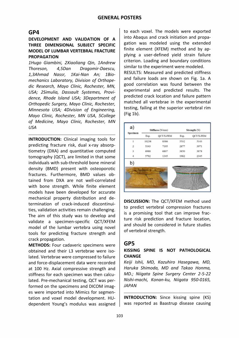
GENERAL POSTERS
103
GP4 DEVELOPMENT AND VALIDATION OF A THREE DIMENSIONAL SUBJECT SPECIFIC MODEL OF LUMBAR VERTEBRAL FRACTURE PROPAGATION 1Hugo Giambini, 2Xiaoliang Qin, 1Andrew Thoreson, 4,5Dan Dragomir‐Daescu, 1,3Ahmad Nassr, 1Kai‐Nan An; 1Bio‐mechanics Laboratory, Division of Orthope‐dic Research, Mayo Clinic, Rochester, MN, USA; 2Simulia, Dassault Systemes, Provi‐dence, Rhode Island USA; 3Department of Orthopedic Surgery, Mayo Clinic, Rochester, Minnesota USA; 4Division of Engineering, Mayo Clinic, Rochester, MN USA, 5College of Medicine, Mayo Clinic, Rochester, MN USA
INTRODUCTION: Clinical imaging tools for predicting fracture risk, dual x‐ray absorp‐tiometry (DXA) and quantitative computed tomography (QCT), are limited in that some individuals with sub‐threshold bone mineral density (BMD) present with osteoporotic fractures. Furthermore, BMD values ob‐tained from DXA are not well‐correlated with bone strength. While finite element models have been developed for accurate mechanical property distribution and de‐termination of crack‐induced discontinui‐ties, validation activities remain challenging. The aim of this study was to develop and validate a specimen‐specific QCT/XFEM model of the lumbar vertebra using novel tools for predicting fracture strength and crack propagation. METHODS: Four cadaveric specimens were obtained and their L3 vertebrae were iso‐lated. Vertebrae were compressed to failure and force‐displacement data were recorded at 100 Hz. Axial compressive strength and stiffness for each specimen was then calcu‐lated. Pre‐mechanical testing, QCT was per‐formed on the specimens and DICOM imag‐es were imported into Mimics for segmen‐tation and voxel model development. HU‐dependent Young’s modulus was assigned
to each voxel. The models were exported into Abaqus and crack initiation and propa‐gation was modeled using the extended finite element (XFEM) method and by ap‐plying a user‐defined yield strain failure criterion. Loading and boundary conditions similar to the experiment were modeled. RESULTS: Measured and predicted stiffness and failure loads are shown on Fig. 1a. A good correlation was found between the experimental and predicted results. The predicted crack location and failure pattern matched all vertebrae in the experimental testing, failing at the superior vertebral rim (Fig 1b).
DISCUSSION: The QCT/XFEM method used to predict vertebral compression fractures is a promising tool that can improve frac‐ture risk prediction and fracture location, and should be considered in future studies of vertebral strength.
GP5 KISSING SPINE IS NOT PATHOLOGICAL CHANGE Keiji Ishii, MD, Kazuhiro Hasegawa, MD, Haruka Shimoda, MD and Takao Honma, MD.; Niigata Spine Surgery Center 2‐5‐22 Nishi‐machi, Konan‐ku, Niigata 950‐0165, JAPAN
INTRODUCTION: Since kissing spine (KS) was reported as Baastrup disease causing

GENERAL POSTERS
104
back pain, some symptomatic cases related with KS had been reported. But it is still unclear that KS is pathological or physiolog‐ical alteration. We hypothesized that the frequency and number of KS increased by age if it was physiological change, and that symptom deteriorated as the number of KS increased if it was pathological. The pur‐pose of this study is to clarify if it is patho‐logical or physiological change. METHODS: One hundred and twenty‐three cases (male 58 female 65) were retrospec‐tively investigated. Patients who had back surgery in past and cases of lumbar disc herniation were excluded. They were divid‐ed into two groups according to the pres‐ence of KS. Age and symptom evaluated by Japan Orthopedic Association Back Pain Questionnaire (JOABPEQ) of KS group and non‐KS (NKS) group were compared. A rela‐tionship among the age, the number of KS, the location of KS, and spondylolisthesis were investigated. RESULTS: KS group had 74 cases (male 31 and female 43) and NKS group had 49 cases (male 27 and female 22). The average age were 69.5 and 62.9 respectively (P<0.001). The average score of JOABPEQ in KS group were greater than that in NKS group in whole category and Visual Analogue Scale (VAS) in KS was lower than that in NKS group without statistically significance. The number of KS and age were positively corre‐lated (R=0.34, P<0.001). L3/4 was the most frequent segment of KS. KS tend to present at upper adjacent segment of spondylolis‐thesis (P<0.001). DISCUSSIONS: The frequency of KS in‐creased by age and the presence of KS did not deteriorate clinical symptoms, suggest‐ing that KS is age‐related change. The result that cases with spondylolisthesis tend to have KS at upper adjacent segment was expected to contribute stabilizing the un‐stable segment or sustaining spinal align‐ment.
GP6 SUPPRESSIVE EFFECT OF LOW‐DENSITY LIPOPROTEIN RECEPTOR‐RELATED PRO‐TEIN‐1 (LRP1) ON SCHWANN CELL APOP‐TOSIS AFTER SCIATIC NERVE INJURY IN MICE Sumihisa Orita*/**, Elisabetta Mantuano*, Kazuyo Yamauchi*/**, Seiji Ohtori**, Sadao Arai**, Kazuhisa Takahashi**, Steven Go‐nias*, Wendy Campana*; *Dept. of Anes‐thesiology, University of California, San Die‐go, USA **Dept. of Orthopaedic Surgery, Graduate School of Medicine, Chiba Univer‐sity, Japan
INTRODUCTION: Schwann cells (SCs) play an essential role in nerve regeneration, and therefore, their survival after peripheral nerve injury (PNI) is important. PNI causes endoplasmic reticulum stress and activates the adaptive unfolded protein response (UPR). The UPR results in expression of C/EBP homology protein (CHOP); CHOP acts as a proapoptotic transcription factor in SCs, unless it is counteracted by low‐density lipoprotein receptor‐related protein‐1 (LRP1), which serves as a major activator of phosphatidylinositol 3‐kinase (PI3K). Thus, UPR can delay and disrupt peripheral nerve regeneration. The present study aimed to explore the role of LRP1 in SCs after UPR activation. METHODS: Sciatic nerve crush injury was induced in C57BL/6J mice (n = 5), and 24 hours after surgery, the injured nerve was resected to obtain SCs. UPR was induced using TNF‐alpha (50 ng/ml) and was con‐firmed by detecting expression of CHOP, an apoptosis biomarker. The role of LRP1 fol‐lowing UPR induction was examined by as‐sessing CHOP levels after addition of the activation ligand of LRP1 (alpha‐2M recep‐tor binding domain [RBD]) and after genetic silencing of LRP1. We considered p < 0.05 as significant. RESULTS: TNF‐alpha addition resulted in increased CHOP expression, indicating UPR

GENERAL POSTERS
105
induction. Addition of RBD significantly in‐creased levels of phospho‐PI3K and de‐creased CHOP levels (p < 0.05). Further, CHOP expression was significantly increased by both PI3K inhibition and LRP gene silenc‐ing, which indicates an association of LRP1 with the UPR pathway. CONCLUSION: Our results support a model in which UPR‐activated signaling pathways pose a major challenge to SC survival after PNI. Further, they support the idea that LRP1 functions as a potent activator of PI3K in SCs, thereby limiting SC apoptosis result‐ing from the increased CHOP expression after PNI.
GP7 SHIFT OF THE PEDICLE SCREW IN THE VER‐TEBRA ACCOMPANIED WITH CHANGE IN THE DISC ANGLE FOLLOWING INSTRU‐MENTED FACET FUSION FOR DEGENERA‐TIVE LUMBAR SPONDYLOLISTHESIS Tomohiro Miyashita *1, Hiromi Ataka *2, Kei Kato *1, Takaaki Tanno *2; *1: Spine Center, Matsudo City Hospital, Matsudo, Japan *2: Spine Center, Matsudo Orthopae‐dic Hospital, Matsudo, Japan
INTRODUCTION: Based on a long‐term clin‐ical and radiological follow‐up study of facet fusion (FF) for degenerative lumbar spondy‐lolisthesis (DLS), we reported that FF is a minimally invasive and suitable method for DLS. However, the disc angle (DA) at the fused level, which became lordotic immedi‐ately after surgery owing to the prone op‐erative position, decreased gradually within 6 months to the same as that in the pre‐operative neutral position. In this study, we assessed the shift of the pedicle screws (PS) in the vertebra to identify the mechanism of the change in the DA. METHODS: Twenty‐six patients who un‐derwent FF for single‐level DLS and CT im‐mediately after surgery and 6 months later were retrospectively reviewed. From these, 7 patients, in whom the DA had decreased
by more than 4° after surgery to the same as that in the preoperative neutral position, were selected to capture the shift of the PS. The patients in whom CT showed PS loosen‐ing and/or pseudarthrosis were excluded. We reconstructed a CT plane, which was vertical to the cranial endplate of the verte‐bra in which the PS was inserted and passed through a cannula of percutaneous PS. We measured the angle between the cranial endplate and the cannula on the plane. RESULTS: Of 14 PSs, the angle of only 1 changed by more than 2° in the upper ver‐tebra of the fused level. The tip of the shift‐ing PS moved cranially. However, in the lower vertebra, the angle of 10 out of 14 PSs changed by 2‐5°, and the tip of all these PSs shifted caudally. DISCUSSION: The change in the DA after FF is thought to be caused by the PS shifting in the vertebra without loosening and by re‐modeling of the bone around it according to the biomechanical load. In other words, the PS itself is limited in its ability to maintain a lordotic DA, and even with a cage, lumbar alignment would return to the preoperative neutral position owing to sinking of the cage. We conclude that in situ fusion is a rational approach in the management of DLS.
GP8 ANATOMICAL BACKGROUND OF “PSEUDO‐SCIATICA” IN CLUNEAL NEURALGIA Tomoyuki Konno, MD1, Yoichi Aota, MD2,Hiroshi Kuniya, MD1, Tomoyuki Saito, MD1, Nei Kyoku, MD3,Shougo Hayashi, MD3, Shinichi Kawada,PhD3, Masahiro Itoh, MD3; 1Department of Orthopaedic Surgery, Yokohama City University Graduate School of Medicine, Yokohama, Kanagawa, Japan; 2 Spine and Spinal cord center Yokohama stoke and brain center, Yokohama, Kana‐gawa, Japan, 3Department of Anatomy, Tokyo Medical University, Shinjyuku, Tokyo, Japan

GENERAL POSTERS
106
INTRODUCTION: Superior cluneal nerves (SCN) are cutaneous nerves distributed at the gluteal region. Medial branch of SCN can become spontaneously entrapped un‐der fascia over iliac crest and cause low back pain. Surgical reports were few and limited to a small number of subjects with low back and/or buttock pain. On the other hand, Trescot stated that SCN entrapment can cause a referred pain dawn the leg, po‐tentially all the way to the foot and this “pseudosciatica” would clinically mimic a radiculopathy (Pain physician 2003). Previ‐ous anatomical studies cannot explain why this clinical entity can cause “pseudosciati‐ca” because the medial branch of SCN com‐prises the cutaneous branches of the dorsal rami of the upper three lumbar nerves. The purpose of the present anatomical study was to improve the understanding of “pseudosciatica” in SCN entrapment. METHODS: Bilateral branches of SCN were observed macroscopically on 6 cadavers (male 3 bodies, female 3 bodies) with a mean death age of 89 years (81‐100) were investigated. All branches of SCN were ex‐posed around the iliac crests (figure). Spe‐cial attention was paid to preserve thin branches and anastomoses. All branches of SCN were proximally explored to identify its emergence from nerve roots.
RESULT: SCN was originating from dorsal roots of from T12 to L5. The medial branch of SCN emerged at three different levels; L3 (n=1), L4 (n=5), and L5 nerve root (n=4). DISCUSSION: In this anatomical study, the medial branch of SCN emerged predomi‐nantly from L4 and L5, which was unlike most previous studies concluding its emer‐gence from T11‐L4 nerve roots. SCNs are potentially an underdiagnosed source of leg pain.
GP9 STENOSIS IN THE LUMBAR SPINE IS ASSO‐CIATED WITH INCREASED HINDFOOT DE‐GENERATION AND ANKLE OSTEOARTHRI‐TIS: A STUDY OF 720 CADAVERIC SPECI‐MENS Joshua T. Anderson, BS (1,2), Alex V. Boiw‐ka, BS (1,2), Navkirat Bajwa, MD (1), Nicho‐las U. Ahn, MD (1); 1 University Hospitals Case Medical Center Department of Ortho‐paedic Surgery 2 Case Western Reserve Uni‐versity School of Medicine
INTRODUCTION: Neural compression due to lumbar spinal stenosis (LSS) is associated with motor and sensory changes and radicular pain in the lower extremities, which can lead to gait changes. This altera‐tion may ultimately lead to deterioration of the ankle and hindfoot joints. METHODS: 720 adult cadaveric specimens age 20‐70 were selected from a collection of human skeletons. All had intact bony elements. In each subject, canal area was calculated at each lumbar level using a pre‐viously validated formula. Ankle arthritis and hindfoot arthritis (i.e., arthritis across the talonavicular [TN], subtalar [ST], and calcaneocuboid joints [CC]) was quantified using the Kellgren‐Lawrence classification system, as determined by presence and size of osteophytes on the articular surfaces. Linear regression analyses, correcting for confounding factors such as age, sex, race, and height were performed to identify the

GENERAL POSTERS
107
association between canal area throughout the lumbar spine and presence and severity of hindfoot arthritis and/or ankle arthritis. RESULTS: A significant negative association was found between total canal area and both the severity of ankle arthritis (p=0.02) and severity of hindfoot arthritis of the TN and CC joints (p=0.01). LSS in the more dis‐tal segments (L4‐S1) was more strongly as‐sociated with severity of ankle arthritis than stenosis in the upper lumbar segments, with higher correlation coefficients. Both were associated with presence and severity of ankle arthritis (p<0.05) and hindfoot ar‐thritis (p<0.05). Subjects with severe LSS (canal area less than 2 SD below the mean) had markedly increased degeneration across the ankle joint, even at ages less than 40. DISCUSSION: The presence of LSS is associ‐ated with degeneration across the ankle joint and joints of the hindfoot. The associa‐tion was most strongly identified at the lower lumbar levels. This correlates physio‐logically, as the lower lumbar levels inner‐vate the distal lower extremities.
GP10 STRESS ANALYSIS IN OSTEOPOROTIC LUM‐BAR VERTEBRA WITH MICRO‐SCALED BEAM‐SHELL TRABECULAR‐CORTICAL STRUCTURE: A FINITE ELEMENT ANALYSIS Yoon Hyuk Kim1, Mengying Wu1, Kyungsoo Kim2; 1Department of Mechanical Engineer‐ing, Kyung Hee University, Korea 2Department of Applied Mathematics, Kyung Hee University, Korea
INTRODUCTION: Due to the difficulties as‐sociated with obtaining bones for in vitro experiments and the limitations on the con‐trol of the experimental parameters, finite element (FE) models including the trabecu‐lar and cortical parts have been developed to investigate biomechanical properties of osteoporotic vertebrae. In this study, FE models of normal and osteoporotic lumbar
vertebrae that incorporate the micro‐scaled trabecular structure of lattice models and the cortical area of shell elements were developed. The von‐Mises stress was ana‐lyzed to predict the risk of the burst frac‐ture in osteoporotic bones of various grades. METHODS: Trabecular models composed of vertical and horizontal struts were devel‐oped for different age groups (young, mid‐dle‐aged, and old), two endplates, and the cortical part of the vertebra were devel‐oped. The trabecular lattice was tied with the cortical shell and endplates. The von‐Mises stress in the trabecular lattice was then analyzed for the three age groups un‐der compressive loadings of 0.15MPa, 0.3MPa, 0.45MPa, 0.6MPa and 0.75MPa. RESULTS: The von‐Mises stress substantially increased as the osteoporosis was getting severe, regardless of the compressive load‐ings. The maximum stress was greater than 50% of the yield stress when compressive loading exceeded 0.45 MPa for middle‐aged group and 0.3 MPa for the old group; in contrast, the maximum stress did not reach 50% of the yield stress even under 0.75 MPa for the young group. DISCUSSION: The results suggest that oste‐oporosis can affect the stress acting on the vertebra even during routine daily activities because the 0.45MPa of compressive load‐ing on the endplate is similar to that pro‐duced during daily activities. The vertebra model that incorporates a realistic trabecu‐lar structure is advantageous because it permits simulation of in vivo specimens for the study of osteoporosis.
GP11 BIOMECHANICAL ANALYSIS OF COMBINED MOTION EXERCISE IN THE LUMBAR SPINE Won Man Park1, Dae Kyung Choi1, Kyungsoo Kim2, Yoon Hyuk Kim1, Jae Lak Yang3; 1Department of Mechanical Engi‐neering, Kyung Hee University, Ko‐rea2Department of Applied Mathematics,

GENERAL POSTERS
108
Kyung Hee University, Korea 3Hanmed Co., Ltd., S. Korea
INTRODUCTION: Spine exercise has been used as one of the most common conserva‐tive treatments for low back pain. However, relatively little is known about the effects of spine exercise on lumbar spine biomechan‐ics. In this study, we investigated biome‐chanical effects of combined motion spine exercise, which consists of the axial traction and the local decompression, on the lumbar spine. METHODS: A validated three‐dimensional finite element model of the lumbar spine was used. Only flexion‐extension motion of the L1 and sacrum, and axial translation of L1 were allowed in the finite element analy‐sis. One‐third of body weight was applied on the center of the L1 vertebra toward the superior direction for axial traction. Transla‐tion of L4 spinal bone toward anterior direc‐tion was considered as the local decom‐pression. Changes in spine biomechanics, such as lordosis angle, intradiscal pressure, and stress on fibers of annulus fibrosus and ligaments, during combined motion spine exercise were investigated using three di‐mensional finite element model of the lum‐bar spine. RESULTS: The lordosis angle between the superior planes of the L1 vertebra and sa‐crum was 44.6° at baseline, 35.2° with glob‐al axial traction, and 46.4° with local de‐compression. The highest average stresses on the fibers of annulus fibrosus were main‐ly shown in the posterior region during axial traction, and decreased during local de‐compression. The intertransverse and pos‐terior longitudinal ligaments experienced stress primarily during global axial traction, these stresses decreased during local de‐compression. DISCUSSION: A combined motion spine exercise of global axial traction and local decompression would be helpful for reduc‐ing tensile stress on the fibers of the annu‐
lus fibrosus and ligaments, and intradiscal pressure in traction therapy. The present study could be used to develop a safer and more effective spine exercise program.
GP12 INCREASE IN TRUNK MUSCLE LOADING WITH SAGITTAL PLANE DEFORMITY: A FI‐NITE ELEMENT ANAYLSIS Yoon Hyuk Kim1, Kyungsoo Kim2, Dae Kyung Choi1, Yongjung J. Kim3; 1Department of Mechanical Engineering, Kyung Hee University, Korea 2Department of Applied Mathematics, Kyung Hee Univer‐sity, Korea 3School of Physicians and Sur‐geons, Columbia University, USA
INTRODUCTION: Sagittal plane deformity has been known to produce significant pain, disability, and poor self‐image in upright posture, which could deteriorate efficiency of energy expenditure by increasing trunk muscle loading in maintaining the posture from biomechanical viewpoint. There is no study to quantify relationship between the sagittal plane deformity and the trunk mus‐cle loading. The purpose of this study is to investigate trunk muscle loading in sagittal plane deformity in the upright posture. METHODS: A three‐dimensional finite ele‐ment model of the intact whole spine by using CT images was developed. Three sag‐ittal plane deformity models (mild, moder‐ate, and severe) by deforming the intact model were obtained. Sagittal plane de‐formity was classified based on sagittal ver‐tical axis (SVA). Sagittal imbalance was de‐fined if SVA is 80 mm or larger from pa‐tients’ X‐ray images. In each model, 117 pairs of trunk muscles (59 pairs of paraspinal muscles and 58 pairs of superfi‐cial muscles) were included. The muscle forces in an upright posture were investi‐gated, where the upper body weight was applied along C7 plumb line. RESULTS: The trunk muscle loadings were 72%, 164%, 251%, and 351% of the body

GENERAL POSTERS
109
weight (BW) in SVA of 0 mm (intact), 80 mm (mild), 155 mm (moderate), and 240 mm (severe), respectively. The paraspinal mus‐cle loadings were 36% (intact), 88% (mild), 174% (moderate), and 256% (severe) of the BW, respectively. DISCUSSION: The results indicated that the larger SVA, the more trunk muscle over‐loadings. Excessive trunk muscle over‐loadings, especially for the paraspinal mus‐cle group, is needed to maintain the pos‐ture which might induce the muscle fatigue, or regional and global posture change to reduce the trunk muscle loading. The find‐ings help to understand biomechanical prin‐ciples in trunk muscle loading related to sagittal plane deformity.
GP13 EFFECT OF LIGATURE PRETENSION IN IN‐TERSPINOUS PROCESS SURGERY Dae Kyung Choi1, Won Man Park1, Yoon Hyuk Kim1, Kyungsoo Kim2; 1Department of Mechanical Engineering, Kyung Hee Univer‐sity, Korea 2Department of Applied Mathe‐matics, Kyung Hee University, Korea
INTRODUCTION: Ligatures to prevent the instability in the flexion movement by wrapping around upper and lower spinous processes are used with many types of in‐terspinous process spacer (IPS). However, the effects of ligature pretension in IPS on the surgical outcomes from the biomechan‐ical point of view have not been analyzed yet. In this study, we investigated the ef‐fects of pretension of ligatures on biome‐chanics of functional spinal unit after inter‐spinous process surgery. METHODS: Three‐dimensional (3D) finite element (FE) models of one pedicle screw system and four different IPSs were devel‐oped. Spinal fusion and interspinous pro‐cess surgeries were virtually performed based on the clinical protocol using the de‐veloped L3‐L4 FE models. Flexion‐extension moments of 7.5 Nm were applied with a
compressive force of 400 N along the direc‐tion from upper vertebra center to the low‐er one on the superior plane of L3 vertebra. The ligature pretension was varied from 0 N to 400 N with an increment of 100 N. Range of motions (ROMs) in flexion and the maxi‐mum von‐Mises stresses at the spinous pro‐cess in extension were analyzed. RESULTS: The ROMs in flexion were sub‐stantially reduced as the ligature pretension was increased while ROMs in flexion in all IPS cases were larger than that in the fusion case, though they were smaller than that in the intact case, regardless of the ligature tension. In contrast, the maximum von‐Mises stresses at spinous process were in‐creased by 22% ‐ 54% in all IPS cases as the ligature tension was increased. DISCUSSION: The results of this study showed that excessive pretension of liga‐ture could increase the risk of subsidence of the osteoporotic bone by increasing maxi‐mum von‐Mises stresses at spinous process and improve the spinal stability by decreas‐ing the ROMs of flexion‐extension move‐ment. Therefore the magnitude of preten‐sion of ligature should be carefully consid‐ered in the interspinous process surgery.
GP14 THE RELATIONSHIP OF THE FRONTAL PLANE TRUNK AND PELVIC MOTION DUR‐ING GAIT AND THE RADIOGRAPHIC SPINOPELVIC MEASUREMENTS OF PA‐TIENTS WITH A UNILATERAL COMPLETELY DISLOCATED HIP Masatsugu Tsukamoto, Tadatsugu Morimo‐to, Tomohito Yoshihara, Masaya Ueno, Shuichi Eto, Shunsuke Kawano, Masaru Kitajima, Motoki Sonohata, Masaaki Mawa‐tari; Department of Orthopedic Surgery, Faculty of Medicine, Saga University, Saga, Japan 5‐1‐1 Nabeshima, Saga 849‐8501, Japan
INTRODUCTION: Patients with a unilateral completely dislocated hip (CDH) often have

GENERAL POSTERS
110
claudication, pelvic tilt and lumbar scoliosis due to their leg length discrepancy. Howev‐er, there have been no previous reports on the frontal plane trunk and pelvic motion from a three‐dimensional (3D) gait analysis in patients with unilateral CDH. Therefore, we have investigated the coronal spinopel‐vic alignment both roentgenologically and by the kinematic pattern on a gait analysis. MATERIALS AND METHODS: A gait analysis was performed on nine patients (mean age, 58 years) with unilateral CDH using the Vi‐con Motion system. We calculated the pel‐vic obliquity (PO), thorax obliquity (TO) and spine lateral flexion (SF) angles to define the frontal plane trunk and pelvic motion angles. The PO was measured in the plane of the laboratory transverse axis and the pelvic frontal axis, and the TO was also measured using these data. The SF was measured between the long axis of the thorax and the pelvis in the frontal plane. The radiographic parameters examined were the coronal Cobb angle of the lumber spine (CA) and the pelvic inclination (PI) measured on standing antero‐posterior X‐ray films. The correlations of the trunk and pelvic motion angles and radiographic measurements were analyzed using Pear‐son’s correlation coefficients. RESULTS: The mean CA and PI were 12.3 and 10.3 degrees, respectively. The mean PO, TO and SF were ‐0.7, 2.5 and 1.8 de‐grees, respectively. The mean arcs of the PO, TO and SF were 6.8, 6.6 and 10.7 de‐grees, respectively. The CA was significantly correlated with the mean TO (r=0.72, p=0.03). DISCUSSION: Because biomechanical stress is applied to the lumbar spine during gait, a lumbar spine deformity may occur in pa‐tients with unilateral CDH. There were no significant correlations between the PI and any of the trunk or pelvic motion angles. This suggests that pelvic and lower limb compensatory actions take place in patients with CDH when they walk.
GP15 TRANSFORMING GROWTH FACTOR‐ß BLOCK THE CONTRIBUTION OF ADAMTS‐1 TO INTERVERTEBRAL DISC DEGENERATION THROUGH THE SMAD/MAPK PATHWAY: A POSSIBLE DEGENERATION REPARATIVE WAY? Zheng Zhaomin, Wang Hua, Wang Jianru, Liu Hui; Department of Orthopeadic, The First Affiliated Hospital of Sun Yat‐ sen Uni‐versity
OBJECTIVE: To investigate how transform‐ing growth factor ß (TGF‐ß) regulation of ADAMTS‐1 expression in nucleus pulposus (NP) cells of the intervertebral disc. METHODS: qRT‐PCR and Western blot were used to measure expression of ADAMTS‐1/4/5 in NP cells treated with TGF‐ß. Trans‐fections were used to measure the effect of Smad3, MAPKs, and activator protein 1 (AP‐1) on TGF‐ß‐mediated ADAMTS‐1 promoter activity. Lentiviral knockdown of Smad2/3 was performed to assess the role of Smad2/3 in ADAMTS‐1 expression. Besides, ADAMTS‐1 role in aggrecan and versican degradation was investigated. RESULTS: TGF‐ß decreased the expression of ADAMTS‐1/4/5 mRNA and protein level in nucleus pulposus cells, besides TGF‐ß suppressed ADAMTS‐1 promoter activity. TNF‐a increased aggrecan or versican deg‐radation, which was inhibited by TGF‐ß, TGF‐ß alone can also decrease aggrecan or versican degradation. Besides, when NP cells were infected with LV‐shADAMTS1, TGF‐ß dependent suppression in NITEGE and G1 secreted protein is significantly blocked. Next to evaluate the potential pathway. Suppression of ADAMTS‐1 pro‐moter activity was evident when the NP cells were co‐transfected with smad2/4 construct, but not smad3. On the other hand, co‐transfection of smad7 restored ADAMTS‐1 promoter activity, and similar result in A‐Fos construct. Supporting the promoter studies, lentiviral delivery of sh‐

GENERAL POSTERS
111
smad2 significantly blocked TGF‐ß suppres‐sion in ADAMTS‐1 expression, but no sh‐smad3. Treatment of cells with MAPK inhib‐itors partially abolished the suppressive effect of TGF‐ß on ADAMTS‐1 mRNA and protein expression, suggesting involvement of these signaling pathways in the regula‐tion of ADAMTS‐1. Gain and loss of function studies confirmed the contribution of JNK1/2 and ERK1/2 to TGF‐ß dependent suppression of ADAMTS‐1 promoter func‐tion. CONCLUSIONS: TGF‐ß, through Smad2, JNK1/2 and ERK1/2, serve as a negative regulator of ADAMTS‐1 expression in the NP, which can inhibit the role of ADAMTS‐1 on NP
GP16 PLIF EXPANDABLE CAGES EFFECTIVELY STABILIZE THE SPINE BETTER THAN TLIF CAGE 1Kodigudla, M; 1Desai, D; 1Agarwal, A; 1Momeni, N; 1Goel, VK; 1Agarwal, AK; 2Schultz, C; 1Engineering Center for Ortho‐paedic Research Excellence (E‐CORE) De‐partments of Bioengineering and Orthopae‐dic Surgery Colleges of Engineering and Medicine University of Toledo, Toledo, OH 43606 2Apex Spine, Munich, Germany
INTRODUCTION: PLIF is a widely used tech‐nique in spinal fusion. Expandable cages are currently being explored in stabilizing the spine, especially as standalone device. The main objective of this study was to biome‐chanically evaluate expandable PLIF cages vs. a standard TLIF cage in fucnctional spinal units (FSUs) with and without additonal posterior fixation. MATERIAL AND METHODS: Twelve liga‐mentous L23 and L45 FSUs were used. The potted caudal (L3/L5) end was fixed to the testing apparatus and pure moments up to 10 Nm at the cranial level were applied in extension (ext), flexion (flex), left and right lateral bending (lb & rb) and left and right
axial rotations (lr & rr). Motion was tracked using the Optotrak motion capture system (NDI, Waterloo, Canada). Following the biomechanical testing of intact specimens, 1 TLIF (Aesculap, Germany) or bilateral ex‐pandable PLIF (Medyssey, Elk Grove, IL) cages were inserted and tested. Next, pedi‐cle screw system (PSS) was implanted to stabilize the segments before further test‐ing. TLIF was performed in 6 L2‐L3 speci‐mens and PLIF was performed in 6 L4‐L5 specimens. RESULTS: Standalone TLIF procedure in‐creased the motion of the segments in ext (26%), lb (16%), rb (10%), lr(25%) and rr(37%) and decreased in flex(7%), com‐pared to intact motion. Standalone bilateral PLIF cages reduced the motion in flex (36%), ext (70%), lb (64%), rb (58%), lr (22%) and rr (5%). The reduction due to standalone PLIF was significant in all loading modes except axial rotation (Figure 1). Implantation of pedicle screw system reduced the motion significantly in all loading modes in TLIF and PLIF groups.
DISCUSSION: The expandable cages effec‐tively stabilized the spine in all modes, ex‐cept axial rotation. Addition of pedicle screw fixation system would further stabi‐lize the spine, especially in rotation. Thus, expandable cages may need additional sta‐bilization, especially in axial rotation to pro‐vide favorable environment for spinal fu‐sion.

GENERAL POSTERS
112
GP17 PEEK TRANSLAMINAR FACET SCREWS PROVIDE LONG TERM STABILITY OF THE LUMBAR SPINE AFTER MICRODISCECTOMY 1Kodigudla, M; 1Desai, D; 1Agarwal, A; 1Goel, VK; 1Agarwal, AK; 2Wanderley, JGPV; 1Engineering Center for Orthopaedic Research Excellence (E‐CORE)Departments of Bioengineering and Orthopaedic Surgery Colleges of Engineering and Medicine Uni‐versity of Toledo, Toledo, OH 43606 2Spine Soft Fusion, Brazil
INTRODUCTION: Tanslaminar facet screw fixation in spine surgery is a minimammy invasive alternative. However the clinical success rate is suboptimal. It might be due to the use of Titanium (Ti) screws which are stiffer than the adjoing bone.The main ob‐jective of this study was to evaluate the stabilization of Ti Vs PEEK screws in fucnc‐tional spinal units (FSUs), pre and post cyclic loading. MATERIAL AND METHODS: 12 ligamentous L2‐L3 FSUs were tested in this study. The potted caudal (L3) end was fixed to the test‐ing apparatus and pure moments were ap‐plied in all loading modes up to 10 Nm at the cranial (L2) end. Motion was tracked using the Optotrak. Following the testing of intact specimens, micro discectomy was performed from right lateral side and test‐ed. Next, translaminar facet screws (TLFS) were inserted bilaterally to stabilize the segments and re‐tested. Ti screws were used in 6 motion segments and PEEK screws were used in the other 6 specimens. Follow‐ing Optotrak testing, all motion segments were loaded cyclically by applying 7.5 Nm in flexion and extension and 5 Nm in axial ro‐tation up to 50,000 cycles at 2 Hz. Post fa‐tigue, the specimens were again tested for range of motion to evaluate the increase in motion if any. RESULTS: TLFS of Ti and PEEK stabilized the segments with micro discectomy. Fatigue loading increased the motion significantly in
flexion, extension, right bending and left rotation without preload and, in flexion with preload for Ti group (Figure 1). The increase in motion post‐fatigue was not significant in all loading modes except in left bending for PEEK group.
DISCUSSION: Post fatigue, the increase in instability in Ti group may be attributed to the loosening of the screw due to high elas‐tic modulus of Ti than bone. In PEEK group the instability was less than Ti suggest that bone screw interface for PEEK screws is more rigid than Ti post fatigue. Thus, PEEK translaminar screws perform better than Ti screws in the short and long‐term
GP18 POSTURAL ACTIVITY OF THE PSOAS MAJOR AND QUADRATUS LUMBORUM MUSCLES IS LESS PRECISELY CONTROLLED IN PEOPLE WITH RECURRING BACK PAIN Rachel J. Park1,2, Henry Tsao1, Andrew G. Cresswell1,2, and Paul W. Hodges1; 1The University of Queensland, NHMRC Centre of Clinical Research Excellence in Spinal Pain, Injury and Health, School of Health and Re‐habilitation Sciences, Brisbane, Queensland, Australia. 2The University of Queensland, Centre for Sensorimotor Neuroscience, School of Human Movement Studies, Bris‐bane, Queensland, Australia
INTRODUCTION: The mechanically unique regions of the psoas major (PM) and quad‐ratus lumborum (QL) muscles are controlled

GENERAL POSTERS
113
independently by the nervous system. So‐phisticated coordination of these regions may be modified in the presence of recur‐ring low back pain (LBP). METHODS: Nine volunteers with recurring episodes of LBP and thirteen pain‐free con‐trols performed rapid bilateral arm flexion and extension, to impose a postural pertur‐bation to the spine. Individuals with LBP were subgrouped into those with high and low erector spinae (ES) electromyography (EMG) when sitting with a lumbar lordosis. Fine‐wire electrodes were used to record EMG from the regions where PM fascicles arise from the transverse process (PM‐t) and vertebral body (PM‐v) and the anterior (QL‐a) and posterior (QL‐p) layers of QL on the right side. RESULTS: Activity of QL‐a did not differ be‐tween directions of arm movement in the LBP groups, unlike controls who had earlier QL‐a EMG onset during arm extension. EMG onset of regions of PM and QL‐p activity did not differ between groups, but the pattern of QL‐p activity differed between LBP sub‐groups. Activity did not differ between PM regions, unlike controls who displayed indi‐vidual control of these regions. DISCUSSION: One interpretation of these data is that people with LBP use a more simplified control strategy for the deep trunk muscles. This strategy involved less discrete control of individual regions of PM and QL, and activation of some regions dif‐fered less between tasks that differed with respect to the directions of reactive mo‐ments from limb movement. This may rep‐resent a simplified strategy to protect the spine in people with recurring LBP and could require consideration in prevention of back pain recurrence.
GP19 THE EFFECTS OF NEUROMUSCULAR ELEC‐TRICAL STIMULATION (NMES) ON CHANGE IN THICKNESS OF DEEP TRUNK MUSCLES IN PATIENTS WITH LOW BACK PAIN. Seung Ok Baek, M.D., Hee Kyung Cho, M.D., Gil Su Jung, Su Min Son, M.D., Yun Woo Cho, M.D., Sang Ho Ahn, M.D., PhD.; Depart‐ments of Physical Medicine and Rehabilita‐tion, College of Medicine, Yeungnam Uni‐versity, Daegu 705‐717, Korea
INTRODUCTION: Neuromuscular electrical stimulation (NMES) can preferentially stimulate contractions in deep lumbar stabi‐lizing muscles and restore muscle function. In current study, we attempted to investi‐gate relation between deep lumbar stabiliz‐ing muscles and NMES during each sessions using real time ultrasound imaging. METHODS: Thirty patients with LBP (12 males, 18 females, mean age 49.2 ± 13.6 years, range 19‐69 years) were recruited. NMES was delivered through abdominal and lumbar electrical stimulation. Ab‐dominal electrical stimulation include 4 hy‐drogel surface electrodes (5cm X 5cm) lo‐cated on both sides of the anterolateral abdominal wall. Lumbar electrical stimula‐tion include 4 surface electrodes according to the cross line on L4/5 interspinous pro‐cess. Session 1 was abdominal electrical stimulation only, session 2 was lumbar elec‐trical stimulation only and session 3 was concurrent stimulation of abdominal and lumbar. During each session, images of ab‐dominal muscle and lumbar multifidus mus‐cles were captured using real time ultra‐sound imaging (RUSI). RESULTS AND DISCUSSION: To investigate relationship between deep lumbar stabiliz‐ing muscles and NMES, we set the specific NMES sessions and used relatively small electrodes. Thicknesses of abdominal mus‐cles (transverse abdominis, oliquus inter‐nus, and obliquus externus) and lumbar multifidus muscles were significantly larger

GENERAL POSTERS
114
during each NMES than at rest for all three sessions (p<0.05). However, there were no significant different between session 1 and 3 in abdominal muscles. And no significant difference was observed between session 2 and 3 in LM. TrA, OI and LM were signifi‐cantly activated together during session 3. The session 3 stimulation optimally activat‐ed deep spinal stabilizing muscles, that is, the TrA, OI and LM, as evidenced by RUSI. The authors recommend that NMES on ab‐dominal wall and lumbar area together would help patients with LBP who had problems with contractions in deep stabi‐lizers.
GP20 ABDOMINAL HOLLOWING DURING GAIT CAN SELECTIVELY ACTIVATE LOCAL TRUNK STABILIZING MUSCLES: A CROSS SECTION‐AL STUDY. Ah Young Lee, M.D., Eun Hyuk Kim, M.D., Seung Ok Baek, M.D., Yun Woo Cho, M.D., Sang Ho Ahn, M.D.; Departments of Physical Medicine and Rehabilitation, College of Medicine, Yeungnam University, Daegu 705‐717, Korea
INTRODUCTION: Trunk muscle exercises are widely performed during clinical and reha‐bilitation programs, and many studies have been performed to examine their effects on different low back pains. However, the ef‐fect of trunk muscles activations during gait with AH on low back pain experienced dur‐ing daily activities has not been clarified. To investigate whether gait with abdominal hollowing (AH) is more effective for pro‐moting local trunk muscle activation than gait without AH using surface electromyog‐raphy (EMG). METHODS: A cross‐sectional comparative study of local trunk muscle activations dur‐ing gait with and without AH in 20 partici‐pants. Twenty healthy men with no history of a cervical, thoracic, or a lumbar spine disorder participated in the study. EMG
amplitudes were normalized by having par‐ticipants perform maximal voluntary con‐tractions (MVC). Surface electrodes were attached to multifidus (MF), lumbar erector spinae (LES), thoracic erector spinae (TES), transverse abdominus ‐ internal oblique abdominals (TrA‐IO), external oblique ab‐dominals (EO), and rectus abdominis (RA). The amplitudes of electromyographic sig‐nals were measured during gait with or without AH. RESULTS: AH during gait was found to result in significant increases in the normalized MVCs of MFs and TrA‐IOs in both sides (p <0.05). Ratios of local muscle activity to global muscle activities were increased dur‐ing gait with AH, and both TrA‐IO/EO were significantly increased by AH (p < 0.05). DISCUSSION: Gait with AH resulted in signif‐icantly more recruitment of local trunk muscles. The study suggests that AH during gait be recommended for low back pain patients in an outpatient setting to improve spinal dynamic stabilization.
GP21 THE RELATION BETWEEN EXPERIMENTAL PAIN AND SPINAL STIFFNESS ‐ A PILOT STUDY Arnold Wong, Greg Kawchuk, Christopher Huang; University of Alberta
INTRODUCTION: A subset of people with low back pain (LBP) have increased spinal stiffness, which decreases as LBP subsides. Currently, the relation between LBP intensi‐ty and spinal stiffness remains unclear. As such, this study investigated the effect of pain on temporal changes of spinal stiff‐ness. METHODS: In separate sessions, nine healthy subjects received injections of hy‐pertonic (0.3ml, 5% NaCl) or isotonic saline (0.3ml, 0.9% NaCl) in random order. On both occasions, saline was injected into the L3/4 and L4/5 interspinous ligaments. Sub‐jects were blinded to saline concentration.

GENERAL POSTERS
115
Spinal stiffness at L3 spinous process was measured by a mechanical indentation de‐vice before, immediately after, and 25‐minute after injection. Pain intensity at the three time points was recorded. Pain inten‐sity and spinal stiffness were compared be‐tween each saline condition using repeated measures analysis of variance adjusted for Fear of Pain Questionnaire‐III scores. Trend analysis and tetrad difference calculation were used as post‐hoc tests. The signifi‐cance level was set at 0.05. RESULTS: Both hypertonic and isotonic sa‐line injections caused significant temporary increase in LBP (p < 0.05). Hypertonic saline induced significantly higher LBP intensity than isotonic saline (p < 0.05). Although there were no significant differences in spi‐nal stiffness between the two saline con‐centrations at all time points, there was a significant interaction between saline con‐centrations and time points after adjusting for the Fear of Pain Questionnaire‐III scores (p < 0.05). Post‐hoc tests showed that both saline injections tended to temporarily in‐crease spinal stiffness but hypertonic saline increased spinal stiffness more than isoton‐ic saline alone. DISCUSSION: Experimental LBP resulted in a transient increase in spinal stiffness beyond pain caused by an inert injection volume. Future work will explore if pain‐related spi‐nal stiffness increases are associated with reflexive muscle activity.
GP22 THE IMPORTANCE OF POSTERIOR OSTE‐OLIGAMENTOUS STRUCTURE TO LUMBAR SPINE STABILITY: AN IN‐VITRO BIOME‐CHANICAL INVESTIGATION Uphar Chamoli [1,2], Mert H. Korkusuz[1], Ashutosh B. Sabnis[1], Andrei R. Manoles‐cu[1], Ashish D. Diwan[1]; 1. Spine Service, Department of Orthopaedic Surgery, St. George Hospital Clinical School, University of New South Wales, Kogarah, Sydney NSW 2217, Australia.
2. School of Mechanical and Manufacturing Engineering, University of New South Wales, Kensington campus, Sydney NSW 2052, Aus‐tralia.
INTRODUCTION: Biomechanical implica‐tions of sequentially sacrificing posterior osteoligamentous structure in a mul‐tisegment (T12‐S1) lumbar spine have not been comprehensively studied before, es‐pecially for any compensatory changes in kinematics at caudal and cranial adjacent levels. The main aim of this study was to evaluate the contribution of posterior liga‐ments and facet joints in stabilizing the lumbar spine during flexion‐extension (FE), lateral bending (LB), and axial torsion (AxT) motions. METHODS: Six fresh frozen cadaveric kan‐garoo lumbar spines were tested in a six‐degrees‐of‐freedom kinematic spine simula‐tor, and the 3D segmental motion was rec‐orded using an infrared motion tracking device. A sinusoidal load (0.01 Hz) of ±5Nm was applied in FE, LB, and AxT motion planes, without a compressive preload. Specimens were tested in the following or‐der: (1) intact state (D0), (2) after interspi‐nous and supraspinous ligament sacrifice between L4‐L5 (D1), (3) further bilateral facetectomy between L4‐L5 (D2). Load vs. deformation curves were plotted to quanti‐fy % changes in range of motion (ROM) and neutral zone (NZ) between D0‐D1, and D0‐D2 states. Paired samples t‐test was used to detect significance in % change (a = 0.05). RESULTS: Global kinematic differences be‐tween D0‐D1 states in all loading planes were found to be small and insignificant, but were highly significant between D0‐D2 states during AxT motion (p<0.01). Between D0‐D1, compensatory decrease (p<0.05) in segmental FE ROM at caudal adjacent, and LB NZ at cranial adjacent levels were ob‐served. Between D0‐D2 states, a compensa‐tory decrease in AxT ROM at caudal adja‐cent level was also observed (p<0.01).
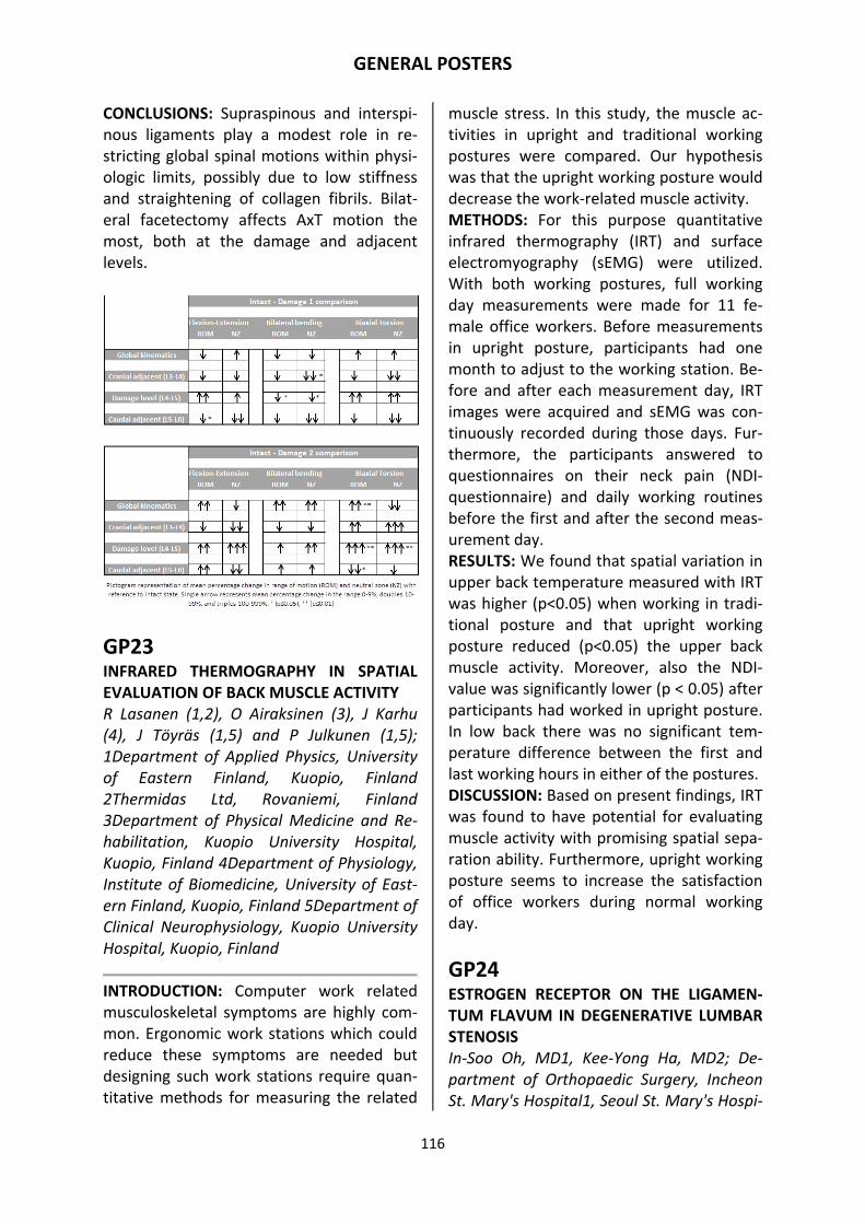
GENERAL POSTERS
116
CONCLUSIONS: Supraspinous and interspi‐nous ligaments play a modest role in re‐stricting global spinal motions within physi‐ologic limits, possibly due to low stiffness and straightening of collagen fibrils. Bilat‐eral facetectomy affects AxT motion the most, both at the damage and adjacent levels.
GP23 INFRARED THERMOGRAPHY IN SPATIAL EVALUATION OF BACK MUSCLE ACTIVITY R Lasanen (1,2), O Airaksinen (3), J Karhu (4), J Töyräs (1,5) and P Julkunen (1,5); 1Department of Applied Physics, University of Eastern Finland, Kuopio, Finland 2Thermidas Ltd, Rovaniemi, Finland 3Department of Physical Medicine and Re‐habilitation, Kuopio University Hospital, Kuopio, Finland 4Department of Physiology, Institute of Biomedicine, University of East‐ern Finland, Kuopio, Finland 5Department of Clinical Neurophysiology, Kuopio University Hospital, Kuopio, Finland
INTRODUCTION: Computer work related musculoskeletal symptoms are highly com‐mon. Ergonomic work stations which could reduce these symptoms are needed but designing such work stations require quan‐titative methods for measuring the related
muscle stress. In this study, the muscle ac‐tivities in upright and traditional working postures were compared. Our hypothesis was that the upright working posture would decrease the work‐related muscle activity. METHODS: For this purpose quantitative infrared thermography (IRT) and surface electromyography (sEMG) were utilized. With both working postures, full working day measurements were made for 11 fe‐male office workers. Before measurements in upright posture, participants had one month to adjust to the working station. Be‐fore and after each measurement day, IRT images were acquired and sEMG was con‐tinuously recorded during those days. Fur‐thermore, the participants answered to questionnaires on their neck pain (NDI‐questionnaire) and daily working routines before the first and after the second meas‐urement day. RESULTS: We found that spatial variation in upper back temperature measured with IRT was higher (p<0.05) when working in tradi‐tional posture and that upright working posture reduced (p<0.05) the upper back muscle activity. Moreover, also the NDI‐value was significantly lower (p < 0.05) after participants had worked in upright posture. In low back there was no significant tem‐perature difference between the first and last working hours in either of the postures. DISCUSSION: Based on present findings, IRT was found to have potential for evaluating muscle activity with promising spatial sepa‐ration ability. Furthermore, upright working posture seems to increase the satisfaction of office workers during normal working day.
GP24 ESTROGEN RECEPTOR ON THE LIGAMEN‐TUM FLAVUM IN DEGENERATIVE LUMBAR STENOSIS In‐Soo Oh, MD1, Kee‐Yong Ha, MD2; De‐partment of Orthopaedic Surgery, Incheon St. Mary's Hospital1, Seoul St. Mary's Hospi‐

GENERAL POSTERS
117
tal2, College of Medicine, The Catholic Uni‐versity of Korea, Seoul, Korea
INTRODUCTION: Although DLS may have several anatomical variations including hy‐pertrophy of the LF, facet joint destruction, and osteophytes, few attempts have been made to investigate the causes of these anatomical variations through an immuno‐histochemical study. The prevalence of DLS in the female population and estrogen‐related degeneration of ligament and ar‐ticular cartilage raise the possibility with the varying expression of the ER. METHODS: A total of 40 patients diagnosed with DLS and who have undergone decom‐pressive laminectomy were considered in this study. 40 patients were divided into 2 groups according to gender with same age. During the surgery, LF was harvested. The expression of the ER‐a and ER‐ß were eval‐uated through immunohistochemistry. Likewise, enzyme immunoassay was ana‐lyzed using an antiestrogen receptor. RESULTS: Enhanced expressions of the es‐trogen receptor were observed in the LF. The amount of the estrogen receptor was 8.7±1.5 fmol in the hypertrophy group and 5.7±3.4 fmol/ml in the control group. The difference was statistically significant (p<0.05). In the facet joint capsule, the amounts of the estrogen receptor were significantly different (p<0.05) for the de‐generative group (8.1±4.5 fmol/ml) and for the control group (4.9±3.3 fmol/ml). DISCUSSION: The varying expression of the ER may be related to the morphological difference in DLS.
GP25 IN‐VITRO BIOMECHANICAL IMPLICATIONS OF A BILATERAL PARS INTERARTICULARIS DEFECT: DO WE NEED A DIFFERENT SPINAL IMPLANT? Alan S. Chen[1], Uphar Chamoli[1,2], Ashish D. Diwan[1]; 1. Spine Service, Department of Orthopaedic Surgery, St. George Hospital
Clinical School, University of New South Wales, Kogarah, Sydney NSW 2217, Austral‐ia. 2. School of Mechanical and Manufactur‐ing Engineering, University of New South Wales, Kensington campus, Sydney NSW 2052, Australia.
INTRODUCTION: The main aim of this study is to further elucidate in vitro biomechanical implications of a bilateral pars defect on global as well as segmental kinematics dur‐ing physiological bending motions, and to further define design requirements for the development of a spinal implant for direct repair of pars interarticularis defect. METHODS: Seven fresh‐frozen kangaroo lumbar spine specimens (L1‐Sacrum) were tested in flexion‐extension, bilateral bend‐ing and left‐right axial torsion under pure moments of ±5 Nm at 0.01 Hz without compressive preload. Two experimental models were tested: 1) intact; 2) L4 bilateral pars interarticularis defect. The range of motion (ROM) and neutral zone (NZ) were measured for the entire spine, at the dam‐aged level, and at the cranial adjacent level. Interpedicular displacement (ID) and inter‐pedicular travel (IPT) were also measured at the cranial and damaged levels. Differences between intact and damaged state were quantified as percentage change from the intact state. Non‐parametric two‐tailed sign testing was performed at 5% level of signifi‐cance (a=0.05). RESULTS: The median global ROM increased significantly by two orders of magnitude in axial torsion. The median ROM and ID at the cranial adjacent level increased and de‐creased significantly by two orders of mag‐nitude in axial torsion and flexion‐extension respectively. The median ROM, NZ and ID at the damaged level increased significantly by at least two orders of magnitude in all load‐ing directions. IPT results revealed no signif‐icant differences. DISCUSSION: The results suggest that the pars defect alters the kinematics most se‐

GENERAL POSTERS
118
verely in axial torsion; and that IPT findings reveal insightful information to consider for development of a dedicated spinal implant for the surgical treatment of pars interartic‐ularis defect.
GP26 APOPTOSIS AND AUTOPHAGY AFTER SPI‐NAL CORD INJURY IN RAT Jun‐Yeong Seo, MD, †Young‐Hoon Kim, MD, †Jang‐Woon Kim, †Kee‐Yong Ha, MD; De‐partment of Orthopaedic Surgery, Jeju Na‐tional University Hospital, School of Medi‐cine, Jeju National University, Jeju, Korea. †Department of Orthopaedic Surgery, Seoul St. Mary's Hospital, College of Medicine, The Catholic University of Korea, Seoul, Korea.
INTRODUCTION: Neuroprotective effect of moderate systemic hypothermia(MSH) has been an important topic in spinal cord inju‐ry. The purpose of this study is to investi‐gate the autophagy and apoptosis in MSH compared to the steroid treatment for the spinal cord injury model in rat. And, which of the intrinsic and extrinsic pathway of apoptosis is more affected by those treat‐ments. METHODS: 69 animals underwent spinal cord contusion with the Multicenter Animal Spinal Cord Injury Study(MASCIS) impactor by 25 g‐cm at T9. Control groups (N=23) have no further treatment after SCI. Group 1(N=23) received MSH treatment for 4 hours; Group 2(N=23) received high dose methylprednisolone treatment after injury. Rats were killed 2 days and 7 days. Apopto‐sis and autophagy was analyzed by im‐munohistochemistry, western blotting and
transmission electron microscopy between three groups. Weekly assessment of loco‐motor function was performed over 6‐week survival period by using the Basso‐Beattie‐Bresnahan locomotor rating scale. RESULTS: Intrinsic and extrinsic pathways of apoptosis in the injured spinal cord were significantly reduced in the MSH and the steroid treated group than the control at 2 days post injury. The intrinsic and extrinsic pathways were still significantly reduced in the MSH group than the steroid treated group and the control at 7 days post injury. In addition, the induction of MSH and ster‐oid treatment significantly decreased the expression levels of LC3 than the control group. The Basso, Beattie, Bresnahan scale in both MSH and steroid treated groups were significantly higher than the control from 3 to 6 weeks. DISCUSSION: MSH and steroid treatment can provide neuroprotective effect on in‐jured spinal cord by inhibiting both apopto‐sis and autophagy. MSH reduced extrinsic pathways and intrinsic pathways much more than steroid treatment for 7 days af‐ter injury. The application of MSH is thought to be superior to the steroid treatment in acute spinal cord injury.
GP27 CAN IN VITRO BIOMECHANICS EXPLAIN THE DEGENERATION OF ADJACENT SEG‐MENTS? ‐ A SYSTEMATIC ANALYSIS‐ David Volkheimer[1], Masoud Mala‐koutian[2], Thomas R. Oxland[2,3], Hans‐Joachim Wilke[1]; [1] Institute of Orthopae‐dic Research and Biomechanics, Center of Musculoskeletal Research, University of Ulm, Germany [2] Department of Mechani‐cal Engineering, University of British Colum‐bia, Vancouver, Canada [3] Department of Orthopaedics, University of British Colum‐bia, Vancouver, Canada
INTRODUCTION: Due to concerns about accelerated degeneration of adjacent seg‐

GENERAL POSTERS
119
ments post‐fusion, motion preserving im‐plants are increasingly promoted. Although in vitro biomechanical studies are frequent‐ly used to support new technologies, they show conflicting results. Our goal in this study is to systematically analyse these in vitro studies to shed light on the strengths and weaknesses of this research. METHODS: A systematic MEDLINE search using the keywords 'adjacent', 'in vitro', and 'spine' identified 227 articles, of which 54 articles contained sufficient detail for analy‐sis. In addition, a mechanical spine model with variable intersegmental stiffness was developed to assess the effect of different test protocols identified in the published studies (Fig. 1).
RESULTS: The analysis identified the test protocol and the method of load application to be the main factors determining the con‐trasting results. When the flexibility proto‐col is used with pure moments, theoretical‐ly no effect on adjacent segments should be found, independent of the implant technol‐ogy. With the stiffness‐ and hybrid method, it is obvious that the adjacent segments have to compensate the motion loss of the treated segment. These findings were vali‐dated with the mechanical spine model. DISCUSSION: The findings of these tests are depending on the choice of the test proto‐cols which are defined by the assumption of the post‐operative motion behavior of the patient. The flexibility protocol implies that he accepts the postoperative confinements to avoid overloading of the spine. For the stiffness‐ and hybrid testing, it is assumed
that the patient tries to reproduce the pre‐operative range of motion of the spine, forcing adjacent segments to compensate for the motion loss of the treated segment. Interpretation of biomechanical in vitro experiments investigating adjacent segment effects requires consideration of the test protocol and load application. Otherwise they may be misleading.
GP28 OSTEOCLASTS INDUCE SENSORY NERVE UPREGULATION OF A NOCICEPTIVE NEU‐ROTRANSMITTER PEPTIDE IN VITRO Kazuyo Yamauchi, Sumihisa Orita, Miyako Suzuki, Yoshihiro Sakuma, Yasuhiro Oikawa, Go Kubota, Kazuhide Inage, Takeshi Sainoh, Jun Sato, Kazuki Fujimoto, Sadao Arai, Kazuhisa Takahashi, Seiji Ohtori; Depart‐ment of Orthopaedics Surgery, Chiba Uni‐versity
INTRODUCTION: We previously reported that postmenopausal osteoporotic pain was reduced after the use of risedronate. How‐ever, the cause of and mechanism underly‐ing pain due to osteoporosis is unclear. In a recent study, we reported that osteoclasts induce the tumor necrosis factor‐a (TNF‐a) inflammatory cytokine in vitro. However, to our knowledge, no studies have reported whether osteoclasts play a role in the sen‐sory nerves in the transmission of osteopo‐rotic pain. Therefore, the aim of this study was to determine the influence of osteo‐clasts on sensory nerves in vitro. METHODS: RAW264.7 cells as hemopoietic stem cells were cultured with a‐MEM and 10% FBS. They were then differentiated into osteoclasts by using macrophage colony stimulating factor (50 ng/mL) and activated by using RANKL (50 ng/mL). Activated oste‐oclasts and isolated rat dorsal rat ganglion cells (DRGs) (P7); 0.7 × 10⁵ cells/mL were co‐cultured for 48 hrs. After culturing, the cells were fixed and subjected to immuno‐cytochemistry with class III betatubulin

GENERAL POSTERS
120
(Tuj1) and calcitonin gene‐related peptide (CGRP), a nociceptive neurotransmitter. We investigated the following three groups: 2 × 10⁶cells/mL, 5 × 10⁶ cells/mL, and 10 × 106 cells/mL of activated osteoclasts. The length of nerve growth of Tuj1‐IR was not statisti‐cally different between groups, while the percentages of CGRP‐IR DRG cells did differ significantly between groups. RESULTS: The length of nerve growth did not differ significantly between groups. The percentage of CGRP‐ and Tuj1‐IR DRG cells increased significantly increased as the numbers of cultured osteoclasts increased. DISCUSSION: It was reported that post‐menopausal osteoporosis is regarded as a high turnover osteoporosis. Therefore, many activated osteoclasts exist in post‐menopausal patients with osteoporosis. We found that inflammatory cytokines in oste‐oclasts sensitized sensory neurons and up‐regulated CGRP. These results suggest a particular mechanism for osteoporotic pain.
GP29 SINGLE LEVEL LUMBAR LAMINECTOMY ALTERS SEGMENTAL BIOMECHANICAL BE‐HAVIOR WITHOUT AFFECTING ADJACENT SEGMENTS Arno Bisschop (1), Susanne J. P. M. van Eng‐elen (2), Idsart Kingma (2), Roderick M. Holewijn (1), Agnita Stadhouder (1), Albert J. van der Veen (3), Jaap H. van Dieen (2) and Barend J. van Royen (1); 1. Research Institute MOVE, Department of Orthopedic Surgery, VU University Medical Center, Am‐sterdam, The Netherlands 2. Research Insti‐tute MOVE, Department of Human Move‐ment Sciences, VU University, Amsterdam, The Netherlands 3. Department of Physics and Medical Technology, VU University Medical Center, Amsterdam, The Nether‐lands
INTRODUCTION: Lumbar laminectomy is a commonly used treatment for symptomatic degenerative spinal stenosis. However, it is
still unknown if and to what extent single level lumbar laminectomy affects the stabil‐ity of treated and adjacent segments. METHODS: Twelve cadaveric human lumbar spines were obtained. After preloading (250 N), spines were tested by applying a 4 Nm load in flexion and extension (FE), lateral bending (LB) and axial rotation (AR) (see Figure for test setup). Subsequently, single level lumbar laminectomy, analogous to clinical practice, was performed. Thereafter, load‐deformation tests were repeated. The range of motion (ROM) and stiffness of the treated motion segments and adjacent segments were calculated before and after laminectomy. Untreated segments were used as control group. Repeated measures ANOVAs with level as between subject fac‐tor and laminectomy as within subject fac‐tor were used. RESULTS: ROM at the level of laminectomy increased significantly for FE (7.3 %), LB (7.5 %), and AR (12.2 %). ROM of adjacent seg‐ments was not affected significantly, with exception of LB (‐7.7 %). Control segments were not affected. Spinal stiffness of treat‐ed, adjacent, and control segments was not affected by laminectomy. DISCUSSION: Despite increased ROM at the level of laminectomy, no substantial effects on adjacent segments were demonstrated. The increase in ROM of 7 ‐ 12 % does not seem to indicate the use of additional in‐strumentation to stabilize the lumbar spine. However, if in an individual case the use of spinal instrumentation is considered, its primary focus should be on re‐stabilizing the level at which the laminectomy was performed.

GENERAL POSTERS
121
GP30 PRECLINICAL EVALUATION OF POSTERIOR SPINAL FIXATORS Luigi La Barbera 1,2, Fabio Galbusera 2, Tomaso Villa 1,2, Francesco Costa 3, An‐nette Kienle 4, Nicolas Graf 4, Hans‐Joachim Wilke 5; 1 LaBS, Department of Chemistry, Materials and Chemical Engineering “G.Natta”, Politecnico di Milano, Milano, Italy 2 IRCCS Istituto Ortopedico Galeazzi, Milano, Italy 3 Department of Neurosur‐gery, Humanitas Clinical and Research Cen‐ter, Rozzano (MI), Italy 4 SpineServ GmbH & Co. KG, Ulm, Germany 5 Institute of Ortho‐paedic Research and Biomechanics, Center of Musculoskeletal Research Ulm (ZMFU), Ulm University, Germany
INTRODUCTION: Preclinical evaluation of spinal implants is a necessary step to ensure their reliability and safety before implanta‐tion. The American Society of Testing and Materials reapproved a standard for the assessment of mechanical properties of posterior spinal fixators [F1717‐2013] which simulates a vertebrectomy model and rec‐ommends to use polyethylene blocks to mimic vertebral bodies. The geometrical configuration (distances and angles) should represent the clinical use, but available data in the literature are a few. This study aims at investigating whether the suggested val‐ues correctly describe the physiological anatomy and how the parameters affect the stress arising in the device. METHODS: Anatomical parameters varying from patient to patient and depending on the spinal level, were compared to pub‐lished data or measured on 14 patients. Others mechanical variables (design of the device) were considered and all parameters were investigated by means of a numerical model. Stress values were calculated either considering the combination of the average values for each parameter and their worst case combination depending on the spinal level.
RESULTS: The standard recommends a set of values which represents quite well the anatomy of an average thoracolumbar segment. The stress on the fixator is signifi‐cantly influenced by the lever arm of the applied load, the position of the center of rotation of the functional spine unit and the pedicular inclination with respect to the sagittal plane. The worst case combination of parameters demonstrates that devices implanted below T5 could undergo higher stresses than according to standard sugges‐tions (Fig. 1), with a maximum increase of 15.2% at L1 level. DISCUSSION: We propose to revise F1717 standard in order to describe the worst case condition we found at L1 level: this will guarantee a higher safety of the implant for a wider population of patients.
GP31 INJECTABLE HYDROGEL TREATMENT STA‐BILIZES HUMAN DISC RANGE OF MOTION UNDER PHYSIOLOGIC CYCLIC LOADING Brent L. Showalter, Dawn M. Elliott, Neil R. Malhotra; University of Pennsylvania and University of Delaware

GENERAL POSTERS
122
INTRODUCTION: We recently developed an injectable nucleus pulposus (NP) implant that restored disc compressive (comp.) range of motion following injury (Malhotra et al, Spine, 2012), can deliver therapeutic agents and be a vehicle for cell delivery (Smith et al, Tissue Eng., in press). Evaluat‐ing implant containment and longer‐term mechanical function under physiological loading is the next important assessment of a NP implant before testing in large animal models. METHODS: Human L5‐S1 bone‐disc‐bone segments (n=14) underwent: 1) Overnight hydration, 2) 10,000 cycles cyclic loading between 0.12 ‐0.96 MPa compression (~2x Body weight) and 3) Overnight hydration. Comp. modulus and strain were measured after each step. Comp. strain is the disc height lost during cyclic loading. Each sam‐ple was tested while intact, following nu‐cleotomy (1.75±0.36 g), and after injection of either saline (PBS, n=7) or implant (hy‐drogel, n=7). RESULTS: The implant was not ejected from the discs for any samples. Nucleotomy de‐creased comp. modulus by 15%, increased comp. strain by 24%, and increased creep strain by 43%. After treatment, modulus of sham samples continued to decrease by 3%, strain increased 3% (Fig 1A), and creep in‐creased 15% (Fig 1B) compared with their nucleotomy values. In contrast, implant samples maintained modulus, decreased comp. strain by 1% (Fig 1A), and decreased creep 3% (Fig 1B). These trends were main‐tained after cyclic loading, but there were no differences between treatments after the second hydration. DISCUSSION: The implant was contained, despite the large annular injury and exten‐sive loading. Implant treatment inhibited deterioration of creep strain, a key indicator of successful degeneration treatment. Compressive strain indicates the implant is initially superior to sham treatment, but did not rehydrate. Future bench tests will ex‐
plore rehydration, along with drug delivery, cell delivery, and implant mechanical func‐tion in a large animal model.
GP32 STRESS CONCENTRATION DEPENDS ON DEFECT ORIENTATION IN BIAXIAL TENSION OF FIBER‐REINFORCED TISSUE John M. Peloquin¹ and Dawn M. Elliott²; ¹University of Pennsylvania, Philadelphia, PA ²University of Delaware, Newark, DE
INTRODUCTION: Fiber‐reinforced soft‐tissues such as intervertebral disc annulus fibrosus have great strength and stiffness, yet may fail at unexpectedly low stress if a crack is present. The presence of a crack produces stress concentrations, potentially causing local failure and crack propagation. These stress concentrations are uncharac‐terized for the annulus fibrosus. This study was designed to examine how the annulus fibers affect crack‐induced stress concentra‐tions; in particular, to identify the crack an‐gle, relative to the fibers, that produces the maximum stress concentration. METHODS: Finite element analysis was conducted, with FEBio, of a center‐cracked transversely isotropic material with proper‐ties representative of annulus fibrosus. Equibiaxial stretch was applied, represent‐ing in situ loading. The fiber angle (with re‐spect to the crack) was varied from 0° to 90° in 15° increments, and the Cauchy stress at the tip of the crack was calculated relative to a crack‐free sample. RESULTS AND DISCUSSION: The maximum stress concentration was observed for cracks angled 45° with respect to the fibers.

GENERAL POSTERS
123
Standard fracture tests orient the crack 90° to the fibers, but this produces a minimal stress concentration. The crack in the 90° case opens widely, blunting the crack and reducing its stress concentration. Samples with angled cracks exhibit local rotations that sharpen the crack and realign the notch roots perpendicular to the fibers (see figure; 45° case). These effects are greatest for the 45° sample, producing the most se‐vere stress concentration and therefore, presumably, the highest risk of failure. This is particularly relevant to the annulus fibro‐sus, where fiber alignment is 25– 45°, as it may influence crack propagation. The smaller stress concentration in the standard 90° oriented fracture test configuration means that this type of test may overesti‐mate the tissue's resistance to fracture.
GP33 SPINE FUNCTION BEFORE AND AFTER TWO WEEKS OF SOFT LUMBAR BRACING Greg Kawchuk, Tiffany Edgecombe, Arnold Wong, Martha Funabashi, Alex Cojocaru, Narasimha Prasad; University of Alberta
INTRODUCTION: A recent province‐wide survey suggests that clinicians’ beliefs about soft lumbar bracing for low back pain (LBP) vary between “useful” and “causes muscle atrophy”. While previous investigation has shown that two weeks of soft lumbar brac‐ing does not alter lumbar muscle volume, this study was designed to determine if muscle function, rather than volume, changed over a two week period. METHODS: Three groups were studied: asymptomatics who did not wear a brace (‐LBP, n = 19), asymptomatics who were braced (‐LBPbr, n = 18) and LBP subjects
who were braced (+LBPbr, n = 17). Braced subjects were instructed to wear the brace continually for two weeks with the excep‐tion of bedroom and bathroom time. Im‐mediately before and after the 2 week peri‐od, three measures of spine function were performed: spinal stiffness via mechanized indentation, a modified Sorensen test (timed lumbar extension against gravity), and the Oswestry Disability Index (ODI). Repeat measures analyses of variance were conducted for all three outcomes with a significance level of 0.05. RESULTS: No significant change in spinal stiffness was observed in any of the three groups. The time of held lumbar extension increased significantly in the –LBP group over the two week period (p = 0.004). Oswestry scores decreased significantly for only the +LBPbr group (p = 0.003) with an average mean decrease approaching the minimal detectable change of the ODI. DISCUSSION: Similar to our prior results that described a lack of change in lumbar muscle volume after two weeks of soft lumbar bracing, this study demonstrated that lumbar function does not worsen in these same bracing conditions. These data add to a growing body of knowledge that suggest that soft lumbar bracing is not harmful, and may be beneficial, for short‐term use in LBP.
GP34 BIOMECHANICAL RATIONALE FOR VARIA‐TIONS IN THE PREVALENCE OF LOW BACK PAIN AND SCHMORL'S NODES FOUND IN DIFFERENT PATTERNS OF MULTI‐LEVEL DISC DEGENERATION Gregory A. Von Forell, MS* Trevor K. Ste‐phens* Todd G Nelson* Dino Samartzis, DSc, † Anton E. Bowden, PhD*; *Department of Mechanical Engineering, Brigham Young University, Provo, Utah, United States of America †Department of Orthopaedics and Traumatology, The University of Hong Kong, Pokfulam, Hong Kong, SAR, China

GENERAL POSTERS
124
INTRODUCTION: Previously published re‐search has shown that contiguous multi‐level disc degeneration (CMDD) has been noted to significantly increase the likelihood of severe low back pain in comparison to skipped levels of disc degeneration (SLDD). Clinical studies have also shown that Schmorl's nodes are more common in pat‐terns of SLDD than with CMDD and that such nodes increase the likelihood of disc degeneration. As such, the purpose of this study was to utilize a nonlinear finite ele‐ment model of the lumbar spine to investi‐gate various patterns of multi‐level disc degeneration and to investigate correla‐tions with clinical incidence of low back pain and Schmorl's nodes. METHODS: A previously validated hexahe‐dral finite element model of a T12‐S1 lum‐bar spine was adapted to simulate the 13 cases shown in the figure. The degenerated discs were modeled based on published material properties. Biomechanics data for each SLDD and CMDD case were compared against each other, against the single‐level degeneration cases, and against the control (non‐degenerated) case. These results were also compared with data from a large co‐hort of patients (the Hong Kong Study) re‐porting low back pain and MRI prevalence of Schmorl’s nodes. RESULTS: The finite element study indicated that CMDD resulted in higher ligament stresses, pedicle stresses, and facet contact forces as compared to the SLDD conditions. Interestingly, comparisons of sequential patterns of disc degeneration (control vs single level degeneration vs contiguous 2 level degeneration vs skipped level 3 level degeneration) showed that the addition of a skipped‐level degenerated disc to a con‐tiguous level degenerated case actually de‐creased stresses. Vertebral strain energy was also shown to be a possible predictor in the development of Schmorl’s nodes.
DISCUSSION: These results provide insight into the variation in clinical studies of symp‐tomatic and asymptomatic multi‐level disc degeneration.
GP35 DO IN VIVO KINEMATIC STUDIES PROVIDE INSIGHT INTO THE DEGENERATION OF SEGMENT ADJACENT TO A SPINAL FUSION? – A SYSTEMATIC ANALYSIS Masoud Malakoutian1, David Volkheimer3, John Street2, Marcel F.S. Dvorak 2, Hans‐Joachim Wilke3, Thomas R. Oxland1,2; 1 Department of Mechanical Engineering, University of British Columbia, Vancouver, Canada. 2 Department of Orthopaedics, University of British Columbia, Vancouver, Canada. 3 Institute of Orthopaedic Research and Biomechanics, Center of Musculoskele‐tal Research, University of Ulm, Germany
INTRODUCTION: Some researchers consid‐er degeneration adjacent to a spinal fusion as a natural progression, while others be‐lieve it is an accelerated process that has roots in altered biomechanics. Many in vitro biomechanical studies reported adjacent level hypermobility; a parallel analysis of in vitro biomechanical studies revealed that many of those studies are based on un‐proven assumptions. The purpose of this study was to review and critically analyze the clinical studies that investigated the in vivo kinematics of the adjacent segments

GENERAL POSTERS
125
and total lumbar spine in patients receiving spinal fusion. METHODS: A systematic search in PubMed database was conducted using the key‐words “adjacent” and “lumbar” in combina‐tion with one of the following keywords: “range of motion”, “ranges of motion”, “kinematic”, “kinematics”, “instability”, “mobility”, “hypermobility”, or “angula‐tion”. Among the 697 articles identified, 29 studies addressed the in vivo kinematics of the segments adjacent to instrumented or non‐instrumented fusion in lumbar spine. Most studies used static radiography for range of motion (ROM) assessment. RESULTS: None of the studies noted a sig‐nificant change of motion at the caudal ad‐jacent segment post‐surgery while two not‐ed a motion decrease. For the cranial seg‐ment where ROM was measured, twelve studies observed no change, nine studies found a significant increase, and three stud‐ies reported a significant decrease in sagit‐tal plane ROM after fusion surgery. Four studies noted the development of excessive motion at the adjacent segment to a spinal fusion in a small subset of patients (~20‐30%). Among the six studies that analyzed total lumbar spine motion, five studies showed a significant decrease and one study reported no change. DISCUSSION: Kinematic changes adjacent to a spinal fusion seem to be rare, but some patients appear to be adversely affected. The detection of such susceptibility should be a focus of future research efforts.
GP36 THE CATABOLIC EFFECTS OF ENDOTHELIAL CELL‐DERIVED MICROPARTICLES ON IN‐TERVERTEBRAL DISC CELLS Pohl PHI*, Lozito T**, Cuperman T**, Yurube T*, Ngo K*, Moon HJ*, Sowa G*, Tuan R**, Kang JD*, Vo N*; * University of Pittsburgh Medical Center ‐ Department of Orthopaedic Surgery, Ferguson Laboratory for Spine and Orthopaedic Research
** University of Pittsburgh ‐ Center for Cel‐lular and Molecular Engineering
INTRODUCTION: Microparticles are cell‐secreted microvesicles or exosomes, parti‐cles smaller than 1 µm that contain mem‐brane and subcellular components of their parental cells. Endothelial cells (ECs) are a primary producer of endothelial microparti‐cles (EMPs). Since ECs are essential for neo‐angiogenesis and neoangiogenesis has been reported in degenerated discs and consid‐ered pathological, the objective of this study is to investigate how secreted prod‐ucts of ECs, including EMPs and secreted proteins, influence the catabolic activities of AF cells in a cell culture model system. METHODS: Endothelial cell line, HMEC‐1, was cultivated in monolayer for isolation of EMPs by ultracentrifugation. EMPs, con‐firmed by electron microscopy, were and labeled with a membrane DiO (Invitrogen) for live cell imaging (LCI). Human annulus fibrosus (AF) cells were treated with 250µg of EMPs or 250µg of SUP protein for 72 hrs. MRNA (qRT‐PCR), protein (Western), and enzymatic activity (fluorophore assay) of key MMPs were measured. RESULTS: LCI revealed that AF cells readily ingested EMPs. PCR analysis demonstrated increased mRNA expression of MMP1 (36 fold), MMP3 (5 fold) and MMP13 (6 fold) in AF cell cultures treated with EMPs com‐pared to untreated control. Western analy‐sis confirmed similar increases in MMP pro‐tein levels. MMP enzymatic activities in conditioned media were also significantly increased in EMPs‐treated AF cells com‐pared to untreated AF cells. DISCUSSION: Cellular communication be‐tween ECs and the resident disc cells is ex‐pected during the pathological process of disc neovascularization. In this study, we found that ECs produced protein factors and EMPs, which dramatically up regulated MMP expression and activity in AF cells. These findings suggest a possible mecha‐

GENERAL POSTERS
126
nism of disc matrix catabolism during neo‐vascularization and the contributive roles of ECs and their EMPs.
GP37 QUANTIFICATION OF WALL PLANK AND ROLL TEST Chiyul Yoon1, Keewon Kim, MD2, Hee Chan Kim, PhD3, Sun Gun Chung, MD, PhD2; 1. Interdisciplinary Program of Bioengineering, Seoul National University Graduate School, Seoul, Korea 2. Department of Rehabilita‐tion Medicine, Seoul National University Hospital, Seoul, Korea 3. Institute of Medical and Biological Engineering, Medical Re‐search Center and the Department of Bio‐medical Engineering, Seoul National Univer‐sity College of Medicine, Seoul, Korea
INTRODUCTION: Dynamic lumbar stability was investigated by quantification of pos‐tural changes of the lumbar spine during wall plank and roll (WPR) activity in healthy young participant and low back pain (LBP) patients. METHODS: Ten LBP patients and 16 con‐trols conducted WPR test with inertial sen‐sors attached to the thoracic spine and sa‐crum. Relative angles between the two sen‐sors characterized lumbar posture as axial twist (AT), kyphosis‐lordosis (KL) and lateral bending (LB). Angular excursions were compared between two groups to reveal any significant differences. RESULTS: The lumbar lordosis increased, and thoracic spine bent away from the wall in both group. The average excursion angles of AT, KL, and LB in LBP patients were 8.3°±14.1, ‐15.3°±5.6, and 11.1°±5.9, and those of controls were 6.9°±12.0, ‐9.5°±6.5, and 7.9°±4.9, respectively. The excursions of KL and LB were significantly larger in LBP than control subjects (p=0.003 and 0.046). DISCUSSION: Both LBP and control subjects demonstrated common lumbar motion pat‐terns: increased lordosis, and lateral bend‐ing of the thoracic spine away from the
wall. However, LBP patients had larger ex‐cursions in sagittal and coronal planes. This finding could be originated from either lumbar instability or compensatory mecha‐nism of LBP patients. With appropriate modification and further studies, quantifica‐tion of WPR test using inertial sensors may provide a relatively simple solution to as‐sess lumbar stability in clinical practice.
GP38 LUMBAR MULTIFIDUS MUSCLE PRESERVA‐TION FOLLOWING LUMBAR INTERSPINOUS IMPLANTATION Marek Szpalski, MD; Laurent G. Fabeck, MD; Robert Gunzburg, MD, PhD; Christopher J. Colloca, DC; Jeb McAviney, DC; Mostafa Afifi, PhD; Brian J. Freeman, MD; Depart‐ment of Orthopedics, Centre Hospitalier Molière Longchamp, Brussels, Belgium and Departments of Orthopaedics and Trauma‐tology, Université Libre de Bruxelles, Brus‐sels, Belgium; PhD; Department of Ortho‐paedic Surgery, Edith Cavell Clinic, Brussels, Belgium; Kinesiology Program, School of Nutrition and Health Promotion, Arizona State University, Tempe, Arizona, U.S.A; Faculty of Kinesiology, University of Calgary, Alberta, Canada; Adelaide Centre for Spinal Research, Adelaide, Australia; School of Medicine, Faculty of Health Sciences, Uni‐versity of Adelaide, Australia
INTRODUCTION: This study examined the effect of an interspinous implant on lumbar spine multifidus muscle responses in an in vivo ovine model. METHODS: Eight anesthetized adolescent Merino wethers (68.4 kg, s.d.=4.5 kg) were biomechanically tested in vivo in the prone and recumbent positions before and follow‐ing insertion of an InSwing® interspinous device at L3‐L4 secured by a tension band. Two, 6, and 12 Hz 40 N oscillatory loads were randomly applied to the L3 spinous process in the prone and oblique positions using a previously validated mechanical

GENERAL POSTERS
127
testing technique. Needle electromyograph‐ic (nEMG) electrodes inserted into the mul‐tifidus muscles at the level of L3 and L4 were used to determine the number of re‐sponses above 3.5x baseline during testing in each position and frequency from the filtered nEMG data. Positive (>3.5x base‐line) nEMG responses for each position, condition, testing frequency were com‐pared using one‐way ANOVA with repeated measures and post‐hoc analyses. RESULTS: In each testing position (prone vs recumbent) and testing condition (normal vs implant) 2 Hz testing was found to have the largest number of positive nEMG re‐sponses and significant differences between 2 Hz and 20 Hz testing were observed (p<.01). Significant decreases in the number of positive nEMG responses also were rec‐orded in the recumbent position for all fre‐quencies examined (p<.01). Positive nEMG responses remained consistent in the im‐plant condition with no significant differ‐ences observed when comparing to normal for prone or recumbent positions nor at or any frequency examined. DISCUSSION: This experimental model pro‐vides novel evidence of muscle preservation following the implantation of an interspi‐nous device. The results confirm differences in muscle activation among the frequency and axis which the lumbar spine is mechan‐ically challenged, but neither of these varia‐bles were found to be influenced by inter‐spinous implantation.
GP39 UP‐REGULATION OF PAIN BEHAVIOR AND GLIAL ACTIVITY IN THE SPINAL CORD AFTER SCIATIC NERVE COMPRESSION AND APPLI‐CATION OF NUCLEUS PULPOSUS IN RATS Masaki Norimoto,Yoshihiro Sakuma, Kazuyo Yamauchi, Sumihisa Orita, Masayuki Miyagi, Miyako Suzuki, Gou Kubota, Yasuhi‐ro Oikawa, Kazuhide Inage, Takeshi Sainoh, Jun Sato, Yukio Nakata, Kazuhisa Takahashi, Seiji Ohtori; Dept. of Orthop. Surg., Chiba
Univ. School of Medicine
INTRODUCTION: Mechanical compression and inflammation caused by prostaglandins and cytokines at disc herniation sites induce pain. Structural changes and pain‐associated cytokines in the dorsal root gan‐glia and spinal dorsal horn contribute to prolonged pain. Glial cells in the spinal dor‐sal horn may also function in pain transmis‐sion. The purpose of this study is to evalu‐ate pain‐related behavior and changes in glial activity in the spinal dorsal horn after combined sciatic nerve compression and nucleus pulposus (NP) application in rats. METHODS: The sciatic nerve was com‐pressed with NP for 2 seconds using forceps in the NP + nerve compression group; the sham‐operated group received neither compression nor NP, and the control group received no operation. Mechanical hyperal‐gesia was measured for 3 weeks using von Frey filaments. Glial activity in the spinal dorsal horn was examined 7 and 14 days postsurgery using anti‐GFAP and anti‐Iba‐1 antibodies to detect astrocytes and micro‐glia, respectively. RESULTS: Mechanical hyperalgesia was de‐tected throughout the 14‐day observation in the NP + nerve compression group, but not in control or sham‐operated groups (P < 0.05). Both astrocytes and microglia were significantly increased in the spinal dorsal horn of the NP + nerve compression group compared to control and sham groups on days 7 and 14 (P < 0.05). DISCUSSION: Nerve compression with NP application produces pain‐related behavior, and up‐regulates astrocytes and microglia in the spinal dorsal horn, suggesting that nerve compression with NP application may induce glial activity and contribute to pain transmission in the spinal cord

GENERAL POSTERS
128
GP40 NOTOCHORDAL CELL‐SECRETED FACTORS STIMULATE MATRIX PRODUCTION BY CA‐NINE NUCLEUS PULPOSUS CELLS AND BONE MARROW DERIVED STROMAL CELLS S.A.H. de Vries(1), E. Potier(1), M. van Doe‐selaar(1), B.P. Meij(2), M.A. Tryfonidou(2), K. Ito(1); (1) Orthopaedic Biomechanics, Department of Biomedical Engineering, Eindhoven University of Technology, Eind‐hoven, Netherlands (2) Department of Clini‐cal Sciences of Companion Animals, Faculty of Veterinary Medicine, Utrecht University, Utrecht, Netherlands
INTRODUCTION: Intervertebral disc degen‐eration is characterized by failure to main‐tain a healthy matrix. Recently, bone mar‐row derived stromal cells (MSCs) have been proposed to replenish the decreasing num‐ber of nucleus pulposus cells (NPCs) in the disc. Bioactive factors, secreted by noto‐chordal cells (NCs) into conditioned medium (NCCM) have also been reported to stimu‐late proteoglycan production. Thus, we as‐sessed, in a canine in vitro model, the stimulatory effects of NCCM on NPCs, MSCs and their mixture (NPCs+MSCs), as well as the effect of MSCs on NPCs alone. METHODS: MSCs and NPCs were harvested from chondrodystrophic (CD) dogs. NCCM was produced from NP tissue of non‐CD dogs. MSCs or NPCs alone (3 million cells/ml) and NPCs+MSCs (6 million cells/ml; 1:1) were cultured for 4 weeks in 1.2% algi‐nate beads and received base medium (BM, high glucose DMEM + 5% stripped FCS + 1% pen/strep) or NCCM (+ 5% stripped FCS + 1% pen/strep). Beads were assessed for GAG and DNA contents by biochemical as‐says, GAG deposition by Alcian Blue stain‐ing, and gene expression (collagen 2, aggre‐can, and SOX9) with RT‐qPCR. RESULTS: GAG content increased in NCCM single cell groups compared to BM (Fig. 1A), whereas it did not with addition of MSCs to NPCs. Alcian Blue staining confirmed these
findings. DNA content decreased in time for all BM groups (Fig. 1B), and did not change for NPCs cultured in NCCM. At day 28, col‐lagen 2, aggrecan, and SOX9 gene expres‐sion increased in NCCM treated NPCs and NPC+MSCs compared to day 0 and BM, whereas no differences were observed for NCCM treated MSCs. DISCUSSION: NCCM induced increased NPC GAG production and directed NPCs towards a healthier phenotype. NCCM also stimulate MSCs, but combining NPCs+MSCs did not have an additive effect. The use of NC‐secreted factors is promising, and identifi‐cation of these factors is of interest. Acknowledgements: This work was sup‐ported by AOSpine International through an AOSpine Research Network grant (SRN2
Figure 1: A) GAG per bead content at day 28 and B) DNA per bead content at day 0 and day 28. • p < 0.05 compared to BM, ∗ p < 0.05 compared to Day 0.
GP41 INCREASED LEVELS OF NERVE GROWTH FACTOR AND BRAIN‐DERIVED NEU‐ROTROPHIC FACTOR IN HUMAN HERNIAT‐ED INTERVERTEBRAL DISCS Yasuchika Aoki MD1, Arata Nakajima MD1, Fusako Watanabe MS1, Masato Sonobe MD1, Fumiaki Terajima MD1, Hiroshi Takahashi MD1, Shinji Taniguchi MD1, Manabu Yamada MD1, Kazuhisa Takahashi

GENERAL POSTERS
129
MD2, Seiji Ohtori MD2, Koichi Nakagawa MD1; 1Department of Orthopaedic Surgery, Toho University Sakura Medical Center 2Department of Orthopaedic Surgery, Graduate School of Medicine, Chiba Univer‐sity
INTRODUCTION: Annular rupture has been reported to trigger discogenic pain. Neu‐rotrophic factors, such as nerve growth fac‐tor (NGF) and brain‐derived neurotrophic factor (BDNF), may play an important role in the generation of discogenic pain. In the present study, the protein levels of NGF and BDNF in discs from patients with disc herni‐ation were examined, and compared with those from discs of patients with other lumbar degenerative disc diseases. METHODS: Patients (n=55) with lumbar degenerative disc disease treated by surgi‐cal intervention were included. Nucleus pulposus tissue (or herniated disc tissue) was surgically removed and homogenized; protein levels were quantified using ELISAs for NGF and BDNF. Levels of NGF and BDNF in the discs were compared between 1) patients with disc herniation and those with other lumbar degenerative disc diseases, 2) patients with back pain and those with slight or no back pain, and 3) moderately degenerated discs (Pfirmann grades 2‐3) and severely degenerated discs (Pfirmann grades 4‐5). RESULTS: Mean levels of NGF and BDNF in discs of patients were: NGF (herniation: 83.4; others: 64.8, pg/mg total protein); BDNF (herniation: 36.5: others 16.2, pg/mg total protein). Herniated discs showed sig‐nificantly higher levels of NGF and BDNF (p<0.05). No significant differences in levels of NGF and BDNF were found between pa‐tients with and without back pain, or be‐tween moderately and severely degenerat‐ed discs. No significant differences were observed when subgroup analysis was per‐formed dividing the discs into herniated discs and others.
DISCUSSION: This study reports that protein levels of NGF and BDNF, which are recog‐nized as pain generators, increased in hu‐man herniated intervertebral discs. The finding suggests these neurotrophins are involved in the generation of discogenic pain. Our results suggest that increased levels of neurotrophins after annular rup‐ture may play an important role in the gen‐eration of discogenic pain.
GP42 EVALUATION OF PAIN BEHAVIOR AND CGRP IMMUNOREACTIVE SENSORY NERVE FIBERS IN THE SPINAL DORSAL HORN AF‐TER SCIATIC NERVE COMPRESSION AND APPLICATION OF NUCLEUS PULPOSUS IN RATS. Yoshihiro Sakuma, MD, Miyako Suzuki, MD, PhD, Sumihisa Orita, MD, PhD, Kazuyo Yamauchi, MD, PhD, Gen Inoue, MD, PhD, Yasuchika Aoki, MD, PhD, Tetsuhiro Ishika‐wa, MD, PhD, Masayuki Miyagi, MD, PhD, Hiroto Kamoda, MD, PhD, Gou Kubota, MD, Yasuhiro Oikawa; Department of Orthopae‐dic Surgery, Graduate School of Medicine Chiba University.
INTRODUCTION: The pathomechanisms of lumbar‐disc herniation pain have not been fully elucidated. Pain‐associated neuropep‐tides, including Substance P and calcitonin gene‐related peptide (CGRP), are produced in DRG neurons and transported to spinal dorsal horn nerve terminals where they function in pain transmission. However, changes in CGRP‐immunoreactive (IR) sen‐sory nerve terminals have not been report‐ed in models of disc herniation. This study evaluated pain‐related behavior and chang‐es in CGRP‐IR terminals in the spinal dorsal horn after combined sciatic nerve compres‐sion and nucleus pulposus (NP) application. METHODS: Three groups of rats underwent either sciatic nerve compression with NP (n = 20), sham operation with neither com‐pression nor NP (n = 20), or no operation

GENERAL POSTERS
130
(controls, n = 20). Mechanical hyperalgesia was measured every second day for three weeks. CGRP‐IR terminals in each spinal dorsal horn lamina were examined 7 and 14 days post‐surgery. Pain behavior and CGRP immunoreactivity were compared among the three groups. RESULTS AND DISCUSSION: Mechanical hyperalgesia was only found in the NP + nerve compression group (P < 0.05). CGRP‐IR nerve terminals in the superficial laminae (I and II), the deep laminae (III‐VI), and lam‐ina X were significantly increased in the NP + nerve compression group compared to control and sham groups (P < 0.05). Our results indicate that nerve compression plus NP application produces pain‐related be‐havior. CGRP‐IR nerve terminals increased in laminae I and II which transmit pain and in laminae III‐VI which transmit propriocep‐tion. Findings suggest that nerve compres‐sion plus NP application induces changes in CGRP expression in the superficial and deep laminae, and these changes are partly re‐sponsible for pathological disc herniation pain.
GP43 EXPRESSION OF THE RANK/RANKL/OPG SYSTEM IN THE RAT INTERVERTEBRAL DISC Norihiko Takegami, Koji Akeda, Koichiro Murata, Akihiro Sudo; Department of Or‐thopaedic Surgery, Mie University Graduate School of Medicine
INTRODUCTION: Receptor activator of NF‐kB ligand (RANKL), a member of the TNF ligand superfamily, is known to regulate bone metabolism. Binding of RANKL to RANK activates T‐RAF6, which stimulates the expression of proinflammatory cyto‐kines through NF‐.B pathways. The RANK‐RANKL signal is considered to be associated with bone matrix catabolization by osteo‐clasts. Osteoprotegerin (OPG) is a decoy receptor for RANKL. It has been recently reported that RANKL is expressed by human
articular cartilage and IVD. However, the expression of each component of the RANK/RANKL/OPG system in the IVD has not been examined in detail. The purpose of this study was to examine the mRNA and immunohistochemical expression of the RANK/RANKL/OPG system in the rat IVD. METHODS: 12‐week‐old male Sprague‐Dawley rats were used in this study. Anulus fibrosus (AF) and nucleus pulposus (NP) cells isolated from dissected thoracolumbar discs were monolayer‐cultured. RANK/RANKL/OPG expressions in rat IVDs were examined using real‐time polymerase chain reaction (cultured cells) and immuno‐histochemistry (cultured cells and IVD tis‐sues, including cartilaginous endplates). RESULTS: mRNA expression levels of RANK, RANKL and OPG were clearly identified in both AF and NP cells. Immunoreactivity to RANK was mainly found in cell membranes of both AF and NP cells, while that to RANKL and OPG was distributed in the cytoplasm of both AF and NP cells. Each component of the RANK/RANKL/OPG system was clearly identified in rat IVD tissues. Intense immu‐noreactivity to RANK, RANKL and OPG was found in the NP and cartilaginous endplate. DISCUSSION: Our study showed that the expression of the RANK/RANKL/OPG system was confirmed both by mRNA and protein levels. To elucidate the association between disc degeneration and this expression, fu‐ture studies are needed to clarify the func‐tion of the RANK/RANKL/OPG system in pathological conditions and differences in its expression in normal and degenerated human IVD tissues.
GP44 ASSOCIATION BETWEEN VERTEBRAL FRAC‐TURE AND DEGENERATIVE CHANGES IN ADJACENT INTERVERTEBRAL DISCS Norihiko Takegami, Koji Akeda, Koichiro Murata, Akihiro Sudo; Department of Or‐thopaedic Surgery, Mie University Graduate

GENERAL POSTERS
131
School of Medicine
INTRODUCTION: Endplate sclerosis (is‐chemic vertebra), found in vertebral col‐lapse/pseudarthrosis, is considered a factor responsible for intervertebral disc (IVD) degeneration. The effect of vertebral frac‐ture (VF) on degenerative changes of adja‐cent IVDs has not been investigated. The purpose of this study was to examine whether VF has an effect on the degenera‐tive changes of adjacent IVDs. METHODS: In 98 patients (50 male, 48 fe‐male; mean age: 68.2 yrs [23‐90]), 584 discs (T12/L1‐L5/S1) were analyzed. The pres‐ence and type (wedge, biconcave, crush) of VF were classified using a semi‐quantitative technique. To examine the association of VF with adjacent IVD degeneration (DD), IVDs were classified into 3 groups: VF cranial to IVD (cranial); VF caudal to IVD (caudal); VF both cranial and caudal (bilateral). IVDs with no adjacent VF (cranial and caudal) were used as controls. The extent of DD was evaluated by MRI (444 discs) and classi‐fied into 2 groups (early and advanced). The presence of vacuum phenomena (VP) was analyzed by multi‐detector CT. The associa‐tion between the presence of VF and extent of DD or the presence of VP was statistically analyzed. RESULTS: VF was identified in 118 vertebrae (20.2%) (wedge: 10.3%, biconcave: 7.7%, crush: 2.2%). 278 IVDs (62.6%) were classi‐fied as advanced DD. Advanced DD in VF‐positive groups (cranial: 59%, caudal: 65%, bilateral: 47%) was significantly higher compared to control (29%) (P<0.05). VP was found in 226 IVDs (38.7%). VP incidence was significantly higher in VF‐positive groups (cranial: 59%, caudal: 65%, bilateral: 47%) compared to control (29%) (P<0.01). VP incidence was significantly higher in bicon‐cave than in wedge type VF (P<0.01). DISCUSSION: The incidence of advanced DD and VP was significantly higher in IVDs with adjacent VF compared to those without
adjacent VF. The results of this study sug‐gest that VF affects the micro‐environment of adjacent IVDs, leading to DD and/or disc rupture.
GP45 IMMUNOHISTOCHEMICAL ANALYSIS OF HYPOXIA‐ AND GLUCOSE METABOLISM‐RELATED FACTORS IN A RABBIT LUMBAR ARTERY LIGATION MODEL Koichiro Murata, Koji Akeda, Norihiko Takegami, Takao Imanishi, Akihiro Sudo; Department of Orthopaedic Surgery, Mie University Graduate School of Medicine
INTRODUCTION: In a rabbit lumbar artery ligation model for investigating the effect of ischemia of lumbar vertebra on interverte‐bral disc (IVD) degeneration, we found that diminished flow in lumbar arteries affects changes in the extracellular matrix (ECM) metabolism of the IVD. Hypoxia inducible factor (HIF)‐1a and its target gene, glucose transporter (GLUT)1 are important for the maintenance of anaerobic‐glycolysis and the response to hypoxia and nutrient stress by nucleus pulposus (NP) cells. The purpose of this study was to examine the immuno‐histological distribution of HIF‐1a and GLUT1 in a rabbit lumbar artery ligation model. METHODS: In 12‐week‐old female New Zea‐land White rabbits (n=4), the 3rd and 4th lumbar arteries were ligated. At 4 weeks after surgery, lumbar spines were separated into disc‐vertebra units. L4/5 discs were used as the ischemic group and L2/3 discs were used as the control (non‐ischemic) group. Sections were stained with antibod‐ies (anti‐HIF‐1a, anti‐GLUT1). Immunoreac‐tive cells were manually counted in each of five fields randomly selected from the fol‐lowing areas: 1. nucleus pulposus (NP), 2. anterior annulus fibrosus (AAF), and 3. pos‐terior annulus fibrosus (PAF). Immunoreac‐tive cells were divided into two groups ac‐

GENERAL POSTERS
132
cording to the intensity of staining: grade 1 (lower) and grade 2 (higher). RESULTS: In the NP, the percentage of grade 2 immunoreactive cells for HIF‐1a tended to be higher in the ischemic discs (65%) than in the control discs (31%). The percentage of GLUT1 immunoreactive cells in the NP was significantly higher in the is‐chemic group (88%) than in the control group (57%). The percentage of grade 2 immunoreactive cells for GLUT1 tended to be higher in ischemic discs (37%) compared to that in the control discs (20%). DISCUSSION: Ischemia of the lumbar spine produced by ligation of lumbar arteries may induce anaerobic and low‐nutrition envi‐ronment, especially in the NP, leading to the enhanced expression of HIF‐1a and GLUT1.
GP46 ALENDRONATE CHONDROPROTECTIVE EF‐FECTS ON DISC CELLS AND ARTICULAR CHONDROCYTES IN VITRO: DOSE‐RESPONSE STUDIES Shigeru Kobayashi MD,PhD, Tsuyoshi Miya‐zaki MD,PhD.; Department of Orthopaedics and Rehabilitation Medicine, Faculty of Medical Sciences, The University of Fukui
INTRODUCTION: Bisphosphonates have been widely used for treatment of osteopo‐rosis. In this study, we examine how con‐centrations of alendronate (ALN) influence the rate of glycosaminoglycan (GAG) accu‐mulation in disc and cartilaginous cells in a three‐dimensional culture system. METHODS: Cells were obtained from nucle‐us pulposus (NP) of caudal discs and articu‐lar cartilage (AC) of metacarpal phalangeal joints of bovine. Cells in alginate beads were cultured in DMEM containing 6% FCS at 370 mOsmol at cell densities of 4 million cells/ml. They were then cultured for 5 days under 21% oxygen with 10‐1‐ 10‐12 mol/L‐ALN, and, without ALN as control. Lactate production was measured enzymatically
and GAG accumulation was measured using a DMB assay. Rate of sulfate GAG synthesis was measured using a standard 35S‐sulfate radioactive method. RESULTS: A bimodal response in NP and AC group was evident with the concentration of GAG accumulated rising as ALN concen‐tration was increased from 0 to 10‐6 mol/L, through it appeared to diminish at very high concentration in 5 days culture. NP and AC samples from control (ALN: 0mol/L) showed GAG concentration in the beads reaching 287.7 and 175.8 µg/ml respectively. At 10‐8mol/L of ALN, the concentration of GAG in the beads reached 332.2 and 305.9µg/ml, respectively, in 5 days. Evidence of greater cellular activity for NP and AC cells cultured at 10‐10 to 10‐6 mol/L was seen from measurement of lactate production per live cell. The potentiated effect of sulfate incor‐poration rate was maximal at 10‐8 mol/L in the both group. DISCUSSION: The largest stimulation of GAG accumulation and energy metabolism was seen after 5 days of culture in cultures containing 10‐8 mol/L‐ALN. It has been sug‐gested that ALN might have either direct or indirect effects on NP and AC cells. This study suggested that treatment with ALN may exert chondroprotective effects on disc and cartilaginous cells, helping to prevent disc degeneration and osteoarthritis.
GP47 DISC HERNIATION DECREASES EXPRESSION OF SEROTONIN RECEPTORS IN THE DORSAL ROOT GANGLION IN A TNF‐DEPENDENT MANNER – A GENE EXPRESSION ANALYSIS IN A RAT MODEL OF DISC HERNIATION. Jonsson D, Finskas O, Fujioka Y, Ståhlberg A, Olmarker K; Musculoskeletal Research, Dept Medical chemistry and Cell biology, Sahlgrenska academy, Gothenburg, Swe‐den. (YF) Dept Orthopaedic Surgery, Gradu‐ate School of Biomedical Sciences, Hiroshi‐ma, Japan, (AS) Dept Pathology, Sahlgren‐ska academy, Gothenburg, Sweden.

GENERAL POSTERS
133
INTRODUCTION: Disc herniation is a com‐mon cause of low back pain and sciatica. During recent decades the knowledge of its pathophysiology has drastically improved, now considered a complex interaction be‐tween leaked nucleus pulposus and the tissue in the spinal canal. An inflammatory interaction has also been demonstrated, with TNF‐inhibition being suggested as a possible future treatment. However, the exact mechanisms of the pathophysiology of disc herniation still remain largely un‐known. The object of the present study was to investigate if experimental disc herni‐ation affects expression of 24 pain‐related genes in the dorsal root ganglion, and if TNF‐inhibition may inhibit any of these changes, following experimental disc herni‐ation in the rat. METHODS: A total of 20 rats were evenly divided into four groups – naïve, sham, disc herniation and disc herniation with TNF‐inhibition (infliximab 4mg/kg i.p. after sur‐gery). Disc herniation was performed by puncturing the L3‐4 disc and then slightly displacing the adjacent nerve root. The dor‐sal root ganglion of the affected nerve root level was harvested 24 hours after surgery and analyzed with a tLDA quantitative real‐time PCR assay. RESULTS: Tendencies of regulation associ‐ated with disc herniation and improvement with treatment could be observed in several genes (Grin2B, Grm3, IL‐1a, Bdkrb2, 5ht2aR). A significant (p<0.05) down‐regulation of the serotonin receptor 2C was seen after disc herniation. This was signifi‐cantly inhibited by TNF‐inhibition. DISCUSSION: Our results suggest that changes in expression of several genes may contribute to the pathophysiology of disc herniation. In particular, changes in sero‐tonergic transmission may contribute to both the pathophysiology as well as the improvement seen in previous studies when treating with TNF‐inhibition. Further evalua‐
tion on the role of serotonin in disc herni‐ation is warranted.
GP48 BIOLOGICAL EVALUATION OF HUMAN DE‐GENERATED NUCLEUS PULPOSUS CELLS IN FUNCTIONALIZED SELF‐ASSEMBLING PEP‐TIDE NANOFIBER HYDROGEL SCAFFOLD Hui Tao, Yan Zhang, Chaofeng Wang, Deli Wang, Chao Zhang, Qin He, Dike Ruan; De‐partment of Orthopedics, Navy general hos‐pital, Beijing, China
INTRODUCTION: Nucleus pulposus tissue engineering (NPTE) has been proposed as an ideal biological approach for interverte‐bral disc degeneration. In this study, we firstly linked the short functional motif (KPSSAPTQLN) of BMP7 to the C‐terminal of RADA16‐I as a functionalized self‐assembling peptide RKP, then mixed with RADA16‐I at a volume rate of 1:1 (RAD‐RKP). Our purpose was to study the effects of RKP and RAD‐RKP for human degenerat‐ed nucleus pulposus cells (hNPCs). METHODS: The hydrogelation ability of RA‐DA16‐I, RKP and RAD‐RKP were firstly ob‐served. Then, the Circular dichroism (CD), atomic force microscopy (AFM) and scan‐ning electron microscopy (SEM) were used to observe the microstructures of RADA16‐I, RKP and RAD‐RKP. Furthermore, the at‐tachment, cytotoxicities, migration, prolif‐eration and gene expression of hNPCs in RADA16‐I, RKP and RAD‐RKP were deter‐mined in vitro. RESULTS: All the peptides could self‐assemble and form transparent viscous hy‐drogels under physiological conditions. CD spectra showed that both RADA16‐I and RAD‐RKP had typical ß‐sheet structures, while RKP exhibited a random structure. AFM and SEM confirmed that all the pep‐tides could self‐assemble into nano‐fiberscaffolds, and had excellent attach‐ment for hNPCs. All the scaffolds had ex‐tremely low cytotoxicities (<14%) for hNPCs.

GENERAL POSTERS
134
The results also showed that the RKP and RAD‐RKP could increase the proliferation and migration of hNPCs after 7 days in comparison with RADA16‐I (p<0.05). QRT‐PCR demonstrated that the expression of collagen II, sox‐9 and aggrecan were up‐regulated, while collagen I were down‐regulated in RKP and RAD‐RKP after 28 days (p<0.05). Moreover, it also confirmed RAD‐RKP with higher proliferation, migration and expression of sox‐9 and aggrecan than RKP (p<0.05). DISCUSSION: The BMP7 designed functional self‐assembling peptide nanofiber hydro‐gels had excellent biocompatibilities and bioactivities for hNPCs, and RAD‐RKP might be have more potential application in hu‐man NPTE.
GP49 IMMUNOHISTOCHEMICAL ANALYSIS OF THE RENIN‐ANGIOTENSIN SYSTEM IN THE HUMAN INTERVERTEBRAL DISC Tatsuhiko Fujiwara1, Koji Akeda2, Tetsuji Kondo1, Koichiro Murata2, Norihiko Takegami2, Tatsuya Kurata1, Akihiro Sudo2; Department of Orthopaedic Surgery, 1Murase hospital and 2Mie University Graduate School of Medicine
INTRODUCTION: Angiotensin II (Ang II), the primary effector of the renin‐angiotensin system (RAS), is known as an endocrine regulator of blood pressure. Locally pro‐duced Ang II, called the “tissue renin‐angiotensin system (tRAS), contributes to the process of inflammatory diseases, and importantly, induces proinflammatory cyto‐kines in several types of tissues. We hy‐pothesized that tRAS is expressed by IVD cells and is involved in the progression of intervertebral disc (IVD) degeneration. However, the expression of tRAS by the human IVD is undetermined. In this study, we assessed the expression of Ang II and its receptors, Ang II receptor type1 (AT1) and type2 (AT2) by human intervertebral discs.
METHODS: Human IVD tissues obtained during surgery (lumbar disc herniation: LDH (n=10, 49.7 yrs‐old [27‐77]) or degenerative disc diseases: DD (n=10, 64.3 yrs‐old [41‐81]) were used. Tissues, fixed in 4% para‐formaldehyde, were processed for paraffin sectioning. Sections were assessed im‐munohistologically using anti‐Ang II, AT1 and AT2 antibodies. Using light‐microscopy (×200), immunoreactive cells were manually counted in each of five fields randomly se‐lected. Immunoreactive cells were divided into two groups according to the intensity of staining: grade 1 (lower) and grade 2 (higher). The percentage of immunoreactive cells relative to the number of total cells was calculated. RESULTS: Immunoreactivity for Ang II, AT1 and AT2 was clearly identified in human IVD tissues. Positive rates for Ang II (LDH: 79%, DD: 76%) and AT2 (LDH: 44%, DD: 39%) were similar; however that of AT1 tended to be higher in DD compared to LDH (LDH: 41%, DD: 58%). The positive rate for AT1‐immunoreactive cells (grade 2) tended to be higher in DD than in LDH. DISCUSSION: For the first time, we have identified immunoreactivity for Ang II, AT1, AT2 in human disc tissues. The association between IVD degeneration and the expres‐sion of tRAS components should be exam‐ined in a future study.
GP50 PRO‐INFLAMMATORY CYTOKINES PRO‐DUCTION INDUCED BY MACROPHAGE‐LIKE THP‐1 CELLS IN INTERVERTEBRAL DISC CELLS DEPENDS ON THE ACTIVATION OF MITOGEN‐ACTIVATED PROTEIN KINASES Joo Han Kim, M.D., Ph.D., Jin Hyun Park, BS., Hong Joo Moon, M.D., Ph.D., Taek Hyun Kwon, M.D., Ph.D., Youn Kwan Park, M.D., Ph.D.; Department of Neurosurgery, Guro Hospital, College of Medicine, Korea Univer‐sity, Seoul, South Korea

GENERAL POSTERS
135
INTRODUCTION: Recent data have suggest‐ed that macrophages are involved in the pathogenesis of intervertebral disc (IVD) degeneration and enhance the secretion of inflammatory mediators in IVD cells. To de‐termine the role of mitogen‐activated pro‐tein kinase (MAPK) signaling in the interac‐tions between macrophages and IVD cells, We hypothesized that MAPK activation will modulate the production of pro‐inflammatory cytokines related to sympto‐matic disc degeneration. METHODS: Human annulus fibrosus (AF) cells and nucleus pulposus (NP) cells were co‐cultured with phorbol myristate acetate‐stimulated macrophage‐like THP‐1 cells with and without SB202190, SP600125, and PD98059. IVD cells and conditioned media from the co‐cultured cells and the stimulat‐ed cells were assayed by western blotting and enzyme‐linked immunosorbent assays (ELISAs). RESULTS: These MAPK inhibitors successful‐ly suppressed the phosphorylation of MAPK. IL‐6, IL‐8, and NO were secreted in greater quantities by the co‐culture group. TNF‐a and IL‐6 production in NP co‐cultured with Macrophages (NP‐M.) were significant‐ly lower than in AF‐M.. SB202190 dose‐dependently suppressed IL‐6 secretion in AF‐M. and NP‐M.. 10 µM SP600125 and PD98059 suppressed the production of TNF‐a and IL‐8 in AF‐M. and NP‐M.. IL‐6 produc‐tion in M.‐exposed NP was significantly blunted by SB202190 and PD98059, while IL‐8 production was significantly blunted by SB202190 in both cell types after M. expo‐sure. DISCUSSION: Symptomatic IVD degenera‐tion can result in macrophage infiltration in the AF and NP, which can cause enhanced inflammatory mediators in these regions. The MAPK pathway signals are selectively responsible for cytokine production in IVD cells exposed to macrophage‐like cells. Spe‐cifically, p38 MAPK is responsible for IL‐6 and IL‐8 production in IVD cells previously
exposed to macrophages, suggesting that the selective blockade of these signals may serve as a therapeutic approach to symp‐tomatic disc degeneration.
GP51 DOES THE HIGH‐INTENSITY ZONE (HIZ) OF LUMBAR INTERVERTEBRAL DISC REALLY MEAN ANNULAR TEAR ? Zhao Fengdong, M.D., Chen Huanhuan MSc, Shan Zhi MSc, Suyou Letu MSc, Fan Shunwu M.D..; Department of Orthopaedics, Sir Run Run Shaw Hospital, School of Medicine, ZheJiang University, Hangzhou 310016, China
INTRODUCTION: the exact essence of HIZ of lumbar intervertebral disc need to be reas‐sessed according to clinical findings and some literatures. By means of CT, MRI and histomorphology: the HIZ was investigated to confirm whether it is annular tear, or calcification of annulous fibrosus? METHODS: the HIZ of the lumbar disc on MRI in 41 patients with low back pain were identified and divided into two groups ac‐cording to the characteristics of HIZ: group A, with high intensity on T2‐weighted MRI and low intensity on T1‐weighted MRI; 29 patients; group B, with HIZ on both T2‐Weighted MRI and T1‐Weighted MRI, 12 patients. All these patients underwent CT scan on the targeted level. 26 patients from group A, 6 patients from group B, were per‐formed discography and provocative test; 15 patients from group A and 7 cases from group B underwent surgery and the poste‐rior annulous fibrosus containing HIZ were harvested for histologic analysis to confirm the essence of HIZ. RESULTS: For these 26 patients of group A who underwent discography, 21 were posi‐tive and annular tear, and 15 of them ac‐cepted operation. Histomorphology exami‐nation showed annular tear combined with granulation tissue in the outer annulus fi‐brosus. In group B, all those targeted discs

GENERAL POSTERS
136
showed calcified or ossified lesion on the posterior annular on CT scan, HE staining showed calcification or ossification of the posterior annulus fibrosus with frontier line. DISCUSSIONS: The HIZ on T2‐Weighted MRI which was found as low intensity zone on T1‐weighted MRI possibly refers as annular tear combined with granulation in‐growth, on the other hand, the HIZ both on T2‐weighted MRI and T1‐weighted MRI might be calcification or ossification of the poste‐rior annulus fibrosus of the targeted discs. From this point of view, the conventional concept of HIZ should be modified as HIZ on T2‐weighted MRI, but low intensity zone on T1‐weighted MRI, in addition, CT scan might be helpful in the distinguished diagnosis.
GP52 EXOGENOUS SIRT1 INHIBITION OF APOP‐TOSIS VIA AUTOPHAGY UNDER LOW NU‐TRITIONAL CONDITION IN HUMAN NUCLE‐US PULPOSUS CELLS Miyazaki, S; Kakutani, K; Maeno, K; Takada, T; Zhang, Z; K; Yurube, T; Hirata, H; Kurakawa, T; Terashima, Y; Kurosaka, M; Nishida, K; Department of Orthopaedic Sur‐gery, Kobe University Graduate School of Medicine, Kobe, Japan
INTRODUCTION: Previously we reported the role of SIRT1 on disc degeneration char‐acterized by the disturbance of nutrition supply. Autophagy is a physiological self digestive process, maintaining energy ho‐meostasis in response to calorie restriction. The aim of this study was to elucidate the effect of exogenous SIRT1 on autophagy and apoptosis under low nutritional (LN) condition in human nucleus pulposus (NP) cells. METHODS: Twenty‐three NP cells were ob‐tained from patients (13‐73 years old) dur‐ing surgical procedures. After the pre‐culture, NP cells were cultured in the fol‐lowing condition; 10% FBS (Group N), 1% FBS (Group LN), 1% FBS with 10µM recom‐
binant human SIRT1 (rhSIRT1) (Group LN+SIRT1), and 1% FBS with 10µM rhSIRT1 and 10mM 3MA, specific inhibitor of au‐tophagy (Group LN+SIRT1+3MA). The total number of NP cells was counted using a microscope. Autophagic activity was assessed by measuring absorbance of MDC and immunostaining for LC3. Apoptotic in‐cidence was assessed by flow cytometry to count apoptotic cells staining positive for Annexin V‐FITC and propidium iodide. RESULTS: On day 7 and 14 after treatment, the total number of NP cells in Group LN and LN+SIRT1+3MA were significantly de‐creased than in Group LN+SIRT1 (day14; Group LN:‐21%, Group LN+SIRT1:‐7%, Group LN+SIRT1+3MA:‐33%, vs Group N). Autophagic activity in Group LN and LN+SIRT1 were significantly up‐regulated than in Group N, whereas this up‐regulation was not seen in Group LN+SIRT1+3MA on day 3 (Group LN:+67%, Group LN+SIRT1:+136%, Group LN+SIRT1+3MA:+21%, vs Group N). The same trend was confirmed by immunostain‐ing for LC3. Both Group LN and LN+SIRT1+3MA showed significant apoptot‐ic alterations compared with Group LN+SIRT1 on day 3, 7, and 14 (day14; Group LN:+99%, Group LN+SIRT1:+47%, Group LN+SIRT1+3MA:+139%, vs Group N). This study revealed that LN condition induced the apoptosis in human NP cells. Moreover rhSIRT1 prevented the apoptosis by accel‐erated autophagy in LN condition.
GP53 DOWN‐REGULATED MIR‐382 CONTRIBUTES TO HUMAN INTERVERTEBRAL DISC DE‐GENERATION BY TARGETING MMP‐16 Hai‐Qiang Wang, Yong‐Zhao Zhang, Yu‐Fei Cheng, Wei‐Lin Zhang, Zhen Sun, Dino Sa‐martzis, Zhuo‐Jing Luo; Department of Or‐thopaedics, Xijing Hospital, Fourth Military Medical University; Department of Ortho‐paedics and Traumatology, University of

GENERAL POSTERS
137
Hong Kong, Pokfulam, Hong Kong, SAR China
INTRODUCTION: We have found that a number of microRNAs are differentially ex‐pressed in IDD, amongst which miR‐382 is down‐regulated and its biologic functions have not been elucidated until now. METHODS: We collected human nucleus pulposus (NP) samples from cadavers as control and patients with IDD as degenera‐tive NP samples. Intervertebral disc speci‐mens were classified as no/minimal degen‐eration and severe degeneration according to histological manifestation. A combination of methodology addressing miRNAs was used, including bioinformatics, quantitative real‐time RCR, Western blotting, immuno‐histochemistry, in situ hybridization with locked nucleic acid, reporter gene analysis and lentiviral vector transfection. RESULTS: Bioinformatics indicated that MMP16 might be the target of miR‐382. The down‐regulation of miR‐382 and in‐crease of MMP16 mRNA were verified using real‐time PCR. Furthermore, miR‐382 inhib‐ited MMP16 expression by directly target‐ing its 3’ UTR, which was abolished by mu‐tation of the miR‐382 binding site. In vitro up‐regulation of miR‐382 in both HEK‐293 cells and human NP cells by transfection with lentiviral pre‐miR‐382 resulted in re‐pression of MMP16; whereas knockdown of miR‐382 with lentiviral antigomiR‐382 led to overexpression of MMP16. As well, a com‐bination of in situ hybridization and im‐munohistochemistry indicated that miR‐382 localized in the cytoplasm of human NP cells with reverse link with MMP16. DISCUSSION: This study is the first address‐ing the biologic functions of miR‐382 and distribution of MMP16 in IDD. Our results indicate that decreased miR‐382 directly targets MMP16 in IDD, implicating an etio‐logic role of miR‐382 in IDD.
GP54 SIMULATION OF DEGENERATIVE AND PRO‐INFLAMMATORY CONDITIONS FOR TEST‐ING OF CELL THERAPY APPROACHES IN A DISC ORGAN CULTURE SYSTEM G.Q. Teixeira1,2,3, R. Goncalves2, A. Boldt1, C. Jahn1, J.A. Mollenhauer4, H.J. Wilke1, M.A. Barbosa2,3, A. Ignatius1, C. Neid‐linger‐Wilke1; 1Institute of Orthopaedic Research and Biomechanics, Center for Musculoskeletal Research, University of Ulm, Ulm, Germany, 2Institute of Biomedi‐cal Engineering‐INEB, R. Campo Alegre, 823, 4150‐180 Porto, Portugal, 3Instituto de Ciências Biomédicas Abel Salazar (ICBAS), R. Jorge Viterbo Ferreira, 228, 4050‐313 Porto, Portugal, 4NMI Natural and Medical Scienc‐es Institute at the University of Tuebingen, Reutlingen, Germany
INTRODUCTION: A bovine disc organ cul‐ture system with simulation of degenerative and pro‐inflammatory conditions was eval‐uated with regard to its suitability for test‐ing the fate of injected cells. METHODS: Disc punches (each 6 from 12 bovine tails) were exposed to needle‐puncture and pro‐inflammatory factors (LPS, IL1ß) while untreated samples served as controls. PGE2 was determined in culture supernatants and gene expression of IL‐6, IL‐8, MMPs, aggrecan and collagen 2 was measured in isolated disc cells. The meta‐bolic profile of disc punches was traced by quantification of glucose and lactic acid. To simulate normal versus degenerative condi‐tions cultures were maintained at 5 mM glucose and 400 mOsm or exposed to low‐glucose (0.05 mM) and reduced osmolarity (300 mOsm). PKH67‐labeled cells were in‐jected in disc explants using an albumin‐based hydrogel as vehicle. Discs were ana‐lyzed by histochemistry and determination of glycosaminoglycan (GAG) content. RESULTS: LPS or IL1ß treatment significantly increased the production of PGE2 (around 6‐fold, p<0.0001), while treated discs pre‐

GENERAL POSTERS
138
sented similar metabolic profile as controls. However, IL1ß increased the expression of IL‐6 (around 3‐fold), IL‐8 (around 2‐fold), MMP‐1 and MMP‐3 (both 2‐4‐fold), while collagen 2 and aggrecan expression was decreased (0.3‐0.1‐fold and 0.7‐0.4‐fold respectively). GAG content of the disc ex‐plants decreased (62.4±9 %) within 2 weeks. Fluorescent cells could be detected at each sampling time point with only little differ‐ences between culture conditions. DISCUSSION: Simulation of degenerative or inflammatory conditions increased PGE2 release and expression of MMPs and inter‐leukins and decreased matrix protein ex‐pression by disc cells. Monitoring of inject‐ed fluorescence‐labelled cells allows charac‐terization of cell reactions in a simulated degenerative environment. This approach is suitable for in vitro testing of regenerative or anti‐inflammatory treatment strategies of disc degeneration.
GP55 BONE MARROW MESENCHYMAL STEM CELLS PROTECTS NUCLEUS PULPOSUS CELLS FROM DEGENERATION BY SUP‐PRESSING NF‐.B SIGNALING VIA TGF‐ß1 Jun Zou, Cheng Cao, Chunshen Wu, Chenxi Yuan, Qin Shi, Huilin Yang; The First Affiliat‐ed Hospital of Soochow University
INTRODUCTION: Many researches have confirmed that BMSCs can arrest degenera‐tive changes in nucleus pulposus cells (NPCs), but its mechanism is not yet clear. It has been reported that immune reactions could play a key role in intervertebral disc degeneration. The purpose of this study is to explore the underlying mechanism of BMSCs for delaying the degenerative pro‐cess of NPCs. METHODS: Human bone marrow mesen‐chymal stem cells and nucleus pulposus cells of lumbar spine were harvested and co‐cultured. The experiment was divided into three groups: pure NPCs culture group
(Group A), pure BMSCs culture group (Group B), NPCs and BMSCs (1:1) co‐culture group (Group C). CCK8 determined the cell proliferation of the three groups of 3, 5 and 7 days. mRNA expression of Collagen II and Aggrecan were detected by PCR, TGF‐ßl levels were measured using ELISA, content of NF‐.B and I.B. was detected by Western‐blot methods, and Collagen II expression was assayed via immunohistochemical staining among groups at each time point. RESULTS: The ability of cell proliferation of group C was greater than the other groups after 3 days. The expression of Aggrecan and Collagen II of group C was higher than the other groups after 7 days. The content of TGF‐ßl and I.B. of group C was higher than the other groups, and the content of NF‐.B was lower after 7 days. Immunohisto‐chemical staining of Collagen II in group C showed strong positive expression on the 7th day. CONCLUSION: TGF‐ß1 is a multifunctional factor that regulates cell growth, adhesion, and differentiation in a wide variety of cell types. It is also a strong immune suppres‐sor. In this study, our results indicate that BMSCs would promote proliferation and the expression of Aggrecan, Collagen II and TGF‐ßl of NPCs. BMSCs delaying NPCs degenera‐tion through inhibiting the NF‐.B pathway via up‐regulated TGF‐ß1. TGF‐ß1 sup‐pressed NF‐.B activation, maintained levels of the inhibitor I.B. expression for prevent‐ing degeneration.
GP56 IN VITRO ASSESSMENT OF THE PROLIFERA‐TIVE AND CHEMOTACTIC POTENTIAL OF BMP‐13 ON A CHONDROCYTE CELL LINE Frank Zhou; Aiqun Wei; Ashish Diwan; Spine Service, St. George Hospital Campus, Uni‐versity of New South Wales, Sydney, Aus‐tralia
INTRODUCTION: Disc degeneration, a major cause of lower back pain, arises mainly from

GENERAL POSTERS
139
matrix and cellular depletion in the nucleus pulposus (NP). Signalling molecules such as BMPs (including BMP‐13) have been theo‐rised to reverse the degenerative process. This project investigates the potential effect of BMP‐13 on cellular proliferation, proteo‐glycan (PG) production and cell motility on a human chondrocytic cell line in vitro. METHODS: A chondrocytic cell line, C28/I2 was cultured in monolayer until confluence. Cells were seeded with/without rhBMP‐13, then analysed for cellular proliferation using MTS assay, PG synthesis using Alcian blue staining, and cellular migration using Boyden chamber assay. In assessing tran‐scriptional effects, RNA of cells with/without BMP‐13 was extracted and purified. RT‐PCR was used to evaluate the expression of chondrogenic markers (Col2, COMP, aggrecan and Sox9), BMP receptors (BMPR‐1a, BMPR‐1b and BMPR‐2) and mi‐gration markers (SNA1, SNA2 and ß‐integrin). RESULTS: BMP‐13 (100 ng/ml) stimulation for 48hrs resulted in significantly increased C28/I2 proliferation and proteoglycan syn‐thesis (p<0.05) compared to control. Cellu‐lar migration significantly increased with BMP‐13 (100 ng/ml) stimulation (4‐fold in‐crease, p<0.05) and under a BMP‐13 (100 ng/ml) induced chemotactic gradient (3‐fold increase, p<0.05). Further, cells cultured with BMP‐13 had upregulated expression of Sox9 (20% increase) and BMPR‐1b (25% increase) compared to control. DISCUSSION: The study demonstrates BMP‐13 as a proliferative and chondrogenic agent and a potential chemoattract‐ant/stimulator for chondrocytes. The study highlights the potential of BMP‐13 to be a chemoattractant to cells of interest when injected into the disc in vivo.
GP57 INTEGRIN ALPHA5/BETA1 MECHANO‐TRANSDUCTION IS INVOLVED WITH NOTO‐
CHORDAL‐CELL DISAPPEARANCE IN EARLY INTERVERTEBRAL DISC DEGENERATION. Takuto Kurakawa1, Kenichiro Kakutani1, Yusuke Morita2, Yuki Kato2, Yoshiki Terashima1, Shingo Miyazaki1, Hiroaki Hira‐ta1, Takashi Yurube1, Toru Takada1, Koichi‐ro Maeno1, Masahiro Kurosaka1, Koichi Masuda3, Nozomu Inoue4, Kotaro Nishida1; 1. Dept. of Orthopaedic Surgery, Kobe Uni‐versity Graduate School of Medicine, Kobe, Japan. 2. Dept. of Biomedical Engineering, Doshisha University, Kyoto, Japan. 3. De‐partment of Orthopaedic Surgery, University of California, San Diego, La Jolla, USA 4. Department of Orthopedic Surgery, Rush University Medical Center, Chicago, USA
INTRODUCTION: In many cell types, trans‐membrane receptors are considered to transmit mechanical stress to cytoskeleton. We previously reported the role of a5ß1 integrin on early intervertebral disc (IVD) degeneration using an ex‐vivo culture sys‐tem in which IVDs were exposed to dynamic mechanical loading. In this study, we hy‐pothesized that a5ß1 integrin is involved with notochordal cell (NC) disappearance because it is considered a key alteration in the early IVD degeneration. METHODS: Forty‐eight coccygeal IVDs were dissected from rats. Each IVD explant was cultured for 6 days in one of following con‐ditions, Group C; unloaded (control group), Group L; with axial mechanical loading (1.3MPa, 1Hz at initial state), Group T; un‐loaded with an inhibitor for a5ß1 integrin (GRGDSP), Group TL; loaded with the inhibi‐tor. Cell viability analysis was examined us‐ing dead/alive staining. Histomorphology was assessed by a degeneration scale. Im‐munopositivity of a NC marker (cytokeratin‐8) and a5/ß1 integrins were evaluated by the percentage of positive cells. mRNA ex‐pressions of aggrecan, collagen type‐1/‐2, MMP‐3/‐13, and a5/ß1 integrins were cal‐culated by real‐time RT‐PCR.

GENERAL POSTERS
140
RESULTS: Cell viability of Group L (69.9%) and Group TL (79.7%) was decreased than Group C (91.6%, P<0.05) in the nucleus pul‐posus (NP). Histological grades of Group L and TL were increased than Group C, but Group TL was significantly lower than Group L. Immunopositivity of cytokeratin‐8 was decreased in Group L (5.4%) and TL (22.1%) compared to Group C (85.3%, P<0.05). Im‐munopositivity of a5/ß1 integrins were sig‐nificantly decreased with NC disappearance. mRNA expressions of aggrecan and collagen type 2 were up‐regulated in Group TL com‐pared to Group L (P<0.05), whereas signifi‐cant up‐regulation of MMPs and integrins in Group L was not seen in Group TL. DISCUSSION: Our results suggested that the loss of NCs due to dynamic mechanical loading was affected by a5ß1 integrin ex‐pressions in rat IVDs.
GP58 ELEVATED VEGF IN DEGENERATIVE INTER‐VERTEBRAL DISCS IN RATS WITH INJURED INTERVERTEBRAL DISCS OF THE CAUDAL VERTEBRAE Sato J, Sakuma Y, Yamauch K, Orita S, Kubo‐ta G, Oikawa Y, Inage K, Saioh T, Fujimoto K, Shiga Y, Arai S, Takahashi K, Ohtori S, Miyagi M; Department of Orthopaedic Sur‐gery, Graduate School of Medicine, Chiba University
INTRODUCTION: Recently, inflammatory cytokines, which are elevated in degenera‐tive intervertebral discs, have been recog‐nized as targets of drug therapy for dis‐cogenic back pain. Some reports have shown that the level of vascular endothelial growth factor (VEGF), a protein involved in angiogenesis, is elevated at inflamed sites, and we believed that VEGF could be a tar‐get for the treatment of discogenic back pain. Thus, we investigated the presence of VEGF in degenerative intervertebral discs in rats with injured intervertebral discs of the caudal vertebrae.
METHODS: Forty male rats (age, 8 weeks) were divided into 2 groups: injured group (intervertebral discs between the 5th and 6th, and 6th and 7th caudal vertebrae were punctured 10 times using a 26G needle) and non‐injured group (intact discs). The inter‐vertebral discs were collected at 1, 4, 7, and 14 days after the procedure (n=5 from each group at each time point), and quantitative evaluation of VEGF was performed using ELISA. RESULTS: The VEGF level was maximum 1 day after the procedure, and then the level gradually decreased. Additionally, the VEGF levels during the entire study period were significantly higher in the injured group compared to those in the non‐injured group (p<0.05). DISCUSSION: In this study, injury to the in‐tervertebral discs resulted in elevated VEGF levels. This finding confirms the increased production of inflammatory cytokines in injured intervertebral discs and indicates that VEGF might be involved in discogenic back pain. Moreover, the presence of ele‐vated VEGF levels during the study period shows that VEGF might be responsible for lingering pain. Anti‐VEGF antibodies are already in use in the ophthalmic field. Based on the findings in this study, a clinical trial involving the administration of anti‐VEGF antibodies into the intervertebral discs for the treatment of low back pain in humans was approved by the review committee in September 2013 and will be performed in the near future
GP59 IS THERE ANY OSTEOARTHRITIC CHANGE OF FACET JOINT AT THE ADJACENT LEVEL AFTER LUMBAR FUSION? WITH COMPARI‐SON OF ADJACENT DISC DEGENERATION (USING MINIMUM 5‐YR FOLLOW‐UP MRI) Chang‐Hoon Jeon, Nam‐Su Chung, Han‐Dong Lee, Hyun‐Suhk Seo; Department of Orthopaedic Surgery, Ajou University School

GENERAL POSTERS
141
of Medicine, Suwon, Republic of Korea
INTRODUCTION: Although adjacent seg‐ment pathology (ASP) after lumbar fusion is commonly found, the etiology of ASP has not been identified yet. The aim of this study was to compare the occurrence of adjacent disc degeneration with osteoar‐thritic change of facet joint after lumbar fusion in comparison with non‐fusion. METHODS: There were 48 patients who had undergone posterolateral fusion (PLF) for lumbar degenerative diseases and had tak‐en MRI at the time of index treatment and at least 5‐year follow‐up were included. The control cohort involved 56 patients with non‐fusion (decompression 24, conservative treatment 32). The Pfirrmann classification was used to grade the adjacent disc degen‐eration. The osteoarthritis of facet joint was graded with Fujiwara’s criteria. On each group, degenerations of the disc and facet joint were evaluated. The correlation be‐tween disc degeneration and osteoarthritic change of facet joint were evaluated. RESULTS: The mean follow‐up duration was 90.5±29.1 months (range, 60~164 months). Disc degeneration at the adjacent level was found in 27.1% of PLF and 23.2% of non‐fusion group (P=0.545). Osteoarthritic change of facet joint was found in 27.1% of PLF and 21.4% of non‐fusion group (P=0.695). Osteoarthritic change of facet joint was not correlated with adjacent disc degeneration (R=‐0.113, P=0.202). DISCUSSION: Adjacent disc degeneration and osteoarthritic change of facet joint oc‐curred similarly at both PLF group and non‐fusion group. These results imply that natu‐ral course degeneration rather than spinal fusion is a more important etiology for ad‐jacent segment pathology.
GP60 DOES THE LUMBAR FUSION GENERATE THE ADJACENT END PLATE PATHOLOGY? A COMPARATIVE STUDY WITH DISC DEGEN‐ERATION USING MINIMUM 5‐YR FOLLOW‐UP MRI Chang‐Hoon Jeon, Nam‐Su Chung, Hyun‐Suhk Seo, Han‐Dong Lee; Department of Orthopaedic Surgery, Ajou University School of Medicine, Suwon, Republic of Korea
INTRODUCTION: In the studies of adjacent segment pathology after lumbar fusion, much attention has been focused on the intervertebral disc. Subsequently less has been documented about the end plate pa‐thology. A retrospective radiological study was performed to investigate the occur‐rence of adjacent end plate pathology and the correlation with the adjacent disc pa‐thology after lumbar fusion in comparison with non‐fusion. METHODS: Forty‐eight patients who had undergone posterolateral fusion (PLF) for lumbar degenerative diseases and had tak‐en MRI at the time of index treatment and at least 5‐year follow‐up were included. The control cohort involved 56 patients with non‐fusion (decompression 24, conservative treatment 32). Using preoperative and fol‐low‐up MRI, Modic grade and other end plate morphologic changes (Schmorl’s node, fracture, erosion, and other irregularity) were observed. The intervertebral disc and end plate degeneration after fusion were compared with those of non‐fusion. RESULTS: The mean follow‐up duration was 90.5±29.1 months (range, 60~164 months). Modic grade change was found in 8.3% of PLF and 5.4% of non‐fusion group (P=0.522). End plate morphologic changes was found in 14.6% of PLF and 14.3% of non‐fusion group (P=0.582). Disc degenera‐tion at the adjacent level was found in 27.1% of PLF and 23.2% of non‐fusion group (P=0.545). The end plate changes were

GENERAL POSTERS
142
highly correlated with adjacent disc degen‐eration (R=0.650, P<0.001). DISCUSSION: Adjacent disc degeneration and end plate pathology occurred similarly at both PLF group and non‐fusion group. These results imply that natural course of spinal degeneration rather than spinal fu‐sion is a more important etiology for adja‐cent segment pathology.
GP61 EXPRESSION OF SCLEROSTIN AND ROLE OF NON‐CANONICAL (PKC) SIGNALING IN THE INTERVERTEBRAL DISC CELLS Young‐Mi Kang 1, 2, Soo‐In Lee2, Ji‐Hye Kim 2, Byung‐Ho Lee 2, Jin‐Oh Park 2, Moon‐Soo Park2 Jae‐Ho Yang2, Sun‐Young Kim2, Kyung‐Soo Suk2, Hak‐Sun Kim2, Hwan‐Mo Lee 2, Seong‐Hwan Moon 1, 2, *; 1BK21 Medical science Graduated School, College of Medicine, Yonsei University, Seoul, Korea 2Department of Orthopaedic Surgery, Col‐lege of Medicine, Yonsei University, Seoul, Korea*[email protected]
INTRODUCTION: Intervertebral disc degen‐eration (IDD) is caused by imbalance in the homeostasis of the extracellular matrix, including type II collagens and proteoglycan in the nucleus pulposus(NP) and leads to low back pain. It has been demonstrated that the activation of protein kinase C (PKC) plays a role in cell growth and differentia‐tion on the intervertebral disc area. Also, it is reported that activation of Wnt signaling suppresses cell proliferation, induces senes‐cence, and contributes to IDD in the NP re‐gion. However, it is not identified that rele‐vance of the Wnt signaling and the non‐canonical Wnt signaling in the homeostasis of intervertebral disc (IVD). Therefore, the objective of this study is to evaluate that non‐canonical pathway is activated by PMA affects on behavior of chondrocyte‐like cells. MATERIALS AND METHODS: Human disc tissues were collected during surgery from
patients and cultured. The IVD cells were seeded at a cell density of 70 cells cm‐1 for protein and RNA isolation in condition treated with Phorbol 12‐myristate 13‐acetate(PMA, 200nM) for 24 hours. RT‐PCR was performed to detect expressions of aggrecan, type II collagen GLUT‐1, GAPDH, SOX9 and HIF‐1a at the mRNA, enzyme linked immune‐sorbent assay was used for sclerostin(Sost). RESULTS: The expressions of type II collagen and SOX9 mRNA increased in condition treated with PMA comparing with condition without PMA. Furthermore, the expression of Sost at protein level was increased in condition treated with PMA. SUMMAY AND CONCLUSION: The activa‐tion of PKC stimulates the expression of SOX9 and type II collagen and induces the expression of Sost on the chondrocyte‐like cells in the IVD. Therefore, the activation of PKC on chondrocyte‐like cells affects senes‐cence and dedifferentiation, which provides the possibility of turnover for degenerated condrocyte‐like cells. It is considered more observations on the relationship between Wnt signaling and their inhibitors and the signal of PKC downstream.
GP62 DIFFERENTIATION OF HUMAN MESEN‐CHYMAL STEM CELLS INTO DISCOGENIC PHENOTYPE USING THREE DIMENSIONAL CULTURE Young‐Mi Kang 1, 2, Soo‐In Lee2, Ji‐Hye Kim 2, Byung‐Ho Lee 2, Jin‐Oh Park 2, Moon‐Soo Park2 Jae‐Ho Yang2, Sun‐Young Kim2, Kyung‐Soo Suk2, Hak‐Sun Kim2, Hwan‐Mo Lee 2, Seong‐Hwan Moon 1, 2, *; 1BK21 Medical science Graduated School, College of Medicine, Yonsei University, Seoul, Korea 2Department of Orthopaedic Surgery, Col‐lege of Medicine, Yonsei University, Seoul, Korea*[email protected]
INTRODUCTION: Intervertebral disc degen‐eration (IDD) is caused by imbalance in the

GENERAL POSTERS
143
homeostasis of the extracellular matrix, including type II collagens and proteoglycan in the nucleus pulposus (NP). For disc tissue engineering, human mesenchymal stem cells (MSCs) provide plausible cell source. Alginates and PLGA are biocompatible ma‐terials and could support a stable cell phe‐notype. Therefore, the objective of this study is evaluated that human mesenchy‐mal stem cells were differentiated into dis‐cogenic phenotype using alginates and PLGA fibers. MATERIALS AND METHODS: Human disc tissues and bone marrow were collected during surgery from patients with lumbar spinal stenosis and the chondrocyte‐like cells (CLCs) from disc tissues and MSCs from bone marrow were cultured. The CLCs and MSCs were seeded at a cell density of 104 cells mm‐3 on alginates and PLGA fibers. Also, the alginate constructs were amassed two layers after one week. Each MSCs and CLCs were seeded on alginates and PLGA fibers were used as control. PLGA fibers were located in the center of Alginate con‐struct. Total RNA was isolated from MSCs and CLCs following manufacturer’s instruc‐tions. The cDNA was amplified for collagen type II, and aggrecan. Glycosaminoglycan (GAG) was measured in protein level using manufacture instruction. RESULTS: The MSCs and CLCs co‐culture is increased in the expressions of type II colla‐gen, aggrecan and GAPDH mRNA compared to control. Also the expression of sGAG in the MSCs and CLCs co‐culture was remained increase in the protein level. SUMMARY AND CONCLUSION: The co‐culture of MSCs and CLCs stimulates the expression of chondrocyte phenotype such as collagen type II and aggrecan and induc‐es GAG expression. Therefore, the co‐culture with MSCs and CLCs affects the MSCs were differentiated into CLCs in the three dimensional structure. Our study sug‐gests that the MSCs can be differentiated into CLCs by co‐culture, which provides ex‐
cellent mechanism for MSC based tissue engineering.
GP63 DIFFUSION TENSOR IMAGING FOR QUAN‐TITATIVE EVALUATION OF LUMBAR INTER‐VERTEBRAL DISC DEGENERATION Oikawa, Yasuhiro1; Eguchi Yawara2; Watanabe, Atsuya1; Orita Sumihisa1; Yamauchi Kazuyo1; Sakuma, Yoshihiro1; Kubota, Go1; Inage, Kazuhide1; Sainoh, Takeshi1; Sato, Jun1; Fujimito, Kazuki1; Arai, Sadao3; Suzuki Miyako1; Takahashi Kazuhisa1; Ohtori Seiji1;; 1. Department of Orthopaedic Surgery, Graduate School of Medicine Chiba University, Japan 2. De‐partment of Orthopaedic Surgery, Shimoshi‐zu National Hospital, Japan 3. Arai Ortho‐paedic Clinic, Japan
INTRODUCTION: Pfirrmann classification, T2 and T1. mapping using MRI are tools to evaluate intervertebral disc (IVD) degenera‐tion. Diffusion tensor imaging (DTI) and dif‐fuion tensor tractography(DTT) are neu‐roimaging tools for visualizing highly aniso‐tropic tissues. Recently, DTI is used for evaluation of degenerated IVDs. The pur‐pose of this study is to provide evidence for the efficacy of DTI and DTT in the quantita‐tive evaluation of degenerated IVDs. METHODS: Three patients with discogenic low back pain underwent DTI and DTT on a 3.0T MR scanner. We evaluated 12 discs from L2‐3 to L5‐S in each patient. Pfirrmann cllasification on conventional MR imaging, fractional anisotropy (FA) value, morpholog‐ical classification on DTT, and number of tracking fibers were evaluated. IVD was divided into 3 layers in a concentric manner from the center; FA value was measured at each layer. DTT was classified into ring, do‐nut and disk type. RESULTS: With Pfirrmann classifications, 6, 2, and 4 discs were grade2, 3 and 4, respec‐tively. With DTT classification, 6, 1 and 5 discs were ring, donut and disk types, re‐

GENERAL POSTERS
144
spedtively. All grade 2 and 1 grade 3 discs corresponded to ring type, and all grade 4 and thepther grade 3 discs corresponded to disk type. FA value of the intermediate layer was 0.10 for ring type, 0.21 for donut, and 0.23 for disk. FA value of the inner layer was 0.06 for ring type, 0.09 for donut, and 0.20 for disk. The number of tracking fibers was increased in disk types. DISCUSSION: This study showed that shape on DTT correlated to Pfirrmann classifica‐tion, and DTT of discs can describe the IVD degeneration process. A previous study showed that water content tended to de‐crease in IVD degeneration. The increasing FA value and number of the tracking fibers may reflect a decreased water content and tissue degeneration. Thus, DTI and DTT can visualize and quantitatively evaluate IVD degeneration. DTI and DTT may detect de‐generation and evaluate repair and regen‐eration.
GP64 IN VITRO LIFESPAN OF NUCLEUS PULPOSUS CHONDROCYTES IN DIFFERENT AGED HU‐MAN INTERVERTEBRAL DISCS Jun‐Seok Lee, MD1,2, Seung‐Chan Kim, MD1, Seo‐Won Chung, BS2, Ki‐Won Kim, MD1,2; 1Department of Orthopedic Surgery, Yeouido St. Mary’s Hospital, The Catholic University of Korea, Seoul, Korea 2Department of Orthopedic Research, Clini‐cal Research Institute, Yeouido St. Mary’s Hospital, The Catholic University of Korea, Seoul, Korea
INTRODUCTION: Normal human cells un‐dergo only a limited number of cell divisions in vitro. We performed a study to investi‐gate the lifespan of NP chondrocytes in dif‐ferent aged human IVDs. We hypothesized that the lifespan of NP chondrocytes may decrease with increasing age or advancing disc degeneration. METHODS: NP specimens remaining in the central region of the intervertebral disc
were obtained from different‐aged 18 pa‐tients (mean: 52.6 years, range: 32‐78 years) undergoing discectomy. The speci‐mens were divided into five groups based on patient’s age decades: 30s (n = 4), 40s (4), 50s (4), 60s (3), and 70s (3). Based on the preoperative magnetic resonance imag‐es, there were 6 patients with grade III de‐generation and 12 patients with grade IV. We serially cultivated NPCs until the cells reached the end of their in vitro lifespan. During each subcultivation, we calculated NPC’s cumulative population doubling level (cPDL). The differences in cPDL among the age groups and between the degeneration grades were analyzed. Correlations be‐tween cPDL and age were also analyzed. RESULTS: Mean cPDL of the NPCs in 30’s, 40’s, 50’s, 60’s and 70’s were 27.0 ± 6.8, 21.7 ± 0.9, 29.7 ± 2.0, 18.3 ± 3.2 and 14.3 ± 8.3, respectively. Mean cPDL in young group (in 30’s, 40’s and 50’s) were significantly higher than those in old group (in 60’s and 70’s) (26.1 ± 5.1 vs. 16.3 ± 6.0, P = 0.002). In addition, there was a significant negative correlation between cPDL and age (r = ‐0.49, P= 0.039). However, there was no dif‐ference in cPDL between the degeneration grades (p = 0.68). CONCLUSIONS: Our study demonstrated that mean PDL of the NPCs in various age groups and in vitro lifespan of NPCs de‐creased with increasing age. Our results may provide baseline data for research in human IVD and be useful in developing bio‐logical treatment for regeneration of de‐generative IVD.
GP65 TELOMERE LENGTH AND TELOMERASE AC‐TIVITY OF NUCLEUS PULPOSUS CHONDRO‐CYTES IN DIFFERENT AGED HUMAN INTER‐VERTEBRAL DISCS DURING LONG‐TERM PASSAGE Jun‐Seok Lee, MD1,2, Sung‐Wook Cho, MD1,2, Seo‐Won Chung, BS2, Ki‐Won Kim, MD1,2; 1Department of Orthopedic Surgery,

GENERAL POSTERS
145
Yeouido St. Mary’s Hospital, The Catholic University of Korea, Seoul, Korea 2Department of Orthopedic Research, Clini‐cal Research Institute, Yeouido St. Mary’s Hospital, The Catholic University of Korea, Seoul, Korea
INTRODUCTION: Normal human cells un‐dergo only a limited number of cell divisions in vitro. We performed a study to investi‐gate the changes of telomere length (TL), telomerase activity (TA), and senescence of nucleus pulposus chondrocytes (NPCs) ob‐tained from different aged donors until the cells reached the end of replicative lifespan in standard cell culture conditions. METHODS: NP tissues were obtained from different‐aged 18 patients (mean: 51.9 years, range: 32‐72 years) undergoing discectomy. The tissues were divided into five groups based on donor’s age decades: 30s (n = 4), 40s (4), 50s (4), 60s (4), and 70s (2). Based on the preoperative magnetic resonance images, there were 7 tissues with grade III degeneration and 11 tissues with grade IV. We serially cultivated NPCs until the cells reached the end of their in vitro lifespan. During each subcultivation, we assessed TL, TA, cumulative population doubling level (cPDL), and expression of senescence‐associated b‐galactosidase ac‐tivity (SA‐b‐gal). RESULTS: Mean values of initial TL and ini‐tial TA were 13.9 ± 4.1 kilobase pairs (kbp), 16.5 ± 7.0, respectively. Mean cPDL was 23.8 ± 7.9 (range, 5 ‐ 37). Initial TL and TA declined as advancing passages but the per‐centages of the SA‐.‐gal‐positive NPCs steadily increased with advancing passages, irrespective of patient’s age. Mean rate of telomere shortening was 319.5 ± 455.6 bp with each population doubling. There was a significant positive correlation between initial TL and last TL (r = 0.64, P = 0.005). In addition, there was a significant negative correlation between age and initial TA (r = ‐0.618, P = 0.006). However, there was no
difference in mean initial TL and mean ini‐tial TA among the age groups and between the degeneration grades. CONCLUSIONS: The present study demon‐strated that the human NPCs had a finite in vitro lifespan. TL and TA declined as advanc‐ing passages and contributed to senescence of NPCs, irrespective of patient’s age.
GP66 DICHOTOMIZING SENSORY NERVE FIBERS INNERVATING BOTH THE LUMBAR VERTE‐BRAL BODY AND THEAREA SURROUNDING THE ILIAC CREST: A POSSIBLE MECHANISM OF REFERRED LATERAL BACK PAIN FROM LUMBAR VERTEBRAL BODY Yasuhiro Shiga, Tatsuya Fujii, Yoshihiro Sa‐kuma, Kazuyo Yamauchi, Sumihisa Orita, Masayuki Miyagi, Miyako Suzuki, Yasuhiro Oikawa, Gou Kubota, Kazuhide Inage, Takeshi Sainoh, Jun Sato, Seiji Ohtori, Kazuhisa Takahashi; Department of Ortho‐paedic Surgery, Graduate School of Medi‐cine, Chiba University
INTRODUCTION: Elderly patients with oste‐oporosis sometimes experience lumbar ver‐tebral fracture and may feel diffuse non‐localized pain in the back, the lateral por‐tion of the trunk, and the area surrounding the iliac crest (ASIC). The pattern of sensory innervation of vertebral bodies remains unclear. DRG neurons with dichotomizing axons have been reported and are thought to be related to referred pain. The purpose of this study was to investigate the exist‐ence of dichotomizing axons to the lumbar vertebral bodies and the ASIC in rats. METHODS: Two kinds of neurotracers [1,1‐dioctadecyl‐3,3,3,3‐tetramethylindocarbo‐cyanine perchlorate (DiI) and fluoro‐gold (FG)]were used. DiI crystals were placed in the left ASIC, and FG was applied into the L2 vertebral body in 10 rats. Four weeks later, left DRGs from L1 to L6 were resected, sec‐tioned and observed under a fluorescence microscope.

GENERAL POSTERS
146
RESULTS: DiI‐labeled DRG neurons innervat‐ing the ASIC and FG‐labeled DRG neurons innervating the vertebral L2 body were dis‐tributed from L1 to L6. The ratio of total double‐labeled per total DiI‐labeled DRG neurons was 10.2%, and that of total dou‐ble‐labeled per total FG‐labeled DRG neu‐rons was 14.7%. These double‐labeled DRG neurons innervating the L2 vertebral body had other axons that extended to the ASIC. DISCUSSION: This finding provides a possi‐ble neuroanatomical explanation for re‐ferred pain in the ASIC from vertebral bod‐ies.
GP67 M1 MACROPHAGE EXISTS IN THE DEGEN‐ERATIVE DISC T. Takada, K. Maeno, K. Kakutani, T. Yurube, H. Hirata, T. Kurakawa, S. Miyazaki, Y. Terashima, M Doita, M. Kurosaka, K. Nishida; Department of Orthopedic Surgery Kobe University Graduate School of Medi‐cine
INTRODUCTION: We have been previously shown that macrophages and intervertebral disc interaction induces inflammatory pain. In this study, we clarified the existence of macrophages and phenotype of the macro‐phages in non‐herniated disc and herniated disc. METHODS: 33 non‐herniated degenerative disc specimen and 23 herniated disc speci‐men slices were stained with anti CD68, CD14 and iNOS monoclonal antibody using immunohistochemical methods. The CD68 positive macrophages were counted in 10 consecutive high‐power (×400) fields, and expressed as cells per high‐power field. We compared correlation between Pfirrman MRI grading scale and CD68‐positive cells per high‐power field using Mann‐Whitney’s U test. Under CD14 (M1 and M2 commonly observed antigen) and iNOS (M1 specific antigen) multistaining procedure, we evalu‐ated M1/M2 ratio in each samples.
RESULTS: In non‐herniated degenerative specimens, 32 in 33 samples showed CD68 positive cells. There was no correlation be‐tween CD68 positive cells in high‐power field and Pfirrman MRI grading scale. M1/M2 ratio was 95.5/4.5. In herniated disc specimen, all of 23 samples showed CD68 positive cells. M1/M2 ratio was 95.6/3.4. DISCUSSION: Non‐herniated degenerative disc and herniated disc consists of macro‐phages which express CD68 and CD14. Al‐most of these macrophages are M1 macro‐phages. These macrophages are thought to be derived from extra disc. M1 macrophag‐es induce inflammatory reaction and for‐eign body elimination. M1 macrophages in non‐herniated disc can play important role in the etiology of disc degeneration and discogenic pain.
GP68 MOLECULAR CHARACTERIZATION OF CHORDOMA CELL LINE U‐CH1‐N: APPLICA‐TIONS FOR INTERVERTEBRAL DISC RE‐SEARCH Nobuyuki Fujita(1), Naobumi Hosogane(2), Ken Ishii(1), Tomohiro Hikata(1), Kota Watanabe(3), Yoshiaki Toyama(1), Takeshi Miyamoto(1), and Morio Matsumoto(1); 1 Department of Orthopaedic Surgery, Keio University School of Medicine, Japan 2 Department of Orthopaedic Surgery, Na‐tional Defence Medical Collage, Japan 3 Department of Advanced Therapy for Spine and Spinal Cord Disorders, Keio Uni‐versity, Japan
INTRODUCTION: Immortalized cell lines are an important tool in disc biology research. However, few nucleus pulposus (NP) cell lines have been established. Our goal was to determine whether chordoma cell line U‐CH1‐N cells had similar molecular character‐istics to primary notochordal nucleus pulpo‐sus (NP) cells. The second objective of the present study was to determine the gene expression profile of TNF‐alpha‐treated U‐

GENERAL POSTERS
147
CH1‐N cells and identify the molecular tar‐get of treatment for intervertebral disc de‐generation. METHODS AND RESULTS: Real‐time RT‐PCR, immunocytochemistry, and flow cytometry analysis showed that notochordal cell marker, CD24 was specifically expressed in U‐CH1‐N over the other cell lines, 293T and MG63. Brachyury was also strongly ex‐pressed in U‐CH1‐N cells at the mRNA and protein levels. A reporter assay revealed the high transcriptional activity of brachyury in U‐CH1‐N cells. Real‐time RT‐PCR analysis and immunocytochemistry showed that U‐CH1‐N cells also expressed high levels of aggrecan and type II collagen. Moreover, U‐CH‐1N cells cultured for a long time were positive for Alcian blue staining. Microarray analysis of TNF‐alpha‐treated U‐CH1‐N cells showed that the scale signal of IL‐6 was the strongest in proinflammatory cytokines. Real‐time RT‐PCR analysis and ELISA con‐firmed the marked induction of IL‐6 expres‐sion by the TNF‐alpha treatment in U‐CH1‐N cells. Moreover, a robust scale signal of STAT3 was identified in these cells. Ten hu‐man samples of degenerative discs con‐firmed the high expression of both IL‐6 and STAT3 in NP. DISCUSSION: The molecular phenotype of U‐CH1‐N cells was similar to that of noto‐chordal NP cells. In the present study, we interestingly found that the expression level of IL‐6 and STAT3 was high in degenerative NP. These findings suggest that U‐CH1‐N is a useful tool for understanding the molecu‐lar mechanisms of disc degeneration and that IL‐6‐STAT3 signal may be also involved in intervertebral disc degeneration.
GP69 INTRACELLULAR CALCIUM SIGNALING IN FIBROUS TISSUES IS BOTH STRAIN‐ AND MICROENVIRONMENT‐DEPENDENT Woojin M. Han (1), Su‐Jin Heo (1,2), Randall L. Duncan (3,4), Robert L. Mauck (1,2),
Dawn M. Elliott (3); (1) Department of Bio‐engineering, (2) Department of Orthopaedic Surgery, University of Pennsylvania, Phila‐delphia, PA, USA; (3) Department of Bio‐medical Engineering, (4) Department of Bio‐logical Sciences, University of Delaware, Newark, DE, USA
INTRODUCTION: Mechanotranduction in‐volves modulation of intracellular calcium. However, the relationship between tissue‐level mechanical deformation and cell re‐sponse remains unclear in fibrous tissues. Also, many studies query this biologic re‐sponse using cells that have been isolated from their native microenvironment, poten‐tially neglecting critical cell‐matrix interac‐tion effects. The objective of this study was to test the hypothesis that the intracellular calcium response of cells is dependent on tensile strain and on the microenvironment. METHODS: Fresh circumferential samples were harvested from bovine fibrocartilage. While knee meniscus fibrocartilage was used here, the same principles will apply for annulus fibrosus. Cells were seeded onto aligned poly(e‐caprolactone) scaffolds. Samples were stained with 15‐µm Cal‐520AM for 1‐h. A custom tensile testing device mounted on a confocal microscope was used to monitor intracellular calcium. Each sample was preloaded to 30‐mN. Base line cell response was recorded for 15‐min at 0.25‐Hz. Subsequently, grip‐to‐grip strain for 0, 3, 6, or 9% (n=4 per strain) was ap‐plied at 0.05%/sec, followed by another 15‐min imaging. RESULTS: Cells in both groups showed cal‐cium transients. At baseline, significantly more cells were spontaneously responding in the native (25%) compared to the scaf‐fold group (10%). With increasing strain, the number of responding cells steadily in‐creased. Amplitude of calcium transients significantly increased in the native group with strain, but not in the scaffold group. Average transient duration in native tissue

GENERAL POSTERS
148
was ~100 seconds, compared to ~40 sec‐onds in scaffolds. Transients were signifi‐cantly more frequent and rapid in scaffolds compared to native tissues. DISCUSSION: This study highlights differ‐ences in strain‐dependent cell signaling be‐tween in situ and in vitro environments, which may be due to differences in cell at‐tachment, pre‐stress, cell‐matrix interac‐tions, and local strain.
GP70 CHEMOKINE EXPRESSION BY ANNULUS FIBROSUS AND NUCLEUS PULPOSUS CELLS Ana V. Chee, PhD 1; David K. Liu, MS 1; Peng Shi, DDS, PhD 1; Ding Chen, MD 2; Zemin Li, MD 3; Di Chen, MD, PhD 4; Howard S. An, MD 1; 1. Department of Orthopedic Surgery, Rush University Medical Center, Chicago, IL, USA; 2.Department of Orthopedics, Xiangya Hospital of Central South University, Hunan, P.R. China; 3. Department of Spine Surgery, First Affiliated Hospital of Sun Yat‐Sen Uni‐versity, Guangzhou, P.R. China; 4. Depart‐ment of Biochemistry, Rush University Med‐ical Center, Chicago, IL, USA.
INTRODUCTION: Higher levels of pro‐inflammatory cytokines and macrophage markers have been detected in the interver‐tebral disc tissues of patients with disc de‐generative disease and discogenic pain. These studies suggest that macrophage recruitment into the disc plays a role in in‐flammation and back pain. The aims of these studies are to determine 1) the ex‐pression profile of chemokines in annulus fibrosus (AF) and nucleus pulposus (NP) cells, 2) the responsiveness of chemokine expression to the treatment of interleukin (IL)‐1, and 3) the correlation of chemokine
expression with macrophage recruitment into the disc. METHODS: Cells were isolated from the NP and AF tissues of 6 donor spines. NP and AF monolayer cells were treated with human recombinant IL‐1 for 24 hours. RNA analysis was performed on chemokine (C‐C motif) ligands, CCL2, CCL3 and CCL5, using real‐time PCR. Protein levels of these chemo‐kines were determined using a Luminex multiplex assay. Chemotaxis of fluorescent‐ly labeled human monocytic cells (THP‐1) was assayed using 3 µm BD fluoroblok in‐serts and the supernatants of the NP and AF cells after treatment. RESULTS: After IL‐1 treatment, NP and AF cells up‐regulated the gene expression of CCL2 (51‐ and 37‐fold), CCL3 (326‐ and 35‐fold) and CCL5 (655‐ and 543‐fold), respec‐tively. Protein levels were also up‐regulated in NP and AF cells: CCL2 (178‐ and 193‐fold), CCL3 (from undetectable to 831 and 447 pg/mL) and CCL5 (126‐ and 165‐fold), re‐spectively. Lastly, supernatants of IL‐1 treated cells were able to induce a higher migration rate of THP‐1 cells than control supernatants. DISCUSSION: These studies show that NP and AF cells express chemokines that at‐tract macrophages into the disc and the expression of these chemokines can be in‐duced by IL‐1 at the RNA and protein level. Migration of human monocytes can also be induced by the chemokines released by IL‐1 treated NP and AF cells.
GP71 AXONAL GROWTH POTENTIAL INDUCED BY INTERACTIONS BETWEEN IVD AND DRG IN RESPONSE TO IL‐1ß Hyunchul Kim 1, Tyler W. Caspar 1, Sameer B. Shah 2, Adam H. Hsieh 1,3; 1 Fischell De‐partment of Bioengineering, University of Maryland, College Park , USA 2 Department of Orthopaedic Surgery & Bioengineering, University of California, San Diego, USA 3

GENERAL POSTERS
149
Department of Orthopaedics, University of Maryland, Baltimore, USA
INTRODUCTION: Sensory nerve fiber origi‐nated from dorsal root ganglia (DRG) has been found within degenerated interverte‐bral disc (IVD) in association with low back pain. Pro‐inflammatory cytokines are one of regulating factors correlated with innerva‐tion in the IVD. For better understanding of pain mechanism, we investigated the role of IL‐1ß on the interactions between AF cells and DRG neurons responsible for nerve in‐growth. METHODS: AF cells obtained from Sprague‐Dawley rat were isolated and pre‐cultured for 1 day, and then replaced with NGF‐free neurobasal media or NGF‐supplemented media containing 0, 1 and 10 ng/ml of IL‐1ß. Harvested adult lumbar DRGs were seeded onto collagen gel construct in Transwell inserts, then were co‐cultured with AF cells in NGF‐free media or cultured in AF cell conditioned media (CM). After 7 days, neu‐rofilament immunostaining was performed to identify neurite outgrowth. RESULTS: Positive neurofilament expression was identified in DRGs cultured in CM ex‐tracted from AF cell cultures with NGF‐free media stimulated by IL‐1ß (1 and 10 ng/mL), but there was little immunoreactivity with‐out IL‐1ß treatment. Neurite growth was consistently observed in all co‐cultured groups without NGF and IL‐1 ß effect. Co‐cultured DRGs showed more axonal out‐growths than DRGs in NGF‐free CM. NGF‐supplemented CM group exhibited high density of axons compared to other groups. DISCUSSION: IL‐1ß had stimulatory effect on AF cells to induce a regulatory soluble factor enhancing axonal growth. Our results suggest that bidirectional crosstalk between AF cells and DRG neurons amplify neuronal growth. We speculate that IL‐1ß plays a role in mediating the nerve growing pathway between IVD and DRG via primary response and bidirectional crosstalk. Neurite length
will be analyzed using Simple Neurite Tracer (Fiji/ImageJ). The potential synergism be‐tween the inflammatory environment, IVD, and DRG can be assessed using our in vitro culture system.
GP72 CHLOROQUINE INCREASES CELLULAR APOPTOSIS, SENESCENCE, AND EXTRACEL‐LULAR MATRIX DEGRADATION IN INTER‐VERTEBRAL DISC CELLS: POSSIBLE ADVERSE EFFECTS OF AUTOPHAGY INHIBITION Takashi Yurube (1,2), William J. Buchser (3), Michael T. Lotze (3), Nam Vo (1), Gwendolyn A. Sowa (1,4), James D. Kang (1); 1. De‐partment of Orthopaedic Surgery, University of Pittsburgh, Pittsburgh, PA 2. Department of Orthopaedic Surgery, Kobe University Graduate School of Medicine, Kobe, Japan 3. Department of Surgery, University of Pittsburgh, Pittsburgh, PA 4. Department of Physical Medicine & Rehabilitation, Univer‐sity of Pittsburgh, Pittsburgh, PA
INTRODUCTION: A common antimalarial agent, chloroquine (CQ), has recently gained increased attention because of its antitumor effect. This principally results from inhibiting the lysosomal protein deg‐radation step of autophagy. However, the role of autophagy on disc cells is largely unknown. Therefore, we designed a study to elucidate the effect of CQ on disc cell fate and matrix homeostasis. METHODS: Rabbit annulus fibrosus cells were cultured with CQ. To characterize cel‐lular responses, cell proliferation and viabil‐ity were measured. To assess autophagy, autophagic flux was longitudinally moni‐tored by imaging cytometry and Western blotting. To analyze cell fate, apoptosis and senescence levels were determined. To evaluate matrix metabolism, catabolic and anti‐catabolic molecule expression and ac‐tivity and matrix synthesis were investigat‐ed.

GENERAL POSTERS
150
RESULTS: Cell proliferation and viability decreased in response to 30‐µM but not 15‐µM CQ. Thus, we selected 15 µM for the remainder of experiments. 15‐µM CQ demonstrated apparent inhibition of au‐tophagy (assessed by LC3, HMGB1, and p62/SQSTM1). 15‐µM CQ showed increased apoptosis at 24 h (by TUNEL, PARP, and caspase‐3) and senescence at 48 h (by p16/INK4a). Furthermore, CQ exhibited a dose‐dependent increase of apoptosis (0–240 µM) and senescence (up to 60 µM). 15‐µM CQ displayed 22.1‐, 5.0‐, and 0.77‐fold up‐regulation of MMP‐3, ‐13, and TIMP‐1 mRNA levels. In zymography, fluorescent assay, and Western blotting, activity and expression of MMP‐2, ‐3, ‐9, and ‐13 in‐creased while those of TIMP‐1 and ‐2 de‐creased. Radioactivity of newly synthesized proteoglycans and collagens decreased in 15‐µM CQ. DISCUSSION: CQ decreases cell prolifera‐tion and viability, suppresses autophagy, increases apoptosis and senescence, and shift matrix metabolism toward catabolism when remaining within a non‐toxic level. This study leads to a possible new treat‐ment strategy for disc disease through un‐derstanding the effect of autophagy on disc cells.
GP73 THE ROLE OF DIABETES TYPE I IN INTER‐VERTEBRAL DISC DEGENERATION Fabrizio Russo, Pedro Pohl, Kevin Ngo, Ta‐kashi Yurube, Qing Dong, Yong Fan, Trucco Massimo, Gwendolyn Sowa, James Kang, Nam Vo; Department of Orthopaedic Sur‐gery, School of Medicine, University of Pitts‐burgh
INTRODUCTION: Diabetes Mellitus (DM) affects 25.8 million people of all ages. Pre‐vious studies suggest a link between DM and several connective tissue pathologies including those of cartilage and bone, how‐ever the role of DM in intervertebral disc
degeneration (IDD) is unclear. The goal of this study was to measure disc degenerative changes in a murine model of human type I diabetes to establish the contributive role of DM in IDD. METHODS: Intervertebral discs (IVDs) were obtained from spines of Wt (C57Bl/6) mice and B6 Akita mouse model of type I DM. B6 Akita mice are hyperglycemic due to the Ins2Akita insulin mutation, which causes the insulin producing beta cells to undergo apoptosis. Total disc proteoglycan (PG) con‐tent was measured by DMMB assay and safranin O/fast green histology. Glut1, a major glucose transporter in discs, was measured by quantitative RT‐PCR, and cell death was assessed by TUNEL assay. RESULTS: DMMB assay and Safranin‐O staining both showed decreased disc GAG content in diabetic B6 Akita mice compared to that in age‐matched nondiabetic Wt con‐trols. Discs of B6 Akita mice also exhibited decreased level (~5 fold) of Glut1 mRNA and increased level of TUNEL‐positive cells. DISCUSSION: IVDs of diabetic B6 Akita mice exhibit an overall decrease in PG content and increased cell death. These changes correlate with a decrease in Glut1 gene ex‐pression. Experiments are being done to evaluate that increased cell death in the IVDs of B6 Akita mice is a result of dimin‐ished glucose uptake (decreased Glut1) in disc tissue. These diabetic mice may repre‐sent a useful model to explore the mecha‐nism of how diabetes affects IDD through its impacts on glucose metabolism in disc tissue.
GP74 REMOVAL OF ADAMTS‐5 PREVENTS TO‐BACCO SMOKING‐INDUCED DISC AGGRECAN BREAKDOWN AND MA‐TRIX PROTEOGLYCAN LOSS Ngo K, Pohl P, Wang D, Dong Q, Leme A, Shapiro S, Roughley P, Sowa G, Kang J, Vo N; Department of Orthopaedic Surgery,

GENERAL POSTERS
151
School of Medicine, University of Pittsburgh
INTRODUCTION: Tobacco smoking is a ma‐jor risk factor of intervertebral disc degen‐eration (IDD). We recently discovered pro‐teolytic destruction of aggrecan and matrix proteoglycan (PG) loss in the intervertebral discs of a mouse model of chronic human tobacco smokers. However, it is still unclear which matrix proteinase (s) are responsible for driving these detrimental effects. Be‐cause ADAMTS5 is the major aggrecanase in cartilaginous tissue, the goal of this study is to examine the contributive role of ADAMTS‐5 in smoking‐induced IDD. METHODS: Three‐month old Wt (C57BL/6) and ADAMTS5 knockout (ADAMTS5‐/‐) mice were exposed to tobacco smoke by direct inhalation (4 cigarettes/day, 5 day/week for 6 months). ADAMTS‐mediated cleavage of disc aggrecan interglobular domain (IGD) terminating in NITEGE‐373 was analyzed by Western using whole disc protein extract. Disc PG content were analyzed by dimethyl methylene blue (DMMB) for total glycosa‐minoglycan (GAG) and by safranin O/fast green histology. RESULTS: Western analysis revealed in‐creased ADAMTS‐mediated disc aggrecan proteolysis in Wt mice (Fig.1A, lane 3) but not ADAMTS5‐/‐ mice (Fig.1A, lane 5) fol‐lowing exposure to tobacco smoke. Com‐pared to unexposed Wt control, smoke‐exposed Wt mice showed a substantial re‐duction (~40%) in total disc GAG content while smoke‐exposed ADAMTS5‐/‐ mice showed only a modest reduction (~20%) in disc GAG content (Fig.1B, 1C). DISCUSSION: Chronic exposure to tobacco smoke promotes disc aggrecan breakdown and matrix PG loss in Wt mice. ADAMTS‐5‐/‐ mice were largely protected from these detrimental effects, indicating that ADAMTS‐5 is a primary aggrecanase medi‐ating smoke‐induced disc aggrecanolysis and matrix loss. Hence, ADAMTS‐5 repre‐sents an important target for the develop‐
ment of therapeutic inhibitors aimed at delaying the onset or ameliorating the se‐verity of IDD in chronic smokers.
GP75 EFFECTS OF THE ANTI‐AGING AGENT RA‐PAMYCIN ON DISC MATRIX HOMEOSTASIS Vo, Nam; Ngo, Kevin; Yurube, Takashi; Pohl, Pedro; Dong, Qing; Miller, Richard; Rough‐ley, Peter; Sowa, Gwendolyn; Kang, James; Department of Orthopaedic Surgery, School of Medicine, University of Pittsburgh
INTRODUCTION: Aging is a major etiologic factor of intervertebral disc degeneration (IDD). Rapamycin has been shown to signifi‐cantly extend lifespan in mice. Rapamycin is a bacterial antifungal product that selective‐ly inhibits mTOR, a central protein kinase that integrates cellular activities in response to nutrients, stress, and extracellular signals which ultimately modulate the organism aging progression. In this study we investi‐gated the effects of rapamycin on disc aging and matrix homeostasis. METHODS: Genetically heterogeneous M‐HET3 mice (n=6) were fed with food con‐taining rapamycin at dose previously demonstrated to extend life span (14 ppm) starting from the age of 4 months until the age of 12 months. Disc proteoglycan (PG) content (DMMB assay), aggrecan proteoly‐sis (Western), proteoglycan synthesis (35S‐sulfate incorporation), and cellular senes‐cence (p16INK4a IHC) were measured.

GENERAL POSTERS
152
RESULTS: Disc PG remained unchanged by rapamycin treatment (Fig. 1A). Disc PG syn‐thesis was decreased by 36% in rapamycin‐treated mice compared to controls (Fig. 1B)Interestingly the levels of disc aggrecan proteolysis (Fig. 1C) as well as the cellular senescence marker p16INK4a (Fig. 1D) de‐creased in rapamycin‐treated animals com‐pared to untreated controls. DISCUSSION: Rapamycin‐mediated sup‐pression of disc PG synthesis is consistent with the well‐known action of rapamycin in inhibiting general protein synthesis through the mTOR pathway. Overall, systemic treatment of mice with rapamycin reduced age‐related disc degenerative changes, in‐cluding disc aggrecanolysis and cellular se‐nescence. Senescent cells are known to produce many catabolic factors such as MMPs. Hence, the reduction in disc aggre‐canolysis might be a result of suppression of disc cellular senescence by rapamycin, though additional confirmatory studies are needed. Rapamycin is a potential therapeu‐tic drug for delaying age‐related disc de‐generative conditions.
GP76 NEURONAL NITRIC OXIDE SYNTHASE INHI‐BITION ATTENUATES INTERVERTEBRAL DISC DEGENERATION Helton L A Defino, Ana Carolina Yssi, Victor Castania, Elaine Del Bell; Department of Biomechanics, Medicine and Rehabilitation‐of the Locomotor System, Ribeirão Preto Medical School, University of São Paulo‐Brazil
ABSTRACT: Intervertebral disc degeneration (IDD) is thought to result from multifactorial causes and is characterized by serial pro‐gressive morphologic and biomechanical functions changes. Although nitric oxide (NO) synthesized through the inducible iso‐form of NO synthase enzyme (iNOS) was associated with the discal degenerative process, the implication of neuronal isoform
of NOS (nNOS) to IDD process is still un‐clear. Using caudal puncture, a method of intervertebral disc degeneration, the in‐volvement of the neuronal isoform of NOS in the discal degeneration was investigated. METHODS: Adult male Wistar rats were anesthetized and submitted to percutane‐ous disc puncture with 21G needle on levels 6‐7 and 8‐9 of coccyges vertebras. The 30‐gauge needle was used for NOS inhibitors injection immediately after the interverte‐bral disc puncture. The NO source from nNOS was selectively inhibited using NPLA, a preferential nNOS inhibitor, or a synthetic small‐interfering RNA (siRNA) against nNOS injected intra disc. Constitutive inhibition of NOS using L‐NAME also was carryout. Re‐sults: The mRNA level for nNOS is increased in 4 times 5 hours after the punctured disc. Pharmacological inhibition of the neuronal NOS through the NPLA caused a significant improvement in the histological and image discal features, while the intradiscal siRNA did not produce a significant difference in the magnetic resonance signal, or in the histological evaluation. CONCLUSION: This study provides evidence that modulation of nNOS may participate in the intervertebral disc degenerative pro‐cess. Therefore, a selective NO inhibition could represent a new therapeutical tool to minimize the disc degeneration, and further investigation is thus required.
GP77 PT MODIFIER OF SRS‐SCHWAB ADULT SPI‐NAL DEFORMITY CLASSIFICATION AND MODE OF PELVIC COMPENSATION Tetsuya Kobayashi, Kiyoshi Aono, Shizuo Jimbo, Issei Senoo, Yuji Atsuta, Hiroshi Ito; Asahikawa Medical University, Dept. of Or‐thopaedic Surgery
INTRODUCTION: Pelvic tilt (PT) represents pelvic retroversion and compensation for sagittal spinal deformity, however, the magnitude of compensation has not been

GENERAL POSTERS
153
well‐documented. Purpose of this study was to clarify the mode of pelvic compensation among community‐based volunteers using SRS‐Schwab classification. METHODS: A final total of 159 healthy fe‐male subjects were recruited from popula‐tion register. Upright entire spine radio‐graphs were used to measure sagittal spinopelvic parameters including LL, SS, PI, SVA and PT. Clinical evaluation included HRQOL, range of back extension (prone‐press), active back extension test (BET), trunk flexor and extensor muscle power using isometric device, and trunk inclination angle at standing (sTIA) and walking (wTIA) using surface markers. RESULTS: Subjects’ age was 50‐79 (mean 64.9) years, and radiographic parameters were as follows; LL 39.6±15.2°, SS 30.7±11.8°, PI 55.2±10.3°, SVA 22.0±35.4 mm, and PT 24.0±11.0°. SVA correlated with both standing TIA (sTIA; r=0.31, p=0.0001) and walking TIA (wTIA; r=0.42, p<0.0001). PT correlated not with sTIA but with wTIA (r=0.48, p<0.0001). SRS‐Schwab PT modifier (0 for PT<20°; + for PT 20‐30°; ++ for PT>30° ) showed significant relationship with wTIA (6.1±3.4°; 7.4±4.3°;10.5±6.6°, p<0.0001), LL (44.4±9.9°; 42.2±12.9°; 29.2±20.5°, p<0.0001), PI (50.6±7.9°; 55.8±7.9°; 60.9±12.7°, p<0.0001), SVA (10.1±23.1mm; 14.1±25.4mm; 51.0±46.7mm, p<0.0001), trunk muscle (780.4±237.3N; 736.5±195.9N; 643.7±224.5N, p=0.0046), and HRQOL (p=0.0031). CONCLUSIONS: Pelvic compensation with PT++ was expressed as; 15.2° decrease in LL, 18% loss of trunk muscle power, 40.9mm forward shift in SVA, and increased wTIA equivalent to additional 13 mm of SVA at walk. Kyphogenic modification with PT++ was inconspicuous at static upright posture, and became significant at walk. SRS‐Schwab classification should be weighed when treating adult spinal deformity.
GP78 02‐13 PROLONGED EXPANSION OF HUMAN NUCLEUS PULPOSUS CELLS EXPRESSING HUMAN TELOMERASE REVERSE TRAN‐SCRIPTASE MEDIATED BY LENTIVIRAL VEC‐TOR Deli Wang, Jianhong Wu, Dike Ruan, Qing He, Yan Zhang, Chaofeng Wang, Hongkui Xin, Department of Orthopedics, Navy gen‐eral hospital, Beijing , China
INTRODUCTION: Lower back pain is a major public health problem in modern society. HNP cells can be expanded in vitro with limited life span. After serial passages in vitro, HNP cells undergo replicative senes‐cence marked with the changes on cell morphology, gene expression, and metabo‐lism; and eventually they also lose their ability to synthesize and secret collagen II and aggrecan .One of strategies to extend the expansion of functional HNP cells in vitro is to ectopically express human te‐lomerase reverse transcriptase (hTERT) in HNP cells. METHODS: To construct a L‐hTERT/EGFP lentiviral transfer vector, Normal HNP tissue was obtained from a lumbar intervertebral disc of a 19‐year‐old man, after washing with PBS, after 12 hours, the HNP cells were harvested. Viral transduction was carried out in an incubator for overnight. we used a semi‐quantitative RT‐PCR to measure the expression of hTERT in lentiviral vector‐transduced HNP cells. Telomerase activity and cellular telomeric length were deter‐mined. real‐time PCR was used to measure Type II collagens and aggrecan expression in HNP cells. we used enzyme‐linked immuno‐sorbent assay to measure the protein levels of collagen 2 and aggrecan. RESULTS: no difference of the telomere length from L‐hTERT/EGFP, L‐EGFP, mock‐transduced HNP cells was observed. But, at day 120 and 210 post viral infection, the telomere length from L‐hTERT/EGFP‐transduced cells was apparently longer than

GENERAL POSTERS
154
the one from bothL‐EGFP‐ and mock‐transduced cells although the telomere length from either group of cells was short‐er at these time points than at day 7 post viral infection.these results suggest that ectopic expression of hTERT extented the expansion capacity of HNP cells in vitro. DISCUSSION: We demonstrated that lentivi‐ral vectors can be used to efficiently,deliver hTERT gene into HNP cells and to exextend these cells' expansion in vitro without the loss of their function.
GP79 EFFICACY OF ANTIBIOTICS SPRAYED INTO SURGICAL SITE FOR PREVENTION OF THE CONTAMINATION IN THE SPINAL SURGERY Bo‐Kyung Suh,1 Seong‐Hwan Moon,2 Hwan‐Mo Lee,2 Seok Woo Kim,3 Tae‐Hwan Kim,3 Yong Shin Kwon,3 Moon Soo Park,3; 1Medical Corps Unit 6878, Republic of Korea Armed Forces San 1‐bunji, Geumgok 2‐ri, Beobwon‐eup, Paju‐si, Gyunggi‐do, 413‐873 Republic of Korea2Department of Ortho‐paedic Surgery Yonsei University College of Medicine 250 Seongsan‐ro, Seodaemun‐gu Seoul 102‐752 Republic of Ko‐rea3Department of Orthopaedic Sur‐geryHallym University Sacred Heart Hospital Medical College of Hallym University 896, Pyeongchon‐dong, Dongan‐gu, Anyang‐si Gyeonggi‐do, 431‐070 Republic of Korea
INTRODUCTION: Postoperative infection is the one of the most devastating complica‐tions of lumbar surgery. There are few re‐ports regarding benefits of additional in‐trawound application of antibiotics during the surgery. However, there is controversy on clinical effectiveness of local antibiotic installation. The purpose is to evaluate ef‐fects of intrawound application of antibiot‐ics on preventing surgical wound contami‐nation during instrumented lumbar surgery METHODS: Eighty‐six patients underwent instrumented lumbar spinal surgery. Mean age was 65.19 years old (range 23‐83 years
old). There were 67 females and 19 males. During the surgery, vancomycin powder was applied into surgical site before closure in 43 patients (antibiotic group) and was not applied in 43 patients (control group). Erythrocyte sedimentation rate (ESR) and C‐reactive protein (CRP) were measured on the 1st, 3rd, 7th, 14th day and one month after operation. The tip of the surgical drain was cultured to evaluate surgical wound contamination. The cultures for the speci‐men of blood, urine, stool and sputum were performed for the identification of other origins of infection if the patient had fever. RESULTS: Preoperative values of ESR and CRP did not show significant difference be‐tween the two groups. However, on the 3rd postoperative day, ESR of the antibiotic group decreased significantly more than that of the control group, while CRP did not show significant difference. We found two patients with contaminated tip culture of surgical drains in antibiotic group, and con‐tamination in one patient in control group. DISCUSSION: Intraoperative appliance of powdered vancomycin into the spinal wound did not significantly decrease the contamination of operative wound in the patients with instrumented lumbar fusion.
GP80 THE PREVALENCE OF IDIOPATHIC SCOLIO‐SIS IN ELEVEN YEAR‐OLD KOREAN ADOLES‐CENTS: A 3 YEAR EPIDEMIOLOGICAL STUDY Moon Soo Park,1 Seong‐Hwan Moon,2 Han Jo Kim,3, Bo‐Kyung Suh,1 Ji Hoon Nam,1 Hwan‐Mo Lee2; 1 Department of Orthopae‐dic Surgery Hallym University Sacred Heart Hospital Medical College of Hallym Universi‐ty 896 Pyeongchon‐dong, Dongan‐gu, An‐yang‐si Gyeonggi‐do, 431‐070 Republic of Korea 2 Department of Orthopaedic Surgery Yonsei University College of Medicine 250 Seongsan‐ro, Seodaemun‐gu Seoul 102‐752 Republic of Korea 3Spine and Scoliosis Ser‐vice Hospital for Special Surgery 535 East 70th Street New York, NY, USA

GENERAL POSTERS
155
INTRODUCTION: School screening enables early detection and early treatment, with the purpose of reducing the patients requir‐ing surgical treatment. Candidates of school screening test for scoliosis are reported to be between 10 and 14 years old. The preva‐lence of idiopathic increased with the in‐creased age of screened population. There‐fore screening might be more effective in the younger age and the age between 11 and 12 is reported the most appropriate for school screening test. The purpose is to evaluate the epidemiological findings of idiopathic scoliosis in the eleven years‐old Korean adolescents. METHODS: A total of 37,856 eleven‐years‐old adolescents were screened for scoliosis. There were 17,110 girls and 20,746 boys. The adolescents who were abnormal by the Moiré topography were assessed subse‐quently by standardized clinical and radio‐logical examinations. The scoliotic curve was defined as 10° or more. RESULTS: The prevalence of scoliosis was 0.19% and most of the curves were small (10° to 19°). The ratio of boys to girls was 1:5.5 over‐all. Sixty adolescents (84.5%) had single curvature. Thoracolumbar curves were the most common type of curve iden‐tified, followed by thoracic and lumbar curves. DISCUSSION: The prevalence of idiopathic scoliosis in the eleven years‐old Korean ado‐lescents was 0.19%.
GP81 ENHANCED BONE REGENERATION EFFI‐CIENCY OF BMP‐2 WITH CONCURRENT RE‐DUCTION OF HETEROTOPIC OSSIFICATION: EVALUATION OF A NEW BMP‐2 CARRIER IN A LARGE ANIMAL MODEL OF LUMBAR IN‐TERBODY FUSION Ming Wang1; Sunny Akogwu Abbah2; Hu Tao1; Soo Yein Toh1; Raymond Wing Moon Lam1; James Goh1,3; Hee‐Kit Wong1; 1. Department of Orthopaedic Surgery, Yong
Loo Lin School of Medicine, National Univer‐sity of Singapore 2. Network of Excellence for Functional Biomaterials, National Uni‐versity of Ireland 3. Department of Bioengi‐neering, Faculty of Engineering, National University of Singapore
INTRODUCTION: Low dose of BMP‐2 deliv‐ered with heparin surface modified alginate microbeads(microbeads) has been shown to achieve consistent fusion with concurrent reduction of heterotopic ossification and seroma formation in rodent model. In the present study, this new combination was further evaluated in porcine lumbar inter‐body fusion model. METHODS: L3‐L6 three level interbody fu‐sion with anterior instrumentation was per‐formed on 6 pigs (average weight 50kg): 1)50 µg/level(n=6);2)150 µg/level(n=6); 3)300 µg/level(n=6). BMP‐2 was delivered by microbeads in a bioabsorbable cage. Spine specimens were harvested for fusion evaluation 3 months after surgery. RESULTS: Manual palpation and microCT imaging demonstrated clinically consistent fusion in all three dosage groups. The quali‐ty of the fusion mass was comparable among different groups as demonstrated by bone volume/tissue volume (BV/TV) (50 µg 0.579 ± 0.108;150 µg 0.645±0.004;300 µg 0.365±0.041). However, significant thicken‐ing of the transverse process was observed on the operated side compared to the non‐operated side (quantified as thickness ra‐tio:50 µg 1.356±0.364;150 µg 1.601±0.292; 300 µg 4.749±0.948) due to heterotopic ossification. Different degrees of bony overgrowth beyond the cage were also ob‐served in all groups. But most importantly, the severity of heterotopic ossification and transverse process thickening were signifi‐cantly reduced by lowering the BMP‐2 dos‐age and superior localization was achieved in 50 µg group. DISCUSSION: Porcine interbody fusion re‐sults supported the previous study that the

GENERAL POSTERS
156
therapeutic efficiency of BMP‐2 was signifi‐cantly enhanced by the new carrier. Moreo‐ver, heterotopic ossification was reduced when lower dose of BMP‐2 was applied. Although BMP‐2 dose tested in porcine model cannot be directly translated to hu‐man usage, significant reduction of effica‐cious BMP‐2 dosage (1/240 of the FDA ap‐proved dose) by the new carrier represents a promising strategy in spinal fusion sur‐gery.
GP82 THERAPEUTIC EFFICACY OF PREGABALIN FOR LOW BACK AND LEG PAIN ASSOCIAT‐ED WITH LUMBAR DISEASE Sakuma, Yoshihiro1; Ohtori, Seiji1; Inoue, Gen2; Yamauchi, Kazuyo1; Orita, Sumihisa1; Kamoda, Hiroto1; Miyagi, Masayuki1; Ishi‐kawa, Tetsuhiro1; Arai, Gen1; Suzuki, Miyako1; Oikawa, Yasuhiro1; Kubota, Go1; Inage, Kazuhide1; Saino, Takeshi1; Sato, Jun1; Na; Dept. of Orthopaedic Surgery, Graduate School of Medicine, Chiba Univer‐sity1 Dept. of Orthopaedic Surgery, Kitasato University, School of Medicine2
INTRODUCTION: Pregabalin(PGB) is increas‐ingly used in Japan to treat neuropathic pain. However, few reports have described its effect in treating the neuropathic pain of lumbar disease. The purpose of this retro‐spective study was to evaluate the efficacy and tolerability of PGB for low back and leg pain in patients with lumbar disease.
METHODS: Between Dec 2010 and Dec 2011, 171 patients (88 males, 83 females) mean age, 68.5 years (range, 20‐91 years) were prescribed PGB to treat low back and leg pain associated with lumbar disease, including lumbar spinal canal stenosis, disk herniation, spondylolisthesis, and compres‐sion fractures. The starting dose was 25 mg once daily or 50 mg twice daily. If there was no effect, the dose was increased in incre‐ments of 25 mg. Visual analog scale (VAS) pain scores were investigated. Patients were divided into two groups: those under 64 years (the younger group) and those more than 65 years (the elderly group). RESULTS: Pain improvement was observed in 67patients (39.1%): in the younger group, 28 of 64 cases (43.6%); in the elderly group, 39 of 107 cases (36.4%). The mean VAS scores were 70 ‐21 mm in the younger group and 83‐35 mm in the elderly group. The mean dose at which pain improvement was seen was 98.9 mg (range, 25‐300 mg):113.8 mg in the younger group; 102.6 mg in the elderly group. Side effects oc‐curred in 26 patients (15%). The frequency of side effects was 17.2% in the younger group and 15.9% in the elderly group. The most common side effect was vertigo, and in 16 patients discontinued due to side ef‐fects. No significant differences in pain im‐provement or side effect frequency were seen between groups. DISCUSSION: PGB provided acceptable pain relief in 40 % of patients with lumbar dis‐ease. While the reported frequency of side effects is typically 30%, we observed lower rates, suggesting the benefit of using a low starting dose of PGB.
GP83 EFFECTIVENESS OF OUTPATIENT MYELOG‐RAPHY FOR PATIENTS WITH LUMBAR DIS‐EASE Sakuma, Yoshihiro1; Ohtori, Seiji1; Inoue, Gen2; Yamauchi, Kazuyo1; Orita, Sumihisa1; Kamoda, Hiroto1; Miyagi, Masayuki1; Ishi‐

GENERAL POSTERS
157
kawa, Tetsuhiro1; Arai, Gen1; Suzuki, Miyako1; Oikawa, Yasuhiro1; Kubota, Go1; Inage, Kazuhide1; Saino, Takeshi1; Sato, Jun1; Na; Dept. of Orthopaedic Surgery, Graduate School of Medicine, Chiba Univer‐sity1 Dept. of Orthopaedic Surgery, Kitasato University, School of Medicine2
INTRODUCTION: In Japan, myelography is generally performed during hospitalization for spinal surgery and is associated with substantial medical expenses and other problems. However, few studies have inves‐tigated the usefulness of performing mye‐lography in outpatient setting in Japan. The aim of this study was to investigate outpa‐tient myelography in more detail and com‐pare it with inpatient examinations. METHODS: Between Aug 2011 and May 2012, 30 patients (13 men, 17 women) mean age, 64 years; (age range, 14‐81 years) underwent myelography before sur‐geries associated with lumbar disease. De‐pending on the mode of examination, the patients were divided into the outpatient and inpatient group. During myelography, intravenous drip replacement was per‐formed for all patients. A 22 gauge spinal needle was used along with 10 ml of Om‐nipaque‐240 as the contrast medium. While outpatients rested for 30 minutes following the examination before returning home, inpatients underwent the examination dur‐ing hospitalization and were discharged the following day. The occurrence of adverse events including headache, the expenses involved, and patient satisfaction were rec‐orded using a questionnaire and compared between the 2 groups. RESULTS: There were 21 and 9 patients in the outpatient and inpatient groups, re‐spectively. Adverse events were observed in 4 cases (19%) in the outpatient group and 1 case (11%) in the inpatient group. The aver‐age expenditure was approximately 104 dollars and 142 dollars in the outpatient and inpatient groups, respectively. Patient
satisfaction was generally good in both groups, with 7 inpatients (76%) and 16 out‐patients (78%) reporting satisfaction. DISCUSSION: The incidence of adverse events in this study population was similar to that in other reports, and patient satis‐faction in both groups was generally good. Furthermore, outpatients paid approxi‐mately 40 dollars less, thus indicating that outpatient examinations are associated with lower medical cost
GP84 STUDY OF THE FACTORS AFFECTING THE LUMBOSACRAL FLEXIBILITY Masaki Imuta, Naoki Ishigaki, and Takato Aihara; Funabashi Orthopedic Hospital, Funabashi‐city, Japan
INTRODUCTION: The purpose of this study was to determine the factors affecting the lumbosacral flexibility (LF). METHODS: Thirty‐one male without a pre‐vious lumbar nor hip disorder (mean age, 24.8±1.85 years) were included in this study. All subjects were provided informed consent and the study was approved by our institutional review board. LF was evaluated using the modified Schober’s test (MST: Fig.). We measured the hardness of three muscles (Fig.), distance from fingertips to floor at maximal flexion position to evaluate the flexibility of the lower leg and the trunk, straight‐leg raising angle to evaluate the flexibility of the hamstrings, and flexion angle of the hip joint (FAH). All measure‐ments were repeated three times and aver‐aged. Bivariate Pearson’s product‐moment correlations were used to determine the relationship between the MST and the oth‐er six measurements. Objective variable was MST and explanatory variables were the other six measurements. Multiple linear regression analysis (MA) was conducted with stepwise procedure. A P value <0.05 was considered statistically significant.

GENERAL POSTERS
158
RESULTS: Mean value of MST was 6.2±0.9 cm. There was a negative correlation be‐tween the values of MST and the hardness of both sides of the thoracic longissimus muscle (TLM, hard side: r = ‐0.556 and soft side: r = ‐0.368). There was a positive corre‐lation between the values of MST and the both sides of FAH (small side: r = 0.557, large side: r = 0.444). From the result of MA, the hardness of the hard side of TLM and the small side of the FAH were the factors affecting the LF. DISCUSSION: From the results of this study, we think that the hard TLM directly restricts the lumbosacral flexibility, and the small FAH was caused by the hardness of the glu‐teus maximus muscle in young adult which restricts the anterior inclination of pelvis and can lead to the restriction of LF. There‐fore, it is important to evaluate and treat the hardness of TLM and FAH for LF.
GP85 THE TOTAL CARE INCLUDING A PRONE EX‐ERCISE AND DIET THERAPY IN LOW BACK SCHOOL WAS EFFECTIVE FOR THE IM‐PROVEMENT OF THE PHYSICAL AND THE PSYCHOSOCIAL PROBLEMS IN THE ELDERLY PATIENTS WITH CHRONIC LOW BACK PAIN. Katsuhiko Sato, Yasufumi Sekiguchi; Dept. of Orthopaedic Surgery, Ohara General Hospi‐tal, Fukushima, Japan
INTRODUCTION: The elderly people having low back pain increase with the advent of an aged society in Japan. The feature is that the low back pain of the elderly person is
accompanied by the spinal kyphosis with intervertebral disc degeneration, back mus‐cle weakness, a lack of proper nutrition, and venous stasis of the lower part of the body due to the disturbance of venous return associated with cardiac dysfunction and the lower limbs muscle atrophy. For the low back pain of such an elderly person, exer‐cise therapy and appropriate diet therapy are required. We devised a prone exercise lying on the bed for the low back pain of the elderly person newly. We introduce our method and report the intervention effect. METHODS: The subjects consisted of 43 patients having chronic low back pain with the degenerative disease in 70 years old or more (38 women, 5 men). In the low back school, the patients were educated mecha‐nisms of the low back pain, the theory of exercise therapy, and how to get nourish‐ment. The patients carried out a prone ex‐ercise and a diet therapy by oneself under the instruction every day for two months. The physical measurement and the evalua‐tion (VAS and JOABPEQ) of the low back pain were performed before and after the intervention. Changes between before and after the intervention were evaluated sta‐tistically. RESULTS: As for the weight and the BMI, there was a significant difference. The pow‐er of back muscle was significantly in‐creased after intervention, and the im‐provement of the posture was accepted, too. However, there was no difference in the pain (VAS). As for JOABPEQ, there was a significant improvement in the criteria of social life and psychological disorder. DISCUSSION: This study indicated that a prone exercise was useful for the physical improvement of the elderly person. And, the possibility that the total care may im‐prove the social life and the psychosocial problem of the elderly with low back pain was shown.

GENERAL POSTERS
159
GP86 CAN BE ABI IN THE LUMBAR SPINAL STE‐NOSIS DIAGNOSIS SUPPORT TOOL SUBSTI‐TUTED BY PALPATION OF POSTERIOR TIBI‐AL ARTERY? ‐ A MULTICENTER CROSS‐SECTIONAL STUDY (DISTO‐PROJECT) Takuya Nikaido1, Miho Sekiguchi1, Koji Yonemoto2, Kazuyuki Watanabe1, Kinshi Kato1, Koji Otani1, Tatsuyuki Kakuma2, Sho‐ji Yabuki1, Shin‐ichi Kikuchi1, Shin‐ichi Kon‐no1, and DISTO‐project working group; 1Department of Orthopaedic Surgery, Fuku‐shima Medical University School of Medi‐cine 2Biostatistics Center, Kurume Universi‐ty
INTRODUCTION: Intermittent claudication is a common symptom of both lumbar spi‐nal stenosis (LSS) and peripheral arterial disease (PAD) in middle‐aged and elderly people. The ankle brachial pressure index (ABI) measurement is required to exclude PAD with the Lumbar Spinal Stenosis Diag‐nosis Support Tool (LSS‐DST) which the Jap‐anese Society for Spine Surgery and Related Research developed (Konno S et al, 2007). However, all doctors may not measure ABI exactly in outpatient department. The pur‐pose of this study is to clarify whether ABI measurement in LSS‐DST can be substituted by posterior tibial artery palpation. METHODS: This study enrolled subjects who were 50 and over from the survey of lumbar spinal stenosis diagnosis support tool (DIS‐TO)‐project in 2,177 hospitals and general practices nationwide. We compared the sensitivity and specificity of the diagnosis of LSS by the LSS‐DST (28,883 effective num‐ber of cases) and by the method that substi‐tuted by posterior tibial artery palpation in ABI measurement of the LSS‐DST (26,998 effective number of cases). RESULTS AND DISCUSSION: Sensitivity 88.2% of LSS diagnosis by LSS‐DST, the spec‐ificity was 83.9%. By the method that sub‐stituted by posterior tibial artery palpation in ABI measurement, as for 87.7% of sensi‐
tivity, the specificity was 78.3%. From the above results, it was shown that the sensi‐tivity and specificity of the diagnosis of LSS by the LSS‐DST and by the method that sub‐stituted by posterior tibial artery palpation in ABI measurement of the LSS‐DST are ap‐proximately equal. Therefore in LSS‐DST, ABI measurement can be substituted by posterior tibial artery palpation, and it may be said that it is useful as a screening tool for LSS. The burden of the doctor reduces by substituting by posterior tibial artery palpation instead of ABI measurement and can expect the further spread of LSS‐DST.
GP87 THE PREVALENCE AND RELATED FACTORS OF LUMBAR LESION IN RHEUMATOID AR‐THRITIS PATIENTS Akinobu Suzuki, MD., PhD., Hiroyuki Yasuda, MD., PhD., Kentaro Yamada, MD., PhD., Shinji Takahashi, MD., PhD., Hidetomi Terai, MD., PhD., Hiromitsu Toyoda, MD., PhD., Hiroaki Nakamura, MD., PhD.,; Department of Orthopaedic Surgery, Osaka City Universi‐ty Graduate School of Medicine
INTRODUCTION: Rheumatoid arthritis (RA) involves not only peripheral joints but also spine. Many previous studies have demon‐strated high prevalence of cervical lesion in RA patients, however, lumbar lesion in RA has not well studied. The purpose of this study is to investigate the current preva‐lence of lumbar lesion in RA patients and to clarify the related factors of the lesion. METHODS: Between September 2010 and June 2011, 201 patients who fulfilled the revised criteria of the American Rheuma‐tism Association were recruited and includ‐ed in this study. We examined radiographs and magnetic resonance images (MRI), and collected clinical data with regard to RA. Low back pain was assessed by visual ana‐log scale (VAS). Lumbar lesion including degenerative lumbar scoliosis (DLS), lumbar spondylolisthesis (LS) and vertebral fracture

GENERAL POSTERS
160
(VF) was evaluated on plain X‐ray, and the disc degeneration and endplate erosion were evaluated on MRI. Multiple logistic regression analysis was performed to inves‐tigate the related factors with the lumbar lesion. RESULTS: The prevalence of DLS, LS and VF were 34.8%, 31.8% and 16.4% respectively. At least one lesion was found in 56.7% of the patients. VAS of low back pain was sig‐nificantly higher in the patient with lumbar lesions than without. Multiple logistic re‐gression analysis revealed that age, disc degeneration, and endplate erosion are the related factors of lumbar lesion. DISCUSSION: The result of the present study revealed that the prevalence of lum‐bar lesion, especially DLS and LS, was high in RA patients compared to that previously reported in elder general population. The result of multivariate analysis suggests that endplate erosion in addition to age related factors synergistically affect the high inci‐dence of lumbar lesion in RA patients.
GP88 COMPARISON OF THE EFFICACY OF SPINAL NERVE ROOT INFILTRATION FOR LUMBAR FORAMINAL STENOSIS AND LUMBAR CA‐NAL STENOSIS Takaaki Tanno 1; Hiromi Ataka 1; Tomohiro Miyashita 2; Kei Kato 2; 1 Matsudo Ortho‐paedic Hospital, Spine Center, Chiba, Japan; 2 Matsudo City Hospital, Spine Center, Chi‐ba, Japan
OBJECTIVE: lumbar foraminal stenosis (LFS) is sometimes difficult to differentiate from central lumbar canal stenosis (LCS), even using modern imaging technology. To char‐acterize the clinical features of LFS, we compared the efficacy of spinal nerve root infiltration (NRI) for radicular symptoms caused by LFS and LCS. MATERIALS AND METHODS: Thirty consec‐utive cases of LFS (L5/S1 lesion, LFS group) with L5 radicular symptoms treated by NRI
since April 2009 were reviewed and com‐pared with 63 cases of LCS (L4/5 lesion, LCS group). There was no statistical difference in sex, mean age at the time of treatment, and RDQ score before treatment between the two groups. NRI was repeated upon patients’ request, and the number of treatments noted. The efficacy of NRI was investigated for at least 2 months after final NRI. The efficacy of NRI was classified into three grades as follows: excellent, pain scale 0–2 (10 being the most severe symp‐toms before NRI) or no need for analgesic medication; good, pain scale 3–6 or less need for medication; and poor, pain scale 7–10 or continuous need for medication. Results: In the LFS group, the mean number of NRI treatments was 2.8 (range, 1 to 5), in LCS group was 2.7 (range, 1 to 6). There was no relationship between the treatment fre‐quency and efficacy in either group. The efficacy for the LFS group was: excellent, 8 cases (27%); good, 6 (20%), and poor, 16 (53%), 14 of 16 poor cases required surgical treatment. The efficacy for the LCS group was: excellent, 37 cases (59%); good, 18 (28%); and poor, 8 (13%). The efficacy of NRI for the LFS group was significantly low‐er than for the LCS group (P < 0.005). CONCLUSION: NRI was less effective for radicular symptoms caused by LFS, com‐pared with LCS. In the LFS group, the poor outcome was more than 50%, although NRI was markedly effective in the LCS group. Therefore, the evaluation of the efficacy of NRI provides useful guidance to avoid miss‐ing lesions of LFS.
GP89 SURVIVAL PROGNOSIS OF SYMPTOMATIC AND ASYMPTOMATIC SPINAL METASTASES BY USE OF TOKUHASHI AND TOMITA SCORES: A RETROSPECTIVE ANALYSIS OF 266 PATIENTS. Kawai Takuya1), Aota Yoichi1), Yamashita Takayuki3), Kono Motonori1), Niimura Takanori1), Thomas E. Mroz3), Isador H.

GENERAL POSTERS
161
Lieberman3), Saito Tomoyuki2); 1) Dept. of Spine and Spinal Cord Surg. Yokohama Stroke and Brain Center,Yokohama, Japan 2) Dept. of Orthop. Surg., Yokohama City Univ., Yokohama, Japan 3) Center for Spine Health, Cleveland Clinic
INTRODUCTION: Most patients with spinal metastasis have a limited life expectancy. Therefore, survival prognosis at an early time is desirable to consider their future treatment strategies in advance. Prognosis systems of Tokuhashi and Tomita for pa‐tients with spinal metastases are widely used. However, these systems were formu‐lated by analyzing only symptomatic cases who required surgery or anticipated for early death. The purpose of this study is to correlate the Tokuhashi and Tomita Scores with patients with spinal metastases includ‐ing asymptomatic cases. METHODS: A total of 266 patients with spi‐nal metastases detected on MR images or bone scans was retrospectively analyzed. The Tokuhashi and Tomita Scores at detec‐tion of their spinal metastases and survival period from detection were reviewed. Pa‐tients were divided into 3 Tokuhashi and 4 Tomita prognostic groups according to both total scores and survival rate in each group was analyzed. Univariate and multivariate analyses were performed to analyze the relationship between survival period and 6 categories of the revised Tokuhashi Score. Results: At the latest follow‐up, 200 pa‐tients died, while 66 were alive. The median survival period was 10.1 months. Kaplan‐Meier curves revealed significant differ‐ences in the mean survival period among Tokuhashi 3 groups (p<0.05). There was significant differences among Tomita 4 groups (p<0.05), except that survival rate in short‐term group was higher than that in middle‐term group. Both Scores were mod‐erately correlated with the actual survival period (Tokuhashi: p<0.05, r=0.57, Tomita: p<0.05, r=0.51). Multivariate analysis re‐
vealed that only performance status, vis‐ceral metastases, and primary site of the cancer were significantly associated with survival period (p<0.01). CONCLUSION: In assessment of life expent‐ancy for patients with spinal metastases including asymptomatic cases, the revised Tokuhashi Score system is likely to be better than Tomita’s system.
GP90 IMPACT OF THE GLUTEAL MUSCLES STRETCH ON LUMBAR REGION Ryousuke Mochizuki, Naoki Ishigaki, and Takato Aihara; Funabashi Orthopedic Hospi‐tal, Funabashi‐city, Japan
INTRODUCTION: In our dairy practice, when we could not directly treat lumbar region for patients with low back pain because of the posture and severe pain, we often ex‐perienced that their low back pain de‐creased by gluteal muscles stretch. The purpose of this study was to assess the im‐pact of the gluteal muscles stretch on lum‐bar region. METHODS: Forty‐two subjects without a previous and present lumbar and hip disor‐der were included in this study. All subjects were provided informed consent, and the study was approved by our institutional review board. They were allocated to either the stretch group (S group: n=23) or the control group (C group: n=19). First, for the purpose of loading on the right low back, all subjects were performed the two sets of 1 minute exercise: four‐point kneeling with left arm and right leg extension (Fig.). Then, we measured the lumbar multifidus muscle hardness (MH1) at the point of 2 cm to‐wards the lateral from the tip of the L4 spi‐nous process using the muscle hardness meter. Secondly, the subjects in the S group were performed the five sets of 30 seconds gluteal muscles stretch (Fig.) and the sub‐jects in the C group were performed the 5 minutes rest at supine position. Then, we
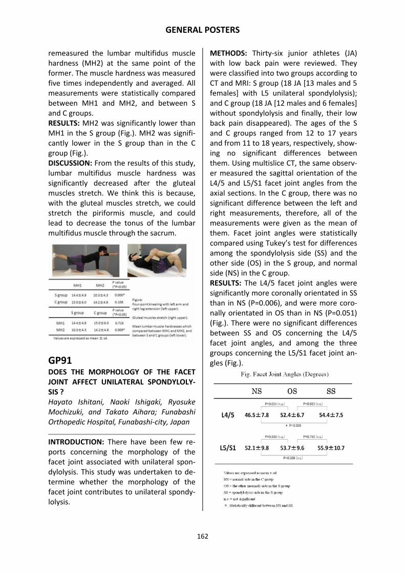
GENERAL POSTERS
162
remeasured the lumbar multifidus muscle hardness (MH2) at the same point of the former. The muscle hardness was measured five times independently and averaged. All measurements were statistically compared between MH1 and MH2, and between S and C groups. RESULTS: MH2 was significantly lower than MH1 in the S group (Fig.). MH2 was signifi‐cantly lower in the S group than in the C group (Fig.). DISCUSSION: From the results of this study, lumbar multifidus muscle hardness was significantly decreased after the gluteal muscles stretch. We think this is because, with the gluteal muscles stretch, we could stretch the piriformis muscle, and could lead to decrease the tonus of the lumbar multifidus muscle through the sacrum.
GP91 DOES THE MORPHOLOGY OF THE FACET JOINT AFFECT UNILATERAL SPONDYLOLY‐SIS ? Hayato Ishitani, Naoki Ishigaki, Ryosuke Mochizuki, and Takato Aihara; Funabashi Orthopedic Hospital, Funabashi‐city, Japan
INTRODUCTION: There have been few re‐ports concerning the morphology of the facet joint associated with unilateral spon‐dylolysis. This study was undertaken to de‐termine whether the morphology of the facet joint contributes to unilateral spondy‐lolysis.
METHODS: Thirty‐six junior athletes (JA) with low back pain were reviewed. They were classified into two groups according to CT and MRI: S group (18 JA [13 males and 5 females] with L5 unilateral spondylolysis); and C group (18 JA [12 males and 6 females] without spondylolysis and finally, their low back pain disappeared). The ages of the S and C groups ranged from 12 to 17 years and from 11 to 18 years, respectively, show‐ing no significant differences between them. Using multislice CT, the same observ‐er measured the sagittal orientation of the L4/5 and L5/S1 facet joint angles from the axial sections. In the C group, there was no significant difference between the left and right measurements, therefore, all of the measurements were given as the mean of them. Facet joint angles were statistically compared using Tukey’s test for differences among the spondylolysis side (SS) and the other side (OS) in the S group, and normal side (NS) in the C group. RESULTS: The L4/5 facet joint angles were significantly more coronally orientated in SS than in NS (P=0.006), and were more coro‐nally orientated in OS than in NS (P=0.051) (Fig.). There were no significant differences between SS and OS concerning the L4/5 facet joint angles, and among the three groups concerning the L5/S1 facet joint an‐gles (Fig.).

GENERAL POSTERS
163
DISCUSSION: We think that a more coronal orientation of the L4/5 facet joint is likely to increase the point loading through the L5 pars interarticularis in extension and rota‐tion. Therefore, if a subject has coronally orientated L4/5 facet joint, spondylolysis at L5 may be occurred on the more coronally orientated side, and the other coronally orientated side may be the risk of L5 spon‐dylolysis.
GP92 VALIDATION OF A DIAGNOSTIC SUPPORT TOOL FOR LUMBAR SPINAL CANAL STENO‐SIS FOR OSTEOARTHRITIS OF THE HIP Morimoto T, Tsukamoto M, Yoshihara T, Sonohata M, Mawatari M; Department of Orthopedic Surgery, Faculty of Medicine, Saga University
INTRODUCTION: A diagnostic support tool (DST) for lumbar spinal stenosis (LSS) based on a self‐administered questionnaire was recently developed (Konno 2007). Since LSS and osteoarthritis of the hip (OAH) demon‐strate similarities in terms of the affected age group and the initial symptoms, both diseases may often lead to a mistaken diag‐nosis. The purpose of this investigation was to evaluate the predictive value of the DST in patients with OAH METHODS: The subjects included in this study consisted of 108 patients with OAH who were treated by total hip arthroplasty (THA) (14 males and 94 females; average age 61 years, range: 36‐88). The hip intensi‐ty was evaluated using a numerical rating scale (NRS). We applied the DST to these patients preoperatively and two months after the operation. The paired t‐test was used to identify changes in the DST before and after THA and the correlations between parameters were evaluated by a regression analysis. A value of P< 0.05 was considered to be significant. RESULTS: The positive rate for the DST was 33% (36 patients) preoperatively and 8%
(nine patients) at two months after THA (P <0.05). The correlation coefficient between the DST and NRS preoperatively was 0.332 (P<0.001). DISCUSSION: The positive rate for the DST in OAH patients decreased from 33% pre‐operatively to 8% at two months after THA, because THA could improve the clinical symptoms of OAH. Therefore, the reason why the positive rate for DST was 33% pre‐operatively may not be the high co‐existence rate of LSS in patients with OAH, but the similarities of the clinical findings of both diseases as a lower leg pain. As a re‐sult, if this DST is applied to patients with OAH, then 33% of the OAH patients may be considered to have LSS. This finding sug‐gests that OAH should be included in the differential diagnosis of LSS.
GP93 INITIAL DIAGNOSIS OF RUPTURED AB‐DOMINAL AORTIC ANEURYSM AND RUP‐TURED INFECTED ABDOMINAL AORTIC AN‐EURYSM WITH LUMBAGO Morimoto T, Tsukamoto M, Yoshihara T, Sonohata M, Mawatari M; Department of Orthopedic Surgery, Faculty of Medicine, Saga University
INTRODUCTION: An infected abdominal aortic aneurysm (IAAA) is a rare and poten‐tially fatal, but poorly recognized, disease. However, the reports of IAAA have been increasing in recent years due to the aging of society and increase in compromised hosts. It has been reported that IAAA, as well as ruptured abdominal aortic aneurysm (AAA), can present as lumbago, and the diagnosis was usually missed in the early stages in these cases. However, few papers have investigated the incidence of lumbago and the participation of orthopedists in the diagnosis of both diseases. The objective of this study was to determine the accuracy and frequency of diagnosing of IAAA and AAA with lumbago.

GENERAL POSTERS
164
METHODS: The medical records of patients with IAAA (n=3, all male, average age 72 years old) and AAA (n=37, 24 males and 13 female, average age 70 years old), the diag‐nosis of which was confirmed by surgery from 2000 to 2010 were reviewed. RESULTS: The initial symptoms were lum‐bago, abdominal pain, consciousness disor‐der, and vital shock in 100%, 33%, 0%, 0% of IAAA cases, and in 44%, 60%, 15%, 22% of AAA cases, respectively. The frequency of the initial examination being performed by orthopedists and the diagnostic accuracy rate by orthopedists were 3% and 100% in AAA cases, 100% and 0% in IAAA cases. The misdiagnoses of IAAA were degenera‐tive spondylosis, lumbago and cholecystitis. DISCUSSION: All IAAA cases presented with lumbago without consciousness disorder or shock vital as the primary symptom, which was less severe than AAA. Therefore, all IAAA cases were initially examined by or‐thopedists, and all were misdiagnosed. These results suggest that orthopedists played in an important role in the diagnosis of cases of IAAA, and that elderly and com‐promised patients with lumbago should be rapidly investigated and treated IAAA if IAAA is a likely possibility.
GP94 TOTAL DISC REPLACEMENT IN THE TREAT‐MENT OF RECURRENT LUMBAR DISC HER‐NIATION Nan Kang, Yong Hai, Shibao Lu Qingyi Wang Chao Kong; China
OBJECTIVE: To evaluate Biomechanical benefits of Total Disc Replacement (TDR) including both the restoration of normal segmental range of motion and the preven‐tion of physiological lumbar lordosis en‐courage spine surgeons to perform TDR for recurrent lumbar disc herniation. Methods: Atotal of twenty first patients (mean age: 44) who had recurrent lumbar disc herniation were operated on between
2000 and 2008. Anterior lumbar discectomy with TDR placement via a extraperitoneal approach were performed. Each patient was evaluated using a VAS and the Oswestry index. Clinical and radiographic results of these patients were evaluated at
each fallow‐up time(1,3,6,12,24
months after operation and the latest).RESULTS: The average visual analogue scales score for pain was 9.40 before opera‐
tion,changed to 4.30 one month after
operation,further declined to 2.70 two years after operation and finally to 1.90 at the latest follow‐up evaluation (P < 0.001)
.Meanwhile,the average Oswestry Disa‐
bility Index was 50.8 before operation,
29.6 one month after operation,13.5 two years after operation and 9.2 at the latest
follow‐up evaluation(P < 0.001).All oper‐ated levels maintained mobile and there was no significant loss of range of motion
observed.Complications such as implant dislocation or significant subsidence of the prosthesis occurred in none case of this
group.96% patients were satisfied with the surgery at the latest follow‐up evalua‐tion CONCLUSIONS: Results from this series are promising and indicate that placement of TDR for recurrent lumbar disc herniation is a valuable alternative to conventional tech‐niques. The main advantages of this appli‐cation are preservation of spinal stability, early mobilization, restoration of normal segmental range of motion and elimination of problems related to intervertebral disc tissue such as discogenic pain and recur‐rence of disc herniation. METHODS: We performed an observational study of patients undergoing fluoroscopy‐guided L5 TFESI in the prone position. A total of 80 patients (40 men and 40 women) with radiating pain of lower limbs were en‐rolled. During TFESI, we measured the angle that the L5 vertebral body forms the rec‐tangular shape and compared between men and women. Then, we measured area of

GENERAL POSTERS
165
safe triangle in tilting angle of fluoroscopy from 15. to 35. and compared between men and women. RESULTS: The mean cephalo‐caudal angle, where the vertebral body takes the shape of rectangle, was 11.0° in men and 13.9° in women.(P=0.007). In men, it was 18.3 mm² at an oblique view angle of 25°, being the greatest. In women, it was 23.6 mm² at an oblique view angle of 30°. At an oblique view angle of 30° and 35°, the area was sig‐nificantly greater in women(P<0.05). DISCUSSION: This results might be due to a difference in the anatomy of the pelvis be‐tween men and women. It can be inferred that the TFESI might be more advanta‐geously used in women as compared with men because the vertebral body cannot be masked by the iliac crest and the area of the target site is greater at a higher degree. Conclusively, when TFESI is performed at the L5 region in the prone position, it would be most reasonable that fluoroscopy is placed at cephalocaudal angle of 11.0° and an oblique angle of 25° in men, cephalo‐caudal angle of 13.9° and an oblique angle of 30° in women.
GP95 SURVIVAL PROGNOSIS IN SYMPTOMATIC AND ASYMPTOMATIC PATIENTS WITH SPI‐NAL METASTASES BY USE OF THE REVISED TOKUHASHI SCORE Kawai Takuya1), Aota Yoichi1), Yamashita Takayuki3), Kono Motonori1), Niimura Takanori1), Thomas E. Mroz3), Isador H. Lieberman3), Saito Tomoyuki2); 1) Dept. of Spine and Spinal Cord Surg. Yokohama Stroke and Brain Center,Yokohama, Japan 2) Dept. of Orthop. Surg., Yokohama City Univ., Yokohama, Japan 3) Center for Spine Health, Cleveland Clinic
INTRODUCTION: Predicting survival for pa‐tients with spinal metastases at early time is desirable to consider their treatment strat‐egies. The revised Tokuhashi Score is widely
used mainly for symptomatic patients who require surgery, while there are no reports regarding the relationship between the To‐kuhashi Score and asymptomatic cases. The purpose of this study is to correlate the re‐vised Tokuhashi score with symptomatic and asymptomatic patients with spinal me‐tastases. METHODS: A total of 266 patients with spi‐nal metastases detected on MR images or bone scans was retrospectively analyzed. They were classified into symptomatic pa‐tients (group A: 187 cases) and asympto‐matic ones (group B: 79 cases). The To‐kuhashi Score at detection of their spinal metastases and survival period from detec‐tion were reviewed. According to the total score (TS), patients in both groups were stratified into 3 categories (TS: 0‐8, 9‐11, 12‐15) and survival rate in each category was analyzed. The Spearman’s correlation coefficient between total Tokuhashi Score and survival period was calculated in both groups. RESULTS: The mean total Tokuhashi score at diagnosis was 7.8 points in Group A and 9.0 in Group B (p<0.05). At the latest follow‐up, 146 patients died and 41 were alive in Group A, while 49 patients died and 30 were alive in Group B. The median survival period was 7.4 months in Group A, 23.4 months in Group B (p<0.05). Kaplan‐Meier curves revealed significant differences in the mean survival period among three cate‐gories in both groups (p<0.05). The total score was significantly correlated with the actual survival period in both groups.(Group A: p<0.05, r=0.58, Group B: p<0.05, r=0.48) DISCUSSION: So far, there has been no study analyzing survival in asymptomatic cases with spinal metastases. This study revealed the revised Tokuhashi Score was significantly correlated with survival period in asymptomatic patients as well as symp‐tomatic ones.

GENERAL POSTERS
166
GP96 TRANSVERSE PROCESS AND NEEDLES OF MEDIAL BRANCH BLOCK TO FACET JOINT AS LANDMARKS FOR ULTRASOUND‐GUIDED SELECTIVE NERVE ROOT BLOCK Yongsoo Choi MD, Daehee Kim MD; De‐partment of Orthopaedic Surgery, Kwangju Christian Hospital, Gwangju, Korea
INTRODUCTION: This study evaluated the usefulness of longitudinal view of trans‐verse process and needles for medial branch block as landmarks under ultraso‐nography. METHODS: We performed selective nerve roots block for 96 nerve roots in 61 patients under guidance of ultrasound. A curved probe was used to identify the facet joints and transverse processes. Identifying the lumbar nerve roots under skin surface and ultrasound landmarks, the cephalad and caudal medial branch blocks were under‐taken under transverse view of sonogram first and a needle for nerve root block was inserted between the two transverse pro‐cesses under longitudinal view while esti‐mating the depth with the needles for me‐dial branch block. Then we injected 1.0ml of contrast medium and checked the distribu‐tion of the nerve root with C‐arm fluoros‐copy to evaluate the accuracy. The visual analog scale (VAS) was used to access the clinical results. RESULTS: Seven SNRBs were performed for the L2 nerve root, 15 for L3, 49 for L4, and 25 for L5 respectively. 86 SNRBs (89.5%) showed successful positioning of the nee‐dles. We failed one case for the L2 nerve root, 2 for L3, 3 for L4, and 4 for L5. The failed needles positioned at wrong leveled segments in 4 cases and inappropriate place in 6 cases. VAS was improved from 7.6±0.6 to 3.5±1.3 after the procedure. DISCUSSION: A needle, which was the same length with the needles for medial branch block, for selective nerve root block was inserted between the two transverse pro‐
cesses under longitudinal view with the same angle of the needles for medial branch block as short axis out of plane ap‐proach. To conform for final depth of the needle, it is possible to get an image for motion echo of final needle tip in ultra‐sound by small movement of the needle tip. For SNRB in lumbar spine, transverse pro‐cesses under longitudinal view as ultra‐sound landmark and the needles of medial branch block can be a promising guidance.
GP97 APPLICATION OF ELECTRIC NERVE STIMU‐LATOR FOR LUMBAR TRANSFORAMINAL EPIDURAL BLOCK Yongsoo Choi, MD , ChaeHyun Lim, MD, Daehee Kim, MD; Department of Orthopedic Surgery, Kwangju Christian Hospital, Gwangju, Korea
INTRODUCTION: The nerve stimulation of nerve roots can recreate the symptoms in the middle of procedure, and can therefore identify the level involved with radicular pain. However, how much amplitude is enough has not been adequately deter‐mined. The purpose of this study is to eval‐uate the clinical feasibility of electric nerve stimulator in lumbar transforaminal epidur‐al block (TFEB). METHODS: Using an electric nerve stimula‐tor, the TFEB were performed in 103 seg‐ments among 49 patients who presented with lower back pain with radiating pain to lower extremities. After positioning an insu‐lated needle at the intervertebral foramen under fluoroscopic guidance, contrast me‐dium was injected to delineate the nerve root. Then, the nerve root was electrically stimulated with the insulated needle to confirm whether or not the same radiating pain was evoked. RESULTS: Of the 103 foraminal segments, the same radiating pain was evoked at 0.5 mAh in 49 (47.6%), at 1.0 mAh in 20(19.4%), at 1.5 mAh in 3(2.9%), at 2.0 mAh in

GENERAL POSTERS
167
17(16.5%), and at 3.0 mAh in 5 segments (4.9%). No response was observed in 9 segments (8.7%). The patients evoked the radiating pain over 2.0 mAh, fluoroscopy revealed successful positioning of the nee‐dle. Visual analogue scale (VAS) for pain improved from a mean of 7.5 to 2.7 after the block (p=0.001). In the 9 cases without response to electrical stimulation, the pa‐tients also showed improvement of VAS, from 7.8 to 3.4(p=0.008). DISCUSSION: There were many studies to establish the relationship between evoked responses and the needle tip‐nerve dis‐tance. According to our results, the patients evoked the radiating pain over 2.0 mAh for successful TFEB as a safety zone, fluorosco‐py revealed successful positioning of the needle. Nerve stimulator can help to predict the accuracy of needle positioning as a sup‐plemental aid for a successful lumbar trans‐foraminal epidural block. It is enough to initiate proper stimulation amplitude of the nerve at 2 mAh.
GP98 ARE HIGH‐HEELED SHOES ASSOCIATED WITH DISC DEGENERATION? Park J‐U, Kim M‐C, Kim U‐C, Lee H‐S, Chung H‐T, Kim M‐W; Department of Orthopaedic Surgery, Bumin Hospital, Busan, Republic of Korea
INTRODUCTION: Although high‐heeled shoes seem to be related with body pos‐ture, most studies report static posture analysis. And few include the consideration of life‐style and occupation. It remains un‐clear whether women wearing high‐heeled shoes during their day‐time may progress sagittal imbalance and disc degeneration or not. We investigated the changes of global sagittal balance, pelvic parameters, and disc degeneration using standing lateral radio‐graphs and MRI in women who frequently wear high‐heeled shoes.
MATERIALS AND METHODS: Our survey asked women, who work in our hospital and visit our out‐patient clinics for medical rea‐sons, about their use of high‐heeled shoes. Among them, we chose women between the ages of 20 and 35 who has not suffered from back pain or radiculopathy. A total of 18 patients who had worn high‐heeled shoes for more than 5 hours a day, more than three times a week, and more than 3 years (H group) were compared with a con‐trol group who had not worn high‐heeled shoes. The changes of global sagittal bal‐ance and pelvic parametes were examined through standing lateral radiographs. The difference of disc degeneration was com‐pared using Pfirrmann grading system in MRI. RESULTS: The C7 plumb line of H group was located more anteriorly than that of control group (P<0.001). The pelvic incidence (PI) between two groups was not statistically different. The pelvic tilt (PT) in H group was increased and the sacral slope (SS) in H group was decreased compared with con‐trol group, but not statistically significant. Pfirrmann grading system showed that disc degeneration was more aggravated in H group(P<0.001). DISCUSSION: As previous reports show that high‐heeled shoes seem to be related with body posture in static state, women who wear high‐heeled shoes during their day‐time for a long period have the changes in sagittal balance and degrees of disc degen‐eration.
GP99 PROSPECTIVE STUDY OF SUPERIOR CLUNEAL NERVE DISORDER AS A POSSIBLE CAUSE OF LOW BACK AND LEG PAIN Hiroshi Kuniya, MD*, Yoichi Aota, MD**, Takuya Kawai, MD**, Kanichiro Kaneko, MD*, Tomoyuki Konno, MD*, Tomoyuki Saito, MD*; *Department of Orthopaedic Surgery, Yokohama City University Graduate School of Medicine, Yokohama, Japan

GENERAL POSTERS
168
**Department of Spine center, Yokohama Stroke and Brain Center, Yokohama, Japan
INTRODUCTION: Although superior cluneal nerve (SCN) can become spontaneously entrapped under fascia over iliac crest, pre‐vious surgical reports were very few and limited to a small number of subjects with low back and/or buttock pain. Trescot in 2003 stated in a review of interventional pain management that cluneal neuralgia is more commonly the result of an entrapped nerve than a nerve injury during iliac crest bone harvest and this clinical entity may be underdiagnosed and should be considered as a potential cause of chronic low back pain (LBP) or leg pain. The purpose of this prospective study was to elucidate the prevalence of SCN disorder and to evaluate the outcomes of trigger point injection. METHODS: A total of 820 consecutive pa‐tients suffering from LBP and/or leg pain who visited authors’ spine clinic were regis‐tered. SCN disorder was suspected when; 1) The maximal tender point was on the pos‐terior iliac crest 70 mm from the midline, and 2) Palpation of the maximal tender point reproduced their chief complaint. For all patients with suspicion of SCN disorders, trigger point injection was done with 5ml of 1% lidocaine. RESULTS: Of 820, 101 patients met the cri‐teria 1) and 2). The mean VAS score was 68.9 ± 19.3 mm and the mean RDQ score was 11.6 ± 7.0 before injection. The mean VAS score significantly decreased to 31.1 ± 27.1 mm at 15 minutes and 45.7 ± 29.1 mm at one week after injection (p<0.05). Strin‐gently diagnosed SCN disorder patients were 87 (11%), provided that patients expe‐rienced more than 20 mm decrease of VAS. Of 87, 44 (51%) had leg symptoms. CONCLUSION: SCN disorder was not rare clinical entity and should be considered as a potential cause of chronic LBP or leg pain. We propose importance of knowledge in this clinical entity.
GP100 DOES LOW BACK PAIN IN PATIENTS WITH EARLY‐STAGE SPONDYLOLYSIS HAVE SPE‐CIFIC CHARACTERISTICS? Shiro Sugiura1, Satoru Nishikawa1, Takeshi Toyooka1, Tetsuo Shiga1, Kazumi Otsuki1, Kazuhisa Kitou1, Yuka Takata1, Tohru Ishi‐zaki1, Yasutaka Omori1, Akito Takata1, Ayako Kote1, Yasuchika Aoki2; 1Nishikawa Orthopaedic Clinic 2Department of Ortho‐paedic Surgery, Toho University Sakura Medical
INTRODUCTION: Magnetic resonance imag‐ing (MRI) is useful for diagnosing early‐stage spondylolysis (ESS). However, MRI cannot be utilized in all adolescent patients with low back pain (LBP). To diagnose ESS as ear‐ly as possible, it would be helpful to clarify specific characteristics of the symptoms. The purpose of this study was, focusing on qualities, extent, and location of LBP, to elucidate the characteristics of LBP in pa‐tients with ESS. METHODS: Patients (n=43, <18 yrs‐old) with acute LBP were included (<1 month). All patients were evaluated by plain radiog‐raphy and MRI; patients who showed obvi‐ous pathological findings other than ESS (e.g., disc herniation, infection, etc.) were excluded. All patients were evaluated by the following examinations: hyperextension and hyperflexion tests (pain provocation tests in standing position), pain quality (sharp/dull), pain extent (fingertip‐sized area/palm‐sized area) and pain location (unilateral/center). To evaluate the useful‐ness of the tests, positive rates for each test were compared between patients with and without ESS.

GENERAL POSTERS
169
RESULTS: Of 43 patients, 28 had ESS (ESS group: mean age: 14.1 yrs‐old; 25 male/3 females) and 15 had no pathological find‐ings that explained the LBP origin [non‐specific LBP group (NS‐LBP): mean age: 14.6 yrs‐old; 8 males/7 females]. Positive rates for each test (ESS/NS‐LBP) were: hyperflex‐ion test (61%/66%, p=0.167); hyperexten‐sion test (92%/100%, p=0.106); pain quality: sharp (71%/39%, p=0.0835), dull (25%/54%, p=0.0699); pain extent: (finger/palm: 59%/41%, 19%/81%, p=0.0133), and pain location: (unilateral/center: 86%/14%, 19%/81%, p=0.0133). DISCUSSION: Pain provocation on hyperex‐tension is generally recognized to be a use‐ful physical sign of ESS. However, our study revealed that positive rates were similar between ESS and NS‐LBP patients, suggest‐ing that it is not useful for diagnosing ESS. Our results indicate that finger‐sized pain and unilateral LBP may be specific charac‐teristics of ESS.
GP101 THE FEATURES OF DEGENERATIVE LUMBAR SCOLIOSIS IN RHEUMATOID ARTHRITIS PATIENTS ‐ MATCHED COHORT STUDY Hiroyuki Yasuda, Akinobu Suzuki, Kentaro Yamada, Shinnji Takahashi, Sho Dozono, Yoshikazu Shinohara, Hidetomi Terai, Hiro‐mitsu Toyoda, Kohji Tamai, Tatsuya Koike, Hiroaki Nakamura; Department of Orthope‐dic Surgery, Osaka City University Graduate School of Medicine
INTRODUCTION: The lumbar lesion in rheumatoid arthritis (RA) have been paid less attention, but some previous studies demonstrated the high prevalence of lum‐bar spondylolisthesis and lumbar scoliosis. The lumbar lesion accompanied with RA is often difficult to treat, and it is important to know the characteristics of lumbar lesion in RA patients. The purpose of this study is to clarify the features of lumbar scoliosis in RA
patients compared with degenerative lum‐bar scoliosis in non‐RA patients. MATERIAL AND METHODS: A total of 54 patients (44 women and 10 men, 69.3 ± 5.4 years, Cobb angle: 14.6 ± 5.9) with scoliosis (Cobb angle of > 10°) who fulfilled the re‐vised criteria of the American Rheumatism Association were included in this study. As control, age, sex, and Cobb angle matched 54 patients without RA were selected and also included. We evaluated superi‐or/inferior end vertebra, apical vertebra and osteophyte formation using Nathan’s classification (1‐4) on plain X‐rays. These parameters were compared between two groups using Man Whitney U‐test. RESULTS: The level of apical vertebra was significantly upper in RA than non‐RA group. The level of superior end vertebra was also significantly upper in RA group, but there was no significant difference in the level of inferior end vertebra between two groups. The levels of curve was more wide in RA groups (RA group: 4.9 ± 1.1 levels, non RA group: 3.6 ± 0.7 levels, P‹0.01). The degree of osteophyte formation was significantly greater in non RA group. DISCUSSION: The present results showed the differences between lumbar scoliosis with RA and that without RA. These differ‐ences may indicate that the process or cause of scoliosis development in RA is dif‐ferent from that of degenerative scoliosis. Further, the less osteophyte formation may suggest that the lumbar scoliosis with RA is more likely to have instability, and these differences should be taken into considera‐tion in the treatment of lumbar scoliosis with RA.
GP102 DO WE NEED HIGH FIELD MR‐SCANNER FORROUTINE IMAGING OF THE LUMBAR SPINE Assheuer J.*, Forutan F.*, Golfman C.*, Trümmler K.H., **Castro W. ***; * Instutut f. Kernspintomographie, Cologne ** Sie‐

GENERAL POSTERS
170
mens AG, Erlangen, *** Orthopädisches Forschungsinstitut, Düsseldorf
AIM: The aim of this study is to compare image quality and diagnostic reliability of the MR examination of the lumbar spine carried out with low, intermediate and high field strength. MATERIALS AND METHODS: Each of 8 vol‐unteers (7 female, 1 male, ageing from 24 y to 57 y) were scanned with 0,3 T, 1,5 T and 3,0T scanner with sequences adjusted to give similar tissue contrast. The image quali‐ty of the following structures were estimat‐ed with a 4 point scale ( 0 = not recognized, 1 = recognized with flat contours, 2 = rec‐ognized and well delineated, 3 = recog‐nized, well delineated and with high con‐trast to adjacent tissues): neuroforaminal structures as fat pad, nerve root and gangli‐on; dural sack and spinal canal; facet joint with the lig. flavum; the disc and lumbar vertebra. Pfirrmann scale was used to clas‐sify disc degeneration. Hydration of the disc was determined by the quotient mean pixel value of the disc devided by mean pixel val‐ue of the adjacent liquor.The evaluation was done by 2 radiologist and 1 orthopaedic spinal surgeon. RESULTS: The review for all 8 volunteers were the same for 3 different scanners re‐garding the disc degeneration and herni‐ation, facet joint and the lig. flavum. Chang‐es of vertebra signal intensities were de‐tected by all scanners. There were some differences in scaling between the scanners for example the lig. longitudionale was bet‐ter delineated by the 1,5 and 3,0 T scanner in the T2 weighted images, whereas more details of the disc were seen by the 0,3 T in the T1 weighted images. Scan time was 45 min for the 0,3 T, 29 min for the 1,5 T and 31 min for the 3,0 T machine. The annual costs calculated for the German market for 7 years of amortization are 135750 Euro for the 0,3 T, 286050 E
GP103 FEASIBILITY OF THE NIJMEGEN DECISION TOOL TO SUPPORT SPINE SURGEONS IN THE TRIAGE OF CHRONIC LOW BACK PAIN PATIENTS M.L. van Hooff (1); J. van Loon (1); M. de Kleuver (1;2); (1) Sint Maartenskliniek, Nij‐megen, The Netherlands (2) VU medical Center, Amsterdam, The Netherlands
INTRODUCTION: In Western Europe Chron‐ic Low Back Pain (CLBP) is responsible for the greatest global burden of all diseases. The CLBP population is heterogeneous and it is unclear who benefits from spinal sur‐gery. The Nijmegen decision tool has been developed to support patient‐triage and is based on evidence and professional (Delphi) consensus. It consists of a web‐based screenings questionnaire and a decision algorithm. Since April 2012 all patients complete the questionnaire and are sys‐tematically followed over time. In this study pre‐intervention patient profiles based on indicators predicting successful outcome (functional status [ODI=22]) of spine surgery and of a Combined Physical and Psychologi‐cal (CPP) program, are evaluated. METHODS: Cross‐sectional design. Diagnos‐tics and decision‐making ‘as usual’. All (1,106) consecutive patients completed the questionnaire, based on 47 predictive indi‐cators: 82 (7.4%) indicated for surgery and 97 (8.8%) for CPP program. Chi2tests for categorical and t‐tests for continuous varia‐bles are used to evaluate baseline differ‐ences. RESULTS: Surgical patients reported more leg pain (t=2.5,p<0.05), larger number of previous surgeries (.2=14.7,p<0.05), and a shorter duration of CLBP (.2=8.1,p<0.05). No differences were found for the number of ‘red flags’ and comorbidities (.2=3.1,p=0.80). DISCUSSION: The screenings questionnaire seems feasible to use. Although some dif‐ferences were found, large cohorts with

GENERAL POSTERS
171
long follow‐up periods are needed to be conclusive about the probability of success in different treatment modalities. At the time of the conference one‐year follow‐up results are available and longitudinal anal‐yses will be performed to identify patient profiles predicting successful treatment outcome. If indeed these profiles can be identified, an efficient referral and treat‐ment allocation may be achieved. This would improve outcomes, and lead to more appropriate use of limited health care re‐sources.
GP104 THE EFFECTS OF DYNAMIC STRETCH FOR TIGHT‐ HAMSTRINGS ON SPINO‐PELVIC RHYTHM Kiyotaka Hasebe(1,2), Yu Okubo(3), Yasushi Hada(1) Kohei Takada(3), Daisuke Suzuki(3) Akira Dezawa(4), Koji Kaneoka(2), Koichi Sairyo(4); (1)Department of Rehabilitation, Teikyo University Mizonokuchi Hospital, Kawasaki, Japan (2)Faculty of Sports Sci‐ence, Waseda University, Japan (3)Faculty of Health and Medical Care, Saitama Medi‐cal University, Saitama, Japan (4)Department of Orthopedic Surgery, Teikyo University Mizonokuchi Hospital, Kawasaki, Japan (5) Department of Ortho‐pedics, The University of Tokushima
INTRODUCTION: Studies have shown that tight hamstrings are related to low back pain. Theoretically, to achieve hamstrings flexibility is essential to prevent low back pain. In this study, we investigated the ef‐fects of stretch intervention on spino‐pelvic rhythm, and discussed the effects of stretching on the prevention. METHODS: Twelve healthy men volun‐teered to participate. The dynamic stretch‐ing machine (Never‐Tight‐Ham; Hogrel Inc., Tokyo, Japan) was used for stretching the hamstrings; stretching was performed for 3 days per week for 6 weeks. The finger‐to‐floor distance (FFD), straight leg raising
(SLR) angle were measured before and after the intervention. Using a SPINAL MOUSE (Index Ltd., Tokyo, Japan), spinal alignment was measured and the spino‐pelvic rhythm was evaluated. RESULTS AND DISCUSSION: After the 6‐week stretching protocol, all subjects achieved hamstrings flexibility. Significant improvements (p < 0.01) were observed in FFD from 5.8 to ‐2.5 cm and in SLR from 71.2° to 79.3°. During complete flexion, to‐tal pelvic motion significantly (p < 0.01) in‐creased from 31.9° to 42.3°, indicating after the stretching intervention, participants could use their pelvis efficiently compared to that before stretching . On the other hand, total lumbar motion remained the same. The lumbar motion increased only during early flexion phase. It is reasonable to use the lumbar spine in the early flexion phase, since this phase is neutral zone at which very less loading to the spine is re‐quired. Pelvic motion increased, especially in the late flexion phase. Lumbo‐pelvic rate was also significantly changed. With im‐proved hamstring flexibility, participants used pelvic motion but not lumbar motion, indicating that dynamic stretch could change the spino‐pelvic rhythm to the pel‐vic dominant pattern. In conclusion, after achieving hamstrings flexibility, the partici‐pants could use the pelvis efficiently. Using stretch exercises, the lumbo‐spine‐rhythm can be changed t
GP105 INTRADISCAL ADMINISTRATION OF THE INTERLEUKIN‐6 RECEPTOR ANTIBODY TO‐CILIZUMAB IMPROVES INTRACTABLE DIS‐COGENIC LOW BACK PAIN Sainoh T, Orita S, Yamauchi K, Kubota G, Inage K, Sato J, Fujimoto K, Shiga Y, Arai S, Takahashi K, Ohtori S; Dept. of Orthopaedic Surgery, Graduate School of Medicine, Chi‐ba University

GENERAL POSTERS
172
INTRODUCTION: Currently, inflammatory cytokines such as interleukin 6 (IL‐6) and tumor necrosis factor a (TNFa) are gaining attention as important factors causing dis‐cogenic lower back pain (LBP). We have previously performed intradiscal admin‐istration of a TNFa inhibitor in cases of re‐fractory discogenic LBP with a degenerated intervertebral disc (IVD), and found that it was safe and effective. In the present study, we aimed to assess the outcomes and ad‐verse events of intradiscal administration of IL‐6 receptor antibody in patients with LBP. METHODS: We examined 30 cases with LBP who were resistant to conservative treat‐ment for >3 months and with 1 degenerat‐ed IVD lesion (17 males, 13 females; aver‐age age, 57.6 years). We injected tocili‐zumab (TCZ; 40 mg) and bupivacaine into the degenerated IVD (TCZ group). The fol‐lowing items were evaluated: (1) compari‐son of the numerical rating scale (NRS) score between the TCZ group and the con‐trol group (bupivacaine injection alone) before, the following day, and 1, 2, and 4 weeks after administration, and of the Oswestry disability index (ODI) score before and 4 weeks after administration; (2) com‐parison of the efficacy of pain relief accord‐ing to the degree of disc degeneration in the TCZ group; and (3) presence or absence of adverse events. RESULTS: In the TCZ group, the NRS and ODI exhibited significant improvements during the 4 weeks of treatment. However, with regard to the comparison of the NRS ac‐cording to the degree of disc degeneration, no significant difference was noted. Local infection (discitis) was observed in 1 of 30 cases. DISCUSSION: We hypothesized that the intradiscal administration of TCZ would have an effect on discogenic LBP. Moreover, we noted a significant improvement in pain and activities during the 4 weeks of treat‐ment. However, we did not identify a dif‐ference in efficacy according to the degree
of IVD degeneration. Thus, we suggest that IL‐6 is possibly involved in discogenic LBP.
GP106 UTILITY OF THE KNEE‐LIFTING TEST FOR SELECTING THE APPROPRIATE DIAGNOS‐ING LUMBAR FACET SYNDROME Kazuji Aoki 1, Toru Uehara 2, Shota Yamada 2, Junko Oishi 2, Atsushi Inada 3; 1 Faculty of Rehabilitation Sciences, Nagoya Gakuin University, Seto, Aichi, Japan. 2 Depart‐ments of Rehabilitation, NTT‐West Tokai Hospital, Nagoya, Aichi, Japan. 3 Departme;
INTRODUCTION: In general, therapy for lumbar disc herniation (LDH) and lumbar facet syndrome (LFS) involves lumbar ex‐tension and flexion exercises, respectively. However, the symptoms of patients with LFS closely resemble those of patients with LDH; therefore, both diagnosis and choice of therapeutic exercises to apply can be very difficult. This study investigated the usefulness of a knee‐lifting test (KL‐t) that we devised as an estimation method for diagnosing LFS. METHODS: For the KL‐t, the patient as‐sumes an upright position against a wall, making contact with the back of their head, thoracic spine, buttocks, and heels. They then try to push their lumbar spine to the wall and alternate lifting each knee. Con‐cerning symptoms after this test, a reduc‐tion in LBP represented a positive reaction, while no reduction represented a negative reaction. Subjects in the study comprised 129 outpatients (77 patients with LFS and 52 patients with LDH) requiring therapeutic exercises. All patients performed the KL‐t before being assigned therapeutic exercis‐es. In addition, 13 patients with LFS and 5 patients with LDH underwent measurement of spinal alignment with SpinalMouse, both before and after KL‐t. RESULTS: In patients with LDH, 17 (32.7%) showed positive results and 35 (67.3%) showed negative results. In patients with
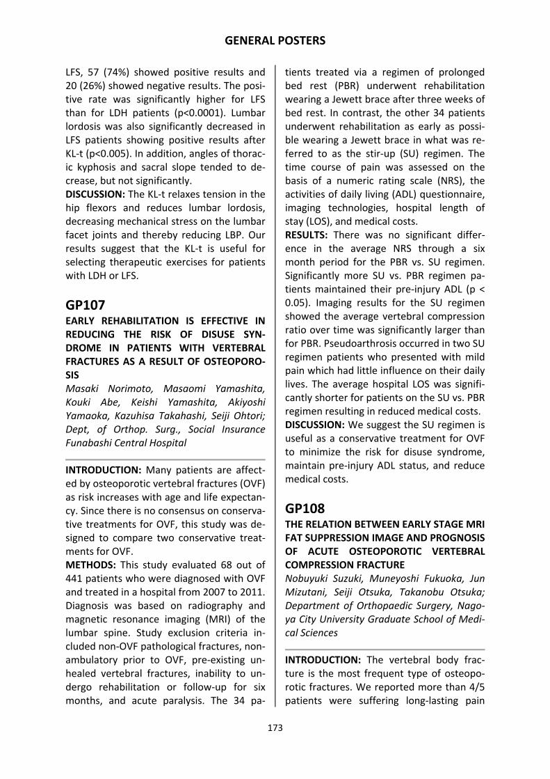
GENERAL POSTERS
173
LFS, 57 (74%) showed positive results and 20 (26%) showed negative results. The posi‐tive rate was significantly higher for LFS than for LDH patients (p<0.0001). Lumbar lordosis was also significantly decreased in LFS patients showing positive results after KL‐t (p<0.005). In addition, angles of thorac‐ic kyphosis and sacral slope tended to de‐crease, but not significantly. DISCUSSION: The KL‐t relaxes tension in the hip flexors and reduces lumbar lordosis, decreasing mechanical stress on the lumbar facet joints and thereby reducing LBP. Our results suggest that the KL‐t is useful for selecting therapeutic exercises for patients with LDH or LFS.
GP107 EARLY REHABILITATION IS EFFECTIVE IN REDUCING THE RISK OF DISUSE SYN‐DROME IN PATIENTS WITH VERTEBRAL FRACTURES AS A RESULT OF OSTEOPORO‐SIS Masaki Norimoto, Masaomi Yamashita, Kouki Abe, Keishi Yamashita, Akiyoshi Yamaoka, Kazuhisa Takahashi, Seiji Ohtori; Dept, of Orthop. Surg., Social Insurance Funabashi Central Hospital
INTRODUCTION: Many patients are affect‐ed by osteoporotic vertebral fractures (OVF) as risk increases with age and life expectan‐cy. Since there is no consensus on conserva‐tive treatments for OVF, this study was de‐signed to compare two conservative treat‐ments for OVF. METHODS: This study evaluated 68 out of 441 patients who were diagnosed with OVF and treated in a hospital from 2007 to 2011. Diagnosis was based on radiography and magnetic resonance imaging (MRI) of the lumbar spine. Study exclusion criteria in‐cluded non‐OVF pathological fractures, non‐ambulatory prior to OVF, pre‐existing un‐healed vertebral fractures, inability to un‐dergo rehabilitation or follow‐up for six months, and acute paralysis. The 34 pa‐
tients treated via a regimen of prolonged bed rest (PBR) underwent rehabilitation wearing a Jewett brace after three weeks of bed rest. In contrast, the other 34 patients underwent rehabilitation as early as possi‐ble wearing a Jewett brace in what was re‐ferred to as the stir‐up (SU) regimen. The time course of pain was assessed on the basis of a numeric rating scale (NRS), the activities of daily living (ADL) questionnaire, imaging technologies, hospital length of stay (LOS), and medical costs. RESULTS: There was no significant differ‐ence in the average NRS through a six month period for the PBR vs. SU regimen. Significantly more SU vs. PBR regimen pa‐tients maintained their pre‐injury ADL (p < 0.05). Imaging results for the SU regimen showed the average vertebral compression ratio over time was significantly larger than for PBR. Pseudoarthrosis occurred in two SU regimen patients who presented with mild pain which had little influence on their daily lives. The average hospital LOS was signifi‐cantly shorter for patients on the SU vs. PBR regimen resulting in reduced medical costs. DISCUSSION: We suggest the SU regimen is useful as a conservative treatment for OVF to minimize the risk for disuse syndrome, maintain pre‐injury ADL status, and reduce medical costs.
GP108 THE RELATION BETWEEN EARLY STAGE MRI FAT SUPPRESSION IMAGE AND PROGNOSIS OF ACUTE OSTEOPOROTIC VERTEBRAL COMPRESSION FRACTURE Nobuyuki Suzuki, Muneyoshi Fukuoka, Jun Mizutani, Seiji Otsuka, Takanobu Otsuka; Department of Orthopaedic Surgery, Nago‐ya City University Graduate School of Medi‐cal Sciences
INTRODUCTION: The vertebral body frac‐ture is the most frequent type of osteopo‐rotic fractures. We reported more than 4/5 patients were suffering long‐lasting pain

GENERAL POSTERS
174
over the year. That is very unsatisfying out‐come from the view of the fracture treat‐ment. If the prognosis of this fracture can predict in early stage, we might be able to choose other treatment options. The pur‐pose of this study was to classify this frac‐ture by first visit MR and predict the prog‐nosis of pain, ADL and QoL after this frac‐ture. METHODS: All the patients over 40 years who visited our department and were diag‐nosed having fresh osteoporotic vertebral fracture were enrolled in this study. Plain X‐ray, CT and MR images were underwent within one week from the first visit and also at 3weeks, 3months and 6months. Pain, ADL and QoL were evaluated by 3 different questionnaires at 3weeks, 3months and 6months. The treatment in all cases was conservative using hard corset. RESULTS: A total of 20 patients were en‐rolled in this study. Mean age was 79.4. All of the case had T2 low intensity at first visit MR image. On the fat suppression image, four cases had diffuse high, four cases had diffuse low, two cases had spot low and ten cases had linear low intensity at first visit. Diffuse fat suppression low and spot low cases had become pseudarthritis and severe collapse. And those patients had severe pain and lower QoL at 6months. Otherwise the patients who had diffuse high intensity had generally good course. DISCUSSION: This study showed the pa‐tients who had diffuse fat suppression low intensity had worse prognosis. The region of the fat suppression low intensity was supposed to cause temporary bone necro‐sis. If the area of the necrosis was extensive, the repair of fracture might become difficult and cause pseudarthritis or severe collapse. So to prevent pseudarthritis or severe col‐lapse, we might be able to apply another treatment options i.e. the vertebro‐ or ky‐phoplasy in early stage.
GP109 VACUUM PHENOMENON OF SACROILIAC JOINT: CORRELATION WITH LUMBO PELVIC ALIGNMENT Yoichiro Takata, Toshinori Sakai, Kosaku Higashino, Yuichiro Goda, Koichi Sairyo; Department of Orthopedics, The University of Tokushima
INTRODUCTION: Lumbo pelvic alignment is reported to be associated with pathomech‐anisms of various spinal disorders. Although the mobility of the sacroiliac joint (SIJ) is small, the vacuum phenomenon (VP), which explains the mobility of SIJ, is often ob‐served in the clinical practice. We analyzed the prevalence of the VP of SIJ from com‐puted tomographic (CT) images, and rela‐tionship between lumbo pelvic alignment and this phenomenon. MATERIALS AND METHOD: We analyzed multi‐detector CT images of 100 subjects (65 men and 35 women). 7 subjects (6 men and 1 woman), who had a transitional ver‐tebra or metastatic pelvic tumor, were ex‐cluded. Pelvic incidence (PI), pelvic tilt (PT) and lumbar lordosis (LL) were measured from multi‐detector computed tomographic (CT) images using 3‐dimentional reconstruc‐tion method. VP of SIJ and intervertebral disc at L5‐S were analyzed from CT images. Originally modified Japanese Orthopedic Association (JOA) score, which focused on subjective symptoms and restriction of ADL, and Roland‐Morris Disability Questionnaire (RDQ) scores were obtained. RESULT: There were 36 subjects with VP of SIJ (VP+ group) and 57 subjects without VP of SIJ (VP‐ group). VP of SIJ was observed in 91% of female subjects, whereas only 8.5% of male subjects had VP of SIJ. Male sub‐jects with VP had significantly lower PI than male without VP (35.1° vs. 46.3°, p < 0.05). There was no correlation between VP of SIJ and JOA score, and RDQ, respectively. DISCUSSION: Almost all female subjects (> 90%) had VP of SIJ, which indicate
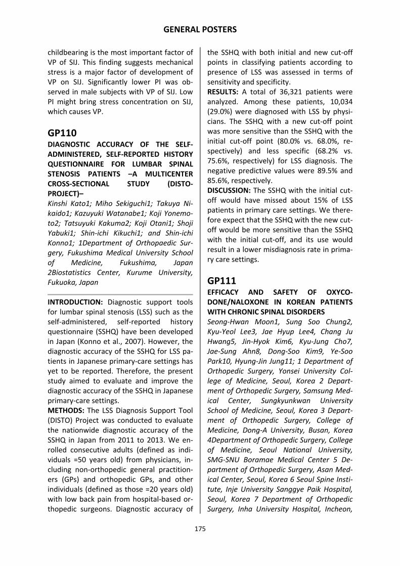
GENERAL POSTERS
175
childbearing is the most important factor of VP of SIJ. This finding suggests mechanical stress is a major factor of development of VP on SIJ. Significantly lower PI was ob‐served in male subjects with VP of SIJ. Low PI might bring stress concentration on SIJ, which causes VP.
GP110 DIAGNOSTIC ACCURACY OF THE SELF‐ADMINISTERED, SELF‐REPORTED HISTORY QUESTIONNAIRE FOR LUMBAR SPINAL STENOSIS PATIENTS –A MULTICENTER CROSS‐SECTIONAL STUDY (DISTO‐PROJECT)– Kinshi Kato1; Miho Sekiguchi1; Takuya Ni‐kaido1; Kazuyuki Watanabe1; Koji Yonemo‐to2; Tatsuyuki Kakuma2; Koji Otani1; Shoji Yabuki1; Shin‐ichi Kikuchi1; and Shin‐ichi Konno1; 1Department of Orthopaedic Sur‐gery, Fukushima Medical University School of Medicine, Fukushima, Japan 2Biostatistics Center, Kurume University, Fukuoka, Japan
INTRODUCTION: Diagnostic support tools for lumbar spinal stenosis (LSS) such as the self‐administered, self‐reported history questionnaire (SSHQ) have been developed in Japan (Konno et al., 2007). However, the diagnostic accuracy of the SSHQ for LSS pa‐tients in Japanese primary‐care settings has yet to be reported. Therefore, the present study aimed to evaluate and improve the diagnostic accuracy of the SSHQ in Japanese primary‐care settings. METHODS: The LSS Diagnosis Support Tool (DISTO) Project was conducted to evaluate the nationwide diagnostic accuracy of the SSHQ in Japan from 2011 to 2013. We en‐rolled consecutive adults (defined as indi‐viduals =50 years old) from physicians, in‐cluding non‐orthopedic general practition‐ers (GPs) and orthopedic GPs, and other individuals (defined as those =20 years old) with low back pain from hospital‐based or‐thopedic surgeons. Diagnostic accuracy of
the SSHQ with both initial and new cut‐off points in classifying patients according to presence of LSS was assessed in terms of sensitivity and specificity. RESULTS: A total of 36,321 patients were analyzed. Among these patients, 10,034 (29.0%) were diagnosed with LSS by physi‐cians. The SSHQ with a new cut‐off point was more sensitive than the SSHQ with the initial cut‐off point (80.0% vs. 68.0%, re‐spectively) and less specific (68.2% vs. 75.6%, respectively) for LSS diagnosis. The negative predictive values were 89.5% and 85.6%, respectively. DISCUSSION: The SSHQ with the initial cut‐off would have missed about 15% of LSS patients in primary care settings. We there‐fore expect that the SSHQ with the new cut‐off would be more sensitive than the SSHQ with the initial cut‐off, and its use would result in a lower misdiagnosis rate in prima‐ry care settings.
GP111 EFFICACY AND SAFETY OF OXYCO‐DONE/NALOXONE IN KOREAN PATIENTS WITH CHRONIC SPINAL DISORDERS Seong‐Hwan Moon1, Sung Soo Chung2, Kyu‐Yeol Lee3, Jae Hyup Lee4, Chang Ju Hwang5, Jin‐Hyok Kim6, Kyu‐Jung Cho7, Jae‐Sung Ahn8, Dong‐Soo Kim9, Ye‐Soo Park10, Hyung‐Jin Jung11; 1 Department of Orthopedic Surgery, Yonsei University Col‐lege of Medicine, Seoul, Korea 2 Depart‐ment of Orthopedic Surgery, Samsung Med‐ical Center, Sungkyunkwan University School of Medicine, Seoul, Korea 3 Depart‐ment of Orthopedic Surgery, College of Medicine, Dong‐A University, Busan, Korea 4Department of Orthopedic Surgery, College of Medicine, Seoul National University, SMG‐SNU Boramae Medical Center 5 De‐partment of Orthopedic Surgery, Asan Med‐ical Center, Seoul, Korea 6 Seoul Spine Insti‐tute, Inje University Sanggye Paik Hospital, Seoul, Korea 7 Department of Orthopedic Surgery, Inha University Hospital, Incheon,

GENERAL POSTERS
176
Korea 8 Department of Orthopedic Surgery, Chungnam National University Hospital, Daejeon, Korea 9 Department of Orthopedic Surgery, Chungbuk National University Hos‐pital, Cheongju, Korea 10 Department of Orthopedic Surgery, Hanyang University Guri Hospital, Guri, Korea 11 Medical Af‐fairs, Mundipharma Korea Ltd, Seoul, Korea
INTRODUCTION: The use of opioids for chronic noncancer pain has increased in recent years. The purpose of this study is to evaluate the efficacy and safety of oxyco‐done/naloxone (Targin®) in chronic, moder‐ate to severe pain due to spinal disorders. METHODS: A multicenter, single‐arm, inter‐ventional study was conducted in patients with chronic spinal disorders not adequate‐ly controlled with weak opioid and/or non‐steroidal anti‐inflammatory drugs (NSAIDs) (Pain Numeric Rating Scale 0‐10; NRS score=4 at baseline). Oxycodone/naloxone was started with 5/2.5mg twice daily, then up‐titrated at the discretion of investiga‐tors. The primary endpoint was the differ‐ence in mean NRS score at 8 weeks from baseline with last observation carried for‐ward method. RESULTS: Total 240 patients were enrolled and 220, 209 patients were included in safety set and full analysis set, respectively. Mean age was 62.3±10.4 years and 61.8% of patients were female. The patients had been treated with weak opioids (62.7%), NSAIDs (58.6%) and adjuvant medications like pregabalin or gabapentin (35%) before enrollment and the baseline mean NRS score was 6.5±1.4. NRS score was reduced to 4.8±2.0 at 8 weeks (difference; ‐1.7±2.2, p<0.05) with mean 18.2±10.2mg dose of oxycodone. EQ‐5D and EQ‐VAS were in‐creased from 0.40±0.33 to 0.55±0.29 and from 50.2±19.5 to 61.2±18.1 at 8 weeks, respectively (p<0.05). 66.5% and 64.1% of the investigators and patients were satisfied with overall pain control at 8 weeks, respec‐tively. 120 adverse events were reported in
77 patients (35.0%) and common adverse drug reactions (ADRs) were nausea (9.1%), dizziness (5.5%), and constipation (5.5%). Most of ADRs were mild and recovered dur‐ing the study period. DISCUSSION: Oxycodone/naloxone of rela‐tively low dose effectively reduced the pain, improved the quality of life and showed tolerable safety profiles in patients with chronic spinal disorders, whose pain was not properly controlled by weak opioids and/or conventional NASI
GP112 THE RELATIONSHIP BETWEEN ROTATION AND TILTING OF VERTEBRAL BODY IN CAS‐ES WITH DEGENERATIVE LUMBAR SCOLIO‐SIS Bunichiro Izumi, Yasushi Fujiwara, Akira Miyauchi, Shinji Kotaka, Hideki Manabe; Department of Orthopaedic Surgery, Hiro‐shima city Asa hospital
INTRODUCTION: In degenerative scoliosis patient, vertebral tilt and rotation are common findings. It is very important to realize the relationship between rotation and tilt of vertebral body, when we do the surgical treatment, especially microscopic posterior decompression surgery, for the lumbar canal stenosis accompanied with degenerative scoliosis (LCSDSc). However, there are few reports about the relation‐ship. Therefore, the purpose of this study was to investigate the rotation and tilting of the vertebral body using the imaging find‐ings of preoperative computed tomography (CT). METHODS: Twenty‐six patients (9 male and 17 female) who underwent microscopic decompression surgery because of LCSDSc. The range of preoperative Cobb angle was between 10 and 20 degrees. The mean age at the time of operation was 70 years old. The mean follow‐up period was 3.3 years. The Cobb angle before and after operation, L4 tilt, level of apex of scoliosis, each verte‐

GENERAL POSTERS
177
bral rotation and tilt, deformities of facet joints were evaluated in preoperative CT. RESULTS: The mean Cobb angle before op‐eration was 14.4 degree (10 ~ 18 degree) and mean angle at the final follow up was 16.5 degrees (12 ~ 21 degree). The mean L4 tilt angle was 8.3 degree (0 ~ 19 degree). The vertebral rotation was observed in 77% levels, and the degenerative change of facet joints were observed in 69% levels. Maxi‐mum intervertebral rotation is most fre‐quently observed at L3/4, Maximum tilt of the vertebral body was more common at L2/3 and L4/5 than L3/4. DISCUSSION: Our results demonstrated that surgeons have to take care of rotation at L3/4 and vertebral tilt at L2/3 and L4/5. It is very difficult to do the decompression sur‐gery because the deformity of the degener‐ative scoliosis is very complex. It is very im‐portant to analyze the deformity well when we treat the degenerative scoliosis.
GP113 SAGITTAL SPINO‐PELVIC ALIGNMENT IN PATIENTS WITH OSTEOARTHRITIS OF THE HIP Yoshihara T, Morimoto T, Tsukamoto M, Sonohata M, Mawatari M; Department of Orthopedic Surgery, Faculty of Medicine, Saga University
INTRODUCTION: Few papers have ade‐quately investigated the spino‐pelvic align‐ment in patients with osteoarthritis of the hip (HOA). The present investigation exam‐ined the sagittal alignment of the spine and pelvis in patients with HOA using the SRS‐Schwab classification. METHODS: The subjects included 32 male and 154 female patients with an average age of 65 years treated by total hip arthro‐plasty due to unilateral HOA. The objects were divided into three groups. Group A included patients under the age of 60 years, group B included those 60‐69 years old and group C included those over the age of 70.
Lateral whole spine radiographs were pro‐spectively taken of all patients before sur‐gery. The parameters of the sagittal spino‐pelvic alignment examined were the sagittal vertical axis (SVA), pelvic tilt (PT), lumbar lordosis (LL) and pelvic incidence (PI). The findings of PI‐LL > 20°, SVA > 9.5 cm and PT > 30° were considered to indicate marked sagittal alignment based on the SRS‐Schwab classification. The significance of differences among the three groups were evaluated by the analyses of variance (ANOVA) with the Bonferroni correction. A value of P<0.05 was considered to be significant. RESULTS: The percentage of patients with PI‐LL > 20°, SVA > 9.5 cm and PT > 30° was 0%, 0% and 0% in group A, 9.3%, 3.1% and 1.6% in group B and 28.1%, 21.9% and 15.6% in group C, respectively. The fre‐quency of marked sagittal alignment in group C was significantly higher than that in group A (P<0.05) and group B (P<0.05). DISCUSSION: In elderly patients with HOA, concurrent disease in both the lumbar spine and hip was not infrequent, and the sagittal spino‐pelvic alignment was decompensated in these cases. Our study suggests that long‐term HOA may affect the sagittal alignment of the spine, or the presence of a spinal deformity may rotate the pelvis backwards and exaggerate the stress on the femoral head, finally causing HOA, which is referred to as secondary hip‐spine syndrome.
GP114 CORONAL SPINO‐PELVIC ALIGNMENT IN CROWE TYPE IV Hirata H, Morimoto T, Yoshihara T, Tsuka‐moto M, Kawano S, Kitajima M, Sonohata M, Mawatari M; Department of Orthopedic Surgery, Faculty of Medicine, Saga Universi‐ty
INTRODUCTION: A leg length discrepancy (LLD) can cause pelvic tilt (PT) and lumbar scoliosis (LS). However, it is controversial whether LS can compensate for LLD with
GENERAL POSTERS
178
the convex shift toward the shorter leg side, probably owing to the fact that these pa‐tients might have a short LLD. We have in‐vestigated the influence of marked LLD in patients with complete hip dislocation of Crowe type IV (more than 100% subluxa‐tion), on the coronal spino‐pelvic alignment. METHODS: The subjects were 51 patients with unilateral complete hip dislocation who were treated by total hip arthroplasty (46 females and 5 males; average age 64 years, range: 49‐84 years). The frontal view of the pelvis and spine in the erect posture were photographed. Measurements were made of the LLD, pelvic tilt (PT), and lumbar scoliosis (LS). The correlation was judged to be positive in cases where there was PT and convexity of the LS toward the side with the hip dislocation, and the findings were oth‐erwise judged to be negative. The correla‐tions between parameters were evaluated by a regression analysis. A value of p<0.05 was considered to be significant. RESULTS: The mean LLD, PT and LS were 5.6 cm, 9.2° and 15.1°. The correlation coeffi‐cients between the LLD and PT, LLD and LS and LS and PT were 0.17(p>0.05), 0.015(p>0.05) and 0.393(p<0.05), respec‐tively. DISCUSSION: In patients with unilateral complete hip dislocation, the LLD did not correlate with the presence of LS and PT. These findings suggest that marked LLD can cause PT and LS due to compensatory changes to maintain the body balance, but that this did not impact the severity of the findings.
GP115 EFFECT OF THE ROCKER EXERCISE ON THE FLEXIBILITY OF THE LEG AND TRUNK, AND ON THE STATIC BALANCE AT STANDING Naoki Ishigaki and Takato Aihara; Dept. of Physical Therapy and Orthopedic surgery, Funabashi Orthopedic Hospital, Funabashi‐city, Japan
INTRODUCTION: The purpose of this study was to compare the flexibility of the leg and trunk, and the sway of the center of gravity (COG) between the conventional crunch exercise (CE) and the rocker exercise (RE). METHODS: Approval of this hospital’s ethics committee and informed consent to inclu‐sion in this study from 40 healthy adults were obtained. They were randomly allo‐cated to either the CE group (n=18) or the RE group (n=22). They were repeated these exercises for 30 seconds and 3 times over (Fig.). Finger‐floor distance (FFD) and straight‐leg raising angle (SLA) were meas‐ured as the indexes of the flexibility of the trunk and leg. The displacement of COG (DOC) and the total length of the sway of COG (LOC) were measured using a postur‐ography. The results were statistically com‐pared between before and after the two exercises, and between the CE and RE groups. RESULTS: FFDs were significantly increased after the CE and RE (Fig.). The SLA after the RE was significantly increased than before the RE (from 69.1° to 80.5°). DOCs were significantly shifted to forward after the CE and RE than before the CE and RE. The LOC after the RE was significantly decreased than before the RE (from 198.8mm to 183.9mm), however, there were no signifi‐cant differences between before and after the CE concerning the SLA and LOC (Fig.). The increase in SLA and the decrease in LOC in the RE group were significantly larger than those in the CE group, however, there were no significant differences between the CE and RE groups concerning the increases in FFD and DOC (Fig.). DISCUSSION: The results indicated that the increase in SLA and the decrease in LOC of RE were significantly larger than those of CE. There have been often reported that the decreased SLA which was caused by the tight‐hamstrings and increased the sway of the COG were the risk factors for low back pain. Therefore, we think that RE can lead

GENERAL POSTERS
179
to a better clinical outcome than CE for the prevention of low back pain.
GP116 CLINICOPATHOLOGIC FEATURES OF LUM‐BAR DISC HERNIATION IN THE ELDERLY Kenichi Kawaguchi*, Kuniyoshi Tsuchiya**, Go Kato*; Department of Orthopaedic Sur‐gery, Saga‐Ken Medical Center Koseikan* Department of Orthopaedic Surgery, Kyushu Koseinenkin Hospital**
INTRODUCTION: Some previous studies reported that the specimens of lumbar disc herniation in elderly were contained hard tissues such as cartilaginous endplate and bone fragment frequency. The aim of the present study was investigated the clinico‐pathologic features of lumbar disc herni‐ation in elderly patients MATERIALS AND METHODS: A total of 117 cases undergoing microscopic discectomy for lumbar disc herniation were reviewed. These are divided in two groups (60 years or more:n=43:group A, less than 60 year:n=74:group B). In these cases, using magnetic resonance imaging, we evaluated for endplate degeneration, disc degenera‐tion, high intensity zone (HIZ), the existence of vertebral corner defect. In addition, his‐topathologically, the frequency of cartilagi‐nous endplate, myxoid change, calcification and inflammatory granulation tissue around herniation tissue were measured.
RESULTS: Endplate degeneration was rec‐ognized in 22 cases (51%) in group A and 17 cases (23%) in group B, and the cases with endplate degeneration were observed carti‐laginous endplate with high frequency (P<0.05). No significant differences were noted in disc degeneration, HIZ and verte‐bral corner defect between two groups. In group A (60%), significantly higher percent‐ages of cartilaginous endplate were found than in group B (31%) (P<0.05). Calcification had a significantly higher incidence in group A (21%) than in group B (11%) (P<0.05). Myxoid change and inflammatory granula‐tion tissue show no significant difference between two groups. DISCUSSION: In elderly patients, avulsion‐type disc herniation caused by endplate degeneration was frequently seen, indicat‐ing that the mechanism of the occurrence of disc herniation may be different with the young patients. Furthermore, considering the result of inflammatory granulation tis‐sue for herniation tissue, clinical symptoms and natural course may be prolonged in elderly patients including hard tissues with high frequency.
GP117 CAN BRIEF SCALE FOR PSYCHIATRIC PROB‐LEMS IN ORTHOPAEDIC PATIENTS (BS‐POP) PREDICT FUNCTIONAL MRI FINDINGS IN PATIENTS WITH CHRONIC LOW BACK PAIN Hideaki Kaneko, Miho Sekiguchi, Shin‐ichi Konno; Department of Orthopaedic surgery, Fukushima Medical University school of medicine, Fukushima, Japan
INTRODUCTION: Regional gray matter atro‐phy, cognitive changes, and unique patterns of brain activity have been demonstrated in patients with chronic low back pain (cLBP). Psychological factors related to abnormal brain conditions are associated with cLBP. The Brief Scale for Psychiatric Problems in Orthopaedic Patients (BS‐POP) is a ques‐tionnaire to assess psychiatric problems.

GENERAL POSTERS
180
The aim of this study was to investigate the utility of BS‐POP for predicting functional MRI findings in patients with cLBP. METHODS: The subjects were twenty‐two cLBP patients who had suffered from persis‐tent LBP for more than 3 months. The pa‐tient version of the BS‐POP was conducted and the subjects were classified into two groups by BS‐POP score, 17 or over (High score: HS group) and under 16 (Non‐high score: Non‐HS group). Each subject was placed in the prone position on a 3 Tesla MRI scanner, and stimulated by manual pressure with the tail of an air‐filled, 20‐mL syringe at 5 cm left of the fourth‐fifth lum‐bar spinal interspace. Three blocks of 30‐second painful stimulus, calibrated at either 5 on the numeric rating scale (NRS), were applied with intervening 30‐second rest conditions during whole‐brain echo‐planar imaging. NRS of unpleasantness was evalu‐ated after each session. Functional imaging was analyzed using a multisubject general linear model with Bonferroni multiple com‐parisons at p<0.05. RESULTS: Activation was observed at the nucleus accumbens (NAc), the prefrontal, insular, and supplementary motor. The sub‐jects in the HS group showed smaller activa‐tion cluster in the NAc than that in the Non‐HS group. There were no difference of acti‐vation clusters in other area. DISCUSSION: It is reported that dysfunction of NAc is found in the patients with cLBP. The cLBP patients with psychiatric problems showed reduction of activation in the NAc in cluster size than the cLBP patients with less psychiatric problems.
GP118 TRUNK MUSCLE ACTIVITIES AT CROSS‐LEGGED SITTING POSTURE IN COMPARI‐SON WITH AT CHAIR SITTING POSTURE Tomofumi Ezure, Takato Aihara; Funabashi Orthopedic Hospital, Funabashi‐city, Japan
INTRODUCTION: We often feel low back pain when we sit with our legs crossed, however, there have been few reports con‐cerning the trunk muscle activities at cross‐legged sitting posture (CLS). The purpose of this study was to compare the trunk muscle activities at CLS with those at chair sitting posture (CS). METHODS: Approval of this hospital’s ethics committee and informed consent to inclu‐sion in the study from 13 healthy men who did not have any previous back and lumbar disorders were obtained. The surface elec‐tromyogram Myosystem 1400 was used for the measurement of the muscle activities. The activities of rectus muscle of abdomen (RA), lumbar multifidus muscle (MF), and the lumbar part of the iliocostalis lumborum muscle (IL) were evaluated. The measure‐ment postures were CS with their pelvis at neutral position and with their hip and knee joints at 90 degrees flexion, and the CLS (Fig.). All subjects were instructed to main‐tain a comfortable posture with their arms in front of the chest. Average amplitudes were normalized to the amplitude in maxi‐mal voluntary contraction (MVC) with their CS and CLS for 20 seconds and %MVC values were calculated. All %MVC values were sta‐tistically compared between at CS and at CLS by Mann‐Whitney U test. A P value <0.05 was considered statistically signifi‐cant. RESULTS: The % MVC of MF at CS (mean, 8.17) was significantly larger than that at CLS (mean, 3.68). The mean %MVC of IL at CS was 6.63 and that at CLS was 9.41, and the mean %MVC of RA at CS was 5.78 and that at CLS was 4.92. There were no statisti‐cally significant differences between at CS and at CLS concerning the %MVC of IL and RA. DISCUSSION: These findings suggest that the activity of the MF was lower at CLS than at CS. There are many reports that MF is important to stabilize the lumbar spine. Therefore, cross‐legged sitting posture

GENERAL POSTERS
181
could lead to low back pain because of the low activity of lumbar multifidus muscles.
GP119 CT CLASSIFICATION AND CLINICAL SIGNIFI‐CANCE OF VACUUM PHENOMENON FOR THE LUMBAR INTERVERTEBRAL DISC K. Nishida, K. Maeno, K. Kakutani, T. Taka‐da, H. Hirata, T. Kurakawa, S. Miyazaki, T. Yurube, Y. Nakanishi, *T. Yano, *J. Yamamo‐to, *T. Iguchi, M. Kurosaka; Dept. of Ortho‐paedic Surgery, Kobe University Graduate School of Medicine, Kobe, Japan *Dept. of Orthopaedic Surgery, Hyogo Rehabilitation Center Hospital, Kobe, Japan
INTRODUCTION: Vacuum phenomenon (VP) in the intervertebral disc has been a well‐recognized phenomenon in the disc degen‐eration process. However, its clinical signifi‐cance has been obscure. Our objective was to classify VP into 5 grades in order to clari‐fy the characteristics and clinical signifi‐cance. METHODS: The study involved 180 lumbar discs from 36 patients (mean 69.7 years) with VP detected by reconstructed sagittal CT images. VP was classified into 5 grades: Grade 1, dot‐like or trace changes in the nucleus pulposus (NP); Grade2, linear cleft in the NP; Grade 3, massive defect in the NP; Grade 4, massive defect from the NP through the annulus fibrosus (AF); and Grade 5, linear cleft through the disc or col‐lapsed disc. Disc height was measured by CT. Segmental instability was evaluated by
flexion/extension radiographs. Correlation of VP grades with Pfirrmann degeneration grades were evaluated. Time course of the grade was evaluated 1 year after the first evaluation. RESULTS: VP was detected in 86 of 180 discs, which were classified into Grade 1, 13; Grade 2, 10; Grade 3, 11; Grade 4, 24; and Grade 5, 26. Disc height was significant‐ly decreased in Grade 4 and 5, and ROM was decreased in Grade 5. In comparison between VP grades and Pfirrmann grades, while no disc with Pfirrmann Grade 1‐2 was observed, 77% of discs with Grade 4 vacu‐um were identified as Pfirrmann Grade 4, and 100% of discs with Grade 5 vacuum were classified as Pfirrmann Grade 5. In functional radiographs, 9.1% of segments with vacuum Grade 3 disc and 16.7% of segments with vacuum Grade 4 disc showed translation over 3mm. Twenty‐one discs were evaluated at 1 year followup, and 6 of them showed progression of 1 grade. DISCUSSION: Our grading system for VP facilitates classifying discs with advanced degeneration in MRI. Vacuum Grade 3‐4 discs may result in segmental instability. It is speculated that the VP of the disc initiated at the NP extends to the AF in the time course.
GP120 ANTI‐BFGF NEUTRALIZING ANTIBODY RE‐DUCES GRIAL CELL ACTIVATION IN DORSAL HORN IN RATS CFA‐ INDUCED DISCITIS. Gen Inoue, Kentaro Uchida, Hisako Fujimaki, Hiroyuki Sekiguchi, Masaki Ueno, Wataru Saito, Toshiyuki Nakazawa, Takayuki Imura, Naonobu Takahira, Masashi Takaso; De‐partment of Orthopaedic Surgery, Kitasato University School of Medicine, Kanagawa, Japan
INTRODUCTION: Basic fibroblast growth factor (bFGF) is a potent trophic factor for neurons and astrocytes that has recently been reported to be a pain transmitter. We

GENERAL POSTERS
182
evaluated the expression of bFGF and glial activation in the rat Complete Freund's ad‐juvant (CFA)‐induced discitis model and investigated the potential of anti‐bFGF neu‐tralizing antibodies as a treatment for in‐flammatory pain. METHODS: Sprague‐Dawley rats (5 weeks old) underwent injection of total 20 ul of CFA to L5‐6 disc. To evaluate the contribu‐tion of bFGF in inflammatory pain, we in‐jected neutralizing antibodies against bFGF (40 ul) intrathecally twice a week (experi‐mental group). Additional rats were inject‐ed with PBS (control group). Two weeks after surgery, bilateral L2 dorsal root ganglia (DRG), which is innervating the disc and spinal cords were processed for immuno‐histochemistry, western blotting, and RT‐qPCR for bFGF, glial fibrillary acidic protein (GFAP, an astrocytic marker), and Iba1 (a marker of microglia or macrophages). RESULTS: Following injection of the neutral‐izing antibody, bFGF expression was signifi‐cantly suppressed in the experimental group at both the mRNA and protein levels. GFAP and Iba1 expression was also sup‐pressed. In immunohistochemistry, bFGF expression in astrocytes in the dorsal horn and in neuronal cells in the DRG was sup‐pressed. Significantly fewer astrocytes and microglia were present in the dorsal horn of the experimental group. DISCUSSION: Neutralizing antibodies sup‐pressed both peripheral and central expres‐sion of bFGF, reduced glial activation. These results suggest that bFGF may play an es‐sential role in inflammatory pain, and that anti‐bFGF treatment may facilitate recovery of inflammatory pain. bFGF could represent a novel therapeutic target for the treatment of inflammatory pain.
GP121 LOW BACK PAIN CAUSES DIFFERENT LEVEL OF DEPRESSIVE TENDENCIES IN VARIOUS LUMBAR DISEASES 1,2,12Shiono Yuta, 1,12Ishii Ken, 3,12Hosogane Naobumi, 1,12Hikata Tomohiro, 1,12Yoshioka Kenji, 5,12Iida Tsuyoshi, 6,12Ozaki Masahiro, 1,12 Ishi‐hama Hiroko, 1,12Horiuchi Yosuke, 1,12 Takahashi Yohei, 7,12Furukawa Mitsuru, 8,12Nagoshi Shigehito, 6,12Shingo Iizuka, 9,12Okada Eijiro, 5,12Kaneko Yasuhito, 10,12Yuichiro Takahashi, 1,12Fujita No‐buyuki, 6,12Kato Hiroyuki, 11,12Daisuke Ichihara, 8,12Kaneko Shinjiro, 1,12 Watanabe Kota, 10,12Kentaro Fukuda, 4,12 Kohno Hitoshi, 7,12Kamata Michihiro, 5,12 Koyanagi Takahiro, 1,12Nakamura Masaya, 1,12Toyama Yoshiaki, 1,12Matsumoto Mo‐rio Department of Orthopedic Surgery, School of Medicine, Keio University, Shinju‐ku, Tokyo, JAPAN2 Nerima General Hospital, Nerima, Tokyo, JAPAN 3 Department of Or‐thopedic Surgery, National Defense Medical College, Tokorozawa, Saitama, JAPAN 4 Keiyu Orthopedic Hospital, Tatebayashi, Gunma, JAPAN 5 Kawasaki Municipal Hospi‐tal, Kawasaki, Kanagawa, JAPAN 6 National Organization Saitama Hospital, Wako, Saitama, JAPAN 7 Keiyu Hospital, Yoko‐hama, Kanagawa, JAPAN 8 National Organ‐ization Murayama Hospital, Musashi‐murayama, Tokyo, JAPAN 9 Saiseikai Cen‐tral Hospital, Minato, Tokyo, JAPAN10 Saiseikai Yokohamashi Tobu Hospital, Yoko‐hama, Kanagawa, JAPAN 11 Saitama Mu‐nicipal Hospital, Saitama, Saitama, JAPAN 12Keio Spine Research Group (KSRG), Tokyo, JAPAN
INTRODUCTION: Chronic low back pain (LBP) is a common presenting symptom. Previous reports showed that depression tendency often causes LBP. However, the relation between LBP due to lumbar spine disorders and depression tendency remains unclear. Here, we investigated the correla‐

GENERAL POSTERS
183
tion between LBP due to various lumbar spine disorders and depression tendency. METHOD: 149 patients with chronic LBP lasting for over 1 year (49 males, 100 fe‐males, mean age 69.5) who visited partici‐pating hospitals in Tokyo over a 2 week pe‐riod were prospectively included. The sub‐ject consisted of spondylosis in 15, lumbar canal stenosis in 62, degenerative lumber scoliosis in 11, lumbar disc hernia in 8, spondylolysis in 5, compression fractures in 7, and miscellaneous in 5. The numeric rat‐ing scale (NRS) and Center for Epidemiologic Studies Depression (CESD: full marks = 60 points) score were collected. We evaluated the correlation between pain and depres‐sion tendency in various in lumbar spine disorders. RESULTS: 143/149 completed the survey. The average NRS and CESD scores were 4.1 and 16.2, respectively. Of all 143 patients, 69 patients (48%) revealed depressive ten‐dency (cut off line: less than 17 points) by CESD. The average NRS scores for those diagnosed with or without depression ten‐dency were 4.6 and 3.7, respectively (p<0.05). History of surgery or age did not affect the NRS and CESD scores. Lower NRS and Higher CESD scores were observed in patients with LCS, while higher NRS and lower CESD were found in patients with LDH. In contrast, patients with compression fractures tended to have relatively high NRS and CESD scores. DISCUSSION AND CONCLUSION: The pre‐sent study showed high prevalence of de‐pression tendency in the patients with lum‐bar spine disorders with LBP. Relationship between NRS and CESD scores is different in each disease.
GP122 CLINICAL COURSE OF MOTOR DEFICITS FROM LUMBAR RADICULOPATHY DUE TO DISC HERNIATION: ANALYSIS OF 6 MONTH OUTCOMES Venu Akuthota, Boimbo, Sandra, MPH. San‐to, Kelly, MS, CCRP Schumacher, Alexandra, BS, Weitzenkamp, David, PhD; University of Colorado School of Medicine Anschutz Med‐ical Campus
INTRODUCTION: Little is known about the natural history of motor deficits from lum‐bar radiculopathy (LR). The objective of this study is to prospectively follow motor defi‐cits from LR for one year. METHODS: 50 patients with LR and motor deficit will be followed at 3 month intervals over one year. During each visit, strength deficit is assessed via manual muscle test (MMT) and handheld dynamometer (HHD). A preliminary analysis of the 3 and 6 month data points for MMT, HHD and functional strength testing was conducted. RESULTS: 28 subjects have been recruited. 20 subjects were available for 6 month data analysis. 16 of 20 underwent non‐operative care while 4 subjects went on to lumbar surgery. With MMT, 11 reached maximal strength improvements at 3 months and 6 additional subjects reached maximal strength improvements at 6 months. 3 sub‐jects worsened with MMT at 3 months. HHD detected more change compared to MMT. With the exception of plantarflexion (PF) strength, HHD showed mild decrement at 3 months and improvement of strength at 6 months. PF strength decreased over 3 and 6 months. Functional testing showed variable changes through 3 and 6 months with no specific trends. Functional testing of PF (heel raises), showing strength im‐provements, did not correlate with HHD of PF, showing strength deficits. DISCUSSION: Subjects with motor deficit from LR showed improvement of strength at 6 months with MMT and HHD. HHD was

GENERAL POSTERS
184
more sensitive than MMT in detecting mo‐tor deficits. HHD showed deficits reached their nadir at 3 months and improvements at 6 months. Functional testing did not pro‐vide additional sensitivity for strength defi‐cit detection except for PF testing. HHD and functional testing of PF did not correlate, suggesting technical reasons for strength decrements with HHD for PF strength. This investigation suggests there is at least a 6 month window where strength deficits may improve even without surgery.
GP123 ADOLESCENT AGONISTIC TENNIS AND SPI‐NAL DESEASES, WHAT'S THE CONNEC‐TION? RESULTS FROM A CROSS‐SECTIONAL STUDY Fabio Zaina (1), Elisa Vantellino (2), Sabrina Donzelli(1), Monia Lusini (1), Salvatore Min‐nella (1), Stefano Negrini (3)(4); (1) ISICO (Italian Scientific Spine Institute), Milan, Italy (2) Università Cattolica del Sacro Cuore, Milan, Italy (3) University of Brescia, Italy (4) IRCCS Don Gnocchi, Milan, Italy
INTRODUCTION: Tennis is widely practiced by adolescent in many countries. Many spi‐nal deformities experts consider this activi‐ty, together with other asymmetrical sport as a risk factor for scoliosis development despite data are missing. Our aim was to verify the prevalence of spinal deformities
and LBP in adolescent agonistic tennis play‐ers compared to healthy controls. METHODS: We designed a cross‐sectional study. A convenience sample of 100 adoles‐cent tennis players (50 girls) was compared to 200 scholars (100 girls) of the same age (12y). We proposed a questionnaire to col‐lect data on LBP and we measured the ATR according to Bunnell to screen for scoliosis, and the plumbline distances for kyphosis and lordosis. The cut‐off defined in litera‐ture for spinal deformities detection has been used. Odds ratios (OR) and 95% Confi‐dence Interval (95CI) have been calculated, and ANOVA was used. RESULTS: Compared to control, tennis play‐ers showed a similar prevalence of low back pain. The OR was not significant. A signifi‐cant difference was found for limitation of usual activity, that was higher for tennis players than controls. We found similar spi‐nal deformities in both groups: ATR female: 3.2°±1 (tennis) vs 2.8°±1 (school), NS; ATR males: 2.8°±1 (tennis) vs 2.6°±1 (school), NS. No differences were found for kyphosis and lordosis. DISCUSSION: There was no correlation be‐tween tennis and LBP, even if there were some differences among groups for limita‐tions of the daily activities. Most relevant, the correlation between Tennis, an asym‐metric sport, and scoliosis that has been postulated by many experts was not con‐fimed by our data. This is a new finding, since until now literature had a grey area on this topic. Adolescent agonistic Tennis showed to be a safe sport without an in‐creased risk of spinal deformities and LBP. Small limitation were noted in the tennis group, but the general effect of this is not so relevant.

GENERAL POSTERS
185
GP124 QUALITY OF LIFE AND LOW BACK IN PAIN IN ADOLESCENT IDIOPATHIC SCOLIOSIS: A CROSS‐SECTIONAL STUDY OF 1354 PA‐TIENTS Fabio Zaina (1), Francesco Negrini (2), Laura Rainoldi (3), Stefano Negrini (4,5); (1) ISICO (Italian Scientific Spine Institute), Milan, Italy (2) Vita‐Salute San Raffaele University, Milan, Italy (3) Fondazione IRCCS Istituto Neurologico C. Besta, Milan, Italy (4) Uni‐versity of Brescia, Italy (5) IRCCS Don Gnoc‐chi, Milan, Italy
INTRODUCTION: Adolescent Idiopatic Scoli‐osis (AIS) can affect the psychological well‐being and quality of life of patients and ac‐cording to some authors it can be related also to back pain. The SRS‐22 Questionnaire is the actual standard to measure psycho‐logical well‐being and quality of life in AIS patients, but it has been mainly used in sur‐gical populations with severe scoliosis or in patients already diagnosed and/or treated. Our aim was to check the correlation be‐tween scoliosis and quality of life and back pain in a large sample of consecutive pa‐tients neither treated nor diagnosed. METHODS: 1354 adolescents (75% female) completed the SRS‐22 before their first evaluation. 5 groups were created: normals <10° Cobb – AIS: 11‐20°; 21‐30°; 31‐40°; >40°). To prevent sample size effect, ANO‐VA with Hochberg's GT2 post‐hoc correction was performed. The two genders were ana‐lyzed separately. Pearson correlations with Cobb degrees have been calculated. RESULTS: In females, statistically significant differences among groups were found for all the domains and the total score; in males, Pain and Mental Health did not show statistically significant differences among groups. In all domains, AIS patients with curves >40° reported significant lower scores compared to normals. Even if statis‐tically significant, the existing negative cor‐relations between Cobb degrees and all
domains scores were negligible (Rho<0.2). All groups showed the highest scores (me‐dian 4.80, range 1.40 – 5.00) for Pain and the lowest scores for Self‐Image (median 3.60, range 1.20 – 5.00). DISCUSSION: The high scores found showed that at the first visit no domain appeared really compromised, with a ceiling effect. Self‐Image appears compromised also in normals, while Pain is not a real issue also in curves >40°. Before AIS diagnosis, signifi‐cant differences between groups were found, but with negligible correlations be‐tween severity of scoliosis and SRS‐22 do‐mains.
GP125 LUMBAR FACET JOINT ORIENTATION IN DEGENERATIVE SPONDYLOLISTHESIS: THE ROLE OF ETHNIC VARIATION IN ASIA PA‐CIFIC (1)Richard Williamd, (2) Shanmuganathan Rajasekaran, (3) Yoshiharu Kawaguchi, (4) Shankar Acharya, (5) Mamoru Kawakami, (6) Shigenobu Satoh, (7) Wen‐Jer Chen, (8) Chun‐Kun Park, (9) Chong‐Suh Lee, (10) Thanit Foocharoen, (11) Hideki Nagashima, (12) Sunguk Kuh, (13) Zhaomin Zheng, (14) Richard Condor, (15) Manabu Ito, (16) Mo‐toki Iwasaki, (17) Je Hoon Jeong, (18) Keith DK Luk, (19) Bambang Prijambodo, (20) Amol Rege, (21) Tae‐Ahn Jahng, (22) Zhu‐ojing Luo, (23) Warat / Anant Tassanawi‐pas, (24) Narayana Acharya, (25) Rohit Pokharel, (26) Yong Shen, (27) Takui Ito, (28) Zhihai Zhang, (29) Janardhana Aithala, (30) Gomatam Vijay Kumar, (31) Rahyussal‐im Ahmad Jabir, (32) Saumyajit Basu, (33) Baojun Li, (34) Vishal Moudgil, (18) Phoebe Sham, (18) Dino Samartzis (1) Department of Orthopaedics, Princess Alexandra Hospi‐tal, Brisbane, Australia (2) Department of Orthopaedics, Ganga Hospital, Coimbatore, India (3) Department of Orthopaedic Sur‐gery, University of Toyama, Toyama, Japan (4) Department of Orthopedics, Sir Gan‐garam Hospital, New Delhi, India (5) De‐
GENERAL POSTERS
186
partment of Orthopaedic Surgery, Waka‐yama Medical University, Kihoku Hospital, Ito‐gun, Japan (6) Department of Spine Sur‐gery, Eniwa Hospital, Hokkaido Japan (7) Orthopaedic Department, Chang Gung Me‐morial Hospital, Taoyuan, Taiwan (8) De‐partment of Neurosurgery, Seoul St. Mary's Hospital, Catholic University of Korea, Seoul, South Korea (9) Department of Orthopedic Surgery, Samsung Medical Center, Sungkyunkwan University School of Medi‐cine, Seoul, South Korea (10) Department of Orthopaedic Surgery, Khonkaen Regional Hospital, Khonkean, Thailand (11) Depart‐ment of Orthopedic Surgery, Faculty of Medicine, Tottori University, Yonago, Japan (12) Department of Neurosurgery, Gang‐nam Severance Hospital, Seoul, South Korea (13) Department of Spine Surgery, The First Hospital Affiliated of Zhongshan University, Guangzhou, China (14) Department of Or‐thopedics, Cebu Orthopaedic Institute, Ce‐bu, Philippines (15) Department of Ad‐vanced Medicine for Spine and Spinal Cord Disorders, Hokkaido University Graduate School of Medicine, Sapporo, Japan (16) Department of Orthopaedic Surgery, Osaka University Graduate School of Medicine, Suita, Japan (17) Department of Neurosur‐gery, College of Medicine, Hangang Sacred Heart Hospital, Hallym University, Seoul, South Korea (18) Department of Orthopae‐dics and Traumatology, University of Hong Kong, Hong Kong, SAR China (19) Depart‐ment of Orthopaedic and Traumatology, Faculty of Medicine Airlargga University, Dr Soetomo Teaching Hospital, Surabaya, In‐donesia (20) Department of Orthopaedics, Deenanath Mangeshkar Hospital, Jehangir Hospital, Pune, India (21) Department of Neurosurgery, Seoul National University Bundang Hospital, Seongnam, South Korea (22) Spine Service, Department of Ortho‐paedic Surgery, Xijing Hospital, the Fourth Military Medical University, Xi'an, China (23) Department of Orthopedics, Phramongkuthklao Army Hospital, Bangkok,
Thailand (24) Dwaraka Institute of Spine Care, Bellary, India (25) Department of Or‐thopedics & Trauma Surgery, Spine Unit, Tribhuvan University, Teaching Hospital, Kathmandu, Nepal (26) Department of Spine Surgery, The Third Hospital of Hebei Medical University of China, Shijiazhuang, China (27) Department of Orthopaedic Sur‐gery, Niigata City General Hospital, Niigata, Japan (28) Department of Orthopaedic Sur‐gery, Beijing 361 Hospital (Aviation General Hospital), Beijing, China (29)Department of Orthopedics, Kasturba Medical College, Manipal University, Mani, Udupi, India (30) Department of Neurosurgery, Fortis Hospi‐tal, Kolkata, India (31) Orthopaedic and Traumatology Department, University of Indonesia / RS Ciptomangunkusumo, Jakar‐ta, Indonesia (32) Neurosciences Division, Park Clinic, Kolkata, India (33) Department of Spine Surgery, The Third Hospital of Hebei Medical University of China, Shijiazhuang, China (34) Department of Orthopedic, Pun‐jab Institute of Medical Sciences Jalandhar, Jalandhar, India
INTRODUCTION: Lumbar facet joint orien‐tation has been reported to be associated with the development of degenerative spondylolisthesis (dSpl). The role of ethnici‐ty regarding facet joint orientation remains uncertain. As such, the following study was performed across a wide‐ranging popula‐tion base to assess the role of ethnicity in facet joint orientation in patients with dSpl in the Asia Pacific region. METHODS: A multi‐national, multi‐ethnic cross‐sectional image‐based study was per‐formed in 34 institutions in Asia Pacific, identifying 448 cases. Lateral standing x‐rays and axial MRIs and/or CT scans were obtained for patients with lumbar dSpl. Magnitude of slip displacement, level of dSpl, and left/right facet joint angulation, width‐curvature ratio, and gap width were noted on image assessment. Facet joint measurements were performed at each

GENERAL POSTERS
187
level from L3‐S1. Gender, age, BMI, and ethnicity were also noted. RESULTS: The study included 389 patients with known ethnic origin (mean age: 61.4 years; 36.7% males, 63.3% females). The mean BMI was 25.6 kg/m2. The level of dSpl was most prevalent at L4/L5 (72.4%). There were 28.8% Indian, 28.5% Japanese, 17.5% Chinese, 8.2% Korean, 6.2% Thai, 4.6% Cau‐casian, 2.3% Filipino, 2.3% Malay, and 1.3 were of mixed Asian origin. Accounting for patient demographics and displacement, there was a statistically significant differ‐ence between ethnicity to that of left/right facet joint angulations, width‐curvature ratios, and gap widths from L3‐S1 between specific ethnic groups (p<0.05). DISCUSSION: This is the largest study to address the role of ethnicity upon lumbar facet joint orientation in dSpl. Ethnicity plays a role in facet joint orientation and may influence the occurrence and severity of dSpl or be a potential consequence. An understanding of ethnic variability may be one factor which assists in identifying those patients at risk of postsurgical development or progression of dSpl.
GP126 THORACOLUMBAR VERTEBRAL ENDPLATE LESIONS IN GYMNASTS: A COMPARISON OF MALE AND FEMALE GYMNASTS Makoto Urushibara, Takato Aihara, Kenji Hatakeyama; Funabashi Orthopedic Hospi‐tal
INTRODUCTION: Our previous reports indi‐cate that young gymnasts demonstrated a higher rate of vertebral endplate lesions than non‐gymnasts who play other sports (gymnasts 20.1%, non‐gymnasts 1.9%). The present study was designed to evaluate the differences in the incidence of vertebral endplate abnormalities between male and female gymnasts. METHODS: We performed radiological ex‐aminations of the thoracolumbar spine in
72 males (mean age, 15.5 years) and 128 females (mean age, 13.7 years). All gym‐nasts visited our hospital with a complaint of low‐back pain. We investigated the inci‐dences of radiological abnormalities in the anterior or posterior portions of the verte‐bral endplate. Furthermore, we divided the involved spinal segment into the following two groups: the thoracolumbar junction (TL) group with T11–L2 lesions and the low‐er lumbar region (LL) group with L3–S1 le‐sions. The incidence of the radiological ab‐normalities was compared between the two groups. RESULTS: The results showed that 40 and 45 vertebral endplate lesions were ob‐served in 23 (32%) male gymnasts and 27 (21%) female gymnasts, respectively. Thus, vertebral end‐plate lesions were more common in the male gymnasts than in the female gymnasts. In addition, no difference was found in the locations of lesions in the male (20 lesions in both the groups) and female gymnasts (23 lesions in the TL group and 22 lesions in the LL group). DISCUSSION: We believe that the differ‐ences in the nature of gymnastic events among male and female gymnasts can be a reason for the vertebral endplate lesions to be more common in male gymnasts. For example, male gymnasts perform swinging on the still rings at high velocity. This can lead to spinal hyperflexion or hyperexten‐sion, which in turn can exert excessive pres‐sure or traction on the spine. It is important for clinicians to understand the features of gymnastics routines in preventing endplate lesions.
GP127 LUMBAR SPINE MAGNETIC RESONANCE (MR) IMAGING IN CHINA: APPROPRIATE‐NESS AND ASSOCIATED FACTORS Yue Wang, Daolie Yu, Xuanwei Wang, Xiangjin Lin; Spine lab, Department of Or‐thopedic Surgery, The 1st Hospital of

GENERAL POSTERS
188
Zhejiang University, Hangzhou, China
INTRODUCTION: Approximately 26% of Chinese adults suffer current back pain. MR is widely used in managing back problems, yet, most patients with back or leg pain do not need a MR scanning. Our clinical expe‐rience suggests that lumbar spine MR may be overused or even abused in China. METHODS: We investigated all lumbar spine MR imaging conducted in 2013 Janu‐ary at 6 leading hospitals in Hangzhou East‐ern China. MR findings which may have clin‐ical significance were defined as positive MR findings, including disc herniation, spi‐nal canal stenosis, spondylolisthesis, infec‐tion, fresh vertebral fractures and tumors. Disc bulging was not counted as a positive MR finding since its association with back pain is weak. Three orthopedic surgeons reviewed these lumbar spine MRIs to de‐termine the rate of positive findings and associated factors. RESULTS: There are 2158 lumbar spines scanned with MR, which were prescribed by orthopedic surgeons (61.8%), neurolo‐gists/neurosurgeons (10.3%), various spe‐cialists of internal medicines (17.4%) and nurses at physical examination center (10.6%). Among them, 35.4% were diag‐nosed as normal spines and 38.8% had some positive MR findings. Disc bulging (25.8%) was the most common finding on lumbar MR images. Simple back pain is the most common complaint (37%) for lumbar spine MR imaging, followed by back and leg pain (27%), and simple leg pain (13%). Posi‐tive findings rates for walking difficulties, back injury and common physical examina‐tion are 67.5%, 65.7% and 10.3%, respec‐tively. Positive MR findings were associated with greater age, but not the duration of various chief complaints. CONCLUSIONS: In Hangzhou China, lumbar MR imaging is overused by various special‐ists. Lumbar spine MR imaging should not be routinely used for the purpose of com‐
mon physical examination. Leg pain, walk‐ing difficulty and back injury are better indi‐cators for lumbar spine MR imaging, as compared with simple back pain.
GP128 LONG‐TERM PROGNOSIS OF OSTEOPOROT‐IC VERTEBRAL FRACTURE ‐ ANALYSIS OF FACTORS AFFECTING MORTALITY, ADL, RESIDUAL BACK PAIN‐ Hoshino M.1, Terai H.2,Tsujio T.1, Suzuki A.2, Namikawa T.3, Kato M.3, Matsumura A.3, Takaoka K. 1, Nakamura H.2,; 1Spine Center, Shiraniwa Hospital, Nara, Japan. 2Dept. of Orthopedic Surgery, Osaka City University Graduate School of Medicine, Osaka, Japan. 3Dept. of Orthopedic Surgery, Osaka City General Hospital, Osaka, Japan.
INTRODUCTION: We have previously re‐ported the factors affecting short‐term out‐comes 6 months after osteoporotic verte‐bral fracture (OVF) in a prospective multi‐center study. The present study aimed to determine the factors affecting long‐term prognosis in OVF patients followed up for a minimum of 5 years. METHODS: A total of 107 OVF patients (17 men and 90 women; mean follow‐up peri‐od, 7.1 years) were enrolled in this clinical study. Outcomes were evaluated at latest follow‐up on the basis of survival, ADL, and back pain, which were used as response variables. Study 1: Factors at the time of injury (sex, age, BMI, middle column injury, previous spine fractures, BMD, level of frac‐ture, dementia, previous use of steroids, and regular exercise before fracture) were evaluated as explanatory variables. Study 2: Factors 6 months after injury (sex, age, nonunion, and vertebral collapse) were evaluated. We performed multivariate lo‐gistic regression analysis and a survival analysis using the Kaplan‐Meier method. RESULTS: At latest follow‐up, 26 patients died; 11 patients were bedridden, 19 were nearly bedridden, and 51 were living inde‐

GENERAL POSTERS
189
pendently; and 32 patients had residual back pain. Study 1: Male sex and advanced age were significantly associated with mor‐tality (P<.05), and middle column injury was marginally associated with mortality (P=.087). There was no significant factor for residual back pain and ADL. Study 2: Ad‐vanced vertebral collapse was significantly associated with ADL (bedridden) and was marginally associated with mortality and residual back pain (P=.034, .074, and .069, respectively). In the survival analysis, male sex, advanced age, and advanced vertebral collapse were significant factors. DISCUSSION: We investigated the long‐term prognosis of OVF, in particular, mortal‐ity, ADL (bedridden), and residual back pain. Middle column injury at the time of OVF and advanced vertebral collapse 6 months after OVF were associated with poor long‐term prognosis.
GP129 COMPARISON OF SPINOUS PROCESS–SPLITTING LAMINECTOMY VERSUS CON‐VENTIONAL LAMINECTOMY FOR LUMBAR SPINAL STENOSIS Masashi Uehara, MD1), Jun Takahashi, MD2), Hiroyuki Hashidate, MD3), Keijiro Mukaiyama, MD2), Shugo Kuraishi, MD2), Masayuki Shimizu, MD2), Shota Ikegami, MD2), Toshimasa Futatsugi, MD2), No‐buhide Ogihara, MD4), Hiroki Hirabayashi, MD5), Hiroyuki Kato,; 1) Department of Or‐thopaedic Surgery, Yodakubo Hospital 2) Department of Orthopaedic Surgery, Shinshu University School of Medicine 3) Department of Orthopaedic Surgery, Shi‐nonoi General Hospital 4) Department of Orthopaedic Surgery, Ina Central Hospital 5) Department of Orthopaedic Surgery, Marunouchi Hospital
INTRODUCTION: Laminectomy is widely performed for treating lumbar spinal steno‐sis (LSS). However, invasion into the para‐vertebral muscle can cause major problems.
To address these problems, we performed spinous process–splitting laminectomy. However, relatively few studies have re‐ported the clinical effects of paraspinal preservation by lumbar spinous process–splitting laminectomy in the postoperative period. So we present a comparative study on decompression of LSS using either the conventional or spinous process–splitting approach. METHODS: This study included 75 patients who underwent laminectomy without fixa‐tion for the treatment of LSS and who were observed for more than 2 years. Fifty‐five patients underwent spinous process–splitting laminectomy (splitting group), and 20 patients underwent conventional lami‐nectomy (conventional group). We evaluat‐ed the clinical and radiographic results of each surgical procedure. RESULTS: In the splitting group, the mean preoperative and 2‐year postoperative JOA scores were 12.8 points and 22.6 points, respectively. In the conventional group, the mean preoperative and 2‐year postopera‐tive JOA scores were 14.3 points and 20.5 points, respectively. JOA score improved significantly 2 years postoperatively in both the splitting and conventional groups (P = .00004 and .021, respectively). When com‐paring both groups, all clinical evaluation items were better in the splitting group than in the conventional group, but not significantly. Mean CRP level on the first postoperative day was significantly higher in the conventional group than in the split‐ting group (P = .006). Radiographic evalua‐tion showed that the mean change in angu‐lation 2 years postoperatively in the split‐ting group was significantly lesser than that in the conventional group (P = .007). CONCLUSIONS: Compared with conven‐tional laminectomy, spinous process–splitting laminectomy might be less invasive and less unstable in patients with LSS with‐out instability.

GENERAL POSTERS
190
GP130 CLINICAL OUTCOME AFTER LUMBAR SPINE SURGERY IN PATIENTS WITH RHEUMATOID ARTHRITIS Hiroki Hirabayashi, Masashi Nawata, Su‐sumu Morioka, Hiroyuki Matsuki, Hideshi Yamazaki, Tetsuo Takanashi, and Yukio Na‐katsuchi; Department of Orthopaedic Sur‐gery, Marunouchi Hospital 1‐7‐45 Nagisa Matsumoto‐shi, Nagano, 390‐8601, Japan
INTRODUCTION: Rheumatoid Arthritis (RA) frequently affects the cervical spine, with the reported frequency of cervical instabil‐ity or subluxation in RA varying from 50% to 70%. In contrast, lumbar involvement is rarely treated surgically, even though pathological studies have found inflamma‐tory involvement of the lumbar facet and lumbar intervertebral discs. Our objective is to describe operative procedures, clinical outcome, postoperative complications, and clinical characteristics in patients with rheumatoid arthritis (RA) who underwent lumbar surgery. METHODS: Nine RA patients with lumbar spinal diseases underwent surgery. These included four with spinal stenosis, three with degenerative spondylolisthesis, one with isthmic spondylolisthesis, and one with burst fracture. The patients were comprised of one man and eight women with a mean age of 60 years and mean follow‐up of 58 months. The medical records and imaging studies of all patients were reviewed retro‐spectively. RESULTS: The operative procedures in‐volved posterior decompression in three patients and posterior decompression and fusion with instrumentation in six. The clini‐cal outcomes at final follow‐up, graded on the scale used by Herkowitz and Kurtz, were no excellent patient, good in three, fair in four and poor in two. Three patients with spinal instrumentations receiving steroid medication had postoperative complica‐tions, including deep infection in two pa‐
tients and adjacent vertebral collapse in one. These patients underwent revision surgeries. Eight patients had concomitant spinal and/or diarthrodial diseases, all of whom were surgically treated. DISCUSSION: Lumbar spine surgery for RA patients was followed by an unfavorable clinical outcome in our case series. Postop‐erative complications, concomitant spinal diseases, and/or diarthrodial diseases may be related to its clinical outcome.
GP131 CORRELATION OF LATERAL STENOSIS IN MRI WITH SYMPTOMS, WALKING CAPACI‐TY AND EMG FINDINGS IN PATIENTS WITH SURGICALLY CONFIRMED LATERAL LUM‐BAR SPINAL CANAL STENOSIS Kuittinen P, Sipola P, Aalto TJ, Määttä S, Parviainen A, Saari T, Sinikallio S, Savolainen S, Turunen V, Kröger H, Airaksinen O, Lei‐nonen V; Department of Neurosurgery, Kuopio University Hospital, Kuopio, Finland and Institute of Clinical Medicine, University of Eastern Finland, Kuopio, Finland Depart‐ment of Clinical Radiology, University of Eastern Finland and Kuopio University Hos‐pital, Kuopio, Finland. Kyyhkylä Rehabilita‐tion Center and Hospital, Mikkeli, Finland. Department of Clinical Neurophysiology, University of Eastern Finland and Kuopio University Hospital, Kuopio, Finland. De‐partment of Clinical Neurophysiology, North Karelia Central Hospital, Joensuu, Finland. University of Eastern Finland, Institute of Public Health and Clinical Nutrition Kuopio, Finland. Department of Orthopedics and Traumatology, Kuopio University Hospital, Kuopio, Finland. Department of Orthopedics and Traumatology, Kuopio University Hospi‐tal and the Bone and Cartilage Research Unit, University of Eastern Finland, Kuopio, Finland. Department of Physical and Reha‐bilitation Medicine, Kuopio University Hospi‐tal, Kuopio, Finland

GENERAL POSTERS
191
PURPOSE: To evaluate the clinical signifi‐cance of lateral lumbar spinal canal stenosis (LLSCS), found by magnetic resonance imag‐ing (MRI), through correlating the imaging findings with patient symptoms, walking capacity and electromyography (EMG) measurements. METHOD: Eighty‐eight patients with symp‐toms of LSS severe enough to indicate op‐erative treatment were studied with MRI. Of these patients, subjects with distinct lateral LSS were included. Accordingly, 140 roots in 14 patients (mean age 58, range 48‐76 years, male 43 %) were evaluated. In MR images the entrance and mid zones of the lateral lumbar nerve root canal were graded as normal, narrowed but not compressed, or compressed. In quantitative analysis, the minimal widths of the lateral recess and mid zone area were measured. Clinical symptoms were recorded with the Oswestry Disability Index (ODI), overall Vis‐ual Analogue Scale (VAS), specific low back pain (LBP; NRS‐11), specific leg pain (LP NRS‐11), Beck Depression Inventory (BDI) and walking distance in the treadmill test. Lumbar paraspinal (L2‐S1) and lower limb needle EMG studies were performed. The findings were classified root by root as 1 = normal, 2 = abnormal. The associations be‐tween radiological, EMG and clinical find‐ings were tested with each other. RESULTS: Severity of the mid zone stenosis in MRI correlated with abnormal EMG find‐ings (p = 0.015). Patients with abnormal EMG had also higher scores in the VAS (41.9 ± 25.7 vs 31.5 ± 18.1; p = 0.018), NRS leg pain (7.5 ± 1.5 vs 6.3 ± 2.1; p = 0.000) and BDI (9.8 ± 3.8 vs 8.0 ± 3.9; p = 0.014). How‐ever, no statistically significant correlations between MRI findings and clinical symp‐toms or walking capacity were found. CONCLUSIONS: MRI findings correlated with abnormal EMG, indicating that a lat‐eral stenosis seen by MRI is a clinically sig‐nificant finding. However, no relationships between the MRI findings and symptoms or
walking capacity were found, suggesting their multifactorial etiology.
GP132 IS IT REAL ADJACENT SEGMENT PATHOLO‐GY BY STRESS CONCENTRATION AFTER LIMITED FUSION IN DEGENERATIVE LUM‐BAR SCOLIOSIS? Kee‐Yong, Ha, Young‐Hoon Kim, Joo‐Hyun Ahn.; Department of Orthopaedic Surgery, Seoul St. Mary’s Hospital College of Medi‐cine, The Catholic University of Korea, Seoul, Korea
INTRODUCTION: The appropriate surgical techniques for degenerative lumbar scolio‐sis (DLS) remain one of the most controver‐sial topics in the spine surgery. However, there has been no study focusing on adja‐cent segment pathology (ASP) showing dif‐ferent patterns according to the different fusion levels. The purpose of this study is to investigate ASP after limited lumbar fusion in the treatment of DLS. METHODS: Fifty nine patients were enrolled and divided into two groups according to the proximal fusion level: group I, consisted of 29 patients undergone fusion below the proximal end vertebrae and group II, con‐sisted of 30 patients undergone fusion to the proximal end vertebrae. Clinical and radiological assessments were performed during an average 67 months follow‐up. The number of radiological findings for ASP was determined based on a 7 radiologic findings and given 1 point for each radiological find‐ing. The two groups were analyzed accord‐ing to radiological ASP (RASP). The ODI and VAS were recorded prospectively. RESULTS: RASP developed in 16 (27.0%). In group 1, 12(41.4%) of 29 patients and 4(13.3%) of 30 in group 2 showed RASP. Group I shows average 4.5 points and1.8 points in group II. RASP scores are much higher in group I than in group II with a sta‐tistical significance (p< 0.005). In group I, 4

GENERAL POSTERS
192
patients underwent revision surgery, but 1 patient in group II. CONCLUSIONS: RASP showed different pat‐terns compared to RASP according to the different fusion level. RASP in group I showed similar patterns to natural progres‐sion of DLS. It is suggested that fusion be included at least over the proximal end ver‐tebrae although RASP with different pat‐terns in group II was shown. KEY WORDS: Degenerative lumbar scoliosis, short fusion, adjacent segment failure, nat‐ural progression.
GP133 BONE CEMENT AUGMENTATION PROCE‐DURES FOR SPINAL PATHOLOGIC FRAC‐TURE BY MULTIPLE MYELOMA. Kee‐Yong Ha, Young‐Hoon Kim, Joo‐Hyun Ahn.; Department of Orthopedic Surgery, Seoul St. Mary's Hospital, College of Medi‐cine, The Catholic University of Korea, Seoul, Korea.
INTRODUCTION: To evaluate efficacy and safety of bone cement augmentations for pathologic fractures related to multiple my‐eloma, the cohort was compared retrospec‐tively with that of conservative treatment. METHODS: Fifty seven patients with patho‐logic fractures of multiple myeloma were enrolled from 2009 to 2011. Fifty one ver‐tebrae from 28 patients were treated with bone cement augmentation procedure (ky‐phoplasty 43, vertebroplasty 8). Forty eight vertebrae from 29 patients were treated with conservative supportive treatments. We assessed clinical results using visual analogue scale (VAS) and Oswestry disabil‐ity index (ODI), and radiologic results using height loss of the affected vertebra and local kyphotic angle, respectively. RESULTS: For clinical decisions on treat‐ment of spinal pathologic fracture, bone scan or single photon emission computed tomography was done for 20 patients who underwent surgery. During mean 15.1
months follow‐up from initial diagnosis of pathologic fracture or surgical intervention, 20 patients died from complications of mul‐tiple myeloma. At one year follow‐up, cu‐mulative survival rate were 77.4% and 74.7% in the operated and conservative groups, respectively (log rank test> 0.05). In terms of clinical results, immediate pain relief was superior in the operated group to that in the conservative group. ODI, maintenance of vertebral height and local kyphotic angle at the last follow‐up were superior in the operated group to the con‐servative group. Among 58 vertebrae in 20 patients, 50 vertebrae (86.2 %) showed ab‐normal uptake at the radionuclide images study. CONCLUSIONS: Bone cement augmentation procedures were safe and effective addi‐tional support for pathologic spinal fracture of multiple myeloma. Radionuclide imaging study was useful for the surgical decision on these procedures.
GP134 LONG‐TERM OUTCOMES OF IN SITU FU‐SION FOR DYSPLASTIC SPONDYLOLISTHESIS Kazuhide Inage(1), Sumihisa Orita(1), Kazuyo Yamauchi(1), Miyako Suzuki(1), Yo‐shihiro Sakuma(1), Go Kubota(1), Yasuhiro Oikawa(1), Takeshi Sainoh(1), Jun Sato(1), Kazuki Fujimoto(1), Sadao Arai(2), Kazuhisa Takahashi(1), Seiji Ohtori(1); (1) Dept. of Orthopaedic Surgery, Graduate School of Medicine, Chiba University (2)Arai Ortho‐paedic Clinic
INTRODUCTION: We aimed to investigate the long‐term outcomes of in situ fusion procedures performed in our hospital for treating dysplastic spondylolisthesis (DS). METHODS: Among the cases who under‐went in situ fusion for DS in our hospital from 1974 to 2004, we examined 12 pa‐tients being followed up in August 2013. Evaluation of the vertebral alignment at the preoperative, postoperative, and final ex‐

GENERAL POSTERS
193
aminations included lumbosacral angle and lumbar lordosis angle measurement, and X‐ray assessment in the profile view in the neutral standing position. Moreover, low back pain and leg pain were assessed using the visual analog scale (VAS) at the different examination periods. We also evaluated the surgical complications. RESULTS: The average follow‐up duration, patient age at the final examination, and patient age at surgery was 20.0, 42.3, and 22.3 years, respectively. At the preopera‐tive, postoperative, and final examinations, the average lumbosacral angle was 32.3°, 33.7°, and 36.5°, and the average lumbar lordosis angle was 51.0°, 48.6°, and 49.6°, respectively; no significant differences were noted among the values in any of the peri‐ods (P<0.05). The average VAS scores for low back pain and leg pain at the final were significantly lower than those at the pre‐operative (P<0.05). None of the patients exhibited any complications (paralysis). DISCUSSION: We noted that the average lumbosacral angle was similar to that of the average angle of a healthy individual (35°), but the lumbar lordosis angle was signifi‐cantly greater than the average angle of a healthy individual (27°); this indicated that the lumbar lordosis compensated for the kyphosis resulting from the advanced spon‐dylolisthesis. And almost patients became free of the preoperative pain at the final. Thus, we recommend the use of in situ fu‐sion to avoid nerve complications that are noted during repositioning surgery and to maintain appropriate sagittal aliment for a long duration (even 20 years after surgery).
GP135 PROSPECTIVE STUDY OF RISK FACTORS RELATED TO REOPERATION AFTER MICRO‐SURGICAL BILATERAL DECOMPRESSION VIA A UNILATERAL APPROACH (MBDU) FOR TREATMENT OF DEGENERATIVE LUM‐BAR DISEASE. Minori Kato1, Akira Matsumura1, Takashi Namikawa1, Kazunori Hayashi1, Hiroaki Nakamura2; 1: Dept. of Orthopaedic Sur‐gery. Osaka City General Hospital. 2: Dept. of Orthopaedic Surgery. Osaka City Universi‐ty Graduate School of Medicine.
INTRODUCTION: Some authors have re‐ported good clinical outcomes following microsurgical bilateral decompression via a unilateral approach (MBDU). This approach entails a less invasive technique that pre‐serves the posterior elements. However, there is no consensus about the inclusion criteria of MBDU for LCS in cases involving AP slippage, lateral listhesis, or scoliotic disc wedging. The purpose of this research was to evaluate the radiographic risk factors of required reoperations after MBDU prospec‐tively. METHODS: Between 2007 and 2010, we performed MBDU 255 patients with lumbar degenerative disease. We followed 207 pa‐tients of them (women/men: 88.119, mean age 70 years) 2 years after the surgery (fol‐low‐up ratio: 81.2%). We investigated the prevalence of cases that required reopera‐tion, these cases’ clinical characteristics, and the risk factors associated with reoper‐ation. RESULTS: Reoperation was needed in 13 cases (6.3%). The cause of reoperation was intraforaminal stenosis in 6 cases, develop‐ment of disc herniation in 4 cases, exacer‐bation of disc degeneration in 2 cases, and low back pain due to intraspinal facet cyst in 1 case. The duration from the initial op‐eration to reoperations for intraforaminal stenosis or due to the development of disc herniation was 6 months. The L4/5 cases

GENERAL POSTERS
194
with reoperation were significantly associ‐ated with scoliotic disc wedging and lateral listhesis in the prone position. The odds ratio of scoliotic disc wedging and lateral listhesis was 9.88 and 12.6, respectively. DISCUSSION: From this study, surgeons should be cautious about indicating MBDU for cases with scoliotic disc wedging or lat‐eral listhesis in L4/5.
GP136 ANATOMY OF LAMINA IN THE THORACOL‐UMBAR SPINE WITH THE SPECIAL REFER‐ENCE TO TRANSLAMINAR SCREWS: CT AND CADAVERIC ANALYSIS WITH SCREW SIMU‐LATION Woojin Cho, MD, PhD; Jason T. Le, BS; Adam L. Shimer, MD; Brian C. Werner, MD; John A. Glaser, MD; Francis H. Shen, MD; Albert Einstein College of Medicine
INTRODUCTION: The use of translaminar (TL) screws has grown to almost all parts of the spine. In terms of examining the specific anatomy important for thoracic and lumbar spine insertion of TL screw, there has been no study to do so. METHODS: T1 to L5 were harvested from 17 cadaveric spines and 3D reconstructions were acquired using 1‐mm CT scans. Kodak Carestream/Pacs Ver 10.2 was used to simulate TL screw insertion points and tra‐jectories bilaterally from T1 to L5. These TL screw simulations were created with the goal of obtaining optimal bony purchase with the 1st screw (1° screw) and the best allowable diameter with the 2nd screw (2° screw) taking into account the trajectory of the 1° screw. For the 2° screw, it was im‐portant to prevent cortical breakage even if the theoretical best diameter for purchase was not obtained. From the CT 3D recon‐structions, anatomical measurements were made based on the simulated 1° and 2° screw trajectories. These measurements included the screw length, vertical angle, horizontal angle, thickness of the narrowest
section of lamina, and the cancellous and cortical thickness of that thinnest portion. From the individually sectioned cadaveric thoracolumbar spine segments (11 out of 17), caliper measurements were obtained that mirrored those from CT. RESULTS: There were no specimens that did not allow the use of the same size 2° screw as was used for the 1° screw. The lengths and the angles of the 1° and the 2° screws are illustrated in Table.
DISCUSSION: This study represents a de‐scription of the lamina anatomy in the thoracolumbar spine in terms of TL screw. This special reference to TL screw in the thoracolumbar spine takes into account the simulated trajectories of 1° and 2° screws using cadaveric and CT analysis. Future studies into the indications for clinical use

GENERAL POSTERS
195
and effectiveness of inserting TL screw into actual thoracolumbar spines would be valu‐able.
GP137 THE INSERTION TECHNIQUE OF TRANS‐LAMINAR SCREWS IN THE LUMBAR SPINE: CT AND CADAVERIC VALIDATION Woojin Cho, MD, PhD; Jason T. Le, BS; Adam L. Shimer, MD; Brian C. Werner, MD; John A. Glaser, MD; Francis H. Shen, MD; Albert Einstein College of Medicine
INTRODUCTION: One of the salvage tech‐niques for revision or severe deformity cas‐es is translaminar screws (TLS). The tech‐nique of TLS insertion in the lumbar spine has not been investigated. METHODS: L1 to L5 were harvested from 11 cadaveric spines. The vertebrae were sepa‐rated from the spine at each level for 7 of the cadavers (Group S), and 4 cadavers were left without separation (Group N‐S). TLS were placed into each vertebral level along the proposed trajectories described in previous studies. The entry point for the 1° screw was at a screw distance above the spinolaminar (SL) jx’s inf margin. A drill was directed toward the middle portion of the contralateral transverse process (TP). The 2° screw entry point was at a screw distance below the SL jx’s sup margin. The screw was directed toward the contralateral TP inf margin. If the 2° screw could not fit due to the space occupied by the 1° screw, the 2° screw was downsized. The group S verte‐brae were checked by observation for cor‐tex breakage. CT scans were done for the group N‐S. RESULTS: There were no vertebrae that did not permit screw insertion. Group S experi‐enced no cortical breakage from the screws. Group N‐S had CT scans that showed only slight cortical breakages in 6 medial and 1 lateral cortex. There were also no facet inju‐ries resulting from screw insertion seen in
group N‐S. The 2° screws never needed to be downsized because of 1° screw blockage.
DISCUSSION: The 1° screw starts at a screw distance above the spinolaminar junction’s inferior margin toward the center of the contralateral transverse process at its base. The 2° screw starts at a screw distance be‐low the spinolaminar junction’s superior margin toward the contralateral transverse process inferior margin. Translaminar screws in the lumbar spine can be inserted relatively safely in the separated specimens, however, in N‐S, medial cortical breakage can be a problem unless the screws are in‐serted percutaneously.
GP138 ANATOMIC GUIDELINE FOR APPROACH TO LATERAL LUMBAR INTERBODY FUSION (LLIF) Woojin Cho, MD, PhD; Gary A. Fantini, MD; Carlos A Castro, MD; Frank P. Cammisa, Jr, MD; Andrew A. Sama, MD; Alex P. Hughes, MD; Federico P. Girardi, MD; Albert Einstein College of Medicine
INTRODUCTION: The LLIF has shown excel‐lent surgical outcome with minimal morbid‐ity. However, in certain settings, adequate lateral access to permit a proper working angle into the disc space may be difficult to achieve. The purpose of this investigation was to establish a clinically robust anatomic guideline for incision location, with respect to laterality and level. METHODS: The operative reports and plain films of the thoraco‐lumbar spine of 512 consecutive patients undergoing LLIF at our

GENERAL POSTERS
196
institute between 2009 and 2011 by the mini‐open transpsoas method were re‐viewed. RESULTS: Distribution of operative levels is depicted in Table. Level of approach T11‐12 and above: transthoracic approach T12‐L1: 10th intercostal space (retropleu‐ral/retroper‐ itoneal approach) L1‐2: 11th intercostal space (retropleural/‐retroperitoneal approach) L2‐3: below 12th rib (retroperitoneal approach) L3‐4: midway between costal margin and iliac crest (ret‐roperitoneal approach) L4‐5: above iliac crest (retroperitoneal approach) L5‐S1: not recommended, even when accessible. The primary determinant of laterality (side) of operative approach involving the L4‐5 disk space was coronal angulation of the L4‐5 disk space. A secondary determinant of fea‐sibility of accessing the L4‐5 disk space is a line drawn through the center of the L4 vertebra, parallel to the inferior end plate of L4. Extension of this line cephalad to the ipsilateral iliac crest indicates that this level will be surgically accessible. In the absence of segmental coronal angulation, the symp‐tomatic side was chosen. In the setting of regional scoliosis, the concavity was the preferred side of approach, as up to three levels can be reached through a single small incision. There were no instances of ap‐proach failure using the method outlined above. DISCUSSION: A clinically robust and worth‐while anatomic guideline to LLIF, with re‐spect to level and laterality of incision placement, is presented.
GP139 DEFORMITY CORRECTION AND MAINTE‐NANCE BY EXTREME LATERAL LUMBAR INTERBODY FUSION (LLIF) AND ITS RELAT‐ED FACTORS Woojin Cho, MD, PhD; Carlos A Castro, MD; Frank P. Cammisa Jr, MD; Andrew A. Sama, MD; Alexander P. Hughes, MD; Russel C. Huang, MD; Federico P. Girardi, MD; Albert
Einstein College of Medicine
INTRODUCTION: The purpose of this study is to report the amount of deformity correc‐
tion and maintenance by LLIF and its effect‐ing factors. METHODS: Out of 512 LLIF cases, 100 stand‐alone LLIF cases for degenerative lumbar scoliosis were identi‐fied after excluding previous fusion, poste‐rior fixation, and cases without scoliosis. Ra‐diographic and clinical data were analyzed retrospectively. In
each patient, overall and segmental de‐formity correction (difference between postop and preop) and maintenance (dif‐ference between final and postop) were measured, and the amount of segmental correction and maintenance was compared in terms of approach side (convex vs. con‐cave). Also, correction was compared be‐tween groups with or without end plate fracture, and maintenance was compared between the groups with or without sub‐sidence. RESULTS: Total 34 patients with follow up were included, and a total of 86 levels were treated. The amount of overall and segmen‐tal correction and maintenance is illustrated in Table. Overall and segmental scoliosis correction was more predictable than lor‐dosis creation. There was no difference in the amount of segmental correction and maintenance in between convex and con‐cave side. The group without the endplate fracture showed more segmental scoliosis correction significantly, the group without the subsidence showed more segmental scoliosis and lordosis maintenance signifi‐cantly.

GENERAL POSTERS
197
DISCCUSION: We report the amount of overall and segmental deformity correction and maintenance by LLIF procedure. Seg‐mental scoliosis correction is very predicta‐ble unless endplate breakage occurs during surgery. Segmental deformity correction maintained well until final follow up unless subsidence occurs. Overall or Segmental lordosis creation is less predictable as we proposed it is dictated by the position of the cage. There was no difference in the amount of segmental correction and maintenance in between convex and con‐cave side.
GP140 CLINICAL RESULTS OF INSTRUMENTED LUMBAR SPINAL SURGERY IN ELDERLY PA‐TIENTS Sakuma, Yoshihiro1; Aramomi, Masaaki2; Masaki, Yutaka2; Ohtori, Seiji1; Inoue, Gen3; Yamauchi, Kazuyo1; Orita, Sumihisa1; Kamoda, Hiroto1; Miyagi, Masayuki1; Ishi‐kawa, Tetsuhiro1; Arai, Gen1; Suzuki, Miyako1; Oikawa, Yasuhiro1; Kubota, Go1; Inage, Kazuhi; Dept. of Orthopaedic Surgery, Graduate School of Medicine, Chiba Univer‐sity1 Dept. of Orthopaedic Surgery, Sammu Medical Center2 Dept. of Orthopaedic Sur‐
gery, Kitasato University, School of Medi‐cine3
INTRODUCTION: Few reports have quanti‐fied the limits of invasive surgery in elderly patients. Sano et al. reported their original age‐specific sliding scale in Japanese to evaluate the limit of invasive surgery based on surgery time and blood loss. Our aim was to evaluate the clinical results of lum‐bar spinal surgery in elderly patients using this sliding scale retrospectively. METHODS: We included 32 patients (10 men, 22 women) aged 75 years or older (mean age, 80.6 years) who had undergone instrumented lumbar spinal surgery. We used the sliding scale to investigate the in‐fluence of surgery time and blood loss on the incidence of general postoperative complications. Clinical outcome was also assessed with the Japanese Orthopaedic Association (JOA) score. RESULTS: The average surgery time and blood loss were 5 hours 46 minutes and 748 mL, respectively, in patients aged 85‐89 years; 5 hours 20 minutes and 625 mL, re‐spectively, in patients aged 80‐84 years; and 4 hours 53 minutes and 588 mL, respective‐ly, in patients aged 75‐79 years. General postoperative complications occurred in 5 cases (15.6%); surgery time exceeded the sliding scale in all 5 cases. In patients over 80 years of age, the incidence of complica‐tions was 25% in cases with excessive blood loss and 33.3% in cases with excessive sur‐gery time. In cases with both excessive blood loss and surgery time, the incidence of complications was 14.3% in all patients and 33.3% in patients over 80 years of age. The mean JOA score improved from 12 be‐fore surgery to 23 after surgery in patients aged 75‐79 years and from 11 before sur‐gery to 21 after surgery in patients aged 80‐84 years, without any significant differences in age. DISCUSSION: Our results demonstrate the utility of the sliding scale. It suggested the

GENERAL POSTERS
198
limits of invasive surgery according to age and could be useful tools in surgical plan‐ning.
GP141 DOES SITE OF SYMPTOMS INFLUENCE THE SURGICAL RESULTS FOR DISCOGENIC LOW BACK PAIN TREATMENT? Ohtori S, Orita S, Yamauchi K, Eguchi Y, Aoki Y, Miyagi M, Suzuki M, Sakuma Y, Kubota G, Oikawa Y, Inage K, Sainoh T, Sato J, Fujimo‐to K, Inoue G, Toyone T, Takahashi K.; De‐partment of Orthopaedic Surgery, Graduate School of Medicine, Chiba University, Japan
PURPOSE: Surgical results for discogenic low back pain (DLBP) treatment remain controversial. The method of diagnosis of DLBP generally used is MRI and discogra‐phy. However, this imaging sometime fails to diagnose DLBP. Symptoms from DLBP are varied and include back pain alone, referred leg pain, and referred inguinal pain. In the current study, we aimed to evaluate whether the site of symptoms influences surgical results for DLBP treatment. METHOD: The site of symptoms before sur‐gery and the surgical result were retrospec‐tively evaluated in 62 DLBP patients. Symp‐tom sites before surgery were divided into (1) back pain alone, (2) back pain + referred inguinal pain, (3) back pain + referred thigh pain, and (4) back pain + referred lower leg pain. The patients showed disc degenera‐tion only at one level (L4‐5 or L5‐S1) on MRI, pain provocation on discography, and underwent anterior interbody fusion. Visual analog scale (VAS) of back pain and the Ro‐land‐Morris Disability Questionnaire (RDQ) were evaluated before and 2 years after surgery. RESULTS: VAS and RDQ were not signifi‐cantly different between the 4 groups be‐fore surgery, but were significantly im‐proved in all 4 groups 2 years after surgery (P < 0.05). The greatest improvement of back pain was found in the group with back
pain + referred inguinal pain compared with the other 3 groups (P < 0.05). Less im‐provement of back pain in the back pain alone group was seen compared with the back pain + referred thigh and back pain + referred lower leg pain groups (P < 0.05). DISCUSSION: If DLBP is strictly diagnosed using several imaging modes, surgical re‐sults were superior in patients with both back pain and either referred inguinal or leg pain compared with those having back pain alone.
GP142 BIOMECHANICAL FUNCTIONAL EVALUA‐TION OF A NOVEL ARTIFICIAL NUCLEUS SYSTEM FOR DISC NUCLEUS REPLACEMENT Tae‐Kyung Lee, Jung‐Hwa Hong, Tae‐Hong Lim*; Department of Control and Instru‐mentation, Korea University, South Korea *Department of Biomedical Engineering, University of Iowa, Iowa, USA
INTRODUCTION: Disc nucleus replacement (DNR) is one of the viable options for de‐generative disc disease (DDD) treatment without fusion. The objective of this was the development of new artificial nucleus system (ANS) and its biomechanical func‐tional evaluation. METHODS: ANS consisting of a balloon and a fluid injection device (FID) was developed for DNR in minimally invasive manner. The balloon has a valve specially designed for in‐situ fluid injection, fluid leakage prevention and in‐situ filling of the hole made in annu‐lus for total nucleotomy. FID is designed to control the injected fluid volume. The im‐plantation procedure is: 1) total nucleotomy via 3mm hole made through the annulus; 2) insertion of folded balloon; 3) fluid injection into the balloon using until the fluid pres‐sure reaches to a desired level. Biomechani‐cal functions of ANS were tested in terms of axial stiffness and rotational flexibility of the segment before and after DNR with our ANS system.

GENERAL POSTERS
199
RESULTS: Total nucleotomy increased seg‐mental axial and rotational displacements significantly from the intact case. DNR with ANS (0.6MPa fluid pressure) was found to restore both axial and rotational stiffness back to those of the intact case. CT images taken after biomechanical tests showed the height of the disc with ANS similar to that of the intact segment. DISCUSSION: It was feasible to perform DNR with ANS through 3 mm hole in annu‐lus, which indicates a minimum incidence of implant back‐out through the annulus inci‐sion. The test results also demonstrated that segmental biomechanical behaviors are similar before and after DNR with ANS. The balloon of ANS was filled with sailine in this study but can be filled with any fluid or more preferably temperature responsive injectable hydrogel. These indicate a great potential for successful clinical use of ANS although further biomechanical tests on fatigue strength of ANS should be per‐formed.
GP143 VERTEBRAL COLLAPSE AFTER LUMBAR SEGMENTAL FUSION *Kanji Sasaki, *Junji Nagano, *Ushio Nosa‐ka, *Tetsutarou Mizuno, *,**Kazuhiro Ha‐segawa; *Spine Center Seirei, Hamamatsu Hosp., **Niigta Spine Surgery Center
INTRODUCTION: Lumbar arthrodesis has been used for large cohorts with successful results. We nonetheless experienced cases of postoperative vertebral collapse (VC) resulting in lumbar kyphosis. We assumed that the cause of VC is not only long fusion but also segmental fusion. This study aims to reveal risk factors of VC after lumbar segmental fusions. METHODS: A total of 69 patients (22 males and 32 females, mean age of 61 years) were selected. Procedures performed included segmental posterior lumbar fusion at L4/5 and at L5/S (54 and 15 cases, respectively)
(6 PLIF, 17 PLF and 46 TLIF). Exclusion crite‐ria were decompression at different levels and/or previous VC. The average follow‐up was 4 years. Change in Cobb angle, pelvic incidence (PI), lumbar lordosis (LL) and sa‐cral slope (SS) were radiographycally as‐sessed. We evaluated disc degeneration of adjacent levels (L1/2 to L5/S) at each fol‐low‐up period. Relations between VC and each factor were assessed by stepwise lo‐gistic regression analysis. Visual analogue scale (VAS) was used to investigate the rela‐tions between VC and back pain. RESULTS AND DISCUSSIONS: VC occurred in 8 patients after fusions (5 patients at Th12, 3 at L1 and 2 at L2). Stepwise analysis re‐vealed that risk factors were advanced age, preoperative low LL and change in Cobb angle by surgical procedures (ROC=0.88). Change in Cobb angle was significantly re‐lated to the presence of VC in particular (Odds ratio=1.3/1 degree). VC occurred in 7 patients (88%) within 2 years, which sug‐gested close relations of VC to lumbar sur‐gery. VC also led to significantly high scores of VAS (back pain), suggesting that exces‐sive change in Cobb angle negatively affects surrounding tissues. Meanwhile, lower LL also put an axial load on the upper seg‐ments. CONCLUSION: Risk factors of VC after lower segment arthrodesis are excessive segmen‐tal correction and original flat back. Fur‐thermore, VC is a risk factor of unsatisfacto‐ry surgical results.
GP144 LOW BACK PAIN INTENSITY IN LUMBAR SPINAL STENOSIS MEASURED BY USING THE JAPANESE ORTHOPAEDIC ASSOCIA‐TION SCORE AND THE VISUAL ANALOGUE SCALE ‐DISTRIBUTION, RESPONSIVENESS TO THE SURGERY AND CORRELATION BE‐TWEEN THE TWO MEASUREMENTS Hiroshi Hashizume (1), Hiroshi Yamada (1), Hiroyuki Oka (2), Akihito Minamide (1), Yukihiro Nakagawa (1), Hideto Nishi (1),

GENERAL POSTERS
200
Hiroshi Iwasaki (1), Shunji Tsutsui (1), Munehito Yoshida (1); 1. Department of Orthopaedic Surgery, Wakayama Medical University, Wakayama, Japan 2. Depart‐ment of Joint Disease Research, 22nd Centu‐ry Medical and Research Center, Graduate School of Medicine, The University of Tokyo, Tokyo, Japan
INTRODUCTION: The Japanese Orthopaedic Association (JOA) score includes a subjec‐tive index for low back pain (LBP) (3, none; 2, occasional mild pain; 1, continuous pain or occasional severe pain; 0, continuous severe pain). In the present study, we aimed to investigate the prevalence of con‐comitant LBP and lumbar spinal stenosis (LSS), and to examine the correlation be‐tween LBP intensity measured by using the JOA score and the Visual Analogue Scale (VAS) in patients with LSS. METHODS: A total of 420 patients with LSS (230 men and 190 women; mean age, 69.2 ± 8.7 years) who were surgically treated was recruited. LBP intensity was measured by using the JOA scores (3–0 points) and VAS values (0–100 mm) before and 1 year after surgery. Concomitant LBP was consid‐ered to be “present” when the JOA score was 1 or 0. The prevalence of LBP, distribu‐tion of the JOA scores and VAS values, and the correlation between the JOA scores and VAS values were examined. RESULTS: The preoperative JOA score was 0 in 22, 1 in 237, 2 in 142, and 3 in 19 pa‐tients. The postoperative JOA score was 0 in 5, 1 in 97, 2 in 210, and 3 in 108 patients. The pre‐surgical prevalence of LBP was 61%, which decreased to 24% after surgery. Simi‐larly, the pre‐surgical median VAS value was 61 (Q1–Q3: 34–79), which decreased to 21 (0–52) after surgery. The average VAS val‐ues corresponding to the JOA scores of 0, 1, 2, and 3 were 86 (95% confidence interval, 77–95), 68 (65–71), 38 (35–42), and 14 (4–24) before surgery, and 89 (70–100), 60 (55–64), 27 (25–30), and 4 (0–8) after sur‐
gery, respectively. The Spearman’s rank correlation coefficient between the JOA scores and VAS values was ‐0.61 before sur‐gery and ‐0.74 after surgery (p < 0.0001). The correlation coefficient between the 2 measurements for the perioperative change in pain intensity was 0.58 (p < 0.0001). DISCUSSION: This study verified the validity of JOA score and indicated the usefulness of the measurement for evaluating LBP inten‐sity in the LSS.
GP145 THE INFLUENCE OF PAIN SENSITIVITY ON THE TREATMENT OUTCOME OF TRANS‐FORAMINAL EPIDURAL STEROID INJECTION IN PATIENTS WITH LUMBAR SPINAL STE‐NOSIS Ho‐Joong Kim, Jin S. Yeom, Joon Woo Lee, Bong‐Soon Chang, Choon‐Ki Lee; Spine Cen‐ter and Department of Orthopaedic Surgery, Seoul National University College of Medi‐cine and Seoul National University Bundang Hospital
INTRODUCTION: Pain sensitivity could be one of the determining factors for symptom severity in the degenerative lumbar spinal stenosis (LSS). The aim of this study was to investigate the effect of individual pain sen‐sitivity on the results of transforaminal epi‐dural steroid injection (TFESI) for the pa‐tients with LSS. METHODS: Seventy‐seven patients with LSS were included in the present study. Pro‐spectively planned evaluations were per‐formed twice consecutively before and 2 months after TFESI. These included a de‐tailed medical history, a physical examina‐tion, and completion of a series of ques‐tionnaires, including pain sensitivity ques‐tionnaire (PSQ), Oswestry disability index (ODI), visual analog scale (VAS) for back and leg pain. The correlations were analyzed among variables between total PSQ/PSQ‐moderate/PSQ‐minor and pain and disabil‐ity level measured by VAS for back/leg pain

GENERAL POSTERS
201
and ODI both before and 2 months after TFESI. RESULTS: Two months after TFESI, there were significant decreases of VAS for back/leg pain and ODI compared to those before injection. Before injection, VAS for back pain and leg pain was highly associated with the PSQ‐scores including total PSQ and PSQ sub‐scores after adjustment for age, BMI, and grade of canal stenosis. However, any sub‐scores of PSQ and total PSQ scores were not correlated with either VAS for back pain/leg pain or ODI two months after TFESI with adjustment made to age, BMI, gender, and grade of canal stenosis. DISCUSSION: The current study highlights that individual pain sensitivity does not in‐fluence the outcomes of TFESI treatment in patients with LSS even though pain sensitiv‐ity has a significant negative correlation with symptom severity of LSS.
GP146 EVALUATION OF BEHAVIOR AND EXPRES‐SION OF RANKL IN DORSAL ROOT GANGLIA AFTER SCIATIC NERVE COMPRESSION AND APPLICATION OF NUCLEUS PULPOSUS IN RATS. Yoshiyuki Matsuyama, MD, Yoshihiro Sa‐kuma, MD, Miyako Suzuki, MD, PhD, Sumi‐hisa Orita, MD, PhD, Kazuyo Yamauchi, MD, PhD, Gen Inoue, MD, PhD, Yasuchika Aoki, MD, PhD, Tetsuhiro Ishikawa, MD, PhD, Ma‐sayuki Miyagi, MD, PhD, Hiroto Kamoda, MD, PhD, Gou Kubot; Department of Ortho‐paedic Surgery, Graduate School of Medi‐cine Chiba University
INTRODUCTION: Radicular pain is a com‐mon symptom of lumbar disc herniation, which is caused by mechanical compression and inflammation of nerve roots in animals and humans. Cytokines are generated at mechanical compression and inflammation site. However, pathomechanisms of pain from lumbar disc herniation have not been fully elucidated. Cytokines generated at the
inflammatory site produce associated pain, but non‐steroidal anti‐inflammatory drugs and steroid are sometimes insufficient in patients. Nuclear factor‐kappa B (NF‐kappaB), receptor activator of NFkB (RANK), its ligand (RANKL) is a gene transcriptional regulator of inflammatory cytokines. The purpose of current study was to evaluate painful behavior and expression of RANKL in dorsal root ganglia (DRGs) after sciatic nerve compression and application of nu‐cleus pulposus (NP) in rats. METHODS: In the model, the sciatic nerve was compressed with NP for 2 seconds us‐ing forceps (n = 20). We used sham‐operated animals (n = 20) and control with‐out any procedure (n=20). Mechanical hy‐peralgesia were measured every second day for 3 weeks using von Frey filaments. RANKL expression in L5 DRGs was examined at 5 and 10 days after surgery using immuno‐histochemistry. Extent of mechanical hy‐peralgesia and the number of RANKL im‐munoreactive DRG neurons was compared among the 3 groups. RESULTS: Mechanical was found in the nerve compression plus application of NP group rats during 12 days, but not in the control and sham‐operated animals (p < 0.05). RANKL was immunostained in nuclei in L5 DRG neurons, and the expression was up‐regulated in the nerve compression plus application of NP group rats compared with the control and sham‐operated rats (p < 0.01). DISCUSSION: Our results indicate that nerve compression plus application of NP pro‐duced pain‐related behavior and up‐regulation of RANKL in DRG neurons. RANKL may play important role to mediate pain from a sciatic nerve which be compressed and applied of NP.

GENERAL POSTERS
202
GP147 NUCLEOPLASTY WITH NUCORE® INJECTA‐BLE NUCLEUS REPLACEMENT FOR HERNI‐ATED LUMBAR DISC: A MULTICENTER STUDY WITH A MINIMUM FIVE‐YEAR FOL‐LOW‐UP U. Berlemann MD1, O. Schwarzenbach MD1, A. Diwan MD PhD2, S. Kitchel MD3, D. Coric MD4; 1The Spine Center, Thun, Switzerland; 2St.Georg Hospital, Sydney, Australia; 3Orthopedic Spine Associates, Eugene OR, USA; 4Carolinas NeuroSurgery and Spine Associates, Charlotte NC, USA
INTRODUCTION: Microdiscectomy has been shown to be highly effective in relieving pain due to lumbar disc herniation. Howev‐er, significant loss of disc height with corre‐sponding recurrent back and/or leg pain may occur over time. This is a report of a minimum five‐year follow‐up data from a clinical multicenter study using an in‐situ curing injectable hydrogel (NuCore® Inject‐able Nucleus, Spine Wave, Inc.) as a nucleus replacement following microdiscectomy. The material is a protein polymer which is injected through the annular defect, replac‐ing nucleus tissue lost to herniation and discectomy. METHODS: Thirty patients (age 18‐60 years) at 4 centers were eligible. All patients un‐derwent a standard lumbar microdiscecto‐my followed by the injection of NuCore® Injectable Nucleus material. Outcomes in‐cluded leg pain and back pain measured on a Visual Analogue Scale (VAS), functioning assessed with the Oswestry Disability Index (ODI), quality‐of‐life assessed with the SF‐36, and disc height on plain radiographs. MRIs taken at different time points were analyzed by an independent reviewer. RESULTS: Twenty‐five patients completed the five‐year follow‐up with an average fol‐low‐up period of 77 months. Three patients required revision surgery with fusion. Over‐all four patients had to be revised due to reherniation of the nucleus. VAS leg pain
decreased from 6.4 (SD ± 2.4) pre‐op to 1.1 (±1.5) at latest follow‐up. VAS back pain decreased from 4.2 (±2.9) to 1.8 (±2.0) and ODI from 42 (±17) to 10 (±8). Disc height loss was 15.9% at latest follow‐up com‐pared to pre‐op. On MRI analysis no rele‐vant reactions of the endplate nor new or worsening of pre‐existing Modic changes were seen. DISCUSSION: Long‐term data of patients with nucleus augmentation with NuCore® Injectable Nucleus following lumbar micro‐discectomy is favourable and in line with the literature. However, potential ad‐vantages over microdiscectomy alone have to be verified in randomized, controlled trials.
GP148 DETECTION OF SPINAL IMPLANT MATERI‐ALS IN SCOLIOSIS PATIENTS WITH LONG‐TERM FOLLOW UP: A QUESTIONNAIRE STUDY Toshiaki Kotani1, Tsutomu Akazawa1, Tsuy‐oshi Sakuma1, Tetsuharu Nemoto1, Kento Nawata1, Atsuro Yamazaki1, Kazuhisa Takahashi2, Shohei Minami1; 1. Dept. of Orthopaedic Surgery, Seirei Sakura Citizen Hospital, Chiba, Japan, 2. Dept. of Ortho‐paedic Surgery, Chiba University, Chiba, Japan
INTRODUCTION: A common concern of scoliosis patients with spinal implants is that their metal implants will activate airport metal detectors. The purpose of the present study was to determine the long‐term de‐tection rate of airport metal detectors to scoliosis patients with implants by mail‐based questionnaire. METHODS: We mailed questionnaires to 299 idiopathic scoliosis patients who had undergone corrective surgery from 1968 to 1988 that asked about the responses of airport metal detectors to their implants. A total of 46 patients who had travelled by airplane after surgery completed the ques‐

GENERAL POSTERS
203
tionnaire. The present analysis was con‐ducted in these 46 patients (4 men, 42 women; mean age, 51.2 ± 4.4 years). RESULTS: The average age at surgery was 16.1 ± 4.0 (range: 8‐29) years. The average follow‐up period was 35.2 ± 3.2 (range: 28.8‐41.1) years. The average number of flights per patient was 13.7 ± 16.7. Overall, 6 patients (13.0%) responded that their implants activated metal detectors. No sig‐nificant difference in detection rates was observed between patients who received anterior versus posterior implants. DISCUSSION: The archway metal detectors used in primary screening have been re‐ported to not detect modern spinal im‐plants, while the handheld metal detectors used in secondary screening do detect all modern posterior spinal implants. A possi‐ble explanation for the detection rate in the present study is that patients may have ac‐tivated handheld metal detectors during secondary screening. Surgeons should edu‐cate their patients about not carrying other metallic objects, such as mobile phones and keys, before passing through archway de‐tectors, so as to not be required to undergo handheld metal detector screening. This information can help the passenger avoid uncomfortable body searching screening procedures, thereby easing air travel anxie‐ty in postoperative scoliosis patients.
GP149 RISK FACTORS FOR BONE NON‐UNION AF‐TER LUMBAR INTERBODY FUSION Ko Ishida, Tomotaka Akamatsu, Yohei Ito, Tomoyuki Katsuhata, Naoto Mitsugi; Yoko‐hama City University Medical Center, Or‐thopaedic Surgery
INTRODUCTION: Although posterior lumbar interbody fusion for lumber stenosis leaded mostly good clinical results, sometimes bone non‐ union occurred. The purpose of this study is to clarify bone union rate and
risk factors for bone non‐union after lumbar interbody fusion. METHODS: One hundred cases who had lumbar stenosis with instability segment, degenerative spondylolisthesis, isthmic spondylolisthesis, or degenerative scoliosis were operated by PLIF or TLIF using pedicle screws and interbody cage with local or iliac bone and followed more than one year from September 2004 to June 2010 in our hospital. Average age was sixty six. Forty two were male and fifty eight were female. Follow up period was 52.6 months and number of fused segments was 1.6. Bone union was defined by bone bridging or bone continuity through interbody cage in CT and less than five degrees in fused segments for flexion and extension X‐rays. Divided into two groups by bone union and non‐union group, several parameters and clinical re‐sults were compared statistically. RESULTS AND DISCUSSION: Eighty four cas‐es acquired bone union and bone union rate was 84%. Bone union rate was 86.1% in L1/2 to 4/5 and 68.8% in L5/S, and union rate was significantly lower in L5/S than other areas. Divided into two groups, age was 65.0 years old in union group and 69.8 in non‐union group and significantly high in non‐union group. BMI, Diabetes, smoking, drinking, bone mineral density in lumbar and femoral neck, presence of crosslink, bone filled area, follow up period, JOA scores and VAS were not significantly dif‐ferent. Number of fused segments was 1.5 in union group and 2.1 in non‐union group and significantly high in non‐union group. CONCLUSION: Bone union rate was 84% for lumbar interbody fusion in our hospital. Risk factors for bone non‐union were high age, multilevel fusion, and L5/S fusion. Relation‐ship between clinical results and bone un‐ion was not significant.

GENERAL POSTERS
204
GP150 ABLATION OF THE BASIVERTEBRAL NERVE: PILOT STUDY RESULTS, CONTINUOUS EX‐PERIENCE AND LONG – TERM RESULTS OF AN INNOVATIVE TREATMENT OF BACK PAIN Becker, Stephan; Institute for Musculoskele‐tal Analysis, Research and Therapy IM‐SART.Ludwig Boltzmann Institute for Exper‐imental and Clinical Traumatology, Austrian Cluster for Tissue Regeneration Vienna Aus‐tria
INTRODUCTION: The basivertebral nerve has been described as one of the major fac‐tors of back pain. This abstract summarizes the basic research around the basivertebral nerve and the outcome of two case series. The Intracept System is a new, needle‐based device developed to ablate the BVN within the vertebral body with RF energy. METHODS: Prospective multicenter study, long – term monocenter follow ‐ up. 16 patients prospectively treated between 2008 and 2009 in a multicenter trial. Long – term (max. 5.5y) f/u of nine patients in a single center (max. 5.5y). All patients pre‐senting for surgical therapy (fusion or TDR) after failed conservative management with erosive osteochondritis (Modic 1 /2) of two to three adjacent vertebrae. The second case series with ten patients (max. F/U 2.5 y). includes patients with spondylolisthesis grade one, black disc grade 2 and 3 after Pfirrmann. A total of 36 levels were treated. F/U with ODI, VAS and SF36. RESULTS: The initial 16 patients showed a significant decrease of ODI scores (31 points) within the first year (p=0.001) . The mean follow – up (3 – 5.5 years) showed still improved parameters (VAS pre 6.3, max F/U 1.1; SF36 PCA pre 36.8, max F/U 47,4; MCA pre 47,4, max F/U 50,2; ODI pre 53,1, max F/U 18,8. The post‐ initial study pa‐tients showed also positive results (VAS max F/U 1, SF36 PCA max F/U 50,5; MCA max F/U 56,1, ODI max F/U 2,7. Sure hit of target
in 31 levels, 2 miss, 1 patient lost to F/U at 6w. Overall all of the on – target patients showed continuing significant improve‐ment, the missed target patients showed no clinical improvement which led to fusion procedures. DISCUSSION: The ablation of the BVN has no negative effect on spine stability. Fusion or TDR could be avoided in all on – target patients. Therefore basivertebral nerve ab‐lation emerges as a very important minimal – invasive tool to treat back pain and avoid larger surgery and should be adapted in anatomical and surgical educational pro‐grams.
GP151 PROGNOSTIC FACTORS OF SURGICAL OUT‐COME AFTER SPINOUS PROCESS‐SPLITTING LAMINECTOMY FOR LUMBAR SPINAL STE‐NOSIS Keishi Maruo, Tokuhide Moriyama, Toshiya Tachibana, Shinichi Inoue, Shinichi Yoshiya; Department of Orthopaedic Surgery, Hyogo College of Medicine
INTRODUCTION: Expansive laminectomy is the most widely used procedure for degen‐erative lumbar spinal stenosis (LSS). Lumbar spinous process‐splitting laminectomy (LSPSL) has been reported to reduce muscle atrophy and postoperative wound pain. The purpose of this study was to assess the clin‐ical and radiographic outcome and to iden‐tify the predictive factors associated with a poor clinical outcome after LSPSL. METHODS: This study was a consecutive single center retrospective case review of patients undergoing LSPSL for LSS with a minimum 2‐year follow‐up. Degenerative lumbar scoliosis (DLS) less than 30 degrees and degenerative spondylolisthesis (DS) less than 20% were included. The Japanese Or‐thopedic Association score (JOA score), re‐covery rate, reoperation, and complications were reviewed to evaluate the clinical out‐

GENERAL POSTERS
205
come. The poor clinical outcome group was defined as a recovery rate less than 50%. RESULTS: A total of 52 consecutive patients (mean age: 72.1 years) met the inclusion criteria with a mean follow‐up of 2.6 years (range: 2‐4.5). The preoperative diagnosis consisted of LSS in 19 cases, DS in 19 cases, DLS in 9 cases, and DS with DLS in 5 cases. The mean JOA score significantly increased from 14.6 points to 23.2 points at the final follow‐up. The mean recovery rate was 60.1%. Thirteen patients (25%) had a recov‐ery rate of less than 50%. A higher rate of DLS was observed in the poor surgical group (Good 15%, Poor 62%, P = 0.003). Progres‐sion of slippage was found in 8 of the 24 (33%) patients with DS. Progression of sco‐liosis was found in 5 of the 14 (36%) pa‐tients with DLS. Progression of scoliosis and slippage did not have an impact on the clin‐ical outcome. DISCUSSION: The clinical and radiographic outcomes of LSPSL for LSS and DS were fa‐vorable, and the progression of slippage and scoliosis were similar to its natural his‐tory. However, the presence of DLS is signif‐icant risk factor associated with poor clinical outcome.
GP152 EFFECTS OF LORDOTIC ANGLE OF A CAGE ON SAGITTAL ALIGNMENT AND CLINICAL OUTCOME IN ONE LEVEL POSTERIOR LUM‐BAR INTERBODY FUSION WITH PEDICLE SCREW FIXATION Jae Hyup Lee, MD,1 Dong‐Oh Lee, MD 1 Ji‐Ho Lee, MD,1 Hyeong‐Seok Lee, MD,2 Choon‐Ki Lee, MD,1 Bong‐Soon Chang, MD,1; 1Department of Orthopedic Surgery, College of Medicine, Seoul National Univer‐sity, Seoul, 156‐707, Korea 2Department of Orthopedic Surgery, Marynoll Medical Cen‐ter, Busan, 600‐730, Korea3
INTRODUCTION: Polyetheretherketone (PEEK) cage increases the fusion rate and maintains lumbar lordosis by securing sta‐
bility in the initial stage in PLIF. However, few clinical studies have been reported the relationship between the lumbar lordosis and the lordotic angles of the cage. This study aims to assess the differences in the radiological and clinical results including segmental lordosis, total lumbar lordosis depending on the lordotic angles of the cage in posterior lumbar interbody fusion (PLIF). METHODS: We reviewed 185 segments which underwent PLIF using two different lordotic angles of 4º (115 segments) and 8º (70 segments) of a PEEK cage on a degener‐ative lumbar spine. The segmental lordosis and total lumbar lordosis of the 4º and 8º cage groups were compared preoperatively, on the immediate postoperatively, on the 6th and 12th postoperative months. Clinical assessment was performed using the Oswestry Disability Index (ODI) and the vis‐ual analogue scale (VAS) of low back pain. RESULTS: There were no differences in terms of age, sex, preoperative diagnosis, or surgical segments in both groups. The pre‐operative and immediate postoperative segmental lordosis angles were 12.9º and 12.6º in the 4º group, and 12º and 12.0º in the 8º group. Both groups exhibited no sig‐nificant different segmental lordosis angle and total lumbar lordosis over period and time. However, the total lumbar lordosis significantly increased from six months postoperatively compared with the imme‐diate postoperative day in the 8º group. The ODI and the VAS in both groups had signifi‐cantly improved postoperatively compared to the preoperative states. However, there were no differences between the two groups. DISCUSSION: Cages with different lordotic angles of 4º and 8º showed insignificant results clinically and radiologically in short‐level PLIF surgery. Clinical improvements and sagittal alignment recovery were signif‐icantly observed in both groups.

GENERAL POSTERS
206
GP153 IS DISCECTOMY WITH LUMBAR DISC HER‐NIATION EFFECTIVE IN DEGENERATIVE LUMBAR SPONDYLOLISTHESIS? Takato Aihara, Kenji Hatakeyama, Makoto Urushibara, and Juntaro Ouchi; Dept. of Orthopedic Surgery, Funabashi Orthopedic Hospital, Funabashi‐city, Japan
INTRODUCTION: The purpose of the pre‐sent prospective study was to assess the effect of discectomy with lumbar disc her‐niation (DH) on degenerative lumbar spon‐dylolisthesis (DS). METHODS: Thirty‐three consecutive pa‐tients with DS were enrolled in this study to compare outcomes of 2 surgical methods. The patients were divided into the following 2 groups: group H (14 DS patients treated by microendoscopic decompression [MED] and discectomy with DH) and group C (19 DS patients treated by MED without DH). All patients were prospectively followed, and the degrees of improvement (DOIs) were calculated by the following formula: post‐operative score ‐ preoperative score of the Japanese Orthopaedic Association Back Pain Evaluation Questionnaire. Lateral radio‐graphs in maximal flexion and extension, and in neutral position were obtained be‐fore and after surgery to measure the inter‐vertebral angle and percentage of slipping, and intervertebral disc height at the level of the slip. The mean duration of follow‐up was 29 months in groups H and C. The pre‐operative and postoperative scores, and all DOIs were statistically compared between groups H and C. All radiological measure‐ments were statistically compared between before and after surgery. RESULTS: All the preoperative scores in group H were lower than those in group C (Fig.).
There were no statistically significant differ‐ences in the postoperative scores and all DOIs between groups H and C. The interver‐tebral disc height in group H was significant‐ly lower after surgery than before surgery. The intervertebral range of motion and per‐centage of slipping were decreased after surgery in group H (Fig.). DISCUSSION: The preoperative scores were lower in group H, however, the surgical out‐come in both groups were similar. We think this is because the intervertebral motions at the level of the slip were stabilized after discectomy in group H. Therefore, discec‐tomy with DH may lead to good clinical out‐come in DS.
GP154 RISK FACTOR ANALYSIS OF ADJACENT SEGMENT DEGENERATION AFTER SINGLE‐LEVEL LUMBAR FUSION *Kanji Sasaki, *Junji Nagano, *Ushio Nosa‐ka, *Tetsutaro Mizuno, *,**Kazuhiro Ha‐segawa; *Spine center, Seirei Hamamatsu Hosp., **Niigata Spine Surgery Center

GENERAL POSTERS
207
BACKGROUND: Lumbar fusion has been widely used and mostly resulted well. How‐ever, we experienced cases of adjacent segment disorders resulting in unsatisfacto‐ry outcomes with unclear risk factors, which led to repeat surgery. This study aims to reveal the risk factors of adjacent segment disorder after segmental fusion. METHODS: A total of 54 patients (22 males and 32 females, mean age of 61 years) with L4/5 fusion (5 PLIF, 15 PLF and 34 TLIF) and 15 (5 males and 10 females, mean age of 51 years) with L5/S fusion as a control were selected. Patients with decompression at different levels were excluded. The average follow‐up was 4 years (range: 2.7‐6.0 years). Angle changes of L4/5, pelvic incidence (PI), lumbar lordosis (LL) and sacral slope (SS) were radiographically assessed. We defined adjacent disc degeneration (AD) as decrease in disc height, osteosclerosis surrounding endplates (XP) and/or intensity change in discs (L1/2 to L5/S) (MRI), and assessed them at each follow‐up period. The Kaplan‐Meier method was used to assess the fail‐ure rate of AD. Radiographic parameters, procedures, sex, ages were statistically ana‐lyzed with the COX hazard model. RESULTS AND DISCUSSIONS: Three patients underwent repeat surgery due to AD (caused by degeneration at L3/4). The Kaplan‐Meier method revealed that AD at L1/2, L2/3 and L5/S occurs before AD at L3/4. No difference was noted between L4/5 and L5/S in the onset timing. The haz‐ard model revealed that risk factors of AD at L3/4 were angle change by procedures and AD at L2/3, whereas those of AD at L2/3 were advanced age and change in SS. Risk factors of AD at L5/S were unclear, which suggested that AD at L5/S is specific for AD at L4/5. CONCLUSION: Adjacent segment disorder at L3/4 is attributable to excessive correc‐tion of lordosis and is usually preceded by AD at L2/3. For prevention of adjacent seg‐
ment disorder, preoperative maintenance of alignment is necessary.
GP155 IMMUNOHISTOCHEMICAL ANALYSIS OF THE RADIOLUCENT ZONE AROUND PEDICLE SCREWS AFTER SPINAL FUSION SURGERY Koji Akeda, Koichiro Murata, Norihiko Takegami, Akihiro Sudo; Department of Or‐thopaedic Surgery, Mie University Graduate School of Medicine
INTRODUCTION: The use of spinal instru‐mentation with a pedicle screw (PS) system is increasingly common. PS loosening, eval‐uated by the presence of a radiolucent zone on radiogram or CT image, has been report‐ed to be associated with pain and/or pseudarthrosis. Although the radiolucent zone around PSs has been reported to be composed of fibrous tissue, a detailed his‐topathology of the radiolucent zone has not been performed. The purpose of this study was to immunohistochemically evaluate the tissue of the radiolucent zone surrounding PSs. METHODS: Five cases of re‐operation after spinal surgery because of loosening of PSs were studied (men: 3; women: 2; mean age: 70.4 yrs). The mean duration from the initial surgery to re‐operation was 8 months. Dur‐ing surgery, fibrous tissues were removed from the screw holes after removal of loos‐ened PSs. The tissues were fixed in 4% para‐formaldehyde and processed for histologi‐cal analysis. Anti‐TRAP (tartrate‐resistant acid phosphatase), RANKL (receptor activa‐tor of nuclear factor kappa‐B ligand), RANK, CD68, p65 (activated NF‐.B) and TNF‐. anti‐bodies were used for immunohistological analyses. RESULTS: Histologically, the radiolucent zone around PSs was composed of fibrous tissue with active proliferation of fibro‐blasts, but also contained an area of mi‐crovessel formation. CD68‐positive macro‐phages were found around the area of mi‐

GENERAL POSTERS
208
crovessel formation, where RANKL‐expressing cells were also identified. At the bone‐tissue boundary, many TRAP‐positive multinucleated giant cells, which also ex‐pressed RANK, were identified. Most of constituent cells within the tissue expressed p65. Immunoreactivity for TNF‐. was also clearly identified (strong: 3 cases, moder‐ate: 2 cases). DISCUSSION: Migration of macrophages, activation of NF‐.B and expression of TNF‐. were found in fibrous tissues around loos‐ened PSs. The expression of pro‐inflammatory cytokines may be responsible for the osteolysis and pain around loosened PSs.
GP156 EVALUATION OF THE EFFECT OF PARATHY‐ROID HORMONE TREATMENT ON CHANG‐ES IN PEDICLE SCREW FIXITY BY COMPUT‐ED TOMOGRAPHY‐BASED FINITE ELEMENT ANALYSIS Kazuhide Inage(1), Yukio Nakata(1), Sumi‐hisa Orita(1), Kazuyo Yamauchi(1), Miyako Suzuki(1), Yoshihiro Sakuma(1), Go Kubo‐ta(1), Yasuhiro Oikawa(1), Takeshi Sainoh(1), Jun Sato(1), Kazuki Fujimoto(1), Sadao Arai(2), Kazuhisa Takahashi(1), Seiji Ohtori(1); (1) Dept. of Orthopaedic Surgery, Graduate School of Medicine, Chiba Univer‐sity (2)Arai Orthopaedic Clinic
INTRODUCTION: After spinal instrumenta‐tion use, reduced pedicle screw (PS) fixity due to osteoporosis and decreased bone strength leads to serious complications such as PS implant failure (loosening, etc.). Alt‐hough the human parathyroid hormone (PTH) medication, teriparatide, reportedly increases bone density, it is unclear wheth‐er PTH treatment has a positive effect on PS fixity. Here, we aimed to evaluate the effect of PTH treatment on PS fixity using a com‐putational finite element method. METHODS: Five osteoporosis patients (5 women; age, 73–75 years; mean age, 74
years) underwent lumbar spinal computed tomography (CT) before and after PTH treatment (treatment duration, 13–18 months; mean, 14 months). We excluded patients with significantly deformed verte‐bral bodies due to implant or compression fracture. A three‐dimensional computation‐al model of the vertebral body was created using acquired CT data. We virtually per‐formed the PS insertion and pull‐out test in the longitudinal axis direction with caudal endplate restriction, using finite element analysis software (Mechanical Finder®). The yield points of the vertebral body were computed from elastic and plastic defor‐mation stresses, and changes in the yield points were compared before and after PTH treatment. RESULTS: After PTH treatment, the pedicle elastic modulus increased from 5358 ± 396.8 kPa to 6843 ± 481.5 kPa (p = 0.0001), and the yield point increased from 1000.7 ± 45.2 kPa to 1147.8 ± 51.5 kPa (p = 0.0017). DISCUSSION: The estimated pedicle stiff‐ness and PS pull‐out load were increased by 25% and 15%, respectively. A previous study indicated that the reduction of PS pull‐out load by using a hook was ‐70 to ‐80 N, whereas the present study indicated that PS fixity was increased by 150 N, suggesting that PTH treatment is effective for increas‐ing PS fixity. Thus, PTH treatment can pre‐vent or reduce PS implant failure by increas‐ing the PS pull‐out strength.
GP157 TWO‐YEAR FOLLOW UP OUTCOMES OF DYNAMIZATION‐PLIF FOR DESTRUCTIVE SPONDYLO‐ARTHROPATHY ~IS DYNAMI‐ZATION PLIF FOR DSA USEFUL METHOD TO REDUCE THE RISK OF INSTRUMENTATION FAILURE?~ Naoki Okamoto, Jun‐ichi Kunogi, Naohiro Kawamura, Shigeru Masuyama, Kazuhiro Masuda, Yujiro Hirao, Kengo Fujii; Depart‐ment of Spine and Orthopedic Surgery, Jap‐

GENERAL POSTERS
209
anese Red Cross Medical Center
INTRODUCTION: There are many problems in PLIF for destructive spondylo‐arthropathy (DSA) of patients with hemodialysis such as instrumentation failure, intervertebral non‐union, progression of kyphosis, and poor post‐operative clinical outcome. The pur‐pose of this study is to evaluate the effi‐ciency of Dynamization‐PLIF (D‐PLIF). METHODS: Between August 2009 and Oc‐tober 2011, 31 patients with lumbar DSA underwent D‐PLIF in our institution. Of these, 16 patients with over 2 year‐follow up were included in this study. Research objects were radiographic findings and clin‐ical outcome (ZCQ and VAS). RESULT: The 16 cases consisted of 7 male and 9 female, mean age was 64.0, mean hemodialysis period was 24.0 years, and mean follow up period was 33.2 months. Operated levels was as follows; 1 level, 8cases; 2 levels, 7cases; 3 level, 1 case. In most of the cases shortening of PLIF seg‐ment stopped within 6 months with aver‐age of 3.6mm. Generally, the fusion level and lumbar lordosis kept good alignment. 15 cases got intervertebral bony union. On‐ly two cases required re‐operation for loos‐ening of pedicle screw and adjacent level impairment. In clinical outcome, ZCQ and VAS improved significantly at the last ob‐servation compared with before operation. In ZCQ, symptom severity score improved 4.0±1.0 to 2.6±0.8, and physical function score improved 3.3±0.6 to 2.1±0.3. VAS(back pain & leg pain) improved 8.3±1.9 to 4.6±2.8. DISCUSSION: The endplate and vertebrae of DSA patients are fragile because of low bone quality, which leads to high frequency of instrumentation failure. In our D‐PLIF, pedicle screws can slide along the rods, and this allows sinking of cages synchronizing the shortening of anterior element. Conse‐quently, loosening of pedicle screw was rare and bone union may have been pro‐
moted by intervertebral compression. In conclusion, D‐PLIF is useful method for DAS, in point of reducing instrumentation failure, keeping good alignment, and getting good clinical outcome.
GP158 CLINICAL OUTCOMES OF ENDOSCOPE‐ASSISTED SPINAL DECOMPRESSION SUR‐GERY FOR DEGENERATIVE LUMBAR SPON‐DYLOLISTHESIS WITH SPINAL INSTABILITY Minamide A, Yoshida M, Yamada H, Hashizume H, Nakagawa Y, Nishi H, Iwasaki H, Tsutsui S. Okada S; Department of Ortho‐paedic Surgery, Wakayama Medical Univer‐sity, Wakayama, Japan
INTRODUCTION: The authors have devel‐oped a minimally invasive laminotomy using spinal endoscope to lumbar spinal disorders since 1998. The novel microendoscopic lam‐inotmy (MEL) technique helps to preserve the facet joints, posterior ligament complex and soft tissues as much as possible. The MEL may not cause postoperative spinal instability. The purpose of this study was to investigate the clinical outcomes of MEL surgery for degenerative lumbar spondylo‐listhesis (DS) with or without spinal instabil‐ity. METHODS: From 2003 to 2010, all patients, who developed a surgical treatment for DS on L3/4 or L4/5 single level, underwent MEL surgery at the authors' institute. A total of 302 patients (126 males, 176 females; mean age: 68.9 years) were reviewed prospective‐ly. They were divided into two groups by with (SI group) or without (Non‐SI group) spinal instability. The spinal instability was defined as %slip > 20% and either slippage > 5% or posterior opening > 5o on lateral stress radiographs. The following items were evaluated preoperatively and 2‐years postoperatively: Japanese Orthopaedic As‐sociation scoring system (JOA score), the recovery rate, VAS for low back pain, and

GENERAL POSTERS
210
clinical affected factors. All parameter were analyzed statistically (p<0.05). RESULTS: Finally, 245 patients (SI: 86pts, Non‐SI: 159pts) were reviewed. There were no significant differences in age and pre‐operative JOA score between two groups. The JOA recovery rate in SI and Non‐SI groups was respectively 61.7±27.2% and 66.5±22.7% (p>0.05). The mean %slip of all patients was 17.1% preoperatively and 17.7% 2‐years postoperatively (p>0.05). In the SI group, there was no postoperative enhancement of spinal instability (p>0.05). Moreover, the spinal stabilization with disc height decrease was postoperatively found in 37% of SI group. CONCLUSIONS: This observation suggests that MEL offered good clinical outcomes of DS with SI, and the postoperative spinal stabilization.
GP159 INCIDENTAL DUROTOMY IN POSTERIOR LUMBAR SURGERY AND ITS PREVENTION USING THE YELLOW LIGAMENT FLOATING METHOD T.Yamazaki, N.Hara, S.Terayama, K.Hayakawa; Department of Orthopedic Surgery, Musashino Red Cross Hospital
Introduction: Incidental durotomy (ID) is a common complication in posterior lumbar surgery and sometimes leads to serious conditions, such as nerve damage or brain hemorrhage. It is important to know how ID occurs and the prevention measures. Methods: ID from 2004 to 2012 was pro‐spectively recorded. We investigated the surgical procedure in which ID occurred and the preventive effect of the yellow ligament floating method. Results and discussion: Fifty IDs including 6 nerve injuries occurred in 1553 posterior lumbar surgeries. ID occurred during lami‐nectomy in 25 cases; while retracting the nerve root to remove disc herniation in 7; while separating scar tissue or yellow liga‐
ment in 5; while removing herniation with forceps in 3; while inserting PLIF cage in 2; and in 1 case each during pedicle screw insertion, while removing soft tissue with forceps during approach, while retracting a nerve root to insert a PLIF cage, while dur‐ing decortication for bone graft using an osteotome, and injecting epinephrine be‐fore incision. ID was not always found when it occurred, but was discovered just before closure in 3 cases, and was recognized by spinal fluid in the drain tube in 1 case. Con‐ventional laminectomy was performed for spinal canal stenosis when discectomy or fusion was unnecessary in 86 cases and ID occurred in 7 cases. After 2006, the yellow ligament floating method was performed for the same condition in 288 cases and ID occurred in 2. The yellow ligament floating method was significantly effective for ID prevention (P=0.0015 chi‐square test) as a decompression procedure. ID can occur during various procedures, and the most frequent surgical procedure was laminectomy. The Japanese Society for Spine Surgery and Related Research report‐ed that full recovery from ID was 84.5%. Heinrich’s law states that preventing a mi‐nor accident may prevent a more serious accident. The yellow ligament floating method is useful for preventing serious ac‐cidents by reducing ID.
GP160 EFFECT OF CERVICAL LAMINOPLASTY ON LOWER BACK PAIN Yujiro Hirao, M.D.; Katsushi Takeshita, M.D., Ph.D.; Hiroyuki Oka, M.D.; Yasushi Oshima, M.D., Ph.D.; Takashi Ono, M.D.; Junichi Ohya, M.D.; Kazuhito Soma, M.D.; Hirotaka Chikuda, M.D., Ph.D.; the Department of Orthopaedic Surgery, the University of To‐kyo, Japan
INTRODUCTION: Axial neck pain after cervi‐cal posterior surgery is a well‐known prob‐lem. However, there are few reports on

GENERAL POSTERS
211
local pain outside the neck and upper ex‐tremities. The purpose of this study was to identify the effect of laminoplasty on pain in areas including the lower back. METHODS: We studied 94 patients (61 men, 33 women) with cervical stenotic myelopathy who underwent laminoplasty in our hospital. Mean age at surgery was 64.7 years, and the mean follow‐up period was 25 months. Numerical Rating Scales (NRS; 0 to 10 points, 0 indicates no pain) for pain in the lower back and other regions were ob‐tained preoperatively and at last follow‐up. We defined pain relief as decrease of NRS by 2 points or more, and pain deterioration as an increase of 2 points or more. We iden‐tified 15 patients who also suffered from thoracic/lumbar disorders, and excluded them from the analysis. To determine whether any radiographic characteristics or psychological factors are related to lower back pain, we evaluated sagittal alignment parameters, the severity of thoracic/lumbar spondylosis by the Kellgren‐Lawrence (K‐L) grade, and the self‐rating scale of Profile of Mood Stated (POMS), calculating Spearman correlation coefficients. RESULTS: Median of lower back pain was 2 points preoperatively, and 3 points at last follow‐up. Though 24% of patients reported pain relief in the lower back, 17% reported pain deterioration. Lower back pain at final follow‐up had no significant relationship with sagittal alignment or K‐L grade. On the other hand, we identified relationships be‐tween lower back pain at final follow‐up and most of the POMS subscales. DISCUSSION: Lower back pain deteriorated in some patients and had little relationship with radiographic characteristics. This study clarified the frequency of post‐laminoplasty axial pain not only in the neck but also in the back to lower back, and a possible rela‐tionship between lower back pain and psy‐chological factors.
GP161 PATIENT CHARACTERISTICS AFFECTING DISCHARGE DISPOSITION AFTER LUMBAR FUSION Alejandro Marquez‐Lara MD, Sreeharsha V. Nandyala BA, Steven J. Fineberg MD, Kern Singh MD; Department of Orthopaedic Sur‐gery, Rush University Medical Center
INTRODUCTION: Lumbar fusion (LF) is commonly utilized to treat degenerative disorders. The purpose of this study was to analyze patient characteristics, comorbidi‐ties, and outcomes that influence the dis‐charge disposition following a LF. METHODS: The Nationwide Inpatient Sam‐ple (NIS) was queried from 2002‐2011. Pa‐tients undergoing elective LF for degenera‐tive pathology were identified and separat‐ed into three cohorts based upon the dis‐charge disposition (home vs home with healthcare vs outside facility). Patient de‐mographics, comorbidity burden (CCI), length of stay (LOS) and costs were as‐sessed. Regression analysis with a 95% con‐fidence interval was utilized to identify in‐dependent predictors for discharge to an‐other facility. SPSS v.20 was utilized for sta‐tistical analysis and a p‐value of <0.001 de‐noted statistical significance. RESULTS: A total of 263,873 LF’s were iden‐tified of which, 190,330 (72.1%) were dis‐charged home, 36,327 (13.8%) were dis‐charged home with healthcare and 37,216 (14.1%) were discharged to an outside facil‐ity. The outside facility cohort was signifi‐cantly older and demonstrated a greater comorbidity burden. In addition, patients discharged to an outside facility were asso‐ciated with a greater number of fusion lev‐els and incurred a significantly greater LOS, costs, and postoperative complications. Regression analysis demonstrated that common chronic conditions (hypertension, diabetes, depression, congestive heart fail‐ure, etc), psycho‐neurological conditions,

GENERAL POSTERS
212
and drug abuse were independent risk fac‐tors for discharge to an outside facility. CONCLUSION: After a lumbar fusion, 14.1% of patients were discharged to an outside facility. This study identified the periopera‐tive parameters that increase the risk for discharge to an outside facility. Further re‐search is warranted to demonstrate if pre‐operative optimization of these risk factors may reduce the need for additional postop‐erative care following a lumbar spinal fu‐sion.
GP162 EFFECT OF HYPERTENSION IN SURGICAL OUTCOMES AFTER LUMBAR SPINE SUR‐GERY Alejandro Marquez‐Lara MD, Sreeharsha V. Nandyala BA, Steven J. Fineberg MD, Kern Singh MD; Department of Orthopaedic Sur‐gery, Rush University Medical Center, Chi‐cago, IL, USA
INTRODUCTION: Hypertension (HTN) is common in patients undergoing lumbar spine surgery (LSS). However, the impact of high blood pressure with regards to patient outcomes and hospital resource utilization after LSS is not well characterized. METHODS: The Nationwide Inpatient Sam‐ple (NIS) database was queried from 2002‐2011. Patients who underwent an elective lumbar decompression (LD) or lumbar fu‐sion (LF) were selected. Patients with HTN were then identified within each surgical cohort. Demographics, comorbidity burden (CCI), length of stay (LOS) and costs were assessed. Regression analysis with a 95% confidence interval was utilized to deter‐mine if HTN was an independent predictor of postoperative complications. SPSS v.20 was utilized for statistical analysis and a p‐value of <0.001 denoted statistical signifi‐cance. RESULTS: A total of 292,835 LDs and 264,944 LFs were identified between 2002‐2011. There were 128,827 (43.9%) and
119,550 (45.1%) hypertensive patients in the LD and LF cohorts respectively. Hyper‐tensive patients were significantly older and demonstrated a greater CCI than normo‐tensive patients. HTN was associated with a greater LOS, hospital costs and postopera‐tive complication. However, there were no significant differences in mortality between hypertensive and normotensive patients. Regression analysis demonstrated that HTN was an independent predictor of postoper‐ative hemorrhagic anemia and urinary tract infection. CONCLUSION: Hypertensive patients were older, demonstrated a greater comorbidity burden, and incurred a greater number of postoperative complications, which likely contributed to the greater LOS and total hospital costs in this patient population. Interestingly, HTN was not associated with a greater mortality rate. In light of these find‐ings, further studies are warranted to de‐termine if adequate peri‐operative blood pressure control can reduce the rate of postoperative complications and the utiliza‐tion of hospital resources.
GP163 CLINICAL DEPRESSION IS ASSOCIATED WITH HIGHER OVERALL MEDICAL COSTS IN WORKER’S COMPENSATION SUBJECTS AT 3 YEARS AFTER SINGLE LEVEL LUMBAR FU‐SION SURGERY Joshua T. Anderson, BS (1,2), Ryan J. Duff (3), Uri M. Ahn, MD (4), Nicholas U. Ahn, MD (1); 1 University Hospitals Case Medical Center Department of Orthopaedic Surgery 2 Case Western Reserve University School of Medicine 3 University of Minnesota Twin Cities 4 New Hampshire Spine Institute
INTRODUCTION: Worker’s compensation (WC) subjects consistently have worse out‐comes following lumbar fusion than non‐WC subjects. Few studies have evaluated predictors of poor fusion outcomes in WC subjects.

GENERAL POSTERS
213
METHODS: We used ICD‐9 and CPT codes to identified 4107 subjects from the Ohio Bu‐reau of Worker’s Compensation that un‐derwent single level lumbar fusion as their index fusion after injury. 720 subjects un‐derwent fusion for the indication of spondy‐lolisthesis, 37 of which had depression be‐fore fusion. 1166 subjects underwent fusion for degenerative disc disease (DDD), 78 of which had depression before fusion. Sub‐jects with a smoking history or failed back syndrome were not considered. We meas‐ured each subject’s total medical costs at 3 years after fusion. We analyzed the impact of depression on medical costs in each group with separate linear regression anal‐yses. RESULTS: Net medical costs were signifi‐cantly higher in subjects diagnosed with depression before fusion in both the spon‐dylolisthesis group (p<0.00) and the DDD group (p<0.00). Among the spondylolisthe‐sis group, subjects without depression at 3 years after fusion had a mean net medical cost of $41,185 (CI+/‐$2783). Those with depression had a mean net medical cost of $66,492 (CI+/‐$21,321). Within this group, personal income also significantly impacted total medical costs (p<0.00). Among the DDD group, subjects without depression at 3 years after fusion had a mean net medical cost of $45,456 (CI+/‐$2472). Those with depression had a mean net medical cost of $61,136 (CI+/‐$10,643). Income did not sig‐nificantly impact the DDD group. Among both groups, the influences of obesity, gen‐der, and age did not significantly impact medical costs. DISCUSSION: Among worker’s compensa‐tion subjects diagnosed with clinical depres‐sion, single level fusion surgery for the indi‐cation of either degenerative disc disease or spondylolisthesis is associated with signifi‐cantly higher medical costs at 3 years fol‐low‐up.
GP164 TWO‐YEAR RESULTS OF X‐STOP INTERSPI‐NOUS IMPLANTS IN THE JAPANESE POPU‐LATIONS WITH LUMBAR SPINAL STENOSIS Kazuhiro Masuda, Jun‐ichi Kunogi, Naohiro Kawamura, Shigeru Masuyama, Yujiro Hirao, Kengo Fujii, Gaku Niitsuma, Naoki Okamoto, Naoki Takahashi; Department of Spine and Orthopedic Surgery, Japanese Red Cross Medical Center
INTRODUCTION: Surgical decompression is the most common procedure to sympto‐matic lumbar spinal stenosis (LSS) and indi‐rectly decompression such as the dynamic stabilization device is not popular in Japan, because spinal canal of Japanese population is significantly narrow compared with those of Westerners. The aims of the present study were to retrospectively assess the clinical outcomes in Japanese population with symptomatic LSS before and at period‐ic intervals following X‐STOP implantation and compared these outcomes with those of decompression surgery. METHODS: We retrospectively analyzed 50 consecutive patients treated with X ‐STOP implantation from January 2011. All pa‐tients were treated at the stenotic segment, which was at one or two levels in each pa‐tient. The Zurich Claudication Questionnaire (ZCQ) were used for clinically evaluating the patients at the preoperative, 1‐year and 2‐year. The outcomes in patients treated with X‐STOP implantation were compared to those in 50 patients treated with decom‐pressive procedures without fusion at one or two levels of stenotic segment. RESULTS: Of the 50 patients enrolled, 46 completed all questionnaires at 1‐year and 42 patients at 2‐years. Clinically significant improvement was attained by 66.7% at 1‐year and 56.7% at 2‐years, compared with 71.3% at 1‐year and 60.7% at 2‐years. More than 60% patients expressed satisfaction (ZCQ patient satisfaction score <2) with

GENERAL POSTERS
214
both procedures.The complication rate was with X‐STOP was 9.5% (4/42). DISCUSSION: Some limitations of our study must be acknowledged. The present study was based on retrospective observational study. Thus, the patient allocation was non‐randomized. However, our study produced novel findings, that improved clinical out‐comes are maintained at 2‐years after X‐STOP implantation in Japanese populations with symptomatic LSS and there were no significant differences between the clinical outcomes of X‐STOP implantation and those of decompression surgery.
GP165 CROWNED DENS SYNDROME AFTER LUM‐BAR SPINAL SURGERY Kazuhiro Masuda, Jun‐ichi Kunogi, Naohiro Kawamura, Shigeru Masuyama, Yujiro Hirao, Kengo Fujii, Gaku Niitsuma, Naoki Okamoto, Naoki Takahashi; Department of Spine and Orthopedic Surgery, Japanese Red Cross Medical Center
INTRODUCTION: Crowned dens syndrome (CDS) is a rare but underrecognized cause of acute cervical pain and calcifications in the periodontoid space. We describe a case of CDS mimicking acute meningitis after lum‐bar spinal surgery. METHODS: A 81‐year‐old woman with lum‐bar spinal canal stenosis secondary to a degenerative spondylolisthesis underwent decompression and fusion with instrumen‐tation from T12 to L4. Surgical procedure was uncomplicated. Three weeks later, She became systemically unwell with headache, restriction of neck motion, and fever 39.. A laboratory examination revealed a markedly increased C‐reactive protein level (22.5 mg/dl), serum amyloid A (2313µg/ml), and a slightly increased white blood‐cell count (9,000/µl). We performed a rachicentesis on the suspicion of acute meningitis, but no relevant cerebrospinal fluid abnormalities were detected.
RESULTS: Cervical computed tomography (CT) showed remarkable crown‐shaped cal‐cium deposits surrounding the odontoid process. The patient was diagnosed with the CDS. The patient’s condition relieved by nonsteroidal anti‐inflammatory drugs and corticosteroids within one week.
DISCUSSION: CDS is diagnosed on clinical and radiological grounds. Sometimes symp‐toms of CDS are misleading and may be misdiagnosed as meningitis, especially hap‐pened after spinal surgery in our case. CT scanning is necessary for diagnosis. Spinal surgeon should be consider CDS as a differ‐ential diagnosis of a possible etiology for fever, headache and cervical pain of un‐known origin, and prevent invasive, expen‐sive and useless investigations.
GP166 ADJACENT SEGMENT PATHOLOGY AFTER SINGLE LEVEL MINIMALLY INVASIVE TRANSFORAMINAL LUMBAR INTERBODY FUSION WITH PERCUTANEOUS SCREW IN‐STRUMENTATION A MINIMUM THREE YEARS FOLLOW‐UP Moon Chan Kim, Jung Wook Park, Woo Chul Kim, Hong Suk Lee ,Do Keun Kim, Hung Tae Chung; Department of orthopaedic surgery of Busan Bumin Hospital
INTRODUCTION: The purpose of this study was to investigate the ASP, especially disc degeneration and Modic change after MI‐

GENERAL POSTERS
215
TLIF with percutaneous screw instrumenta‐tion. METHODS: Fourty‐four patients treated with single level MI‐TLIF who had taken MRI at preoperative periods and at least 3‐years follow‐up were included between Dec.2008 and Aug.2010. Disc degeneration grading through MRI was based on a modified Pfirrmann grade by observing the T2‐weighted midsagittal image.Vertebral body endplate and bone marrow change grading through MRI was based on Modic change by observing the T1 and T2 weighted midsagit‐tal image. The progressive pattern of Pfirr‐mann grade and Modic change three years after surgery was analysed in adjacent level of fusion segment. RESULTS: All patients had progressive pat‐tern of disc degeneration. Although caudal level of fusion segment except L5‐S1 fusion was observed in 27(90.0%) cases. The pro‐gressive adjacent disc degeneration of cra‐nial level was found in all level. The pro‐gressive disc degeneration of caudal level was 4(80%) in L3/4 fusion, 23(92%) in L4/5 fusion. The progressive pattern of disc de‐generation was occurred more frequently in cranial level of fusion segment. Also, 40(90.9%) patients had progressive pattern of Modic change.The progressive adjacent Modic change of cranial level was found in 3(60%) of L3‐4 fusion,18(72%) of L4‐5 fu‐sion, 12(85.7%) of L5‐S1 fusion. The pro‐gressive pattern of Modic change of caudal level was found in 4(80%) of L3‐4 fusion, 22(88%) of L4‐5 fusion. The progressive pat‐tern of Modic change was more frequent in caudal level of fusion segment. DISCUSSION: We found different progres‐sive patterns of disc degeneration & modic change according to adjacent segment of MI‐TLIF. This method can be presume the occurrence of ASP to be delayed compared to other posterior fusion methods. However in this study we have verified the progres‐sion of ASP so studies comparing other pos‐terior fusion methods will be needed.
GP167 THE INFLUENCE OF SPINAL SAGITTAL BAL‐ANCE ON CLINICAL OUTCOMES AFTER MI‐CROENDOSCOPIC LAMINOTOMY FOR PA‐TIENTS WITH LUMBAR SPINAL CANAL STE‐NOSIS. Dohzono S, Toyoda H, Terai H, Suzuki A, Yasuda H, Shinohara Y, Tamai K, Nakamura H; Department of Orthopaedic Surgery, Osaka City University Graduate School of Medicine
INTRODUCTION: Little is known about the relation between anterior translation of the C7 plumb line and clinical outcomes after decompression surgery for patients with lumbar spinal canal stenosis (LSS). The pur‐pose of this study was to evaluate whether spinal sagittal balance affects low back pain (LBP) and clinical outcomes after microen‐doscopic laminotomy. METHODS: The study subjects were 88 pa‐tients (47 male and 41 female; mean age, 68.7 years) who were diagnosed with LSS. All patients underwent microendoscopic laminotomy at our institution. Patients were observed for an average of 16.0 months (6‐48 months). All patients evaluat‐ed the JOA score and VAS for LBP, leg pain and leg numbness before and after surgery. The distance between C7 plumb line and the posterior corner of the sacrum (sagittal vertical axis, SVA) and L1S1 angle before surgery were measured using lateral stand‐ing films of the entire spine. Patients were divided into two groups according to SVA: Forward bending trunk group (F group) showed a value of SVA was more than 50mm; and Control group showed less than 50mm. Radiologic factors and clinical out‐comes were compared between two groups. RESULTS AND DISCUSSION: 35 patients were allocated to F group, and 53 to Control group. The average SVA was 81.0mm in F group and 22.0mm in Control group. L1S1

GENERAL POSTERS
216
angle of the F group (22.9degree) was sig‐nificantly lower than that of Control group (32.8 degree). There was no significant dif‐ference between two groups in the im‐provement ratio of JOA scores and VAS for leg numbness at the final follow‐up. VAS for LBP and leg pain of the F group at the final follow‐up (21.1/18.9mm) was significantly higher than that of Control group (11.0/9.4mm), though there was no signifi‐cant difference between groups preopera‐tively. CONCLUSION: Microendoscopic surgery itself is minimally invasive to the paraspinal muscles. However preoperative abnormal translation of SVA related to residual low back pain after this procedure.
GP168 EVALUATION OF LOW DOSE RHBMP‐2 AND BONE‐MARROW DERIVED MESENCHYMAL STEM CELLS IN POSTEROLATERAL SPINAL FUSION‐DIFFERENCES WITH AND WITHOUT OSTEOGENIC DIFFERENTIATION Tao Hu 1, Soo Yein Toh 1, Raymond Wing Moon Lam 1, Ming Wang 1, Sunny Akogwu Abbah 1, James Cho‐Hong Goh 2, Jun Li 2, Kishore Bhakoo 1,3, Simon Cool 1,4, Hee‐Kit Wong 1*; 1 Department of Orthopaedic Surgery, Yong Loo Lin School of Medicine, National University of Singapore, Singapore. 2 Department of Biomedical Engineering, Faculty of Engineering, National University of Singapore, Singapore. 3 Singapore Bioim‐aging Consortium, Agency for Science, Technology and Research, Singapore. 4 In‐stitute of Medical Biology, Agency for Sci‐ence, Technology and Research, Singapore. *Corresponding author.
INTRODUCTION: High dose rhBMP‐2 has been reported to result in complications, such as seroma formation, etc. Dose reduc‐tion can reduce complications, but this in‐fluences fusion rate in a dose dependent manner. Both osteogenic differentiated and undifferentiated bone‐marrow derived
mesenchymal stem cells (BMSCs) have been reported to enhance bone regeneration. We proposed that low dose rhBMP‐2 com‐bined with osteogenic differentiat‐ed/undifferentiated BMSCs may induce posterolateral spinal fusion. METHODS: Forty‐four syngeneic Fischer rat were divided into 6 groups: 1)10µg rhBMP‐2 with undifferentiated BMSCs, 2)2.5µg rhBMP‐2 with undifferentiated BMSCs, 3)0.5µg rhBMP‐2 with undifferentiated BMSCs, 4)10µg rhBMP‐2 with osteogenic‐differentiated BMSCs, 5)2.5µg rhBMP‐2 with osteogenic‐differentiated BMSCs, 6)0.5µg rhBMP‐2 with osteogenic‐differentiated BMSCs. Each rat underwent posterolateral spinal fusion between L4 and L5 transverse processes on the both sides following implantation of 1M cells. The lumbar spines were harvested and evaluat‐ed by manual palpation test, microCT and histological analysis at 6 weeks postopera‐tively. RESULTS: Through manual palpation of the harvested spine, both differentiated and undifferentiated BMSCs groups with 10µg rhBMP‐2 achieved 100% (6/6) solid fusion. In 2.5µg rhBMP‐2 groups, 4 of 8 rats (50%) in the undifferentiated BMSCs group, and 1 of 8 rats (12.5%) in the differentiated BMSCs group achieved fusion. In 0.5µg rhBMP‐2 groups, no solid fusion was achieved (0%). MicroCT and histology re‐sults confirmed the findings of the manual palpation.
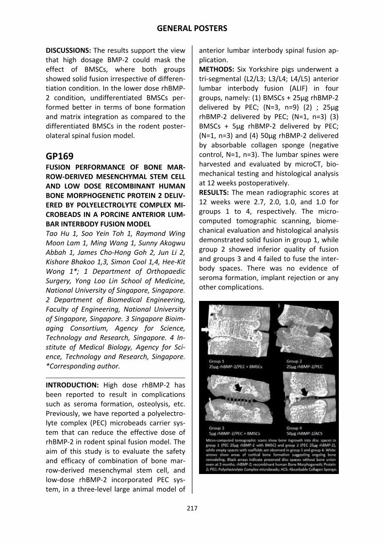
GENERAL POSTERS
217
DISCUSSIONS: The results support the view that high dosage BMP‐2 could mask the effect of BMSCs, where both groups showed solid fusion irrespective of differen‐tiation condition. In the lower dose rhBMP‐2 condition, undifferentiated BMSCs per‐formed better in terms of bone formation and matrix integration as compared to the differentiated BMSCs in the rodent poster‐olateral spinal fusion model.
GP169 FUSION PERFORMANCE OF BONE MAR‐ROW‐DERIVED MESENCHYMAL STEM CELL AND LOW DOSE RECOMBINANT HUMAN BONE MORPHOGENETIC PROTEIN 2 DELIV‐ERED BY POLYELECTROLYTE COMPLEX MI‐CROBEADS IN A PORCINE ANTERIOR LUM‐BAR INTERBODY FUSION MODEL Tao Hu 1, Soo Yein Toh 1, Raymond Wing Moon Lam 1, Ming Wang 1, Sunny Akogwu Abbah 1, James Cho‐Hong Goh 2, Jun Li 2, Kishore Bhakoo 1,3, Simon Cool 1,4, Hee‐Kit Wong 1*; 1 Department of Orthopaedic Surgery, Yong Loo Lin School of Medicine, National University of Singapore, Singapore. 2 Department of Biomedical Engineering, Faculty of Engineering, National University of Singapore, Singapore. 3 Singapore Bioim‐aging Consortium, Agency for Science, Technology and Research, Singapore. 4 In‐stitute of Medical Biology, Agency for Sci‐ence, Technology and Research, Singapore. *Corresponding author.
INTRODUCTION: High dose rhBMP‐2 has been reported to result in complications such as seroma formation, osteolysis, etc. Previously, we have reported a polyelectro‐lyte complex (PEC) microbeads carrier sys‐tem that can reduce the effective dose of rhBMP‐2 in rodent spinal fusion model. The aim of this study is to evaluate the safety and efficacy of combination of bone mar‐row‐derived mesenchymal stem cell, and low‐dose rhBMP‐2 incorporated PEC sys‐tem, in a three‐level large animal model of
anterior lumbar interbody spinal fusion ap‐plication. METHODS: Six Yorkshire pigs underwent a tri‐segmental (L2/L3; L3/L4; L4/L5) anterior lumbar interbody fusion (ALIF) in four groups, namely: (1) BMSCs + 25µg rhBMP‐2 delivered by PEC; (N=3, n=9) (2) ; 25µg rhBMP‐2 delivered by PEC; (N=1, n=3) (3) BMSCs + 5µg rhBMP‐2 delivered by PEC; (N=1, n=3) and (4) 50µg rhBMP‐2 delivered by absorbable collagen sponge (negative control, N=1, n=3). The lumbar spines were harvested and evaluated by microCT, bio‐mechanical testing and histological analysis at 12 weeks postoperatively. RESULTS: The mean radiographic scores at 12 weeks were 2.7, 2.0, 1.0, and 1.0 for groups 1 to 4, respectively. The micro‐computed tomographic scanning, biome‐chanical evaluation and histological analysis demonstrated solid fusion in group 1, while group 2 showed inferior quality of fusion and groups 3 and 4 failed to fuse the inter‐body spaces. There was no evidence of seroma formation, implant rejection or any other complications.

GENERAL POSTERS
218
DISCUSSIONS: The results of this study sug‐gest that combination of the BMSCs and low dose rhBMP‐2 delivered by PEC could be used as a bone graft substitute in this large animal ALIF model, and may be a via‐ble alternative to conventional autograft bone.
GP170 COMPARISON OF RADIOLOGICAL AND CLINICAL RESULTS OF BALLOON KYPHO‐PLASTY ACCORDING TO ANTERIOR HEIGHT LOSS IN THE OSTEOPOROTIC VERTEBRAL FRACTURE Jae Hyup Lee, MD,1 Dong‐Oh Lee, MD,1 Ji‐Ho Lee, MD,1 Hyeong‐Seok Lee, MD,2 Bong‐Soon Chang, MD,1 Choon‐Ki Lee, MD,1; 1Department of Orthopedic Surgery, College of Medicine, Seoul National University, Seoul, 156‐707, Korea 2Department of Or‐thopedic Surgery, Marynoll Medical Center, Busan, 600‐730, Korea
INTRODUCTION: The purpose of this study was to compare the vertebral height reduc‐tion rate, kyphotic angle and clinical results according to the degree of anterior verte‐bral height loss, and to determine the feasi‐bility and effects of kyphoplasty on severely collapsed osteoporotic vertebral fracture. METHODS: A total of 129 patients (145 ver‐tebrae) who had kyphoplasty due to osteo‐porotic vertebral fracture and were fol‐lowed up for more than 1 year were re‐cruited for analysis. The patients’ kyphotic angle, anterior vertebral height and anterior height reduction ratio 1 year after surgery were compared. Pain around the fractured vertebra and the radiological and clinical results according to bone mineral density were also compared. Patients were divided into three groups according to anterior ver‐tebral compression rate; compression rate exceeding 70% were placed in Group I, compression rate of 50~70% in Group II, and compression rate of 30~50% or less in Group III.
RESULTS: Group I had a higher vertebral height reduction rate immediately after surgery compared to the other groups, but the reduction rate of vertebral height in Group I noticeably decreased with time. All three groups showed a significant reduction in the anterior height between pre‐ and post‐operation. The anterior height 1 year after surgery did not differ between Group I and II, but Group III showed a significantly higher anterior height compared to the other groups. Pain around the fractured vertebra at 1 year after surgery did not dif‐fer significantly between groups. DISCUSSION: Kyphoplasty on patients with an anterior vertebral compression rate ex‐ceeding 70% due to osteoporotic vertebral fracture significantly improved the degree of pain, anterior vertebral height reduction and kyphotic angle correction, with no dif‐ferences in these parameters being found compared with other groups. Therefore, kyphoplasty can be a useful approach in patients with an anterior vertebral com‐pression rate exceeding 70%.
GP171 MRI EVALUATION OF DURAL SAC EN‐LARGEMENT BY INTERSPINOUS SPACERS (X‐STOP): DOES LIMITING THE SPINAL EX‐TENSION CHANGE THE GLOBAL SAGITTAL ALIGNMENT? Akazawa T., Kotani T., Sakuma T., Nemoto T., Nawata K., Yamazaki A., Minami S., Ohtori S., Takahashi Ka.; Dept. Orthopedic Surgery, Seirei Sakura Citizen Hospital, Sa‐kura; Dept. Orthopedic Surgery, Graduate School of Medicine, Chiba University, Chiba, Japan.
INTRODUCTION: This study aimed to an‐swer the following question regarding sur‐gery using interspinous spacers (X‐STOP). 1. Does the dural sac enlarge? 2. Does flexing the affected spine have an effect on the global alignment? 3. Do clinical symptoms improve?

GENERAL POSTERS
219
METHODS: The subjects were 11 patients who had lumbar spinal stenosis and had undergone surgery using interspinous spac‐ers. There were 6 men and 5 women and their mean age was 72.6 years (range: 63‐82 years). The evaluation of pre‐ and post‐operative imaging involved MRI, plain x‐rays. The VAS scores were examined. Three‐dimensional MRI was used for evaluation, and the area of the dural sac was measured using a cross‐sectional image of the superi‐or endplate of the lower vertebral body of the affected spine. The plain x‐ray of the whole spine was to measure t thoracic ky‐phosis (TK), lumbar lordosis (LL), pelvic pa‐rameters, affected intervertebral angle, disc height, and percentage of slip. RESULTS: MRI showed that the pre‐ and postoperative areas of the dural sac were 54.3±23.6 mm2 and 79.2±26.7 mm2, re‐spectively, indicating a significant enlarge‐ment (p<0.001). There was no significant difference between the pre‐ and postopera‐tive flexion disc angles, but the extension disc angles were 9.4o and 6.2o, respectively (p=0.002). There was no significant differ‐ence in pre‐ and postoperative TK, LL, PT, SS, or SVA. The pre‐ and postoperative in‐tervertebral angles were 4.7o and 3.3o, respectively, indicating a decrease (p=0.219). The VAS scores were significantly improved postoperatively: low back pain (preoperative: 4.6 and postoperative: 2.2, p=0.024), buttock and lower leg pain (7.3 and 1.7, p<0.001), and buttock and lower leg numbness (5.4 and 1.8, p=0.043). DISCUSSION: MRI showed that the dural tube enlarged 1.46 fold, and indirect de‐compression was achieved using the inter‐spinous spacer. The disc angle in a standing position was slightly decreased postopera‐tively, but the global sagittal alignment was not affected.
GP172 THE CLINICAL OUTCOMES AND FEATURES AFTER MICROENDOSCOPIC LAMINOTOMY FOR ELDER PATIENTS WITH LUMBAR SPI‐NAL STENOSIS. Hiroyuki Yasuda, Hiromitsu Toyoda, Sho Dohzono, Tomiya Matsumoto, Hidetomi Terai, Akinobu Suzuki, Kazuhiro Shinohara, Kohji Tamai, Hiroaki Nakamura; Depart‐ment of Orthopedic Surgery, Osaka City University Graduate School of Medicine
INTRODUCTION: With increased aging of the population, spine surgeons have more opportunity to treat elder patients for lum‐bar spinal stenosis (LSS). The purpose of this study was to clarify the clinical features and to evaluate clinical outcomes after micro‐endoscopic laminotomy (MEL) with LSS in the elder patients aged 75 years or older. MATERIALS AND METHODS: We retrospec‐tively reviewed 93 consecutive patients with LSS who underwent MEL. We excluded patients with other conditions that could affect functional status and finally 81 pa‐tients (47 male and 41 female), aged 68.5 ± 10.2 years, were analyzed in this study. The number of single level decompression was 72, two levels was 8 and three levels was 1. Follow up period was 18.8 ± 8.8 months. JOA score (0‐29), visual analog scale (VAS) for low back pain/leg pain/ leg numbness were evaluated before surgery and at 3, 6, 12 months after the surgery and the final follow up. Patients were divided into three groups according to age at the time of sur‐gery, group A : less than 65 years, group B : from 65 years to 74 years, group C : 75 years or more. The data was analyzed and compared among three groups. RESULTS: JOA score at 3 and 6 months after surgery in group B and C were significantly lower than that in group A (at 3M: 26.5/ 24.2 / 23.2, at 6M: 27.2/ 25.0/ 25.5,) and the clinical courses of VAS for low back pain/ leg pain were similar. There were no significant differences in VAS for leg numb‐
GENERAL POSTERS
220
ness among the 3 group at any time during follow up. At the final follow‐up, JOA score, VAS for low back pain/ leg numbness were not significantly different among the 3 groups, but VAS for leg pain in group C was higher than that in group A. CONCLUSION: After MEL for LSS, improve‐ment of clinical outcomes such as JOA score and VAS was poor at early postoperative period in elder patient. However, JOA score at the final follow up was almost similar among the 3 groups. Thus, elderly patients could obtain reasonable recovery after MEL.
GP173 SPINAL OSTEOTOMY AND CORRECTION USING SUK DVR SYSTEM FOR ADULT SAG‐ITTAL PLANE DEFORMITY Masahiro Kanayama, MD* Motoya Ikawa, MD** Tomoyuki Hashimoto, MD* Keiichi Shigenobu, MD* Fumihiro Oha, MD* Akira Iwata, MD* Shingo Onda, MD* Masaru Tanaka, MD* Hiromi Tsunekawa, MD**; * Spine Center, Hakodate Central General Hospital, Hakodate, Japan ** Dept. of Or‐thopaedic Surgery, Toyooka Central Hospi‐tal, Asahikawa, Japan
INTRODUCTION: SUK DVR system was de‐veloped for vertebral derotation maneuver in scoliosis surgery. We applied this device for sagittal‐plane correction in adult spinal deformity. This study aims to introduce sur‐gical techniques and report radiographic and clinical results. METHODS: Thirteen patients who under‐went spinal osteotomy (PSO in 11; PVCR in 2) using SUK DVR system for adult sagittal plane deformity were reviewed. Surgical procedures included (1) preparing secure proximal and distal vertebral anchors using multisegmental pedicle screw‐rod fixation, (2) decancellating only posterior two‐thirds of vertebral body and leaving anterior one‐third for anterior column support, and (3) closing the osteotomy by approximation of SUK tubes secured to the proximal‐ and
distal‐endmost screws. Radiographic pa‐rameters included anterior deviation of C7 plumb line and kyphosis correction. Clinical outcomes were evaluated by VAS of back pain and RDQ. Mean follow‐up period was 14 months (5‐24 months). RESULTS: Distal fixation ended at L4 in six, L5 in six, and L6 in one patient; no patients required fixation to the sacrum and/or pel‐vis. Kyphosis angle improved from +24 de‐grees to ‐21 degrees; mean correction was 45 degrees. Anterior deviation of C7 plumb line was 12.1 cm before surgery, 4.1 cm after surgery, and 5.8 cm at the final follow‐up. VAS of back pain improves from 68/100 to 21/100. RDQ were 17.3 before surgery, and improved to 9.3 at the final follow‐up. DISCUSSION: The current procedures have biomechanical advantages and resulted in favorable clinical outcomes. SUK DVR sys‐tem can provide and control a powerful correction force as intended. Multisegmen‐tal pedicle screw anchoring can maximize the correction force in patients with osteo‐porosis and poor bone stock.
GP174 COMPARISON OF PERI‐OPERATIVE OUT‐COMES OF LUMBAR FUSION: ORTHOPAE‐DIC SURGERY VERSUS NEUROSURGERY Alejandro Marquez‐Lara MD, Sreeharsha V. Nandyala BA, Steven J. Fineberg MD, Kern Singh MD; Department of Orthopaedic Sur‐gery, Rush University Medical Center, Chi‐cago, IL, USA
INTRODUCTION: The influence of the train‐ing specialty on the outcomes associated with spine surgery is not well characterized. The purpose of this study was to compare the peri‐operative outcomes in lumbar fu‐sions (LF) between Orthopaedic surgeons and Neurosurgeons. METHODS: The National Surgical Quality Improvement Program (NSQIP) database was searched to identify patients undergo‐ing a LF for degenerative pathologies be‐

GENERAL POSTERS
221
tween 2006 and 2011. The selected cohort was divided based upon the primary sur‐geon's specialty (Orthopaedic surgery or Neurosurgery). Preoperative patient charac‐teristics and peri‐operative outcomes were assessed. SPSS v.20 was utilized for statisti‐cal analysis with a p‐value of = 0.05 to de‐note significance. RESULTS: A total of 7,909 LF were identified from 2006‐2011, of which 3,887 (49.1%) were performed by Orthopaedic surgeons and 4,022 (50.9%) by Neurosurgeons. There were significant differences in patient comorbidities between Orthopaedic and Neurosurgery patients. The Neurosurgery cohort more often underwent ambulatory procedures and demonstrated a longer op‐erative time. However, Orthopaedic pa‐tients incurred a longer hospital stay than Neurosurgery patients. In addition, there were significant differences in the incidence and type of postoperative complications between specialties. However, the mortality rate did not significantly differ between groups.
DISCUSSION: The results in this study demonstrate distinct patient profiles and
peri‐operative events associated with each spine surgery specialty. Despite these dif‐ferences, the mortality rate did not differ between the surgical cohorts. In light of these findings, further studies are warrant‐ed to better characterize the risk factors associated with each specialty and their impact in patient outcomes and hospital resource utilization after LF procedures.
GP175 IMPACT OF RESIDENT INVOLVEMENT ON COMPLICATIONS FOLLOWING LUMBAR DECOMPRESSION SURGERY Alejandro Marquez‐Lara MD, Sreeharsha V. Nandyala BA, Steven J. Fineberg MD, Bryan D. Haughom MD, Kern Singh MD; Depart‐ment of Orthopaedic Surgery, Rush Universi‐ty Medical Center, Chicago, IL, USA
INTRODUCTION: Although fundamental to the practice of medicine, the need to train the next generation of surgeons may com‐promise the quality of care delivered to patients. The goal of this study was to char‐acterize the impact of resident involvement on the rates of complications following lumbar decompression (LD) surgery. METHODS: The National Surgical Quality Improvement Program (NSQIP) database was searched to identify patients undergo‐ing a LD for degenerative conditions be‐tween 2006 and 2011. Patients were divid‐ed into two cohorts based upon resident participation during the surgical procedure. Preoperative patient characteristics and peri‐operative outcomes were assessed. SPSS v.20 was utilized for statistical analysis with a p‐value of = 0.05 to denote signifi‐cance. RESULTS: A total of 11,055 LDs were identi‐fied between 2006 and 2011, including 3,339 (30%) cases with resident involve‐ment. The resident cohort demonstrated a lower BMI and a higher incidence of alcohol abuse than those cases without resident involvement. There was a higher rate of

GENERAL POSTERS
222
clean‐contaminated and contaminated sur‐gical wounds in those cases with resident involvement. Cases with resident involve‐ment were associated with a longer opera‐tive time and hospitalization, a greater inci‐dence of postoperative complications, and greater re‐operation rates. There were no differences in mortality between the two cohorts. DISCUSSION: Thirty‐day outcomes data indicated higher rates of re‐operation, and postoperative complications in those cases with resident involvement. Differences in preoperative wound class and a longer op‐erative time may help explain these differ‐ences. Despite these findings, there were no differences in mortality between the two cohorts. In light of these findings, further analysis is warranted in order to determine preoperative measures that should be un‐dertaken to help mitigate the peri‐operative complications associated with resident in‐volvement during a LD procedure.
GP176 INCIDENCE, PREDICTORS AND OUTCOMES OF PNEUMONIA AFTER LUMBAR SPINE SURGERY Alejandro Marquez‐Lara MD, Sreeharsha V. Nandyala BA, Steven J. Fineberg MD, Kern Singh MD; Department of Orthopaedic Sur‐gery, Rush University Medical Center, Chi‐cago, IL, USA
INTRODUCTION: Postoperative pneumonia (PNA) is a frequent complication following orthopaedic procedures. However, there is limited data in the published literature re‐garding the peri‐operative risk factors asso‐ciated with PNA after lumbar spine surgery (LSS). METHODS: Patients who underwent LSS were identified in the National Surgical Quality Improvement Program (NSQIP) da‐tabase. Patients with postoperative PNA were identified and compared to unaffect‐ed patients. Patient characteristics, lab val‐ues, and perioperative outcomes were as‐sessed. Regression analysis was performed to identify predictors for postoperative PNA with a 95% confidence interval. SPSS v.20 was utilized for statistical analysis with a p‐value of = 0.05 to denote significance. RESULTS: A total of 22,676 LSS were identi‐fied between 2006‐2011, of which 85 (0.4%) incurred postoperative PNA. Affected pa‐tients were significantly older, and demon‐strated a higher BMI and a greater number of comorbidities. The PNA cohort under‐went a greater proportion of non‐ambulatory surgeries and demonstrated an increased number of blood transfusions and a longer operative time. In addition, affect‐ed patients demonstrated a longer hospital‐ization, greater readmission and reopera‐tion rates, and a higher incidence of post‐operative complications. Risk factors asso‐ciated with postoperative PNA included obesity, chronic obstructive pulmonary dis‐ease (COPD), peripheral vascular disease (PVD), and a longer operative time.

GENERAL POSTERS
223
DISCUSSION: In this study postoperative PNA was associated with a greater inci‐dence of perioperative complications, and longer hospitalization. In addition, multiple risk factors including obesity COPD, PVD and a longer operative time were associat‐ed with an increased risk of postoperative PNA. In light of these findings, further re‐search is warranted to determine if optimi‐zation of these perioperative risk factors can mitigate the impact of PNA on hospital resource utilization and postoperative out‐comes.
GP177 INCIDENCE, PREDICTORS AND OUTCOMES OF PULMONARY EMBOLISM AFTER LUM‐BAR SPINE SURGERY Alejandro Marquez‐Lara MD, Sreeharsha V. Nandyala BA, Steven J. Fineberg MD, Kern Singh MD; Department of Orthopaedic Sur‐gery, Rush University Medical Center, Chi‐cago, IL, USA
INTRODUCTION: Postoperative pulmonary embolism (PE) is a well‐known life‐threatening complication following ortho‐paedic procedures. However, there is lim‐
ited data on the incidence, risk factors, and peri‐operative outcomes for PE following lumbar spine surgery (LSS). METHODS: The National Surgical Quality Improvement Program (NSQIP) database was utilized to identify patients who un‐derwent elective LSS. Patients with a PE were compared to unaffected patients. Preoperative patient characteristics, lab values, and peri‐operative outcomes were assessed. Regression analysis was per‐formed to identify predictors for a postop‐erative PE with a 95% confidence interval. SPSS v.20 was utilized for statistical analysis with a p‐value of = 0.05 to denote signifi‐cance.
RESULTS: A total of 22,610 LSS were identi‐fied between 2006‐2011, of which 79 (0.3%) demonstrated a postoperative PE. The af‐fected cohort was older, and demonstrated a higher BMI and a greater number of comorbidities. PE patients underwent a greater proportion of non‐ambulatory sur‐geries and incurred a longer operative time than non‐PE patients. In addition, affected patients incurred a longer hospitalization, greater readmission rates, and a higher in‐cidence of postoperative deep vein throm‐

GENERAL POSTERS
224
bosis and mortality than unaffected pa‐tients. Risk factors associated with a post‐operative PE included obesity, elevated white blood cell count (WBC), and pro‐longed operative time. Interestingly, smok‐ing was associated with a reduced risk for a postoperative PE. DISCUSSION: This study demonstrated that patients with a postoperative PE incurred a longer hospitalization, a higher readmission rate, and greater mortality than unaffected patients. Furthermore, risk factors associat‐ed with a postoperative PE included obesi‐ty, elevated WBC and a longer operative time. Understanding the incidence and risk factors associated with a PE may assist in the prevention, diagnosis, and treatment of this potentially fatal complication.
GP178 OUTCOMES AFTER LUMBAR DECOMPRES‐SION OR FUSION IN PATIENTS WITH AN ELEVATED WHITE BLOOD CELL COUNT (>10K) Alejandro Marquez‐Lara MD, Sreeharsha V. Nandyala BA, Steven J. Fineberg MD, Kern Singh MD; Department of Orthopaedic Sur‐gery, Rush University Medical Center, Chi‐cago, IL, USA
INTRODUCTION: Patients undergoing lum‐bar spine surgery (LSS) may present with an elevated white blood cell (WBC) count. However, little has been reported on the effects of this abnormal lab value in pa‐tients undergoing elective LSS. METHODS: The National Surgical Quality Improvement Program (NSQIP) database was queried to select patients who under‐went LSS between 2006‐2011. Patients with an elevated WBC count (>10,000 cell/µL) were identified. Preoperative patient char‐acteristics and peri‐operative outcomes were assessed. Regression analysis, with a 95% confidence interval, was performed to determine the association between an ele‐vated WBC and postoperative complications
after controlling for demographics and comorbidities. SPSS v.20 was utilized for statistical analysis with a p‐value of = 0.001 to denote significance. RESULTS: A total of 20,188 LSS were identi‐fied of which 2,684 (13.3%) demonstrated an elevated WBC count. Patients with leu‐kocytosis were older and demonstrated a greater number of comorbidities. Patients with an elevated WBC count more often underwent outpatient procedures, and demonstrated fewer intraoperative blood transfusions and shorter operative times. There were no significant differences in the hospital length of stay or postoperative complications between the two cohorts. Regression analysis demonstrated that an elevated WBC count was not associated with a greater risk of complication or mor‐tality.
DISCUSSION: Despite an older and more comorbid patient population in patients with leukocytosis, the peri‐operative out‐comes did not significantly differ between the two cohorts. In fact, an elevated WBC count demonstrated favorable intraopera‐tive parameters. The results in this study suggest that an elevated WBC does not sig‐nificantly affect outcomes after LSS and

GENERAL POSTERS
225
may even contribute to a faster operative time and recovery. Further research is war‐ranted to better characterize the impact of an elevated WBC count in LSS.
GP179 OUTCOMES AFTER LUMBAR SPINE SUR‐GERY IN PATIENTS WITH AN ELEVATED CREATININE Alejandro Marquez‐Lara MD, Sreeharsha V. Nandyala BA, Steven J. Fineberg MD, Kern Singh MD; Department of Orthopaedic Sur‐gery, Rush University Medical Center, Chi‐cago, IL, USA
INTRODUCTION: Routine preoperative blood testing may reveal an elevated creat‐inine (Cr) level. However, the impact of this abnormal lab value on peri‐operative out‐comes in lumbar spine surgery (LSS) is not well characterized. METHODS: The National Surgical Quality Improvement Program (NSQIP) database was queried between 2006‐2011. Patients undergoing LSS were identified and divided based upon the preoperative Cr level (Males >1.3mg/dL, Females >1.1mg/dL). Preoperative patient characteristics and peri‐operative outcomes were assessed. Regression analysis, with a 95% confidence interval, was performed to determine the association between an elevated Cr and postoperative complications after control‐ling for demographics and comorbidities. SPSS v.20 was utilized for statistical analysis with a p‐value of = 0.001 to denote signifi‐cance. RESULTS: A total of 18,647 LSS were identi‐fied from 2006‐2011, of which 1,656 (8.9%) demonstrated an elevated Cr. These pa‐tients were significantly older and demon‐strated a greater number of comorbidities than the control group. In addition, patients with an elevated Cr underwent fewer out‐patient procedures, demonstrated a greater number of blood transfusions, and incurred a longer operative time. In addition, a high
Cr was associated with a longer hospitaliza‐tion and a greater incidence of postopera‐tive complications. However, regression analysis demonstrated that an elevated Cr was not associated with a greater risk of postoperative complications. DISCUSSION: Patients with an elevated Cr demonstrated a greater incidence of post‐operative complications and incurred a longer hospitalization. However, after con‐trolling for demographics and comorbidi‐ties, an elevated Cr was not associated with a greater risk of postoperative complica‐tions. In view of these results, further inves‐tigation is warranted to clarify the impact of an elevated Cr level on peri‐operative out‐comes in LSS.
GP180 OUTCOMES AFTER LUMBAR SPINE SUR‐GERY IN PATIENTS WITH PREOPERATIVE ANEMIA Alejandro Marquez‐Lara MD, Sreeharsha V. Nandyala BA, Steven J. Fineberg MD, Kern Singh MD; Department of Orthopaedic Sur‐gery, Rush University Medical Center, Chi‐cago, IL, USA

GENERAL POSTERS
226
INTRODUCTION: Anemia is thought to in‐crease the risk for postoperative complica‐tions after spine surgery. However, there is limited published data describing the im‐pact of a low hematocrit (Hct) in patients undergoing lumbar spine surgery (LSS). METHODS: The National Surgical Quality Improvement Program (NSQIP) database was queried between 2006‐2011. Patients who underwent LSS were selected and di‐vided into cohorts based upon the preoper‐ative Hct level (Males: Hct <40.6g/dL, Fe‐males: <36.1g/dL). Patient characteristics and peri‐operative outcomes were as‐sessed. Regression analysis, with a 95% con‐fidence interval, was performed to deter‐mine the association between anemia and postoperative complications after control‐ling for demographics and comorbidities. SPSS v.20 was utilized for statistical analysis with a p‐value of = 0.001 to denote signifi‐cance.
RESULTS: A total of 20,280 LSS cases were identified between 2006‐2011 of which 4,522 (22.3%) demonstrated a low Hct. Anemic patients were significantly older and demonstrated a greater number of
comorbidities. In addition, patients with anemia underwent fewer outpatient proce‐dures, and demonstrated a greater number of blood transfusions, and a longer opera‐tive time. Anemic patient incurred greater postoperative complications, readmission rates and incurred a longer hospitalization. Regression analysis demonstrated that a preoperative anemia was associated with a greater risk for postoperative blood trans‐fusions. DISCUSSION: Anemic patients undergoing LSS demonstrated a greater comorbidity burden, longer operative time, and greater intraoperative blood transfusions, which likely influenced the greater postoperative complication rates and longer hospitaliza‐tion. The results of this study demonstrate a logical association between preoperative anemia and peri‐operative blood transfu‐sions. However, further research is war‐ranted to better characterize the associa‐tion between anemia and the peri‐operative outcomes in LSS.
GP181 OUTCOMES AFTER SINGLE‐LEVEL LUMBAR DECOMPRESSION OR FUSION IN PATIENTS WITH HYPERTENSION Alejandro Marquez‐Lara MD, Sreeharsha V. Nandyala BA, Steven J. Fineberg MD, Kern Singh MD; Department of Orthopaedic Sur‐gery, Rush University Medical Center, Chi‐cago, IL, USA
INTRODUCTION: Hypertension (HTN) is common among patients undergoing lum‐bar spine surgery (LSS). However, there is limited data in the literature describing the impact of HTN on the peri‐operative out‐comes following elective LSS. METHODS: The National Surgical Quality Improvement Program (NSQIP) database was queried between 2006‐2011. Patients who underwent a single‐level lumbar fusion (LF) or lumbar decompression (LD) were identified. Patients with a history of HTN

GENERAL POSTERS
227
were compared to normotensive patients. Preoperative patient characteristics, lab values, and perioperative outcomes were assessed. Regression analysis was per‐formed, with a 95% confidence interval, to identify the association between HTN and postoperative complications after control‐ling for demographics and comorbidities. SPSS v.20 was utilized for statistical analysis with a p‐value of = 0.001 to denote signifi‐cance. RESULTS: A total of 17,283 single‐level LD and LF cases were identified of which 45.9% had HTN. These patients were significantly older and demonstrated a higher number of comorbidities than normotensive patients. Hypertensive patients underwent fewer outpatient procedures, and demonstrated greater intraoperative blood transfusions and longer operative times. In addition HTN was associated with longer hospital stay and a greater incidence of postoperative complications. Regression analysis demon‐strated a strong association between HTN and organ space infections.
DISCUSSION: This study demonstrated that HTN was associated with a greater number of comorbidities and longer operative
times, which likely influenced the longer hospitalization and greater incidence of postoperative complications. The results in this study suggest that HTN significantly impacts postoperative outcomes after LSS. However, the various associated comorbidi‐ties in patients with HTN warrants further studies to minimize possible confounding factors with aims to reduce complications and improve outcomes in LSS.
GP182 RISK FACTORS ASSOCIATED WITH SURGI‐CAL SITE INFECTIONS FOLLOWING LUMBAR SPINE SURGERY FOR DEGENERATIVE PA‐THOLOGIES Alejandro Marquez‐Lara MD, Sreeharsha V. Nandyala BA, Steven J. Fineberg MD, Kern Singh MD; Department of Orthopaedic Sur‐gery, Rush University Medical Center
INTRODUCTION: A surgical site infection (SSI) is one of the most common complica‐tions after orthopaedic procedures. Howev‐er, the incidence as well as the risk factors associated with a SSI after lumbar spine surgery (LSS) has been poorly reported. METHODS: The National Surgical Quality Improvement Program (NSQIP) database was queried to identify patients who un‐derwent elective LSS between 2006‐2011. The resulting cohort was divided between patients who had a SSI and those who did not. Preoperative patient characteristics and peri‐operative outcomes were as‐sessed. Regression analysis, with a 95% con‐fidence interval, was performed to deter‐mine the risks associated with SSI. Statistical analysis was performed with SPSS v.20 and a p‐value of <0.001 denoted significance. RESULTS: A total of 22,676 lumbar spine surgeries were identified from 2006‐2011, of which 462 (2.0%) developed a postopera‐tive SSI. There were no significant differ‐ences in patient age between cohorts, how‐ever infected patients demonstrated a greater comorbidity burden. Wound com‐

GENERAL POSTERS
228
plications were also associated with longer operative times, greater resident involve‐ment and greater intraoperative blood transfusions. In addition, the affected co‐hort demonstrated a longer hospitalization and incurred greater rates of readmissions, reoperations and sepsis. Regression analysis demonstrated that obesity, diabetes, dialy‐sis, steroid use, bleeding disorders, resident involvement, and increased operative times were associated with an increased risk of SSI.
DISCUSSION: This study demonstrated that SSI are associated with greater peri‐operative complications and hospital re‐source utilization. In addition a number of risk factors were identified to be strongly associated with a SSI. The development of peri‐operative protocols based upon these identified risk factors may help mitigate the incidence of SSI and the impact of this oc‐currence on peri‐operative outcomes and hospital resource utilization.
GP183 SENTINEL EVENTS IN LUMBAR SPINE SUR‐GERY Alejandro Marquez‐Lara MD, Sreeharsha V. Nandyala BA, Hamid Hassanzadeh, MD, Kern Singh MD; Department of Orthopaedic Surgery, Rush University Medical Center, Chicago, IL, USA
INTRODUCTION: Sentinel events in lumbar spine surgery can have significant medical, social, economic, and legal implications. The incidence and peri‐operative outcomes as‐sociated with these complications have not been well characterized in the published spine literature. METHODS: The Nationwide Inpatient Sam‐ple was queried from 2002‐2011. Patients who underwent elective lumbar spinal sur‐gery were identified. Sentinel events includ‐ing bowel or peritoneal injury, vascular inju‐ry, nerve injury, retention of foreign ob‐jects, and wrong‐site surgery were identi‐fied. Patient demographics, comorbidities (CCI), number of fusion levels, length of stay, total costs, and postoperative out‐comes were assessed. The risk for in‐hospital mortality associated with each complication was calculated utilizing a 95% confidence interval (CI). Statistical analysis was performed with SPSS v.20 and a p‐value of <0.001 denoted significance. RESULTS: A total of 543,146 lumbar spine surgeries were recorded from 2002‐2011 of which 414 (0.8 per 1,000 cases) incurred sentinel events. There were no significant differences in the mean age or CCI between the two cohorts. However, affected patients more often underwent 3+ level fusion pro‐cedures. The sentinel events cohort in‐curred a longer hospitalization, greater costs, and a greater incidence of in‐hospital complications. Patients with a bowel or per‐itoneal injury, vascular injury, and wrong‐site surgery demonstrated a greater risk of

GENERAL POSTERS
229
mortality relative to unaffected patients.
DISCUSSION: This NIS analysis demon‐strates that sentinel events are associated with a significant increase in hospital re‐source utilization and worsened postopera‐tive outcomes including death. This study demonstrates the financial and medical burden associated with peri‐operative com‐plications. Further research is warranted to develop strategies to prevent and mitigate the impact of these events in both short and long‐term outcomes.
GP184 PERIOPERATVE COMPLICATIONS OF LUM‐BAR SPINE SURGERY IN PATIENTS OVER 85 YEARS. 1 Shingo Onda, Masahiro Kanayama, Tomoyuki Hashimoto, Keiichi Shigenobu, Fumihiro Ooha, Akira Iwata, Masaru TanakaC 2 Kazuo Kaneko; 1 Spine Center, Hakodate Central General Hospital 2 De‐partment of Orthopedics Surgery, Juntendo Medical University
INTRODUCTION: We investigated the rate of perioperative complications in decom‐pression surgery versus instrumented fu‐sion over‐85‐year population. METHODS: Of 907 patients who underwent lumbar spine surgery for degenerative dis‐orders from January 2006 to June 2012, 33 patients (3.6% of the entire population) were over 85 years (85 to 94 years). Surgical indications were determined solely based on the pathology regardless of age. Spinal stenosis without instability was treated by laminoplasty; instrumented fusion was indi‐cated in the case of neuroforaminal decom‐pression and/or spinal instability. With re‐spect to the type of surgery, we investigat‐ed co‐morbidities, ASA classification, peri‐operative complications, and JOA score. RESULTS: 19 patients underwent posterior decompression without fusion. Instrument‐ed fusion was performed in 14 patients (PLIF in 6, TLIF in 4 and PLF in 4). Co‐morbidities were hypertension in 52%, dia‐betes in 39%, heart disease in 36%, old cer‐ebral infarction in 15%, and history of ma‐lignant tumor in 12%. All the patients were classified as ASA 2 or less. Overall perioper‐ative complication rate was 24%: the rate was 21% in decompression group (deep infection, dural tear, GI tract complication, death due to cerebral infarction in one each) and 28.5% in the fusion group (deep infection in 1, implant dislodgement in 2, GI tract complication in 1). The rates of peri‐operative complications were not statisti‐cally different between the two groups. Both group showed significant improve‐ment in JOA score. DISCUSSION: In the treatment of lumbar degenerative disorders in elderly patients, surgeons favor less invasive decompression over instrumentation surgery. The current study showed that the rate of perioperative complication was comparable between de‐compression alone and instrumented fu‐sion. If the pathology requires spinal fusion, surgeons should not hesitate to perform
GENERAL POSTERS
230
instrumentation even in the patients over 85 years.
GP185 SURGICAL DEBRIDEMENT WITH RETEN‐TION OF SPINAL INSTRUMENTATION AND LONG‐TERM ANTIMICROBIAL THERAPY FOR MULTIDRUG RESISTANT SURGICAL SITE INFECTIONS AFTER SPINAL SURGERY: A CASE SERIES Miyazaki, S; Kakutani, K; Maeno, K; Takada, T; Hirata, H; Kurakawa, T; Terashima, Y; Kurosaka, M; Nishida, K; Department of Orthopaedic Surgery, Kobe University Grad‐uate School of Medicine, Kobe, Japan
INTRODUCTION: Multidrug resistant surgi‐cal site infection (SSI) is one of the most significant complication after instrumented spinal surgery. However, there is no con‐sensus on treatment strategies, in particu‐lar, surgical procedures, whether retention or removal of instrumentation, are still con‐troversial. In this study, we present our ex‐perience with the debridement with reten‐tion of instrumentation and long‐term an‐timicrobial therapy (more than 3 months) for multidrug resistant SSI. METHODS: This is a retrospective case se‐ries of the clinical results of 12 cases (8 men and 4 women, 69.6 years) with multidrug resistant SSI occurred in 409 consecutive cases in which spinal instrumentation was used between 2007 and 2013. The microbi‐al pathogen included 8 cases of methicillin‐resistant staphylococcus aureus, 2 cases of multiple‐drug resistant corynebacterium, 1 case of methicillin‐resistant coagulase nega‐tive staphylococcus aureus, and 1 case of methicillin‐resistant staphylococcus epi‐dermidis. RESULTS: Eleven patients with SSI were di‐agnosed within 90 days postoperatively and 1 patient on day 171. The mean time from the diagnosis to surgery was 2.9 days (range1‐6). Ten patients underwent surgical debridement with implant retention and 2
patients didn’t need any surgery. Further‐more, no patients required multiple opera‐tions. All patients were given antimicrobial treatment. Intravenous antimicrobials (VCM, VCM+TAZ, or GM) were administrat‐ed for average 64.2 days (12‐352) and oral antimicrobials (RFP+SMX/TMP, SMX/TMP, or MINO) for average 318 days (89‐1673). The mean time to recover clinical presenta‐tion, such as pain relief, wound‐healing, and decline of fever, was 29.3 days (7‐55) and the mean time until C‐reactive protain lev‐els returned to normal was 56.7 days (7‐105). All SSI were controlled and never re‐cur during follow up. DISCUSSION: Multidrug resistant SSI is suc‐cessfully treated with the debridement with implant retention and long‐term antimicro‐bial therapy.
GP186 PROGNOSTIC FACTORS IN MICROENDO‐SCOPIC LAMINOTOMY FOR PATIENTS WITH LUMBAR SPINAL STENOSIS Toyoda H, Dohzono S, Matsumoto T, Terai H, Suzuki S, Yasuda H, Nakamura H.; Dept. Orthopaedic Surgery, Osaka City University Graduate School of Medicine, Osaka, Japan
INTRODUCTION: Decompressive surgery for lumbar spinal stenosis is the most frequent‐ly performed spine operation, but there have been few studies on factors that may predict outcome. We tried to find predic‐tors of outcome in microendoscopic lam‐inotomy (MEL) for lumbar spinal stenosis (LSS). MATERIALS AND METHODS: We retrospec‐tively reviewed 93 consecutive patients with LSS who underwent MEL. We excluded patients with other conditions that could affect functional status and finally 88 pa‐tients (aged 68.5 ± 10.2 years) were ana‐lyzed. Mean follow up period was 18.8 ± 8.8 months. We investigated factors predicting inferior function (JOA score was less than 25 points) and residual back and leg pain

GENERAL POSTERS
231
(VAS scores was more than 30mm) after MEL using multivariate regression analysis. RESULTS: The mean JOA score was 14.0±4.2 points preoperatively and improved to 25.2±3.5 points at the latest follow up. The mean VAS for leg pain was 59.1±28.9 pre‐operatively and improved to 13.2±22.6 at the latest follow up. The mean VAS for low back pain was 45.4±30.9 preoperatively and improved to 15.1±20.6 at the latest follow up. In multivariate analysis, sex (OR1.2), ASA physical classification system (OR2.1), posterior angulation(OR2.1)were signifi‐cantly associated with inferior function. Duration of symptoms (OR1.9), sagittal ver‐tical axis (SVA)(OR1.4)were significantly associated with residual low back pain. SVA(OR1.4)were significantly associated with residual leg pain. CONCLUSION: Multiple factors predict out‐come in MEL, most importantly ASA, poste‐rior angulation and SVA.
GP187 GOOD CLINICAL OUTCOMES AND FUSION RATE OF LESS INVASIVE FACET FUSION WITH A PERCUTANEOUS PEDICLE SCREW SYSTEM FOR DEGENERATIVE LUMBAR SPONDYLOLISTHESIS Tomohiro Miyashita *1, Hiromi Ataka *2, Kei Kato *1, Takaaki Tanno *2; *1: Spine Center, Matsudo City Hospital, Matsudo, Japan *2: Spine Center, Matsudo Orthopae‐dic Hospital, Matsudo, Japan
INTRODUCTION: Based on our long‐term clinical and radiological follow‐up study of posterolateral fusion (PLF) for degenerative lumbar spondylolisthesis (DLS), we recog‐nized that facet fusion (FF) alone would be sufficient for spinal fusion. We devised a new, simple, less invasive FF using a percu‐taneous pedicle screw (PPS) system for DLS. In this study, we assessed the fusion rate and clinical outcomes of this procedure. METHODS: Eighty‐two patients (51 women, 31 men; average age, 68.1 years), who un‐
derwent FF for single‐level DLS, were retro‐spectively reviewed after a minimum 1‐year follow‐up. The surgical method involved, making a 5cm skin incision, bilateral laminar fenestration, and FF with autologous bone harvested from the spinous process. PPS were then inserted. We evaluated the FF rate using CT, the range of motion (ROM) at the fused level on a flexion‐extension lateral X‐ray, preoperatively and at the final follow‐up, and the therapeutic effectiveness of FF using the Japanese Orthopaedic Association Back Pain Evaluation Questionnaire (JO‐ABPEQ). RESULTS: The FF rate was 85.4%. The ROM at the fused level was less than 2 degrees at the final follow‐up in all the patients with adequate FF on CT. In those (11 patients) with inadequate FF, the average ROM sig‐nificantly decreased from preoperative 13.4 degrees to postoperative 3.9 degrees. Ther‐apeutic effectiveness on the JOABPEQ was demonstrated in the Walking ability score by 92.3% of the patients and in the Low back pain score by 71.9%. DISCUSSION: Park et al reported that FF alone is an effective surgical option for DLS. However, in their method, a bone graft was harvested from the iliac crest and a conven‐tional pedicle screw system was used. We devised a less invasive FF using a local bone graft and a PPS system. The clinical out‐comes and the fusion rate of FF with the PPS system were comparable to those with conventional PLF. Thus, FF with a PPS sys‐tem is a useful technique for the manage‐ment of DLS.
GP188 SSI SURVEILLANCE FOLLOWED BY VANCO‐MYCIN POWDER PROPHYLAXIS OF HIGH‐RISK CASES TO REDUCE SSI RATES AFTER SPINAL SURGERY. Kentaro Takagi, Satoshi Ogihara, Masuda Kimio, Iwasawa Mitsuyasu, Takamure Kouji, Uchida Yoshio, Tabira Yoshihiko, Hirai Shi‐ma, Makabe Kenta, Mori Toshihito; De‐

GENERAL POSTERS
232
partment of Orthopaedic Surgery, Spine Center, Sagamihara National Hospital
INTRODUCTION: Prevention of surgical site infection (SSI) is particularly important in spinal surgery. Vancomycin powder (VP) is effective in preventing SSI before closing surgical wounds. However, VP is not pre‐ferred as a prophylactic in all cases because vancomycin is an MRSA therapeutic drug. In our hospital, we conducted prospective SSI surveillance following spinal surgery. After identifying the high‐risk group, we used VP for SSI prophylaxis in these pat METHODS: Phase 1: SSI surveillance of spi‐nal surgery began in April 2011. Factors recorded included sex, age, BMI, primary disease, ASA classification, diabetes, smok‐ing, rheumatoid arthritis (RA), steroids, op‐erative time, spinal instrumentation, amount of bleeding, and dural tear. Phase 2: based on phase 1 results, from Septem‐ber 2012 we defined a high‐risk group as patients with RA undergoing instrumenta‐tion fusion surgery, and started selective VP use in this group. VP was used with 1000mg/operation dosage after hospital IRB approval. SSI rate was studied prospec‐tively and a significant difference was ob‐served in the VP treated group (P<0.05). RESULTS: Phase 1: 152 cases of spinal sur‐gery (59, cervical spine; 93, thoracolumbar spine; 102 decompression; 50 fusion) were registered with occurrence of deep SSI ob‐served in 4 (2.7%). Phase 2: 108 cases of spinal surgery (35, cervical spine; 73, thora‐columbar spine; 76, decompression; 32, fusion) were registered with only 1 (lumbar decompression with no previous history: 0.9%) developing SSI. In phase 2, no SSI de‐veloped out of 12 spinal fusion cases in RA patients. SSI incidence was reduced signifi‐cantly (P = 0.036, Chi‐square test) in the phase 2. DISCUSSION: Surveillance of SSI is reported to be useful in the investigation and preven‐tion of SSI. In our hospital, it was also useful
for identifying high‐risk cases, leading to SSI reduction. After identifying the SSI high‐risk group, selective prophylactic VP usage may help prevent SSI.
GP189 ANALYSIS OF REOPERATIONS AFTER MI‐CROENDOSCOPIC LUMBAR DECOMPRES‐SION Yuzawa Yohei; Spine Center Tokyo‐West Tokushukai Hospital 3‐1‐1 Matsubara‐cho Akishima, Tokyo, 196‐0003 Ja
INTRODUCTION: This study analyzed re‐operation cases after microendoscopic lumbar decompression (MED). METHODS: We studied all patients who had undergone MED more than 6 months previ‐ously. The number of patients was 250, the mean age was 45.0, with 170 males and 80 females. Preoperative diagnoses were 225 intra‐canal disc herniations, 23 spinal steno‐sises, 2 lateral disc herniations. Reoperation ratio, duration from primal surgery, site of reoperation and reoperation procedure were investigated. Operation outcome was measured by improvement in the JOA score using Hirabayashi's formula. RESULTS AND DISCUSSION: The number of reoperation cases among the MED patients was 7 (4 males, 3 females), which account‐ed for 2.8% of all the MED patients. The mean age was 40.9. The mean duration from primal surgery was 20.5 (14 to 469) days. All pre‐reoperative diagnoses were recurrence of disc herniation. Regarding the sites of reoperation, 6 cases were of the same level and same side and one case was of a different level. Reoperation procedures were 5 MEDs, 1 laminotomy and 1 fusion. Laminotomy was performed for the case with spinal stenosis and fusion was per‐formed for the case whose job involved very heavy manual labor. The improvement rate of all MED patients was 77.2% with reoperation patients accounting for 69.1%. The reported reoperation rate of standard

GENERAL POSTERS
233
open or microsurgical methods was 2% to 5%. In our study the reoperation rate was 2.8%. In some literatures, the clinical suc‐cess ratios of reoperation surgery were in‐ferior to primary discectomy. In our study, the improvement rate of primal MED was 77% and the improvement rate of reopera‐tion was 69%. Regarding the selection of the surgical methods for recurrent disc her‐niation, many authors do not recommend fusion after a first‐time recurrent disc her‐niation. Reoperation can be performed with the same microendoscopic procedure.
GP190 CAN POVIDONE‐IODINE SOLUTION PRE‐VENT SURGICAL SITE INFECTION IN SPINE SURGERY? Naoki Takahashi,Jun‐ichi Kunogi,Naohiro Kawamura,Shigeru Masuyama,Kazuhiro Masuda,Yujiro Hirao,Kengo Fujii,Gaku Niitsuma,Naoki Okamoto; Japanese Red Cross Medical Center, Department of Spine and Orthopedic surgery
INTRODUCTION: Surgical site infection (SSI) is one of the most devastating perioperative complications, prevention is important as well as treatment. The purpose of this study is to examine the effectiveness of in‐traoperative irrigation with povidone‐iodine solution. METHODS: We irrigated the surgical wounds of patients undergoing spine sur‐gery with the solution composed of 10cc povidone‐iodine 10% + 50cc physiological sodium chloride solution from January 2012. The irrigation was performed before wound closure. The surgical site was filled with the solution for one minute, and then the substance was washed out by copious irrigation with physiological sodium chloride solution. We prospectively recorded the clinical data of patients who underwent spine surgery between January 2011 and December 2012, and compared the rate of SSI. Then, the effect of povidone‐iodine
disinfection was accessed by chi‐square test. RESULTS: We performed 443 and 575 spine surgeries during 2011 and 2012 respective‐ly. There were no significant difference in the patient’s background (age, sex, diabetes mellitus, and hemodialysis), operation time, intraoperative bleeding, and the variety of operative procedures performed. Five SSI were observed in patients operated during 2011 (1.13%). In contrast, four SSI were observed during 2012 (0.70%). But, we found no significant difference in the rate of SSI between two groups. DISCUSSION: Although several studies indi‐cated the effectiveness of povidone‐iodine irrigation in spine surgery, our results failed to confirm them. It may be attributed to the difference of the way of administration. Future study may confirm the efficacy by modifying the way of administration.
GP191 ENTRAPMENT OF THE SUPERIOR CLUNEAL NERVE CAN CAUSE LEG PAIN AND/OR TIN‐GLING MIMICKING LUMBO‐SACRAL SPINAL DISORDERS. REPSORT OF 19 SURGICAL CASES. Yoichi Aota, Takuya Kawai, Hiroshi Kuniya, Tomoyuki Konnno, Tomoyuki Saito; Yoko‐hama stoke and brain center, Yokohama, Kanagawa, Japan and Department of Or‐thopaedic Surgery, Yokohama City Universi‐ty Graduate School of Medicine, Yokohama, Kanagawa, Japan
INTRODUCTION: Entrapment of superior cluneal nerve (SCN) can become spontane‐ously entrapped over the iliac crest. Previ‐ous reports were few and limited to sub‐jects with low back or buttock pain. We report clinical symptoms, surgical findings and outcome on consecutive patients who required surgical exploration because of intractable pain. METHODS: Diagnosis of SCN entrapment was made by palpation of the iliac crest

GENERAL POSTERS
234
resulting markedly tender and replicated leg pain and by pain relief after local anaes‐thetic injection. A total of 19 patients (F/M=9/10) with a mean age of 66 (41 ‐ 83) was surgically treated. RESULTS: Of 19, 12 had leg pain or tingling spreading below the knee, mimicking a lumbar radiculopathy. Six had been misdi‐agnosed as lumbar spine disorders and had prior unnecessary spine surgeries. Range of lumbar motion was limited in flexion (n=7), extension (n=3), or both (n=9). Forward bending with rotation contralateral to the lesion induced pain in 11 patients. Release was done unilaterally (n=9) or bilaterally (n=10). At least, two branches were ex‐plored at the tender point. Under micro‐scopic observation, entrapment was noted under the thoracolumbar and/or gluteus maximus fascia often with adhesion. Satis‐factory outcome was obtained in all pa‐tients. Seven experienced complete disap‐pearance of symptoms. The mean VAS scores were 71 ± 23 mm before and 13 ±15 at 3 months after surgery. Revision was done in 2 patients at 1 month and 3 years after surgery with satisfactory outcome. DISCUSSION: This clinical entity is underdi‐agnosed and should be considered as a po‐tential cause of severe low‐back or leg pain. Combination of limited lumbar motion with radiating leg symptoms mimics lumbo‐sacral radiculopathy. A specific physical sign for entrapment of SCN does not exist. How‐ever, forward bending with rotation contra‐lateral to the lesion may replicate pain. Spine surgeons should be aware of this clin‐ical entity and preclude unnecessary spine surgeries.
GP192 CLOSED SUBARACHNOID DRAINAGE FOR MANAGEMENT OF CEREBROSPINAL FLUID LEAKAGE ‐ THE EFFICACY OF THE PUMP‐REGULATED LUMBAR SUBARACHNOID DRAINAGE‐ Nobuyuki Suzuki, Muneyoshi Fukuoka, Jun Mizutani, Seiji Otsuka, Takanobu Otsuka; Department of Orthopaedic Surgery, Nago‐ya City University Graduate School of Medi‐cal Sciences
INTRODUCTION: The frequency of cerebro‐spinal fluid(CSF) leakage in spine surgery is 1‐3.1%. At the second surgery the rate be‐comes 8.1 %. The spinal drainage is one of the most effective treatments for CSF leak‐age. But the pressure regulated drainage forced the patients to be bed rest and very limited activities, because it's drainage amount is highly sensitive to patient posi‐tioning. To allow the patients more active and avoid over drainage, we present a safe and effective method of pump‐controlled lumbar subarachnoid drainage. METHODS: From July 2010 to September 2012, the patients who were underwent spinal surgery and needed to be treated by spinal drainage because of CSF leakage were involved in this study. Pump‐controlled lumbar subarachnoid drainage were used for all those cases. The drainage system consists of SILASCON® spinal drain‐age kit, infusion pump, extension tube de‐signed for infusion pump, three‐way stop‐cock and IVH bag. All those instruments are daily prepared in a hospital. RESULTS: During that period 6 patients were treated with a pump‐controlled lum‐bar subarachnoid drainage. Patient popula‐tion consisted of 3 men and 3 women, rang‐ing in age from 18 to 75 years. The average duration of lumbar drainage was 8 days (range, 5‐13days). In this series, 2 patients developed a CSF fistula after tumor resec‐tion and the remaining 4 patients devel‐oped iatrogenic CSF fistulae after surgery

GENERAL POSTERS
235
for decompression. All those leakages were successfully closed. No patient in this study developed headache, infection or deep ve‐nous thrombosis. DISCUSSION: This system addresses the issue of over‐ and under‐drainage because changes in position of the drain chamber or patient position have no effect on the rate or amount of CSF is withdrawn. That de‐creased complications i.e. headache, infec‐tion, tension pneumocephalus and patient satisfaction was improved. So we recom‐mend this method for the patients who need drainage after CSF fistula.
GP193 USE OF AMNIOTIC MEMBRANE ANTI‐ADHESION BARRIER FOR LUMBAR DISCEC‐TOMY Jeffrey Beihn, D.O., Jessica Shellock, M.D., Richard D. Guyer, M.D., Donna D. Ohnmeiss, Dr.Med., Scott L. Blumenthal, M.D., Jack E. Zigler, M.D.; Texas Back Institute Research Foundation and the Texas Back Institute, Plano, TX
INTRODUCTION: Animal studies found that anti‐adhesion barriers reduced scar after surgery. Human amniotic membranes have been used to prevent peridural fibrosis but clinical utility has been debated. There is also concern if these materials increase complications of dural tears, hematoma, or early recurrent disc herniation. The purpose of this study was to compare early compli‐cations and clinical outcomes in discectomy patients who received amniotic membrane barrier to those who did not. METHODS: A consecutive series of 282 pa‐tients undergoing single‐level lumbar discectomy was identified with 114 patients receiving amniotic membrane compared with 112 who did not (56 patients excluded for use of fat graft, gel foam, or other mate‐rial (n=42), not clear if barrier material used (n=9), or charts could not be located (n=5)). Data collected included: complications, re‐
operations, pre‐ and post‐operative Oswestry Disability Index (ODI), and visual analog scores assessing back pain leg pain recorded for early follow‐up (2‐4 weeks) and most recent office visit. RESULTS: There was no significant differ‐ence in re‐operation rates for disc re‐herniation among patients receiving amni‐otic membrane barrier and those not (3.5% vs. 6.3%; p>0.30). There was also no signifi‐cant difference in post‐operative hematoma or other surgery‐related complication rates. In both groups, back pain, leg pain, and ODI scores improved significantly at early and last follow‐up (p<0.01) with no significant differences between groups (p>0.15). CONCLUSION: Patients treated with amni‐otic membrane anti‐adhesion barrier did not have increased adverse effects, nor did they have a better outcome. Only 4 of 114 patients receiving the membrane under‐went re‐operation for recurrent herniation; too few to make a meaningful evaluation of the ease of approach for subsequent inter‐vention. There does not appear to be any significant difference with/without using amniotic membrane for single‐level discec‐tomy.
GP194 IMPLEMENTATION OF A SPINE SURGERY QUALITY ASSURANCE PROGRAM IN A MULTI‐SITE PRIVATE PRACTICE Richard D. Guyer, M.D., Ray Baker, M.D., Scott L. Blumenthal, M.D., Jack E. Zigler, M.D., Jennifer Shivers, P.A., Donna D. Ohnmeiss, Dr.Med., Sunita Verma‐Kurvari, Ph.D.; Texas Back Institute, Plano, TX; Washington Interventional Spine Assoc., Kirkland, WA; and the Texas Back Institute Research Foundation, Plano, TX
INTRODUCTION: There are growing de‐mands for quality assessment in spine care including comparisons between providers. The purpose of this study was to describe implementation of a spine surgery quality

GENERAL POSTERS
236
assurance (QA) program within a multi‐site spine practice. METHODS: Data included procedure type, surgeon, assistant, hospital, intra‐operative complications/events (designated “other” such as change in surgical plan, unanticipat‐ed challenges, etc.), post‐operative compli‐cations/events, and re‐operations. At each post‐operative visit, clinical staff completed a form on complications/events. Quarterly, a summary report was generated for the peer review committee and each surgeon with the number of cases performed for the practice and individual, number and inci‐dence of dural tears, infections, re‐operations, and “other” to allow compari‐sons of individuals to overall practice and each other. Cumulative reports were pre‐pared annually for the period beginning with the program’s inception in January 2009. RESULTS: QA reports were successfully generated and reviewed by peer review committee and individual surgeons. Any outlying value was investigated in greater detail and reviewed with the individual. An example of data generated from cumulative annual reporting was an infection rate for the practice of 1.7%, ranging from 0.0% to 4.7% among surgeons. DISCUSSION: The QA program was success‐fully implemented and remains active. Benchmarking surgeon performance is pos‐sible within the practice. Trending individual surgeon’s performance allows for peer analysis, and remedial measures for con‐sistent outliers. External benchmarking can be performed using published complication rates for dural tears and infections; howev‐er, differences in definitions applied and procedures limit exact comparisons. This program may serve as a step toward com‐prehensive QA monitoring for spine surgery on a larger scale.
GP195 THE IDEAL TRAJECTORY FOR BALLOON KY‐PHOPLASTY USING RAY‐SUM 3D IMAGING Atsuro Yamazaki1, Toshiaki Kotani1, Tsuto‐mu Akazawa1, Tsuyoshi Sakuma1, Tetsu‐haru Nemoto1, Kento Nawata1, Kazuhisa Takahashi2, Shohei Minami1; 1. Dept. of Orthopaedic Surgery, Seirei Sakura Citizen Hospital, Chiba, Japan, 2. Dept. of Ortho‐paedic Surgery, Chiba University, Chiba, Japan
INTRODUCTION: Balloon kyphoplasty (BKP) is a common procedure for spinal compres‐sion fractures; however, few studies have investigated the anatomical trajectory of BKP. The purpose of the present study was to investigate the ideal entry points and inclination angles for BKP using ray‐sum images by postprocessing CT data sets. METHODS: We identified 58 patients with a vertebral compression fracture in T12, L1, or L2 who underwent BKP. We excluded 28 patients for severe collapsed fracture or fractures involving the posterior wall and pedicles. Of the 58 patients, 90 vertebrae (T12, L1, L2) of 30 patients were included in the study. Axial planes that passed the cen‐ter of both pedicles and one of the anterior walls of vertebral bodies at T12, L1, or L2 were reconstructed. On the reconstructed images, we defined paths between the cen‐ter of the anterior wall of the vertebral body and center of the pedicle as the ideal trajectory for BKP. Intersection of the ideal trajectories and posterior cortexes of the vertebra were plotted on reconstructed images, and indicated the entry points for BKP. These intersections of the entry points were displayed on 3D ray‐sum imaging, which has an appearance similar to that of conventional fluoroscopy. Insertion angles on the axial plane were also evaluated on the reconstructed CT. RESULTS: The left and right entry points were located 2.8 ± 1.4 and 2.3 ± 1.2 mm lateral and 4.0 ± 2.5 and 4.1 ± 2.3 mm cra‐

GENERAL POSTERS
237
nial from the outer and superior edge of the pedicles, respectively. There was no signifi‐cant difference in the distance from the outer edge of the pedicle between the lev‐els of vertebrae. The distance between the entry points and the outer edge of the pedi‐cle was 37.2 ± 23.3% of the transverse di‐ameter of the pedicle. The insertion angle was 22.9 ± 1.7 degrees without any signifi‐cant difference between vertebrae.
DISCUSSION: Ray‐sum 3D imaging can be useful to surgeons for preoperative plan‐ning for BKP.
GP196 PREGNANCY AFTER LUMBAR TOTAL DISC REPLACEMENT Anthony Owusu, M.D., Jack E. Zigler, M.D., Richard D. Guyer, M.D., Scott L. Blumenthal, M.D., Donna D. Ohnmeiss, Dr.Med.,; Texas Back Institute Research Foundation and the Texas Back Institute, Plano, TX
INTRODUCTION: The age of patients indi‐cated for lumbar total disc replacement (TDR) for the treatment of painful disc de‐generation begins with 18 years and in most clinical TDR studies the mean age ranges from 40 to 45 years. This suggests that there are females of child baring age who may receive, or be interested in receiving, TDR. There is much literature on back pain during pregnancy, but a paucity of infor‐mation for patients contemplating surgery who may wish to become pregnant later.
Most TDRs are designed for implantation using an anterior lumbar spine approach which may increase patient concerns about future pregnancy. The purpose of this study was to evaluate the course of back pain and safety of pregnancy after lumbar TDR. METHODS: From a single center, 10 pa‐tients were identified who became preg‐nant after lumbar TDR surgery. Data were recorded from study records to determine if the patients experienced increased pain or had any complications with their pregnancy or delivery. The mean age of the patients was 29.9 years, ranging from 25 to 38 years. RESULTS: TDR procedures were performed at a single level, with 9 at L5‐S1 level and 1 at L4‐5. Half of the patients had delivery via Cesarean section and the others underwent vaginal delivery. There were no pregnancy‐related complications. One patient reported moderately increased back pain during pregnancy which resolved after delivery. DISCUSSION: Although based on a small patient sample, pregnancy after lumbar TDR followed a typical course. One patient re‐ported moderate back pain during pregnan‐cy which resolved post‐delivery. Planning to become pregnant was an exclusion criterion in FDA IDE trials for TDRs, primarily to main‐tain study group homogeneity and avoiding issues related to not being able to radio‐graph pregnant patients. It is hoped that this study may provide reassurance to women who become pregnant after TDR surgery that there should not be undue concern about safety of pregnancy and de‐livery.
GP197 SPINAL ALIGNMENT AND LOW BACK PAIN AFTER TOTAL HIP REPLACEMENT ARTHRO‐PLASTY IN PATIENTS WITH SEVERE HIP OS‐TEOARTHRITIS Yawara Eguchi1, Satoshi Iida2, Chiho Suzu‐ki2, Yoshiyuki Shinada2, Seiji Ohtori2, Kazuhisa Takahashi3; 1Department of Or‐thopeadic surgery, Shimoshizu National

GENERAL POSTERS
238
Hospital, Chiba, Japan 2 Department of Or‐thopeadic surgery, Matsudo City Hospital, Chiba, Japan 3 Department of Orthopaedic Surgery, Graduate School of Medicine, Chi‐ba University, Chiba, Japan
INTRODUCTION: The influence of hip oste‐oarthritis on spinal sagittal alignmemt and the low back pain (LBP) was first recognized as hip‐spine syndrome by Offerski and MacNab in 1983. The purpose of this study was to evaluate the affect of total hip re‐placement arthroplasty (THA) on the spinopelvic alignment and the low back pain (LBP) in patients with severe hip osteoar‐thritis. METHODS: 30 patients (29 females, one male; median age, 63.5 years) with severe hip osteoarthritis who were performed with THA were studied in this study. Exclusion criteria were previous lumbar surgery and current medical treatment for LBP. Sagittal spinopelvic alignments such as sacral slope (SS), lumbar lordosis (LL), pelvic tilt (PT), and pelvic incidence (PI) and coronal alignmetns such as lumbar scoliosis were assessed be‐fore surgery and one and 6 months after THA. In addition, visual analogue scales (VAS) score of low back pain and the coxal‐gia and Japanese Orthopaedic Association Back Pain Evaluation Questionnaire (JO‐ABPEQ) were also investigated. RESULTS AND DISCUSSION: Lumbar scolio‐sis significantly decreased from 6.56 to 3.87 degrees and SS also significantly decreased from 35.6 to 33.2 degrees after THA. On the other hand, there were no significant dif‐ference in LL, PT and PI. The incidence of LBP before THA was 66%( 20 of 30 pa‐tients). Following THA, VAS score of LBP and coxalgia and JOABPEQ significantly im‐proved after THA. It was previously report‐ed that the THA improved LBP but not spi‐nal sagittal alignment in patients with de‐generative hip joint syndrome. In this study, both LBP and spinal alignment such as lum‐bar scoliosis and sacral slope were im‐
proved by THA, demonstrating that abnor‐mal lumbosacral alignment affected LBP.
GP198 PROSPECTIVE COHORT STUDY OF QUALITY OF LIFE IN PATIENTS WITH METASTATIC SPINE TUMORS Kakutani K1; Sakai Y2, Maeno K1 Yurube T1; Hirata H1; Kurakawa T1; Miyazaki S1; Te‐rashima Y1, Takada T1; Iguchi T3 Kurosaka M1; Nishida K1; 1. Department of Ortho‐paedic Surgery, Kobe University Graduate School of Medicine 2. Division of Rehabili‐taion Medicine, Kobe University Graduate School of Medicine 3. Department of Reha‐bilitation Science, Kobe University Graduate School of Medicine in Hyogo Rehabilitation Center
INTRODUCTION: Metastatic spinal tumor declined quality of life (QOL), the effect of spinal surgery on deteriorated QOL is still obscure. The aim of this study is to clarify the comprehensive effect of spinal surgery for metastatic spinal tumor. METHODS: Consecutive 55 patients with progressive paralysis due to metastatic spi‐nal tumors were prospectively followed. 39 patients received spine surgery (Group S); 16 patients chose conservative treatment (Group C). Survival analysis was performed by Kaplan‐Meier method. At the day of sur‐gical indication, 1,3 and 6 month later, gen‐eral condition and ADL were evaluated by the Eastern Cooperative Oncology Group Performance Status (PS) and Barthel index (BI). Neurological status was assessed by the Frankel classification. RESULTS: Median survival time of Group S and C was 288 and 120 days respectively, Group S is significantly longer than Group C. Group S patients reduced PS with time (mean: 3.5.1.9.1.7.1.6) but Group C patients deteriorated PS (mean:3.5.3.8.3.7.3.5). In addition, BI was increased in the Group S (mean:40.0.71.1.71.7.84.5) and reduced in the Group C (mean:40.6.25.4.26.6.34.5). At

GENERAL POSTERS
239
every time point after surgery, Group S sig‐nificantly improved PS and BI compared to Group C. Whereas 15% of improved pa‐tients re‐deteriorated during the follow up. 85% of Group S patients improved 1= scores for the Frankel classification but 9% re‐deteriorated, while no patient in the Group C improved Frankel classification. In addi‐tion, the patients with high grade cancer (lung and eshophageal cancer) demonstrat‐ed the improvement as well as low grade cancer patients. DISCUSSION: Surgery for metastatic spinal tumors maintained true survival, further‐more it elevated and maintained PS and BI in more than 80% patients. This study sug‐gests that surgical treatment can improve QOL as well as neurological deficit in pa‐tients with metastatic spinal tumors.
GP199 BOWEL/BLADDER DYSFUNCTION AND NUMBNESS IN THE SOLE OF THE BOTH FEET IN LUMBAR SPINAL STENOSIS ‐ A MULTICENTER CROSS‐SECTIONAL STUDY (DISTO‐PROJECT) – Kazuyuki Watanabe1, Miho Sekiguchi1, Koji Yonemoto2, Takuya Nikaido1 , Kinshi Kato1, Koji Otani1, Shoji Yabuki1, Tatsuyuki Ka‐kuma2, Shin‐ichi Kikuchi1, Shin‐ichi Konno1 ,DISTO‐project working group; 1Department of Orthopaedic Surgery, Fukushima Medical University School of Medicine 2Biostatistics Center, Kurume University
INTRODUCTION: In LSS, numbness in the sole of the both feet or bowel/bladder dys‐function appear at rest especially for cauda equine lesion. The purpose of this study was to clarify the relationship between symptoms at rest and severity of lumbar spinal stenosis (LSS). METHODS: The research group consisted of Lumbar Spinal Stenosis Diagnosis Support Tool (DISTO)‐project members. The survey was conducted in 564 hospital and general practices nationwide from December 1,
2011 to December 31, 2012. The patients who visited to clinic or hospital because of their low back pain were included in this study. Patients were diagnosed with LSS by using LSS‐diagnostic support tool (LSS‐DST)(table1). Japanese Orthopaedic associ‐ation back pain evaluation questionnaire (JOABPEQ) was examined. Presence of both sole numbness and bowel/ bladder dysfunc‐tion were evaluated by medical interview. The subjects were divided into 4 groups according to their symptoms; the group of both sole numbness and bowel/bladder dysfunction (SN+BD group), the group of only sole numbness (SN group), the group of only bowel/bladder dysfunction (BD group), and the group of no symptoms at rest (Non group). JOABPEQ were compared among 4 groups RESULTS: 3647 patients were predicted to have LSS according to the results of LSS‐DST. 1159 of 3647 patients (69.9%) indicat‐ed to have the symptoms at rest; 411 pa‐tients had numbness of the both soles, 180 patients had bowel/bladder dysfunction. There were significant differences in each domains of JOABEQ among 4 groups. The SN group showed lower JOABPEQ score compared with the Non group. The BD group showed slightly low score compared with the Non group. The SN+BD group showed the lowest score in each domains of JOABPEQ compared with other three groups (p<0.05). DISCUSSION: LSS patients having sole numbness and bowel/bladder dysfunction at rest showed significantly lower QOL and activities of daily living. These symptoms might be related with severity of LSS.

GENERAL POSTERS
240
GP200 PROSPECTIVE MULTICENTER SURVEIL‐LANCE AND RISK FACTOR ANALYSIS OF DEEP SURGICAL SITE INFECTION AFTER POSTERIOR THORACIC AND/OR LUMBAR SPINAL SURGERY IN ADULTS Satoshi Ogihara, MD1, Takashi Yamazaki, MD2, Hiroyuki Oka, MD3, Kota Miyoshi, MD4, Seiichi Azuma, MD5, Takashi Yamada, MD6, Motoaki Murakami, MD7, Naohiro Kawamura, MD8, Nobuhiro Hara, MD9, Sei Terayama, MD2, Jiro Morii, MD10, So Kato, MD11,and Toru Maruy; 1Department of Orthopaedic Surgery, Spine Center, Sagami‐hara National Hospital 2Department of Or‐thopaedic Surgery, Musashino Red Cross Hospital 3Department of Joint Disease Re‐search, 22nd Century Medical and Research Center, the University of Tokyo 4Department of Orthopaedic Surgery, Yoko‐hama Rosai Hospital 5Department of Or‐thopaedic Surgery, Saitama Red Cross Hos‐pital 6Department of Orthopaedic Surgery, NTT Kanto Hospital 7Department of Ortho‐paedic Surgery, Toranomon Hospital 8Department of Spine and Orthopaedic Sur‐gery, Japanese Red Cross Medical Center 9Department of Orthopaedic Surgery, Facul‐ty of Medicine, University of Tokyo 10Department of Orthopaedic Surgery, Sanraku Hospital 11Department of Ortho‐paedic Surgery and Musculoskeletal Oncol‐ogy, Tokyo Metropolitan Komagome Hospi‐tal 12Department of Orthopaedic Surgery, Saitama Medical Center, Saitama Medical University
INTRODUCTION: SSI is one of the most seri‐ous and significant complications after spi‐nal surgery that is associated with high morbidity rates, high healthcare costs, and poor patient outcomes. The accurate identi‐fication of risk factors is essential in devel‐oping strategies to prevent potentially dev‐astating infections. The purpose of this study was to identify independent risk fac‐tors for adult patients who develop deep
SSI after posterior thoracic and/or lumbar spinal surgery using a prospective multicen‐ter surveillance research method. METHODS: From July 2011 to June 2012, we performed a prospective surveillance study in adult patients who had developed SSI after undergoing thoracic and/or lumbar posterior spinal surgery at participating 11 hospitals. Detailed data on preoperative and operative patient characteristics were recorded prospectively using a standardized data collection format. In all the participat‐ing hospitals, a patient was considered to have SSI based on the definition set forth by the Centers for Disease Control. RESULTS: A total of 2,736 consecutive adult patients undergoing posterior thoracic and/or lumbar spinal surgeries were en‐rolled, among whom 24 (0.9%) developed postoperative deep SSI. Multivariate regres‐sion analysis indicated 4 independent risk factors: Preoperative steroid therapy (P = 0.001), spinal trauma (P = 0.048), and male gender (P = 0.02) were statistically signifi‐cant independent patient‐related risk fac‐tors, whereas operating time = 3 hours (P < 0.001) was a surgery‐related independent risk factor. DISCUSSION: Preoperative steroid therapy, spinal trauma, male gender, and operating time = 3 hours were 4 independent risk fac‐tors for deep SSI after thoracic and/or lum‐bar spinal surgeries in adult patients. Identi‐fication of these risk factors should serve as a basis for developing protocols for decreas‐ing the risk of SSI in future patients.
GP201 LOWER URINARY TRACT SYMPTOM IN PA‐TIENTS WITH LUMBAR CANAL STENOSIS WHO HAVE UNDERGONE DECOMPRESSION SURGERY Naohiro Kawamura, Jun‐ichi Kunogi, Shigeru Masuyama, Kazuhiro Masuda, Yujiro Hirao, Kengo Fujii, Zaika Tei, Masakazu Kanetaka, Gaku Niitsuma, Naoki Okamoto, Naoki Takahashi; Department of Spine and Ortho‐

GENERAL POSTERS
241
pedic Surgery, Japanese Red Cross Medical Center
INTRODUCTION: Patients with lumbar spi‐nal canal stenosis (LCS) not only experience leg neuropathy but also lower urinary tract symptoms (LUTS). But there are few reports concerning the prevalence and severity of bladder symptoms especially on patient‐based outcome which is easy to obtain in daily practice. METHODS: From January to June 2013, among the patients who underwent poste‐rior decompression surgery with/without fusion for LCS, a total of 40 male patients aged 40‐85 (mean 68.1) years, and 33 fe‐male patients aged 50‐84 (mean 70.2) years completed the following questionnaires; the Zurich Claudication Questionnaire (ZCQ), International Prostate Symptom Score (IPSS), overactive bladder symptom score (OABSS), International Consultation on Incontinence Questionnaire‐Short Form (ICIQ‐SF). Spearman’s rank‐correlation coef‐ficient was used to examine the correlation. RESULTS: The mean number of levels de‐compressed was 2.3 in male and 2.0 in fe‐male. Spinal fusion with instrumentation was applied to 10 male (25%) and 22 female (69%). Notably, 22 male patients (55%) showed moderate‐to‐severe symptoms by IPSS, although 14 of them had no history of prostatic hypertrophy. In OABSS 14 males (35%) and 8 females (25%) had moderate‐to severe symptoms. In male patients, IPSS had weak correlation with ZCQ (r, 0.381) and the number of levels decompressed. DISCUSSION: It was suggested from the results that the severity of symptom for LCS may correlate with LUTS. The present pa‐tient‐based outcome scales for LCS lacks the assessment for LUTS although patient have them in relatively high frequency. In the future study the response for the treatment and the postoperative course are to be ex‐amined. Further study is necessary to eluci‐
date the etiology of LUST in LCS and to identify proper instruments to access it.
GP202 COMPARISON OF COMORBIDITY AND SURGICAL OUTCOMES IN LUMBAR FUSION SURGERY Sreeharsha V. Nandyala BA, Alejandro Marquez‐Lara MD, Hamid Hassanzadeh MD, Kern Singh MD; Department of Ortho‐paedic Surgery, Rush University Medical Center
INTRODUCTION: Few studies have elucidat‐ed the impact of the patient’s comorbidity burden on the surgical characteristics, costs, and length of stay with regards to lumbar arthrodesis. METHODS: Data from the Nationwide Inpa‐tient Sample was queried from 2002‐2011. Patients undergoing an index anterior (ALIF), posterior (PLIF), or anterior/posterior (APLF) lumbar fusion for degenerative con‐ditions were identified and stratified. A modified Charlson Comorbidity Index (CCI) was calculated for each patient and the pa‐tients were grouped according to their score. Patient demographics, length of stay (LOS), costs, surgical complexity (levels of fusion), and rhBMP‐2 utilization were as‐sessed within each CCI cohort. SPSS v.20 was utilized for statistical analysis with a Pearson Correlation Co‐efficient with an alpha‐level of <0.01. RESULTS: A total of 315,597 lumbar fusion procedures were identified from 2002‐2011 of which 71.2%, 46.7%, and 64.2% of cases were performed in patients with a CCI of =2 in the ALIF, PLIF, and APLF cohorts, respec‐tively. With an increasing co‐morbidity, pa‐tients incurred a greater hospitalization and total hospital costs. In contrast, BMP utiliza‐tion significantly decreased with rising comorbidity in the ALIF cohort. Lastly, with greater comorbidity, the proportion of 3+ level fusion procedures significantly in‐creased in all surgical cohorts.

GENERAL POSTERS
242
DISCUSSION: This analysis of the NIS data‐base demonstrates that the majority of lumbar arthrodesis procedures are per‐formed in relatively healthy patients with a CCI of =2. With greater comorbidity, the surgical complexity increased as evidenced with the rise of 3+ level fusions. The greater surgical complexity and hospitalization may, in part, explain the increased total hospital costs associated with rising comorbidity across the spectrum of lumbar arthrodesis. However, further investigation is warranted to explain the trends of BMP utilization as a function of patient comorbidity.
GP203 COMPARISON OF COMORBIDITY AND THE RELATIVE RISK INCREASE FOR IN‐HOSPITAL MORTALITY AND COMPLICATIONS FOL‐LOWING LUMBAR SPINE SURGERY Sreeharsha V. Nandyala BA, Alejandro Marquez‐Lara MD, Hamid Hassanzadeh MD, Kern Singh MD; Department of Ortho‐paedic Surgery, Rush University Medical Center
INTRODUCTION: The published literature lacks data evaluating the association be‐tween a rising level of comorbidity and the relative risk for postoperative complications and mortality following a lumbar fusion. METHODS: Data from the Nationwide Inpa‐tient Sample was queried from 2002‐2011. Patients who underwent an index lumbar fusion were identified and stratified based upon each patient’s modified Charlson Comorbidity Index (CCI). Postoperative in‐patient mortality, thromboembolism, wound infection, cardiac event, renal fail‐ure, and ileus were assessed. The relative risks in each CCI cohort were calculated in
reference to the previous respective CCI group in order to investigate the risk in‐crease with a rising comorbidty burden. SPSS v.20 was utilized for statistical analysis with a 95% confidence interval for relative risk. RESULTS: A total of 315,597 lumbar ar‐throdesis procedures were recorded from 2002‐2011. The initial relative risk increase for inpatient mortality, pulmonary embo‐lism (PE), wound infection, cardiac events, and renal failure was significant beginning with a CCI score of 2 and compounded for the subsequent CCI cohorts. The risk in‐crease for mortality was most dramatic for patients with a CCI score >5. The initial sig‐nificant increase in relative risk for deep vein thrombosis (DVT) was demonstrated in less comorbid patients with the CCI of 1, and was consistently significant for the sub‐sequent CCI groups. DISCUSSION: This analysis of the NIS data‐base demonstrates that the CCI can be uti‐lized to estimate the relative risk increase for in‐hospital mortality and complications following lumbar arthrodesis. This study suggests that the risk increase for in‐hospital mortality and catastrophic compli‐cations initiates in less comorbid patients with a CCI of 1 and 2.
Further studies are warranted to investigate the etiologies for this risk increase of com‐plications following lumbar arthrodesis.

GENERAL POSTERS
243
GP204 COMPARISON OF PATIENT DE‐MOGRAPHICS AND SURGICAL CHARACTER‐ISTICS BETWEEN AGE GROUPS IN LUMBAR FUSION SURGERY Sreeharsha V. Nandyala BA, Hamid Has‐sanzadeh MD, Alejandro Marquez‐Lara MD, Kern Singh MD; Department of Orthopaedic Surgery, Rush University Medical Center
INTRODUCTION: Despite the rapid expan‐sion and popularity of lumbar fusion proce‐dures, few studies have characterized the changing patient demographics and surgical characteristics between age groups. METHODS: Data from the Nationwide Inpa‐tient Sample was queried from 2002‐2011. Patients undergoing an index anterior (ALIF), posterior (PLIF), or anterior/posterior (APLF) lumbar fusion were identified and separated into cohorts. Patients in each surgical cohort was stratified into age groups as follows: 18‐39, 40‐49, 50‐59, 60‐69, 70‐79, and 80+. Patient demographics, comorbidities (CCI), length of stay (LOS), costs, surgical complexity (levels of fusion), and rhBMP‐2 utilization were assessed within each age cohort. SPSS v.20 was uti‐lized for statistical analysis with a Pearson Correlation Co‐efficient with an alpha‐level of <0.01. RESULTS: A total of 315,597 lumbar ar‐throdesis procedures were recorded from 2002‐2011 of which 80%, 55.8%, and 73.3% of cases were performed prior to the sixth decade in the ALIF, PLIF, and APLF cohorts, respectively. In all approaches, a greater proportion of females underwent lumbar fusion with increasing age. In addition, with greater age, the patient’s CCI, mean hospi‐tal LOS, and total costs increased signifi‐cantly. Furthermore, with greater age, the proportion of 3+ level fusion procedures significantly increased. Lastly, BMP utiliza‐tion decreased with age in the ALIF cohort, but increased slightly in the PLIF approach.
Discussion: This analysis of the NIS database demonstrates that with greater age, the proportion of females undergoing lumbar fusion, the comorbidity burden and the sur‐gical complexity significantly increased. The greater comorbidity burden and surgical complexity may, in part, explain the greater hospital stay and the total costs across the spectrum of lumbar arthrodesis. However, the dichotomy in BMP utilization as a func‐tion of age between the ALIF and PLIF co‐horts remains subject to further investiga‐tion.
GP205 EPIDEMIOLOGICAL TRENDS IN INTERSPI‐NOUS PROCESS, PEDICLE‐BASED DYNAMIC STABILIZATION AND FACET REPLACEMENT DEVICES BETWEEN 2007‐2011 Sreeharsha V. Nandyala BA, Alejandro Marquez‐Lara MD, Steven J. Fineberg MD, Miguel A. Pelton BS, Kern Singh MD; De‐partment of Orthopaedic Surgery, Rush Uni‐versity Medical Center
INTRODUCTION: Interspinous process spacers (ISP), pedicle based dynamic stabili‐zation devices (PBDS), and facet replace‐ment (FR) devices were introduced as min‐imally invasive and motion‐sparing alterna‐tives to treat lumbar degenerative patholo‐gies. The purpose of this study was to char‐acterize the national trends of these proce‐dures in regards to the incidence, de‐mographics, comorbidities, costs, and mor‐tality. METHODS: Data from the National Inpa‐tient Sample was queried from 2007‐2011. Patients undergoing an insertion of an ISP, PBDS, or a FR for degenerative conditions

GENERAL POSTERS
244
were identified. The patient demographics, comorbidity burden (CCI) hospital length of stay (LOS), costs, and mortality were as‐sessed in each cohort during this period. SPSS v.20 was utilized for statistical analysis with a Pearson Correlation Co‐efficient with an alpha‐level of < 0.05. RESULTS: The national weighted estimate of dynamic stabilization cases peaked in 2008 (4,695) and steadily declined to 1,212 in 2011. The ISP and PBDS cohorts demon‐strated the greatest decrease in incidence. In addition, the mean age at surgery in the ISP and FR cohorts demonstrated a signifi‐cant downtrend. In all cohorts from 2007‐2011, the mean CCI, LOS, and total hospital costs significantly increased. Lastly, there were no significant changes in mortality during this period. DISCUSSION: From 2007‐2011, the utiliza‐tion of interspinous spacers, pedicle based dynamic stabilization systems, and facet replacement devices has declined since their peak in 2008. Patients undergoing these procedures during this period were associated with a mounting comorbidity burden and demonstrated a significant up‐trend in the mean LOS and total hospital costs. None of the surgical cohorts were associated with significant changes in mor‐tality. Further studies are warranted to characterize the decline of these proce‐dures and the associated long‐term out‐comes compared to traditional lumbar de‐compression and fusion techniques.
GP206 THE INFLUENCE OF TIMING AND CONTEXT ON PAIN ASSESSMENT IN PATIENTS WITH LUMBAR SPINAL STENOSIS Matthew Smuck, MD(1), Patricia Z. Zheng, MD(1), Ming‐Chih J. Kao, PhD, MD(2), Rich‐ard W. Hu, MD(3), Christy Tomkins‐Lane, PhD(4); (1) PM&R Section, Department of Orthopaedic Surgery, Stanford University, Redwood City, CA, USA (2) Pain Medicine Division, Department of Anesthesiology, Stanford University, Redwood City, CA, USA (3) Division of Orthopaedics, Department of Surgery, University of Calgary, Calgary, Al‐berta, Canada (4) Department of Physical Education and Recreation, Mount Royal University, Calgary, Alberta, Canada
INTRODUCTION: Standardized pain assess‐ment is vital for outcomes research, but is difficult in conditions like lumbar spinal ste‐nosis (LSS) where pain variability is a hall‐mark symptom. Here we investigate the impact of context and timing on pain out‐comes in LSS. METHODS: 21 consecutive LSS patients were tested 1 week pre‐ and 6 months post‐decompression with the SF36, SSSQ, ODI, and the Self‐Paced Walking Test (SPWT). During the SPWT, current back and leg pain were assessed before starting (pain at rest) and at maximum tolerated walk (provoked pain). Percent change after sur‐gery was calculated for all current pain measures, and for recent historical pain including prior 7‐day pain (ODI pain ques‐tion) and prior month pain (SF36 and SSSQ pain subscales). Significance of postop changes in pain were determined using Welch Two Sample t‐test; and correlations of change between the different pain measures were calculated using Spearman’s Roh. RESULTS: Significant pre‐ to postop im‐provements were observed in all pain measures. Greatest reductions were ob‐served for the SF36 pain subscale and pro‐

GENERAL POSTERS
245
voked leg pain (both >70%). Least change occurred for the ODI pain question and back pain at rest (both <30%). No correlations were detected between the current pain measures (leg or back, at rest or provoked) and the recent historical pain (month or 7‐day). Among the recent historical pain measures, only weak correlation existed between the ODI pain question and the SF36 pain subscale (r=0.47). DISCUSSION: In patients with LSS, the con‐text and timing of pain assessment strongly influences pain severity and proportion of change following surgery. Despite collection of all measures in the same setting, only scant and weak correlations were observed between pain measures involving different timing and contexts. While the subjectivity of pain likely plays a role in this variability, changes in the timing and context of pain assessments may reflect different disease constructs in LSS.
GP207 USE OF THE SPINE ADVERSE EVENTS SE‐VERITY (SAVES) SYSTEM TO CATEGORIZE AND REPORT ADVERSE EVENTS IN SPINE SURGERY Wagner PJ1, Ailon T2, Zhou H1, Pascal S1, Harrigan MB1, Stauff M1, Lapinsky A1, Connolly PJ1, DiPaola CP1; 1 UMass Memo‐rial Healthcare, University of Massa‐chusse6s Medical School, Worcester MA 2 Harvard School of Public Health, Boston MA
INTRODUCTION: Analysis of adverse events (AEs) in spine surgery has historically been retrospective, utilizing hospital administra‐tive data. Our objective was to determine the incidence, severity and effect on hospi‐tal length of stay (LOS) for AEs in spine sur‐gery using the Spine AdVerse Events Severi‐ty (SAVES V2) system. METHODS: AEs for all surgical spine pa‐tients at our institution were prospectively collected for 18 months and correlated with retrospective data from operative reports
and H&Ps. Statistical analyses compared patient demographics, diagnoses, and sur‐gical characteristics to hospital length of stay and likelihood of adverse events. RESULTS: This system captured 75% (765/977) of surgical cases for all indica‐tions over the study period. 73% (541/743) of patients experienced at least one AE, with an average of 1.2 AEs per patient (range 0‐5). The most common AEs were pain control (31%), urinary retention (9.7%), wound infection (6.3%), and incidental du‐rotomy (5.8%). For patients experiencing at least one AE, 30% had no effect on LOS, 48% increased LOS by 1‐2 days, 15% in‐creased LOS by 3‐7 days, and 7% had pro‐longed LOS greater than 8 days. Our system captured 25.4% more adverse events (60.0% vs. 34.6%) than hospital administra‐tive data. Univariate analysis revealed pa‐tient age, emergent surgery, diagnostic and surgical categories, and spine region to be predictors of both AEs and LOS. Instrumen‐tation was predictive of increased LOS but not AEs. The type of AE was strongly associ‐ated with LOS. Multivariable analysis of AE likelihood demonstrated emergent surgery to be the strongest independent predictor with an adjusted odds ratio of 8.5 versus elective surgery. DISCUSSION: Spine surgery is associated with a high incidence of adverse events, which often prolong hospital length of stay. Better characterization of adverse events and their predictors could lead to improved management strategies that reduce patient morbidity and mortality.
GP208 THE DISC DEGENERATION HAS LITTLE IM‐PACT ON SURGICAL OUTCOMES FOR LUM‐BAR SPONDYLOLYSIS. Kazuki Fujimoto1, Masatsune Yamagata1, Ko Shimizu1, Yoshikazu Ikeda1, Fumitake Nakajima1, Mitsuhiro Hashimoto1, Takuro Moriya1, Koji Akimoto1, Yuya Ogawa1, Seiji Ohtori2, Kazuhisa Takahashi2; 1 .Chiba Ro‐

GENERAL POSTERS
246
sai Hospital 2.Dept. of Orthopaedic Surgery, Graduate School of Medicine, Chiba Univer‐sity
INTRODUCTION: In case of spondylolysis with disc degeneration, the surgical choice of interbody fusion procedure or repair of defect is controversial. METHODS: 13 patients were included to this study, 12 male and 1 female, with a mean age of 23.5 years, ranging from 13 to 55 years. Among these, 10 cases engaged in sports activities, including 5 baseball play‐ers, 3 soccer players, 1 tennis player, and 1 high jumper, and the others were light labor workers. 7 cases had a segmental screw and wire fixation, 4 cases had a Buck’s fusion, 2 cases had a segmental screw and hook fixa‐tion. JOA score and VAS were evaluated before and six months after surgery, and the period of return to sports activity or work was also evaluated. Rate of bony un‐ion and preoperative disc degeneration was investigated. The relation between pre‐operative disc degeneration and surgical outcome was assessed. RESULTS: The mean preoperative JOA score was 21.5 points and the mean JOA score at 6 months after surgery was 28.6 points. The mean preoperative VAS indicating 8.3 points significantly improved to 1.9 points at 6 months after surgery. All cases re‐turned to their previous sports activities or works after surgery. The average period of return to sports activities or works was 4.2 months, ranging from 3 to 7 months. 9 of 13 cases (69%) achieved solid bony union. The number of patients with disc degeneration in preoperative MRI was five. There was no significant difference between those with and without preoperative disc degeneration in terms of surgical outcome. There were no significant changes in disc height and spinal instability between preoperative and post‐operative radiograms. CONCLUSIONS: This study showed favora‐ble clinical outcome after surgical treatment
for lumbar spondylolysis. In choosing surgi‐cal treatment for lumbar spondylolysis, preoperative disc degeneration has little impact on postoperative surgical outcome.
GP209 DOES THE TYPE OF SURGERY INFLUENCE THE SURGICAL RESULTS FOR METASTATIC SPINE DISEASE? Kee‐Yong Ha, MD, PhD, Jan Noel Molon, MD, Young‐Hoon Kim, MD, PhD, Joo‐Hyun Ahn, MD, and Nak‐Min Hyun MD; Depart‐ment of Orthopedic Surgery, Seoul St. Mary’s Hospital, College of Medicine, The Catholic University of Korea, Seoul, Korea.
INTRODUCTION: Surgical treatment has been getting a more prominent role in the treatment options for metastatic spine dis‐ease. We evaluated whether the type of surgery may influence surgical results. By comparing surgical results of different sur‐gical options, we want to know whether the type of surgery may influence surgical re‐sults for metastatic spine disease. METHODS: 48 patients with metastatic spi‐nal disease who had undergone palliative surgery were enrolled. Posterior decom‐pression with fusion (n= 34), and anterior‐posterior reconstruction (n=14) were per‐formed. Visual analogue scale, Karnofsky performance score, American spinal cord association (ASIA) scale, and Spinal Instabil‐ity Neoplastic Score(SINS) were compared. The survival period and related hazard fac‐tors were also assessed. Separately, lung and hepatocelluar cancer were investigated to reduce bias. RESULTS: Most patients experienced im‐provement in pain and performance status (18.5 ± 16.5 %) 3 months post‐operatively. 12 patients (25%) graded ASIA D experi‐enced neurological improvement to ASIA E. There were no significant difference in pa‐tient demographics, 6 months‐cumulative survival rates (P >0.05). Survival was not

GENERAL POSTERS
247
affected by the preoperative pain scale, Tokuhashi score, neurologic status and op‐eration type. Improvement of performance status (hazard ratio 0.84, 95 % CI 0.77‐0.92) and SINS (hazard ratio 1.9, 95 % CI 1.2‐2.9) significantly affected survival after opera‐tion in metastatic spine disease of lung and hepatocellular cancer. DISCUSSION: There was no significant dif‐ference in survival rates between posterior and antero‐posterior surgery for metastatic lesions resulting from lung and hepatocellu‐lar cancer. Improvement of performance status and SINS had a significant impact on the survival rate following surgical treat‐ment for these metastatic spine lesions.
GP210 LUMBAR SPINAL CANAL STENOSIS ASSOCI‐ATED WITH COMPRESSION FRACTURE OF LOWER LUMBAR SPINE Kota Watanabe1,6, Kenya Nojiri2,6, Hideaki Yoshida3,6, Eijiro Okada4,6, Tomohiro Hika‐ra5,6, Nobuyuki Fujita5,6, Akio Iwanami5,6, Naobumi Hosogane5,6, Ken Ishii5,6, Ma‐saya Nakamura5,6, Yoshiaki Toyama5,6, Morio Matsumoto5,6; 1. Dept. of Advance Treatment for Spine and Spinal Cord disor‐ders, Keio Univ., Tokyo, Japan 2. Dept. of Orthop. Surg., Isehara Kyodo Hosp., Kana‐gawa, Japan 3. Dept. of Orthop. Surg., Fussa Hosp., Tokyo, Japan 4. Dept. of Orthop. Surg., Tokyo Saiseikai Central Hosp., Tokyo, Japan 5. Dept. of Orthop. Surg., Keio Univ., Tokyo, Japan 6. Keio Spine Research Group (KSRG), Tokyo, Japan
BACKGROUND: Lumbar spinal canal steno‐sis associated with compression fracture of the lower lumbar spine (CFLSS) is relatively rare, and the optimal surgical treatment for CFLSS remains to be elucidated. The pur‐pose of this study was to evaluate the surgi‐cal results of CFLSS with illustration of its clinical features. MATERIALS AND METHODS: 68 patients who underwent surgical treatment for
CFLSS were included in this study. L3, 4, or 5 vertebrae were involved in all CFLSS pa‐tients. The mean age at the time of surgery was 74.5 years. Characteristic of the radio‐graphic findings and the surgical results were retrospective reviewed. 55 patients who underwent decompression surgery for degenerative spondylolisthesis without compression fractures were also evaluated as a control group for this study. RESULTS: A single vertebra was involved in 31 patients (46%) and multiple vertebrae in 37 (54%). Anterior and/or lateral listhesis more than 10% at the affected levels was observed in 51 patients (75%).The slippage occurred at intervertebral level above the fractured vertebra in 38 patients (56%) and at level below the fractured vertebra in 13 (19%). 43 patients underwent decompres‐sion surgery alone (LAM), 17 posterior in‐tervertebral fusion (PLIF) and 8 posterol‐ateral fusion with spinal implants (PLF). At 2 years after surgery, the mean recovery rate of LAM was 56±29% in Japanese orthopead‐ic association score, 58% in PLIF, 36% in PLF, and 75% in the control group. Complica‐tions occurred in 27 patients (40%), 12 in LAM, 11 in PLIF and 5 in PLF. A majority of the complications were related to bone fragility. While in the control group, the complications occurred 17% of the patients. DISCUSSIONS: Since 75% of CFLSS devel‐oped spondylolisthesis at the upper or low‐er intervertebral levels, the onset of symp‐toms may be mainly caused by the instabil‐ity of the affected segments. The clinical results were not favorable either in decom‐pression alone or fixation with spinal im‐plants.

GENERAL POSTERS
248
GP211 ASSESSMENT OF RISK FACTOR OF ROD FRACTURE AFTER POSTERIOR SPINAL FU‐SION FOR ADULT SPINAL DEFORMITY Naobumi Hosogane1,2, Kota Watanabe3, Hitoshi Kono4, Mitsuru Yagi5, Hideaki Ima‐bayashi1, Masashi Saito4, Yoshiaki Toya‐ma2, Morio Matsumoto2; 1 Department of Orthopedic Surgery, National Defense Medi‐cal College 2 Department of Orthopedic Surgery, Keio University 3 Department of Advanced Therapy for Spine and Spinal Cord Disorders, Keio University 4 Keiyu Spine Cen‐ter 5 Orthopedic Surgery, National Hospital Organization, Murayama Medical Center
INTRODUCTION: Surgical outcomes of cor‐rection surgery in adult spinal deformity (ASD) has been improved, however postop‐erative complications such as rod fracture (RF) remain unresolved. The purpose of this study was to clarify the risk factors of RF after posterior spinal fusion (PSF) for ASD. METHODS: One‐hundred‐twenty‐one ASD patients (14 males 107 females, mean 65.3 years) who underwent PSF > 5 levels and followed > 1 year (mean 2.6 years) were included in the study. Spinal osteotomy was performed in 26 patients (22 PSO, 3 VCR, 1 wedge osteotomy). There were 17 patients (14%, 5 males 12 females) who developed RF during follow‐up period. Radiological parameters were compared between pa‐tients with RF and without RF (NRF). Risk factors of RF were assessed with uni‐ and multivariate logistic regression analysis. RESULTS: Among 24 patients with osteoto‐my at lumbar spine, 8 patients developed RF. Furthermore, 6 patients had RF in 17 (35.3%) osteotomy patients with discs pre‐served at both cephalad and caudal to os‐teotomy level (floating PSO), while 2 RF in 7 non‐floating PSO (28.6%). In sagittal plane, preoperative lumbar lordosis (LL) was signif‐icantly smaller in RF (‐0.3°) compared with NRF (‐15.6°) and surgical correction angle of LL was larger in RF (31.2°) than NRF (16.6°).
There was no significant difference in sagit‐tal vertical axis preoperatively and at final follow‐up. The number of level of interbody fusion was 2.7 in RF and 1.7 in NRF which was significant difference. Multivariate lo‐gistic regression analysis revealed floating PSO at lumbar spine (OR 45.6), male gender (OR 59.6) and number of interbody fusion (OR 5.2) were significantly associated with RF. DISCUSSION: PSO/VCR are useful technique for the correction of rigid spine. However, as these techniques require resection of both anterior and posterior elements, the risk of RF may increase. Surgical strategies are required to avoid the floating fusion or to preserve the spinal elements as much as possible.
GP212 SEVERE WOUND PAIN IMMEDIATELY AF‐TER FUSION SURGERY USING PERCUTANE‐OUS PEDICLE SCREW: A COMPARATIVE STUDY WITH WILTSE APPROACH. Tanaka H.¹, Hoshino M.¹, Tujio T.¹, Seki M.¹, Ando Y.¹, Iwakiri K.¹, Kobayashi A.¹, Nakamu‐ra H.²; ¹Spine Center, Shiraniwa Hospital, Nara; ²Dept. of Orthopedic Surgery, Osaka City University Graduate School of Medicine, Osaka, Japan
INTRODUCTION: Percutaneous pedicle screw (PPS) is a typical procedure in mini‐mally invasive spine stabilization surgery, and it was reported to have many benefits including reduction in infection rates, paraspinal muscle damage, and residual low back pain. However, the outcomes (wound pain in particular) immediately after surgery are currently poorly defined. The purpose of this study was to analyze the difference in early postoperative outcomes between PPS and pedicle screw (PS) using Wiltse ap‐proach. METHODS: Eleven patients (5 men and 6 women, mean age 67.2 years) who under‐went 1‐ or 2‐level PLIF using PPS technique

GENERAL POSTERS
249
were enrolled (PPS group). As Control, 11 patients who underwent PLIF with PS inser‐tion using Wiltse approach were selected (Wiltse group) after matching for sex, age, and number of levels fused. Postoperative usage of analgesics (diclofenac, pentazo‐cine), intraoperative usage of an anesthetic, operative time, blood loss (intra‐ and post‐operative), and blood test data (CK, CRP, WBC, RBC, and Hb; pre‐ and postoperative day 1, day 4, and day 7) were compared between the two groups. Student's t‐test was used for statistical comparison. RESULTS: The average usage of analgesics (pentazocine) immediately after surgery was marginally higher in PPS group than in Wiltse group (1.1 ± 1.2 and 0.3 ± 0.5 times, respectively; P = 0.08). There were no signif‐icant differences in other outcome measures (usage of diclofenac, intraopera‐tive usage of anesthetics, operative time, blood loss, and blood test data. DISCUSSION: Compared to the Wiltse group, patients in the PPS group required analgesics more frequently after surgery. Because the other outcome measures showed no significant differences, differ‐ences in the number of skin incision (5 vs. 1) and the PS insertion route (intra‐ vs. inter‐muscular) caused a difference in the severi‐ty of wound pain after surgery. To achieve minimal invasiveness in fusion surgery using PPS, improvement of wound pain control is needed.
GP213 RELATIONSHIPS OF PRE‐OPERATIVE PARA‐VERTEBRAL MUSCLE VOLUME, FAT INFIL‐TRATION, AND LUMBAR LORDOSIS WITH POST‐OPERATIVE FUNCTION IN THE PA‐TIENTS WHO UNDERWENT SPINAL SUR‐GERY FOR LUMBAR DEGENERATIVE DIS‐EASE Dong‐Eun Shin, Ahn T‐K , Kyung‐Chung Kang, Chul‐Kee Hong; Department of Or‐thopaedic surgery, CHA Bundang Medical Center, Korea, KyungHee University Medical
Colloge in Seoul, Korea
Back muscle is considered to be a critical factor for back pain and functional disability in patients with lumbar degenerative dis‐eases. However, there are few reports re‐garding relationships between pre‐operative paravertebral muscle status and post‐operative dysfunction. Between 2010 and 2011, we retrospectively reviewed 20 patients with degenerative lumbar spinal disease who underwent posterior lumbar interbody fusion (PLIF). The evaluation in‐cluded pre‐operative paravertebral muscle size (CSA, Cross sectional area), fat infiltra‐tion grade (I, II, III) on MRI image and lum‐bar lordosis and post‐operative Oswestry Disability Index(ODI). The relationships among them were analyzed. The mean CSA of paravertebral muscle at L3‐4 and L4‐5 level were 21.94±3.35cm2, 21.36±3.27cm2 The fat infiltration grade were grade I(10%), II(55%), and III(35%). The mean lumbar lor‐dotic angle of pre‐ and post‐operative were 41.02±17.54°, and 42.30±11.13°. The CSA of paravertebral muscle showed inverse corre‐lation with post‐operative ODI (L3‐4: r=‐.582, p=.036, L4‐5:r= ‐.568, p=0.006), but fat infiltration grade and lumbar lordotic angle did not show correlation with post‐operative ODI (p>.05). In patients who un‐derwent PLIF for lumbar degenerative lum‐bar disease, pre‐operative paravertebral muscle size is considered to have significant effects on post‐operative functional disabil‐ity
GP214 THE EVALUATION OF THE PAIN ORIGIN OF NON‐UNION OSTEOPOROTIC VERTEBRAL FRACTURE USING THE VERTEBRAL BLOCK PROCEDURE BEFORE THE OPERATION Tsuyoshi Kato, Toshitaka Yoshii, Hiroyuki Inose, Tsuyoshi Yamada, Satoshi Sumiya, Takuya Oyaizu, Takashi Hirai, Kenichiro Sa‐kai, Atsushi Okawa; Tokyo Medical and Den‐

GENERAL POSTERS
250
tal University, Dept. of Orthopaedic Surgery
INTRODUCTION: In recent years, vertebro‐plasty (VP) using CPC or PMMA is one of the most common minimally invasive surgeries as the treatment for nonunion of osteopo‐rotic vertebral body fracture (OVF). Howev‐er, the evaluation for the cause of the pain has not been made clearly. This time, we made preoperative evaluation about the pain origin before VP by vertebral body block using anesthesia. METHODS: Inclusion criteria were primary OVF with back pain over 40mm VAS. We checked the contrast inside the OVF cleft which were injected by trans‐pedicle injec‐tion about the leakage to around vertebral body or inside spinal canal. And then 0.5% bupivacaine was injected and we evaluate the block effectiveness by checking the VAS change about low back pain. Using CT imag‐ing to confirm the posterior wall of the ver‐tebral body injuries, or the size and shape of the nonunion lumen, we could decide how we operate the OVF, by BKP or CPC‐VP, w/wo posterior instrumentation and/or w/wo decompression. RESULTS AND DISCUSSION: 41 cases (male 11, female 30 cases, average age 76), T12: 9, L1: 16 cases, the other 16 cases, were carried out. There were the spinal canal leakage in 3 cases, and we determined that there is no adaptation of VP and went the decompression and fusion. 33 of the 38 cases the vertebral block were effective, so we made VP operation. Of other 5 cases 3 cases were treated conservatively, and the other 2 cases were evaluated the pain oc‐curred from kyphosis. The need for mini‐mally invasive surgery the like VP is dramat‐ically increasing in our country, however, evaluation for the back pain was difficult, due to disuse or osteoporosis, or kyphosis or non‐union OVF. This time, it was effec‐tive as an evaluation before surgery per‐formed vertebral body contrast and verte‐bral block in all cases to consider the VP. It
is also so simple prognosis that would be considered very useful.
GP215 ANALYSIS OF FACTORS AFFECTING ACTIVI‐TY OF DAILY LIFE AT DISCHARGE IN BAL‐LOON KYPHOPLASTY PATIENTS Tomohiro Shirai1, Nao Shimizu1, Mai Toyo‐da1, Noriko Fukuda1, Toshiaki Kotani2; 1.Dept. of Rehabilitation, Seirei Sakura Citi‐zen Hospital, Chiba, Japan, 2.Dept. of Or‐thopaedic Surgery, Seirei Sakura Citizen Hospital, Chiba, Japan
INTRODUCTION: Balloon kyphoplasty (BKP) is an effective procedure for reducing pain in patients with vertebral compression frac‐tures; however, few studies have investi‐gated the activities of daily life (ADL) after BKP. The purpose of the present study is to investigate the factors affecting ADL at dis‐charge after BKP. METHODS: Twenty‐eight vertebral com‐pression fracture patients who had BKP were included in this study. Patients were divided into two groups at discharge: inde‐pendent ADL (I group) and non‐independent ADL (non‐I group). There were 16 and 12 patients in the I and non‐I groups, respectively. Age, sex, BMI, VAS (pre‐BKP, 2 days post‐BKP, and at discharge), ability for basic movement scale (ABMS) and Barthel Index (BI) (pre‐BKP, 2 days post‐BKP), num‐ber of hospitalized days, and the days needed to walk 50 meters post‐BKP were compared between the two groups. Logistic regression analysis was also performed. RESULTS: The patients who performed in‐dependent ADL at discharge had higher pre‐ and post‐BKP ADL scores. In the I group, patients had the following significantly dif‐ferent findings compared with the non‐I group: a lower age and VAS (2 days post‐BKP and at discharge); higher ABMS and BI (pre‐BKP and 2 days post‐BKP); and fewer hospitalized days and fewer days needed to walk 50 meters. Logistic regression analysis

GENERAL POSTERS
251
revealed that the following factors affected ADL at discharge: BI pre‐BKP (odds ratio: 1.087, P < 0.05) and ABMS 2 days post‐BKP (odds ratio: 3.108, P < 0.05). DISCUSSION: The factors affecting ADL at discharge were the pre‐BKP BI and the ABMS 2 days post‐BKP. For BKP patients, pre‐ and post‐BKP movement ability can affect the movement ability at discharge.
GP216 MASSIVE DISC HERNIATION: RISK FACTOR ANALYSIS Sang Soo Eun M.D., Yun suk Hong M.D., Sang Ho Lee M.D., Ph.D.*; Department of Orthopaedic surgery, Wooridul Hospital, Seoul, Korea *Department of Neurosurgery, Wooridul Hospital, Seoul, Korea
OBJECTIVE: To find characteristics and risk factors of massive disc herination.(MDH) BACKGROUND: Some previous study showed clinical course and conservative treatment effects of MDH. But there is no data of risk factor anylysis. This study is to find characteristics and risk factors for MDH. METHODS: From June 2009 to October 2010, 99 patient diagnosed as MDH under‐went disc removal surgery. Age, sex matched control group (1:1) with disc her‐niation who underwent disc removal sur‐gery were compared. Level, direction, mi‐gration, and location of disc herniation were recorded. Symptom duration, presence of motor weakness, voiding difficulty, previous trauma history, operation history and pres‐ence of lysis were analyzed. Patient’s height, weight , BMI were also compared. VAS‐B, VAS‐L, ODI scores and recurrence rate were obtained. Chi‐square test and Mann‐Whintney U test and multivariate analysis were performed statistically. RESULTS: There were 65 males and 34 fe‐males. The mean age was 44 years(range 22‐77 years). Levels of involvement, migra‐tion, and location of disc herniation were
not different in two groups. Direction of herniation was different in two groups sta‐tistically. (P<0.05) Symptom duration, ag‐gravation time was shorter in MDH group. (P<0.05) MDH showed higher incidence of motor weakness. (P<0.05) Voiding difficulty did not showed significant difference. Histo‐ry of trauma was higher in MDH.(P<0.05) Height, weight and BMI do not seem to af‐fect incidence of massive disc herniation. Previous operation history and presence of lysis was higher in MDH. (P<0.05) VAS‐B, VAS‐L, ODI did not show significant differ‐ence. Recurrence rate was higher in MDH.(P<0.05) DISCUSSION & CONCLUSIONS: This is first study to analyze the characteristics and risk factor in MDH. Symptom duration, aggrava‐tion time, and motor weakness are related to MDH. The history of trauma, previous surgery in index level and presence of lysis in lower level of vertebra are the risk factor for MDH.
GP217 MRI VERSUS CT FOR THE DIAGNOSIS OF LUMBAR SPINAL STENOSIS Sang Soo Eun M.D.*, Wei Chiang Liu M.D.; *Department of Orthopaedic surgery De‐partment of Radiology
OBJECTIVE: The purpose of the study was to compare the effectiveness of CT and MRI in visualizing soft tissues in lumbar spinal ste‐nosis (LSS), and to correlate the images with preoperative symptoms. MATERIALS AND METHODS: A total of 163 patients who had undergone unilateral lam‐inotomy for bilateral decompression to treat LSS at L4‐5 were retrospectively ana‐lyzed. The narrowed spinal canal area was measured on axial images with CT and MRI, and compared with the acquired dimen‐sions from preoperative Visual Analog Scale (VAS) and Oswestry Disability Index (ODI) scores.

GENERAL POSTERS
252
RESULTS: The mean compromised spinal canal areas were 75.08 mm(2) on MRI and 63.13 mm(2) on CT, which were significantly different. Mean VAS for back pain was 5.37, and 7.94 for leg pain. Mean ODI was 55.17%. There was no significant correlation noted between clinical parameters and nar‐rowed spinal canal area. CONCLUSION: Spinal canal area was more narrowed on CT than on MRI in axial cuts. This finding can be explained by the superi‐or ability of multidetector CT to discrimi‐nate cortical bone from soft tissue such as the ligamentum flavum. Our study high‐lights the value of CT examination in combi‐nation with MRI prior to LSS surgery.
GP218 DOES TRANEXAMIC ACID EFFECTIVELY RE‐DUCE BLEEDING AFTER SPINAL FUSION SURGERY IN PATIENTS WHO TAKING LOW DOSE ASPIRIN? Kyu‐Jung Cho, M.D., Young‐Tae Kim, M.D., Beom‐Ho Seo, M.D.; Department of Ortho‐pedic Surgery, College of Medicine, Inha University, Incheon, Korea
INTRODUCTION: Low dose aspirin is com‐monly used for preventive purposes in pa‐tients who have had myocardial infarction and stroke. However, in spinal surgery, aspi‐rin has been reported to increase the bleed‐ing tendency. The objective of this retro‐spective study was to evaluate the efficacy of tranexamic acid (TXA) in reducing blood loss after spinal fusion surgery in patients who taking low dose aspirin MATERIALS AND METHODS: Fifty‐five pa‐tients who taking low dose aspirin undergo‐ing spinal fusion surgery for spinal stenosis were included in this study. Twenty‐five patients who administered TXA were com‐pared with 30 patients who had not taken TXA. Blood loss through drain, amount of blood transfusion, and hematological labor‐atory findings were evaluated.
RESULTS: For a mean period of 38.7 months, 100mg aspirin was administered. It was discontinued at least 7 days before sur‐gery. There were no differences in patient demographics and number of fused verte‐bral levels. The first 24 postoperative hours blood loss through drain showed a signifi‐cant difference between 766.58±346.88ml in the TXA group and 1036.14±558.93ml in the control group (p=0.006). The amount of total blood loss through drain was signifi‐cantly less in the TXA versus control group, 970.44±521.66ml vs 1286.41±663.80ml (p=0.027). However, the period until the removal of hemovac drain after surgery was 2.96±1.08 days in the TXA group and 2.32±0.94 days was in the control group with no significant difference (p=0.074). In the TXA group, the packed red blood cell (pRBC) transfused volumes for the first 24 postoperative hours was significantly lower than in the control group (725.21±251.4ml vs 1107.69±530.5ml, p=0.038). There were no differences in the amount of transfused fresh frozen plasma volumes and hemato‐logical profiles. CONCLUSIONS: TXA had an effect to reduce bleeding after spinal fusion surgery in pa‐tients who taking low dose aspirin.
GP219 POSTERIOR LUMBAR INTERBODY FUSION USING SINGLE CAGE AND UNILATERAL POSTEROLATERAL FUSION WITH LOCAL BONE.‐ AVERAGE 5 YEARS FOLLOW UP RE‐SULTS‐ Kyu‐Jung Cho, M.D., Young‐Tae Kim, M.D., Geon‐Ho Kim, M.D., Jong‐Hyeok Yang, M.D.; Department of Orthopedic Surgery, College of Medicine, Inha University, Incheon, Korea
INTRODUCTION: To evaluate the mid‐term clinical and radiological results of posterior lumbar interbody fusion using single cage and unilateral posterolateral fusion with local bone in degenerative lumbar spine diseases. (Fig. 1)

GENERAL POSTERS
253
MATERIALS AND METHODS: We evaluated 52 patients who had been followed up for at least 3 years after surgery. Clinical results were evaluated using Oswestry Disability Indices (ODIs), and bony fusion, lumbar lor‐dosis angles, segmental lordosis angles, and disc space height and adjacent segment disease (ASD) were assessed in the radio‐graphs. RESULTS: The average age at the time of surgery was 62 years‐old. The follow‐up period was 60.6 months (range 3‐9year). The bony fusion was achieved in 49 of 52 patients with 94 % fusion rate. ODI im‐proved from 58.6±21.08% before surgery to 32.13±17.45% at the final follow‐up (p=0.002). Preoperative lumbar lordosis angles & segmental lordosis angle were significantly improved from 33.6o±9.3 to 36.4o±11.8 and from 14.4o±5.9 to 16.5o±5.6, respectively with surgery (p=0.019, p=0.0004). The disc space height was also improved with surgery from 9.1±3.1mm to 12.6±2.4 mm (p=0.047). Ra‐diographic ASD occurred in 23% (12 of 52) of patients, and clinical ASD developed in 7.6% (4 of 52) patients. Revision surgery was performed in 3 of 4 patients with clini‐cal ASD. CONCLUSION: Posterior lumbar interbody fusion using single cage and unilateral pos‐terolateral fusion with local bones demon‐strated satisfactory bony fusions and good clinical results in the mid‐term follow‐up, and are considered effective in the restora‐tion of lumbar lordosis angles, segmental lordosis angles, and disc space height.
GP220 SURGICAL OUTCOME OF POSTERIOR FU‐SION FOR OSTEOPOROTIC VERTEBRAL COMPRESSION FRACTURE AT MIDDLE TO LOWER LUMBAR SPINE 1,11Satoshi Suzuki, 2,11Naobumi Hosogane, 3,11Ken Ishii, 3,11Tomohiro Hikata, 3,11Nobuyuki Fujita, 4,11Kenya Nojiri, 5,11Hitoshi Kono, 6,11Etsuro Yorimitsu, 7,11Masahiro Kato, 8,11Daisuke Ichihara, 9,11Eijiro Okada, 3,8,11Hiroko Isihihama, 10,11Kota Watan; 1 Department of Ortho‐pedic Surgery, Sano Kosei General Hospital, Sano, Tochigi, Japan 2 Department of Or‐thopedic Surgery, National Defense Medical College, Tokorozawa, Saitama, Japan 3 De‐partment of Orthopedic Surgery, Keio Uni‐versity, Shinjuku, Tokyo, Japan 4 Depart‐ment of Orthopedic Surgery, Isehara Kyodo Hospital, Isehara, Kanagawa, Japan 5 De‐partment of Orthopedic Surgery, Keiyu Or‐thopedic Hospital, Tatebayashi, Gunma, Japan 6 Department of Orthopedic Surgery, Hino Municipal Hospital, Hino, Tokyo, Japan 7 Department of Orthopedic Surgery, Saisei‐kai Utsunomiya Hospital, Utsunomiya, Tochigi, Japan 8 Department of Orthopedic Surgery, Saitama City Hospital, Saitama, Saitama, Japan 9 Department of Orthopedic Surgery, Saiseikai Central Hospital, Minato‐ku, Tokyo, Japan 10 Department of Ad‐vanced Therapy for Spine and Spinal Cord Disorders, Keio University 11 Keio Spine Research Group (KSRG), To‐kyo, Japan
INTRODUCTION: Surgical treatment of os‐teoporotic vertebral compression fracture (OVCF) at middle to lower lumbar spine is challenging. The purpose of this study was to evaluate the surgical outcomes of poste‐rior fusion for OVCF at middle to lower lumbar spine. METHODS: This is a multicenter retrospec‐tive study of 18 patients over 60 years old who underwent posterior surgery for OVCF at L3‐5. There were 11 females and 7 males

GENERAL POSTERS
254
with a mean age of 74.5 years (60‐90 years). OCVF was located at L3 in 9 patients, L4 in 7 patients, L5 in 2 patients. Morphology of the fractured vertebra, local kyphosis, lum‐bar lordosis, and surgical procedures were evaluated with X‐ray. We defined radiologi‐cal outcome as poor in the patients with back out of screws, progression of vertebral collapse or patients who required revision surgery. The rest of the patients were de‐fined as fair and compared with poor group. RESULTS: Vertebroplasty (VP) augmented posterior fusion was performed in 5 pa‐tients, VP and PLF without instrumentation in 2 patients, PLF in 7 patients, shortening osteotomy in 3 patients, and VCR in 1 pa‐tient. Mean level of fusion was 2.6 levels (2 to 5). There were 7 patients categorized as poor (back out of screws and progression of collapse in 2 patients, revision in 3 pa‐tients.) and 11 patients as fair. In fair group, anterior intervention such as augmentation with VP, VCR, or shortening was performed in 8 patients, while only in 1 patients in poor group, which showed significant dif‐ference in number of cases. There were no significant differences in the preoperative morphology of collapsed vertebra or sagit‐tal alignments between two groups. DISCUSSION: OVCF at middle to lower lum‐bar spine is relatively rare and its surgical management is challenging due to anatomi‐cal restriction to obtain sufficient anchoring points especially in lower lumbar spine. Our study suggests that adding anterior inter‐vention to augment anterior stability may help to improve surgical outcome.
GP221 Efficacy of Minimally Invasive spine Stabili‐zation for Spinal Infection Shinichi Ishihara1.2.5, Ken Ishii2.5, Yasuhito Kaneko4.5, Haruki Funao3.5, Yasuyuki Fu‐kui1.5 Yoshiaki Toyama2.5, Takahiro Ko‐yanagi4.5, and Morio Matsumoto2.5; 1 De‐partment of Spine and Spinal Cord Center, International University of Health and Wel‐fare Mita Hospital 2 Department of Ortho‐pedic Surgery, School of Medicine, Keio Uni‐versity 3 Department of Orthopedic Surgery, Nerima General Hospital 4 Department of Orthopedic Surgery, Kawasaki Municipal Kawasaki Hospital, Kanagawa, Japan; 5 Keio Spine Research Group (KSRG), Japan
INTRODUCTION: Though most patients with spinal infection are typically treated with antimicrobial therapy, several cases re‐quired surgical treatment such as an anteri‐or approach for debridement and fusion with autologous bone graft. Minimally inva‐sive spine stabilization (MISt) using percu‐taneous pedicle screws (PPS) has recently become available. Here, we report clinical outcomes of MISt with PPS in the patients with spinal infection. METHODS: Eight patients who underwent MISt using PPS for spinal infection were retrospectively reviewed. The subjects in‐cluded 4 men and 4 women with a mean age of 64.6 years (39‐85 years). The mean follow‐up (f‐u) period was 31.3 months (minimum 6M f‐u). The subjects consisted of pyogenic spondylitis in 7 cases and tu‐berculous spondylitis in 1 case. Fusion levels were T6‐L1.T7‐L2.T12‐L2.L1‐3.L1‐S1.L3‐5.L4‐5.L4‐S1, respectively. MISt with PPS com‐bined w/wo anterior spinal fusion was per‐formed in 4 cases each. RESULTS: Isolated pathogens were Staphy‐lococcus aureus, Enterobacter aerogens, Streptococcus intermedius and tuberculo‐sis. The level of C‐reactive protein de‐creased immediately after stabilization of MISt and it became normalized at the aver‐

GENERAL POSTERS
255
age period of 39.0 days. Average recovery rate of JOA scores at the final f‐u was 73.8 (25‐100) %. VAS for back pain significantly improved from 8.3 to 0.8. Bony union was obtained in all patients. There are no com‐plications associated with the surgical pro‐cedure. DISCUSSIONS: MISt procedure provides less invasive and effective stabilization for un‐stable and/or destructive spines. In the pre‐sent study, intractable spinal infections were cured by MISt procedure, suggesting that stabilization of unstable spine by MISt reduced the local inflammation and infec‐tion. MISt can be a promising treatment option for intractable spinal infection.
GP222 EFFICACY OF ANKLE‐BRACHIAL INDEX AS A PREOPERATIVE SCREENING IN SPINE SUR‐GERY *Tanaka Masaru, *Oha Fumihiro, *Kanayama Masahiro, *Hashimoto Tomo‐yuki, *Shigenobu Keiichi , *Iwata Akira, *Onda Shingo, **Kaneko Kazuo; *Spine Center, Hakodate Central General Hospital **Dept. of Orthopaedic Surgery, Juntendo University
INTRODUCTION: Ankle‐Brachial Index (ABI) is widely used for the screening of periph‐eral arterial disease (PAD). The aim of this study is to investigate the efficacy of ABI as a preoperative screening in spine surgery. METHODS: For the screening of PAD, ABI was routinely measured in all the candi‐dates for spine surgery in our institution. A total of 1427 patients who had undergone spine surgeries between June 2010 and June 2013 were included in this study. They were 730 males and 697 females with a mean age of 63 years. We reviewed ABI, age, co‐morbidities (diabetes mellitus: DM, cerebrovascular disease: CVD, and ischemic heart disease: IHD) and smoking status. CT‐ or MR‐angiography was used for a definitive diagnosis of PAD when ABI was less than
0.9. Fisher’s exact test or chi‐square test was used for statistical comparison. RESULTS: 36 patients (2.5%) showed less than 0.9 of ABI. PAD was diagnosed in 23 patients (1.6%) through CT‐ or MR‐angiography. The prevalence of PAD was 3.5% in the patients over 70 years, and 0.6% in those under 70 years; the difference was significant (P<0.05). The rate of DM was 56.5% in the PAD group, which was signifi‐cantly higher than 18.9% in the non‐PAD group. Patients with CVD or IHD were more likely to have PAD, however the differences were not significant. Smoking rate was 17.4% in the PAD group versus 2.8% in the non‐PAD group; the difference is significant (P<0.05). Patients under 70 years with PAD had any co‐morbidities and/or smoking habit. DISCUSSION: Elastic stocking is widely used to prevent deep vein thrombosis and pul‐monary embolism, but is a potential risk to cause acute limb ischemia in case of PAD. The current study showed that advanced age, DM and smoking habit were the risk factors for PAD. In the patients under 70 years, however, ABI measurement might be unrequisite as a preoperative PAD screening when they have no co‐morbidities and/or smoking habit.
GP223 CLINICAL OUTCOMES OF RECAPPING HEMILAMINOPLASTY IN THE TREATMENT OF LUMBAR FORAMINAL LESIONS *Tanaka Masaru, *Oha Fumihiro, *Kanayama Masahiro, *Hashimoto Tomoyuki , *Shigenobu Keiichi, *Iwata Akira , *Onda Shingo, **Kaneko Kazuo; Spine Center, Hakodate Central General Hospital Dept. of Orthopaedic Surgery, Juntendo University
INTRODUCTION: We examined clinical out‐comes of recapping hemilaminoplasty in the treatment of lumbar foraminal lesions.

GENERAL POSTERS
256
METHODS: Surgical procedures included (1) detaching hemilamina by T‐saw, (2) neu‐roforaminal decompression, and (3) replac‐ing the lamina to the original position with poly‐L‐lactic acid (PLLA) screws. A total of 45 patients (25 males, 20 females, 58 years) were retrospectively reviewed with a mini‐mum one‐year follow‐up. The pathologies included foraminal disc herniation in 42 patients, and foraminal stenosis in three. Clinical outcomes were evaluated by VAS (0‐100) of low back pain, leg pain and leg numbness, and JOA score. In 23 patients (10 males, 13 females, 62 years) with a mini‐mum two‐year follow‐up, intervertebral instability and facet joint degeneration were evaluated using functional radiog‐raphy and CT scan. Mean follow‐up period was 64 months. Paired t‐test was used for statistical comparison. RESULTS: VAS of low back pain, leg pain and numbness were 50, 71 and 46 preoperative‐ly, which were improved to 24, 22 and 22, respectively. JOA score is also improved from 14/29 to 22/29. In 23 patients with a minimum 2‐year follow‐up, 3 patients (13%) had ipsilateral facet joint space narrowing; 5 patients (22%) had contralateral facet joint osteoscrelosis and growing osteo‐phyte. Only 3 cases were reoperated due to recurrent disc herniation. No additional surgeries were required for intervertebral instability and/or osteoarthritis. DISCUSSION: Recapping hemilaminoplasty is a safe technique by directly observing exiting nerve root during neuroforaminal decompression without spinal fusion. How‐ever, there might be a potential risk for progression of spinal instability and/or facet joint degeneration. The current study showed that 35% of the patients developed facet joint degeneration but did not re‐quired additional surgery. The current pro‐cedure is advantageous in the treatment of lumbar foraminal lesions without spinal instability.
GP224 RISK FACTORS FOR ADJACENT SEGMENT DEGENERATION AND CHANGE OF SAGIT‐TAL PROFILE AFTER SURGICAL CORREC‐TION OF LUMBAR DEGENERATIVE KY‐PHOSCOLIOSIS WITH ILIAC SCREW In‐Soo Oh, MD1, Kee‐Yong Ha, MD2; De‐partment of Orthopaedic Surgery, Incheon St. Mary's Hospital1, Seoul St. Mary's Hospi‐tal2, College of Medicine,The Catholic Uni‐versity of Korea, Seoul, Korea
INTRODUCTION: Although various proce‐dures were introduced to correct deformity and fusion using iliac screw in LDK, there have been no clinical reports on degree of surgical correction and on changes of adja‐cent segment. METHOD: A total of 32 patients, who had undergone surgical correction and lum‐bar/thoracolumbar fusion with pedicle and iliac screw instrumentation for LDK with a minimum 2‐year follow‐up were included for the present investigation. 10 cases with ASD (group 1) and 22 cases without ASD (group2) were compared to see pre and postoperative change of sagittal view of spine and change of adjacent segment. As coronal spinal parameter, we used kyphosis of thoracic spine (T4‐T12)(TK), lordosis of lumbar spine (L1‐S1) (LL) and T9 offset. As pelvic parameter, sacral slope (SS), pelvic slope (PS) and pelvic incidence (PI) angle were measured. As coronal parameter, Cobb’s angle was measured. All deformity type of patients was classified using SRS‐Schwab classification. Risk factors of ASD were evaluated using variables of SRS‐Schwab classification. RESULTS AND DISCUSSION: Patients were average 68.8(53~81). Among ASD, there were 6 cases of proximal junctional kypho‐sis (60%), 4 cases of vertebral fracture (40%). There is no statistical difference in age. Group 1, rather than group 2, showed significant increment in PI (preoperative: 62.69±9.39° vs. 53.66±8.66°, postoperative:

GENERAL POSTERS
257
62.81±9.39° vs. 53.06±12.14°) (P=0.017, 0.017). Postoperative SS and TK showed more statistical significant in group 2. Curve types, classified using SRS‐Schwab classifica‐tion, are all corrected using iliac screw and after correction, coronal curve type is N curve, PI minus LL are all increased, PT showed tendency to decrease. Cases with preoperative SVA more than 9.5cm and with post operative PI minus LL less than 20 degree were at higher risk of developing ASD.
GP225 LONG‐TERM RADIOLOGICAL OUTCOMES IN SHORT SEGMENT POSTERIOR LUMBAR INTERBODY FUSION USING EXPANDABLE CAGES Toshitaka Yoshii MD, PhD, Kiyoshi Mochida MD, PhD, Akio Tsuchiya MD, PhD, Tsuyoshi Kato MD, PhD, Yoshiyasu Arai MD, PhD, Atsushi Okawa MD, PhD; Department of Orthopaedic Surgery, Tokyo Medical and Dental University, Tokyo, Japan
INTRODUCTION: Previous studies in poste‐rior lumbar interbody fusion (PLIF) have reported that wedge‐shaped cages have advantages over rectangular cages to obtain segmental lordosis. L‐Varlock is an expand‐able cage: parallel‐sided at insertion, wedge‐shaped after expansion, which can be useful to achieve successful sagittal alignment. In this study, we investigated the efficacy of expandable cages to enhance and maintain segmental and lumbar lordo‐sis in PLIF. METHODS: 26 consecutive patients (30 segments), who underwent one‐ or two‐level PLIF using expandable cages, were prospectively evaluated. Intraoperative changes in segmental lordosis were as‐sessed using fluoroscopy, at the prone posi‐tion, after decompression, after insertion and after expansion of the cages, and after posterior compression. Radiological out‐comes were evaluated with a minimum of
7‐year follow‐up and were compared with control group of patients using rectangular cages. RESULTS AND DISCUSSION: The mean lor‐dosis of the fused segment was 10.1±4.7 degrees (deg) preoperatively, increased in the prone position under general anesthe‐sia (12.7±4.7 deg), increased after posterior decompression (14.6±5.3 deg), decreased with cage insertion (12.2±5.0 deg), in‐creased with cage expansion (17.2±5.0 deg), and further increased with posterior com‐pression (20.9±5.3 deg). All of these chang‐es were statistically significant. The postop‐erative increase in segmental lordosis was higher than the control group. The segmen‐tal lordosis was well preserved after weight bearing (18.4±5.6 deg) and during the fol‐low‐up period: 17.9±5.1 degrees at 1 year and 16.8±4.8 degrees at the final follow‐up. The lumbar lordosis at L1/S1 (pre: 38.3±12.3 deg) was significantly enhanced postoperatively (45.3±13.4 deg) and well preserved at the final follow‐up (43.6±13.6 deg). In conclusion, expandable cages were useful to enhance segmental lordosis and maintain sagittal alignment during the long‐term follow‐up in short segment PLIF.
GP226 ENTRY ZONE OF ILIAC SCREW TO MAIN‐TAIN THE MAXIMAL LENGTH Soo‐An Park1, Dai‐Soon Kwak2; 1Department of Orthopedic Surgery, Ui‐jeongbu St. Mary’s Hospital, The Catholic University of Korea, Uijeongbu‐si, Gyeonggi do, South Koreaq 2Catholic Institute for Ap‐plied Anatomy, The Catholic University of Korea, Seoul, South Korea
INTRODUCTION: Spinopelvic fixation some‐times demand longer, multiple iliac screws or one‐rod connection. However, traditional technique is not clear enough to plan it preoperatively. This study evaluated the entry zone of iliac screw to maintain the maximal length.

GENERAL POSTERS
258
METHODS: The 3‐D model of L5‐Sacrum‐Pelvic bone was reconstructed with CT scans of 100 cadaveric bodies using the Mimics software. One virtual iliac screw (10mm‐diameter) was applied to the poste‐rior superior iliac spine (PSIS) aiming the anterior inferior iliac spine (AIIS). In each model, multiple axial views that cross from the PSIS (with extending 20mm superiorly and inferiorly with 10mm interval) to the AIIS were collected. On each view, the max‐imal screw length (MSL: the length to perfo‐rate the cortex or to touch the anterior cor‐tex) and the protruding width of posterior iliac spine (PW) were measured. Consecu‐tive difference of each parameter between adjacent entry points (EP) was analyzed statistically. RESULTS: The MSL was longest at the EP of inferior 10mm (112.3mm/SD15.1), was not significantly different with that of the PSIS (111.3mm/20.7), and was significantly long‐er than that at the inferior 20mm (103.3mm/17.6). Superiorly, the MSL at the EPs of 10mm (99.4mm/33.0) and 20mm (76.7mm/39.7) became significantly shorter than those of inferior adjacent EPs. The PW was greatest at the EP of superior 10mm (19.1mm/3.9), and became significantly shorter at those of superior 20mm (18.4mm/4.4) and PSIS (16.2mm/3.2). Infe‐riorly, the PW at the EPs of 10mm (9.6mm/3.9) and 20mm (4.7mm/3.0) be‐came significantly shorter than those of superior adjacent EPs. DISCUSSION: Starting the iliac screw inferi‐orly allows the long screw, but the PW be‐comes thinner inferiorly. Starting it superi‐orly may not allow the long screw, but the PW is thicker superiorly. Inferiorly ap‐proached iliac screw may allow the longer length, the space for second iliac screw su‐periorly, one‐rod connection, but may vio‐late the sacroiliac joint.
GP227 ASSOCIATION BETWEEN PSYCHOLOGICAL DISORDER AND LOW BACK PAIN AFTER DECOMPRESSION SURGERY FOR LUMBAR SPINAL STENOSIS Nobuyuki Fujita(1), Tomohiro Hikata(1), Kota Watanabe(2), Ken Ishii(1), Akio Iwanami(1), Masaya Nakamura(1), Yoshiaki Toyama(1) and Morio Matsumoto(1); 1 De‐partment of Orthopaedic Surgery, Keio Uni‐versity School of Medicine, Japan 2 Depart‐ment of Advanced Therapy for Spine and Spinal Cord Disorders, Keio University, Japan
INTRODUCTION: We conducted a retro‐spective study on risk factors of low back pain after surgery for lumbar spinal steno‐sis. METHODS: 99 patients (60 males and 39 females with a mean age of 69.4 years) who underwent laminectomy between Septem‐ber 2008 and June 2012 were included in the study. We assessed ages at surgery, operative time, estimated blood loss, the number of surgical levels and pre‐operative imaging findings, as well as JOA scores be‐fore and one year after surgery, Roland Morris Disability Questionnaire (RDQ), and JOA Back Pain Evaluation Questionnaire (JOABPEQ). Comparisons were made on one‐year post‐operative JOABPEQ between a group with Visual Analog Scale (VAS) for low back pain below 5 (Group A) and that with VAS of 5 or above (Group B). RESULTS: The number of subjects in Groups A and B were 61 and 38, with a mean age of 68.9 and 70.4, respectively. No significant difference was observed in operative time, estimated blood loss, the average number of surgical levels and the numbers of slip‐page. Moreover, there was no significant difference in the average numbers of severe degenerative facet joints on CT (0.97/0.86) and severe degenerative discs on MRI (1.28/1.14). The score of pre‐operative RDQ were significantly higher in Group B (9.5/12.1). In pre‐operative JOABPEQ, no

GENERAL POSTERS
259
significant difference was observed in mean scores for lumbar function, walking ability, social life function. The mean scores for low back pain (51.3/35.7) and mental health (51.2/40.7) were significantly lower in Group B. Especially, a strong correlation was observed between post‐operative low back pain and mental health in pre‐operative JOABPEQ. DISCUSSION: There was no association be‐tween low back pain 1 year after decom‐pression surgery and pre‐operative spondy‐lolisthesis, degenerative discs or facet joint OA. Interestingly, we found that low back pain after decompression surgery was asso‐ciated with pre‐operative psychological dis‐orders.
GP228 DECOMPRESSION WITH COFLEX INTER‐LAMINAR DYNAMIC STABILIZATION FOR DEGENERATIVE LUMBAR STENOSIS: PRO‐SPECTIVE 3‐5 YEARS FOLLOW‐UP EVALUA‐TION. Yong Hai, Lijin Zhou, Yuzeng Liu, Shibao Lu, Qingjun Su, Jincai Yang, Li Guan, Nan Kang.; Orthopedic Surgery, Beijing Chaoyang Hos‐pital, Capital Medical University, Beijing, China
INTRODUCTION: To evaluate the clinical efficacy and safety of posterior Coflex dy‐namic stabilization for the treatment of degenerative lumbar stenosis, a perspective clinical and radiographic study was con‐ducted and 3‐5 years outcome was report‐ed. METHODS: Between September, 2007 and August, 2009, 48 consecutive patients with degenerative lumbar stenosis were pro‐spectively enrolled and treated with selec‐tive decompression and Coflex inter‐laminar dynamic stabilization. There were 23 male and 25 female (age 60.3 years 48‐79). 46 patients underwent single level pro‐cedure and 2 patients with two level proce‐dures. The outcome evaluations were utiliz‐
ing VAS scale, Modified ODI score, and radi‐ographic results with the ROM of surgery and adjacent level as well as complications. All data were analyzed and patient’s satis‐faction was evaluated at final follow‐up examination. RESULTS: Follow‐up examinations were conducted at 1, 3, 6, 12, 24, 36 months post‐operatively and clinical and radio‐graphic evaluations were carried out. All patients were followed at least 36 months (36‐60 months, average 45.2 months). The average VAS scale was 8.1 pre‐op, 2.9 post‐op and 2.5 at latest follow up. The ODI score was 54.3 pre‐op, 21.8 post‐op and 24.5 at the latest follow up. All data above were statistically significant improved. Dur‐ing follow up, the range of motion (ROM) of operated levels were maintained (2‐6°,average 3.6°) and there was no signifi‐cant increase of ROM in adjacent level. Sev‐en cases of Coflex device related complica‐tions (14.6%) were occurred but no patient underwent revision surgery for it. DISCUSSION: From this prospective study from single institute, mid‐long term satis‐factory clinical outcome could be achieved in patients with single level of DLS treated by selective decompression and posterior Coflex inter‐laminar dynamic stabiliza‐tion,with few device related complications and ASD occurred.
GP229 EVALUATION OF SAGITTAL BALANCE IN LUMBAR DEGENERATIVE DISEASES USING CURVE HARMONY ANGLES Zhaomin Zheng M.D. Ph.D, Hui Liu M.D. , Sibei Li M.D. , Jiranru Wang M.D. , Hao Yang M.D., Hua Wang M.D., Zemin Li M.D.; De‐partment of Spine Surgery, The First Affiliat‐ed Hospital of Sun Yat‐sen University, 58 Zhongshan 2nd Road, Guangzhou, 510080, China.
INTRODUCTION: The relationship between adjacent curves on spinopelvic sagittal

GENERAL POSTERS
260
plane is important to maintain sagittal bal‐ance.Parameters quantifying this relation‐ship is still lacking.The current study is to use Curve Harmony Angle(CHA) to quantify the relationship between adjacent curves and pelvis for sagittal balance evaluation. METHODS: Radiographic analysis of 68 normal population(NP) and 52 patients of Lumbar Spinal Stenosis(LSS) and Degenera‐tive Scoliosis(DS) are performed by measur‐ing sagittal parameters including Sacrum Kyphosis Angle(SKA).Correlation study among parameters was performed. CHAs (Cervical‐Thoracic Angle,CTA;Thoracic‐Lumbar Angle,TLA;and Lumbar‐Sacral An‐gle,LSA) were identified and comparative study among NP, LSS and DS were per‐formed. Comparative study according to Schwab Adult Deformity Classification of CHA in different balance status was per‐formed to test the reliability of CHA. RESULTS: All subjects showed typical changes of sagittal parameters. Sacral Ky‐phosis Angle(SKA)was shown closely corre‐lated to important to sagittal positioning parameters. In NP, LL was correlative with PI and SS;PI was correlated with PT and SS(P<0.01); TK was correlated with LL and Cervical Lordosis (CL).CHAs were found within small ranges in NP.LSS and DS showed characteristic patterns of CHAs. In LSS, with its typical change(LL decreases, PT increases), TLA became greater and LSA smaller. In DS, the change was more severe, and the cervical spine was involved in the compensation, leading to CTA decrease. SVA of LSS and DS became greater to differ‐ent extension. Compensatory Balance group showed smaller LSA than Balance group; Imbalance group showed smaller CTA,LSA and greater TLA. DISCUSSION: CHA between normal popula‐tion and different lumbar disorders or among different balance status showed characteristic changes. CHA can be used to evaluate the clinical sagittal balance and
outcomes of surgical correction in the treatment of lumbar degenerative disease.
GP230 DEVELOPMENT OF L4‐5 DISC DEGENERA‐TION INTO DEGENERATIVE SPONDYLOLIS‐THESIS OR SPINAL STENOSIS Zhaomin Zheng M.D. Ph.D, Hui Liu M.D., Sibei Li M.D., Jiranru Wang M.D., Hao Yang M.D. Zemin Li M.D., Hua Wang M.D., Xiang Li M.D.; Department of Spine Surgery, The First Affiliated Hospital of Sun Yat‐sen Uni‐versity, 58 Zhongshan 2nd Road, Guang‐zhou, 510080, China.
INTRODUCTION: Degenerative Spondylolis‐thesis (DS) and Lumbar Spinal Stenosis (LSS) are important types of spondylolisthesis and lumbar degenerative diseases causing low back pain (LBP). Spinopelvic sagittal balance has been emphasized recently and considered of importance in pathological mechanism of many spinal disorders, and spinopelvic sagittal malalignment in DS and LSS has also been noticed. But the role of spinopelvic sagittal alignment in the devel‐opment of L4‐5 disc degeneration into DS or LSS has not yet been fully clarified. The pur‐pose of the current study is to compare the spinopelvic sagittal balance parameters among Normal Population (NP), LSS and DS, and investigate the role of sagittal align‐ment in the pathological mechanism of DS and LSS development. METHODS: 40 NP, 30 single segment L4‐5 DS without LSS and 25 LSS without spondy‐lolisthesis patients were enrolled. Compara‐tive analysis of sagittal parameters and disc degeneration grades among NP,DS and LSS were performed. RESULTS: Compared to NP and LSS, DS showed significantly greater Pelvic Inci‐dence (PI), Sacral Slope (SS) and Lumbar Lordosis (LL), while LSS showed significantly smaller Pelvic Tilt (PT) and Ratio of PT and SS (PT/SS). DS showed significantly greater L5 slope than NP and LSS. In both Great‐PI

GENERAL POSTERS
261
group and Small‐PI group, all above results remained. LSS showed significantly higher degenerative grade of each adjacent disc than DS. Population with adjacent segment degeneration (ASD) showed higher inci‐dence of pelvic retroversion (PT/SS =1), and LSS showed greater proportion of ASD than DS. DISCUSSION: Lumbar spine morphology of great LL determined by great PI is a risk fac‐tor of L4‐5 DS. L5 slope is a parameter can be used to predict the risk of L4‐5 DS. Adja‐cent Segments Degeneration (ASD) is the driving factor of pelvic retroversion for compensation of lumbar sagittal malalign‐ment and differs the development of L4‐5 disc degeneration into degenerative spon‐dylolisthesis or spinal stenosis.
GP231 LMP‐1 SUPPRESSES INTERVERTEBRAL DISC DEGENERATION BY INHIBITING MMPS EX‐PRESSION Zhaomin Zheng M.D. Ph.D, Hui Liu M.D., Hehai Pan M.D., Jiranru Wang M.D., Hua Wang M.D., Wenbin Ding M.D., Hao Yang M.D., Zemin Li M.D.; Department of Spine Surgery, The First Affiliated Hospital of Sun Yat‐sen University, 58 Zhongshan 2nd Road, Guangzhou, 510080, China.
INTRODUCTION: LIM mineralization pro‐tein‐1(LMP‐1) is an intracellular regulatory molecule in bone formation. Previous stud‐ies have proved that it has beneficial effects on intervertebral disc regeneration in vitro and in vivo. However the mechanism of LMP‐1 suppresses intervertebral disc de‐generation remains unknown. The purpose of the present study was to identify the protective role of LMP‐1 in a TNF‐a induced intervertebral disc degeneration cell model. METHODS: TNF‐a induced extracellular ma‐trix degradation of rat nucleus pulposus cells (NPCs) was used to imitate interverte‐bral disc degeneration. Cultured NPCs were transfected with rLMP‐1 siRNA to knock
down the LMP‐1 expression, sulfated gly‐cosaminoglycan (sGAG) dyeing, real‐time polymerase chain reaction (RT‐PCR), and western blotting were used to analyze pro‐teoglycan, mRNA, and protein levels, re‐spectively. Two days later, the mRNA levels of MMPs were measured. RESULTS: In vitro experiments revealed that the sulfated glycosaminoglycan (sGAG) level was significantly reduced with the rLMP‐1 siRNA treatment, while the mRNA level of MMP3 and MMP13 increased. TNF‐a down‐regulated the proteoglycan expression in NPCs, knocking down LMP‐1 expression had significantly elevated levels of MMP3 and MMP13 mRNA, accompanied with the de‐crease of sGAG level. DISCUSSION: LMP‐1 suppresses TNF‐a in‐duced intervertebral disc degeneration by inhibiting MMPs expression.
GP232 RECOMPRESSION OF CEMENTED VERTE‐BRAE AND ITS RISK FACTORS FOLLOWING MIDTERM FOLLOW‐UP AFTER PERCUTA‐NEOUS KYPHOPLASTY Chongyan Wang; Fengdong Zhao; Depart‐ment of Orthopaedics, Sir Run Run Shaw Hospital, School of Medicine, Zhejiang Uni‐versity
INTRODUCTION: Percutaneous kyphoplasty is widely used for osteoporotic vertebral compression fracture since its significant postoperative pain relief and excellent res‐toration of vertebral body height, while relatively high costs and complications in‐cluding neurological deficit have been not‐ed these years. However, a high incidence of recompression in cemented vertebrae during follow‐up was always ignored. METHOD: The anterior vertebral height, middle vertebral height, kyphotic angle of treated vertebrae and Visual Analog Scale scores were measured preoperatively, 1 day postoperatively and at final follow‐up(3‐34 months postoperatively). We also assessed

GENERAL POSTERS
262
the following covariates: age, sex, body weight, height, treated vertebral level, lum‐bar spine bone mineral density, intraverte‐bral cleft, anterior vertebral body restora‐tion, middle vertebral body restoration, the minimum distance between cement and endplate, antiosteoporotic treatment after surgery. Paired t‐test and multiple logistic regression analysis were used to analyse data. RESULTS: We continuously analysed 93 ver‐tebrae of 75 patients with a mean follow‐up of 9.8 months (3‐34 months). The mean restoration of anterior and middle vertebral height were 14.8% and 10.2% respectively, while the mean loss of anterior and middle vertebral height were 8.1% and 9.3% re‐spectively at final follow‐up. Restoration of anterior vertebral height and middle verte‐bral height, intravertebral cleft, the distance between cement and endplate were posi‐tively associated with recompression during the follow‐ups (P<0.05). DISCUSSION: The restoration of cemented vertebral height after percutaneous kypho‐plasty could be partly offset by recompres‐sion during mid‐term follow‐up, which could be as a consequence of insufficient fill of vertebrae and repeated “microfractures” during kyphoplasty. Percutaneous kypho‐plasty is an effective treatment for osteopo‐rotic vertebral compression fracture but needs proper patient selection.
GP233 THE LOCAL APPLICATION OF VANCOMYCIN IN SPINE SURGERY AND CHANGES TO BAC‐TERIAL RESISTANCE PROFILES Frank Valone III M.D.; Shane Burch M.D., Sigurd H. Berven M.D., Bobby Tay M.D., Vedat Deverin M.D., Serena S. Hu M.D.; Uni‐versity of California, San Francisco Department of Orthopaedic Surgery
INTRODUCTION: Vancomycin placement in the surgical wound has come under recent investigation. Sweet et al. showed a signifi‐
cant reduction in spine surgical site infec‐tions. No data is available on how this novel treatment has altered bacterial resistance. The aims of this study are to: analyze bacte‐ria and resistance patterns in spine surgery infections, and determine if there is an as‐sociation between Vancomcyin placement and Vancomycin resistant bacteria. METHODS: All surgical site infections within an academic tertiary care spine center were analyzed from 2007 – 2013. Antibiotic re‐sistance profiles as well as bacteria type were analyzed by epoch. Additionally, a retrospective consecutive case series analy‐sis was performed on 1013 surgical cases completed from 2011‐2013 with varying application of Vancomycin. RESULTS: 126 bacteria were isolated from 81 surgical site infections from 2007‐2013. Resistance profiles and bacteria type were analyzed (Table 1). The most prevalent bac‐teria isolated: 2007: MSSA (36%); 2008: MSSA, MRSE (13.5%); 2009: MRSE (41.2%); 2010: MRSE (33.3%); 2011: MSSA (54.6%); 2012: MSSA (18.8%); 2013: MSSE (40%). Only two Vancomycin resistant bacteria were found: Enterococcus Faecium 2008 & 2012. Additionally, from 2011‐2013 Vanco‐mycin was placed 475 times (47%). The sin‐gle patient with the Vancomycin resistant bacteria in 2012 did not receive Vancomy‐cin in the surgical wound prior to the devel‐opment of the infection. DISCUSSION: This study provides retrospec‐tive data that suggests that Vancomycin resistant bacteria are rare in spine surgery infections. Additionally, there has not been an increase in Vancomycin resistant surgical site infections. Lastly, there was not a signif‐icant association between Vancomycin placed intra‐operatively and Vancomycin resistant bacteria. This study is an integral first step in monitoring how the application of Vancomycin will alter the bacteria, and resistance profiles in spine surgical site in‐fections.

GENERAL POSTERS
263
GP234 A CLINICAL STUDY ON PRESERVING SPI‐NOUS PROCESS AT DECOMPRESSION SUR‐GERY FOR LUMBAR SPINAL STENOSIS Hatakeyama Kenji, Aihara Takato, Urushi‐bara Makoto; Funabashi Orthopedic Hospi‐tal
INTRODUCTION: Minimally invasive spine surgery (MIS) has become common for re‐cent years. However, few reports revealed superiority in clinical outcomes compared to conventional spine surgery. The purpose of this study was to evaluate the clinical results of bilateral decompression via uni‐lateral approach (BDU) as MIS for preserv‐ing spinous process. METHODS: We evaluated the clinical out‐come of BDU (n=24) and those of lam‐inotomy with cutting the basal part of spi‐nous process (ML; n=26). All of these sur‐geries were performed using microscopy by one surgeon from 2010 to 2012. As clinical index, we used Japanese Orthopaedic Asso‐ciation Back Pain Questionnaire (JOABPEQ) score and VAS score of low back pain (LBP), leg pain and leg numbness. We compared them at before‐surgery, 1 month, 3 months, 6 months and 12 months after surgery be‐tween two groups. RESULTS: There was no difference between two groups for age, the number of levels decompressed, all five factors of JOABPEQ
score and VAS scores before surgery (p>0.2). Improvement of JOABPEQ score of BDU group tended to be better than that of ML group at all times, but not significantly different (p>1.0). As to VAS scores, there was also a tendency that BDU was superior to ML at all times but not significant, either. In both groups, JOABPEQ scores were bet‐ter at 3 or 6 months after surgery and worst at 1 month. Average VAS scores of BDU were lower than ML at all times, but not significant. DISCUSSION: We make an effort to pre‐serve spinous process, because we think that it is important for spinal stability and has function of posterior loading. However, within one‐year follow up, no statistical superiority was seen on this study for pre‐serving spinous process. We think further follow up and more cases are needed to prove the superiority of this surgery.
GP235 THE RISK FACTORS OF SURGICAL SITE IN‐FECTION AFTER LUMBAR INSTRUMENTA‐TION SURGERY Keiichi Sato, Kenji Hatakeyama; Funabashi Orthopedic Hospital, Funabashi‐city, Japan
INTRODUCTION: Surgical Site Infection (SSI) has a possibility to lead to serious sequelae. It would suffer the patients and medical staffs as well as increasing hospital stay and cost. Therefore, prevention is important. METHODS: Three hundred thirty‐nine cas‐es, who underwent lumbar instrumentation surgery between January 2008 and April 2013 in our hospital, were evaluated for SSI risk factors. For deciding SSI; we used crite‐ria of SSI prevention guidelines by the U.S. Centers for Disease Control and Prevention (CDC). RESULT: The SSI incidence was 3.8% (13/339 cases). Men and low BMI were identified as independent risk factors by multiple logistic regression analysis (odds ratio Men; 28.4, low BMI; 0.7). There was a

GENERAL POSTERS
264
significant differences in the point of sex; men 7.6% (12/156 cases) women 0.5% (1/183 cases) (p<0.05). Also there was a significant differences in BMI; slender 23.0% (3/13 cases), standard 3.6% (8/217 cases), obesity 1.8% (2/109 cases) (p<0.05). DISCUSSION: SSI prevalence was reported from 3 to 7% in previous reports. Therefore we thought that of our hospital (3.8%) might be appropriate. In this study, “men” and “slender” were risk factors for SSI. However, not only we did not find any re‐port describing those risk factors, but also many previous reports and CDC guideline insisted that obesity was major risk factor. We considered, “men” had thin layer of subcutaneous fat and “slender” had thin muscles in addition to thin fat so that more compressive stress was generated between instruments and back tissues, resulting in minute injury. There was no poor nutrition case with low serum total protein among SSI group. However, especially in case of slender, we thought there were some kinds of poor conditions that cannot be meas‐ured.
GP236 PATHOMECHANISMS OF SCIATICA IN‐DUCED BY DISC HERNIATION: ULTRA‐STRUCTURAL CHANGES OF VASCULAR COMPONENT IN THE PERIRADICULAR AD‐HESIVE TISSUE. Shigeru Kobayashi, MD,PhD, Kenichi Takeno, MD,PhD.; Department of Ortho‐paedics and Rehabilitation Medicine, Facul‐ty of Medical Sciences, The University of Fukui.
INTRODUCTION: The straight‐leg‐raising (SLR) test is one of the most significant signs for making a clinical diagnosis of lumbar disc herniation. Our previous study showed that intraradicular blood flow and electro‐physiological values apparently disturbed during the SLR test in patients with disc herniation. In this study, the mechanisms
responsible for the adhesive nerve root were studied by examining herniated tissue collected at operation from patients with lumbar disc herniation. METHODS: The subjects were 18 patients with lumbar disc herniation who underwent microdiscectomy (11 men and 7 women, aged range, 17‐59). The preoperative SLR test revealed that 12 patients experienced sciatica below an angle of 30 degrees and 6 patients from 30 to 60 degrees. Nerve root movement was clearly disturbed, being only 0 ~ 1 mm (0.2 ± 0.5 mm on average) during intraoperative SLR test. The periradicular and hernia specimens collected during sur‐gery were examined by light and electron microscope. RESULTS: In the histological examination, adhesions of transligamentous and seques‐trated hernias showed the severe inflam‐matory changes with granulation between nerve root and herniated tissue. In the elec‐tron micrograph, high endothelial venules, development capillaries, and lymphatic ves‐sels and were present in the adhesive tis‐sue. Many macrophages and fibroblasts infiltration observed adjacent to the dura mater of nerve root, and many fibroblasts producing collagen fibers were observed in the adhesive tissue. DISCUSSION: Periradicular vascularization will occur at site of inflammation by some chemical factors of herniated disc. The presence of periradicular adhesions will compound the nerve root pain by fixing the nerve in one position and thus increasing the susceptibility of the nerve root to ten‐sion or compression. Control of the periradicular inflammatory reaction is an important challenge when treating patients with disc herniation.

GENERAL POSTERS
265
GP237 IMPACT OF RESIDENT INVOLVEMENT ON COMPLICATIONS FOLLOWING LUMBAR FUSION SURGERY Sreeharsha V. Nandyala BA, Alejandro Marquez‐Lara MD, Steven J. Fineberg MD, Kern Singh MD; Department of Orthopaedic Surgery, Rush University Medical Center
INTRODUCTION: Fundamental to the per‐petuation of quality care in medicine is the need to train the next generation of medical practitioners. The goal of this study is to characterize the impact of trainee involve‐ment on the rates of complications follow‐ing lumbar spine fusion (LF) surgery. METHODS: The National Surgical Quality Improvement Program (NSQIP) database was searched to identify patients undergo‐ing LFs between 2006 and 2011. Patients were divided into two cohorts based upon resident participation during the surgical procedure. Patient preoperative character‐istics and peri‐operative outcomes were assessed. SPSS v.20 was utilized for statisti‐cal analysis with a p‐value of < 0.05 to de‐note significance.
RESULTS: A total of 5,680 LFs were identi‐fied between 2006 and 2011, including 1,917 (33.7%) with resident involvement. The resident cohort was older and demon‐strated a greater number of comorbidities than the non‐resident cohort. Operative time, length of stay, and intraoperative transfusions were greater in those cases with resident involvement. The incidences of postoperative superficial wound infec‐tion, blood transfusion, DVT, and sepsis were greater with resident involvement. DISCUSSION: This study demonstrated sev‐eral differences in outcome between the groups, notably an increased length of stay, and peri‐operative complications in those cases with resident involvement. A signifi‐cant limitation of the study is the inability to identify the extent of resident involvement during the surgical procedure. As such, fur‐ther analysis is needed before definitive conclusions can be made regarding surgical outcomes associated with resident in‐volvement.
GP238 PERIOPERATIVE OUTCOMES OF MULTI‐LEVEL ANTERIOR‐POSTERIOR LUMBAR FU‐SIONS Sreeharsha V. Nandyala BA, Alejandro Marquez‐Lara MD, Steven J. Fineberg MD, Kern Singh MD; Department of Orthopaedic Surgery, Rush University Medical Center
INTRODUCTION: Circumferential (anteri‐or/posterior) lumbar fusion (APLF) is utilized for robust internal fixation of the lumbar spine. The surgical outcomes may vary based upon the number of spinal levels that require fusion. METHODS: Data from the Nationwide Inpa‐tient Sample (NIS) was queried from 2002‐2011. Patients undergoing an APLF were identified and separated into three co‐horts:1‐2 levels, 3‐7 levels, and 8+ levels. Patient demographics, comorbidities (CCI), length of stay (LOS), costs, and periopera‐

GENERAL POSTERS
266
tive outcomes were assessed. SPSS v.20 was utilized for statistical analysis with a p‐value of < 0.001 to denote significance. RESULTS: A total of 24,594 APLFs were identified in the NIS of which 20,398 (82.9%) were 1‐2 level fusions, 3,964 (16.1%) were 3‐7 level fusions, and 232 (0.9%) were 8+ level fusions. The 8+ level fusion cohort comprised of older patients with a significantly greater CCI than the other cohorts. In addition, patients in the 8+ level cohort incurred a significantly greater LOS and total hospital costs. Furthermore, the 8+ level fusion cohort demonstrated a greater incidence of postoperative compli‐cations. Lastly, the 3‐7 level and 8+ level fusion cohorts demonstrated a significantly greater incidence of in‐hospital mortality than the 1‐2 level fusion cohort.
DISCUSSION: This study highlights an in‐creasing trend of complication rates associ‐ated with a greater number of levels in APLF. The 8+ level fusion cohort incurred the greatest LOS and total hospital costs. These findings are likely due to the greater incidence of postoperative complications, an older and more comorbid patient popu‐lation, and the invasiveness associated with an 8+ level fusion. The 3‐7 level and 8+ level
fusion cohorts demonstrated a significantly greater incidence of in‐hospital mortality. In light of this data, further studies are war‐ranted to characterize the risk factors for postoperative complications and mortality after circumferential lumbar fusion surgery.
GP239 PERIOPERATIVE OUTCOMES OF MULTI‐LEVEL VERSUS SINGLE LEVEL LUMBAR SPINE SURGERY Sreeharsha V. Nandyala BA, Alejandro Marquez‐Lara MD, Steven J. Fineberg MD, Kern Singh MD; Department of Orthopaedic Surgery, Rush University Medical Center
INTRODUCTION: Lumbar fusion (LF) is commonly performed for both single‐ and multi‐level degenerative pathology. The surgical outcomes may vary based upon the complexity of the LF. METHODS: Data from the Nationwide Inpa‐tient Sample was queried from 2002‐2011. Patients undergoing a LF were identified and separated into two cohorts: 1‐2 level and 3+ level fusions. Patient demographics, comorbidities (CCI), length of stay, costs, and perioperative outcomes were assessed. Regression analysis, with a 95% confidence interval, was utilized to demonstrate inde‐pendent predictors of in‐hospital mortality after LF. SPSS v.20 was utilized for statistical analysis with a p‐value of < 0.001 to denote significance. RESULTS: A total of 265,334 LFs were identi‐fied in the NIS of which 233,009 (87.8%) were 1‐2 level fusions and 32,325 (12.2%) were 3+ level fusions. The 3+ level fusion cohort was significantly older and demon‐strated a greater CCI than the 1‐2 level co‐hort. In addition, patients requiring 3+ level fusions incurred a significantly longer hospi‐talization and greater total hospital costs. Furthermore, the 3+ level fusion cohort demonstrated a significantly greater inci‐dence of postoperative complications and mortality. Lastly, regression analysis

GENERAL POSTERS
267
demonstrated that age >65 years, coag‐ulopathy, electrolyte disturbances, and pulmonary disorders were significant pre‐dictors of in hospital mortality after a LF. DISCUSSION: Patients undergoing a multi‐level LF demonstrated a longer hospitaliza‐tion, greater hospital costs, and incurred an increased incidence of postoperative com‐plications and mortality. These findings are likely due to an older and more comorbid patient population and the greater com‐plexity associated with a 3+ level lumbar fusion. Further studies are warranted to help mitigate these risk factors and to ac‐count for the greater incidence of postop‐erative complications after multilevel LF surgery.
GP240 RISK FACTORS FOR URINARY TRACT INFEC‐TIONS FOLLOWING LUMBAR SPINE SUR‐GERY Sreeharsha V. Nandyala BA, Alejandro Marquez‐Lara MD, Steven J. Fineberg MD, Kern Singh MD; Department of Orthopaedic Surgery, Rush University Medical Center
INTRODUCTION: A urinary tract infection (UTI) is an important and frequent postop‐erative complication. The purpose of this study is to analyze the risk factors associat‐ed with a UTI following lumbar spine sur‐gery using data from the National Surgical Quality Improvement Program (NSQIP) da‐tabase. METHODS: The NSQIP database was que‐ried to identify patients who underwent a lumbar fusion or decompression from 2006‐2011. Two study cohorts were developed: patients with and without documented postoperative UTIs. Preoperative patient characteristics, surgery and peri‐operative outcomes were assessed. Regression analy‐sis, with a 95% confidence interval, was utilized to identify risk factors for a postop‐erative UTI. SPSS v.20 was utilized for statis‐tical analysis with a p‐value of <0.001 to denote significance.
RESULTS: 22,676 lumbar spine procedures were identified in the NSQIP database, of which 336 (1.5%) developed a postopera‐tive UTI. The UTI cohort was significantly older and demonstrated a greater number of comorbidities than the control group. Patients with a postoperative UTI demon‐

GENERAL POSTERS
268
strated a longer operative time, greater intraoperative blood transfusions, a longer hospitalization, and a greater incidence of postoperative complications and mortality than unaffected patients. Regression analy‐sis demonstrated that age >65 years, fe‐male gender, obesity, hypertension, a histo‐ry of stroke, and a greater operative time were associated with an increased risk of postoperative UTI. DISCUSSION: This study demonstrates that UTIs after lumbar spine procedures are as‐sociated with worsened peri‐operative out‐comes including death. A number of signifi‐cant risk factors were demonstrated to in‐crease the risk of a postoperative UTI. Fur‐ther studies are warranted to better charac‐terize the associated risk factors in order to mitigate the incidence of postoperative UTIs and their impact on the peri‐operative outcomes in lumbar spine surgery.
GP241 COST OF “ZERO EVENT” COMPLICATIONS: COMPARISON OF ANTERIOR CERVICAL DISKECTOMY AND FUSION TO COMMON ORTHOPAEDIC PROCEDURES Kawaguchi, Satoshi1; Waagmeester, Gar‐rett1; Anderson, Paul2; Arthur, Melanie3; Hart, Robert A.1; 1. Oregon Health and Sci‐ence University, Portland, OR, United States 2. University of Wisconsin Hospital and Clin‐ics, Madison, WI, United States 3. University of Alaska Fairbanks, Fairbanks, AK, United States
INTRO: The current healthcare economic environment has led to a designation of Deep Vein Thrombosis (DVT), Pulmonary Embolus (PE), and Surgical Site Infection (SSI) as “zero events”, implying that their incidence can be reduced to zero. It is pro‐posed that the costs of managing these complications be borne by hospitals and health care providers, rather than billing to health care payers. Data regarding potential costs of such complications have not previ‐
ously been reported. We used a billing da‐tabase to compare the relative charges for said complications in association with Ante‐rior Cervical Discectomy and Fusion (ACDF) and several other orthopaedic procedures. METHODS: All patients with primary proce‐dure codes indicating ACDF, Lumbar Inter‐body Fusion (LIF), Lumbar Laminectomy (LL), Total Knee Replacement (TKR) and To‐tal Hip Replacement (THR) in the California State Inpatient Database from 2008‐2009 were analyzed. Patients with diagnostic and/or treatment codes for DVT, PE, and SSI were separated from patients without these codes. Patients with more than one primary procedure or complication code were ex‐cluded. Hospitalization and treatment charges from primary surgery through 3 months post‐op were calculated. RESULTS: Incidence of “zero event” compli‐cations were lower for ACDF than for other orthopaedic procedures (0.6%, 0.1%, and 0.1% for DVT, PE, and SSI respectively). Me‐dian charges for ACDF were $73,432, com‐parable to THR and TKR and less than those for LIF for uncomplicated primary proce‐dures (Table 1).
DISCUSSION: While incidence is low, “zero event” complications of orthopedic proce‐dures increase the cost of care substantial‐ly. Charges for patients experiencing DVT, PE, and SSI in association with ACDF in‐crease by factors of 4.2x, 2.9x, and 2.4x those for patients without such complica‐

GENERAL POSTERS
269
tions, respectively. If the shift occurs, cost projections by health care providers will need to incorporate added cost of care for patients experiencing complications.
GP242 IS STANDALONE LATERAL INTERBODY FU‐SION SAFE FOR MULTILEVEL ADULT DE‐GENERATIVE SPINAL STENOSIS? Kanwarpaul Grewal, DO; Vedat Deviren, MD; Murat Pekmezci, MD; Lindsey Sheffrey, MD; University of California, San Francisco, California
INTRODUCTION: Lateral interbody fusion (LIF) is frequently utilized in spinal surgery. There is still debate on the ideal patient for stand‐alone LIF procedures. The authors propose that patients with degenerative multilevel spondylosis with facet hypertro‐phy in the absence of adult deformity and osteoporosis are candidates for stand‐alond LIF procedures. METHODS: A retrospective review of con‐secutive patients treated with LIF between 2009‐2012 was performed. Clinical out‐comes were assessed post‐operatively at 3 week, 6 week, 3 month, 6 month and 1 year intervals with radiographs and CT scan for evaluation of subsidence and fusion. Oswestry Disability Index (ODI) and EQ‐5D scores were collected at preoperative and latest follow‐up visits. RESULTS: Of the 178 patients who under‐went LIF, five met the inclusion criteria. All patients had neurological claudication, radiculopathy and back pain. Average age was 79.2 years (70‐88). The mean DEXA scan T‐score was 1.06 (0.8‐1.275) for the spine and 0.68 (‐1.8‐0.75) for the femoral neck. Three patients had 4 level (L1‐L5) and two patients had 3 level (L2‐L5) LIF proce‐dure. Average length of stay was 3.4 days; estimated EBL was 50ml and no patient required blood products. No infections were reported. One patient had kyphoplas‐ty for endplate fracture at 6 weeks. The
mean follow‐up was 1.1 years (9 months‐1.6 yrs). Disc heights were restored from 2.17 mm to 6.05 mm postoperatively. Mean lumbar lordosis increased by 7 degrees (Avg 49.6° vs. 42.8° preop.) The ODI score im‐proved from mean of 39.2 (range 28‐58) to 21 (range 14‐32) at final follow‐up. The EQ‐5D score improved from mean of 0.66 (0.44‐0.77) to 0.77 (0.69‐0.80) at final fol‐low‐up. DISCUSSION: Stand‐alone LIF has minimal perioperative complications and could be safely performed in selected patients re‐quiring multilevel lumbar fusion. The pro‐posed selection criteria resulted in de‐creased subsidence rates as well as good clinical outcomes at 1 year follow‐up.
GP243 FOUR RODS PREVENT ROD BREAKAGE AND PSEUDARTHROSIS IN PEDICLE SUBTRAC‐TION OSTEOTOMIES. Sachin Gupta, Murat Sakir Eski , Blythe Dur‐bin‐Johnson, Christopher Ames, Vedat Devi‐ren , Munish Gupta; University of California at Davis, Sacramento ,CA University of Cali‐fornia San Francisco, San Francisco,CA
INTRODUCTION: Pedicle Subtraction Oste‐otomies have been widely used to treat sagittal plane deformities. The purpose of this study was to assess two methods of posterior instrumentation (2 rods vs. 4 rods) used in the surgical technique when per‐forming pedicle subtraction osteotomies. METHODS: A retrospective review of con‐secutive pedicle subtraction osteotomies was performed at two centers where the only major difference in the technique was the use of 2 rods vs. 4 rods. Center 1using 4 rods had 29 pts and Center 2 using 2 rods had 20 pts that were analyzed. The clinical as well as the radiographic data was re‐viewed. Statistical methods used were two‐sample t‐tests and Fisher's Exact Test using R,version 2.13.0 (R Core Team, 2013).

GENERAL POSTERS
270
RESULTS: All cases at both centers were revision cases. The mean preoperative SVA (p=0.014), CSVL (P=0.004), and PI + TK + LL (P=0.033) were significantly larger for Cen‐ter 1 than for Center 2. Similarly, the mean postoperative thoracic kyphosis (p=0.001), SVA (P=0.049), CSVL (P=0.042), and PI + TK + LL (P<0.001) were significantly larger for Center 1 than for Center 2. Changes from preoperative to postoperative radiographic measurements did not differ significantly including the PSO angle between institu‐tions. A rate of pseudarthrosis of 5 out of 20 with 2 rods was significantly greater than 1 out of 29 with 4 rods (P = 0.035). Rod breakage was higher 5 out of 20 with 2 rods than 0 out of 29 with 4 rods (P = 0.008). The broken rods were Stainless (5.5 mm & 6.35 mm) and CoCr (5.5 mm, 6.0 mm & 6.0 mm). Three out of five patients from Center 2 with a pseudoarthrosis had BMP used. The patient with a pseudarthrosis from Center 1 had an infection and developed a pseudar‐throsis after rod removal. Rates of other major and minor complications did not dif‐fer significantly. DISCUSSION: Both techniques can be suc‐cessfully used to correct sagittal plane de‐formity. Four rods are more successful in avoiding pseudarthrosis and rod breakage.
GP244 NUCLEOUS PULPOSUS TISSUE ENGINEER‐ING USING A NOVEL PHOTOPOLYMERIZA‐BLE HYDROGEL AND MINIMALLY INVASIVE DELIVERY Henrikson KJ*, Pohl PHI*, Lin H**, Hartman RA*, Tuan RS**, Kang JD*, Vo N*, Sowa G*; * University of Pittsburgh Medical Center ‐ Department of Orthopaedic Surgery, Fergu‐son Laboratory for Spine and Orthopaedic Research ** University of Pittsburgh ‐ Cen‐ter for Cellular and Molecular Engineering
INTRODUCTION: A recently developed pho‐toinitiator lithium 2,4,6‐phenylbenzoylphos‐phinate (LAP) (Fairbank et al. Biomaterials.
2009;30(35):6702‐7) has improved the bio‐compatibility of photopolymerization by eliminating the requirements for UV irradia‐tion or non‐aqueous condition. UV light poses a concern for mutagenicity. In this study, methacrylated gelatin (MA‐gelatin)/LAP monomer solution seeded with rabbit mesenchymal stem cells (MSC’s) was delivered to the disc via 30‐Gauge needle with minimal disruption of the annulus fi‐brosus (AF). Visual light photopolyermiza‐tion was then performed via optical fiber illumination through a 30‐Gauge needle. METHODS: 1) 10% w/v MA‐gelatin/LAP gels seeded with MSC’s (1x10⁶ cells/mL) were studied at 2 weeks with MTS proliferation assay. 2) 10% w/v MA‐gelatin/LAP with and without with MSC’s (1x10⁶ cells/mL) was delivered to the NP and photopolymerized. Whole disc organ culture was conducted for 1 week, after which samples were harvest‐ed for DMMB assay for total glycosamino‐glycan (GAG). RESULTS: 1) MSCs were viable and prolifer‐ative (23% increase in total viable cells) at 2 weeks. 2) Total GAG assay demonstrated increased GAG in the AF (26%) and NP (12%) with hydrogel plus MSC’s versus hy‐drogel alone. Discussion: Intervertebral disc tissue engineering still encounters challeng‐es. Vadala et al. reported leakage of MSCs delivered to NP without a hydrogel carrier with a risk of osteophyte formation and compression of important structures such as the spinal cord. The present study con‐firms the potential utility of MA‐gelatin/LAP hydrogels by showing cell viability, prolifer‐ation, GAG production, and no leakage. This technology may allow for nucleus pulposus tissue engineering with minimal disruption of the AF.

GENERAL POSTERS
271
GP245 DIAGNOSTIC AND THERAPEUTIC EFFECT OF ANALGESIC DISCOGRAM WITH STEROID AND LIDOCAINE INJECTION Jin Hwan Kim, M.D.,PhD. Jung Hoon Kim, M.D.,PhD. Jae Kwang Song, M.D.; Dept of Orthopedic Surgery, INJE University, ILSAN PAIK hospital
INTRODUCTION: We used analgesic discog‐ram with steroid and lidocaine injection to pain relief of lumbar radiculopathy and fur‐thermore determinate the fusion level for spinal stenosis with radiculopathy. METHODS: From 2001 to 2010, 615 pa‐tients who had impressed radiculopathy and low back pain with spinal stenosis or IDD (Internal disc disruption) underwent discogram at our institution. Among them, 51 patients underwent surgical decompres‐sion and posterior interbody fusion at least 2 years follow up. We analyzed patients demographic data including Visual Analog Scale scores measuring as well as follow‐up questionnaires. A good surgical outcome was defined as a residual VAS score of =3 and the patient’s opinion that s/he would do the surgery again and was either satis‐fied or very satisfied with the surgical out‐come. Discogram was done the single nee‐dle technique with triangulation method and injected analgesics (1cc depomedrol and 2% lidocaine 0.5cc) after radiopaque dye provocation. RESULTS: Over the 70%, 418 out of 615 pa‐tients showed temporally pain score im‐provement but score and duration of pain relief were varied. Among 47 patients who underwent surgery except loss of follow up, the degeneration type of discogram were irregular types in 40 cases, fissured types in 2 cases. When we injected nonionic con‐trast, only 4 patients reported a similar pain that they experienced. But, 36 patients had pain relief after concomitant steroid and lidocaine injection. Of the 47 patients, 36 (76%) had positive and 11 (24%) negative
with analgesic discogram. Among 36 pa‐tients with positive discogram, 30 (83%) had good surgical outcomes after 2 years follow up. Whereas, 5 of the 11 patients with a negative discogram, had good surgical out‐comes. DISCUSSION: Analgesic discogram with steroid and lidocaine injection had tempo‐rally therapeutic effect of lumbar radicu‐lopathy and disc degenerative disease. An‐algesic discogram can be useful for deter‐mination of appropriate fusion level.
GP246 EFFICACY OF TOPPING‐OFF SURGERY IN PREVENTING ADJACENT SEGMENT DEGEN‐ERATIVE STENOSIS OF THE LUMBAR SPINE: A RADIOGRAPHIC AND CLINICAL STUDY Yang Jincai, Hai Yong, Chen Xiaolong, Guan Li, Su Qingjun, Kang Nan; Dept. of Orthope‐dic Surgery, Beijing Chao‐Yang Hospital, Capital Medical University, Beijing, China
INTRODUCTION: To investigate the radio‐graphic change of adjacent segment degen‐eration after Topping‐off (posterior lumbar interbody fusion+above segment interspi‐nous implant). METHODS: All lumbar spinal stenosis cases from the department of orthopedics of Bei‐jing Chaoyang hospital that recieved PLIF+above segement Coflex between Au‐gust 2008 and August 2012 were analyzed retrospectively. Analysis the gender, age, body mass index of all the patients. The X‐ray films before and after surgery were measured and got some data (total lordosis, the height of disc, range of motion, lordosis and olithesis). One year follow‐up were ex‐amined by MRI, observed the degeneration of intervertebral disc. All data used SPSS 12.0 to analysis. RESULTS: There were 32 patients in Top‐ping‐off (male 21, female 11), including group one (L4/5 PLIF+L5S1 Coflex) 12 pa‐tients and group 2 (L3/4 PLIF+ L4/5 Coflex) 20 patients. Age averaged 61.5 years old

GENERAL POSTERS
272
(range, 30‐79 years), follow‐up averaged 24.6 months (range, 63‐14 months). The average surgery time was (110±21)min. The average blood loss was (401±110)ml. Post‐operation the ODI, JOA and VAS score im‐proved. There was no significant difference in the height of disc of the Coflex segment and above segment between before and after surgery. Segmental lordosis of Coflex and total lordosis were all increased signifi‐cantly(t=‐1.9 and ‐2.0,P<0.05). The Coflex and above segment’s range of motion and olisthesis did not significantly. There was no significant difference in the number of the disc ch DISCUSSION: Topping‐off surgery is fit for the patients who have serious stenosis of lumbar spinal cord with mild and moderate stenosis of adjacent segment, which can restrict the adjacent segment’s range of motion and prevent excessive olisthesis of adjacent segment in both extension and flexion, also can sustain the height of Coflex segment. It shows a good result after short‐term follow‐up. Topping‐off surgery plays an active role in preventing the adjacent.
GP247 THE APPLICATION OF MINIMALLY INVA‐SIVE SPINE STABILIZATION (MIST) FOR METASTATIC SPINAL TUMORS Kazuo Nakanishi, Toru Hasegawa; Kawasaki Medical School
INTRODUCTION: While the prognosis of cancer patients is getting better due to the improvement of cancer treatment, patients suffering from the generation of bone me‐tastasis are on the increase. A technique of minimally invasive spine stabilization (here‐inafter, MISt) is spreading in Japan, and this is being applied to infections and metastatic spinal tumors in addition to degenerative disorders. In this study, MISt was carried out for the treatment of metastatic spinal tumors, and the usefulness of this modality
was investigated, although the outcomes are for short‐term results. METHODS: The subjects comprised 15 cases that underwent surgery using the MISt technique against metastatic spinal tumors. The cases included 8 men and 7 women, with a mean age at surgery of 64 years old. The primary tumor was: breast cancer in 3 cases, prostate cancer in 2 cases, thyroid cancer in 3 case, renal cancer in 1 case, and another cancer in 6 cases. RESULTS: The mean postoperative follow‐up period was 6.6 months, with death in: 3 cases following surgery. The number of fu‐sion areas was 5.1. The mean surgery time was 171 minutes and the mean bleeding amount was 117 ml, and a mean time taken until recovery of 5.6 days. There were no complications. Regarding paralysis, the same or an improvement of 1 grade or more was observed compared to postoper‐ative Frankel classification. CONCLUSIONS: The following benefits of the MISt technique against metastatic spi‐nal tumor exist. 1) Few infections. 2) Little bleeding. 3) Early recovery .4) No need to open the wound. 5) There is little invasion, so there are few complications to the radia‐tion therapy. At the same time, there are drawbacks to MISt. 1) Learning curve. 2) Connecting the rod is difficult. 3) Bone grafting cannot be carried out. This tech‐nique allows for low‐invasiveness, early recovery, as well as maintenance of ADL, and is believed to be a useful remedy when systemic conditions permit.
GP248 POSTOPERATIVE TRANSLATION OF UPPER INSTRUMENTED VERTEBRA IS A RISK FAC‐TOR FOR PROXIMAL JUNCTIONAL DE‐FORMITY IN THE CORONAL PLANE AFTER POSTERIOR LUMBAR FUSION SURGERY 1,4Isogai Norihiro, 2,4Ishii Ken, 3,4Hosogane Naobumi, 2,4Hikata Tomohi‐ro, 2,4Fujita Nobuyuki, 1,4Kaneko Yasuhito, 1,4Koyanagi Takahiro, 2,4Nakamura Ma‐

GENERAL POSTERS
273
saya, 2,4Toyama Yoshiaki, 2,4Matsumoto Morio; 1 Kawasaki Municipal Hospital, Ka‐wasaki, Kanagawa, Japan 2 Department of Orthopedic Surgery, School of Medicine, Keio University, Shinjuku, Tokyo, Japan 3 Department of Orthopedic Surgery, National Defense Medical College, Tokorozawa, Saitama, Japan 4 Keio Spine Research Group (KSRG), Tokyo, Japan
INTRODUCTION: The proximal junctional deformity in coronal plane(PJDC) is a poten‐tial complication after posterior lumbar fusion. There are few studies investigating the PJDC in detail. The purpose of this study was to identify risk factors of PJDC after posterior lumbar fusion. METHODS: From January 2009 to Decem‐ber 2010, among 51 patients who under‐went posterior spinal fusion for lumbar de‐generative diseases with over 1 year follow‐up(f‐u) period, 26 patients with more than 5 of proximal junctional(PJ) angle which was identified as wedging angle between upper instrumented vertebra(UIV) and proximal adjacent vertebra in coronal plane or with more than 3mm of translation of UIV which was identified as distance between central sacral vertical line and center of UIV in cor‐onal plane before surgery were retrospec‐tively reviewed. Nine patients with a pro‐gression of more than 5 of PJ angle be‐tween immediately after surgery and the latest f‐u were classified as PJDC group and other 17 patients were included in the con‐trol group. Age, Gender, diagnosis, fusion levels, radiographic parameters, and clinical course were evaluated. Radiographic pa‐rameters including PJ angle, translation of UIV and UIV tilt on coronal plane before surgery and during f‐u were measured and compared between both groups. RESULTS: The average translation of UIV immediately after surgery was 14.8mm in PJDC group and 6.7mm in the control group, which was significantly differ‐ent(p<0.05). There were no differences in
the other factors between the two groups. Eight patients in PJDC group complained new clinical symptoms due to PJDC at the mean f‐u of 17.9 months after surgery and 4 of them required additional surgery. DISCUSSION: In this study, we found that postoperative translation of UIV was a risk factor for PJDC after posterior lumbar fu‐sion surgery. Attention should be paid to minimize postoperative translation of UIV in surgical planning to avoid PJDC and further poor outcome.
GP249 COMPLICATIONS FOLLOWING MINIMAL INVASIVE LATERAL INTERBOBY FUSION FOR DEGENERATIVE LUMBAR DISEASE Keung Nyun Kim, Do Heum Yoon, Yoon Ha, Seong Yi, Dong Ah Shin; Department of Neu‐rosurgery, Yonsei University, Seoul, Korea
INTRODUCTION: Recently introduced MISS technique is the lateral transpsoas ap‐proach for lumbar interbody fusion. This MISS technique, which has been referred to as direct lumbar interbody fusion (DLIF) or extreme lumbar interbody fusion (XLIF), has been used in the surgical treatment of de‐generative lumbar disease. The object of this study is to examine the complications during and following DLIF procedure. METHODS: We performed DLIF procedures for 60 consecutive patients between May 2011 and Dec. 2012 for the following diag‐nosis: degenerative disc disease; adjacent segment degeneration; degenerative spon‐dylolisthesis; lumbar degenerative scoliosis. Stand alone DLIF was done in 12 patients, and 43 patients underwent DLIF with poste‐rior fixation. All posterior fixations were done with pedicle screw system. All the record and radiologic data were reviewed retrospectively focused on complications. RESULTS: Overall complication rate was 23.3% including minor transient complica‐tions and major permanent ones. Most fre‐quent complication was sensory change of

GENERAL POSTERS
274
anterior thigh (6 cases) followed by psoas weakness (4 cases). These weakness and sensory changes were developed frequently after multi‐level DLIF procedures. Although EMG monitoring was performed in all cases, one case of lumbar plexus injury was devel‐oped. A contralateral disc herniation and cage migration were occurred and addition‐al surgeries were performed. On radiologi‐cal evaluation, definite cage subsidence was occurred in 42% of stand‐alone group and 15% of posterior fixation group (p<0.05). DISCUSSION: DLIF is a safe and effective procedure for degenerative lumbar diseases with good clinical outcome. But complica‐tions can be occurred especially in multi‐level procedures. End plate damage may be critical risk factor of cage subsidence follow‐ing DLIF. Minimization of psoas muscle inju‐ry, careful EMG monitoring and skillful end‐plate preparation is recommended for pre‐venting complications in DLIF procedures.
GP250 EVALUATION OF BEHAVIOR AND EXPRES‐SION OF NAV1.7 IN DORSAL ROOT GAN‐GLIA AFTER SCIATIC NERVE COMPRESSION AND APPLICATION OF NUCLEUS PULPOSUS IN RATS Yoshihiro Sakuma, MD, Miyako Suzuki, MD, PhD, Sumihisa Orita, MD, PhD, Kazuyo Yamauchi, MD, PhD, Gen Inoue, MD, PhD, Yasuchika Aoki, MD, PhD, Tetsuhiro Ishika‐wa, MD, PhD, Masayuki Miyagi, MD, PhD, Hiroto Kamoda, MD, PhD, Gou Kubota, MD, Yasuhiro Oikawa, M;
PURPOSE: Pathomechanisms of pain from lumbar disc herniation have not been fully elucidated. Prostaglandins and cytokines generated at the inflammatory site produce associated pain, however, non‐steroidal anti‐inflammatory drugs and steroid are sometimes insufficient in patients. Tetro‐dotoxin‐sensitive voltage‐gated sodium (NaV) channels are related to sensory
transmission in primary sensory nerve. Es‐pecially, sodium channel Nav1.7 has emerged as one of analgesic targets. The purpose of current study was to evaluate painful behavior and expression of NaV1.7 in dorsal root ganglia (DRGs) after sciatic nerve compression and application of nu‐cleus pulposus (NP) in rats. METHODS: In the model, the sciatic nerve was compressed with NP for 2 seconds us‐ing forceps (n = 20), but without compres‐sion nor NP in sham‐operated animals (n = 20). We used additional control group with‐out any procedure (n=20) Mechanical hy‐peralgesia were measured every second day for 3 weeks using von Frey filaments. NaV1.7 expression in L5 DRGs was exam‐ined at 7 and 14 days after surgery using immunohistochemistry. The number of neurons immunoreactive for NaV1.7was compared among the 3 groups. RESULTS: Mechanical (during 14 days) was found in the nerve compression plus appli‐cation of NP group rats, but not in the con‐trol and sham‐operated animals (p <0.05). NaV1.7 expression in L5 DRGs was up‐regulated in the nerve compression plus application of NP group rats compared with the control and sham‐operated rats (p <0.01). CONCLUSIONS: Our results indicate that nerve compression plus application of NP produced pain‐related behavior. We con‐cluded that NaV1.7 expression in DRG neu‐rons may play important role to mediate pain from a sciatic nerve which be com‐pressed and applied of NP.

INDEX
275
A Aalto T O75, GP131 Abbah SA GP168‐169 Abdu WA O01,O20,O71, O79 Abe K GP107 Acharya N GP125 Acharya S GP125 Adams MA O05,O38‐39,O55, SP18 AdinarayanaRao M O33 Adjei OB O63 Afifi M GP38 Agarwal A GP16‐17 Agarwal AK GP16‐17 Ahn J‐H GP132‐133,209 Ahn J‐S GP111 Ahn NU O19,O46,O50,O73, SP02,GP9,163 Ahn SH SP38, GP19‐20 Ahn T‐K GP213 Ahn UM O19,O46,O50,O73, SP02, GP163 Ahn Y SP58 Aichmair A SP01,SP44 Aihara T GP84,90‐91,115,118, 126,153,234 Ailon T GP207 Airaksinen O O75, GP23,131 Aithala J GP125 Aizawa M O65 Akagi M O13 Akamatsu T GP149 Akazawa T GP148,171,195 Akeda K SP23,SP51, GP43‐45, 49,155 Akimoto K GP208 Akira I GP222‐223 Akogwu Abbah S GP81 Akune T SP60 Akuthota V GP122 Alini M SP53 Ames C O17,O51,063,SP19, GP243 An HS O27,O40, GP70 An K‐N SP59, GP4 Andersen T O18,O19,O21,O46, O50,O73,SP02 Anderson JT GP9,163 Anderson P GP241 Andersson GBJ O27,O40,SP37 Ando Y GP212 Aoki K GP106 Aoki Y SP11,SP17,SP36, GP41, 100,141,146,250 Aono K GP77 Aota Y GP8,99,191 Arai D SP49
Arai G GP6,28,58,63, 82‐83,105,134,140,156 Arai S SP16,SP31,SP48 Arai Y SP03,GP225 Aramomi M GP140 Arima H O12 Arthur M GP241 Arts MP O70 Assheuer J GP102 Ataka H GP7,88,187 Atsushi I GP106 Atsuta Y GP77 Azuma S GP200
B Baek SO GP19‐20 Bagade S O09 Bajwa N GP9 Baker R O15, GP194 Balkovec C O05 Baluch D SP43 Banno T O12 Barbosa MA GP54 Basu S GP125 Battié MC O03,SP52 Becce F SP06 Becker S GP150 Beihn J GP193 Bell E GP76 Bell KM O58 Berger‐Roscher N O44 Berlemann U GP147 Bernardini M SP53 Berven SH GP233 Bess S O17,O51,O63,S19 Bhakoo K GP168‐169 Bisschop A GP29 Blomster L O28 Blood E O10,O20 Blumenthal SL O15,GP193‐194,196 Boachie‐Adjei O SP19,SP20 Boimbo S GP122 Boiwka AV GP9 Boldt A GP54 Bowden AE GP34 Boyd SK SP52 Brayda‐Bruno M SP22 Broom ND O04 Brox J‐I O54 Bruno‐Brayda M O03 Buchser WJ SP14, GP72 Bünger C O18,O21 Burch B GP233 Burkhardt JK O25 Burton D O17,O51,O63,SP19
C Cammisa FP SP01,SP44, GP138‐139 Campana W GP6 Campbell D O69 Cao C GP55 Carandang G O62,SP43 Carter T O14 Caspar TW GP71 Castro CA GP138‐139 Castro W GP102 Chamoli U GP22,25 Chan D O07,O69 Chang B‐S O72,SP10,SP29,SP39, GP3,145,152,170 Cheah K O69 Chee AV GP70 Chen AS GP25 Chen D GP70 Chen H SP46, GP51 Chen J GP1 Chen W‐J GP125 Chen X GP246 Chen Y O08 Cheng Y‐F GP53 Cheung JPY O24 Cheung KMC O02,O07,O24, O66‐67,O69 Chiba K O31 Chikuda H GP160 Cho CH SP29 Cho HG SP10 Cho HK SP38, GP19 Cho K‐J GP111,218‐219 Cho S‐W GP65 Cho W SP20, GP136‐139 Cho YE O16,O26,SP09 Cho YW SP38, GP19‐20 Cho‐Hong Goh J GP168‐169 Choi DK SP26, GP11‐13 Choi Y GP96 Choi YS GP97 Cholewicki J O80 Christensen A O21 Christolias GC SP30 Chung HT GP98,166 Chung NS GP59‐60 Chung S SP15 Chung SG GP37 Chung SS GP111 Chung SW GP64‐65 Claireaux HA SP18 Cojocaru A GP33 Colloca CJ GP38 Condor R GP125 Connolly PJ GP207

INDEX
276
Cool S GP16‐169 Coric D GP147 Cortes DH O37,SP25 Costa F GP30 Cresswell AG GP18 Cunningham JE O47 Cunningham M SP20 Cuperman T GP36
D Daisuke I SP28, GP121 Danco AT O56 De Benedictis G SP53 de Kleuver M GP103 de Oliveira CI GP76 De Strobel F SP53 de Vries SAH GP40 Debski RE SP50 Defino HLA GP76 DeLucca JF O37,SP25 Denaro L SP53 Denaro V O58,SP53 Desai D GP16‐17 Deviren V O63,SP19, GP233, 242‐243 Deyo RA O22 Dezawa A GP104 Ding W GP231 DiPaola CP GP207 Diwan AD SP15, GP22,25,56,147 Dohzono S SP57, GP167,172,186 Doita M GP67 Dolan P O05,O38‐39,O55,SP18 Don AS O47 Dong Q GP73‐75 Donzelli S GP123 Dozono S GP101 Dragomir‐Daescu D SP59, GP4 Du JY SP01 Duff RJ O19,O46,O50,O73, GP163 Duncan RL O37, GP69 Durbin‐Johnson B SP12, GP243 Durr P O23 Dvorak MFS GP35
E Edgecombe T GP33 Edwards K O29 Eglin D SP53 Eguchi Y SP11,SP36,SP49, GP63, 141,197 Elliott DM O37,SP2,GP31‐32,69 Enomoto M SP03 Espinoza Orías AA O27,O40 Eto S GP14 Eun SS, GP216‐217
Evangelisti G SP01 Ezure T GP118
F Fabeck LG GP38 Fairbank JCT O29,O03,O54 Faloon M SP20 Fan S O06,O57,SP46, GP51 Fan Y GP73 Fantini GA GP138 Farrokhi F O22 Fekete T O25,O48,O74,O81 Ferlic P O81 Fields AJ O30 Fineberg SJ SP41‐42, GP161‐162,174‐182,205,237‐240 Finskas O GP47 Flood JN O80 Fogg L O27,O40 Foocharoen T GP125 Forutan F GP102 Freeman BJ GP38 Friedmann A SP07 Fritzell P O54 Fujii K O11,GP157,164‐165,190,201 Fujii T GP66 Fujimaki H O41, GP120 Fujimito K SP49, GP28,58,63,105, 134,141,156,208 Fujimiya M SP45 Fujimoto K SP11,SP31,SP36 Fujimoto T SP48 Fujioka Y GP47 Fujita N SP24,SP28, GP68,121, 210,220,227,248 Fujiwara T GP49 Fujiwara Y SP55, GP112 Fukuda N GP215 Fukui Y GP221 Fukuoka M GP108,192 Fumihiro O GP222‐223 Funabashi M GP33 Funao H GP221 Furukawa M SP28, GP121 Futatsugi T GP129
G Galbusera F O44,SP22, GP30 Ghanayem AJ SP43 Giambini H SP59, GP4 Gibbons L O03 Girardi FP SP01,SP44, GP138‐139 Glaser JA GP136‐137 Goda Y O53,SP45, GP109 Goehre F SP07
Goel VK GP16‐17 Goh J GP81 Golfman C GP102 Goncalves R GP54 Gonias S GP6 Goto M SP51 Gou Kubota GP42 Graca M SP12 Graf N GP30 Gregg C O14 Grewal K GP242 Grove MR O20 Grycel B O18 Guan L GP228,246 Gulati T SP15 Gunzburg R GP38 Gupta K SP12 Gupta M SP12, GP243 Gupta S SP12, GP243 Guyer RD O15, GP193,194,196
H Ha K‐Y GP24,26,132‐133,209,224 Ha Y GP249 Hada Y GP104 Hägg O O54 Hai Y GP228,246 Hall H O14 Hall L O28 Hamilton DK O17,O51,O63,SP19 Han M O30 Han SK O08 Han WM GP69 Handa J SP05 Hansen ES O21 Hansson T O52 Hara N GP159,200 Harada M O53 Harding IJ O05,SP18 Harrigan MB GP207 Hart R O17,O51,O63,SP19, GP241 Hartman RA O58,SP14,SP35, SP50, GP244 Haschtmann D O25,O81 Hasebe K GP104 Hasegawa K O78, GP5,143,154 Hasegawa T O12, GP247 Hashidate H GP129 Hashimoto K O13,SP47 Hashimoto M GP208 Hashimoto T O59, GP173‐184 Hashizume H SP60, GP144,158 Hassanzadeh H O77, GP183, 202‐204 Hatakeyama K GP126,153,234‐235 Haughom BD GP175

INDEX
277
Havey RM O62,SP43 Hayakawa K GP159 Hayashi K GP135 Hayashi S GP8 He Q GP48,78 He Y SP46 Hebelka H O52 Hee HT SP32 Heider F O45 Heilmann A SP07 Helmig P O18,O21 Henrikson KJ GP244 Heo S‐J GP69 Herkowitz H O01,071,079 Herno A O75 Heywood C O03 Higashino K O53,SP45, GP109 Hikara T GP68,121,210,220 227,248 Hikata T SP24,SP28 Hildebolt C O09 Hirabayashi H GP129‐130 Hirai T SP03, GP214 Hirao Y O11,GP157,160,164‐165,190,201 Hirata H GP52,57,67,114, 119,185,198 Hiratzka J O17,O51,O63,SP19 Hodges PW O28, GP18 Hoffman C O14 Holewijn RM GP29 Homma T O78 Hong C‐K GP213 Hong J‐H GP142 Hong Y‐S GP216 Honma T GP5 Hori Y O68 Horiuchi Y SP28, GP121 Hoshino H O12 Hoshino M GP128,212 Hosogane N SP24,SP28, GP68,121,210‐211,220,248 Hostin R O17,O51 Høy K O18,O21 Hsieh AH O08, GP71 Hu RW GP206 Hu SS GP233 Hu T SP13, GP168‐169 Huang C GP21 Huang RC GP139 Huang Y‐C O36 Huang Z GP1 Huddleston PM SP59 Hughes AP SP01,SP44, GP138‐139 Hutton WC O34, SP45 Hwang CJ GP111 Hyun N‐M GP209
I Ichihara D GP220 Ide K O12 Ignatius A GP54 Iguchi T GP119,198 Iida S GP121,197 Iida T SP28 Ikawa M GP173 Ikeda S O68 Ikeda T O13 Ikeda Y GP208 Ikegami M O11 Ikegami S GP129 Ikuchi S GP110 Imabayashi H GP211 Imanishi T GP45 Imura T GP120 Imuta M GP84 Inage K SP11,SP16‐17,SP31, SP36, SP48‐49, GP28,39,42,58,63,66,82‐83,105,134,140‐141,146,156,250 Inose, H GP214 Inoue G O41,SP11,SP17,SP36, GP42,82‐83,120,140‐141,146,250 Inoue N O27,O40, GP57 Inoue S GP151 Inoue T SP47 Ishida K GP149 Ishigaki N GP84,90‐91,115 Ishihama H SP28, GP121 Ishihara S GP221 Ishii K O65,O78,SP24,SP28,SP56, GP5,68,121,210,220‐221,227,248 Ishikawa T SP17, GP82‐83,140, 146,250 Ishimoto Y SP60 Ishitani H GP91 Ishizaki T GP100 Ishizuka R SP47 Isihihama H GP220 Isogai N GP248 Isomura T O60 Ito H GP77 Ito K GP40 Ito M GP125 Ito T GP125 Ito Y GP149 Itoh M GP8 Iwakiri K GP212 Iwanami A SP24, GP210,220,227 Iwasaki H GP144,158 Iwasaki M GP125 Iwasaki N O59 Iwata A O59, GP173,184 Izumi B SP55, GP112
J Jabir RA GP125 Jacobs NT O37 Jacobs WCH O70 Jahn C GP54 Jahng T‐A GP125 James G O28 Jamison R O23 Jenrette J O23 Jeon C‐H GP59‐60 Jeong JH GP125 Jeszenszky D O25,O74,O81 Jimbo S GP77 Jin T SP35 Jönsson B O49 Jonsson D GP47 Julkunen P GP23 Jun Li J GP168‐169 Jun S GP42 Jung GS GP19 Jung H‐J GP111 Junichi N GP42
K Kaaikala KH O80 Kagotani R SP60 Kakinuma Y O65 Kakuma T O76, GP86,110,199 Kakutani K GP52,57,67,119, 185,198 Kamata M GP121 Kamoda H SP17,SP48, GP42,82‐83,140,146,250 Kan K‐T O72 Kanayama M O59, GP173,184 Kaneko H GP117 Kaneko K GP99,184 Kaneko S SP28, GP121 Kaneko Y SP28, GP121,221,248 Kanematsu F O68 Kaneoka K GP104 Kanetaka M GP201 Kang JD O58,SP14,SP35,SP50, GP36,72‐75,244 Kang K‐C GP213 Kang KT SP29,SP39 Kang N GP228,246 Kang YJ SP32 Kang Y‐M GP61‐62 Kanna RM O07,SP54 Kannippan N SP54 Kao M‐C O42,O61, GP206 Kao P O07 Karayannis N O61 Karhu J GP23 Karppinen J O02,O66‐67,O69

INDEX
278
Kato G GP116 Kato H SP28, GP121,129 Kato K O76,SP04,SP23,SP27,SP40, GP7,86,88,110,187,199 Kato M GP128,135,220 Kato S GP200 Kato T SP03,SP23, GP214,225 Kato Y GP57 Katsuhata T GP149 Kawabata S SP03 Kawada S GP8 Kawaguchi K GP116 Kawaguchi S GP241 Kawaguchi Y O35,GP125 Kawai T GP99,191 Kawakami M GP125 Kawamura N O11,GP157,164‐165,190,200‐201 Kawano S GP14,114 Kawchuk G GP21,33 Kazuo K GP222‐223 Keh E O23 Keiichi S GP222‐223 Kenta M GP188 Kentaro F SP28, GP121 Kida Y SP47 Kiely PD SP44 Kienle A GP30 Kikuchi K SP04 Kikuchi N O43 Kikuchi S O76,SP05,SP27,SP40, GP86,199 Kim CW O23 Kim D O26 Kim DH GP96‐97 Kim DK GP166 Kim DS GP111 Kim EH GP20 Kim GH GP219 Kim H SP58 Kim HC O08, GP37,71 Kim HJ O72,SP10,SP29,SP39, GP80,145 Kim HS O64, GP61‐62 Kim JH O16,O64,SP09, GP50,61‐62,111,245 Kim Jung H GP245 Kim JW GP26 Kim K SP26, GP10‐13 Kim KN GP249 Kim KW GP37,64‐65 Kim M O26 Kim MC GP98,166 Kim MW GP98 Kim PD O23 Kim SC SP39, GP64 Kim SW GP79
Kim SY O16,O64,SP09, GP61‐62 Kim TH GP79 Kim UC GP98 Kim WC GP166 Kim YH SP26, GP10‐13,26,132‐133,209 Kim YJ GP12 Kim YT GP218‐219 Kimio M GP188 Kimura T O35 Kingma I GP29 Kita T O68 Kitahara K O78 Kitajima M GP14,114 Kitchel S GP147 Kitou K GP100 Kleinstück FS O25,O74,O81 Kletsas D SP34 Klineberg E O17,O51,O63,SP19 Kobayashi A GP212 Kobayashi S O12,O32, GP46,236 Kobayashi T SP45, GP77 Kodigudla M GP16‐17 Koes BW O70 Kohno H SP28, GP121 Koike T GP101 Kokubo T SP51 Kondo T GP49 Konno S O76,SP04‐05,SP27,SP40, GP86,110,117,199 Konno T GP8,99,191 Kono H GP211,220 Korge A O45 Korkusuz MH GP22 Kotaka S SP55, GP112 Kotani T GP148,171,195,215 Kote A GP100 Kothe R O80 Kouji T GP188 Koyanagi T SP28, GP221,248 Koyasu S O65 Kröger H O75, GP131 Krug R O30 Kubota G SP11,SP16‐17,SP31, SP36,SP48‐49,GP28,39,58,63,66, 82‐83,105,134,140‐141,146, 156, 250 Kuh S GP125 Kuittinen P GP131 Kulik G SP06 Kumar GV GP125 Kuniya H GP8,99,191 Kunogi J O11, GP157,164‐165, 190,201 Kuraishi S GP129 Kurakawa T GP52,57,67,119, 185,198
Kurata T GP49 Kurosaka M GP52,57,67,119, 185,198 Kwak DS GP226 Kwon TH GP50 Kwon YS GP79 Kyoku N GP8
L La Barbera L GP30 Lafage V O17,O51,O63,SP19 Lama P O05,SP18 Lancaster S O09 Landham PR O47 Lapinsky A GP207 Lasanen R GP23 Lazary A O03 Le C‐K O72 Le JT GP136‐137 Le Maitre C SP18 Lebl DR SP01,SP44 Lee A O80,SP20 Lee BH O64,SP09, GP61‐62 Lee CK SP10,SP29,SP39, GP3,145,152,170 Lee C‐S GP125 Lee D‐O GP3,152,170 Lee H‐D GP59‐60 Lee HM O16,O64,SP09, GP61‐62,79‐80 Lee H‐S GP98,152,166,170 Lee J‐H GP152,170 Lee JH GP3,111,152,170 Lee Ji‐Ho GP3 Lee J‐S GP64‐65 Lee JW GP145 Lee KW SP10 Lee K‐Y GP111 Lee SH SP58, GP216 Lee S‐I GP61‐62 Lee T‐K GP142 Lee Y‐S GP3 Lee AY GP20 Leinonen V O75, GP131 Leivseth G O54 Leme A GP74 Leung VYL O36 Li B GP125 Li H O18 Li S GP229‐230 Li X GP230 Li Y O69 Li Z GP70,229‐231 Lieberman IH GP89,95 Lim CH GP97 Lim T‐H GP2,142

INDEX
279
Lin H GP244 Lin X GP127 Lin Z GP1 Ling W SP35 Little C O28 Liu DK GP70 Liu H GP15,229‐231 Liu J O06,O57 Liu WC GP217 Liu Y GP228 Lotz JC O30 Lotze MT SP14, GP72 Lozito T GP36 Lu S GP228 Lu WW O36 Luca A O48 Luk KDK O02,O36,O67,O69, GP125 Lullo B SP43 Luo J O55 Luo Z GP125 Luo Z‐J GP53 Lurie JD O01,O10,O20,O22, O71,O79 Lusini M GP123 Lykissas MG SP44
M Määttä J O66 Määttä S GP131 Maeno K GP52,57,67,119,185,198 Magland JF SP25 Maitra S SP12 Makino H O35 Malakoutian M GP27,35 Malhotra NR GP31 Manabe H SP55, GP112 Mannion AF O25,O48,O54, O74, O81 Manolescu AR GP22 Manson JF O47 Mantuano E GP6 Marbacher S O25 Marquez‐Lara A O77,SP41‐42, GP161‐162,174‐183,202‐205, 237‐240 Marshall J O39 Martin BI O22 Marumo K SP47 Maruo K GP151 Maruyama T GP200 Masahiro K GP222‐223 Masaki Y GP140 Masaru T GP222‐223 Masayuki M GP42 Massimo T GP73
Masuda K O11, GP57,157,164‐165,190,201 Masuyama S O11,GP157,164‐165,190,201 Matsudaira K O43,O60 Matsuki H GP130 Matsumine A SP51 Matsumoto M O65,SP24,SP28, SP56,GP68,210‐211,220‐221, 227,248 Matsumoto T GP172,186 Matsumura A GP128,135 Matsuo Y SP27 Matsushita T SP51 Matsuura T SP45 Matsuyama Y O12,O31, GP146 Mauck RL GP69 Mavrogonatou E SP34 Mawatari M GP14,92‐93,113‐114 Mayer HM O45 McAviney J GP38 McCal Il O03 McCarthy I O17,O51 McIntosh G O14 Mehren C O45 Mei BP GP40 Meisel H‐J SP07 Melrose J O28 Miettinen H O75 Millecamps M O56 Miller R GP75 Min W‐K GP94 Minami S GP148,171,195 Minamide A SP60, GP144,158 Minnella S GP123 Mirza SK O22 Mitsugi N GP149 Mitsuyasu I GP188 Miyagi M O56,SP17,SP11,SP36, GP39,58,66,82‐83,140‐141,146,250 Miyamoto H O13 Miyamoto T GP68 Miyashita T GP7,88,187 Miyauchi A GP112 Miyazaki S GP52,57,67,119, 185,198 Miyazaki T GP46 Miyoshi K GP200 Mizuno T GP143,154 Mizutani J GP108,192 Mochida K GP225 Mochizuki R GP90‐91 Modo M SP35 Mollenhauer JA GP54 Molon JN GP209 Momeni N GP16
Montgomery AS O47 Moojen WA O70 Moon HJ GP36,50 Moon Lam RW SP13, GP81, 168‐169 Moon SH O16,O64,SP09, GP61‐62,79‐80,111 Morgan TS O01,O71,079 Morii J GP200 Morimoto T GP14,92‐93,113‐114 Morio M GP121 Morioka S GP130 Morita Y GP57 Moriya T GP208 Moriyama T GP151 Motonori K GP89,95 Moudgil V GP125 Mroz TE GP89,95 Mukaiyama K GP129 Mummaneni PV O17,O51 Mundis G O63,SP19 Murakami M GP200 Muraki S SP60 Murata K SP23,SP51, GP43‐45,49,155 Muriuki M O62 Musumeci M SP53 Mysliwiec LW O80
N Nagai S O65 Nagano J GP143,154 Nagashima H GP125 Nagata K SP60 Nagoshi S SP28, GP121 Nakagawa K GP41 Nakagawa Y GP144,158 Nakajima A GP41 Nakajima F GP208 Nakamura H O68,SP21,SP57, GP87,101,128,135,146,167,172, 186,212 Nakamura J SP17, GP146,250 Nakamura M SP24,SP28, GP121,210,220,227,248 Nakanishi K GP247 Nakanishi Y GP119 Nakano M O35 Nakashima D SP56 Nakata Y SP16, GP39,82‐83,140,156 Nakatsuchi Y GP130 Nakazawa T GP120 Nam JH GP80 Namikawa T GP128,135

INDEX
280
Nandyala SV O77,SP41‐42,GP161‐162,174‐183,202‐205,237‐240 Naresh‐Babu J O33 Nassr A SP59, GP4 Natarajan RN SP37 Nawata K GP148,171,195 Nawata M GP130 Neelima G O33 Negrini F GP124 Negrini S GP123,124 Neidlinger‐Wilke C GP54 Nelson TG GP34 Nemoto T GP148,171,195 Ngo K SP50, GP36,73‐75 Nguyen N‐L SP43 Nicholls F SP35 Niederman B O18 Niitsuma G O11,GP164‐165, 190,201 Nikaido T O76,SP04‐05,SP27,SP40, GP86,110,199 Nishi H GP144,158 Nishida K GP52,57,67,119,185,198 Nishida M SP56 Nishikawa S GP100 Nishimura A SP23 Nishiori H SP16 Nishisho T O53 Nishiwaki Y SP28 Noel‐Barker N O39 Nojiri H O27,O40 Nojiri K GP210,220 Norimoto M GP39,107 Nosaka U GP143,154
O Ogawa J SP56 Ogawa Y GP208 Ogihara N GP129 Ogihara S GP188,200 Oh H‐K SP58 Oh I‐S GP24,224 Oh KW GP3 Oha F O59, GP173 Ohnmeiss DD O15, GP193‐194,196 Ohtori S O56,SP11,SP16‐17,SP31, SP36,SP48‐49, GP6,28,39, 41,58, 63,66,82‐83,105,107,134,140‐141,146,156,171,197,208,250 Ohya J GP160 Oikawa Y SP11,SP16‐17,SP36, SP48‐49,GP28,39,58,63,66,82‐83,134,140‐141,146,156,250 Oishi J GP106 Oka H SP60, GP144,160,200 Okada E SP28, GP121,210,220
Okada S GP158 Okada Y O65 Okamoto N GP157,164‐165, 190, 201 Okawa A SP03, GP214,225 Okubo Y GP104 Olmarker K GP47 Omori Y GP100 Onda S O59,SP11,SP16‐17,SP31, SP36,SP48‐49, GP173,184 O'Neill CW O23 Ono T GP160 Ooha F GP184 Orita S GP6,63,66,82‐83,28,39,42, 58,105,134,140‐141,146,156,250 Oshima Y GP160 Otani K O76,SP04‐05,SP40, GP86, 110,199 Otoshi K SP27 Otsuka S GP108,192 Otsuka T GP108,192 Otsuki K GP100 Ouchi J GP153 Owusu A GP196 Oxland TR GP27,35 Oyaizu T GP214 Ozaki M SP28, GP121
P Pakarinen M O75 Pan H GP231 Papadopoulos V SP34 Park C‐K GP125 Park EY SP33 Park JB SP33 Park JH GP50 Park J‐O GP61‐62 Park J‐U GP98 Park JW GP166 Park JY O72 Park MS O16,O64,SP09,GP61‐62,79‐80 Park RJ GP18 Park S‐A GP226 Park SE O64 Park WM SP26, GP11,13 Park YK GP50 Park Y‐S GP111 Parviainen A GP131 Pascal S GP207 Patel A SP43 Pathak PK O80 Patwardhan A O62, SP43 Pawar A SP20 Pekmezci M GP242 Peloquin JM GP32
Pelton MA GP205 Perelson G O23 Peul WC O70 Pierzchala K SP06 Pohl PPI SP14, GP36,73‐75,244 Pokharel RGP125 Pollintine P O55 Popovich Jr. JM O80 Porchet F O25,O74,O81 Potier E GP40 Prasad N GP33 Prijambodo B GP125
Q Qiang Glen LZ SP13 Qin X GP4
R Ra I‐H GP94 Rainoldi L GP124 Raiszadeh K O23 Raiszadeh R O23 Rajasekaran S O07,SP54, GP125 Rasche V O44 Raveendran M O07 Rawlins B SP20 Reeves NP O80 Rege A GP125 ReshmaBegum SK O33 Roberts S O03 Robertson PA O04,O47 Robson‐Brown K O38 Roughley P GP74‐75 Ruan D GP48‐78 Russo F O58,SP53, GP73
S Saari T GP131 Sabnis AB GP22 Sadamasu A SP17 Saino T GP82‐83,140 Saino, K SP16 Sainoh T SP11,SP17,SP31,SP36, SP48‐49, GP28,39,58,63,66,105, 134,141,146,156,250 Sairyo K O53,SP45, GP104,109 Saito M GP211,220 Saito T GP8,99,191 Saito W O41, GP120 Sakai K SP03, GP214 Sakai T O53, GP109 Sakai Y GP198 Sakanaka H O68 Sakir Eski M GP243

INDEX
281
Sakuma T GP28.39,42,58,63,66, 82‐83,134,140‐141,146,148, 156,181,195,250 Sakuma Y SP11,SP16‐17, SP36,SP48‐49 Sama AA SP01,SP44, GP138‐139 Samartzis D O02,O24,O66‐67,O69, GP34,53,125 Santo K GP122 Saro J SP17 Sasaki A O65 Sasaki K GP143,154 Sato E O43,O60 Sato J SP11,SP16,SP31,SP36,SP48‐49, GP28,39,58,63,66,82‐83,105, 134,140‐141,146,156 Sato K GP84,235 Satoh S GP125 Savolainen S O75, GP131 Sawada T O43,O60 Schär R O25 Schizas A SP06 Schizas C O48,SP06 Schmid A O28 Schmit A SP06 Schneider MJ SP50 Schultz C GP16 Schumacher A GP122 Schwab F O17,051,O63,SP19 Schwan S SP07 Schwarzenbach O GP147 Se JY GP26 Segar A O29 Seki M GP212 Seki S O35 Sekiguchi H O41, GP120 Sekiguchi M O76,SP27, GP86,110, 117,199 Sekiguchi Y GP85 Senoo I GP77 Senthil N O07 Seo B‐H GP218 Seo H‐S GP59‐60 Shaffrey C O17,O51,O63,SP19 Shah SB GP71 Sham P O69, GP125 Shan Z O06,O57,SP46, GP51 Shapiro S GP74 Sharma A O09 Sheffrey L GP242 Shellock J GP193 Shen FH GP136‐137 Shen YGP 125 Shetty AP SP54 Shi P GP70 Shi Q GP55 Shiga T GP100
Shiga Y SP31, GP58,68,105 Shigematsu H O24 Shigenobu K O59, GP173,184 Shima H GP188 Shimer AL GP136‐137 Shimizu K GP208 Shimizu M GP129 Shimizu N GP215 Shimoda H O78, GP5 Shin DA GP249 Shin D‐E GP213 Shinada Y GP197 Shingo I SP28, GP121 Shingo O GP222‐223 Shinohara A SP47 Shinohara K GP172 Shinohara Y SP57, GP101,167 Shinomiya K SP03 Shinozaki Y SP56 Shiono Y SP28, GP121 Shirai T GP215 Shivers J GP194 Showalter BL GP31 Shu C O28 Shue J SP01 Siepe CJ O45 Sigmundsson FG O49 Singh JR SP30 Singh K O77,SP41‐42, GP161‐162, 174‐183,202‐205,237‐240 Sinikallio S O75, GP131 Sipola P GP131 SivaLeela V O33 Smith J O17,O51,O63,SP19 Smuck M O42,O61, GP206 Søgaard R O21 Soma K GP160 Son SM GP19 Song J‐K GP245 Sonobe M GP41 Sonohata M GP14,92‐93,113,114 Soshi S SP47 Sowa GA O58,SP14,SP35,SP50, GP36,72‐75,244 Spratt K O10 Stadhouder A GP29 Ståhlberg A GP47 Stauff M GP207 Stefanakis M O55 Stender Hansen E O18 Stephens TK GP34 Stone LS O56 Street J GP35 Strömqvist B O49 Su Q GP228, 246 Sudo A SP23,SP51, GP43‐45, 49,155
Sugiura S GP100 Suh B‐K GP79‐80 Suk KS O16,O64,SP09, GP61‐62 Sullivan TB SP02 Sumiya S GP214 Sun S GP53 Suyou L O06,O57,SP46, GP51 Suzuki A SP57, GP87,101,128, 167,172 Suzuki C GP197 Suzuki D SP45, GP104 Suzuki M O43,O60,SP11,SP16‐17, SP36,SP49, GP28,39,42,63,66,82‐83,134,140‐141,146,156,250 Suzuki N GP108,192 Suzuki S GP186,220 Szpalski M GP38
T Tachibana T GP151 Tak HJ SP38 Takada K GP104 Takada T GP52,57,67,119,185,198 Takagi K GP188 Takahash K GP39 Takahashi H GP41 Takahashi J GP129 Takahashi K O56,SP11,SP16‐17, SP31,SP36,SP48‐49, GP6,28,41, 58,63,66,82‐83,105, 107,134,140‐141,146,148,156, 171,195,197, 208,250 Takahashi M SP03 Takahashi N GP164‐165,190,201 Takahashi N O11 Takahashi S SP21, GP87,101 Takahashi Y SP28,SP56, GP121 Takahata T GP220 Takahira N O41, GP120 Takahiro K GP121 Takanashi T GP130 Takanori N GP89,95 Takao S O53 Takaoka K GP128 Takaso M O41, GP120,250 Takata A GP100 Takata Y O53, GP100,109 Takayama K O68 Takayuki Y GP89,95 Takegami N SP23,SP51, GP43‐45,49,155 Takeno K O32, GP236 Takeshi Sainoh GP42 Takeshita K GP160 Takuya K GP89,95 Tamai K SP57, GP101,167,172

INDEX
282
Tanaka H GP212 Tanaka M O59, GP173,184 Tang QG O08 Taniguchi S GP41 Tanno T GP7,88,187 Tao H GP48,81 Tarlton J SP18 Tassanawipas WA GP125 Tay B GP233 Tei Z GP201 Teixeira GQ GP54 Teraguchi M SP60 Terai H SP21,SP57, GP87,101, 128,167,172,186 Terajima F GP41 Terashima Y GP52,57,67,185,198 Terayama S GP159,200 Tetsuhiro Ishikawa GP42 Tezuk F O53 Thambyah A O04 Thomkins‐Lane C O42 Thoreson A GP4 Togawa D O12 Toh SY SP13, GP81,168‐169 Toki S SP45 Tomizawa S SP03 Tomkins‐Lane C GP206 Tomoaki T SP17 Tomoyuki H GP222‐223 Tomoyuki S GP89,95 Toshihito M GP188 Tosteson ANA O01,010,O20, O22,O71,O79 Tosteson TD O01,O10,O20, O71,O79 Toyama Y O31,O65,SP24, SP28, SP56, GP68,210‐211,220‐21, 227,248 Toyoda H SP21,SP57, GP87, 101,167,172,186 Toyoda M GP215 Toyone T SP11,SP36, GP141, 146,250 Toyooka T GP100 Töyräs J GP23 Trümmler KH GP102 Tryfonidou MA GP40 Tsao H GP18 Tsuchiya A GP225 Tsuchiya K GP116 Tsujio T GP128 Tsukamoto M GP14,92‐93, 113‐114 Tsunekawa H GP173 Tsutsui S GP144,158 Tuan R GP36,244 Tujio T GP212
Tuomainen I O75 Turunen V O75, GP131 Ty Bae K SP35
U Uchida A SP51 Uchida K O41, GP120 Uehara M GP129 Uehara T GP106 Ueno M O41, GP14,120 Ueyama H O68 Urban J O29,SP34 Urushibara M GP126,153,234 Ushiku C SP47
V Vadala G O58,SP53 Valone III F GP233 van den Akker O70 van der Veen AJ GP29 van Dieen JH GP29 van Doeselaar M GP40 van Engelen SJPM GP29 van Hooff ML GP103 van Loon J GP103 van Marle ME O70 van Royen BJ GP29 van Zwet EW O70 Vanhanen S O75 Vantellino E GP123 Varga PP O03 Verma‐Kurvari S GP194 Victor Castania V GP76 Videman T SP52 Villa T GP30 Vleggeert‐Lankamp CLAM O70 Vo N O58,SP14,SP35, GP36, 72‐75,244 Volkheimer D GP27,35 Von Forell GA GP34 Vorono LI SP43 Voronov L O62
W Waagmeester G GP241 Wade KR O04 Wagner PJ GP207 Wanderley JGPV GP17 Wang C O06,O57, GP48,78,232 Wang D GP48,74,78 Wang H GP15,229‐231 Wang H‐Q GP53 Wang J GP15,229‐231 Wang M GP81,168‐169 Wang T GP2 Wang X GP127
Wang Y SP52,GP127 Wang Z O34 Watanabe A SP49, GP63 Watanabe F GP41 Watanabe K O76,SP04,SP24, SP28,SP40, GP68,86,110, 121, 199,210‐211,220,277 Watanabe Y O12 Wei A SP15, GP56 Weinstein JN O01,O10,O20, O71,O79, Weitzenkamp D GP122 Werner BC GP136‐137 Wertli M O74 Wetpiriyakul P SP12 Whang CJ O16,SP09 Wiechert K O45 Wilke H‐J O44,SP22, GP27,30, 35,54 Williams R GP125 Winkelpleck MD O80 Wong A GP21,33 Wong H‐K SP13, GP81,168‐169 Wright AA SP25 Wu C GP55 Wu JH GP78 Wu M GP10
X Xiao J O36 Xie Q O06 Xin H GP78 Yabuki S O76,SP04,SP40, GP86, 110,199
Y Yagi M GP211 Yamada H SP60, GP144,158 Yamada K SP21, GP87,101 Yamada M GP41 Yamada S GP106 Yamada T O12, GP200,214 Yamagata M GP208 Yamaguchi S SP51 Yamamoto J GP119 Yamamoto K O68 Yamano Y O68 Yamaoka A GP107 Yamashita K GP107 Yamashita M GP107 Yamato Y O12 Yamauch K GP39,58 Yamauchi K SP11,SP16‐17,SP31, SP36,SP48‐49,GP6,28,42,63,66,82‐83,105,134,140‐141,146,156,250 Yamazaki A GP148,171,195

INDEX
283
Yamazaki H GP130 Yamazaki T GP159,200 Yang H GP55,229‐231 Yang J GP228,246 Yang JH O16,O64,SP09, GP61‐62,219 Yang JL GP11 Yano T GP119 Yasuchika Aoki GP42 Yasuda H SP57, GP87,101,167, 172,186 Yasuda T O12,O35 Yasuhiro Oikawa GP42 Yasui N SP45 Yasunami O68 Yee A O07 Yeom JS O72,SP10,SP29, SP39, GP145 Yi JM SP10 Yi S GP249 Yoichi A GP89,95 Yonemoto K O76, GP86,110,199
Yoon C GP37 Yoon DH GP249 Yoon ST O34 Yorimitsu E GP220 Yoshiaki T GP121 Yoshida H GP210 Yoshida M SP60, GP144,158 Yoshihara T GP14,92‐93, 113, 114, 188 Yoshii T SP03, GP214,225 Yoshimura N SP60 Yoshio U GP188 Yoshioka K O65, SP28, GP121 Yoshiya S GP151 Yu D GP127 Yuan C GP55 Yuichiro T SP28, GP121 Yurube T SP14,SP50, GP36,52,57, 67,72‐73,75,119,198 Yuzawa Y GP189
Z Zaina F GP123‐124 Zehra U O05,O38‐39 Zhang C GP48 Zhang W‐L GP53 Zhang Y GP48,78 Zhang Y‐Z GP53 Zhang Z GP52,125 Zhao F O06,O57,SP46, GP51, GP232 Zhao W O01,O10,O20,O71,O79 Zheng P O61 Zheng PZ GP206 Zheng Z GP15,125229‐231 Zhou F GP56 Zhou H GP207 Zhou L GP228 Zhu Q GP1 Zigler JE O15, GP193‐194,196 Zou J GP55