o (pbc) o 1° gvhd, sos vod, nrh, 2° · turnover: 500cc/d secreted down biliary tree into gb where...
TRANSCRIPT

General Liver
Other o Check Ig panel for unexplained hepatitis: IgG (autoimmune), IgA (alcohol), IgM (PBC) o Post BM Transplant Liver Injury: 1° GVHD, SOS‐VOD, NRH, 2° DILI, Sepsis, Infection esp Adenovirus, HSV, Fungal
Acute GVHD (lymphocytic intrahepatic bile duct destruction) Chronic GVHD (similar to acute but w/ more bile duct drop out, fibrosis, necrosis, cholestasis, et al ~PBC)
o Tubes Percutaneous cholecystostomy tube (PCT, C‐Tube) often placed in pts who cannot get a cholecystectomy Percutaneous choledochocystostomy tube (T‐tube) sometimes placed after liver transplant CT/US Guided Percutaneous Biliary Access w/ Catheter/Stent Placement by IR
External Drainage Catheter (catheter only extends into mid CBD b/c of severe obstruction, bile exits retrograde thru holes in catheter externally into bag)
Internal‐External Drainage Catheter (catheter extends into duodenum, bile exits antegrade/retrograde thru holes in catheter internally into duodenum and externally into bag)
Stent (IR docs can also place stents) o Albumin
technically you give 0.5‐1 g/kg per dose w/ max of 6g/kg/d nevertheless most typically just give 12.5‐25g using 5% (mainly only used for shock b/c you want to give volume) or 25% (used in most circumstances where the pt is intravascularly depleted b/c of low albumin resulting in edema and third spacing) solution IV Q8hrs x1d
NB 1% solution = 1g of solute dissolved in a final volume solution of 100mL NB expensive, r/o allergic rxn, no study has shown a benefit of colloid over crystalloid in volume resuscitation
o CAM Milk Thistle (Silybum marianum) Licorice (Glycyrrhiza glabra) Indian Herb (Picrorrhiza kurroa) SAM (S‐Adenosyl‐L‐Methionine) Various Chinese Herbs
Anatomy o Bile
25% of GB empties b/t meals (w/ the MMC) so as to prevent stone formation vs 75% of GB empties during meals via CCK stimulation
Turnover: 500cc/d secreted down biliary tree into GB where it is stored and concentrated, during fast the GB stores 30mL of bile, during a meal CCK is released and the GB contracts and empties bile in a controlled manner, CCK also relaxes SO, >90% of BA are absorbed by the TI
Composition: water, electrolyte, bile salts/acids, phospholipids, cholesterol (only pathway for excretion), bilirubin, toxins/meds, divalent cations (only pathway for copper excretion), heavy metals
Creation: hepatocytes convert cholesterol to primary bile acids (cholic and chenodeoxycholic acid), bacteria convert primary bile acids to secondary bile acids (deoxycholic and lithocholic acid), hepatocytes subsequently conjugate secondary bile acids w/ taurine (80%) / glycine (20%) which increases the solubility at a more alkaline pH
Regulation: bile acid synthesis is regulated by feedback inhibition from reabsorbed bile acids reaching the liver via the portal vein following absorption in the terminal quarter of the SI (enterohepatic circulation)
Functions: (1) solubilizes large fat globules into soluble emulsified micelles, (2) excrete phospholipids, cholesterol, bilirubin, toxins, et al, (3) contribute to antimicrobial defense in the intestine by inducing expression of antimicrobial genes in the SI, (4) prevent formation of calcium gallstones and oxalate kidney stones, (5) acts as a hormone regulating fat/carb homeostasis
o 5% of body weight vs 20% of CO o Blood
Portal: 2/3 volume, 1/3 oxygen, mainly supplies nutrients Hepatic Artery: 1/3 volume, 2/3 oxygen NB biliary system only gets blood from hepatic artery NOT portal vein NB hence thrombosis of portal vein rarely causes ischemic injury NB hepatic artery blood is regulated by adenosine (vasodilator released by portal triad when portal blood flow
decreases thus maintaining stable overall hepatic blood volume by dilated hepatic artery) o Three hepatic veins (left 2‐3, right 4, right 5‐8)
NB caudate lobe drains directly to the IVC separate from the hepatic veins o Hepatic artery is a branch of the celiac artery o Three lobes: right (segments 4‐8), left (segments 2‐3), caudate (segments 1, behind and underneath liver wrapping
around IVC) o Liver can be divided into 8 vascular/biliary segments based on the Couinaud (“kwee‐NO”) system which can be surgically
resected w/o damaging the remainder of the liver, each segment has its own vascular/biliary supply/drainage o Liver is divided into lobules/acinus which are in turn divided into three Rappaport zones (I: periportal/centriacinar w/ the
portal triad, II, III: periacinar/centrilobular w/ the central hepatic vein) o Hepatocytes are aligned into 1‐2 cell wide cords

Meds
o Liver dose meds in cirrhotics o Analgesics: tramadol, tylenol <2g/d, no NSAIDs b/c of increased r/o precipitating HRS and GIB, no opiates b/c increased
r/o HE but if you have to use then use hydromorphone/fentanyl o Insomnia: trazodone o Agitation: zyprexa (avoid benzo) o OCP: progesterone based (avoid estrogen based)
Ways to assess Fibrosis o APRI (AST/ULN / Plt x100 and when >3 then likely cirrhosis) o FibroTest (blood markers: alpha2‐macroglobulin, haptoglobulin, GGT, TB, apolipoprotein A1) o FibroSpect (blood markers: hyaluronanic acid dihydroepiandrosterone) o FibroScan (US test looking at elastography)
Ways to assess Function o Cholate Shunt (give cholate and measure clearance, it is kind of like a “GFR” for the liver) o Indocyanine Green Clearance o Galactose Elimination Capacity o Caffeine Clearance
Imaging o TIPSogram o Ab US w/ Doppler
assess liver size and masses
assess Portal (nl hepatopetal – flow toward liver vs abnl hepatofugal “fugax” = “away” – flow away from liver), Splenic, Hepatic Vein, IVC patency along w/ Hepatic Artery Resistive Index (lower better?)
o Riedel’s Lobe (female anatomic variation w/ a prominent R lobe extending below umbilicus)
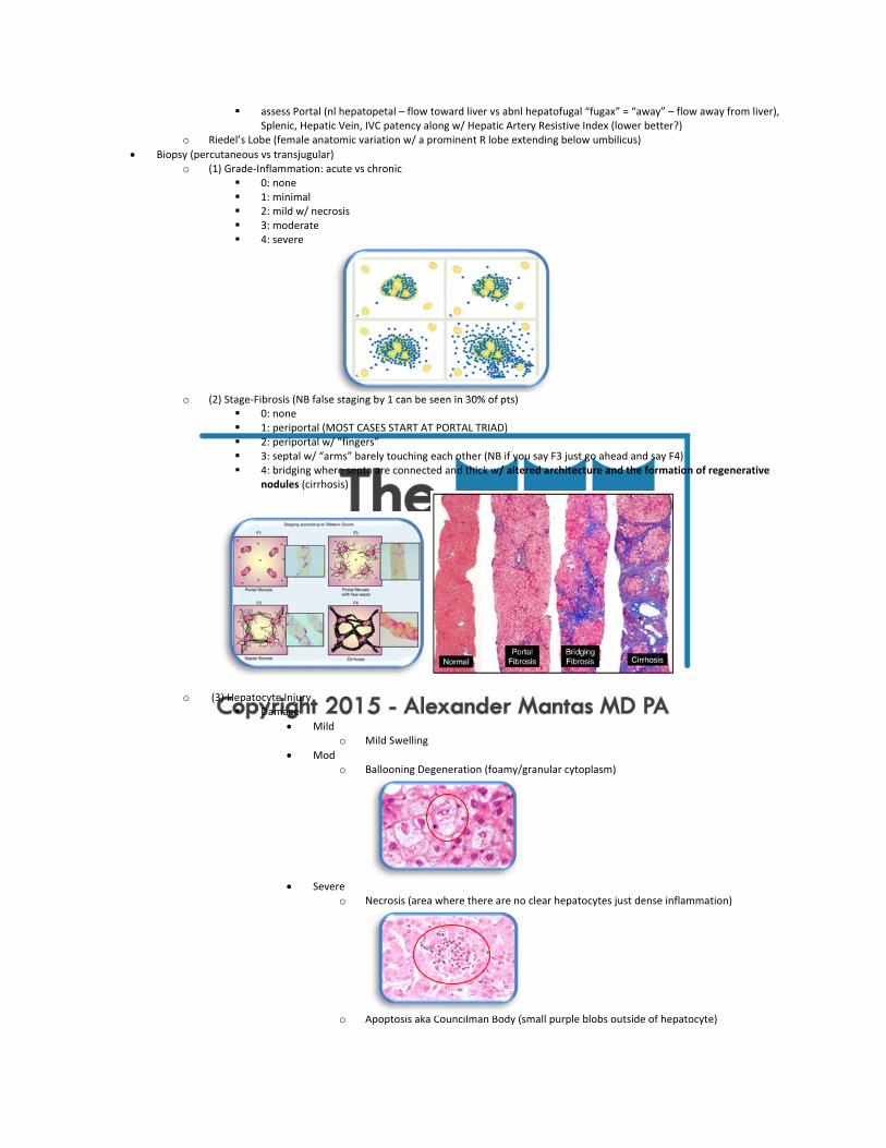
Biopsy (percutaneous vs transjugular) o (1) Grade‐Inflammation: acute vs chronic
0: none 1: minimal 2: mild w/ necrosis 3: moderate 4: severe
o (2) Stage‐Fibrosis (NB false staging by 1 can be seen in 30% of pts)
0: none 1: periportal (MOST CASES START AT PORTAL TRIAD) 2: periportal w/ “fingers” 3: septal w/ “arms” barely touching each other (NB if you say F3 just go ahead and say F4) 4: bridging where septa are connected and thick w/ altered architecture and the formation of regenerative
nodules (cirrhosis)
o (3) Hepatocyte Injury Damage
Mild o Mild Swelling
Mod o Ballooning Degeneration (foamy/granular cytoplasm)
Severe
o Necrosis (area where there are no clear hepatocytes just dense inflammation)
o Apoptosis aka Councilman Body (small purple blobs outside of hepatocyte)
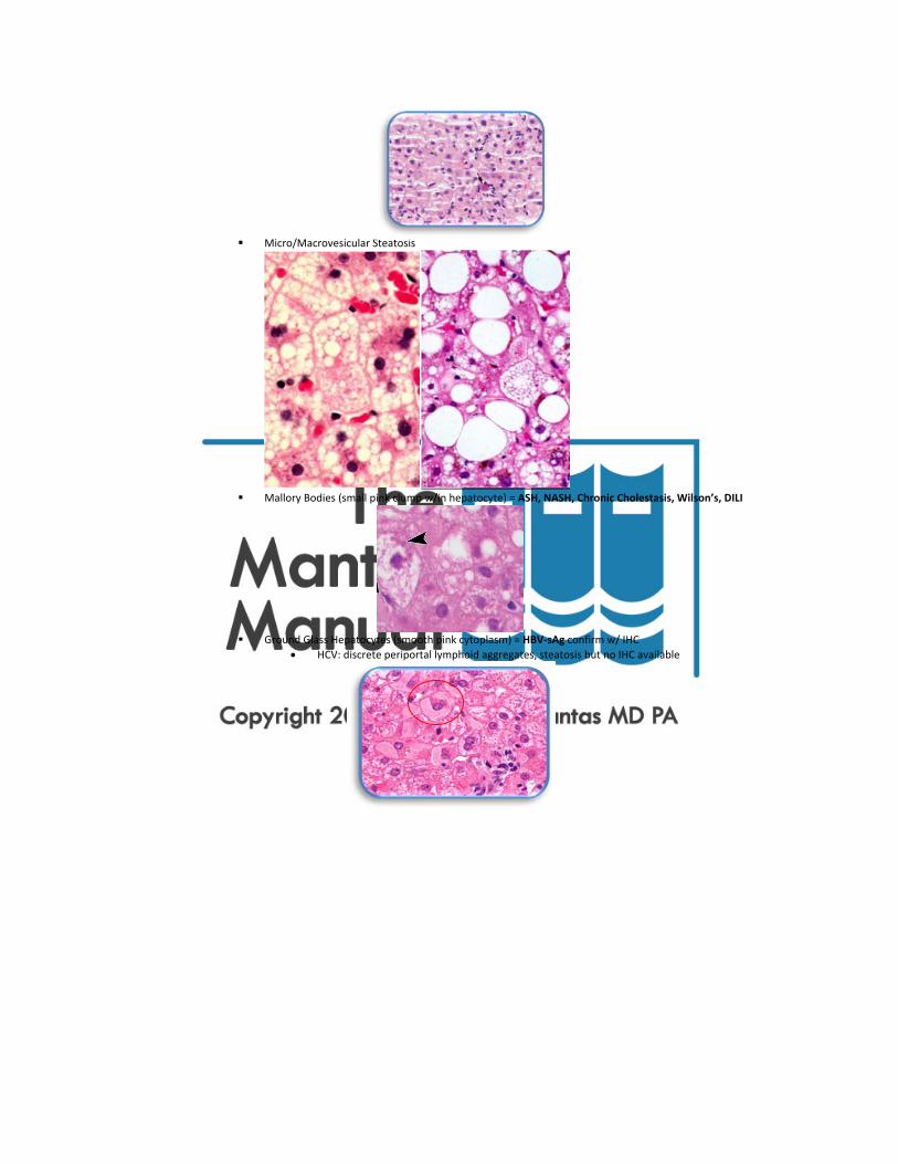
Micro/Macrovesicular Steatosis
Mallory Bodies (small pink clump w/in hepatocyte) = ASH, NASH, Chronic Cholestasis, Wilson’s, DILI
Ground Glass Hepatocytes (smooth pink cytoplasm) = HBV‐sAg confirm w/ IHC
HCV: discrete periportal lymphoid aggregates, steatosis but no IHC available

Interlobular Duct Injury (PBC, PSC, HCV) Pigments
Bile in canaliculi
Hemosiderin
Lipofuscin (non‐specific/pathologic “liver aging” characterized by the presence of yellow‐brown granules)
Iron
Stains
H&E (95% of dx are made w/ this stain)
Collagen o Reticulin: stains collagen that lines sinusoids therefore good to establish subtle
architecture (not done much anymore) Normal Hepatocyte Loss = sinusoids collapse creating reticulin crowding Heptocyte Regeneration = sinusoids spread apart creating reticulin spacing
Normal Hepatocyte Loss Hepatocyte Regeneration
o Masson Trichrome: three separate dyes which highlight collagen, cytoplasm, nuclei therefore good to establish overall architecture in cirrhosis

o (4) Portal Triad
Cholestasis (Dilation, Portal Triad Edema, Inflammation)
Acute (centrilobular around central veins w/ canaliculi filled w/ bile)
Chronic (cholate stasis w/ “pale/feathery/foamy degenerating” periportal hepatocytes, in addition
there is increased copper)
o Always order pressures when doing a Transjugular Liver Biopsy
WHVP (Wedge Hepatic Venous Pressure, nl <10mmHg, wedge a catheter into one of the hepatic veins and inflate balloon = portal pressure + intra‐abdominal pressure) – FHVP (Free Hepatic Venous Pressure, nl <5mmHg, deflate balloon catheter = intra‐abdominal pressure) = HVPG (Hepatic Venous Pressure Gradient)
5 II
10‐5 = 5 * after eliminating intra‐ab pressure the pressure in the portal vein to the heart should be the same *
RA FHVP WHVP HVPG
Normal <5 <5 <10 <5
Post HepaticPost Sinusoidal
=

Intra‐Sinusoidal = = Pre‐SinusoidalPre‐Hepatic
= = = =
HVPG ~ Portal BP (nl <5mmHg) o Portal HTN >5 o EV Formation >10 o EV Bleed and Ascites Formation >12 (highest risk of re‐bleed when >20)
NB this method is not accurate for pre‐hepatic/pre‐sinusoidal portal hypertension AND post‐hepatic portal hypertension hence only good for sinusoidal and post‐sinusoidal portal hypertension
Pruritus o Etiology
Cholestasis: bile acid and opioid peptide skin deposition Other: Dermatitis, Xerosis aka Dry Skin, Iron Deficiency, PV esp after hot shower, Scabies, Lichen Planus, Psych,
HIV, HepC, MS, Parasitic Disease, Hypo/HyperTH, DM, Uremia, Opioids, PV, Parasitophobia NB secondary skin lesions can develop including nummular eczema, prurigo nodularis, lichenified plaques, etc
o Tx General
moisturizing lotions esp Sarna Lotion
humidifier at night
wear lightweight clothing
avoid precipitants (above)
keep nails short to avoid scratching
topical (menthol, camphor, phenol, calamine lotion, topical H1B, capsaicin, local anesthetics) Cholestasis
1° bile acid resins/binders (cholestyramine) and antihistamines (hydroxyzin (Atarax), pamoate (Vistaril), doxepin (Silenor))
o NB take antihistamines at night o NB bile acid dissolution (eg. URSO) is not actually effective and may make it
paradoxically worse except in cholestasis of pregnancy nevertheless it is often given
2° rifampin (inhibits bile acid uptake and as an abx it decreases production of secondary BAs but there is a 15% risk of acute hepatitis)
3° opiate antagonists (eg naloxone, naltrexone, ondansetrone)
4° SSRIs (eg sertraline, paroxetine, mirtazapine)
5° UVB/PUVA, charcoal hemoperfusion, plasmapharesis, barbiturates, liver transplant
Op Eval
Pre‐Op o Every pt should undergo a pre‐op H&P to look for liver dz AND if H&P suggests liver disease then check LFTs/Imaging
otherwise the routine check of LFTs/Imaging is NOT recommended o Avoid elective surgery when there is acute hepatitis and decompensated cirrhosis o In pts with cirrhosis determine Child Class (A/B/C= 10/30/80% peri‐operative mortality) & Modified MELD Score
(www.mayoclinic.org/meld/mayomeld9.html, includes MELD and Age/ASA Class to give you 7d/30d/90d/1yr/5yr mortality) but these scores change w/ emergency/type of surgery and other comorbidities
o Cardiac surgery and hepatic resection are the worst surgeries o Goal INR <1.5 and Plt >50k o Ascites can compromise respiration during surgery, increases risk of wound dehiscence and ab wall herniation, etc
therefore minimize w/ diuretics/LVP/salt restriction and consider TIPS o Avoid HRS by giving albumin o Some give prophylactic rifaximin/lactulose o Improve nutritional status
Intra‐Op o In a normal pt decreased intra‐operative hepatic blood flow (2/2 anesthesia, blood loss, etc) results in a brief asymp spike
in LFTs but in a pt w/ liver disease there can be intra/post‐operative decompensation b/c hepatic blood flow at baseline is already compromised
o Increased r/o hypoxemia 2/2 decreased lung volume from ascites & hepatic hydrothorax, HPS, hepatopulmonary HTN o NEVER use halokanes as it can cause acute hepatitis, avoid narcotics/benzos, best to use propofol
Post‐Op o Etiology of Problems: underlying liver dz, benign post op cholestasis or if bad consider TPN/sepsis induced cholestasis,
hepatotoxic inhalational anesthetics (halokanes), impaired perioperative hepatic perfusion, transfusions, DILI, bile‐duct injury
o Best to follow INR as it is the best marker for hepatic fxn Liver Biopsy
Approaches: percutaneous by GI, transjugular by IR, laparoscopic by surgeons
Adequacy: >1.5cm length, >1.2mm diameter
Prior: hold aspirin x1wk and NSAIDs x3d, fast ON
Contraindications: INR >1.5, Plt <60k, ascites, suspected hemangioma
Types: various kinds (Jamshidi, Klastkin, Menghini, Tru‐Cut, Vim‐Silverman) but the most common one used is the BARD Max‐Core Spring loaded biopsy (16G/18G) (NB also obtain a Safe‐T‐Bx tray kit which has lidocaine, surgical blade, specimen bottle, etc)
Position: give Versed 1mg IV, place pt supine w/o pillows, elevate R arm behind head, angle legs to left to open up R intercostals spaces
Find Location: percuss R trunk along midaxillary line to the point of dullness during both inspiration/expiration, usually 2nd or 3rd intercostal space above costal margin, confirm w/ US noting gallbladder and lungs, mark site w/ pen
Procedure: sterilize site, anesthetize w/ 1% lidocaine in both superficial and deep planes (pt might feel pain when you anesthetize the capsule), nick skin w/ blade (not always done), introduce BARD Bx right above rib to avoid damage to neurovascular bundle, resistance will be encountered when you hit the liver capsule, have pt expire and hold (shrinks lungs and brings liver to thorax wall), do Bx, remove needle and then apply pressure w/ gauze for a few minutes then bandage, put specimen into specimen bottle w/ saline, roll pt onto R side and stay there for 1hr, observe for 1 more hour
Complications: pain (30%), parenchymal or subcapsular hematoma (25%, incidental, asymptomatic, conservative Tx), hemobilia (rare, p/w GIB, biliary colic, jaundice 5d later, conservative Tx), intraperitoneal bleed (rare, most serious, p/w shock and pain w/in a few hours, Tx angiography), pneumo/hemothorax, bile peritonitis, abscess/sepsis, Bx of other organs (kidney, lung, GB, colon)
Paracentesis
NB PATIENTS ON LACTULOSE TEND TO HAVE A DISTENDED CECUM HENCE AVOID THE RLQ
Contraindications: skin infection, most of the time these pts are cirrhotics and have a coagulopathy but the r/o bleeding is very low in paracentesis so you don’t need FFP or Plt unless pt has active DIC!!! otherwise there is no data on INR/Plt cut‐offs
Equipment: quick tap paracentesis kit (pink lettering), 6 vacutainer bottles, 2 sets of blood Cx bottles, 4x4 gauze, pressure tape, alcohol swabs, 3 pairs of 7.5 gloves, biohazard bag, albumin
Risk: bleeding, infection, organ injury, tracking of fluid through tissue into groin, continued leak (place ostomy bag over site) but most concerning is "post‐paracentesis circulatory dysfunction"
o max reabsorption capacity of peritoneal surface is ~1L/d thus if you remove more than this than you are removing intravascular volume therefore give 5g of albumin for every 1L of fluid removed and only if pt has infection, renal insufficiency, hypoNa, or albumin <2.5g/dL, no need to give it before
Perform: full gown/gloves/mask/drapes, have pt lie supine with head of bed slightly elevated, tap out dullness, choose site of entry (lower quadrants 2‐4cm medial and cephalad to the ASIS lateral to the rectus abdominus to avoid artery OR midline 2‐4cm below umbilicus (not done anymore b/c most pts have thick pannus making it difficult) NB do not use old surgery scars and avoid varicosities in the skin which are common in cirrhotics, clean/drape, anesthetize skin with 25G needle and everything underneath with 22G needle esp the sensitive peritoneum, advance needle/catheter assembly into ab using a "Z" towards perineum or pinch skin in this direction and go straight in so after pinch is released the skin and peritoneum are off, either of these two approaches minimizes ascitic fluid leak, when peritoneal fluid is obtained advance catheter and remove needle, attach a 50cc syringe with a three‐way stopcock, if paracentesis is done for therapeutic fluid removal attach a connecting tube to the sidearm of the three way stopcock and connect it to 1L vacuum bottles, manipulate catheter or reposition pt if fluid flow stops, remove catheter, bandage site
Lab Analysis: Routine: Cell Count w/ Diff (normally lymphocyte predominant, usefulness of mesothelial cells is unclear, in general diuresis does not change the numbers), Albumin, Total Protein, Cx/GS (direct BCx bottles increases yield to 80%), Cytology, AFB, LDH (nl <1/2serum), Amylase (nl <1/2serum), Triglycerides (nl <serum, high TGL may interfere w/ albumin assays creating a falsely high SAAG), Bilirubin (nl <serum, if SABG is >1 or ascetic bili >6 then consider biliary perforation) Glu (~serum, if lower then infection), NO VALUE: pH, lactate, NB dipsticks are being developed, NB high serum globulin levels can spill over into ascites pulling water with it lowering ascetic albumin creating a falsely high SAAG)
World Record: 41L but in general do NOT take more than 5L off at one time b/c paracentesis removes opsonins whereas diuresis concentrates them hence just take off enough fluid to make pt comfortable and then diuretics
Liver Biochemical Tests
Types o Hepatocyte Integrity (AT, LDH)
Aminotransferases – AT: enzymes used in gluconeogenesis converting amino acids to keto acids, AST (aspartate transaminase, mitochondria) = SGOT (serum glutamic oxaloacetic transaminase) = AST is also found in heart muscle, skeletal muscle, kidney, brain, etc vs ALT (alanine transaminase, cytoplasm) = SGPT (serum glutamic pyruvic transaminase) = ALT more specific for liver and last longer but some is found in small amounts in skeletal muscle
NB statin induced myopathy (AST high ALT low) vs hepatitis (AST and ALT high)
NB L>S in all cases of acute hepatitis b/c you generally lyse cells releasing cytoplasmic ALT EXCEPT alcohol which selectively damage mitochondria
NB in cirrhosis the ratios actually flip such that S>L in advanced cases
NB mild AT can increase w/ Celiac Sprue, Addisons, Hyper/Hypothyroidism, damage to other organs (refer above)
NB recent evidence suggests that the ULN of ALT is likely too high, a better ULN is 30M/19F
NB levels below LLN is of no significance but can be seen after dialysis and with VitB6 deficiency
T1/2 3d Lactate Dehydrogenase – LDH: found in many different tissues
o Hepatocyte Synthetic Function (Alb, INR) Coagulation Factors
All clotting factors are made by liver except VIII
2,7,9,10,C,S require VitK vs 1,5,8,AT‐3 do not require VitK
VitK is low in cholestasis, malabsorption, maldigestion, chronic abx use BUT NOT cirrhosis Albumin
t1/2 = 14‐21d o Hepatocyte Excretory Function (Bilirubin)
TB (1mg/dL) = IB (0.85mg/dL, 85%) + DB (0.15mg/dL, 15%), NB visibly jaundice when TB >3mg/dL
Lipid Soluble Indirect/Unconjugated Bili o made from the breakdown of hemoglobin o bound to albumin and thus cannot be filtered by glomerulus o it goes to liver and is converted to direct/conjugated bili
Water Soluble Direct/Conjugated Bili o UDP conjugates bili o can enter back into blood and be filtered by glomerulus (creating abnl dark urine) or go
into stool and be converted to stercobilin (creating nl dark stool) but if it cannot go into stool then the stool is pale/clay like (creating abnl light stool)
NB DB can directly be detected by adding diazotized sulfanilic acid which cleaves conjugated bilirubin forming azo‐dipyrrole which appears red, the degree of which can be calculated by spectrophotometery (Diazo Van Den Bergh Reaction), IB can indirectly be detected by adding alcohol before the Diazo Van Den Bergh Reaction which allows for cleavage of BOTH conjugated and unconjugated giving you a total bilirubin level, unconjugated can be derived by subtracting conjugated from total hence “indirect”
Cholesterol Copper Bile Salts
o Cholangiocyte Integrity (AlkPhos, GGTP, 5’NT) Alkaline Phosphatase – AP: AlkPhos also seen in 1° bone 2° SI/kidney/placenta (esp when infarcted), action is
unknown, higher levels during states when you have high metabolism (after eating, growth, etc hence children, pregnancy, cancer esp lung/ovarin), slowly rises with age w/ bone breakdown, after a fatty meal, CKD b/c decreased clearance, t1/2 7d, “inducible” hence it is slow to rise and once high it is slow to come back down, you can check a “Fractionated AlkPhos” (Total = Bone (“Burn” = heal labile) + Liver (heat stable)), low in Wilson’s
Gamma Glutamyl TransPeptidase – GGTP: can also be increased w/ EtOH and Drugs (Coumadin, AEDs, etc) esp if GGT/AP ratio is >2.5, also made in kidney/spleen/pancreas/heart/seminal‐vesicle/brain but NOT in bone hence good to measure if you are trying to determine if AP is from bone or liver and it is also more sensitive than AP except in BRIC where GGT is normal but AP is high
5’‐Nucleosidase – 5’‐NT: found in liver/heart/pancreas/brain/vessels NOT in bone
Hepatocellular Injury (AT > AP/Bili, rate/peak/ratio are important) o Mild <100 (<2x ULN): Cirrhosis and Any Chronic Hepatitis o Mod 100‐500 (2‐5x ULN): Steatosis, Autoimmune, Vascular, Metabolic o Severe >500 (>5x ULN): Viral & Toxin (gradual/prolonged, nl LDH) vs Ischemia (abrupt/brief, elevated LDH)
Check a Creatine Kinase and Aldolase to rule out muscle disease Drug Induced Liver Injury (DILI)
General o www.livertox.nih.gov o More common and more severe in older, obese, malnourished, alcoholic women but
having chronic liver dz does not increase one’s r/o DILI but may make it more severe if it does occur
Types
Toxic/Intrinsic Idiosyncratic
less common more common
occurs in any pt therefore predictable occurs randomly in some pts therefore not predictable
worse the higher the dose no relation to dose
injury occurs w/in hours latency period is variable but generally longer in the order of several weeks
not genetic genetic
usually hepatocellular injury Not only hepatocellular injury but also cholestasis, granulomatous, autoimmune, cancer, steatosis, vascular (refer)
class effect non‐class effect
directly toxic process of drug or its metabolite thus knowing P450 inducers/inhibitors is also important
allergic process w/ systemic hypersensitivity Sx
true idiosyncratic reaction

Not many with the main ones beingTylenol, INH, Chemo, Fungal Toxins, VitA, MTX, Heavy Metals
Not many with the main ones being Phenytoin, Sulfa, Dapsone
Many
Important Points o “Adaptation”: initially abnormal LFTs but after awhile they return to normal o Some drugs cause cause abnl LFTs but no actual injury (eg. elevated TB w/ rifampin,
cyclosporine, indinavir and elevated AP w/ phenytoin, warfarin) while some drugs cause no change in LFTs but still cause damage (eg. sex steroids, MTX, hypervitaminosis A)
o If AT <3xULN then just observe vs if AT >3xULN then 10% r/o ALF if TB becomes abnormal
Drugs that cause Hepatocellular Injury o Meds: NSAIDs (esp diclofenac, sulindac), Anti‐Bacterials (Macrobid, Minocycline,
Bactrim), HAART, Anti‐TB (INH, Rifampin), Anti‐Fungals (Azoles), Endocrine (Niacin, TZDs, Statins), Neuro (Valproic Acid, SSRIs, Muscle Relaxants, Phenytoin), CV (Methyldopa, Amiodarone, BB, CCB, Plavix), GI (H2B) Anesthetics, PTU/MMI, Any Sulfa Drug, Disulfiram, Retinoids, MTX
o Work‐Exposure Agents: CCl4, HCFCs, Vinyl Chloride, Haloaromatics, Phosphorus, Iron, Copper, Arsenic, Toluene, NB insecticides/herbicides/pesticides generally do not cause liver injury
o Illicit Drugs (Any Stimulant) o CAM: Herbals (www.nccam.nih.gov), Steroids (Androgens), Vitamins (Hypervitaminosis
A), Weight Reduction Agents (Hydroxycut, Lipokinetix, Ma Huang, Kombucha) o Foods: mushrooms, fruits, contamination (refer to acute)
Pathology: very non‐specific but suggestive if necrosis is more severe than the clinical picture
Tx: steroids are generally not helpful, consider NAC and other Tx for specific toxins, generally pts do better when drug is stopped but some (azoles and amiodarone) take months to improve
Cholestasis (AP/Bili > AT, first start w/ Ab‐US or CT to look for ductal dilatation and if + then proceed w/ Cholangiography (ERCP, MRCP, PTC) for Dx of extrahepatic obstruction but if negative then proceed w/ Direct Liver Injury Assessment (Labs, Bx) for Dx of intrahepatic obstruction
o Other if partial obstruction then bili will be normal thus making you think infiltrative dz acute: high transaminases vs chronic: mildly high transaminases as obstruction clears AT will actually still continue to rise for a while b/c it is inducible and takes a while for
it stop being made CA 19‐9 >1000 (DDx: biliary/pancreatic cancer or florid cholangitis) vs <1000 (DDx: “ “ + pancreatitis) complete obstruction will raise TB to 20 but not >20 so if >20 then consider hepatocellular injury chronic obstruction will lead to high INR b/c of vitamin K deficiency sometimes hepatocellular injury can result in a cholestatic picture copper tests will be abnormal in any chronic cholestatic disorder
o Extrahepatic Cholestasis w/ Ductal Dilation = Complication: segmental liver atrophy, secondary biliary cirrhosis, cholangitis, choledocholithiasis
Wall Thickening aka Stricture
Cancer = irregular not smooth, abrupt shelf like borders, >14mm in length, intrahepatic duct dilation, intraductal polypoid areas
o Distal Cancer: pancreatic, ampullary, bile duct cancer (but don’t forget surrounding malignant LAD and mets to the bile duct)
o Proximal Cancer: bile duct, gallbladder, liver cancer (but don’t forget surrounding malignant hilar LAD and mets to the liver w/ colon, gastric, pancreatic, breast, melanoma)
Autoimmune (Large Duct PSC, Autoimmune Cholangiopathy)
Alloimmune diseases (Liver Transplant Rejection, GVHD)
Chronic Pancreatitis
Injury o Post Sphincterotomy Stenosis o Chronic Choledocholithiasis o Post Op: Iatrogenic Injury during Cholecystectomy 0.6‐2.2% risk vs OLTx 15‐20% risk o Trauma o Radiation o Chemo o Ischemia (important) o Infection: CMV after OLTx
Infiltrative (Sarcoidosis)
AIDS Cholangiopathy o secondary sclerosing cholangitis w/ multiple strictures and/or papillary stenosis

o Mechanism: (1) infection w/ 1° Crypto, 2° Micro/Cyclo, CMV, (2) ischemic injury from vasculitis, (3) HAART DILI, typically seen when CD4 <100
o Dx: ERCP brushings/aspirate/biopsy o Tx: sphincterotomy and stenting, NB Tx of infection has not shown to decrease Sx nor
improve cholangiographic changes but obviously still do Extrinsic Compression
Psuedocyst
RP Fibrosis
LAD
Cancer Intraluminal Occlusion
Choledocholithiasis
Cystic Fibrosis
Infection o Ascaris lumbricoides o Clonorchis sinensis, Opisthorchis viverrini, Fasciola hepatica/gigantica (“Liver Fluke”)
Epidemiology: Clonorchis/Opisthorchis (freshwater fish) vs Fasciola (freshwater plants esp watercress), Asia/Russia
Mechanism: eggs passed into feces, eggs ingested by snails, eggs hatch and mature into cercariae which penetrate freshwater fish/plants and develop into metacercariae, humans ingest raw, pickled, or undercooked fish/plants, metacercariae pass into bile ducts and develop into worms which then create eggs
S/S: usually asymptomatic but can cause fever, malaise, RUQ pain Complications:
physical presence of worms in bile dict = biliary obstruction
chronic irritation of worms in bile duct = cholangiocarcinoma Dx: Stool O&P Tx: Praziquantel or Abendazole
Bile Plug Syndrome o Def: plug of thick inspissated bile and mucus in the CBD, occurs in sick premature
infants, Tx w/ irrigation during surgery
Biliary Atresia o Def: destruction or absence of all or a portion of the extrahepatic bile ducts o Epidemiology: 1/10‐15k births, most common cause of neonatal cholestasis, pediatric
liver transplant, cause of pediatric liver death o Etiology: unknown but RFs include black, spring conception, low VitE/Copper/Phos,
infection esp Reovirus‐3 o S/S: uneventful delivery, prolonged jaundice otherwise the infant is normal, if unTx then
death w/in 2yrs, 10‐25% have other defects o Types and Tx
Type I (5%) CBD (surgically correctable) Type II (5%) “ “ + Cystic/CHD (surgically correctable) Type III (90%) “ “ + Intra‐Hepatic Bile Ducts (was once considered NOT
surgically correctable until the Kasai Operation in which jejunum is anastomosed to liver porta hepatis and if that fails then liver transplant)
o Intrahepatic Cholestasis w/o Ductal Dilation = initially there is bile duct proliferation w/ accumulation of bile pigmented plugs in Zone 1 on histology but after time there is “Vanishing Bile Duct Syndrome” aka Ductopenia aka loss of intralobular bile ducts
Post‐Op TPN Infection
general sepsis (very important)
viral (any hepatotropic virus but especially HAV/EBV/CMV) DILI
“Bland” Cholestasis aka w/o Hepatitis: H2B/PPI, NSAIDs, Sex Steroids, Augmentin, Sulfa, Griseofulvin, Ketoconazole, Tamoxifen, Coumadin, Cyclosporine, Azathioprine
o NB ceftriaxone complexes w/ bile to form sludge
“Conspicuous” Cholestasis aka w/ Hepatitis: Chlorpromazine, Flucloxacillin, Macrolides, Bactrim, Clindamycin, Sulfa, FQ, Ketoconazole, Azathioprine, Metformin, Statins, ACE‐I, TCAs, Methyldopa, Work‐Exposure Agents
GVHD Autoimmune (PBC, Small Duct PSC) Intrahepatic Cholestasis of Pregnancy Neonatal Cholestasis

Idiopathic (most common)
Infections: TORCH, Coxsackie, Echo, Syphilis, Rubella, CMV
Metabolic: A1AT, Galactosemia, Fructosemia, HypoTH, CF, Niemann‐Pick, Glycogen Storage Dz
Syndromes: o Benign Recurrent Intrahepatic Cholestasis (BRIC)
Mech: AR mutation of FIC‐1 gene (Dutch population) Presentation: episodic pruritus/jaundice w/ first episode before 30yo, last
wks‐mos, resolve spontaneously, in b/t episodes Sx, labs and histology normal lasting mos‐yrs
Tx: supportive NB does not lead to progressive liver injury NB unlike other cholestasis disorders GGT remains normal
o Progressive Familial Intrahepatic Cholestasis (PFIC) aka Byler’s Syndrome o Dubin Johnson Syndrome o Rotor Syndrome o Sitosterolemia o Alagille’s Syndrome
Mech: AD mutation of JAGGED‐1 gene resulting in a paucity of bile ducts Presentation: variable presentation from liver failure during first few months
of life to mild Sx at adulthood
General (developmental delay)
GI (intrahepatic cholestasis w/ a paucity of intralobular ducts, HSM, xanthomata, pruritus)
Face (dysphormic facies w/ prominent forehead, hypertelorism, deep set eyes, small pointy chin, elongated nose w/ bulbous tip)
CV (pulmonic stenosis)
MS (butterfly vertebra, short stature)
Eye (posterior embryotoxin w/ dysgenesis of the iris/cornea) Tx: supportive until liver failure then transplant
Infiltrative (AP, LDH) o Granulomas
Autoimmune: Sarcoidosis, PBC, Crohn’s, Wegener’s Cancer: Lymphoma Foreign Body: Berylliosis, Talc Infection
Viral: CMV, EBV
Bacteria: TB, MAC, Brucellosis, Nocardia, Tularemia, Rickettsia
Fungal: Histo
Parasitic: Toxo, Schisto DILI
1° Allopurinol, Phenytoin, Hydralazine, Quinidine
2° Methyldopa, Nitrofurantoin, TZDs, Amiodarone, Augmentin o Cancer (uniquely lymphoma and small cell cancers cause a more infiltrative process rather than discrete masses) o Amyloid o Abscess o Stauffer’s Syndrome (renal cell carcinoma paraneoplastic syndrome resulting in an increase in the “Regan Isoenzyme”)
Disordered Bilirubin Metabolism (Isolated Hyperbilirubinemia) o If TB is high and IB/DB is high aka high IB >85% and low DB <15%
GENERAL Hepatocellular Damage SPECIFIC Decreased Hepatocellular Uptake/Conjugation
Drugs (Indinavir, Rifampin, Probenecid)
Infections (any type of infection but esp CMV)
Uridine DiPhosphatate glucuronyl transferase (UDP) Disorder o Crigler‐Najjar Syndrome I/II (Type I is severe w/ IB ~20 and used to be fatal at infancy
w/ kernicterus until the use of isolated hepatocyte transplant into native liver, Type II is mild w/ IB ~5 and most pts are asymptomatic w/ nl liver histology, no Tx but phenobarbital can be used to lower bilirubin)
o Gilbert’s Syndrome (9% of population, mutation of UDP resulting in decreased activity, at baseline TB is 2‐4 but during episodes rises to 7‐8, nl liver histology, no Tx as benign, intermittent and often brought on by physical stress, fasting, alcohol, best test is to have pt fast and see if bili doubles and then return to baseline levels after eating)
o If TB is high but the ratio of IB/DB is normal aka IB =85% and DB =15% (DB is never >15% b/c you can’t stimulate the liver to conjugate more IB to DB)
Any Type of General Cholestasis Increase in Production

Hemolysis (refer but always remember Wilson’s)
Hematoma Resorption
Muscle Injury
Profound Problems w/ RBC production (eg. really bad IDA, SCD, HS) o Newborn Jaundice
Types
Physiologic (occurs b/t 1‐7d post‐partum, never <1d, bili always <15mg/dL and always rise <5mg/dL/d, returns to nl after a 1‐3wks, no encephalopathy, RFs: prematurity, AA, Asian, male, bruising 2/2 traumatic delivery, high altitude, seen in 50% of newborns)
o Newborn Jaundice (immature UDP) o Breast Milk Jaundice (high FA content in milk by some unknown mechanism inhibits
conjugation)
Pathologic (occurs <1d or >7d) o Poor Breastfeeding Jaundice (there is not enough food for newborn to keep passing
stool keeping chyme in bowel and thus allowing more time for absorption) o Hemolysis
Complications: Kernicterus aka Bilirubin Encephalophy aka Bilirubin Induced Neurologic Dyfunction (BIND), occurs when bilirubin detaches from albumin (>20mg/dl exceeds albumin binding capacity) and diffuses across BBB collecting in basal ganglia causing motor problems NOT cognitive problems
Early: lethargy, hypotonia, poor sucking, decreased feeding
Later: opisthotonos (backward arching neck/back w/ body resting on heels/head), hypertonia, high‐pitched crying then apnea, seizure
Long Term: athetoid movement (arms/hands writhing in a “dancing” quality), cerebral palsy, sensorineural deafness, speech deficits, painful spasms decreased upward gaze, dental dysplasia
Tx
Check bili b/f d/c 72hrs after delivery (if d/c before 72hrs infant must be seen by pediatrician in 2‐5d) and if fine then send home and educate parents of newborn jaundice and proper breastfeeding and if still jaundice then alert doctor b/c likely pathologic
Phototherapy if >15: conjugates bilirubin to a water soluble form which can be excreted
Double Volume Exchange Transfusion if >20 or S/S of Kernicterus