nutrition, inflammation and insulin resistance in end ... · a schematic diagram of the proposed...
TRANSCRIPT
Protocol Date: 12/16/2015
Nutrition, Inflammation and Insulin Resistance in End Stage Renal Disease—Aim 2
T. Alp Ikizler, MD, Professor Division of Nephrology
Vanderbilt University Medical Center S-3223 Medical Center North
2
Table of Contents: Study Schema 1.0 Background 2.0 Rationale and Specific Aims 3.0 Animal Studies and Previous Human Studies 4.0 Inclusion/Exclusion Criteria 5.0 Enrollment/Randomization 6.0 Study Procedures 7.0 Risks of Investigational Agents/Devices (side effects) 8.0 Reporting of Adverse Events or Unanticipated Problems involving Risk to
Participants or Others 9.0 Study Withdrawal/Discontinuation 10.0 Statistical Considerations 11.0 Privacy/Confidentiality Issues 12.0 Follow-up and Record Retention Appendix References
3
Uremic Wasting
Inflammation
Insulin resistance
?
?
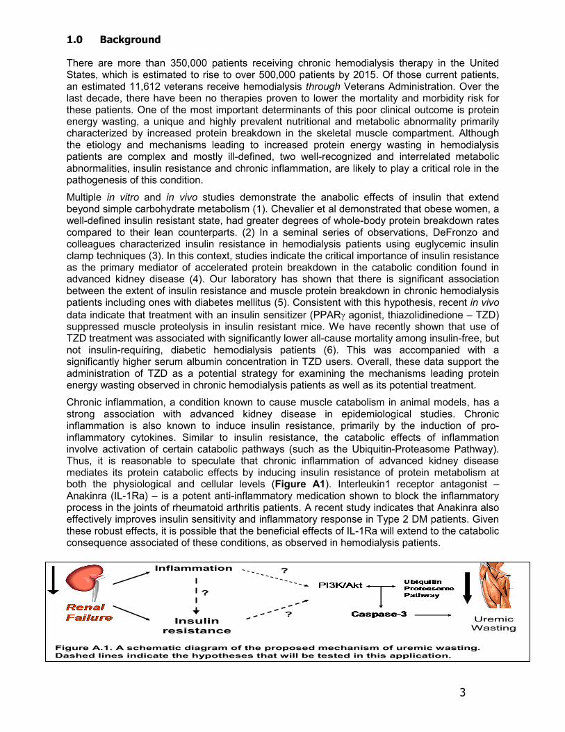
Figure A.1. A schematic diagram of the proposed mechanism of uremic wasting. Dashed lines indicate the hypotheses that will be tested in this application.
?
1.0 Background There are more than 350,000 patients receiving chronic hemodialysis therapy in the United States, which is estimated to rise to over 500,000 patients by 2015. Of those current patients, an estimated 11,612 veterans receive hemodialysis through Veterans Administration. Over the last decade, there have been no therapies proven to lower the mortality and morbidity risk for these patients. One of the most important determinants of this poor clinical outcome is protein energy wasting, a unique and highly prevalent nutritional and metabolic abnormality primarily characterized by increased protein breakdown in the skeletal muscle compartment. Although the etiology and mechanisms leading to increased protein energy wasting in hemodialysis patients are complex and mostly ill-defined, two well-recognized and interrelated metabolic abnormalities, insulin resistance and chronic inflammation, are likely to play a critical role in the pathogenesis of this condition.
Multiple in vitro and in vivo studies demonstrate the anabolic effects of insulin that extend beyond simple carbohydrate metabolism (1). Chevalier et al demonstrated that obese women, a well-defined insulin resistant state, had greater degrees of whole-body protein breakdown rates compared to their lean counterparts. (2) In a seminal series of observations, DeFronzo and colleagues characterized insulin resistance in hemodialysis patients using euglycemic insulin clamp techniques (3). In this context, studies indicate the critical importance of insulin resistance as the primary mediator of accelerated protein breakdown in the catabolic condition found in advanced kidney disease (4). Our laboratory has shown that there is significant association between the extent of insulin resistance and muscle protein breakdown in chronic hemodialysis patients including ones with diabetes mellitus (5). Consistent with this hypothesis, recent in vivo data indicate that treatment with an insulin sensitizer (PPARγ agonist, thiazolidinedione – TZD) suppressed muscle proteolysis in insulin resistant mice. We have recently shown that use of TZD treatment was associated with significantly lower all-cause mortality among insulin-free, but not insulin-requiring, diabetic hemodialysis patients (6). This was accompanied with a significantly higher serum albumin concentration in TZD users. Overall, these data support the administration of TZD as a potential strategy for examining the mechanisms leading protein energy wasting observed in chronic hemodialysis patients as well as its potential treatment.
Chronic inflammation, a condition known to cause muscle catabolism in animal models, has a strong association with advanced kidney disease in epidemiological studies. Chronic inflammation is also known to induce insulin resistance, primarily by the induction of pro-inflammatory cytokines. Similar to insulin resistance, the catabolic effects of inflammation involve activation of certain catabolic pathways (such as the Ubiquitin-Proteasome Pathway). Thus, it is reasonable to speculate that chronic inflammation of advanced kidney disease mediates its protein catabolic effects by inducing insulin resistance of protein metabolism at both the physiological and cellular levels (Figure A1). Interleukin1 receptor antagonist – Anakinra (IL-1Ra) – is a potent anti-inflammatory medication shown to block the inflammatory process in the joints of rheumatoid arthritis patients. A recent study indicates that Anakinra also effectively improves insulin sensitivity and inflammatory response in Type 2 DM patients. Given these robust effects, it is possible that the beneficial effects of IL-1Ra will extend to the catabolic consequence associated of these conditions, as observed in hemodialysis patients.
4
1.1 Abbreviations and terms used in this protocol.
CHD Chronic hemodialysis ESRD End-stage renal disease
CKD Chronic Kidney Disease PEW Protein-energy wasting
TZD Thiazolidinedione IL1-Ra Inetrleukin-1 receptor antagonist
1.2. The mortality rate of treated chronic hemodialysis patients remains high in the U.S. The life-expectancy of hemodialysis patients is 20-25 years less than the normal age-sex-race matched U.S. population over the age of 45, worse than what is observed in patients with newly diagnosed colon cancer (7). The recent advances in our understanding of the uremic state, as well as the improvements in the science and technology of renal replacement therapy, have not significantly impacted these outcomes. Moreover, the health care cost of treating the U.S. end-stage renal disease program exceeds $17 billion dollars annually (7). Therefore, it is vital to develop and implement new and effective strategies to reduce these unacceptably high death and hospitalization rates.
1.3. ESRD is an important health problem within Veterans in the U. S. Due to the increasing number of patients with diabetes and obesity, it is likely that this cohort will expand within the VA patient population in the future. By 2030 an estimated 2 million people in the US will need dialysis or transplantation for advanced kidney failure. In terms of the DVA Health care system, there are an estimated 11,612 veterans receiving hemodialysis through VA, of which 5,147 enrolled veterans were receiving HD at VA facilities in FY 2008. An even more disturbing statistic is that mortality in ESRD is six times higher than in the general Medicare population with adjustment for age, gender and ethnicity. 1.4. Chronic hemodialysis patients display multiple metabolic and nutritional derangements, which can be collectively termed as protein energy wasting of kidney disease. Despite vigorous attempts to prevent these derangements and their consequences, protein energy wasting is present in approximately 20 - 50% of patients on chronic hemodialysis. (8-14). Protein energy wasting is primarily characterized by insidious loss of somatic protein stores (reflected by changes in lean body mass and serum creatinine) and decreased visceral protein concentrations (reflected by changes in serum albumin and prealbumin). Several studies have demonstrated that the presence of protein energy wasting, especially the degree of loss of muscle mass, sharply increases mortality and hospitalization rate in chronic hemodialysis patients (15-21).
1.5. Protein energy wasting is common in multiple chronic disease states and is a significant risk factor for mortality. Chronic hemodialysis is not the only condition where a similar nutritional derangement can be observed. Specifically, in many chronic disease states such as advanced cancer, congestive heart failure, chronic infections and chronic lung disease, a comparable loss of visceral and somatic protein stores occur. Usually, serum albumin is decreased and somatic protein stores are either depleted or non-functional in these patients. There seems to be several shared pathways leading to PEW in these patients. For example, decreased appetite, inappropriately increased resting energy expenditure, and elevated concentrations of inflammatory cytokines are observed in most, if not all, of these chronic disease states. A common observation that is also applicable to ESRD is that the extent of protein energy wasting, regardless of its etiology or disease state, is highly associated with hospitalization and death risk.
5
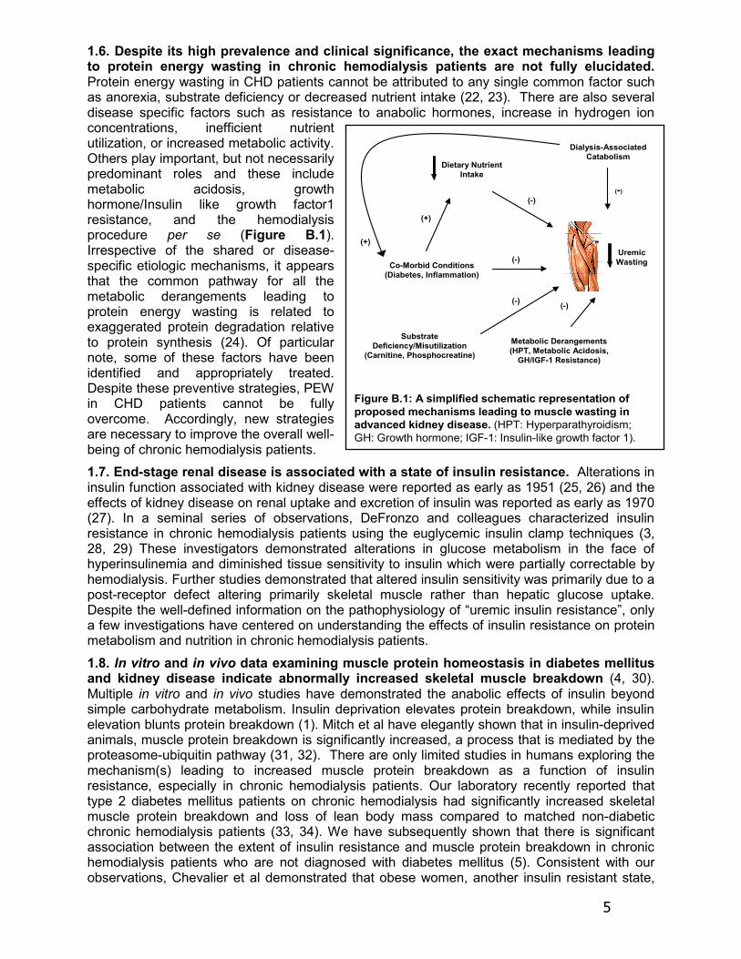
Dietary Nutrient Intake
(-)
Dialysis-Associated Catabolism
(-)
Co-Morbid Conditions(Diabetes, Inflammation)
(-)
(+)
(+)
Substrate Deficiency/Misutilization
(Carnitine, Phosphocreatine)
(-)
Metabolic Derangements(HPT, Metabolic Acidosis,
GH/IGF-1 Resistance)
(-)
Figure B.1: A simplified schematic representation of proposed mechanisms leading to muscle wasting in advanced kidney disease. (HPT: Hyperparathyroidism; GH: Growth hormone; IGF-1: Insulin-like growth factor 1).
Uremic Wasting
1.6. Despite its high prevalence and clinical significance, the exact mechanisms leading to protein energy wasting in chronic hemodialysis patients are not fully elucidated. Protein energy wasting in CHD patients cannot be attributed to any single common factor such as anorexia, substrate deficiency or decreased nutrient intake (22, 23). There are also several disease specific factors such as resistance to anabolic hormones, increase in hydrogen ion concentrations, inefficient nutrient utilization, or increased metabolic activity. Others play important, but not necessarily predominant roles and these include metabolic acidosis, growth hormone/Insulin like growth factor1 resistance, and the hemodialysis procedure per se (Figure B.1). Irrespective of the shared or disease-specific etiologic mechanisms, it appears that the common pathway for all the metabolic derangements leading to protein energy wasting is related to exaggerated protein degradation relative to protein synthesis (24). Of particular note, some of these factors have been identified and appropriately treated. Despite these preventive strategies, PEW in CHD patients cannot be fully overcome. Accordingly, new strategies are necessary to improve the overall well-being of chronic hemodialysis patients. 1.7. End-stage renal disease is associated with a state of insulin resistance. Alterations in insulin function associated with kidney disease were reported as early as 1951 (25, 26) and the effects of kidney disease on renal uptake and excretion of insulin was reported as early as 1970 (27). In a seminal series of observations, DeFronzo and colleagues characterized insulin resistance in chronic hemodialysis patients using the euglycemic insulin clamp techniques (3, 28, 29) These investigators demonstrated alterations in glucose metabolism in the face of hyperinsulinemia and diminished tissue sensitivity to insulin which were partially correctable by hemodialysis. Further studies demonstrated that altered insulin sensitivity was primarily due to a post-receptor defect altering primarily skeletal muscle rather than hepatic glucose uptake. Despite the well-defined information on the pathophysiology of “uremic insulin resistance”, only a few investigations have centered on understanding the effects of insulin resistance on protein metabolism and nutrition in chronic hemodialysis patients.
1.8. In vitro and in vivo data examining muscle protein homeostasis in diabetes mellitus and kidney disease indicate abnormally increased skeletal muscle breakdown (4, 30). Multiple in vitro and in vivo studies have demonstrated the anabolic effects of insulin beyond simple carbohydrate metabolism. Insulin deprivation elevates protein breakdown, while insulin elevation blunts protein breakdown (1). Mitch et al have elegantly shown that in insulin-deprived animals, muscle protein breakdown is significantly increased, a process that is mediated by the proteasome-ubiquitin pathway (31, 32). There are only limited studies in humans exploring the mechanism(s) leading to increased muscle protein breakdown as a function of insulin resistance, especially in chronic hemodialysis patients. Our laboratory recently reported that type 2 diabetes mellitus patients on chronic hemodialysis had significantly increased skeletal muscle protein breakdown and loss of lean body mass compared to matched non-diabetic chronic hemodialysis patients (33, 34). We have subsequently shown that there is significant association between the extent of insulin resistance and muscle protein breakdown in chronic hemodialysis patients who are not diagnosed with diabetes mellitus (5). Consistent with our observations, Chevalier et al demonstrated that obese women, another insulin resistant state,

6
had greater degrees of whole-body protein breakdown rates after overnight fast compared to their lean counterparts, and also had suppressed anabolic responses to insulin as measured by hyperinsulinemic, euglycemic, isoaminoacidemic clamp studies (2). Overall, these data indicate the critical importance of insulin resistance as a significant mediator of the accelerated protein breakdown in conditions such as diabetes mellitus and end-stage renal disease (Figure B.2) (31).
1.9. Thiazolidinediones (TZDs) are commonly used insulin sensitizing agents with intriguing pleotrophic effects. PPARs belong to a family of ligand-activated nuclear transcription factors and are involved in a variety of human diseases including diabetes mellitus, obesity, hypertension, inflammation, atherosclerosis, and cancer. Three isoforms (PPARα, PPARβ/δ, and PPARγ) have been cloned and characterized by their distinct expression patterns, different ligand binding specificities, and metabolic functions. Antidiabetic thiazolidinediones (TZDs) promote adipocyte differentiation through activation of PPARγ and sensitize the cells to the effects of insulin, underlying their success in the treatment of patients with type II diabetes. PPARγ is also found in cells of the immune system and in human atheroma (localized in monocyte-derived macrophages, foam cells, and smooth muscle cells) where they exert overall atheroprotective effects through regulation of monocytes as well as their response to inflammatory stimuli within the arterial wall. PPARγ has also been shown to inhibit the production of the pro-inflammatory mediators TNF-α, IL-1β, and IL-6 from activated human macrophages at the transcriptional level. [6]. 1.10. Thiazolidinediones (TZDs) represent a novel intervention strategy to improve protein energy wasting in non-diabetic populations, such as chronic hemodialysis patients. In a recent study, Wang et al reported that in muscle of db/db mice, a model for insulin resistance, protein degradation and activities of the major proteolytic systems, caspase-3 and the proteasome, were increased. Treatment of these mice with rosiglitazone improved indices of insulin resistance and of suppression of proteolysis. We recently studied the effect of thiazolidinedione use on survival among chronic hemodialysis patients in a national cohort of 5290 incident dialysis patients with diabetes (35). Thiazolidinedione use was assessed according to prescription data, and the analyses were stratified based on insulin use due to observed interaction. In the primary analysis, thiazolidinedione treatment was associated with significantly lower all- cause mortality among insulin-free but not insulin- requiring subjects, with adjusted hazards ratios of 0.53 (0.31-0.89) and 0.82 (0.46-1.47) respectively. These encouraging data provide a strong rationale to utilize TZDs as a therapeutic strategy to elucidate the mechanisms involved in protein catabolism associated with wasting, especially in the setting of insulin resistance. (36) 1.11. The presence and the extent of chronic inflammation is a potential catabolic factor that worsens the nutritional status of ESRD patients. Inflammation, more correctly termed
Figure B.2. A proposed mechanism depicting mechanisms leading to muscle wasting in catabolic diseases. Ubiquitin E3 enzymes atrogin-1 and muscle ring finger-1 (MuRF-1) seem to play a important role in protein degradation based on the originating stimuli. Adapted from Lecker JASN 2006
Figure B.2. A proposed mechanism depicting mechanisms leading to muscle wasting in catabolic diseases. Ubiquitin E3 enzymes atrogin-1 and muscle ring finger-1 (MuRF-1) seem to play a important role in protein degradation based on the originating stimuli. Adapted from Lecker JASN 2006
7
systemic inflammatory response syndrome, is a complex combination of physiological, immunological and metabolic effects occurring in response to a variety of stimulators resulting from tissue injury or disease processes. Cytokines, such as IL-1, IL-6 and TNF-α are the primary mediators of these catabolic effects. Advanced kidney disease, similar to other chronic diseases, is characterized by ineffectual anti-inflammatory responses associated with repetitive cytokine releases resulting in significant adverse effects to the host (37). In the case of advanced kidney disease, there is decreased clearance of uremic toxins (middle molecules, iPTH, leptin) leading to cytokine release and activation. Contributing factors in addition to decreased renal function include, but are not limited to, activation of complement factors in response to certain dialytic factors such as hemodialysis membrane, endotoxin transfer, peritoneal dialysate bio-incompatibility and vascular access (PTFE grafts, catheters). The metabolic influence of such factors are worsened by the presence of co-morbidities such as chronic infections (periodontal, peritonitis), diabetes mellitus, atherosclerosis and congestive heart failure.(38-40) 1.12. Pro-inflammatory cytokines play an integral role in muscle catabolism in models of inflammatory diseases. The metabolic and nutritional effects of chronic inflammation are many and include anorexia, increased skeletal muscle protein breakdown, increased whole body protein catabolism, cytokine-mediated hypermetabolism and disruption of the growth hormone and IGF-1 axis leading to decreased anabolism (38, 41). IL-1, IL-6 and TNF-α are the primary mediators of the metabolic effects of the inflammatory response in animal models (37, 41-43). Several studies have shown the specific involvement of the pro-inflammatory cytokine network in the signaling cascade of Ub-Prot proteolytic pathway. E3α-II is the pro-inflammatory cytokine-inducible form of the E3α family (44). These findings lead us to believe that chronic inflammation could be a major (if not causative) factor for the poor nutritional status observed in these patients (Figure B.2). No study to date has examined the involvement of these proteins, in terms of both expression and function, in the development of protein energy wasting of kidney disease.
1.13. Interleukin-1 (IL-1) is associated with development of protein energy wasting in chronic hemodialysis patients. The adverse metabolic and nutritional effects of IL1 ß are well established (45, 46). IL-1 suppresses dietary nutrient intake and increases resting energy expenditure. It is also associated with net negative protein balance in animal models and surgical patients. In a longitudinal study of 54 CHD patients, Johansen et al showed that higher IL-1ß concentrations were associated with a narrower phase angle (P = 0.004) and with a more rapid decline in phase angle with time (time x log IL-1ß interaction, P = 0.01), suggesting that IL 1 ß beta was associated with the reduction in muscle mass in CHD patients. Kalantar-Zadeh showed that appetite is strongly associated with inflammatory cytokine concentrations, including IL-1. They also showed a strong relationship between IL-1 concentrations and mortality in CHD patients.
1.14. Anti-inflammatory strategies to improve protein energy wasting in chronic hemodialysis patients are limited and IL-1 receptor antagonist, Anakinra, represents a promising strategy. Studies in patients with rheumatoid arthritis have shown that administration of IL-1Ra blocks the inflammatory process in the joints and prevents destructive process associated with IL-1. In a recent study in Type 2 diabetes mellitus patients, 14 weeks of IL-1Ra administration resulted in significant improvements in inflammatory state and was accompanied by robust improvement in insulin sensitivity as well. Given its strong effects on inflammation and insulin activity, it is likely that administration of IL-1Ra in chronic hemodialysis patients might lead to blockade of protein catabolic consequences of these conditions. To our knowledge, there are no studies examining the nutritional effects of IL-1Ra (other than the preliminary studies performed by our laboratory). Based on these observations, we propose to study the short-term effects of IL-Ra on the inflammatory state and protein and energy metabolism in chronically inflamed CHD patients. It is also clear that there is significant overlap in the mechanisms by which chronic inflammation and insulin resistance exert their adverse nutritional effects, namely exaggerated protein breakdown. Thus the need for more meticulous

8
examination of the relative contribution of these particular metabolic derangements to protein energy wasting of advanced uremia. 2.0 Rationale and Specific Aims The overall goal is to elucidate the mechanisms by which chronic inflammation and insulin resistance influence the development of protein energy wasting in hemodialysis patients.
Specific Aim: To test the hypothesis that inhibiting inflammatory response by administration of an IL-1r antagonist (Anakinra) or increasing insulin sensitivity by administration of a PPARγ agonist (Actos) will improve net protein metabolism.
Hypothesis: The chronic inflammatory component of PEW observed in hemodialysis patients is, at least in part, mediated by insulin resistance.
ESRD represents an important health problem in the VA patient population. Due to the increasing number of patients with diabetes and obesity, it is likely that this cohort will expand in the future, which renders this project highly relevant to the VA patient population. By 2030 an estimated 2 million people in the US will need dialysis or transplantation for advanced kidney failure. An even more disturbing statistic is that mortality in ESRD is six times higher than in the general Medicare population with adjustment for age, gender and ethnicity. Protein energy wasting is highly prevalent in these patients and is one of the most important determinants of their poor clinical outcome. Despite its well-recognized occurrence, the etiology and the mechanisms leading to protein energy wasting observed in chronic hemodialysis patients cannot be attributed to any single factor. However, irrespective of the specific etiologic mechanisms, it appears that the common pathway for all the metabolic derangements is related to exaggerated protein degradation relative to protein synthesis (47).
Two well-recognized and presumably interrelated metabolic abnormalities, insulin resistance and chronic inflammation, may be the major determinants of protein catabolism in CHD patients. There are no studies examining the effects of anti-inflammatory interventions and/or insulin sensitizers on protein homeostasis in CHD. Due to their established anti-inflammatory and other pleotrophic effects, Interleukin-1 receptor antagonist Anakinra and insulin sensitizer PPARγ agonist Actos represent two such promising interventions. By modulating inflammatory response and insulin signaling through two pharmacological interventions, we will have the unique opportunity to clarify mechanisms contributing of these two particular metabolic derangements in the development of protein energy wasting observed in chronic hemodialysis patients. Assessment of protein and energy turnover by stable isotope tracer techniques in CHD patients with different levels of inflammatory response and insulin resistance can elucidate the influence of these metabolic derangements on protein homeostasis, as well as substrate metabolism and oxidation. The combined dual glucose and amino acid (AA) clamp technique, first developed by Abumrad et al (65) was devised to compensate for the confounding reduction in plasma amino acids with insulin administration due to its suppressive effect on protein breakdown, providing the most precise estimation of components of protein homeostasis.
If successful, our proposed studies have great potential to influence clinical practices in chronic hemodialysis patients because the proposed intervention protocol could be easily accessible and can ultimately lead to improvements in the hospitalization and death rates. Further, since insulin resistance and inflammation are common in other chronic disease states associated with protein energy wasting, the application of these medications could be more generalized to those conditions.
The proposed study will therefore represent an important step in identifying predictors of adverse renal outcomes and potential intervention strategies. Hence, it is expected that the

9
results of this proposal will have a great impact on Veterans Health Care and make important contributions to the research mission of the DVA, providing new knowledge in metabolic and nutritional aspects of ESRD patients. 3.0 Animal Studies and Previous Human Studies The primary focus of the PI’s laboratory is to study the nutritional and metabolic consequences of chronic kidney disease, especially in chronic hemodialysis patients. Around that theme, we have published a number of studies on this area providing important observations (11, 18, 48-58) related to the adverse nutritional and metabolic effects of the hemodialysis procedure as a model for protein catabolism; etiology, extent and adverse consequences of chronic inflammation; and the influence of diabetes mellitus and insulin resistance on protein metabolism in advanced kidney disease as preliminary data applicable to this grant proposal. The hypothesis put forward in this grant application is directly based on these data and the proposed experiments are logical extension of these studies.
3.1. Studies focused on inflammation 3.1.1. The extent inflammation in chronic hemodialysis patients As a part of ongoing research efforts on this area, we have recently completed the most comprehensive assessment of chronic inflammatory patterns in CHD patients. In brief, we measured monthly CRP levels in 128 prevalent CHD patients [56.6 yrs (range 19-90), 68% African Americans, 39% diabetics (DM)] over 2 years. There were a total of 2405 CRP measurements (median 5.7 mg/L; IQR 2.4-16.6 mg/L). We defined persistent inflammation as 3 consecutive measurements greater than pre-determined cutoff levels at least once during the study. Accordingly, 77% of the patients had CRP > 3mg/L for 3 consecutive measurements (Figure C.1) Using univariate linear mixed effect model we identified (a) the presence of hemodialysis catheter (p<0.0001), (b) diabetes mellitus (p<0.0001), (c) elevated WBC (p<0.0001), (d) older age (p=0.04), and (d) reduced serum albumin (p<0.02) as independent predictors of elevated CRP. CRP was not associated with gender, race, dialysis vintage, hemoglobin, calcium or creatinine. These results are published (59). 3.1.2. Etiology of high prevalence of chronic inflammation in CHD patients 3.1.2.a. Loss of renal function leads to increased prevalence of chronic inflammation in renal disease: In an attempt to understand the etiology of increased prevalence of inflammation in renal failure patients, we studied patients with varying degrees of kidney function. We examined a group of patients with chronic kidney disease (n = 60), not requiring dialysis, extending over a lower range of kidney function (5–60 ml/min). We measured plasma CRP and IL-6 concentrations as markers of the inflammatory state. The plasma CRP and IL-6 concentrations differed significantly between patients with advanced chronic kidney disease and healthy subjects. This study suggests that chronic inflammation is prevalent in patients with chronic kidney disease well before the initiation of renal replacement therapy and demonstrates a high prevalence of this condition.
3.1.2.b Chronic hemodialysis therapy is ineffective in controlling chronic inflammation and leads to an incremental worsening of chronic inflammatory state overtime: We performed a
0
5
10
15
20
25
30
1 3 5 7 9 11 13 15 17 19 21 23 25
Month of Follow up
C Re
ativ
e Pr
otei
n, m
g/L
25th
50th
75th
Figure C.1. Median, 25th, and 75th percentiles of 2405 CRP measurements obtained every month over 26 months in a cohort 128 prevalent CHD patients
0
5
10
15
20
25
30
1 3 5 7 9 11 13 15 17 19 21 23 25
Month of Follow up
C Re
ativ
e Pr
otei
n, m
g/L
25th
50th
75th
Figure C.1. Median, 25th, and 75th percentiles of 2405 CRP measurements obtained every month over 26 months in a cohort 128 prevalent CHD patients
10
prospective observational study to examine the influence of maintenance hemodialysis therapy (with biocompatible membranes and pure dialysate) on inflammatory markers. We measured CRP, IL-6, IL-10 before the first hemodialysis treatment at the start of maintenance hemodialysis therapy, and every 3 months over a period of 12 months in 50 CHD patients. While there was no significant trend in mean IL-6 concentrations over the 12 months, CHD patients with IL-6 concentrations at the lowest 2 quartiles displayed significant increases overtime. Similar patterns were observed for CRP concentrations. This observation suggests hemodialysis is not only ineffective in controlling inflammation in advanced uremia, but also leads to an incremental worsening of the chronic inflammatory state over time. This latter phenomenon is observed in patients who initiate maintenance HD at lower concentrations of inflammatory markers.
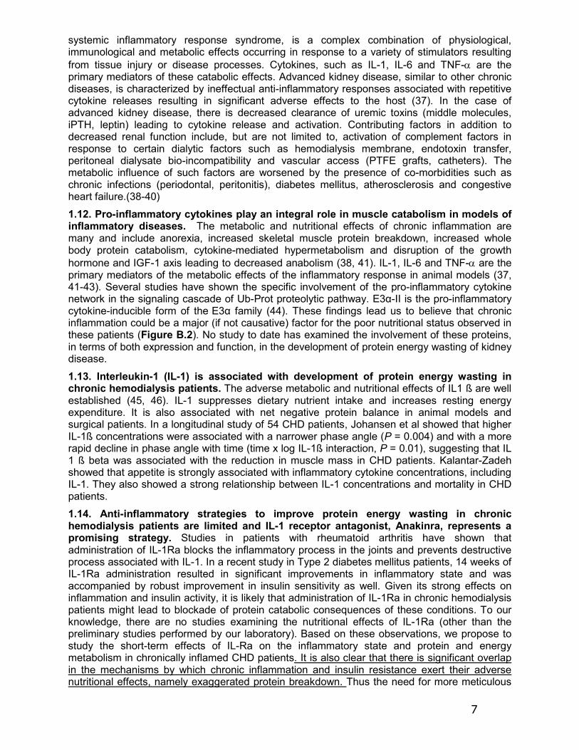
3.1.2.c. The hemodialysis procedure leads to acute inflammatory response in dialysis patients: We examined the effects of a single hemodialysis session on inflammatory markers in 7 hemodialysis patients before, during and 2 hours after a hemodialysis treatment by measurements of serum IL-6 concentrations and fractional synthetic rate of fibrinogen. There was a 70% increase in IL-6 concentrations during the 2-hour post-dialysis period (P < 0.01 compared to pre-dialysis period, Figure C.2) There was also a comparable significant increase in fibrinogen fractional synthetic rate during the two-hour post dialysis period. These results suggest activation of an inflammatory response during a single hemodialysis session and provide evidence of an independent contribution of hemodialysis to the chronic inflammatory state in hemodialysis patients.
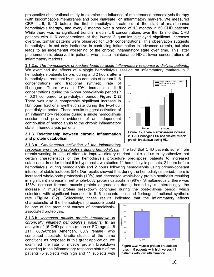
3.1.3. Relationship between chronic inflammation and protein catabolism 3.1.3.a. Simultaneous activation of the inflammatory response and muscle proteolysis during hemodialysis: The fact that CHD patients suffer from uremic wasting in spite of efforts to improve dietary nutrient intake led us to hypothesize that certain characteristics of the hemodialysis procedure predispose patients to increased catabolism. In order to test this hypothesis, we studied 11 hemodialysis patients, 2 hours before hemodialysis, during hemodialysis and 2 hours following hemodialysis using primed-constant infusion of stable isotopes (54). Our results showed that during the hemodialysis period, there is increased whole-body proteolysis (10%) and decreased whole-body protein synthesis resulting in significant increase in net whole-body protein catabolism (96%). Simultaneously, there was 133% increase forearm muscle protein degradation during hemodialysis. Interestingly, the increase in muscle protein breakdown continued during the post-dialysis period, which coincided with significant increases in IL-6 concentrations and fibrinogen fractional synthetic rate (Figure C.2). Collectively, these results indicated that the inflammatory effects characteristic of the hemodialysis procedure could be one of the prominent causes of hemodialysis-associated proteolysis. 3.1.3.b. Increased muscle protein breakdown in chronically inflamed hemodialysis patients: In an analysis of 16 CHD patients (mean (± SD) age 41.8 ±11, 80%African American, 80% female) who completed substrate kinetic studies at the same conditions as proposed in this grant application, we examined the rate of muscle protein breakdown according to the inflammatory response status of the patients (5 subjects with high and 11 subjects with
99
77
0
20
40
60
80
100
120
High CRP Low CRP
ug/1
00m
l/min
Figure C.3: Muscle protein breakdown rates in 5 patients with high versus 11 patients with low inflammation
99
77
0
20
40
60
80
100
120
High CRP Low CRP
ug/1
00m
l/min
Figure C.3: Muscle protein breakdown rates in 5 patients with high versus 11 patients with low inflammation
Figure C.2. There is simultaneous increase in IL-6, Fibrinogen FSR and skeletal muscle protein breakdown during HD.
Figure C.2. There is simultaneous increase in IL-6, Fibrinogen FSR and skeletal muscle protein breakdown during HD.

11
low inflammation). Our preliminary data showed a numerical difference between the muscle protein breakdown rates supportive of our hypothesis as depicted in Figure C.3 (98.8 ± 44.27 vs. 77 ± 36.0 ug/100ml/min (54)). While these experiments are preliminary in nature, they provide reasonable rationale supporting our hypothesis. 3.2. Studies focused on insulin resistance 3.2.1. Uremia is a state of Insulin resistance. Earlier studies by De Fronzo indicated that advanced kidney disease is associated with insulin resistance. In an initial attempt to confirm these findings in our patient population, we examined the degree of insulin resistance by practical measures in 33 non-diabetic (by ADA criteria) patients on CHD (mean (± SD) age 46.2±1 2.3, 78.8% African American, 39% females; BMI 27.4±6.3; albumin 3.6±0.35; CRP 7.7±10.5; CO2 26.4±3.9; Calcium 8.4±1.5).Fasting blood samples were taken to examine concentrations of glucose and insulin. We calculated insulin resistance using Homeostasis model assessment of insulin resistance (HOMA). Mean HOMA was 2.66±2.61 and median (interquartile range) was 1.6 (0.99–3.6). HOMA was associated with BUN, CO2 and BMI by spearman correlations. In the multivariate analysis, the only significant determinant of HOMA levels was BMI.
3.2.2. Measures of insulin sensitivity in chronic hemodialysis patients. Hyperinsulinemic euglycemic glucose clamp method (HEGC) is the gold standard to determine insulin sensitivity (IS) during a stimulated state. Insulin sensitivity can also be measured indirectly during a non-stimulated state using HOMA, QUICKI, and HOMA-AD (HOMA corrected by adiponectin). The comparison of the various indirect methods of measuring insulin resistance in CHD patients has not been properly studied in ESRD, nor have the extent and the determinants of insulin resistance as measured by HEGC in this population. In an attempt to explore this issue, 12 African American chronic hemodialysis patients (50±9 years old, 33% females, 40% with type 2 Diabetes Mellitus) were studied 3 consecutive times, 8 weeks apart. Laboratory metabolic parameters were obtained after 12 hours of fasting at each time point. Body composition was measured by DEXA at baseline. Insulin resistance was assessed by HEGC (glucose disposal rate - GDR), HOMA, HOMA-AD (corrected by adiponectin), QUICKI, and the McAuley’s index (triglyceride based).
In this cohort, the mean (±SD) values were: BMI 34.4±7.6 kg/m2, HA1c 5.48±0.52, adiponectin 19.4 ±8.6 μg/mL and median IL-6 was 9.51(5.09,18.80) pg/mL. The mean (±SD) values for measures of IR were: GDR 5.22 (4.33, 6.23) mg/kg/min, HOMA 4.7±3.4, QUICKI 0.31±0.02, HOMA-AD 115±97, and McAuley’s 1.51±0.33. Table C.1. depicts the correlation matrix between these indices. Using GEE model, the best predictor of HEGC was HOMA-AD (p=0.001), followed by HOMA (p=0.008) and QUICKI (p=0.03). Truncal fat % by DEXA, interleukin-6 and leptin were strongly associated with GDR by HEGC. These data indicated that 1) Insulin resistance is common in CHD patients; 2) HOMA corrected by adiponectin (HOMA-AD) is the best correlate of IR measured by the gold standard HEGC method; 3) While the etiology of IR is complex in CHD patients, central obesity and inflammation seem to play important roles.
Table C1 HEGC HOMA-AD HOMA-IR QUICKI McAuley's HEGC 1
HOMA-AD -0.671 *P<0.001 1
HOMA-IR -0.584 *P=0.002
0.88 *P<0.001 1
QUICKI 0.584 *P=0.004
-0.88 *P<0.001
-0.996 *P<0.001 1
McAuley's 0.49 *P=0.03
-0.47 *P=0.06
-0.851 *P<0.001
0.845 *P<0.001 1

12
3.2.3. High dose insulin administration leads to a decrease in plasma amino acid concentrations. An important aspect of our study protocol is utilization of dual clamp technique to assess insulin resistance of protein metabolism. In order to provide rationale for this approach, we studied plasma amino acid concentrations consecutively during 12 HEGC studies. As can be seen in Figure C.4., high dose insulin administration led to an average of 25% decrease in all plasma total amino acid concentrations. The decrease was in the range of 6% to 41%. Similar observations were seen in essential, branched-chain and non-essential amino acid concentrations, albeit it was more prominent for essential amino acid levels. These results indicate the need to develop euaminoacidemia in order to appropriately assess the extent of insulin resistance of protein metabolism. 3.2.4. Insulin resistance is associated with skeletal muscle protein breakdown in non-diabetic chronic hemodialysis patients. (5) Based on the data indicating that insulin resistance might be a modulator of protein metabolism in chronic dialysis patients (33, 34), we hypothesized that resistance to the anti-catabolic effects of insulin as a possible mechanism underlying the muscle wasting seen in chronic dialysis patients who have not been diagnosed by overt DM. In order to test this hypothesis, we examined the relationship between HOMA, a measure of insulin resistance, and fasting whole body and skeletal muscle protein metabolism in 18 non-diabetic CHD patients with BMI<35 using primed-constant infusions of L-(1-13C) leucine and L-(ring-2H5) phenylalanine. Mean (±SD) fasting glucose and BMI were 80.6±9.8 mg/dl and 25.4±4.4 kg/m2. Median [IQR] HOMA was 1.6 [1.4, 3.9]. Mean skeletal muscle protein synthesis, breakdown, and net balance were 89.6±11.7, 97±13.3, and -7.4±7.1 μg/100 ml/min, respectively. Using simple linear regression, we observed (a) a positive correlation between HOMA and skeletal muscle protein synthesis (R2 =0.28; p=0.024), (b) a positive correlation between HOMA and protein breakdown (R2 = 0.49; p=0.001; Figure C.5); and (c) an inverse association between net skeletal muscle protein balance and HOMA (R2 = 0.20; p = 0.066). The results of this study demonstrated evidence of insulin resistance in the dialysis population and this is associated with enhanced skeletal muscle protein breakdown. These findings underscore the therapeutic implication for targeting insulin resistance in patients with uremia to prevent wasting. 3.2.5. Increased muscle protein breakdown in CHD patients with uncontrolled diabetes mellitus: We next examined whole-body and skeletal muscle protein metabolism in six CHD patients with Type 2 DM (5 Male, 44.4 ± 6.1 years old, 2 White/4 AA HbA1C=9.5 ± 1.1%), and 6 non-diabetic CHD patients (5 Male, 43.3 ± 6.7 years old, 2 W/4 AA) in a fasted state, using same methodology, in order to examine whether the
Figure C.6. Forearm muscle protein components comparing DM with non-DM patients. *Denotes significant difference between T2DM versusnon-DM.
Figure C.6. Forearm muscle protein components comparing DM with non-DM patients. *Denotes significant difference between T2DM versusnon-DM.
1200
1600
2000
2400
2800
0 135 170 190 210 240 260
Time (min)
Tot
al [A
A] (
mM
)
Figure C.4. Changes in mean Total Plasma Amino Acid concentrations during Hyperinsulinemic Euglycemic Glucose Clamp. There is an average 25% decrease in plasma TAA concentrations during the steady state. Data are obtained from 32 clamp studies in 12 CHD patients.
R2 = 0.49
0
50
100
150
200
250
0 1 2 3 4 5 6HOMA
Mu
scle
Pro
tein
Bre
ak
do
wn
µ
g/1
00
ml/
min
.
p = 0.001
Figure C.5 Simple linear regression revealed a direct relationship between HOMA and skeletal muscle protein breakdown (p=0.001).
R2 = 0.49
0
50
100
150
200
250
0 1 2 3 4 5 6HOMA
Mu
scle
Pro
tein
Bre
ak
do
wn
µ
g/1
00
ml/
min
.
p = 0.001
Figure C.5 Simple linear regression revealed a direct relationship between HOMA and skeletal muscle protein breakdown (p=0.001).
13
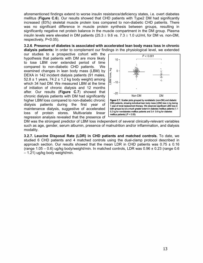
aforementioned findings extend to worse insulin resistance/deficiency states, i.e. overt diabetes mellitus (Figure C.6). Our results showed that CHD patients with Type2 DM had significantly increased (83%) skeletal muscle protein loss compared to non-diabetic CHD patients. There was no significant difference in muscle protein synthesis between groups, resulting in significantly negative net protein balance in the muscle compartment in the DM group. Plasma insulin levels were elevated in DM patients (25.3 ± 9.6 vs. 7.3 ± 1.0 uU/ml, for DM vs. non-DM, respectively, P<0.05). 3.2.6. Presence of diabetes is associated with accelerated lean body mass loss in chronic dialysis patients: In order to complement our findings in the physiological level, we extended our studies to a prospective cohort with the hypothesis that patients with DM are more likely to lose LBM over extended period of time compared to non-diabetic CHD patients. We examined changes in lean body mass (LBM) by DEXA in 142 incident dialysis patients (91 males, 52.8 ± 1 years, 74.2 ± 1.2 kg body weight) among which 34 had DM. We measured LBM at the time of initiation of chronic dialysis and 12 months after. Our results (Figure C.7) showed that chronic dialysis patients with DM had significantly higher LBM loss compared to non-diabetic chronic dialysis patients during the first year of maintenance dialysis, suggestive of accelerated loss of protein stores. Multivariate linear regression analysis revealed that the presence of DM was the strongest predictor of LBM loss independent of several clinically-relevant variables such as age, gender, serum albumin, presence of malnutrition and/or inflammation, and dialysis modality. 3.2.7. Leucine Disposal Rate (LDR) in CHD patients and matched controls. To date, we studied 6 CHD patients and 4 matched controls using the dual-clamp protocol described in approach section. Our results showed that the mean LDR in CHD patients was 0.75 ± 0.16 (range 1.05 – 0.6) ug/kg body/weight/min. In matched controls, LDR was 0.96 ± 0.23 (range 0.6 – 1.21) ug/kg body weight/min.
Figure C.7. Scatter plots grouped by nondiabetic (non-DM) and diabetic (DM) patients, showing individual lean body mass (LBM) loss in kg during 1 year of renal replacement therapy. We observed significant LBM loss in both groups but at a much greater extent in diabetes mellitus patients (1.1 0.2 kg for nondiabetes mellitus patients and 3.4 0.6 kg for diabetes mellitus patients) (P < 0.05)
Figure C.7. Scatter plots grouped by nondiabetic (non-DM) and diabetic (DM) patients, showing individual lean body mass (LBM) loss in kg during 1 year of renal replacement therapy. We observed significant LBM loss in both groups but at a much greater extent in diabetes mellitus patients (1.1 0.2 kg for nondiabetes mellitus patients and 3.4 0.6 kg for diabetes mellitus patients) (P < 0.05)

14
4.0 Inclusion/Exclusion Criteria Human Subjects Involvement and Characteristics: We propose to study 45 CHD subjects for this specific aim. The study subjects will be primarily selected from Vanderbilt’s outpatient dialysis center and the Nashville VA outpatient dialysis unit. There will be no restriction on gender (except pregnant women), race, and age (except less than 21 years old) for subject selection.
Inclusion Criteria: 1. Patients on CHD undergoing three time a week therapy for more than 6 months;
2. Age ≥ 21 years old;
3. Acceptable dialysis adequacy (spKt/V > 1.2);
4. A patent, well-functioning, arterio-venous dialysis access;
5. Ability to give informed consent;
6. Life expectancy greater than 6 months; 7. BMI ≥ 20 and ≤ 45.
Exclusion Criteria: 1. Pregnancy;
2. Intolerance or allergy to the study medication (including the metabolic clamp studies);
3. Severe, unstable, active inflammatory disease (active infection, active connective tissue disorder), active cancer or cancer history in the prior 5 years except skin cancer, AIDS-HIV, active or history of liver disease (including HBV and HCV);
4. Hospitalization or infection within 1 month prior to the study;
5. Patients receiving steroids and/or other immunosuppressive agents (Prednisone > 5 mg/day; excluding inhaled and topical steroids);
6. Diabetes Mellitus on insulin therapy;
7. Previous history of TB with or without documented adequate therapy;
8. Patients with recent close exposure to an individual with active TB;
9. Females using oral contraceptives;
10. Patients with NYHA Class III or IV heart failure;
11. Patients with a history of angina, myocardial infarction, transient ischemic attacks, or strokes within the last 6 months.
Stopping criteria: If subjects have any signs or symptoms of an infectious process (pneumonia, cellulitis, urinary tract infection, etc.) they will be dropped from the study. If a subject starts inhaled steroids or high potency topical steroids (with the exception of over the counter hydrocortisone cream) the subject may be dropped. In addition, if a subject has any signs or symptoms of decline or deterioration of their health status, the subject will be dropped. Additional stopping criteria are detailed in Section 7.0 under the risks associated with the two study drugs.

15
5.0 Enrollment/Randomization All studies will be conducted at the Vanderbilt University Medical Center General Clinical Research Center (GCRC). There will be no restriction on sex, race, age (≥ 21 years) or disease etiology (except as stated for each protocol) for subject selection or exclusion. Institutional review board approval and written informed consent will be obtained from all study subjects. We propose to study a total of 45 hemodialysis subjects. We expect 60% of subjects to be African Americans. The most common causes of ESRD in our clinic are diabetes (40%), hypertension (24%), and glomerulonephritis (24%).
Recruitment strategies: The study subjects will be primarily selected from Vanderbilt’s outpatient dialysis center and the Nashville VA outpatient dialysis unit. These units serve a total of 360 CHD patients (240 VU affiliated and 160 VA affiliated). The growth rate (incident patients) for our patient pool is 5-7% per year, providing a total of approximately 450 subjects over 4 years and approximately 300 subjects that would be predicted to be eligible for our study. Accordingly, we need 15-20% recruitment rate for sample size proposed for Specific Aim 2.
Diabetes Mellitus: In this study, we have elected to include subjects with the known diagnosis of diabetes mellitus. We will exclude subjects who are receiving insulin. Our preliminary data is highly suggestive that this patient cohort is specifically influenced by these abnormalities and represent appropriate target for intervention.
Randomization: Subjects will be randomly assigned to one of the study arms (Anakinra versus Actos versus Placebo). Randomization will be determined by a computer-generated sequence and will be stratified by diabetes status.
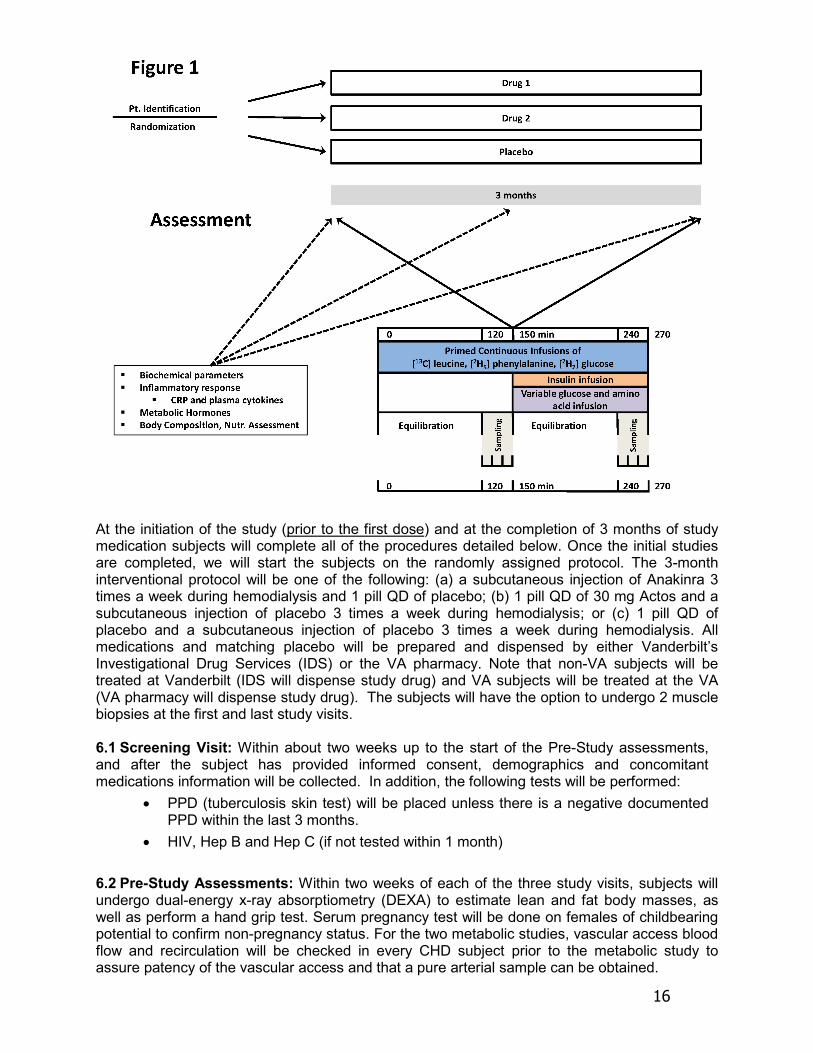
Standard follow-up measures for study subjects: All preventive measures such as optimal doses of dialysis, provision of adequate dietary protein and energy intake and appropriate control of metabolic abnormalities, such as metabolic acidosis, will be employed (11, 60). Further, treatment of metabolic acidosis improves nutritional markers in dialysis patients (72). Additional bicarbonate will be prescribed to keep pre-dialysis total bicarbonate levels above 22 mmol/L (above 24 mmol/L post-dialysis). Patients’ calcium, phosphorus, and hematocrit levels will also be followed closely and kept within accepted ranges. Patients will be prescribed phosphate binders to control hyperphosphatemia to levels below 6.0 mg/dl. Hematocrit will be followed more closely (twice per month) and erythropoietin will be prescribed to keep hematocrit within the range of 33 - 36% per recommended clinical practice guidelines. Also, all participants will have a baseline assessment, as well as on-going monitoring, of their health status as documented in their medical record. The study duration is approximately 4 months (2-4 weeks of baseline assessment followed by 3 months of treatment and 2 weeks post study follow up). 6.0 Study Procedures The design is randomized, double-blind, placebo-controlled (Figure 1). Once the subject is determined to be eligible for the study, we will randomly assign him/her to one of the study arms (Anakinra versus Actos versus Placebo). Each subject will be treated for 3 months. During the study, subjects will receive both subcutaneous injection of Anakinra or matching placebo and oral administration of Actos or matching placebo. The investigators and the study subjects will be blinded to the treatment. Subjects will be assessed for body composition and biochemical parameters at the beginning, at 6 weeks and at the end of the study. A dual-clamp metabolic study will be performed at the beginning and at the end of the study.
16
At the initiation of the study (prior to the first dose) and at the completion of 3 months of study medication subjects will complete all of the procedures detailed below. Once the initial studies are completed, we will start the subjects on the randomly assigned protocol. The 3-month interventional protocol will be one of the following: (a) a subcutaneous injection of Anakinra 3 times a week during hemodialysis and 1 pill QD of placebo; (b) 1 pill QD of 30 mg Actos and a subcutaneous injection of placebo 3 times a week during hemodialysis; or (c) 1 pill QD of placebo and a subcutaneous injection of placebo 3 times a week during hemodialysis. All medications and matching placebo will be prepared and dispensed by either Vanderbilt’s Investigational Drug Services (IDS) or the VA pharmacy. Note that non-VA subjects will be treated at Vanderbilt (IDS will dispense study drug) and VA subjects will be treated at the VA (VA pharmacy will dispense study drug). The subjects will have the option to undergo 2 muscle biopsies at the first and last study visits. 6.1 Screening Visit: Within about two weeks up to the start of the Pre-Study assessments, and after the subject has provided informed consent, demographics and concomitant medications information will be collected. In addition, the following tests will be performed:
• PPD (tuberculosis skin test) will be placed unless there is a negative documented PPD within the last 3 months.
• HIV, Hep B and Hep C (if not tested within 1 month)
6.2 Pre-Study Assessments: Within two weeks of each of the three study visits, subjects will undergo dual-energy x-ray absorptiometry (DEXA) to estimate lean and fat body masses, as well as perform a hand grip test. Serum pregnancy test will be done on females of childbearing potential to confirm non-pregnancy status. For the two metabolic studies, vascular access blood flow and recirculation will be checked in every CHD subject prior to the metabolic study to assure patency of the vascular access and that a pure arterial sample can be obtained.

17
6.3 Pre-Study Diet: Within two weeks prior to each metabolic study, subjects will be interviewed using the NDS-R system for 2 random food recalls. For 2 weeks prior to each metabolic study, subjects will be asked to eat a diet based on recommended dietary protein and calorie intakes for their weights (1.2 g/kg BW/day of protein, 30 kcal/kg BW/day energy, and low Na) [47]. This is critically important as changes in nutrient intake can alter the responses measured in the metabolic studies. The subjects will be fed a meal at the CRC the evening before the study. 6.4 Relation to Dialysis Procedure: Subjects will be studied on a non-dialysis day. 6.5 Metabolic Clamp Study: Day 1: Subjects will be admitted to the GCRC the night prior to the study. The last meal will be about 8 hours prior to the study but ad lib water consumption will be allowed (post-absorptive state). Day 2: All hemodialysis patients will have either a forearm arterio-venous (AV) shunt created for the dialysis treatment, either in the form of AV fistula or synthetic graft or AV shunt in the thigh. A needle will be placed at the venous site of the AV access to collect a baseline blood sample (for isotopic backgrounds) and once the baseline sampling is completed, to initiate isotopes infusion. The arterial vascular access obtained at the arterial side of the AV access will be used to sample arterial blood. If possible, another catheter will be placed into the contra lateral forearm vein (preferably in a retrograde fashion) to obtain samples of venous blood. The hematocrit will be checked prior to starting the study. If pre-study hematocrit is less than 30%, then a concentrated blood draw schedule will be employed (70 ml blood draw maximum). If the pre-study hematocrit is less than 27%, the study will be postponed and the subject’s primary nephrologist will be informed. Also prior to starting the study, metabolic cart will be performed to measure simultaneous energy expenditure and respiratory quotient over 20 minutes, and arm (or leg) blood flow will be estimated using capacitance plethysmography. A schematic diagram of the metabolic clamp study is depicted in Figure 2. In brief, at time zero (T=0) baseline blood sampling will be done, baseline amino acid levels will be checked by bedside HPLC, and a baseline breath sample will be obtained. Subjects will then receive a bolus injection of NaH13CO3 (0.12 mg/kg), L-(1-13C) Leucine (7.2 μmol/kg), L-(ring-2H5) Phenylalanine (3.6 μmol/kg), and 6,6-2H2 Glucose (3.6 mg/kg) to prime the CO2, Leucine, Phenylalanine, and Glucose pools, respectively. A continuous infusion of labeled Leucine (0.12 μmol/kg/min), Phenylalanine (0.06 μmol/kg/min) and Glucose (0.06 mg/kg/min) will then be started and continued throughout the remainder of the study. The initial 2 hours will be for tracer equilibration. During this period, arm (or leg) blood flow will be estimated again using capacitance plethysmography.
After two hours (T=120), we will obtain samples every 10 minutes over 30 minutes for determination of protein turnover. We will also obtain 2 breath samples. At time 150 minutes, subjects will receive insulin infusion at the concentration of 60 mU/m2/min. Blood glucose will be checked every 5 minutes to achieve normoglycemia (95-105 mg/dl) using variable infusion of 20% dextrose, and also amino acid levels will be checked every 10 minutes using rapid beside HPLC methodology [48] to achieve
Figure 2: Schematic diagram of the metabolic study day protocol. Metabolic cart and plethysmography measurements will be performed during the equilibration phase. A primed constant infusion of L-(1-13C) leucine, L-(ring-2H5) phenylalanine and 6,6-2H2 glucose will be maintained throughout the entire study (270 minutes).

18
baseline amino acid concentrations of leucine and phenylalanine using variable infusion of amino acid supplementation, the start of which may be delayed to allow achieving an interim state of normoglycemia (per PI discretion). It is anticipated that normoglycemia and normo-amino acidemia will be achieved within 90-180 minutes. At time 240 minutes, we will obtain additional blood samples every 10 minutes over 30 minutes for measurement of protein turnover. We will also obtain 2 breath samples. Blood samples obtained every 5 minutes for glucose and every 10 minutes for amino acid concentrations over the same 30 minute period will allow determination of insulin sensitivity, as well as carbohydrate and protein metabolism, respectively. A similar set of samples will also be collected if the interim normoglycemia state is achieved.
We will also monitor serum potassium levels every 30 minutes and infuse potassium chloride to maintain eukalemia.
Optional muscle biopsy may be obtained before and after the study. Also, if the subject is able to produce urine, we will collect a sample.
Once the study is completed, the stable isotope, amino acids and insulin infusions will be discontinued and subjects will be fed. The dextrose infusion will be gradually decreased over time with blood glucose being checked every 15 minutes. Once the blood glucose level has stabilized, the dextrose infusion will be discontinued and the blood sugar will be monitored every 15 minutes for about another hour. At this point, depending on the clinical status of the hemodialysis subjects, we may perform a post-study dialysis procedure either at the GCRC or at one of the outpatient dialysis clinics. Standard discharge instructions will be provided to the subject by the GCRC including contact information. After the baseline study visit subject will be followed up with at dialysis for first injection. After the end of study (Month 3) we will follow up within about 2 weeks at their dialysis session. 6.6 Other assessments: We will assess the following parameters at baseline, week 6 and end of study. Biochemical parameters: comprehensive metabolic panel, phosphorous, prealbumin,
lipid panel, transferrin, HbA1c Inflammatory response: CRP and plasma cytokines (IL-1, IL-6, IL-10, IL-12, TNFα) Metabolic hormones: insulin, adiponectin, resistin, leptin Oxidative stress markers: F2-isoprostane, ADMA, cysteine, homocysteine, and
glutathione Plasma amino acids and free fatty acids Body composition assessment: DEXA Hand grip test
6.7 Methods: All methods used in our studies have been standardized in our and co-investigators’ laboratories [23, 35, 49, 50]. Standard techniques Nutritional Assessment: We will use standard and ESRD modified techniques to assess nutritional status of the patients. These will include visceral protein concentrations (serum albumin, prealbumin, cholesterol, and transferrin) and somatic protein stores (anthropometrics and body composition).
Body composition analysis: We will primarily use DEXA for body composition analysis. The studies will be performed within 2 weeks of each of the three study visits. Our group has extensive experience with these methodologies in CHD patients and published a manuscript regarding their utility (91). While these methodologies have their inherent problems, the repeated measures in the same patients will minimize the variability.

19
Dietary Protein and Energy Intake: Prior to each metabolic clamp study, the subjects will be interviewed using the NDS-R system for 2-day random food recalls during a two-week period. This software is available at the GCRC.
Metabolic cart: A metabolic cart will be used to measure simultaneous resting energy expenditure and respiratory quotient.
Breath sampling: During the metabolic study we will collect breath samples several times from the subject via a Douglas bag with duplicate 25 ml samples placed into non-siliconized glass vacutainer tubes for measurement of breath 13CO2 enrichment. Additional samples may be collected if an interim normoglycemia state is achieved. Blood flow measurements: Blood flow measurements will be obtained twice—once before the metabolic study begins and the second during the equilibration phase. Arm (or leg) blood flow will be estimated using capacitance plethysmography. 7.0 Risks of Investigational Agents/Devices (side effects) Chronic hemodialysis patients are at high risk of morbidity and mortality due to the co-morbidities associated with the chronic kidney disease.
• Inconvenience of reporting to the GCRC and staying one night for each metabolic clamp study.
• The insulin infusion can lower blood potassium levels. If this drops too low, your heart rhythm may be affected. We will closely monitor potassium levels (every 30 minutes), and place the subject on a machine that monitors the heart to decrease this risk and to observe any complications. If a decrease in potassium occurs, it will be replaced intravenously.
• Replacing the potassium may cause slight burning or irritation and the subject’s heart rhythm may be affected. Again, we will monitor the subject’s heart to decrease this risk and adjust the rate of replacement accordingly.
• There is a small risk of low blood sugar. If this occurs, the subject may experience any of these symptoms: feeling lightheaded, headache, fast heartbeat, sweating, blurred vision, hunger, and rarely seizures. We will check blood sugar every 5 minutes. If the blood sugar level drops below normal, it will be restored intravenously.
• Inconvenience of having gelled electrodes placed on the chest and being connected to a heart monitor. The gelled electrodes used for ECG monitoring may cause skin irritation.
• Having to lie still for 10-15 minutes during the DEXA scan may be uncomfortable.
• The DEXA scan uses x-rays (or radioactivity). You will be exposed to a small amount of radiation. The radiation exposure that you would likely receive from a total of three DEXA scans is approximately equal to one month of exposure from natural background sources. If you are a female of childbearing potential, we will do a pregnancy test before the DEXA to make sure you are not pregnant.
• The IV placed in the forearm may cause a slight bruise and carries a risk of infection which is rare.
• Eating a recommended or stable diet for 2 weeks and providing 2 dietary recalls may be inconvenient.
• Breathing through a mouthpiece may be inconvenient for the subject and may make the subject feel lightheaded or dizzy.
20
• The blood pressure cuffs placed on the forearm/leg may be uncomfortable when inflated. For each blood flow test, one cuff will remain inflated up to 2 minutes. Another cuff will be inflated/deflated about 9 times over 2 minutes. The third cuff will remain uninflated.
• Inhaling and exhaling into a 1-liter Douglas bag may be an inconvenience for the subject, but it does not pose any risk.
• The isotopes used in the metabolic studies are not radioactive and do not present any additional risk to the subject.
• The fistula needles placed in the subject’s arm or leg carry a risk of infection but it is rare. All necessary aseptic procedures will be taken for needle insertion.
• The lidocaine used for local anesthetic prior to IV insertion or muscle biopsy may cause numbness, burning and/or local rash or irritation, if the subject is allergic. There is a risk that lidocaine may cause problems with heart rhythm, but this is unlikely given the amount the subject will receive.
• Subjects will be asked not to take birth control pills during the study due to possible drug interactions.
• Subjects should avoid getting flu shots or other vaccines during the study period.
• The pills used in this study (Actos) can cause or make congestive heart failure worse in some patients. There have been reports of liver toxicity, anemia and bone fractures. Subjects will be monitored closely for signs of any of these adverse events. Subjects may be withdrawn from the study if they experience any of these events.
• The injection used in this study (Anakinra) has some adverse effects such headache and redness/pain at the injection site. If these effects persist or worsen, subjects will be asked to notify the study doctor promptly. Other less common side effects include nausea, diarrhea, abdominal pain, sinus problems, and/or flu-like symptoms. If these effects occur, subjects will be instructed to contact the study team immediately. This drug may rarely cause a decrease in the white blood cell (WBC) count, which could be life threatening. We will monitor WBC count on a weekly basis to make sure the WBC is within normal limits. Subjects may be withdrawn from the study if they experience any of these events.
• Risk of injected placebo includes redness/pain at the injection site.
• The PPD tuberculosis skin test may cause mild pain at the injection site.
• Taking the placebo pills may be an inconvenience.
• Optional Muscle Biopsy. The muscle biopsy may cause muscle soreness, bruising, infection or a small blood filled bump (hematoma) at the biopsy site. There is a small chance that some slight bleeding may also occur. The soreness may last as long as 48 hours.
As with all research studies, there are unidentifiable and unforeseeable risks that may occur during this study. All women of child-bearing potential will have a serum pregnancy test prior to continuing in the study. 8.0 Reporting of Adverse Events or Unanticipated Problems involving Risk to
Participants or Others Safety information will be assessed by subject interview. As these results are collected, all adverse events (AE) will be identified and reported to the principle investigator within fourteen
21
(14) days. Adverse events will be reported as described below. The principal investigator (PI) is responsible for evaluating each AE and for determining whether the AE affects the risk/benefit ratio of the study and whether modifications to the protocol and consent form are required. Treatment will be discontinued for any subject who develops conditions during the course of the study that are felt to significantly alter the risk/benefit profile for the individual subject (including but not limited to the development of angina, myocardial infarction, stroke, TIA, ulcer disease, gastrointestinal bleeding, worsening liver function, or signs/symptoms of increasing extracellular fluid volume overload). Adverse event grading. All adverse events will be graded as follows:
Severity 0 = No adverse event or within normal limits 1 = Mild—did not require treatment 2 = Moderate—resolved with treatment 3 = Severe—required professional medical attention 4 = Life-threatening or disabling 5 = Death
Related to study drug (there is a reasonable possibility that the experience may have been caused by the drug)
0 = Unrelated 1 = Unknown 2 = Related
Unexpected event (an AE with specificity or severity not consistent with the risk information in the protocol/application or an AE that has not been previously observed)
0 = No 1 = Yes
Serious (any AE occurring at any dose that results in death; a life-threatening adverse drug experience; inpatient hospitalization or prolongation of existing hospitalization; a persistent or significant disability/incapacity; a congenital anomaly/birth defect; or any important medical event that, based on medical judgment, jeopardizes the subject and may require medical or surgical intervention to prevent one of the above outcomes)
0 = No 1 = Yes
Adverse event reporting. Any adverse experience associated with the use of drug that is serious, unexpected and possibly or definitely study related will be reported in writing to the IRB within 10 working days of the PI’s first knowledge of the occurrence. The PI will also notify DVA by telephone or by FAX of any unexpected fatal or life-threatening experience associated with the use of the drug within 5 business days after initial receipt of the information. The PI will review all expedited adverse event reports. In addition, any deviations from protocol will be reported to the IRB.
The annual summary of all adverse events and any audit reports will be sent to the IRB and to the study DSMB. A written annual report will also be submitted to the DVA. Data Safety and Monitoring Board (DSMB). We will establish a DSMB for the purposes of this study. The board will consist of the following members from the Vanderbilt University Medical Center:
Naji Abumrad, MD (Department of Surgery); Chair Satish Raj, MD (Division of Clinical Pharmacology) Chang Yu, PhD (Department of Biostatistics)

22
The members of the DSMB will meet after enrollment of 15, 30 and 45 subjects or annually (whichever comes first) via conference call, email, or face-to-face. The DSMB will review study progress, adverse event logs and any preliminary data. DSMB summary reports will be provided to the IRB according to the IRB’s policies and procedures, as well as annually to the DVA. 9.0 Study Withdrawal/Discontinuation Patients who are non-compliant with the study protocol or no longer wish to participate in the study will be withdrawn from the study. 10.0 Statistical Considerations 10.1. Power Calculations Sample size computation was based on the following matched group comparisons using independent t-tests between an insulin sensitizer vs. placebo. We also anticipate similar effects with anti-inflammatory agent vs. placebo. For each comparison, sample size of 15 was used.
Specific Aim: To test the hypothesis that inhibiting inflammatory response by administration of an IL-1r antagonist (Anakinra) or increasing insulin sensitivity by administration of a PPARγ agonist (Actos) will improve net protein metabolism. To compare the chronic effects of an insulin sensitizer versus placebo on the rate of amino acid (Leucine) disposal rate (LDR) in dialysis subjects.
We are planning a study with 15 pairs of subjects. Prior data indicate that LDR is normally distributed with standard deviation ranging from 0.16 to 0.23 in CHD and control patients, respectively. If the true difference in the experimental and control means is 0.21, we will be able to reject the null hypothesis that the population means of the experimental and control groups are equal with probability (power) 0.933. The Type I error probability associated with this test of this null hypothesis is 0.05. These differences are biologically plausible since our preliminary data in healthy controls showed LDR of 9.6 ± 0.23 ug/kg/min, an absolute difference of 0.21 ug/kg/min. When we used the SD of 0.23 considering the true difference in the experimental and control means of 0.21, we will be able to reject the null hypothesis that the population means of the experimental and control groups are equal with probability (power) 0.673. 10.2. Data analysis plan Baseline characteristics will be compared using one-way analysis of variance (ANOVA) or Kruskal-Wallis test for continuous variables according to their distributions and chi-square or Fisher’s exact test for categorical variables in order to assess the success of randomization. In order to assess improvement by the intervention, independent sample t-test or Wilcoxon rank sum test will be used to compare rate of change “before” and “after“ leucine disposal rate separately between placebo to CHD subjects who are randomized to either anti-inflammatory or to insulin sensitizer agent. We will also perform ANCOVA (as described under Data analysis plan for the aim 1) analysis using multiple linear regression adjusting baseline values to assess difference in % of improvement from baseline between groups.
When distribution of baseline characteristics variables is not equally distributed nevertheless of randomization, we will adjust the un-evenly distributed variables in a regression analysis as potential confounders. For the data with 45 subjects, we will include up to 4 covariates such as BMI, age, gender and inflammatory state at entry if they are distributed un-evenly between arms plus two dummy variables for intervention groups to avoid over-fitting. Normality in residuals in the multiple regression model will be assessed, and we will attempt transformation of outcome variable to achieve normality. Changes during experimental protocols within and inter-groups involving more than 2 time-point measures of outcomes will be analyzed by linear mixed effect regression models.
23
11.0 Privacy/Confidentiality Issues All efforts, within reason, will be made to keep personal information in the research record confidential but total confidentiality cannot be guaranteed. Each participant will be assigned a unique study identification number and will be referred to by this number to protect their identity over the course of the study. The numbers will be assigned sequentially. Only key study personnel will have access to these numbers. The codes will be kept in a password protected database on the VU server. No information will be given to anyone without permission from the subject. All data will be identified with the assigned study identification number unique to the subject.
All access to protected health information (PHI) as defined by current and future federal standards will be carefully managed. Best effort will be made to de-identify PHI in all case report forms. All physical records (including case report forms that contain PHI) are stored in a locked office and only study personnel will have access. All computers containing patient information are behind locked doors in rooms with limited access.
The database is housed on the Vanderbilt University secured computer network and is password protected. Only members of the study team will have access to this database.
Adverse event reports and annual summaries will not include subject-identifiable material but only the assigned study identification numbers. 12.0 Follow-up and Record Retention The study will be conducted over the course of three years. Records will be retained indefinitely. Data will be stored on the Vanderbilt University computer network in a password protected database. Only members of the study team will have access. Only Key Study Personnel will have access to the subject’s code. The code will be destroyed after study closure. Paper case report forms and other pertinent paper documentation will be kept in a locked office and only study personnel will have access. Only personnel directly involved with the study will have access to source data and the electronic database. All research data will be maintained by the PI after study closure. The PI will maintain the research data indefinitely. REFERENCES
1. Flakoll PJ, Jensen MD, Cherrington AC. Physiologic action of insulin. In: Leroith D, I. TS, Olefsky J (eds). Diabetes Mellitus: A Fundamental and Clinical Text, 3 edn. Lippcott Williams & Wilkins: Philadelphia, PA, 2004, pp 165-181.
2. Chevalier S, Marliss EB, Morais JA, Lamarche M, et al. Whole-body protein anabolic response is resistant to the action of insulin in obese women. Am J Clin Nutr 2005; 82: 355-365.
3. DeFronzo RA, Alvestrand A, Smith D, Hendler R, et al. Insulin resistance in uremia. J Clin Invest 1981; 67: 563-568.
4. Lee SW, Dai G, Hu Z, Wang X, et al. Regulation of muscle protein degradation: coordinated control of apoptotic and ubiquitin-proteasome systems by phosphatidylinositol 3 kinase. Journal of the American Society of Nephrology 2004; 15: 1537-1545.
5. Siew ED, Pupim LB, Majchrzak KM, Shintani A, et al. Insulin resistance is associated with skeletal muscle protein breakdown in non-diabetic chronic hemodialysis patients. Kidney Int 2006.
24
6. Brunelli SM, Thadhani R, Ikizler TA, Feldman HI. Thiazolidinedione use is associated with better survival in hemodialysis patients with non-insulin dependent diabetes. Kidney Int 2009.
7. US Renal Data System. Excerpts from the USRDS 2001 Annual Data Report. Am J Kidney Dis 2001; 38: S1- S248.
8. Bergstrom J. Protein catabolic factors in patients on renal replacement therapy. Adv Exp Med Biol 1989; 260: 1-9.
9. Bergstrom J. Nutrition and mortality in hemodialysis. J Am Soc Nephrol 1995; 6: 1329-1341.
10. Hakim RM, Levin N. Malnutrition in hemodialysis patients. Am J Kidney Dis 1993; 21: 125-137.
11. Ikizler TA, Hakim RM. Nutrition in end-stage renal disease. Kidney Int 1996; 50: 343-357.
12. Kopple JD. Effect of nutrition on morbidity and mortality in maintenance dialysis patients. Am J Kidney Dis 1994; 24: 1002-1009.
13. Mitch WE, Walser M. Nutritional therapy of the uremic patient. In: B.M.Brenner, F.C.Rector (eds). The Kidney, 4 edn. Saunders: Philadelphia, 1991, pp 2186-2222.
14. Mitch WE, Goldberg AL. Mechanism of muscle wasting:The role of ubiquitin-proteasome pathway. N Engl J Med 1997; 335: 1897-1905.
15. Churchill DN, Taylor DW, Cook RJ, al e. Canadian hemodialysis morbidity study. Am J Kidney Dis 1992; 19: 214-234.
16. Iseki K, Kawazoe N, Fukiyama K. Serum albumin is a strong predictor of death in chronic dialysis patients. Kidney Int 1993; 44: 115-119.
17. Lowrie EG, Huang WH, Lew NL, Liu Y. The relative contribution of measured variables to death risk among hemodialysis patients. In: E.A.Friedman (ed). Death on Hemodialysis. Kluwer Academic Publishers: Amsterdam, 1994, pp 121-141.
18. Ikizler TA, Wingard RL, Harvell J, Shyr Y, et al. Association of morbidity with markers of nutrition and inflammation in chronic hemodialysis patients: a prospective study. Kidney Int 1999; 55: 1945-1951.
19. Collins AJ, Ma JZ, Umen A, Keshaviah P. Urea index and other predictors of hemodialysis patient survival. Am J Kidney Dis 1993; 23: 272-282.
20. Goldwasser P, Mittman M, Antignani A, Burrel D, et al. Predictors of mortality on hemodialysis. J Am Soc Nephrol 1993; 3: 1613-1622.
21. Owen Jr. WF, Lew NL, Liu Y, Lowrie EG, et al. The urea reduction ratio and serum albumin concentrations as predictors of mortality in patients undergoing hemodialysis. New England Journal of Medicine 1993; 329: 1001-1006.
22. Kopple JD, Coburn JW. Metabolic studies of low protein diets in uremia. I. Nitrogen and potassium. Medicine (Baltimore) 1973; 52: 583-595.
23. Mitch WE. Dietary requirements of predialysis patients for protein and calories. In: M.E.Mitch, Klahr S (eds). Handbook of Nutrition and the Kidney, 5th edn. Lippincott-Williams & Wilkins: Philadelphia, 2005, pp 115-137.
24. Motil KJ, Matthews DE, Bier DM, Burke JF, et al. Whole-body leucine and lysine metabolism: response to dietary protein intake in young men. Am J Physiol 1981; 240: E712-721.
25. Runyan JW, Jr., Hurwitz D, Robbins SL. Effect of Kimmelstiel-Wilson syndrome on insulin requirements in diabetes. N Engl J Med 1955; 252: 388-391.
26. Zubrod CG, Eversole SL, Dana GW. Amelioration of diabetes and striking rarity of acidosis in patients with Kimmelstiel-Wilson lesions. N Engl J Med 1951; 245: 518-528.
27. Rabkin R, Simon NM, Steiner S, Colwell JA. Effect of renal disease on renal uptake and excretion of insulin in man. N Engl J Med 1970; 282: 182-187.
28. DeFronzo RA, Tobin JD, Rowe JW, Andres R. Quantification of pancreatic beta cell sensitivity to glucose and tissue sensitivity to insulin. J Clin Invest 1978; 62: 425-435.
29. DeFronzo RA, Smith D, Alvestrand A. Insulin action in uremia. Kidney Int Suppl 1983; 16: S102-114.

25
30. Du J, Hu Z, Mitch WE. Molecular mechanisms activating muscle protein degradation in chronic kidney disease and other catabolic conditions. Eur J Clin Invest 2005; 35: 157-163.
31. Lee SW, Dai G, Hu Z, Wang X, et al. Regulation of muscle protein degradation: coordinated control of apoptotic and ubiquitin-proteasome systems by phosphatidylinositol 3 kinase. J Am Soc Nephrol 2004; 15: 1537-1545.
32. Du J, Mitch WE. Identification of pathways controlling muscle protein metabolism in uremia and other catabolic conditions. Curr Opin Nephrol Hypertens 2005; 14: 378-382.
33. Pupim LB, Flakoll PJ, Majchrzak KM, Aftab Guy DL, et al. Increased muscle protein breakdown in chronic hemodialysis patients with type 2 diabetes mellitus. Kidney Int 2005; 68: 1857-1865.
34. Pupim LB, Heimburger O, Qureshi AR, Ikizler TA, et al. Accelerated lean body mass loss in incident chronic dialysis patients with diabetes mellitus. Kidney Int 2005; 68: 2368-2374.
35. Brunelli SM, Thadhani R, Ikizler TA, Feldman HI. Thiazolidinedione use is associated with better survival in hemodialysis patients with non-insulin dependent diabetes. Kidney Int 2009; 75: 961-968.
36. Chalasani NP, Sanyal AJ, Kowdley KV, Robuck PR, et al. Pioglitazone versus vitamin E versus placebo for the treatment of non-diabetic patients with non-alcoholic steatohepatitis: PIVENS trial design. Contemporary Clinical Trials 2009; 30: 88-96.
37. Kaysen GA. The microinflammatory state in uremia: causes and potential consequences. J Am Soc Nephrol 2001; 12: 1549-1557.
38. Bergstrom J, Lindholm B, Lacson E, Jr., Owen W, Jr., et al. What are the causes and consequences of the chronic inflammatory state in chronic dialysis patients? Semin Dial 2000; 13: 163-175.
39. Panichi V, Tetta C, Rindi P, Palla R, et al. Plasma C-reactive protein is linked to backfiltration associated interleukin-6 production. Asaio J 1998; 44: M415-417.
40. Ross R. Atherosclerosis--an inflammatory disease. N Engl J Med 1999; 340: 115-126. 41. Bistrian BR. Role of the systemic inflammatory response syndrome in the development
of protein-calorie malnutrition in ESRD. Am J Kidney Dis 1998; 32: S113-117. 42. Martin F, Santolaria F, Batista N, Milena A, et al. Cytokine levels (IL-6 and IFN-gamma),
acute phase response and nutritional status as prognostic factors in lung cancer. Cytokine 1999; 11: 80-86.
43. de Martino M, Galli L, Chiarelli F, Verrotti A, et al. Interleukin-6 release by cultured peripheral blood mononuclear cells inversely correlates with height velocity, bone age, insulin-like growth factor-I, and insulin-like growth factor binding protein-3 serum levels in children with perinatal HIV-1 infection. Clin Immunol 2000; 94: 212-218.
44. Kwak KS, Zhou X, Solomon V, Baracos VE, et al. Regulation of protein catabolism by muscle-specific and cytokine-inducible ubiquitin ligase E3alpha-II during cancer cachexia. Cancer Res 2004; 64: 8193-8198.
45. Abad LW, Schmitz HR, Parker R, Roubenoff R. Cytokine responses differ by compartment and wasting status in patients with HIV infection and healthy controls. Cytokine 2002; 18: 286-293.
46. Nova E, Gomez-Martinez S, Morande G, Marcos A. Cytokine production by blood mononuclear cells from in-patients with anorexia nervosa. Br J Nutr 2002; 88: 183-188.
47. Ikizler TA. Nutrition, inflammation and chronic kidney disease. Curr Opin Nephrol Hypertens 2008; 17: 162-167.
48. Caglar K, Fedje L, Dimmitt R, Hakim RM, et al. Therapeutic effects of oral nutritional supplementation during hemodialysis. Kidney Int 2002.
49. Caglar K, Peng Y, Pupim LB, Flakoll PJ, et al. Inflammatory signals associated with hemodialysis. Kidney Int In press 2002.
50. Danielski M, Ikizler TA, McMonagle E, Kane JC, et al. Linkage of hypoalbuminemia, inflammation, and oxidative stress in patients receiving maintenance hemodialysis therapy. Am J Kidney Dis 2003; 42: 286-294.
26
51. Ikizler TA, Flakoll PJ, Parker RA, Hakim RM. Amino acid and albumin losses during hemodialysis. Kidney Int 1994; 46: 830-837.
52. Ikizler TA, Greene J, Wingard RL, Parker RA, et al. Spontaneous dietary protein intake during progression of chronic renal failure. J Am Soc Nephrol 1995; 6: 1386-1391.
53. Ikizler TA, Greene JH, Wingard RL, Hakim RM. Nitrogen balance in hospitalized chronic hemodialysis patients. Kidney Int Suppl 57 1996; 50: S53-S56.
54. Ikizler TA, Pupim RB, Brouillette JR, Levenhagen DK, et al. Hemodialysis stimulates muscle and whole-body protein loss and alters substrate oxidation. Am J Physiol Endocrinol Metab 2002; 282: E107-E116.
55. Ikizler TA, Wingard RL, Sun M, Harvell J, et al. Increased energy expenditure in hemodialysis patients. J Am Soc Nephrol 1996; 7: 2646-2653.
56. Parker III TF, Wingard RL, Husni L, Ikizler TA, et al. Effect of the membrane biocompatibility on nutritional parameters in chronic hemodialysis patients. Kidney Int 1996; 49: 551-556.
57. Pupim LB, Evanson JA, Hakim RM, Ikizler TA. The extent of uremic malnutrition at the time of initiation of maintenance hemodialysis is associated with subsequent hospitalization. J Ren Nutr 2003; 13: 259-266.
58. Pupim LB, Kent P, Caglar K, Shyr Y, et al. Improvement in nutritional parameters after initiation of chronic hemodialysis. Am J Kidney Dis 2002; 40: 143-151.
59. Hung A, Pupim L, Yu C, Shintani A, et al. Determinants of C-reactive protein in chronic hemodialysis patients: Relevance of dialysis catheter utilization. Hemodial Int 2008; 12: 236-243.
60. May RC, Kelly RA, Mitch WE. Mechanisms for defects in muscle protein metabolism in rats with chronic uremia: The influence of metabolic acidosis. J Clin Invest 1987; 79: 1099-1103.
61. Kimmel PL, Phillips TM, Simmens SJ, Peterson RA, et al. Immunologic function and survival in hemodialysis patients. Kidney Int 1998; 54: 236-244.
62. Owen WF, Lowrie EG. C-reactive protein as an outcome predictor for maintenance hemodialysis patients. Kidney Int 1998; 54: 627-636.
63. Stenvinkel P, Heimburger O, Paultre F, Diczfalusy U, et al. Strong association between malnutrition, inflammation, and atherosclerosis in chronic renal failure. Kidney Int 1999; 55: 1899-1911.
64. Qureshi AR, Alvestrand A, Danielsson A, Divino-Filho JC, et al. Factors predicting malnutrition in hemodialysis patients: a cross-sectional study. Kidney Int 1998; 53: 773-782.
65. Flakoll PJ, Brown LL, Frexes-Steed M, Abumrad NN. Use of amino acid clamps to investigate the role of insulin in regulating protein breakdown in vivo. JPEN J Parenter Enteral Nutr 1991; 15: 81S-85S.
66. Kopple JD. National kidney foundation K/DOQI clinical practice guidelines for nutrition in chronic renal failure. Am J Kidney Dis 2001; 37: S66-70.
67. DeFronzo RA, Tobin JD, Andres R. Glucose clamp technique: a method for quantifying insulin secretion and resistance. Am J Physiol 1979; 237: E214-223.
68. Brown LL, Williams PE, Becker TA, Ensley RJ, et al. Rapid high-performance liquid chromatographic method to measure plasma leucine: importance in the study of leucine kinetics in vivo. J Chromatogr 1988; 426: 370-375.
69. Lim VS, Yarasheski KE, Crowley JR, Fangman J, et al. Insulin is protein-anabolic in chronic renal failure patients. J Am Soc Nephrol 2003; 14: 2297-2304.
70. Workeneh BT, Rondon-Berrios H, Zhang L, Hu Z, et al. Development of a diagnostic method for detecting increased muscle protein degradation in patients with catabolic conditions. J Am Soc Nephrol 2006; 17: 3233-3239.
71. Frison L, Pocock SJ. Repeated measures in clinical trials: analysis using mean summary statistics and its implications for design. Stat Med 1992; 11: 1685-1704.
72. Pickering WP, Price SR, Bircher G, Marinovic AC, et al. Nutrition in CAPD: serum bicarbonate and the ubiquitin-proteasome system in muscle. Kidney Int 2002; 61: 1286-1292.
27
73. Bailey JL, Wang X, England BK, Price SR, et al. The acidosis of chronic renal failure activates muscle proteolysis in rats by augmenting transcription of genes encoding proteins of the ATP-dependent ubiquitin-proteasome pathway. J Clin Invest 1996; 97: 1447-1453.
74. Du J, Wang X, Miereles C, Bailey JL, et al. Activation of caspase-3 is an initial step triggering accelerated muscle proteolysis in catabolic conditions. J Clin Invest 2004; 113: 115-123.
75. Song YH, Li Y, Du J, Mitch WE, et al. Muscle-specific expression of IGF-1 blocks angiotensin II-induced skeletal muscle wasting. J Clin Invest 2005; 115: 451-458.
76. Hotamisligil GS, Peraldi P, Budavari A, Ellis R, et al. IRS-1-mediated inhibition of insulin receptor tyrosine kinase activity in TNF-alpha- and obesity-induced insulin resistance. Science 1996; 271: 665-668.
77. Du J, Hu Z, Mitch WE. Cellular signals activating muscle proteolysis in chronic kidney disease: a two-stage process. Int J Biochem Cell Biol 2005; 37: 2147-2155.
78. Glass DJ. Molecular mechanisms modulating muscle mass. Trends Mol Med 2003; 9: 344-350.
79. Kulik G, Klippel A, Weber MJ. Antiapoptotic signalling by the insulin-like growth factor I receptor, phosphatidylinositol 3-kinase, and Akt. Mol Cell Biol 1997; 17: 1595-1606.
80. Wang X, Hu Z, Hu J, Du J, et al. Insulin resistance accelerates muscle protein degradation: activation of the ubiquitin-proteasome pathway by defects in muscle cell signaling. Endocrinology 2006.
81. Caglar K, Peng Y, Pupim LB, Flakoll PJ, et al. Inflammatory signals associated with hemodialysis. Kidney Int 2002; 62 1408-1416.
82. Pupim LB, Himmelfarb J, McMonagle E, Shyr Y, et al. Influence of Initiation of Maintenance Hemodialysis on Biomarkers of Inflammation and Oxidative Stress. Kidney Int 2004; 65: 2371-2379.
83. Simmons EM, Langone A, Sezer MT, Vella JP, et al. Effect of renal transplantation on biomarkers of inflammation and oxidative stress in end-stage renal disease patients. Transplantation 2005; 79: 914-919.
84. Hasselgren PO, Fischer JE. Muscle cachexia: current concepts of intracellular mechanisms and molecular regulation. Ann Surg 2001; 233: 9-17.
85. Llovera M, Carbo N, Lopez-Soriano J, Garcia-Martinez C, et al. Different cytokines modulate ubiquitin gene expression in rat skeletal muscle. Cancer Lett 1998; 133: 83-87.
86. Tisdale MJ. Loss of skeletal muscle in cancer: biochemical mechanisms. Front Biosci 2001; 6: D164-174.
87. Guan Y, Breyer MD. Peroxisome proliferator-activated receptors (PPARs): Novel therapeutic targets in renal disease. Kidney Int 2001; 60: 14-30.
88. Fonseca V. Effect of thiazolidinediones on body weight in patients with diabetes mellitus. Am J Med 2003; 115 Suppl 8A: 42S-48S.
89. Majchrzak KM, Pupim LB, Chen K, Martin CJ, et al. Physical activity patterns in chronic hemodialysis patients: comparison of dialysis and nondialysis days. J Ren Nutr 2005; 15: 217-224.
90. Pupim LB, Flakoll PJ, Brouillette JR, Levenhagen DK, et al. Intradialytic parenteral nutrition improves protein and energy homeostasis in chronic hemodialysis patients. J Clin Invest 2002; 110: 483-492.
91. Flakoll PJ, Kent P, Neyra R, Levenhagen D, et al. Bioelectrical impedance vs air displacement plethysmography and dual-energy X-ray absorptiometry to determine body composition in patients with end-stage renal disease. JPEN J Parenter Enteral Nutr 2004; 28: 13-21.
92. Heinrikson RI, Meredith SC. Amino acid analysis by reverse-phase high pressure liquid chromatography: Precolumn derivatization with phenylisothiocyanate. Analytical Biochemistry 1984; 136: 65-74.
93. Garlick PJ, M. A. McNurlan, K. C. McHardy, A. G. Calder, E. Milne, L. M. Fearns, and J. Broom. Rates of nutrient utilization in man measured by combined respiratory gas
28
analysis and stable isotopic labeling: effect of food intake. Hum Nutr Clin Nutr 1987; 41: 177-191.
94. Biolo G, Maggi SP, Williams BD, Tipton KD, et al. Increased rates of muscle protein turnover and amino acid transport after resistance exercise in humans. Am J Physiol 1995; 268: E514-520.
95. Gelfand RA, and E. J. Barrett. Effect of physiologic hyperinsulinemia on skeletal muscle protein synthesis and breakdown in man. Journal of Clinical Investigation 1987; 80: 1-6.
96. Rooyackers OE, Nair KS. Hormonal regulation of human muscle protein metabolism. Annu Rev Nutr 1997; 17: 457-485.
97. Jaleel A, Nehra V, Persson XM, Boirie Y, et al. In vivo measurement of synthesis rate of multiple plasma proteins in humans. Am J Physiol Endocrinol Metab 2006; 291: E190-197.
98. Jequier E, K. Acheson, and Y. Schutz. Assessment of energy expenditure and fuel utilization in man. Annu Rev Nutr 1987; 7: 187-208.
99. Pilegaard H, Ordway GA, Saltin B, Neufer PD. Transcriptional regulation of gene expression in human skeletal muscle during recovery from exercise. Am J Physiol Endocrinol Metab 2000; 279: E806-814.
100. Bailey JL, Zheng B, Hu Z, Price SR, et al. Chronic kidney disease causes defects in signaling through the insulin receptor substrate/phosphatidylinositol 3-kinase/Akt pathway: implications for muscle atrophy. J Am Soc Nephrol 2006; 17: 1388-1394.
101. Guan Y, Hao C, Cha DR, Rao R, et al. Thiazolidinediones expand body fluid volume through PPARgamma stimulation of ENaC-mediated renal salt absorption. Nat Med 2005; 11: 861-866.
102. Gilbert RE. Rosiglitazone: opening Pandora's black box? Clin J Am Soc Nephrol 2007; 2: 1329-1331.