nutrition and fertilitydiposit.ub.edu/dspace/bitstream/2445/150981/1/tfg_matas_anaïs.pdf ·...
TRANSCRIPT

UNIVERSITAT DE BARCELONA
FACULTAT DE FARMÀCIA I CIÈNCIES DE L'ALIMENTACIÓ
TREBALL DE FI DE GRAU
NUTRITION AND FERTILITY
ANAÏS MATAS AYALA
Bibliographic research
Department of biochemistry and physiology
January, 2020
This work is licensed under a Creative Commons license.

INDEX
1. Summary and key words…………………………………….….………………………………….……………… 1
2. Introduction………………………………………………………………………………………………………………. 2
2.1. Physiology of the reproductive system…………………………….……………………………. 2
2.1.1. Female reproductive system…………………………….…………………………….. 2
2.1.2. Male reproductive system…………………………….………………………………… 5
2.2. Physiology of fertilization: ovum and spermatozoon role………………………………. 7
2.3. Infertility…………………………….…………………………………………………………………….…. 9
2.3.1. Definition and prevalence…………………………….………………………………… 9
2.3.2. Causes…………………………….……………………………………………………………… 9
2.3.3. Approaches to treat infertility…………………………….…………………………… 11
2.4. Importance of nutrients on fertility……………………………………………………………….. 12
3. Objectives………………….………………………………….…………………………….…………………………….. 13
4. Material and methods……………..…………………….…………………………….……………………………. 14
5. Detailed description of the actual knowledge of the topic………………………………………..… 15
5.1. Macronutrients…………….…………………………….………………………………………………… 15
5.1.1. Carbohydrates…………….…………………………….………..……………….………… 15
5.1.2. Fats…………….…………………………….……………………..……………………………. 16
5.1.3. Protein…………….……..……………………..…….……………………………………….. 17
5.1.3.1. Dairy…………….…………………………….……………………….…………… 18
5.1.3.2. Animal protein…………….…………………………….………..…………… 18
5.1.3.3. Soy…………….…………………………….………………………………………. 19
5.2. Micronutrients…………….………………………………………….………………….………………… 20
5.2.1. Folic acid…………….…………………………….……………………………………………. 20
5.2.2. Vitamin D…………….…………………………….…………………………………………… 21
5.2.3. Vitamin A…………….…………………………….…………………………………………… 22
5.2.4. Vitamins E and C…………….…………………………….……………………….….……. 22
5.2.5. Calcium…………….…………………………….………………….……………………….…. 23

5.2.6. Iron…………….………………………….…………………………………..…………….…… 23
5.2.7. Zinc…………….…………………………….……………….……………………………..…… 23
5.2.8. Selenium…………….…………………………….…………………………………….…….. 24
5.2.9. Iodine…………….…………………….…….………………………………………..……….. 25
5.2.10. Vitamin B12…………….…………………………….……………………………………… 25
5.2.11. Antioxidants…………….…………………………….……………………………….……. 25
5.3. Toxic substances…………….…………………………….……………………………….……………… 26
5.4. Body weight: overweight and underweight…………….…………………………….………. 27
5.5. Dietary patterns and overall diet quality……………….………………………………………. 28
6. Discussion…………….…………………………….………………………………………………………………….….. 30
7. Contributions and suggestions to the topic: elaboration of a leaflet…………….…………….. 33
7.1. Rationale for the leaflet…………….………………………….……………………………………... 33
7.2. Leaflet design…………….…………………………….………………………………………………….. 34
8. Conclusions …………………………………………….…………………………….…………………………………… 35
9. Bibliography…………………………………………….…………………………….………………………………..…
10. Annex…………………………………………….…………………………….………………………………..…………
36
41
1
1. SUMMARY AND KEY WORDS
Infertility is a disease that currently affects 1 out of 6 citizens worldwide in their fertile lifespan.
The main strategy to treat it is the use of ART (Assisted Reproductive Technology), which has a huge impact and economic, emotional and psychologic costs on society. It is because of this that
other approaches are being studied to prevent infertility and help improve the fertility taxes, with
a special focus on promoting changes in both lifestyle and nutrition patterns. There is literature demonstrating that actions targeted to improve the overall diet quality help increase fertility,
whereas unhealthy diets are detrimental for fertility in both men and women. There is evidence
showing positive effects of nutrients such as n-3 Polyunsaturated Fatty Acids (PUFAs) and folic acid and a negative impact of Trans Fatty Acids. However, evidence regarding the benefits of
other nutrients such as n-6 PUFAs, monounsaturated and saturated fats, vitamins (D, E, C, A) and
minerals and trace elements (Fe, Zn, I, Se, Ca) is still limited. Further investigations are needed to clarify the topic and elaborate specific recommendations and clinical guidelines to boost fertility.
Key words: fertility, sterility, infertility, nutrition, diet.
RESUM I PARAULES CLAU
La infertilitat és una patologia que actualment afecta a nivell mundial a 1 de cada 6 ciutadans en la seva etapa fèrtil. La principal estratègia per tractar-la són les tècniques de reproducció assistida
(en anglès ART- Assisted Reproductive Techology), les quals tenen un elevat impacte i cost en la
societat, tant a nivell econòmic com a nivell emocional i psicològic. És per això que s’estan estudiant altres estratègies per prevenir la infertilitat i ajudar a millorar les taxes de fertilitat, amb un èmfasi especial en la promoció de canvis en l’estil de vida i en els patrons alimentaris i
dietètics. Hi ha literatura que demostra que accions orientades a seguir una dieta més saludable ajuden a millorar la fertilitat, mentre que patrons dietètics poc saludables són perjudicials tant
en l’home com en la dona. Hi ha evidència dels efectes positius de nutrients com els àcids grassos
poliinsaturats n-3 i l’àcid fòlic i també de l’impacte negatiu dels àcids grassos trans. Tot i així, l’evidència sobre els beneficis d’altres nutrients com els àcids grassos poliinsaturats n-6,
monoinsaturats i saturats, les vitamines (D, E, C, A) i els minerals i elements traça (Fe, Zn, I, Se,
Ca) és molt limitada. En el futur calen més investigacions per clarificar aquest tema i per poder elaborar recomanacions específiques i guies clíniques per potenciar la fertilitat.
Paraules clau: fertilitat, esterilitat, infertilitat, nutrició, dieta.

2
2. INTRODUCTION
2.1. Physiology of the reproductive system
2.1.1. Female reproductive system
The feminine reproductive system includes two ovaries (female gonads), two Fallopian tubes
(also called uterine tubes; they contain the infundibulum with fimbriae, the ampulla and the
isthmus), the uterus (that includes fundus, the uterine body and the cervix- the lowest part of
the uterus that connects the uterine cavity with the lumen of the vagina) and the vagina. It also
has accessory glands called the Bartholin’s glands (1,2). (Figure 1)
The functional structure of the ovaries are the ovary follicles - they contain a primary oocyte covered by
follicular cells - which have endocrine functions and
are also responsible for the gametogenesis process (2). Unlike masculine reproductive system, the
feminine one undergoes cyclic changes, as it regularly
prepares for fertilization and pregnancy. It is called
the menstrual cycle and, on average, it lasts 28 days from the beginning of one cycle to the initiation of the
next one (1).
If fertilization has not happened, menstruation occurs, which is a periodic vaginal hemorrhage
that mainly contains blood, prostaglandins and endometrium tissue from the uterus. On average,
the menstrual phase lasts 3 to 5 days, although it can go up to 8 days on healthy women. The
amount of blood lost is approximately 30 mL, although up to 80 mL is considered normal (1). The
menstrual cycle includes the ovary cycle and the uterine cycle. The ovary cycle is divided in three
different stages - the follicular phase, ovulation and the luteal phase. At the same time, the uterus
undergoes three different phases – the menstrual, the proliferative and the secretory ones (1).
After birth, girls have around 7 million primordial follicles, which have a single layer of cells and
contain the primary oocyte. In puberty, only around 300,000 primordial follicles are left (3).
During the entire fertile lifespan in women, at the beginning of every menstrual cycle, there is an
increase on the FSH and LH levels, which leads to the development of 6 to 12 primordial follicles
Figure 1. Female reproductive system. Extracted from (2).

3
into primary follicles, which contain the primary oocyte and an outer layer of cells called
granulosa cells. Afterwards, primary follicles evolve into secondary follicles through a process
that involves the creation of the theca (a mass of cells that surrounds the granulosa cells), which
is divided into the inner theca - secretes estrogens and progestogens – and the outer theca – a
connective tissue layer that protects the follicle structure (2-4).
After this, the granulosa cells start to produce follicular liquid rich in nutrients and estrogens, which accumulates in a cavity called antrum and surrounds the primary oocyte. The secondary
follicle also contains the zona pellucida, which is a glycoprotein layer surrounding the primary
oocyte. Finally, around the zona pellucida there is an arrangement of granulosa cells that form what is called the Corona radiata (4).
Each menstrual cycle there is growth of multiple follicles but by day 6 or 7 only one
– called the mature or dominant follicle - rapidly grows and it contains the
secondary oocyte, which produces ovulation. The other 5 to 11 follicles
degenerate in an apoptosis process called
follicular atresia (1,3). (Figure 2)
During the ovary cycle, the follicular phase occurs from day 1 to day 13 and the development of the secondary follicles happens on the first 6 days. Afterwards, from day 7 to day 13, the
secondary follicle evolves to Graafian follicle. The main source of estrogens are the granulosa cells, although the theca inner cells are also necessary for their production (1,4).
On day 14th ovulation happens: the secondary oocyte with its corona radiata is expelled out of
the follicle to the Fallopian tubes. If fertilization happens, the fertilized egg will be implanted in
the uterus. Otherwise, it will be discharged through the vagina during menstruation (1,4).
Finally, from day 15th to day 28th (end of the cycle) the luteal phase takes place, in which the broken follicle is filled with blood, both the granulosa and theca cells rapidly grow, and luteal
cells proliferate, leading to the creation of the corpus luteum. Luteal cells, which are lipid rich
Figure 2. Follicle development in the ovary. Extracted from (1).

4
and give a characteristic yellowish color, secrete both estrogens and progestogens. If ovum
fertilization happens, the corpus luteum is maintained thanks to the hCG (human chorionic
gonadotropin) until the end of the pregnancy. Otherwise, the corpus luteum degenerates into
fibrous scar tissue called the corpus albicans, which is going to be discharged (1,4).
Regarding the uterine changes, during the menstrual phase (day 1 to 5 of the menstrual cycle)
the endometrium thickness is quite small, as almost all the layers and tissue created have already been discharged through the vagina (1,2,4).
Afterwards, the proliferative phase (also
called preovulatory phase) starts, as the
estrogens produced by the ovary follicles promote the creation of a new endometrium layer, with a remarkable
thickening during the 14 and 15 day. The ovulation usually happens around day 14th
and, after it, the secretory phase starts and lasts until the 28th day. During this stage
there is a huge increase on the
vascularization thanks to both the estrogens and progestogens produced by the corpus luteum (1,2,4). (Figure 3)
Finally, if no fertilization occurs, the secretory phase takes place, in which the corpus luteum disappears and there is a decline on the estrogens and progestogens levels that leads to the
endometrium layers’ discharge. This is the starting point (day 1) of the new menstruation cycle
(1,2,4).
Sometimes, there can be anovulatory cycles, in which no ovulation occurs and no corpus luteum develops. However, the estrogens would promote the endometrium growth and its posterior
discharge (1).
Estrogens are sexual hormones produced by the ovary follicles, the corpus luteum and the
placenta. They promote ovarian follicle growth and increase the motility of the uterine tubes and
they also participate on the cyclic changes that occur in the endometrium, cervix and vagina
Figure 3. Ovary and uterus changes during the menstruation cycle. Extracted from (1).
5
during the menstrual cycle (1,4). Progestogens are also sexual hormones produced by the corpus
luteum, the placenta and the follicles (but in smaller quantities in comparison to the estrogens).
They promote endometrium changes and also participate in the cyclic changes that occur in the
uterus and the vagina during the menstrual cycle (1,4). Cholesterol is the precursor of both the
estrogens and progestogens (1).
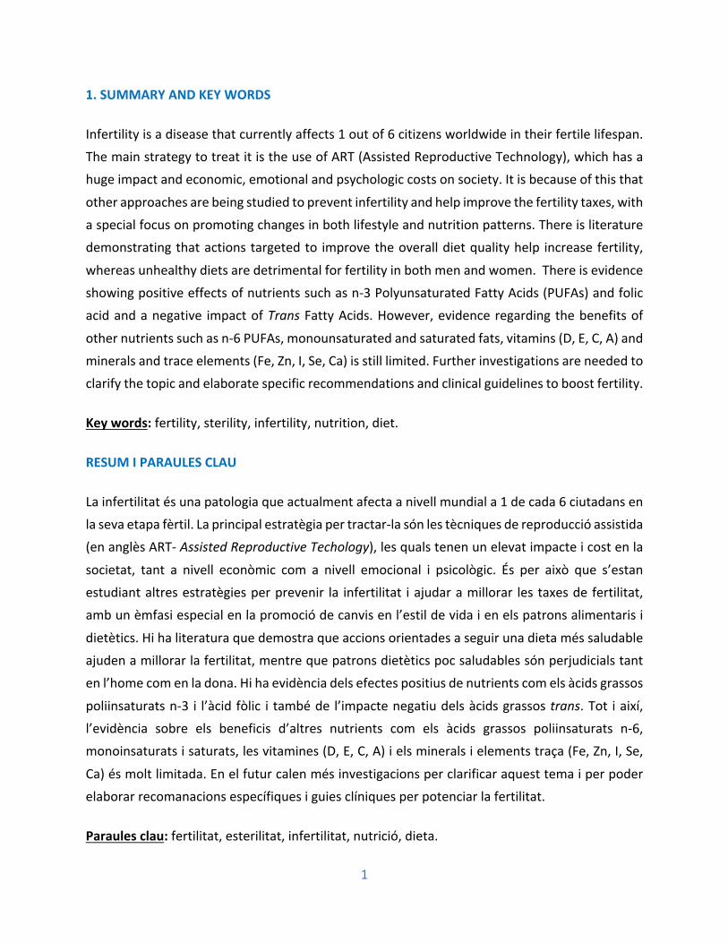
Ovary function is regulated by the hypothalamus-hypophysis-ovary axis through a negative feedback. The hypothalamus produces GnRH, which stimulates the
LH (luteinizing hormone) and FSH (follicle-stimulating hormone)
production in the adenohypophysis. FSH stimulates the granulosa cells, whereas LH stimulates both the granulosa and the theca inner
cells. Then, granulosa cells secrete inhibin B, a substance that inhibits
FSH production whereas the theca cells produce estrogens, which will inhibit LH, FSH and GnRH production (1,2,4). (Figure 4)
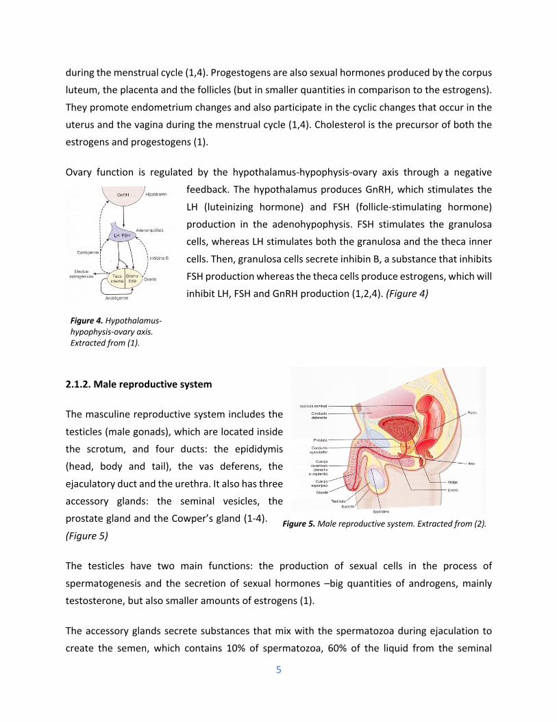
2.1.2. Male reproductive system
The masculine reproductive system includes the
testicles (male gonads), which are located inside the scrotum, and four ducts: the epididymis
(head, body and tail), the vas deferens, the
ejaculatory duct and the urethra. It also has three
accessory glands: the seminal vesicles, the
prostate gland and the Cowper’s gland (1-4).
(Figure 5)
The testicles have two main functions: the production of sexual cells in the process of
spermatogenesis and the secretion of sexual hormones –big quantities of androgens, mainly
testosterone, but also smaller amounts of estrogens (1).
The accessory glands secrete substances that mix with the spermatozoa during ejaculation to create the semen, which contains 10% of spermatozoa, 60% of the liquid from the seminal
Figure 4. Hypothalamus-hypophysis-ovary axis. Extracted from (1).
Figure 5. Male reproductive system. Extracted from (2).

6
vesicles, 30% of the liquid from the prostate gland and small quantities of substances from the
Cowper’s gland (2,3).
The spermatogenesis - process in which the spermatogonium (primitive germ cell) evolve into different
stages (primary spermatocyte, secondary spermatocyte and
spermatid) to end up developing the mature spermatozoa - occurs at the seminiferous tubules, which are little
canaliculus located inside the testicles (1-4). This process
duration is approximately 74 days (3). (Figure 6)
Once formed, the spermatozoa go through the rete testis
to the efferent ducts to the epididymis head. Then, spermatozoa undergo a maturation process and become
mobile once they arrive to the epididymis tail.
Afterwards, they continue to the vas deferens and they may be stored more than one month in both the
epididymis tail and the vas deferens lumen without losing
their viability. Finally, they travel along the ejaculatory duct and arrive to the urethra and the prostate during
ejaculation (1-4). (Figure 7)
The seminiferous tubules contain two types of cells: the germ cells - which undergo maturation
to produce the mature spermatozoon - and the Sertoli cells - which provide protection (through the tight junctions that form the blood-testis barrier) and nutrients (especially glycogen) for the
spermatozoa formation. These cells also have an important endocrine function, as they produce
the Anti-Müllerian hormone (AMH), which inhibits the development of the female reproductive system in the male body (1,2).
In between the seminiferous tubules Leydig cells are found, which produce testosterone (from
cholesterol) to the blood. This sexual hormone participates in different processes in the
reproductive system, including spermatogenesis maintenance, semen production and secondary
sexual characters development (1,2). Testosterone also regulates the Sertoli cells function (2).
Figure 6. Stages of the spermatozoa development. Extracted from (3).
Figure 7. Structure of the testicle. Extracted from (2).

7
Each spermatozoon formed has some motility, but it is not until they go through the epididymis
when they reach full ability to move. Spermatozoa contain a tail, which has a lot of mitochondria,
and a head, that stores the DNA and is protected by the acrosome – a structure that contains big
quantities of enzymatic substances (including the hyaluronidase – degrades proteoglycans, and
the proteolytic enzymes – degrade proteins) that enable the acrosome reaction in the
spermatozoa, which consists on the ovum outer layer’s break down that will enable penetration and fertilization processes (1-3).
The endocrine function of the gonads is regulated by the hypothalamus-hypophysis-testicle axis
through a negative feedback process (2,4). The hypothalamus produces GnRH, which stimulates the LH (luteinizing hormone)
and FSH (follicle-stimulating hormone) production in the
adenohypophysis. FSH stimulates the Sertoli cells, whereas LH stimulates the Leydig cells proliferation. Then, Sertoli cells
secrete inhibin B, which selectively inhibits FSH production
whereas the Leydig cells produce testosterone, which inhibits
FSH, LH and GnRH production (1,2,4). (Figure 8)
2.2. Physiology of fertilization: ovum and spermatozoon role
During men ejaculation inside the women vagina during the sexual intercourse, there is a release
of approximately 3,5 mL of semen. Although one single spermatozoon is necessary for
fertilization, there are around 120 million spermatozoa/mL of semen, although this number can range from 35 to 200 million. Therefore, around 400 millions of spermatozoa are present in the
quantity of semen ejaculated (3). The reduction of spermatozoa number in the semen is
associated with infertility, as half of the men with 20-40 millions/mL and almost all of the men with less than 20 millions/mL of semen are sterile (1).
Sometimes, men with normal quantities of spermatozoa in the semen are sterile due to the
presence of morphologic alterations in the spermatozoa, such as presence of two tails, two heads
or abnormal shapes, as well as lack of motility (3).
Figure 8. Hypothalamus-hypophysis-testicles axis. Extracted from (1).

8
After ejaculation, the spermatozoa go up through the uterus to the Fallopian tubes’ ampulla
thanks to the uterus’ and Fallopian tubes’ contractions, the prostaglandins of the semen and the
oxytocin released by the feminine hypophysis (1,3). In the ampulla, their speed is reduced and
they undergo Capacitation, which is the last step of the spermatozoa maturation. This process
increases their motility and also promotes the acrosome reaction and, thus, gives them the ability
to fertilize an egg cell (1,3,4).
During ovulation, the ovum and its Corona Radiata go out of the ovary, travel through the
Fallopian tubes and finally to the uterus (3). Fertilization usually occurs at the Fallopian tubes’
ampulla and, once the spermatozoon meets the egg cell in this region (thanks to the production of attractive chemical molecules), subsequent steps need to happen for success (2-4).
Firstly, the spermatozoon breaks down the extracellular matrix (Corona radiata) located around the ovum thanks to the hyaluronidase enzymes.
Then, the acrosome reaction occurs, in which the acrosome is separated from the spermatozoon’s
head and all the enzymatic substances are freed so that they can help break down the zona
pellucida (2-4). Once the spermatozoon is
adhered to the ovum’s cellular membrane, the spermatozoon nucleus is freed into the egg cell’s cytoplasm and the calcium-mediated signal
transduction starts. In the ovum, there is a huge calcium release that will participate in its
activation (2). (Figure 9)
Inside the egg cell, both the spermatozoon and the ovum undergo specific changes to create the
feminine and masculine pronucleus and, finally, they completely merge, so that the 23 feminine
chromosomes and the 23 masculine chromosomes create a new structure with 46 chromosomes (23 pairs) that is called zygote (1,3,4).
Afterwards, the zygote will travel along the Fallopian tubes through the uterus, where blastocyte
implantation in the endometrium will take place around day 5 to 7 after ovulation (3). During
pregnancy, an organ called placenta develops, which is special because it is formed by two
Figure 9. Fertilization process. Extracted from (2)

9
individuals (the mother and the fetus) and its functions include exchange of nutrients (glucose,
amino acids, fatty acids…) and metabolic residues, hormone production to ensure pregnancy
maintenance as well as protection against most of the microorganisms, although some viruses
and substances (medicines and alcohol) can go through it (4).
2.3. Infertility:
2.3.1. Definition and prevalence
The World Health Organization (WHO) clinically defines infertility as "a disease of the
reproductive system defined by the failure to achieve a clinical pregnancy after 12 months or
more of regular unprotected sexual intercourse". Infertility is included in the International Classification of Diseases (ICD 10), which is a diagnostic tool that provides standardized basis for
identifying and reporting health conditions and diseases globally (5). In the English language,
infertility is considered a synonym of sterility.
Subfertility is a situation of reduced fertility with prolonged time without achieving conception.
(6) Most of the pregnancies happen during the first six cycles of the fertile phase during the
menstrual cycle (6), so that subfertility is usually described as the ability to become pregnant
without medical help but in a period higher than a year, which is after 12 unsuccessful cycles (7). 50% of the couples with subfertility conceive spontaneously in the next 36 months after the first
year, while the other half of the group that does not conceive is considered infertile (6).
Data from 2017 established that in the European Union (EU) more than 25 million individuals in
a reproductive age have infertility (8). Worldwide, in the developed countries, about 1 out of 6
citizens suffer it during their reproductive lifespan (7,8).
The EU average total fertility rate is 1.58, while in Spain it is of 1.32. In our country, it is estimated
that more than 800,000 couples suffer from infertility and the average age of women at the first
childbirth is 31.8 years (8).
2.3.2. Causes
One of the main factors impacting the ability to have a child is the age of the progenitors.
Although men account for about 25-35% of the infertility cases, women age is more crucial, as
the maximum fecundity ages are between 20 and 30 years old and some epidemiologic studies

10
have shown fertility hugely declines in women older than 38-40 years old, although at the age of
35 the reproductive capacity is already diminished. For men, the age factor is less clear and
relevant, but some data establishes that the reproductive ability declines after the age of 40 (7).
Apart from advanced age, causes of infertility include toxic compounds exposure, diseases (such
as Polycystic Ovary Syndrome (PCOS), endometriosis and hypothyroidism) and lifestyle habits
(lack of physical exercise, chronic stress, unbalanced diets and drug consumption such a tobacco, alcohol, weeds and cocaine) (13).
There are factors that completely limit the reproductive ability, such as lack of sperm or
obstruction of the Fallopian tube. There are others, though, that only reduce partially the
likelihood of a pregnancy, as they affect the production of gametes (in men, reduced sperm motility or quantity; in women, anovulation related to PCOS or Premature Ovarian Failure), the interaction of gametes (alterations in the fertility process, in the sperm transport, in the ovum
capacitation in the Fallopian tubes…) or the implantation of the fertilized egg (7).
The most common causes of infertility in developed countries are seminal alterations in men (25-
35% of the cases) and ovulatory disorders (25% of the cases) and endometriosis (5-15% of the cases) in women. Finally, fertility from unknown causes accounts for up to 20% of the cases (7).
In men, erectile disfunction is also a cause of infertility and may be caused by different factors
such as neurovascular diseases (for example, diabetes mellitus), insufficient androgen
production, structural alterations or drug (including alcohol and tobacco) consumption (2).
In women, some cases of infertility are due to physical alterations on the feminine reproductive
organs but other times they are related to either an altered physiological function of the
reproductive tract or an abnormal development of the egg cell. The most frequent cause of
infertility is the lack of ovulation (anovulation), which can be due to a reduced secretion of
gonadotropins or it can be related to an abnormality on the ovaries, such as the presence of thick
capsules that block the ovum expulsion (3). Another frequent cause of feminine sterility is
endometriosis, in which the uterine endometrium grows out of it and surrounds the Fallopian
tubes, ovaries and the pelvic cavity around the uterus. It leads to fibrosis and occlusion of the
tubes, making it impossible for the egg cell to be freed from the ovary (3). Moreover,
endometriosis can result in ovarian function impairment (7).

11
Other causes include Salpingitis – an inflammation of the Fallopian tubes usually caused by an
infection; and the secretion of abnormally thick mucus – which makes it harder for sperm to
move in the feminine reproductive tract (3).
PCOS is an heterogenous endocrine disorder characterized by abnormally high androgen levels
(hyperandrogenism), presence of multiple cysts in the ovaries, ovulatory dysfunction and
irregular menstruations (9,10). It is usually associated to obesity and insulin resistance (in 50 to 70% of the cases) (10, 11). Its prevalence varies depending on the diagnosis criteria and it is up
to 15-20% of the population when using the European Society for Human Reproduction and Embriology (ESHRE)/ American Society for Reproductive Medicine (ASRM) criteria (Rotterdam criteria, 2004). Women are diagnosed if they meet two of the following criteria:
clinical/biochemical hyperandrogenism; oligo or anovulation; polycystic ovaries (10). Clinically,
women present hirsutism, oligomenorrhea (abnormally infrequent menstrual flow) or amenorrhea (abnormal absence of menstruation) and, frequently, infertility (10). PCOS is the
most common cause of anovulation in women (12).
It has been stablished that extremes on body weight may be one of the causes of infertility, as women with IMC lower than 20 have higher risk of anovulation, whereas both women and men
presenting overweight have higher rates of subfertility (14). As some cases of infertility or
subfertility are related to excessive body weight, weight loss and practice of physical activity may help increase likelihood of pregnancies (14).
2.3.3. Approaches to treat infertility
There is a wide range of Assisted Reproductive Techniques (ART) treatments, which have
different levels of complexity. In Spain, the available methods include Intrauterine Insemination (IUI); In Vitro Fertilization (IVF); Intracytoplasmic Sperm Injection (ICSI) and pre-embryo transfer;
Embryo Freezing and Frozen Embryo Transfer (FET); Preimplantation Genetic Diagnosis (PGD);
Preimplantation Genetic Screening (PGS); Gamete, Double Gamete and anonymous Embryo Donation. Surrogacy is not available. Currently, ICSI is the most widely used (8).
However, ART techniques have high emotional and economic costs, and this is why emerging
studies are focusing on identifying modifiable lifestyle factors, including dietary patterns, that
may promote fertility on specific disorders and diseases (15).

12
In women suffering PCOS, treatment includes usage of oral contraceptives (they reduce hirsutism
and menstrual cycle alterations) and anti-diabetic drugs such as metformin (it improves insulin
sensitivity). Furthermore, lifestyle changes such as weight loss have shown to improve menstrual
irregularities, hyperandrogenism-related symptoms, insulin sensitivity and infertility (10,11).
2.4. Importance of nutrients on fertility
Both macronutrients (carbohydrates, proteins and fats) and micronutrients are needed to
maintain normal energetic, structural and regulatory functions in the body. Vitamins and minerals are essential molecules, as humans cannot synthetize them – or at least in adequate
quantities – so that they need to obtain these micronutrients from the diet. Furthermore, some
foods contain phytochemicals, which also participate on many human body processes.
Emerging studies are focusing on the importance of an overall optimal nutritional status and an
adequate body weight and composition for fertility. More specifically, nutrients such as vit D, E, C, A, Ca, Fe, Zn, Se, I, folic acid, vit B12 and omega-3 fatty acids are thought to be necessary to
boost fertility and ensure a successful pregnancy, as they are involved in the fertilization process
(13). It has been demonstrated that specific dietary changes help decrease the frequency of ovulatory disorders and, thus, improve fertility (14).
13
3. OBJECTIVES
Based on the fact that infertility affects a significant part of the population worldwide and the
promising positive impact diet and specific nutrients may have on it, the main objective of this project is to understand if diet modifications can help improve the fertility on both female and
male.
The secondary objectives of this bibliographic research are:
- Understand the role and function of specific nutrients on the fertilization process.
- Establish foods or nutrients that may be positively and negatively affecting fertility.
- Search for possible nutritional approaches and dietary changes to improve fertility in
both male and female.
- Provide recommendations for the general population to improve fertility and pregnancy
likelihood.

14
4. MATERIAL AND METHODS
This work has been completed by using the Information from both bibliographic databases and
physical books.
The bibliographic database used for the search was Pubmed, accessed through the website of the "Centre de Recursos per a l'Aprenentatge i la Investigació" (CRAI) from the Universitat de Barcelona (UB). The information extracted comes from articles and their selection has been done
based on the most relevant ones for the topic, trying to prioritize the most recent ones.
The search was conducted in English to ensure the finding of all the important articles, as
literature and studies are usually in this language and the terms introduced include (nutrition OR nutrient) AND (fertility OR fecundity), food AND (fertility OR fecundity), fertility AND (causes OR
factors), Zinc AND (fertility OR fecundity), vit D AND (fertility OR fecundity), (omega-3 OR PUFAs)
AND (fertility OR fecundity), (folic acid OR B9) AND (fertility OR fecundity), B12 AND (fertility OR fecundity), (Se OR Selenium) AND (fertility OR fecundity), vit E AND (fertility OR fecundity), etc.
The filters applied during the search were the data of publication (articles published from 1999 -
during the last 20 years) and the species of the studies (studies only in humans).
The search was first restricted to reviews and metanalyses to get an overall understanding of the
topic and, then, the search was focused on finding primary articles, giving priority to the best
quality studies - randomized clinical trials and prospective cohort studies, although evidence was limited and other studies - such as case-control - were considered as well.
The physical books used were borrowed from the Library of the Universitat de Farmàcia i Ciències de l'Alimentació - Campus Torribera, Universitat de Barcelona (UB). The sections used for the elaboration of this project were "Nutrition and Dietetics" and "Physiology and Physiopathology".
This project includes three main sections: the introduction, with the objective of determining
basic physiology and physiopathology of the human reproductive tract to later understand the
results; the results and discussion, which provides specific data of the most relevant and recent studies, providing the currently available scientific evidence of the topic; and the author's
contribution and suggestions for the topic, focusing on the elaboration of a leaflet summarizing
the most important findings that could be provided to the general population to promote specific dietary patterns and lifestyle habits to improve fertility.
15
5. DETAILED DESCRIPTION OF THE ACTUAL KNOWLEDGE OF THE TOPIC
5.1. Macronutrients
5.1.1. Carbohydrates
It has been established that the quantity and quality of dietary carbohydrates may have an impact
on fertility (15).
The NHS-II (Nurses’ Health Study-II) - a prospective cohort study – found an association between glycemic load and the risk of anovulation. It also found a positive relationship between total
carbohydrate intake and ovulatory infertility when this macronutrient was eaten in higher
amounts in substitution for naturally occurring fats: after adjustments were done, women in the highest quintile of carbohydrate intake had a 78% higher risk of ovulatory infertility in comparison
to those women on the lowest quintile. Statistically significant data showed that, the higher
intake of total carbohydrate, the higher risk of anovulation (12).
However no statistically significant association was found regarding total fiber intake, fiber intake from different foods or overall high glycemic index foods intake with ovulatory infertility (12).
Also, the association between fiber-rich diets and anovulation is not clear, as different studies
have shown opposed effects (15).
It has been demonstrated that women suffering PCOS have superior high-glycemic index foods’ intake in comparison to healthy women (15). A controlled intervention study showed that a
dietary pattern based on reducing dietary carbohydrates in a weight-maintaining diet was
associated with an improvement on the metabolic profile, including reduced fasting glucose and
fasting insulin levels and increased insulin sensitivity. Reduced fasting insulin was statistically associated with a reduction on testosterone levels, potentially enhancing ovary function (9).
Another controlled intervention study that gave participants an eucaloric low-carbohydrate diet
also showed an improvement on insulin sensitivity (11). However, other studies showed that total carbohydrate intake did not seem to have any effect on the hormone levels in healthy
women (15).
Although these studies consisted on controlled interventions, some limitations were noticed –
such as small sample size and restrictive inclusion criteria, which limits the generalizability of the results.

16
Regarding men fertility, lower high-glycemic index foods consumption has also been associated
with better semen quality in comparison to diets rich in refined and high glycemic index
carbohydrates (13).
5.1.2. Fats
Fats play an important role on the reproductive function. Omega-3 (n-3) PUFAs (polyunsaturated fatty acids) are necessary for female fertility, as they are involved in oocyte maturation and
embryo development and are also needed for the synthesis of substances involved in the implantation and pregnancy maintenance. Conversely, TFAs (trans fatty acids) are detrimental
for fertility purposes, as they promote insulin resistance, which can lead to alterations on the
ovary function and to ovulatory infertility (15,16).
Therefore, the type of fats greatly influence fertility, as a diet high in PUFAs from the omega-3
series combined with a diet low in TFAs has been associated with better fertility. There is still little evidence, though, regarding the impact of SFAs (Saturated Fatty Acids), MUFAs
(monounsaturated fatty acids) and omega-6 PUFAs on female fertility (15).
The NHS-II study showed that ovulatory infertility was not associated with total fat, cholesterol
and most types of fatty acid consumption, but was associated with TFAs, as each 2% of the daily energy obtained from this type of fat rather than from carbohydrates was correlated with a 73%
higher risk (Relative risk (RR) = 1.73; 95% CI: 1.09, 2.73) of suffering ovulatory infertility after
adjusting for confounding factors. The relative risk for ovulatory infertility when TFAs was consumed instead of omega-6 fatty acids was similar to the previous one (17).
Furthermore, when obtaining the 2% of the daily energy from TFAs instead of MUFAs, the risk of
ovulatory infertility doubled (RR=2.31; 95% CI: 1.09, 4.87). The TFAs substitutions were made in
an isocaloric diet, to avoid the diet being a confounding variable (17).
This same cohort study showed women consuming higher amounts of TFAs had 48% higher risk of suffering endometriosis. Conversely, an inverse association between PUFAs and endometriosis
risk was observed, as women in the highest fifth of long-term PUFAs consumption had 22% less
likelihood of suffering endometriosis in comparison to women in the lowest fifth of intake (18).
Moreover, statistically significant data showed that each 1% of energy coming from TFAs instead of any other fat source was associated with higher risk of endometriosis, whereas each 1% of

17
energy coming from n-3 PUFAs consumption rather than TFAs correlated with almost 50% lower
risk of endometriosis. No significant associations where found when each 1% of additional energy
came from omega-3 PUFAs rather than MUFAs, n-6 PUFAs or SFAs.
Another large prospective cohort study also associated high TFAs consumption and low n-3
PUFAs with reduced fertility (19).
A cohort study showed that the higher tertile of DHA (Docosapentaenoic acid) consumption,
which is a type of n-3 PUFA, was associated with reduced risk of anovulation in comparison to the lowest tertile (RR = 0.42; 95% CI:0.18-0.95). (20) Another US and Canada cohort study showed
increased fecundability with higher n-3 consumption, although this association was not found in
a Denmark cohort that already had much higher n-3 PUFAs baseline intake (19).
TFAs are associated with negative fertility outcomes - reduced sperm quantity and increased
insulin resistance, which also negatively impact ovulation and sperm quality (13).
In men, diets high in saturated fat have been associated with lower sperm counts and overall
sperm concentration. One study found that the highest quartile for saturated fat consumption
had a 41% reduction on sperm count. Another study found that a higher saturated fat
consumption was associated with 43% lower sperm count and 38% lower sperm concentration. Moreover, men with higher TFAs levels in seminal fluid showed reduced sperm count as well (21).
Regarding omega-3 fatty acids, those who had a higher intake of them showed a mild
improvement in sperm morphology (21).
Supplementation with omega-3 has shown to help improve the insulin sensitivity in women
suffering PCOS (13) and to improve the sperm motility in men (22).
5.1.3. Protein
Currently there is a lot of controversy about the effects dairy, animal protein and soy products
may have on fertility, as they have been linked with an increased intake of pesticides, endocrine-
disrupting substances as well as steroid hormones and growth factors, which can all lead to alterations in the reproductive function and the hypothalamus-hypophysis-gonad axis (15).
18
5.1.3.1. Dairy
The detrimental impact dairy may have on fertility is still controversial, but the most recent
studies have not found conclusive evidence of a negative impact on overall fertility nor ovulatory infertility (15,16).
For instance, the NHS-II study showed no correlation between total dairy intake and risk of
anovulatory infertility, but did show that low-fat dairy intake increased the risk of anovulatory
infertility whereas high-fat dairy intake reduced this risk (23). Conversely, a study using two different cohorts (one in Denmark and one in North America) did not support this hypothesis, as
it did not show any clear effect of low-fat or high-fat dairy consumption on fecundability in either
cohorts. Only in women younger than 30 years old, cheese and high dairy intake were associated with enhanced fertility (24).
Another cohort study observed associations between intakes of yogurt (RR: 2.1; 95% CI: 1.2, 3.7) and cream (RR: 1.8; 95% CI: 1.0, 3.2) and sporadic anovulation when compared with no intake,
but did not find association of dairy fat consumption with ovulatory function alterations in
women with regular menstrual cycles (25).
In a case-control study in Wisconsin, high consumption of milk (³3 glasses/day) showed
protection for female fertility (OR=0.3; CI:0.1-0.7) (26).
5.1.3.2. Animal protein
Although there is also controversy on the effect meat may have on fertility, current evidence
potentially suggests that red meat intake is associated with an increased risk of infertility (16),
whereas replacing animal protein by vegetable protein may improve fertility and ovulation (13).
Among the NHS-II participants, increased meat consumption was positively associated with
ovulatory infertility, as adding one serving of meat daily significantly increased the risk by 32%.
Also, higher intake of protein from vegetable sources was associated with a 50% less risk of suffering ovulatory infertility and increasing the protein intake from vegetable foods by 5%
instead of protein from animal foods, showed a RR = 0.42 (95%; CI:0.24-0.76) of ovulatory
infertility. This study concluded that risk of anovulation may be reduced by replacing animal sources of protein, especially chicken and red meats, with vegetable sources (27).

19
Contrarily to the results for meat consumption, fish consumption has been associated with higher
likelihood of pregnancy (16). For instance, a cohort study found that high seafood consumption
(8 servings/cycle) was strongly associated with reduced time to pregnancy – improved fecundity
– in comparison to couples with low intake (28).
Although presence of methylmercury – a toxicant found in big predator fishes- on the blood has
been associated with infertility, currently there are no strong conclusions on the negative impact mercury and other contaminants present on the fish may have on fertility (16). Furthermore,
recent recommendations from the American College of Obstetricians and Gynecologists (ACOG) for women who may become pregnant, pregnant women and breastfeeding mothers promoted an intake of 2-3 times/week of a variety of low-contaminated fish, as this animal product is one
of the main sources of n-3 PUFAs. However, they did highlight the need to restrict to one serving
fish such as albacore tuna, halibut and carp and to completely avoid fishes such as bigeye tuna, swordfish, king mackerel and tile fish due to their big mercury concentration (29). (Figure 10)
5.1.3.3. Soy
Although soy and phytoestrogens supplements were thought to be detrimental for endocrine
pathways, ovarian reserve and fertility, no conclusive studies have shown a negative impact on
it (16). Available studies are small and heterogenous and are lacking evaluation of specific ovarian
reserve markers, leading to the impossibility of drawing conclusive results (15).
Figure 10. ACOG’s seafood consumption guide. Extracted from (29)

20
From current data, no association has been found between soy isoflavones’ consumption and
acceleration of age at menopause nor fecundity (15).
Some evidence has shown to provide positive effects on fertility in couples undergoing infertility treatments, as the supplementation improved the likelihood of becoming pregnant, the live birth
rates and the endometrial thickness (16).
5.2. Micronutrients
5.2.1. Folic acid
Folic acid rich foods include vegetables (especially the dark green leafy ones), fruits, nuts, beans,
peas, grains, seafood, eggs, dairy, meat and poultry. Liver, spinach, asparagus and Brussel sprouts are among the foods with a higher content of it (36).
This nutrient is crucial for DNA synthesis, gametogenesis, fertilization and pregnancy (15) and its lack is related to alterations of the ovulation in women and reduction of the quantity and quality
of sperm in men (13). Moreover, the folic acid deficiency during pregnancy is associated with neural tube defects, premature birth and reduced intrauterine growth (13).
Its supplementation is essential to prevent neural tube defects (NTD) in the fetus but different studies -including RCTs (Randomized Control Trial)- have shown that, beyond this function, this
B-vitamin may also be beneficial for female fertility, as supplementation with higher doses (>800 μg/day) than those taken only to prevent NTD (usually 400 μg/day) have been associated with
improvements on fertilization (15,16). Some prospective cohort studies found lower risk of
ovulatory infertility (30), decreased frequency of anovulation periods (31) and shorter time to
pregnancy (32) in women taking a folic acid supplement.
Also, higher reproductive success (increased implantation, clinical pregnancy and live births) in ART was observed in women in the higher quartile of folate intake and of serum folates (33,34).
Although some studies established a higher risk of fetal loss with high folic acid doses (>800
μg/day), the latest Cochrane evidence concluded that there is not higher risk of miscarriage
(15,16). Furthermore, later observational studies even showed a decreased risk for spontaneous
abortion (35,37,38). For instance, the NHS-II study, which included more than 11,000 women in
the cohort, established women taking a high-dose (700-800 μg/day) folic acid supplement had a
21
relative risk of 0.80 (95%; CI 0.71-0.90) of spontaneous abortion in comparison to women without
supplemental folate intake (35).
5.2.2. Vitamin D
The main source of vit D for humans is sun exposure and there are only few foods with significant amounts of Vit D. The best dietary sources are fatty fish (salmon, tuna and mackerel) and fish
liver oils and other foods with smaller amounts include beef liver, cheese and egg yolks. There
are also mushrooms with enhanced vit D2 levels thanks to ultraviolet light exposure (36).
This vitamin has been thought to be important for fertility, as vitamin D receptors (VDR) have
been found in the ovary, uterus, endometrium and placenta (14,15) and it promotes the synthesis of the Anti-Müllerian hormone (AMH) (13). Also, this liposoluble vitamin stimulates follicular
maturation in the ovary and regulates the implantation process through gene regulation (15). Vit
D may be related with infertility in both men and women, as its deficiency is associated with the pathogenesis (including metabolic syndrome and insulin resistance) of PCOS and endometriosis
in women and with lower testosterone levels and decreased sperm quality in men (39).
It was thought that vit D may be a key nutrient to improve human fertility due to its participation
on reproductive functions and also in relation to some promising animal studies (15). However, currently there is a lot of controversy around the possible positive impact of vit D on human
fertility, as most recent studies have heterogenous outcomes and have not showed a strong
correlation of vit D supplementation and the likelihood to become pregnant, especially on women who have sufficient vit D serum levels (16).
For instance, the NHS-II study could not establish any relationship between vit D intake and
anovulatory infertility (15). The Danish cohort study did not find correlations between vit D serum
levels and overall conception rates as well, although it showed that low 25-OH-vitD levels may
increase the risk of late miscarriage (40).
Supplementation may be indicated and improve the conditions during severe vit D deficiency,
obesity, insulin resistance, low AMH concentrations and oligospermia (13), but the current
available evidence has not established any causality – only correlations- nor the ability to treat infertility through vit D (13).
22
Although vitamin D deficiency may be detrimental for fertility purposes, more evidence,
especially from RCT is needed to clear the topic and establish causality, if existing (16).
5.2.3. Vitamin A
Vit A richest foods are liver and fish oil and other sources include eggs and milk. Provitamin A is found in vegetables such as leafy greens, orange, yellow and red vegetables, tomatoes and fruits
(36).
It has many functions associated with the reproduction and it is necessary to ensure adequate
sexual hormones’ synthesis and the spermatogenesis process, to protect the ovum and sperm
from oxidative stress as well as to promote the successful implantation of the fertilized egg cell. Moreover, it promotes the correct development of the placenta and also participates in the
embryogenesis and organogenesis. The use of b-carotenes (provitamin-A) supplementation has
been associated with better sperm motility and quality (13).
5.2.4. Vitamins E and C
The best sources of vit E include nuts, seeds and vegetable oils but it can also be found in some
green leafy vegetables and fortified cereals (36). Vit C is found vastly in fruits and vegetables,
including citrus fruits, berries, peppers, kiwis, broccoli, Brussels sprouts, tomatoes and potatoes (36).
They are antioxidant vitamins that protect the ovum and sperm from oxidative stress. Vit E, which
is a liposoluble vitamin, participates in the fertilized egg cell implantation and the placenta
development (13).
Vit C is essential for collagen biosynthesis, which is extremely important for adequate ovarian follicle growth and also for the ovulation and luteal phase due to the high amounts of collagen
present on the ovarian tissue (22). A few studies associated high doses of vit C (ranging from 500
to 750 mg/day) with improved fertility and higher pregnancy rates (22) and Vit E and C supplementation may help increase sperm quality and quantity (13).
23
5.2.5. Calcium
Dairy products (milk, yogurt and cheese) are rich natural sources of calcium, but it can also be
found in non-dairy products such as Chinese cabbage, kale, broccoli, almonds, tofu and sardines with bones (36).
Calcium is a mineral that plays a key role in the hyperactivation and the acrosome reaction on
the sperm, processes that are essential for fertilization success. Moreover, it also participates on
the spermatogenesis process and the sperm motility (13,14).
5.2.6. Iron
Heme iron is found in meat, fish and seafood, whereas non-hem iron is found in eggs, milk, nuts, beans, vegetables and specific fortified products such as cereals and breads (36).
Iron is a crucial mineral for fertility, as it helps the fertilized ovum implantation process. Also, it reduces pregnancy complications and is essential for the fetus nervous system development (13).
The NHS-II studied the effect of iron on fertility and found that women with a higher iron intake
either from supplementation or from greater dietary intakes had improved fertility. The authors
established this could be due to the fact that the ovary uses iron for its functions (22).
5.2.7. Zinc
A wide variety of foods contain Zinc (Zn), including oysters, eggs, red meat, poultry, seafood (crab
and lobster), beans, nuts, whole grains and dairy products (36).
Zn is a trace element that plays a key role in fertility for both female and male, but has a greater
importance for men, as it promotes testicle development, testosterone synthesis and sperm
viability - motility, maturity, structure, function, quality and quantity (14,41). It is also involved in the interactions that happen in the female reproductive tract - including capacitation and
fertilization (41).
Moreover, it protects both the sperm and the egg cell from oxidative stress, participates in the
embryogenesis process and the placenta formation. Finally, it is also crucial for the fetus' nervous

24
system growth and development (13). Supplementation with Zn has shown benefits for the
fertility in men (41).
5.2.8. Selenium
Foods rich in Selenium (Se) include Brazil nuts, seafood and fish (tuna, halibut, sardines, shrimp), muscle meats (beef, ham), cereals and other grains and dairy products (36).
Se is a trace element that has many potential implications in reproductive functions, as
selenoproteins play an important role in both female and male fertility (42,43). In men, Se helps
maintain the spermatozoa integrity and viability by protecting them from oxidative damage (43).
It also participates in testosterone biosynthesis and in spermatozoa development and maturation (42,43). In female, Se is necessary for follicle growth, maturation and dominance, for embryonic
development and for protecting against oxidative stress both the dominant follicle and the
endometrial remodeling for later implantation (42). It also helps in the placenta development and is necessary for the adequate development of the fetus' nervous system (13).
Some evidence suggests that a higher Se dietary intake improves the antioxidant activity and,
thus, male fertility. There are some studies carried out in men showing a relationship between
Se supplementation and increased fertility, sperm motility, semen quality and spermatozoa motility and morphology. Conversely, some other studies showed Se supplementation did not
have any benefit and some even showed a decrease on sperm motility (43).
In women, evidence is even more scarce, as most of the studies are centered on the importance
of Se during pregnancy and only few focus on its impact on fertility and, more specifically, on
oocyte development and ovarian physiology (42).
Although evidence is growing, most of the current available studies are done under in vitro
conditions and there is still a lack of high-quality studies on humans to draw solid conclusions to
do clinical and guideline recommendations (42). Studies regarding Se supplementation show that it is beneficial for Se-deficient individuals, but it can be harmful if excessive doses are given.
Therefore, health care providers should be careful when recommending its supplementation,
especially on those individuals who may already have sufficient or high levels (42). Moreover, other two crucial factors that should be taken into account are the dosages and the Se status of

25
the population, as Se content of specific food hugely varies depending on the soil (for instance,
in the US soils are richer in Se, whereas Europe soils provide a poorer source of Se) (36,43).
5.2.9. Iodine
Iodine can be found in seaweed, seafood and dairy products. Fruits and vegetables also contain iodine, but their concentration varies depending on the soil content (36). Generalized use of
iodized table salt is recommended to avoid deficiencies (22).
It is necessary for maintaining fertility and developing the placenta. It is also needed for the
growth and development of the fetus' nervous system (13). A study showed that hypothyroidism
was related to an increased risk of subfertility and established that adequate iodine intake is really important not only for pregnancy and child development, but also for conception (22).
5.2.10. Vitamin B12
Vitamin B12 is only found in animal products, including fish, meat, poultry, eggs, milk and milk
products (36).
This vitamin participates on DNA synthesis and is necessary for the development and
functionality of the placenta. A good nutritional status thanks to its supplementation has shown to improve the quality of sperm and to prevent spontaneous abortion (13).
5.2.11. Antioxidants
Although there is an association between oxidative stress and reduced spermatogenesis,
evidence regarding the positive benefits of antioxidants on fertility is weak and inconclusive (14).
There are only limited studies about the impact of antioxidants in fertility and the existing ones
have many limitations and low quality. Furthermore, the term "antioxidant" is so wide that every
study made used a different compound and dosage, making it really difficult to draw conclusions
(16).
Some studies have established that oral supplementation with antioxidants may provide benefits on male fertility. For instance, a double-blinded RCT showed that the supplementation with 66
mg of Zinc and 5mg of folic acid together produced a significant 74% increase on the total normal
sperm count in subfertile men after the 26 weeks intervention (44). A Spanish case-control study

26
established that a reduced intake of antioxidants such as lycopene, folates and vitamin C was
associated with a poorer semen quality (45).
A review study including 35 articles indicated that diets high in nutrients such as omega-3 fatty
acids, vit D, folate, antioxidants (such as vit E, C, Se, Zn, b-carotene, lycopene and cryptoxanthin) and low in SFAs and TFAs had an inverse association with low semen quality parameters (46).
From the current evidence available, it can be established that supplementation may be beneficial in improving male fertility, but it does not provide improvements on females
undergoing ART. However, even if the supplementation may be positive for the male partner,
there is still lack of evidence regarding the dosage and the specific antioxidants associated with
the benefits (13,16). Also, it is still unknown if supplementation is as effective as receiving them from an antioxidant rich diet (13).
It is important to determine that, although semen quality is evaluated and associated with
specific nutrient consumption, there is no strong association between semen quality and fertility or vice versa. Therefore, producing changes on semen quality through nutrition does not strongly
imply producing an effect on the couple fertility (16).
5.3. Toxic substances
It has been well stablished that alcohol and caffeine consumption during pregnancy time has many risks associated, including miscarriage, growth impairment and development of fetal
alcohol syndrome. However, surprisingly to what is generally thought, the evidence demonstrating negative effects of caffeine and alcohol on fertility is inconsistent, probably due
to the low quality and bias of many of the studies, making it difficult to draw a conclusion
regarding the caffeine and alcohol harm for fertility (16).
Some evidence has shown detrimental effects, such as a review study which found that a high
male intake of alcohol and caffeine had a detrimental impact on the chances of pregnancy in
their female partner (46). Conversely, others such as the Environment and Reproductive Health Study, which is an ongoing prospective cohort study, have not found any association between
male alcohol and caffeine consumption and semen quality among fertility patients (47).
Evidence regarding to what extent alcohol affects male reproductive health is less clear than the
one regarding tobacco. Specifically, a meta-analysis showed a clinical association between
27
cigarette smoking and infertility, as it reduced sperm count and motility and increased abnormal
sperm morphology (21).
Although there is a lack of interventional studies and evidence is not solid enough due to the presence of multiple confounding factors, there is an existing association between all these toxic
substances consumption and impaired male fertility, including reduced spermatogenesis and
sperm parameters as well as increased DNA oxidative stress. Therefore, it would be safe to assume that recommendations of tobacco smoking cessation and avoidance of alcohol and
recreational drug intake in male would be the best course of action for couples trying to achieve
pregnancy (48).
Moreover, recent studies are starting to show that environmental toxicants may interact with food nutrients and may impact the human health and reproductive success. For instance, it has been stablished that high pesticide levels on fruits and vegetables may negatively impact
reproductive success (pregnancy events and live births) in ART (49). Thus, human exposure to environmental contaminants such as methylmercury on fish, hormones and antibiotics in animal
products and pesticides in produce should be considered in the future when studying the impact of foods and nutrients on human fertility (15).
5.4. Body weight: overweight and underweight
Maintaining an adequate body weight is extremely important for fertility purposes, as both
underweight and overweight have shown to be detrimental for pregnancy success (14,50).
In obese or overweight individuals, the excessive adipose tissue produces higher amounts of
leptin, a hormone that alters the hypothalamus-hypophysis-ovary/testicle axis and leads to
higher testosterone concentrations and lower estrogen concentration in women and to higher
estrogen concentrations and lower testosterone concentration in men, producing an inversion
of the normal amounts of sexual hormones (13). Moreover, the excess of free fatty acids in the
blood leads to insulin resistance, which negatively impacts the ovulation and the sperm quality
(13). Obesity negatively affects the oocyte quality and uterine receptivity (50). Also, obesity
clearly increases infertility risk and is associated with menstrual disorders such as PCOS (50).
Some studies established that in ovulatory women suffering subfertility and with a BMI > 29
Kg/m", the chances of conception were decreased by 5% for each increased unit of BMI (50).

28
In this regard, weight loss is a strategy recommended in order to enhance fertility status in
overweight or obese women and moderate physical activity practice has shown to improve
metabolic function and hormonal profiles (50).
On the other hand, underweight status is associated with anovulation, amenorrhea and reduced
luteal phases in women and with decreased production, motility and viability of sperm in men
(13). Also, the initiation of the menses requires a minimum of fat mass in order to maintain ovulatory function. Eating disorders, malnutrition as well as practicing exercise to exhaustion
have been associated with subfecundity and infertility (50). One study determined that
underweight women (considering a BMI < 19 Kg/m") had an average 29-month long time to
pregnancy, which is considerably longer than the 6.8 months needed for those women with normal weight (50).
Regarding men, a meta-analysis from 2012 that included 13,077 patients and 21 studies found that obese or overweight men had a higher risk of suffering infertility. Also, another cohort study
including more than 10,000 men found that as BMI increased, the concentration and number of sperm as well as the semen volume decreased. Finally, another meta-analysis including over 115,000 patients concluded that obesity was related to increased infertility rates (OR = 1.66) (21).
5.5. Dietary patterns and overall diet quality
Different studies have investigated the positive effects of an overall “healthy” diet on fertility.
For instance, the NHS-II study showed that women consuming a “fertility” diet had significant lower risk (RR=0.35; 95% CI:0.23-0.48) of suffering ovulatory infertility; a Spanish case-control
study showed higher adherence to the Mediterranean diet decreased the likelihood of having
difficulties becoming pregnant (OR=0.56; 95% CI: 0.35-0.95) in comparison to low adherence to this diet (51,52).
A review that included 23 observational studies concluded that dietary patterns including fruits
and vegetables, fish and whole grains, were associated with improved semen quality in men (53).
Another review including 35 articles indicated that intake of foods such as shellfish, seafood, poultry, cereals, vegetables, fruits, low-fat dairy and skimmed milk were associated with a benefit
on different semen quality parameters, whereas diets rich in processed meats, soy foods, full-fat
dairy, cheese, sweets and sugar-sweetened beverages were correlated with negative effects on semen quality (46).

29
Although studies slightly differed on what was defined as a "healthy diet", they all had in common
a higher intake of protein from vegetable sources, a higher intake of animal protein sources from
fish and poultry rather than meats and processed meats, a higher intake of MUFAs and n-3 PUFAs
in comparison to TFAs, a higher intake of whole grains, fruits and vegetables and a lower intake
of processed foods. They all showed benefits for fertility in women and for semen quality in men
as well as increased odds of pregnancy events (15,16).
On the other hand, "unhealthy diets", which may be defined as the ones high in processed meat
and processed foods, fried foods and sugar-sweetened beverages, are thought to negatively
impact women fertility and are associated with lower semen quality and poor testicular function (16). This was shown in a multi-center retrospective study in which higher fast food intake
frequency as well as reduced fruit intake was associated to longer time to pregnancy and
infertility (54).
Proposed dietary recommendations for improving male fertility include providing high fiber and low glycemic index carbohydrates, promoting MUFAs and avoiding TFAs, reducing animal protein
intake and increasing the one from vegetable sources. Being moderately physically active may help as well (13).

30
6. DISCUSSION
Evidence shows many nutrients (both macronutrients – carbohydrates, fats and protein - and
micronutrients – vitamins, minerals and trace elements) regulate and participate on the different stages of the fertilization process and that dietary factors impact human fertility. However, there
is not the same level of evidence for all the nutrients. Furthermore, differences regarding their
impact on male and female fertility have been found.
Currently there is more evidence on the impact different nutrients have on women fertility, whereas fewer studies have investigated the impact of a variety of nutrients on the male
population. For men, most of the available literature focuses on the impact and possible benefits
of antioxidants, but studies are extremely heterogenous, as they use a wide range of supplements (16). Other nutrients that should be considered when tackling male fertility include Se and Zn, as they both have shown to be especially important for male fertility processes
(13,14,41-43) in comparison to other micronutrients that are less studied. However, it is worth highlighting that most of the studies focus on semen quality and parameters improvement, which
have very limited predictive value for spontaneous pregnancy events, so that further studies need to be conducted to assess how diet may impact overall fecundity on the male partner.
On the one hand, for the general population it is frequently recommended to follow an overall
healthy diet, which promotes a higher intake of whole grains, legumes, nuts, fruits, vegetables and fish and a reduced consumption of trans fatty acids and red meat to improve fertility (15,16).
There is evidence supporting it, as the positive outcomes on fertility have been established on
the literature - different studies show associations between this nutritional approach and a reduction of ovulatory infertility (51,52) in women and an improvement on semen quality
parameters on men (53).
On the other hand, conversely to the results found for a healthy diet, an unhealthy diet that
includes high amounts of processed foods and sugar-sweetened beverages and is high in TFAs and SFAs has been associated with detrimental effects on semen quality (16,46) and worse
fertility outcomes, including longer time to pregnancy (54).
It has also been shown that BMI is a factor that should be taken into account for both men and
women when considering fertility, as both under and overweight are detrimental (14,21,50).
Therefore, promoting a healthy weight should be another strategy to boost fertility and the

31
regular practice of physical activity targeted to weight loss should be encouraged on patients
suffering obesity, insulin resistance or PCOS (in female patients) (50).
Although causality cannot be established, as an overall healthier diet has shown to be beneficial not only for fertility purposes but for the prevention of many other chronic diseases – including
certain cancers, diabetes mellitus type II and cardiovascular diseases – it would be wise to
promote a change of dietary patterns on the general population and, more specifically, on the adults in their fertile lifespan, to promote better health status, prevent specific diseases and,
likely, boost their fertility. Apart from potentiating healthier nutrition habits, promoting lifestyle
changes - such as the practice of physical activity - focused at maintaining a normal BMI should also be considered based on the current evidence (13,50).
Evidence is more solid on the benefits of an overall nutritional approach, whereas the possible benefits of specific nutrients on fertility are still more unknown and less established. There is
literature showing their participation on fertility processes (such as spermatogenesis, follicle maturation, implantation…) but the clinical significance and impact of these nutrients on fertility
has not been well established yet. Currently there is more information regarding the benefits of n-3 PUFAs (18,19,21,22) and the detrimental effects of TFAs (13,15,16,19,21). There is also strong
evidence showing the benefits of taking a prenatal folic acid supplementation to prevent neural
tube defects, as well as to promote fertility and maintain a healthy pregnancy (15,16,30,31-34).
However, there are still few evidence-based and high-quality studies showing an impact of some
macronutrients on fertility. For example, there is inconclusive evidence about whether there are
different outcomes with high and low-glycemic diets or not (12,15). Also, currently there is controversy about the positive, negative or neutral effect of proteins from dairy, animal sources
and soy products on fertility (15,16, 23-25). Finally, little literature is available for fat types such
as n-6 PUFAs, MUFAs and SFAs (15).
Similarly, little literature focuses on the benefits of most vitamins (including vitamin A, D, E, C and B12) and minerals (including Fe, I, Ca) on fertility. For instance, although vitamin D was
thought to be a nutrient with potential therapeutic effects, most recent evidence is inconclusive
and has not shown as many benefits as expected (13,16), especially considering that vitamin D is involved in many stages of the fertilization process (14,15). Calcium is another nutrient thought
to be necessary for fertility (13,14), as it participates on crucial steps, but evidence about the
impact on clinical outcomes is extremely limited.

32
Regarding toxic substances, some studies have shown opposite results, especially for alcohol and
caffeine, so that a strong conclusion cannot be made for now (46-47). However, in terms of public
health promotion it would be accepted to promote a reduction of alcohol and tobacco intake in
the general population (48).
Finally, it is needed to highlight that most of the studies are observational, so that it should be
taken into account that confounding factors may influence the results and no causality can be established. It is because of this that there is a need to conduct future interventional studies to
establish causality, especially for those nutrients which may have more promising results. If a
positive benefit can be demonstrated and a relationship dose-response can be established, more specific recommendations could be given to patients to improve their fertility outcomes.

33
7. CONTRIBUTIONS AND SUGGESTIONS TO THE TOPIC: ELABORATION OF A LEAFLET
7.1. Rationale for the leaflet elaboration
Currently, there are guidelines for pregnancy, including the "Guia per embarassades", published
on 2018 by the Generalitat de Catalunya. However, there are no guidelines or information available at the state (Catalonia, Generalitat de Catalunya) nor national (Spain, Sociedad Española de Nutrición) level to provide specific and useful information to the general public
regarding the usefulness of nutrition to improve fecundity and the importance of diet for fertility purposes.
By this time there are still a lot of gaps regarding the importance of specific nutrients and their dosages on fertility and more studies need to be conducted to be able to make specific
recommendations for the population. Nowadays public health recommendations for the
periconceptional period are mainly based on folic acid supplementation but there is evidence showing that many other nutrients are important in this period of the lifecycle (14).
It has been stablished that many women entering pregnancy have a suboptimal nutritional status
related to low intake of nutrients such as Fe, Zn and PUFAs (13), which leads me to consider that
periconceptional guidance should be broadened and the importance of having an adequate diet to ensure adequate intake of all the needed nutrients and not only folic acid should be
highlighted. Therefore, although the evidence is inconclusive about whether specific nutrients
may improve infertility, the importance of having an overall optimal nutritional status before pregnancy is clear. Therefore, I have thought it would be useful to elaborate specific information for the general population to promote appropriate lifestyle and dietary habits.

34
7.2. Leaflet design
In this part of my work I have elaborated a leaflet (Annex) containing the most essential and
crucial messages to be provided to the public in an easy and simple way to be understood. It is mainly focused on raising awareness of the importance of switching towards an overall healthier
diet for fertility purposes and it provides some examples of specific foods that should be included
to the diet to provide ideas to make it easier for the individuals to make small changes in their diet. It also presents groups of foods whose intake should be limited in the daily diet.
Furthermore, it includes advice on reducing alcohol, tobacco, drugs and caffeine consumption
because, although evidence on their negative impact on male fertility is inconclusive, I consider it is cautious to remind the population about it as a general public health recommendation.
This piece of information could be displayed in medical centers, hospitals as well as other public spaces to reach the target population, which are both adult female and male in their fertile
lifespan. The title is called “Nutrition & fertility”, the subtitle establishes “Small changes to boost your fecundity” and it includes a section of “Include more of..” and a section of “reduce the intake
of…”. This design has been inspired by the newly published “Petits canvis per menjar millor” from the Generalitat de Catalunya (55).
I have elaborated an English version, as this work has been elaborated entirely in English, but also
a version in Catalan, as the information would be handed in centers of Catalonia, so that providing it in this language would facilitate the understanding for the target population. Both
versions can be found in the Annex of this project.

35
8. CONCLUSIONS
- Many nutrients participate in the fertilization process and are necessary for achieving a healthy
pregnancy. These include folic acid, vitamins D, A, E and C, Calcium, Iron, Zinc, Selenium, Iodine and vitamin B12. They play important roles in a wide range of processes, such as the sexual
hormones synthesis, gametogenesis, follicle maturation, ovulation, acrosome reaction, fertilized
egg implantation and protection of the sperm and the ovum from oxidative stress.
- There is strong evidence on the impact on fertility only for few nutrients. Folic acid and n-3 PUFAs have proved to be beneficial, whereas TFAs have shown to be detrimental for fertility
purposes. Regarding the effect of many other macronutrients (including fat types such as n-6
PUFAs, MUFAs and SFAs and also protein from dairy, animals and soy products) and micronutrients (including vitamins D, A, C, E and Se, Fe, I), literature is still very limited. Furthermore, toxic substances such as alcohol and caffeine do not have strong and convincing
evidence as there are only few studies and some are contradictory.
- An overall "healthy" diet - based on a higher intake of protein from vegetable sources, that
prioritizes fish and poultry from animal protein sources and promotes a higher intake of fruits, vegetables, whole grains, MUFAs and n-3 PUFAs - has shown to generally have a positive impact
on fertility and, therefore, should be recommended to the general population. Conversely, an
"unhealthy" diet, in which there is a high intake of sugar-sweetened beverages, processed foods, red and processed meats, SFAs and TFAs, has shown the opposite effect, as it has proved to be
detrimental for fertility purposes.
- Promoting a switch towards an overall healthier diet as well as the practice of physical activity
regularly should be recommended for both male and female in reproductive age to help improve their fertility. Health professionals should be encouraged to provide advice on the importance of
an adequate nutritional status before conception beyond the folic acid supplementation.
Presenting the information in a leaflet targeted to couples wanting to achieve pregnancy could be a useful strategy to increase awareness about the topic.
All in all, evidence shows dietary patterns as well as lifestyle habits have an impact on fertility.
However, more studies need to be conducted to have stronger evidence, especially double-
blinded RCT when possible, to establish causality on the effects of specific nutrients on fertility
as well as the benefits of supplementation and the adequate doses.

36
9. BIBLIOGRAPHY
1. Barrett KE, Barman SM, Boitano S, Brooks HL. Ganong Fisiología médica. 25a ed. México DF: McGraw-Hill Interamericana editores;2016.
2. Koeppen BM, Stanton BA. Berne y Levy Fisiología. 7a ed. Barcelona: Elsevier;2018.
3. Hall JE. Guyton y Hall tratado de fisiología médica: decimotercera edición. 13ª ed. Barcelona: Elsevier;2016.
4. Tortora GJ, Derrickson B. Principios de anatomía y fisiología. 15a ed. Buenos Aires: Médica Panamericana; 2018.
5. World Health Organization [Internet]. Ginebra; 2019 [citated on November 15th of 2019]. Available at: https://www.who.int/reproductivehealth/topics/infertility/en/
6. Gnoth C, Godehardt E, Frank-Herrmann P, Friol K, Tigges J, Freundl G. Definition and prevalence of subfertility and infertility. Human Reproduction. 2005;20(5):1144-1147.
7. Sociedad Española de Fertilidad. Saber más sobre fertilidad y reproducción asistida. Madrid; 2011. [citated on November 15th of 2019]. Available at: https://www.sefertilidad.net/docs/pacientes/spr_sef_fertilidad.pdf
8. European Society of Human Reproduction and Embryology. A policy audit on fertility: analysis of 9 EU countries. March 2017. [citated on November 15th of 2019]. Available at: http://www.fertilityeurope.eu/our-projects/policy-audit/
9. Gower BA, Chandler-Laney PC, Ovalle F, Goree LL, Azziz R, Desmond RA, et al. Favourable metabolic effects of a eucaloric lower-carbohydrate diet in women with PCOS. Clin Endocrinol (Oxf). 2013;79(4):550–7.
10. Sirmans SM, Pate KA. Epidemiology, diagnosis, and management of polycystic ovary syndrome. Clin Epidemiol. 2014;6(1):1–13.
11. Douglas CC, Gower BA, Darnell BE, Ovalle F, Oster RA, Azziz R. Role of diet in the treatment of polycystic ovary syndrome. Fertil Steril. 2006;85(3):679–88.
12. Chavarro JE, Rich-Edwards JW, Rosner BA, Willett WC. A prospective study of dietary carbohydrate quantity and quality in relation to risk of ovulatory infertility. Eur J Clin Nutr. 2009;63(1):78–86.

37
13. González-Rodríguez L, López-Sobaler A, Perea Sánchez J, Ortega R. Nutrición y fertilidad. Nutr Hosp. 2018;35(6):7-10.
14. Mahan K, Raymond J. Krause’s food & the nutrition care process. 14th ed. St Louis, Missouri: Elsevier;2017.
15. Chiu Y, Chavarro J, Souter I. Diet and female fertility: doctor, what should I eat?. Fertil Steril. 2018;110 (4):560-569.
16. Gaskins A, Chavarro J. Diet and fertility: a review. Am J Obstet Gynecol. 2018; 218(4): 379–389.
17. Chavarro JE, Rich-Edwards JW, Rosner BA, Willett WC. Dietary fatty acid intakes and the risk of ovulatory infertility. Am J Clin Nutr. 2007;85:231-7.
18. Missmer SA, Chavarro JE, Malspeis S, Bertone-Johnson ER, Hornstein MD, Spiegelman D, et al. A prospective study of dietary fat consumption and endometriosis risk. Hum Reprod. 2010;25(6):1528–35.
19. Wise LA, Wesselink AK, Tucker KL, Saklani S, Mikkelsen EM, Cueto H, et al. Dietary fat intake and fecundability in 2 preconception cohort studies. Am J Epidemiol. 2018;187(1):60–74.
20. Mumford SL, Chavarro JE, Zhang C, Perkins NJ, Sjaarda LA, Pollack AZ, et al. Dietary fat intake and reproductive hormone concentrations and ovulation in regularly menstruating women. Am J Clin Nutr. 2016;103(3):868–77.
21. Hart K, Tadros NN. The role of environmental factors and lifestyle on male reproductive health, the epigenome, and resulting offspring. Panminerva Med. 2019 Jun;61(2):187-195.
22. Buhling K, Grajecki D. The effect of micronutrient supplements on female fertility. Curr Opin Obstet Gynecol. 2013, 25(3):173-180.
23. Chavarro JE, Rich-Edwards JW, Rosner B, Willett WC. A prospective study of dairy foods intake and anovulatory infertility. Hum Reprod. 2007;22 (5): 1340–1347.
24. Wise LA, Wesselink AK, Mikkelsen EM, Cueto H, Hahn KA, Rothman KJ, et al. Dairy intake and fecundability in 2 preconception cohort studies. Am J Clin Nutr. 2017;105(1):100–110.

38
25. Kim K, Wactawski-Wende J, Michels KA, Plowden TC, Chaljub EN, Sjaarda LA, et al. Dairy food intake is associated with reproductive hormones and sporadic anovulation among healthy premenopausal women. J Nutr. 2017;147(2):218–26.
26. Greenlee AR, Arbuckle TE, Chyou PH. Risk factors for female infertility in an agricultural region. Epidemiology. 2003;14(4):429–36.
27. Chavarro JE, Rich-Edwards JW, Rosner BA, Willett WC. Protein intake and ovulatory infertility. Am J Obstet Gynecol. 2008;198(2):210.e1–7.
28. Gaskins AJ, Sundaram R, Buck Louis GM, Chavarro JE. Seafood intake, sexual activity, and time to pregnancy. J Clin Endocrinol Metab. 2018;103(7):2680–2688.
29. American College of Obstetricians and Gynecologists. ACOG Practice advisory: update on seafood consumption during pregnancy. 2017. [citated on November 15th of 2019]. Available at: https://www.acog.org/Clinical-Guidance-and-Publications/Practice-Advisories/ACOG-Practice-Advisory-Seafood-Consumption-During-Pregnancy
30. Chavarro JE, Rich-Edwards JW, Rosner BA, Willett WC. Use of multivitamins, intake of B vitamins, and risk of ovulatory infertility. Fertil Steril. 2008;89(3):668–76.
31. Gaskins AJ, Mumford SL, Chavarro JE, Zhang C, Pollack AZ, Wactawski- Wende J, et al. The impact of dietary folate intake on reproductive function in premenopausal women: a prospective cohort study. PLoS One. 2012;7(9): e46276.
32. Cueto HT, Riis AH, Hatch EE, Wise LA, Rothman KJ, Sorensen HT, et al. Folic acid supplementation and fecundability: a Danish prospective cohort study. Eur J Clin Nutr. 2016;70(1):66–71.
33. Gaskins AJ, Afeiche MC, Wright DL, Toth TL, Williams PL, Gillman MW, et al. Dietary folate and reproductive success among women undergoing assisted reproduction. Obstet Gynecol. 2014;124(4):801–9.
34. Gaskins AJ, Chiu YH, Williams PL, Ford JB, Toth TL, Hauser R, et al. Association between serum folate and vitamin B-12 and outcomes of assisted reproductive technologies. Am J Clin Nutr. 2015;102(4):943–50.
35. Gaskins AJ, Rich-Edwards JW, Hauser R, Williams PL, Gillman MW, Ginsburg ES, et al. Maternal prepregnancy folate intake and risk of spontaneous abortion and stillbirth. Obstet Gynecol 2014;124:23–31.

39
36. National Institutes of Health (NIH). Vitamin and Mineral Supplement Fact Sheets. 2019. [citated on November 15th of 2019]. Available at: https://ods.od.nih.gov/factsheets/list-VitaminsMinerals/
37. Hasan R, Olshan AF, Herring AH, Savitz DA, Siega-Riz AM, Hartmann KE. Self-reported vitamin supplementation in early pregnancy and risk of miscarriage. Am J Epidemiol. 2009;169(11):1312–8.
38. Byrne J. Periconceptional folic acid prevents miscarriage in Irish families with neural tube defects. Ir J Med Sci. 2011;180(1):59–62.
39. Anagnostis P, Karras S, Goulis DG. Vitamin D in human reproduction: a narrative review. Int J Clin Pract. 2013;67(3):225–235.
40. Moller UK, Streym S, Heickendorff L, Mosekilde L, Rejnmark L. Effects of 25OHD concentrations on chances of pregnancy and pregnancy out comes: a cohort study in healthy Danish women. Eur J Clin Nutr. 2012; 66(7):862–8.
41. Kerns K, Zigo M, Sutovsky P. Zinc: A necessary ion for mammalian sperm fertilization competency. Int J Mol Sci. 2018; 19 (12): 1-18.
42. Qazi I, Angel C, Yang H, Pan B, Zoidis E, Zeng C, Han H, Zhou G. Selenium, selenoproteins, and female reproduction: A review. Molecules. 2018; 23(12).
43. Mistry HD, Broughton Pipkin F, Redman CW, Poston L. Selenium in reproductive health. Am J Obstet Gynecol. 2012 Jan;206(1):21-30.
44. Wong WY, Merkus HM, Thomas CM, Menkveld R, Zielhuis GA, Steegers-Theunissen RP. Effects of folic acid and zinc sulfate on male factor subfertility: a double-blind, randomized, placebo-controlled trial. Fertil Steril. 2002 Mar;77(3):491–8.
45. Mendiola J, Torres-Cantero AM, Vioque J, Moreno-Grau JM, Ten J, Roca M, et al. A low intake of antioxidant nutrients is associated with poor semen quality in patients attending fertility clinics. Fertil Steril. 2010 Mar 1;93(4):1128-33.
46. Salas-Huetos A, Bulló M, Salas-Salvadó J. Dietary patterns, foods and nutrients in male fertility parameters and fecundability: a systematic review of observational studies.Hum Reprod Update. 2017 Jul 1;23(4):371-389.

40
47. Karmon AE, Toth TL, Chiu YH, Gaskins AJ, Tanrikut C, Wright DL, et al. Male caffeine and alcohol intake in relation to semen parameters and in vitro fertilization outcomes among fertility patients. Andrology. 2017 Mar;5(2):354-361.
48. Sansone A, Di Dato C, de Angelis C, Menafra D, Pozza C, Pivonello R, et al. Smoke, alcohol and drug addiction and male fertility. Reprod Biol Endocrinol. 2018 Jan 15;16(1):3.
49. Chiu YH, Williams PL, Gillman MW, Gaskins AJ, Minguez-Alarcon L, Souter I, et al. Association between pesticide residue intake from consumption of fruits and vegetables and pregnancy outcomes among women un- dergoing infertility treatment with assisted reproductive technology. JAMA Intern Med. 2018;178(1):17–26.
50. Fontana R, Della Torre S. The Deep Correlation between Energy Metabolism and Reproduction: A View on the Effects of Nutrition for Women Fertility. Nutrients. 2016 Feb 11;8(2):87.
51. Chavarro JE, Rich-Edwards JW, Rosner BA, Willett WC. Diet and lifestyle in the prevention of ovulatory disorder infertility. Obstet Gynecol. 2007;110(5):1050–8.
52. Toledo E, Lopez-del Burgo C, Ruiz-Zambrana A, Donazar M, Navarro- Blasco I, Martinez-Gonzalez MA, et al. Dietary patterns and difficulty conceiving: a nested case-control study. Fertil Steril. 2011;96(5):1149–53.
53. Ricci E, Al-Beitawi S, Cipriani S, Alteri A, Chiaffarino F, Candiani M, et al. Dietary habits and semen parameters: a systematic narrative review. Andrology. 2018 Jan;6(1):104-116.
54. Grieger JA, Grzeskowiak LE, Bianco-Miotto T, Jankovic-Karasoulos T, Moran LJ, Wilson RL, et al. Pre-pregnancy fast food and fruit intake is asso- ciated with time to pregnancy. Hum Reprod. 2018;33(6):1063–1070.
55. Agència de Salut Pública de Catalunya. Petits canvis per menjar millor. Barcelona; 2018. [citated on November 15th of 2019]. Available at: http://canalsalut.gencat.cat/ca/vida-saludable/alimentacio/petits-canvis-per-menjar-millor/

41
10. ANNEX
Leaflet – english version
Image 1. Front part of the leaflet

42
T
Image 2. Back part of the leaflet

43
Tríptic informatiu – versió en català
Image 3. Part frontal del tríptic informatiu

44
Image 4. Part posterior del tríptic informatiu