nutraceutical uses of sorghum bran - nu life market
TRANSCRIPT

NUTRACEUTICAL USES OF SORGHUM BRAN (SORGHUM BICOLOR)
by
AMY L. BURDETTE
(Under the Direction of Phillip Greenspan)
ABSTRACT
Select varieties of sorghum grain (Sorghum bicolor) are excellent sources of antioxidants,
phytochemicals and very long chain fatty aldehydes, alcohols and acids. Studies have linked the
consumption of food sources enriched in these beneficial compounds to reduced incidence of
inflammatory and cardiovascular diseases. Although sorghum grain is one of the top five most
important grains produced worldwide, the health promoting effects of sorghum have not received
significant attention. In this dissertation, the anti-inflammatory effects of ethanol extracts of
sorghum bran varieties are investigated. The hypocholesterolemic action of the wax and bran
fractions of sorghum grain alone and in combination with other nutraceutical ingredients is also
examined. The secretion of cytokines TNF-α and IL-1 from LPS-stimulated human peripheral
mononuclear cells was significantly reduced by extracts of black sorghum bran. In the phorbol
myristate (TPA) inflamed mouse ear edema model, markers for acute inflammation were
measured after the treatment with various varieties of sorghum bran extracts or indomethacin.
Ethanolic extracts of black and sumac sorghum bran produced reductions in both ear edema and
myleoperoxidase activity. No effect was observed with white or mycogen sorghum bran
varieties. The anti-inflammatory activity positively correlated with the high phenolic contents
and antioxidant activies of black and sumac sorghum brans. Black bran extract did not affect

COX-2 protein expression, an enzyme involved in prostaglandin biosynthesis. Black sorghum
bran extract in combination with indomethacin did not produce a greater effect than each agent
alone suggesting that this bran may also block an enzyme involved in prostanoid production.
Additional studies undertaken demonstrated that integration of sorghum wax into
hypercholesterolemic diets did not produce any lipid-altering effects. A diet composed of 20%
sorghum bran significantly lowered plasma cholesterol. A formulation comprised of a blend of
phytosterols, Spirulina, turmeric and pantethine, produced lipid-lowering effects in the
hypercholesterolemic hamster. The addition of 5% or 10% sorghum bran to this formulation
produced a further modest lowering of plasma cholesterol levels. In conclusion, select sorghum
bran varieties may be a beneficial food/nutraceutical ingredient for the moderation of
inflammatory diseases and maintaining healthy plasma cholesterol concentrations.
INDEX WORDS: Sorghum bran, cholesterol, anti-inflammatory, nutraceutical and
phytochemicals

NUTRACEUTICAL USES OF SORGHUM BRAN (SORGHUM BICOLOR)
by
AMY L. BURDETTE
B.S., Georgia College & State University, 2003
A Dissertation Submitted to the Graduate Faculty of The University of Georgia in Partial
Fulfillment of the Requirements for the Degree
DOCTOR OF PHILOSOPHPY
ATHENS, GEORGIA
2007

© 2007
Amy L. Burdette
All Rights Reserved

NUTRACEUTICAL USES OF SORGHUM BRAN (SORGHUM BICOLOR)
by
AMY L. BURDETTE
Major Professor: Phillip Greenspan
Committee: Diane K. Hartle James L. Hargrove Opal Bunce Roger Dean
Electronic Version Approved: Maureen Grasso Dean of the Graduate School The University of Georgia December 2007

iv
DEDICATION
To my grandparents that I have lost along this journey… Your love and friendship has had a large impact in the person I am today.

v
ACKNOWLEDGMENTS
I would like to thank first my major advisor, Dr. Phillip Greenspan. It has been a very
rewarding experience working under your direction. Not only have you helped me become a
better scientist but you demonstrated the best remedy for many of life’s problems is laughter.
You truly are a gifted teacher.
I would also like to thank my committee members Dr. Hartle, Dr. Hargrove, Dr. Bunce
and Dr. Dean. I owe special thanks to Dr. Hartle and Dr. Hargrove for their support.
I am forever appreciative of my professors at Georgia College & State University for
encouraging me to continue my education.
To my labmates, Linda Duncan and Eve Bralley, I have always believed that people
come into our lives to fulfill a certain purpose and meaning. Your presence in my life only
solidifies this belief. I cannot convey how much your friendship has meant to me. Although I
will miss our talks over lunch, animal care procedures and lab time; I will forever be grateful for
the lasting relationships we have built.
I am grateful to the American Foundation for Pharmaceutical Education for their
generous monetary support.
I would also like to thank Mary Eubanks and Joy Wilson for their assistance throughout
my graduate program.
To my sister Dena, the person who has always known me best, I cannot thank you
enough for your love and friendship. I am a stronger person knowing you are always there for

vi
me. Thank you for the many phone calls and your calming voice. Thank you for being my
angel.
To my parents, Dave and Diane, thank you for believing in me, encouraging me and
unconditional love. Most importantly, thank you for inspiring me to dream and supporting my
decisions. You both model the type of the person I aspire to be.
Finally, I would like to thank my husband, Cory, for your patience, understanding and
sacrifices you’ve made only to support my career. Your love and support has helped make this
possible. Achieving my goals would not be as rewarding without you in my life. Most of all,
thank you for bringing to my life laughter, compassion, friendship and for putting everything into
perspective. As we end one milestone in our lives, I am excited to see what our future holds.

vii
TABLE OF CONTENTS
Page
ACKNOWLEDGMENTS ...............................................................................................................v
CHAPTER
1 INTRODUCTION AND LITERATURE REVIEW .....................................................1
Sorghum Grain ..........................................................................................................4
Phytochemicals..........................................................................................................7
Inflammation ...........................................................................................................15
Cardiovascular Disease ...........................................................................................24
Hypothesis Tested ...................................................................................................34
References ...............................................................................................................36
2 ANTI-INFLAMMATORY EFFECTS OF SELECT VARIETIES OF SORGHUM
BRAN EXTRACTS ................................................................................................57
Abstract ...................................................................................................................58
Introduction .............................................................................................................59
Methods ...................................................................................................................60
Results .....................................................................................................................66
Discussion ...............................................................................................................70
Acknowledgments ...................................................................................................76
References ...............................................................................................................77

viii
3 EFFECT OF SORGHUM BRAN AND WAX FRACTIONS ON PLASMA
CHOLESTEROL CONCENTRATIONS IN HAMSTERS..................................101
Abstract .................................................................................................................102
Introduction ...........................................................................................................103
Methods .................................................................................................................104
Results ...................................................................................................................106
Discussion .............................................................................................................107
Acknowledgments .................................................................................................111
References .............................................................................................................112
4 A NUTRACETICAL FORMULATION LOWERS PLASMA LIPIDS IN THE
HYPERLIPIDEMIC HAMSTER..........................................................................122
Abstract .................................................................................................................123
Introduction ...........................................................................................................124
Methods .................................................................................................................125
Results ...................................................................................................................128
Discussion .............................................................................................................130
Acknowledgments .................................................................................................134
References .............................................................................................................135
5 CONCLUSIONS........................................................................................................147

1
CHAPTER ONE
INTRODUCTION AND LITERATURE REVIEW
The human diet has consisted of whole grains for many thousands of years. However,
recently a shift from whole grain to refined grains has occurred in the human diet [1]. The
grindmills that were used previously to grind the grain did not separate the bran and germ from
the endosperm. With the introduction of the roller mill, a machine that more efficiently
separated the bran and germ from the endosperm, refined grain entered the human diet [1, 2].
Refined grain contains much more starch and fewer phytochemicals and vitamins than whole
grains [3]. However, in the 1970’s consumption of whole grain increased due to the ‘fiber
hypothesis’ which suggested that whole foods that provide fiber result in significant health
benefits.
Today, whole grain consumption has been associated with a decreased incidence of
cancer, cardiovascular disease (CVD), diabetes and obesity [1]. In fact, the consumption of
whole grains is associated with a decrease in the incidence of cardiovascular events [4-7]. The
2006 diet and lifestyle recommendations by the American Heart Association states that a diet
rich in whole grains can decrease the risk of CVD and that half of all grain consumption should
consist of whole grains [8]. The cholesterol lowering and improved glucose response is
associated with the effects of soluble fiber, although insoluble fiber is well known for its effects
on bowel stimulation [1].

2
However, the fiber hypothesis has not tested well in accounting for benefits of whole
grains. Anderson and colleagues [9] have shown that protective effects of whole grains on CVD
are independent of fiber, suggesting that additional components in the bran and germ fraction
have important biological effects. Whole grains contain many antioxidant compounds including
fat and water-soluble vitamins, vitamin E, phenolic acids and phytoestrogens which may account
for other mechanisms by which the consumption of grains can benefit human health [1].
Additionally, the glycemic index, a system that ranks carbohydrates on their effects on blood
glucose, is lower for whole grains due to the slower rate of both digestion and absorption [3]. In
response to the overwhelming evidence that the consumption of whole grains is health-
promoting, the current United States Dietary guidelines are 3 servings of whole grain foods daily
rather than refined grains.
The most common cereal grains include wheat, rice and maize, oats, rye, barley, millet
and sorghum [2]. Grain is composed of three parts: bran, germ and endosperm (Figure 1.1).
The endosperm is the bulk of the kernel and contains proteins and starch. The germ contains the
plant embryo and is thus the part where new plants sprout. The bran consists of the seed coat or
testa and the pericarp. The bran fraction is rich in phytochemicals, fiber, vitamins, minerals and
nutrients located in the pericarp [1]. The pericarp can be further divided into three sections:
epicarp, mesocarp and endocarp. The outermost layer or epicarp is covered with a thin layer of
wax [10] that contains constituents such as very long chain fatty acids and alcohols that may be
biologically active [11]. A study conducted by Jensen et al. found that there was an even
stronger inverse relationship between bran consumption and a decrease risk of CVD then with
whole grain consumption [5].

3
The research presented in this dissertation focuses on the hypocholesterolemic activity of
the sorghum wax and bran fractions and the anti-inflammatory effects of sorghum bran
originating from the sorghum grain (Sorghum bicolor).
Figure 1.1: Structure of a whole grain. Adapted from Slavin et al., 2004.

4
SORGHUM GRAIN
Sorghum belongs to the grass family and has long been an important food staple in the
semi-arid tropics of Africa. Archeological evidence dates the usage of sorghum as food back to
6500 B.C. Cultivation of sorghum began around 2000 B.C. and later spread to arid areas of
China, India and Asia [12]. Sorghum arrived in America in the middle of the nineteenth century
[13]. One remarkable characteristic of sorghum grain is its drought tolerance [14].
Sorghum is one of the five top cereal grains produced worldwide, along with maize,
wheat, rice and barley. The United States is responsible for nearly 25% of worldwide sorghum
production followed closely by India at 21%. The majority of USA sorghum is produced in
Kansas, Nebraska, Texas and Arkansas. Although human cereal grain consumption has
increased significantly within the last thirty years in the USA, the consumption of sorghum has
remained stagnant. While sorghum is widely eaten by humans in developing countries, it
remains primarily an animal feed in developed countries. In fact, the use of sorghum grain for
animal feed is one of the main driving forces behind its global production [15]. Sorghum has the
advantage of being gluten-free and is therefore a safe grain in breads, cookies, and snacks for
celiac patients [13, 16].
The genus Sorghum is a genetically diverse with both wild (Sorghum halepense and
Sorghum propinquum) and cultivated (Sorghum bicolor) species [12]. Select varieties of
sorghum have considerably high concentrations of phenolic compounds and antioxidant
capacities that are located primarily in the bran fraction of the grain. Flavonoids, phenolic acids
and tannins are three phenolic categories found in sorghum. The flavonoids consist of
anthocyanins, flavanols, flavones, and flavanones and the phenolic acids are benzoic and
cinnamic derivatives [17]. One type of anthocyanin that is unique to sorghum is 3-

5
deoxyanthocyanin [18]. Sorghum contains only non-hydrolysable tannins as proanthocyanidins.
Of the total anthocyanin content, nearly all black and brown varieties of sorghum contain 36-
50% of apigeninidin & luteolinidin, two types of 3-deoxyanthocyanins [19]. The
proanthocyanidins are found in highest concentrations in brown varieties of sorghum. The
phenolic, flavonoid and tannin contents vary greatly among the different varieties of sorghum.
Biological Activities of Sorghum
Decreased risks of CVD are associated with the increased consumption of cereal grains.
This may be due to the phytosterol, polyphenolic and fiber content of whole grains. Sorghum
has relatively low amounts of β-glucans, a soluble fiber component that is responsible for the
hypocholesterolemic actions in other cereal grains [20, 21]. An increased fecal bile acid
secretion and increased high-density lipoprotein (HDL) cholesterol was observed when animals
were fed a diet composed of 30% sorghum [22]. In rat microsomes, hexane extracts of sorghum
dose-dependently inhibited 3-hydroxy-3-methylglutaryl CoA (HMG-CoA) reductase, the rate-
limiting enzymatic step in the synthesis of cholesterol [22].
Carr et al. [23] supplemented the diets of hamsters with sorghum lipids extracted from
the grain with hexane. Plasma cholesterol and plasma non-HDL-cholesterol fractions, consisting
mostly of low-density lipoprotein (LDL) cholesterol, were significantly lowered in a dose-
dependent manner with grain sorghum lipids at 1% of the diet. A further increase of grain
sorghum lipids in the diet to 5% significantly reduced plasma triglycerides [23]. Long-chain
fatty alcohols, long chain fatty aldehydes, long chain fatty acids, phenolic compounds and plant
sterols in sorghum are present in this hexane preparation and could participate in the
hypocholesterolemic activity of the sorghum extract.

6
Ethanolic sorghum bran was shown to inhibit the formation of advanced glycation end
(AGE) products in vitro [24]. The formation of AGE products can lead to the generation of
reactive oxygen species, activation of nuclear factor-κB (NF-κB) and the production of pro-
inflammatory cytokines. All of these events have been linked to the pathogenesis of diabetes.
The high-proanthocyanidin varieties of sorghum bran (i.e.sumac bran) were more potent
inhibitors than the lower tannin varieties (black bran) and very low total phenolic varieties (white
and mycogen brans). Additionally, sorghum bran was more efficacious in inhibiting AGE
product formation than wheat, rice and oat bran extracts [24].
Obesity is a large problem in the western world and is related to several disease
conditions such as diabetes and CVD. Tannins in sorghum are thought to bind to proteins and
carbohydrates to form complexes that cannot be further broken down by digestive enzymes,
thereby reducing the feed efficiency in animals without significantly affecting the absorption of
nutrients [19]. Typically animals must consume more feed to produce the same weight gain.
Although this is not ideal for livestock, sorghum may be used to help lower calorie absorption to
help manage obesity.
Another mechanism by which tannins may slow digestion is by directly binding to
sucrase, amylase, trypsin, chymotrypsin, lipase, other digestive enzymes [25] and to intestinal
brush-border bound amino acid transporters [26], thereby rendering them less active. In fact,
certain cultures in Africa prefer the consumption of high-tannin sorghum to other types of cereal
grains due to its ‘staying power’ in the stomach. This may be related due to its slower
digestibility.
Epidemiological studies indicate possible protective effects of sorghum consumption on
the incidence of cancer. Countries that consume large quantities of sorghum (Africa, India,

7
China, etc.) have a low incidence of esophageal cancer, whereas, wheat and corn consumption is
associated with an elevated incidence of this disease. In addition to a low glycemic index,
sorghum may contain chemopreventative compounds not present in wheat and corn [25].
Very little work has been done on the anti-inflammatory actions of sorghum bran. Our
laboratory, previous previously found that sorghum bran extracts are potent inhibitors of
hyaluronidase activity. This enzyme is responsible for generating low molecular weight
hyaluronan (HA) which is involved in intensifying the inflammatory processes by up-regulating
CD44 receptors, pro-inflammatory cytokines, and matrix metalloproteinases. Therefore, the
balance between the synthesis and degradation of HA is critical in the maintenance of joint
function [27]. High-phenolic sorghum bran (black and sumac varieties) was more effective in
inhibiting hyaluronidase activity than the low-phenolic containing sorghum varieties. In
contrast, two of the most common brans consumed in the human diet, rice and wheat bran, had
very little anti-hyaluronidase activity.
PHYTOCHEMICALS
Figure 1.2 illustrates the classification of the many different types of phytochemicals.
Phytochemicals are defined as bioactive non-nutrient plant compounds in fruits, vegetables,
grains and other plant foods that are linked to reducing the risk of many chronic diseases. More
than 5000 phytochemicals have been identified but a large percentage still remains
undocumented [28]. Phenolic compounds are one of the most studied phytochemicals. Sorghum
brans can contain large concentrations of phenolic compounds [25].

8
Figure 1.2: Classification of dietary phytochemicals. Adapted from Liu, R.H. J. Nutr. 2004:134:3479S-3485S.
Phenolic Acids
Phenolic acids are further divided into hydroxylbenzoic acids and hydroxycinnamic acids
(Figure 1.3) and are present in all types of cereal grains. Hydroxybenzoic acids are derived from
benzoic acid and include gallic, p-hydroxybenzoic, vanillic, syringic, and protocatechuic acids.
Caffeic, ferulic, and sinapic acids constitute the hydroxycinnamic acids derived from cinnamic
acid [19]. Phenolic acids occur in cereal in both free and bound states. Free phenolic acids are
located in the pericarp and bound phenolic acids are esterified to cell walls in the endosperm

9
[29]. Brown sorghum (a high-tannin category) is a particularly rich source of ferulic and
coumaric acids [30].
Figure 1.3: Structure of a) benzoic acid and derivatives and b) cinnamic acid and derivatives. Adapted from Liu, R.H. J. Nutr. 2004:134:3479S-3485S.

10
In agreement with Hahn et al. [30] our laboratory identified high concentrations of
protocatechuic, ferulic and caffeic acid in sumac bran (a brown sorghum) and ferulic, p-coumaric
and caffeic phenolic acids in black sorghum bran (see chapter 2). Phenolic acids found in grains,
fruits and vegetables and are known for their strong antioxidant capabilities [25]. For instance,
ferulic acid significantly prevented DNA damage in rat peripheral blood lymphocytes and
improved plasma antioxidant status of rats during nicotinic toxicity [31]. Ferulic, caffeic and p-
coumaric protect LDL from oxidation and inhibit the formation of plasma thiobarbituric acid
reactive substances [32]. Protocatechuic acid has been shown to effectively reduce the actions of
nitrosoamines, known carcinogens in rats [33]. Nitric oxide production was significantly
inhibited by gallic acid [34] and ferulic acid [35] in LPS-stimulated macrophages
Flavonoids
Flavonoids are compounds with a C6-C3-C6 skeleton that consists of two aromatic rings
joined by a three carbon link. They are subdivided by their differences in the generic structure of
the heterocycle C ring as flavonols, flavones, flavanols (catechins), flavanones, anthocyanidins,
and isoflavonoids (Figure 1.4).
Some of the common types of flavonoids found in fruits and vegetables include flavonols
(quercetin, kaempferol, and myricetin), flavones (luteolin and apigenin), flavanols (catechin,
epicatechin, epigallocatechin, epicatechin gallate, and epigallocatechin gallate), flavanones
(naringenin), anthocyanidins, and isoflavonoids (genistein) [28]. Flavonoids contribute the blue,
purple and red colors in plants. Flavonoids possess various biological activities including
anticancer, antimicrobial, antiviral, anti-inflammatory, immunomodulatory and antithrombotic
effects [36].

11
Figure 1.4: Structure of flavonoids. Adapted from Adapted from Liu, R.H. J. Nutr. 2004:134:3479S-3485S.
There is not a vast amount of literature concerning identification of flavonoids in
sorghum. It is known, however, that different varieties of sorghum have significant differences
in both their type and content of flavonoids. For example, black sorghum contains the highest
amount of 3-deoxyanthocyanins, an anthocyanin lacking a C-3 hydroxylation in the C ring, when
compared to red and brown varieties. Additionally, 3-deoxyanthocanins apigeninidin and
lutelinidin account for nearly 50% of the total anthocyanin content. The 3-deoxyanthocyanins
are synthesized within the sorghum plant to help protect it from bacterial and fungal infections
[37]. These compounds may prove useful as natural food colorants as they have increased
stability in acidic solutions compared to other common anthocyanins found in fruits and
vegetables that are easily converted to their anthocyanidin counterpart [19]. Lutelinidin, isolated
from sorghum seedlings, was more cytotoxic than other anthocyanins in reducing human cancer
cell viability of HL-60 and HepG2 cells [38]. Similar results were obtained with apigeninidin.

12
Six of the other common anthocyanins in sorghum include cyanidin, delphinidin,
malvidin, pelargonidin, petunidin, and peonidin. Relative to fruits such as blueberries,
strawberries and grapes, black sorghum bran was reported to have a higher anthocyanins content
and antioxidant activity. Evidence that anthocyanins in black sorghum are responsible for its
high antioxidant activity was confirmed by a positive correlation between total anthocyanin
content and antioxidant activity [39]. Black sorghum bran was reported to contain the highest
amount of flavan-4-ols compared to other varieties of sorghum with luteoforol and apiforol
predominating [40]. Two common flavones isolated and identified in sorghum include apigenin
and luteolin [19]. Inducible nitric oxide synthase (iNOS) expression is down-regulated by
apigenin in LPS-induced macrophages [41]. Naringenin and kaempferol are two flavanones that
have been identified in sorghum [29]. Kaempferol and apigenin bind to the transcription factor,
peroxisome proliferator activated factor (PPAR), acting as a possible ligand in activating the
transcription of numerous genes [41].
Tannins
Tannins convert animal skin to leather during the tanning process, hence their name. The
two types are condensed tannins or proanthocyanidins and hydrolysable tannins. The latter type
has never been found in sorghum. Proanthocyanidins were named because anthocyanidins are
released when tannin polymers are treated with mineral acids [30]. The proanthocyanidins in
sorghum are mainly polymerized flavan-3-ol and/or flavan-3,4-diol units, consisting largely of
catechin (88%) and epicatechin (12%) subunits as seen in Figure 1.5 [42]. Sumac bran
(sorghum variety) contains nearly 4% tannins [43]; this stands in contrast to other varieties which
may be relatively free of such molecules.

13
Figure 1.5: Structure of condensed tannin. Adapted from www.taninos.tripod.com/aa4.jpg
As a sorghum kernel matures, the flavonoid monomer units are condensed, forming
oligomeric proanthocyanidin polymers. Due to the bitter taste of tannins in foods, many types of
high-tannin sorghum may prove useful in astringent tasting foods such as dark chocolate. In
addition, the bitter-tasting, high-tannin content in sorghum imparts a degree of bird resistance
and protects the grain from mold growth [30].

14
Over 60% of the proanthocyanindins in sorghum have a degree of polymerization over 10
[43]. Since absorption of molecules either by the skin or GI tract is largely dependent upon the
size of molecules, the bioavailability of these large compounds is in question. It has been
reported that proanthocyanindins with degrees of polymerization measuring less than 7 were
readily “absorbed” in the intestinal epithelium cell monolayer [44]. Deprez et al. [45] showed
that large polymers are degraded by the colonic microflora into lower molecular weight phenolic
acid compounds that can be easily absorbed. The significance of these metabolites that are
bioavailability renews interest in the biological effects of high molecular weight tannins.
Condensed tannins have many beneficial pharmacological and biological effects; among
these are significant anti-cancer, anti-inflammatory and antioxidant properties. Tannins have
potent oxygen radical scavenging capabilities [46, 47] and can effectively inhibit oxidation of
lipids contained in LDLs [48]. Tannins have also been reported to inhibit platelet aggregation in
vitro [49] and are efficient iron and copper metal chelators, reducing oxidative damage to the
myocardium [49]. Studies have shown that anthocyanidins can inhibit the formation of IL-1β
and TNF-α cytokines in vitro [50].
Phytosterols
Cholesterol-like compounds that are natural components of plant cell membranes are
called phytosterols. Phytosterols mimic and compete with cholesterol absorption in the GI tract
and can produce a cholesterol-lowering effect. These compounds are primarily found in the wax
and bran fractions of cereal grains. Sitosterol, campesterol and stigmasterol have been identified
in sorghum. Singh et al. [25] reported nearly 0.5 mg/g of phytosterols in sorghum while corn
contains 0.9 mg/g.

15
INFLAMMATION
Inflammation is a response initiated by the activation of the immune system occurring
when the host is threatened with infection, irritation or injury. Inflammation plays a central role
in health and disease. Typically, inflammation is the first crucial step in fighting off an infection.
Acute inflammation is highly protective. However, chronic inflammation participates in the
development of many diseases. Cardiovascular disease, diabetes, arthritis, cancer, asthma,
allergies, irritable bowel syndrome and many others are known to involve chronic inflammation.
The humoral and cellular components of the immune system interact in such a way as to
result in inflammation. This event is initiated by the recognition of a foreign object by the host,
followed by the reaction of the object with humoral components and inflammatory cells,
resulting in the production of numerous cytokines, chemokines and other mediators [51].
Lipopolysaccharide and Inflammation
Lipopolysaccharide (LPS) is a major component in the outer cell wall of gram-negative
bacteria which serves dual roles by providing great structural integrity to the bacteria and
protecting the membrane from certain kinds of chemical attacks. Many of the inflammatory
events caused by gram-negative bacteria can be mimicked by LPS administration, making this a
well characterized model to study inflammation [52-54].
LPS is a large molecule composed of three parts: the lipid A, O-antigen and core
oligosaccharides. Lipid A consists of fatty acids that are embedded in the cell membrane while
the rest of molecule projects outward towards the surface. The core oligosaccharides containing
various sugars such as heptose are attached to the lipid A. This region of the molecule is partly
responsible for stimulating the immune system to fight off infection. The polysaccharide chain

16
that extends outward from the core polysaccharide is easily recognized by the host and is
referred to as the O-antigen [51].
LPS induces local and systematic inflammation by forming a complex with LPS-binding
protein (LPB) (Figure 1.6). The LPS-LPB complex then binds to CD14, a receptor located on
monocytic cells. This complex then activates Toll-like receptors that function as signal–
transducing components. MyD88, also a Toll-like molecule, binds to this complex. This event
is followed by the recruitment and dissociation of inactive receptor-associated kinases resulting
in the interaction with tumor necrosis factor receptor associated factor 6 (TRAF6) and the
activation of transcription factors namely, nuclear factor-κB (NF-κB) [51, 55]. Numerous genes
are activated by the transcription factors that encode for cytokines and enzymes leading to an
inflammatory response. The macrophage expression and secretion of the cytokines TNF-α and
IL-1β are increased [53, 56-58]. Cyclooxygenase II (COX-II), an enzyme that catalyzes the
synthesis of prostaglandins is also upregulated upon the activation of NF-κB [54, 58-63].
Alternatively, in chronic inflammatory states in which external pathogenic stimuli are absent,
TNF-α and IL-1β have been shown to promote the dissociation of IκB, rendering NF-κB active.
Therefore the production of these cytokines can further perpetuate the inflammatory responses
[64].

17
Figure 1.6: Signaling by LPS. Adapted from http://crobm.iadrjournals.org/cgi/content-nw/full/13/2/132/F6.

18
12-O-tetradecanoylphorbol-13-acetate and Inflammation
Mouse ear edema induced by phorbol ester 12-O-tetradecanoylphorbol-13-acetate (TPA)
has been widely used as an animal model for testing anti-inflammatory activity. In this model,
TPA has been associated with an increase in ear edema and hyperplasia by an increased
production of many inflammatory mediators including prostaglandin E2 (PGE2) and leukotriene
B4 (LTB4). These eicosanoids are responsible for recruiting macrophages, neutrophils and other
leukocytes ultimately leading the release of histamine and bradykinin, two mediators of
inflammation [65, 66]. TPA also stimulates keratinocytes to release cytokines TNF-α and IL-1β
which have been reported to exacerbate edema in the mouse ear [67, 68].
The mechanism of TPA activity is not completely elucidated but is thought to start with
the activation of protein kinase C [69]. PKC has been implicated in events that lead to
keratinocyte differentiation, epidermal tumor growth and cutaneous inflammation [70]. The
PKC pathway is part of a signal transduction system (Figure 1.7) that is dependent upon the
turnover of phosphatidylinositol bisphosphate (PIP2). PKC is activated by diacylglycerol
(DAG), a second messenger that is formed by the actions of phospholipase C. PKC contains
binding sites for both DAG and phorbol esters. Therefore, TPA activates PKC. Activated PKC
phosphorylates various cellular enzymes and receptors, such as the nuclear transcription factor
NF-κB [71, 72].

19
Figure 1.7: Signaling pathway of protein kinase C.
NF-κB
NF-κB, when complexed with its inhibitory protein IκB, remains in a silent state in the
cytosol. Phorbol esters, bacterial toxins, free radicals, ultraviolet radiation and cytokines can
phosphorylate IκB, thereby rendering NF-κB active. Following the degradation of IκB, the p50
and p65 subunits of NF-κB translocate to the nucleus and participate in initiating transcription of
over 200 genes that have a broad range of functions (Figure 1.8) [73]. NF-κB is one of the most
pivotal regulators of pro-inflammatory gene expression [73] promoting chemotaxis of

20
neutrophils [69], cyclooxygenase and lipoxygenase induction [74, 75] and pro-inflammatory
cytokine production (IL-1, TNF-α, IL-6) [67]. The activation of NF-κB has been associated
with cancer, autoimmune diseases, atherosclerosis, diabetes and other inflammatory diseases
[76-81].
Inflammatory mediators such as prostaglandins and leukotrienes are synthesized during
the metabolism of arachidonic acid (AA) by cyclooxygenase (COX) and lipoxygenase (LOX)
enzyme pathways. These mediators promote inflammation by recruiting macrophages,
neutrophils, and other leukocytes that release histamine and bradykinins [83].
AA is a 20-carbon fatty acid that is localized in cellular membrane phospholipids.
Phospholipse A2 catalyzes the release of AA from the membrane phospholipids. This reaction
can be activated by the phosphorylation of phospholipase A2 which is regulated by PKCα [82],
cytokines [83] and by nuclear transcription factors such as NF-κB [75, 84-86]. The subsequent
release of AA from the phospholipids pools allows for AA to be metabolized by either the COX
pathway or the LOX pathway (Figure 1.9).

21
Arachidonic Acid
Prostaglandins and leukotrienes affect the initial responses to inflammation by increasing
vasodilatation (redness) and vascular permeability (swelling) [87]. LTB4 promotes neutrophil
chemotaxis and PGE2 can increase free intracellular calcium resulting in keratinocyte
differentiation and vascular permeability changes produced by histamine and bradykinin [85].
Prostaglandins and leukotrienes play a large role in inflammation. The topical application of
prostaglandins and leukotrienes, specifically PGE2 and LTC4 abolished the anti-edematous
effects of indomethacin and zileuton, respectively, in AA-injured mouse ears. The loss of the
protective effects of indomethacin and zileuton by PGE2 and LTC4 application resulted in ear
edema similar to the TPA values [88].
Figure1. 8: Activation of NF-κB pathway from www.biochemsoctrans.org
Gene
Products

22
Figure 1.9: Pathway of prostaglandin and leukotriene synthesis from www.sigmaaldrich.com
Pharmaceutical Agents and Inflammation
Non-steroidal anti-inflammatory drugs (NSAIDs; e.g. aspirin, ibuprofen, and
indomethacin) are pharmaceutical agents widely used in the treatment of inflammation and
management of pain. NSAIDs decrease prostaglandin synthesis by inhibiting the COX enzymes.
Two isoforms of COX have been identified. The constitutively expressed COX-1 enzyme is a
housekeeping enzyme that plays a critical role in the protection of the gastric mucosa and platelet
aggregation [85, 87]. COX-2 enzyme is induced by a number of stimuli including cytokines,

23
tumor promoters, carcinogens and endotoxins [89]. Aspirin acetylates and inactivates COX
activity in an irreversible manner, whereas all other members of the NSAID class act by
competitive inhibition at the active site. As a consequence of non-selective COX inhibition,
NSAIDs cause toxic side effects including gastrointestinal effects (gastric bleeding, abdominal
pain, and hemorrhages) and renal toxicities [90]. These effects are due largely to the inhibition
of the housekeeping enzyme, COX-1.
A newer generation of NSAID drugs are the selective COX-2 enzyme inhibitors. These
pharmaceutical agents have been called the coxibs (e.g. celecoxib and rofecoxib) and are used
clinically for the management of arthritis and pain. It was thought that the select inhibition of
COX-2 enzyme and the preservation of COX-1 activity would decrease many of the toxicities
encountered with NSAID usage. Although coxibs decrease gastrointestinal toxicities, they also
suppressed the production of prostacyclin, an inhibitor of platelet aggregation. In addition, the
balance of prostanoid synthesis then favors the formation of eicosanoids produced by COX-1
such as pro-thrombotic thromboxanes. These imbalances due to selective COX-2 inhibition may
explain the increase in cardiovascular complications in many patients taking coxibs.
Subsequently coxibs have been pulled off the market [87, 91]. Other classes of anti-
inflammatory agents such as corticosteroids and disease modifying anti-rheumatic agents, all
produce adverse side effects.
Natural Agents and Inflammation
Phenolic acids have also been shown to be anti-inflammatory agents but are not
associated with many of the side effects of pharmaceutical agents. Phenolics are capable of
changing gene expression and inhibiting both enzyme expression and enzyme activity.
Protocatechuic [70] and ferulic acids [92] inhibited PKC activity in an experimental model of

24
TPA-induced inflammation by gene exression. Flavones such as apigenin [41] have been shown
to inhibit the activation of NF-κB in LPS-activated macrophages, thereby suppressing the
promotion of the COX-2 gene. Delphinidin, an anthocyanin, inhibited NF-κB activation by
blocking LPS-induced IκB-α degradation and p65 translocation to the nucleus [93]. In LPS-
activated macrophages, gallic acid [94], ferulic acid [95], and catechin [94] significantly
inhibited the production of TNF-α . Apigenin, luteolin, kaempferol and naringenin were all
found to inhibit the release of AA from rat neutrophil membranes [96]. Gallic acid [94] and
derivatives of ferulic acid [97] have been shown to inhibit the activity of the COX-2 enzyme.
The transcription of COX-2 was down-regulated by both kaempferol and cyanidin in LPS-
activated macrophages [41]. Each of these phytochemicals are known constituents in the bran
fraction of many varieties of sorghum.
CARDIOVASCULAR DISEASE
Cardiovascular disease (CVD) is the number one cause of death in the United States. It is
estimated that every 26 seconds an American will suffer from a coronary event. CVD also
comes with a high price tag putting a major financial strain on many Americans. It is estimated
that nearly $432 billion dollars will be spent on healthcare or loss of productivity as a result of
CVD in 2007 [98]. CVD is a broad term that describes many different diseases that affect the
heart and/or arteries such as coronary artery disease, heart attack, heart failure, high blood
pressure and stroke. The pathology of many of these diseases is believed to involve
inflammation. There are many risk factors to CVD such as cigarette smoking, family history,
hypercholesterolemia, diabetes, hypertension, age and obesity [99].

25
Pathogenesis of Cardiovascular Disease
Atherosclerosis is a disease affecting the arteries in which plaque composed of a
fatty/wax-like substances is deposited on the sides of the arterial walls. The building of the
plaque subsequently results in the narrowing of the arterials over time. This narrowing causes
blood flow to become abnormally turbulent. A heart attack or stroke can occur when part of the
plaque breaks off or a blood clot forms, at the site of a plaque and block blood flow. The
formation of a plaque can start at a very young age and can progressively worsen in individuals
in their 30s [100].
While the exact etiology of atherosclerosis is unknown, cholesterol in the atherosclerotic
plaque is derived from circulating blood lipoproteins. There is an association between high
plasma cholesterol levels and increased CVD risks [101]. Low-density lipoprotein (LDL) is
often referred to as the ‘bad cholesterol’. LDL contains a surface monolayer of phospholipids
and free cholesterol, and TG and cholesterol esters are located in the core of the particle.
Cholesterol and triglycerides are packaged into very-low density lipoproteins (VLDL) and are
secreted by liver (Figure 1.10). Free fatty acids and intermediate-density lipoproteins (IDL) are
yielded from the VLDL through lipolysis. The fatty acids are used primarily for energy
production while the IDL is eventually converted to LDL by further loss of triglycerides. It is
here that the cholesterol-rich LDL particle has two fates: 1) uptake by cells in the periphery or 2)
uptake by hepatocytes via the LDL-receptor (LDLR) [102].

26
Figure 1.10: Overview of lipoprotein metabolism.
Once the LDL particles move from the plasma to the arterial walls, they can become modified
and/or oxidized. The oxidized LDL is highly atherogenic [103] and subendothelium space
(Figure 1.11). Modified LDL then promotes recruitment of monocytes from the blood to the
artery walls and into the endothelium by activating the expression monocyte chemotatic protein-
1 (MCP-1) on the surface of endothelial cells. Once in the subendothelial space, modified LDL
stimulates monocytes to differentiate into macrophages. The macrophages also express and
recruit numerous cytokines including TNF-α and IL-1 which are responsible for activating
endothelial cells to express adhesion molecules (i.e. intercellular cell adhesion molecule and
vascular cell adhesion molecule). This viscous cycle promotes accelerates the progression of
atherosclerosis [102]. Macrophages aggressively endocytose modified LDL via scavenger
receptors resulting in the formation of foam cells. These foam cells occur in growing
atherosclerotic plaques. Foam cells also release growth factors and metalloproteinases, resulting
Hepatocyte
Cholesterol
Trig. VLDL
IDL Lypolysis
Triglycerides
LDL
LDL Receptor
Peripheral (arterial)
cells
FFA

27
in matrix degradation and cell proliferation. As macrophages and foam cells accumulate, they
develop into fibro-fatty plaques in human arteries and progresses into atherosclerotic lesions that
have the potential to rupture [104] and precipitate myocardial infarctions and strokes.
High-density lipoprotein (HDL) is often referred to as the ‘good cholesterol’ due to its
anti-atherogenic activities. This particle plays a role in removing excess lipids from the vascular
walls and foam cells, inhibiting MCP-1 and adhesion molecule expression, promoting the efflux
of cholesterol from foam cells, and protecting LDL from modifications [102].
Figure 1.11: Steps in atherogenesis. Picture adapted from www.lipidsonline.gov.
For these reasons, the strategy of many lipid altering pharmaceuticals focuses on
lowering plasma LDL and/or increasing plasma HDL concentrations to decrease risks of CVD.

28
The National Cholesterol Education Program, Adult Treatment Panel III (NCEP ATP III) has
suggested an optimal target value of LDL cholesterol should be less than 100 mg/dl, total
cholesterol less than 200 mg/dl, and HDL cholesterol should be higher than 40 mg/dl to lower
the risk of CVD [105].
Lipoprotein levels are not the only cardiovascular disease risk markers. Several other
biomarkers that serve as indicators of high cardiovascular risks include plasma levels of
homocysteine, fibrinogen and C-reactive protein (CRP). High levels of homocysteine, an amino
acid intermediate in methionine metabolism, are known to damage endothelial cells, promote
artery damage and increase potential for abnormal blood clotting. One study found that men
with high levels of homocysteine were three times more likely to have a heart attack [106].
Fibrinogen, a protein involved in blood clotting, has been associated with artery disease by
restricting blood flow, accelerating plaque deposits, and promoting damage to arteries [107,
108]. CRP, a marker for systemic inflammation, is a strong predictor of heart attacks and strokes
demonstrating the relation between atherosclerosis and inflammation [109]. CRP was found to
be one of the best markers for future cardiac events in a study that evaluated over 28,000 women.
Women with the highest CRP levels had a four-fold risk of experiencing a cardiac event in
contrast to the women with lowest levels of this protein [110].
Hamster Model of Hypercholesterolemia
Hamsters are a widely used animal in experiment models of hypercholesterolemia. Bile
acid excretion is not altered in response to dietary cholesterol in this animal [111]. Ultimately,
hepatic LDLR activity is reduced resulting in increased plasma cholesterol. These responses in
the hamster mimic that seen in some humans in which the downregulation of cholesterol
synthesis and increased bile acid secretion are unable to prevent increases in plasma cholesterol.

29
A diet enriched in coconut oil and cholesterol will induce hypercholesterolemia in hamsters
[112-117]. Coconut oil is composed of 92% saturated fats and of this nearly 44% is lauric acid,
17% myristic acid and 8% palmitic acid [118]. These fatty acids have been shown
experimentally to decrease LDLR activity and mRNA expression, thereby increasing plasma
LDL cholesterol concentrations in hamsters [118-121]. This model has been used to test the
efficacy of many cholesterol-lowering pharmaceuticals such as statins [122].
Pharmaceutical Agents Used to Treat Hypercholesterolemia
While hypercholesterolemia is associated with CVD, there are pharmaceutical agents
available that lower cholesterol levels. The statins are one class of lipid altering agents that
inhibit 3-hydroxy-3-methylglutaryl coenzyme A reductase (HMG CoA reductase), the rate
limiting step of cholesterol synthesis. Less cholesterol is packaged into VLDL and secreted from
the liver. Since VLDL is a precursor to LDL, blood concentrations of LDL are decreased. A
homeostatic response of the liver to decreased cholesterol synthesis is to increase hepatic LDL
receptors thereby clearing more LDL cholesterol from the bloodstream [123]. Statins have been
shown to lower LDL cholesterol concentrations by more than 50% [124], blood triglyceride
levels [125], and CVD morbidity [126]. Statin drugs include lovastatin, rosuvastatin,
atorvastatin, simvastatin, and pravastatin. Although statin therapy is one of the most popular
cholesterol-lowering avenues in the management of hyperlipidemia, there are many side effects.
Statin therapy can result in liver toxicities which are detected by measuring transaminase levels.
Therefore, monitoring of transaminase levels is recommended to those undergoing statin therapy.
Rhabdomyolysis, a serious muscle/kidney toxicity has been documented with statin usage.
Additionally, patients complain of muscle aches and tenderness, [127] memory loss and
alterations in behavior [128].

30
Bile acids are metabolites of cholesterol that are secreted by the liver into the bile. Once
bile acids are secreted into the intestine, nearly 95% are reabsorbed into circulation and returned
to the liver for reutilization. Bile acid resins bind to bile acids in the intestine preventing their
reabsorption, and thereby promoting their excretion in the feces. In response to lower
concentrations of bile acids, the liver upregulates HMG-CoA reductase and 7-α-hydroxylase, the
rate-limiting enzyme in bile acid synthesis. The conversion of more cholesterol into bile acids
then promotes increased expression of LDLR pulling LDL out of the plasma to replenish hepatic
cholesterol stores [123]. However, the upregulation of HMG-CoA reductase increases
cholesterol synthesis resulting in an increase in triglyceride levels which is counter productive
[129]. Bile acids resins have been shown to effectively lower plasma LDL and can slightly
increase plasma HDL concentrations [130-132]. Unfortunately, the use of bile acid resins is
limited by their high incidence of adverse gastrointestinal effects [133] and their ability to block
the absorption of many commonly used drugs [123]. Bile acid resins are often used in
combination with other lipid altering agents such as statins [130].
Ezetimibe is a pharmaceutical agent approved by the U.S. Food and Drug Association in
2002 to lower blood lipid levels by blocking the absorption of both hepatic excreted biliary
cholesterol and dietary cholesterol in the intestine without inhibiting the absorption of other fats.
It is thought that the site of inhibition occurs at the tips of the intestinal villi [134]. Ezetimibe
has been shown to effectively decrease plasma LDL concentration by 18% when used in
monotherapy [135]. Often, ezetimibe is used in combination with other cholesterol-lowering
agents such as the statins. A further decrease (10-20%) in plasma LDL is observed when used
with statin therapy. There are no significant adverse effects compared to placebo with ezetimibe
treatment [136].

31
Although fibric acid derivative or fibrates do not produce favorable reduction in LDL
cholesterol they have been shown to effectively reduce plasma triglycerides, increase plasma
HDL levels and stimulate bile acid secretion. Fibrates appear to facilitate these effects by
activating of transcription factors known as peroxisome proliferators activated receptors (PPAR),
specifically PPARα, which is expressed in the liver. PPARs modulate the transcription of many
genes involved in lipoprotein metabolism through peroxisome proliferator response elements.
Additionally, plasma fibrinogen levels are significantly reduced by 25% with fibrate therapy.
The mechanism of this response still needs to be elucidated. Fibrates can produce adverse
gastrointestinal effects such as nausea and diarrhea and liver toxicity. The formation of
gallstones can occur with the long-term usage of fibrates [137]
Natural Agents Used to Treat Hypercholesterolemia
Dietary intervention is an important means of treating hypercholesterolemia. A diet low
in saturated fat and high in fiber is currently emphasized in reducing the risk of CVD. However,
other dietary components may have equivalent or larger effects on lipid profiles without the
many disadvantages associated with pharmaceutical agents, including the expensive costs and
adverse side effects. Natural compounds that decrease cholesterol when incorporated in a daily
food product may contribute to a reduction in the risk of CVD.
Niacin is a B vitamin that lowers plasma LDL cholesterol up to 25%, significantly lowers
plasma triglycerides and increases plasma HDL levels up to 35% and is as efficacious as
prescription drugs. Cholesterol is synthesized, packaged in VLDL and secreted by the liver.
Niacin produces these lipid-lowering effects by blocking the secretion of VLDL from the liver
[138]. More recently, it has been shown that niacin has a high affinity for a receptor that is
highly expressed in adipose tissues, GPR109A. The alterations in the plasma lipid profiles may
32
be mediated through GPR109A receptor but the exact mechanism remains to be elucidated [139].
The usage of niacin in the treatment of hypercholesterolemia is limited due to its low patient
compliance. Many people taking niacin complain of a burning sensation and flushing [123].
Slow release forms of niacin maintenance have fewer side effects [123].
The cholesterol-lowering capabilities of plant sterols have been recognized since the late
1950s. Plant sterols and stanols are collectively referred to as phytosterols or phytostanols [140].
For dietary cholesterol to be absorbed in the GI tract, it must be incorporated into micelles which
partition cholesterol from the oil phase to the aqueous micellar phase. The chemical structure of
phytosterols is similar to that of cholesterol allowing for the phytosterols to compete with
cholesterol in this process. The affinity of phytosterols in micelles is greater than that of
cholesterol. The reduction in cholesterol absorption with the GI tract increases hepatic LDLR
activity thereby decreasing plasma LDL by 10% with the consumption of 2 g per day of
phytosterols [123, 140, 141].
Policosanol is a mixture of very long chain fatty alcohols isolated from sugar cane wax
(Saccharum officinarum L.) that has been incorporated into Bayer’s One-A-Day® Cholesterol
PlusTM and many other products to help maintain healthy cholesterol levels. The very long chain
fatty alcohols must first be metabolized to very long chain fatty acids to exert their effects. The
bioavailability of the very long chain fatty alcohols is low; therefore, to increase bioavailability,
these constituents must be metabolized to very long chain fatty acids [11]. For metabolism in
gut to occur, chylomicrons deliver the very long chain fatty alcohols to the liver where they enter
the endoplasmic reticulum and undergo oxidative metabolized to their acid counterparts [142].
The acid then undergoes β-oxidation in the peroxisome [11]. Studies show that policosanol
produces beneficial changes in plasma LDL, plasma triglycerides, plasma HDL and total

33
cholesterol concentrations in both experimental animals [143, 144] and humans [145-147]. It is
believed that policosanol mediates these changes by inhibiting cholesterol biosynthesis.
Although policosanol does not directly inhibit HMG-CoA reductase, the key enzyme involved in
cholesterol biosynthesis, it may occur at a step between acetate utilization and mevalonate
formation [148]. Additionally, policosanol may increase the uptake of LDL by the LDL-
receptor, consequently lowering the concentration of circulating LDL [149]. However, these
mechanisms have never been elucidated. In contrast, many recent studies undertaken to
investigate the hypocholesterolemic actions of policosanol failed to produce any significantly
effects on plasma levels [150-157]. The controversy surrounding policosanol as an effective
lipid-lowering supplement is discussed in further detail in later chapters of this dissertation.
Turmeric (Curcuma longa) is a member of the ginger family. Turmeric is traditionally
powdered and used as a major ingredient in curry powder. Nearly 5% of turmeric is composed
of curcumin, an orange-yellowish oil that is believed to be one active component in turmeric.
Turmeric has many pharmacological activities [158]. Turmeric prevents the development of
atherosclerosis by: (1) producing cholesterol lowering effects [159], (2) blocking platelet
aggregation, and (3) inhibiting LDL oxidation [160]. Curcumin is believed to reduce plasma
LDL and triglycerides by significantly increasing the activity of cholesterol 7-α-hydroxylase, the
rate-limiting enzyme involved in bile acid synthesis. Thus, the rate of cholesterol catabolism is
increased [161, 162]. LDLR mRNA expression is dose-dependently increased in HepG2 cells
treated with curcumin. These results suggest that curcumin may decrease plasma LDL by
increasing the amount of LDL cleared from the plasma [163].
Spirulina is a dried form of the nutrient-rich blue-green alga or cyanobacterium. The
U.S. Food and Drug Administration in 2003 approved Spirulina as a whole food in 2003.

34
Traditionally, Spirulina has been used as food dating back to the Aztec civilization [164].
Spirulina has many therapeutic effects such as anti-viral, immunomodulatory, anti-cancer, anti-
diabetic, and hypocholesterolemic effects [165]. Consumption of Spirulina in rats reduces
plasma LDL, plasma VLDL, and total cholesterol concentrations [164]. Spirulina contains a
high chlorophyll content which may be responsible for its pharmacological actions. Chlorophyll
is metabolized to phytanic (3,7,11,15-tetramethylhexadecanoic) acid, an isoprenoid-derived fatty
acid [166]. This fatty acid has been shown to have a high affinity for the nuclear transcription
factor, PPAR-α [167]. The beneficial lipoprotein effects have been shown to be mediated via the
activation of PPAR-α by stimulating lipoprotein lipases; subsequently the hydrolysis of VLDL
triglycerides is increased [168]. Therefore, the ligand-binding properties of phytanic acid to
PPAR may be responsible for Spirulina’s hypocholesterolemic actions.
Pantetheine is a major component and precursor of coenzyme A and is further
metabolized to pantothenic acid and cysteamine. Pantethine, the stable form of pantetheine, has
produced hypolipidemic activities in both experimental animals [169, 170] and humans [171]. In
humans, a significant reduction in plasma lipoproteins was observed which was accompanied by
an increase in plasma HDL concentrations [171]. Pantethine when added to human skin
fibroblast cells decreased cholesterol synthesis by 80% [172]. This inhibition occurred at the
steps between the conversions of lanosterol to cholesterol [172]. Additionally, pantethine
decreased fatty acid synthesis by 50% and decreased oxidation of plasma LDL [173].
HYPOTHESES TESTED
In Chapter Two the following hypothesis was tested:
Sorghum bran of several varieties will inhibit acute inflammation associated with the topical
application of phorbol myristate, a potent inflammatory tumor promoter, to mouse ears. It is

35
anticipated that highest phenolic acid and antioxidant capacity brans will have the greatest anti-
inflammatory activity and certain sorghum brans will be more efficacious inhibitors than wheat,
rice and oat brans.
In Chapter Three the following hypothesis was tested:
The very long chain fatty acids, alcohols and aldehydes in the wax fraction of sorghum will
produce cholesterol-lowering effects when integrated into the diets of hypercholesterolemic
hamsters. In addition, the consumption of sorghum bran will decrease plasma cholesterol
concentrations in hamsters consuming a high fat diet.
In Chapter Four the following hypothesis was tested:
The combination of four natural components (a blend of phytosterols, turmeric, Spirulina, and
pantethine) into the diets of hypercholesterolemic hamsters will reduce plasma cholesterol to a
greater extent than previously reported for each agent alone. The formulation will produce
additive pharmacological effects allowing for the administration of significantly smaller doses
than previously documented. The integration of sorghum bran into the cholesterol-lowering
formulation will produce a further decrease in plasma cholesterol concentrations in animals fed a
high fat diet.

36
REFERENCES
1. Slavin, J., Whole grains and human health. Nutrition Research Reviews, 2004. 17(1): p.
99-110.
2. Seal, C.J., Whole grains and CVD risk. Proceedings of the Nutrition Society, 2006. 65(1):
p. 24-34.
3. Hu, F.B., Plant-based foods and prevention of cardiovascular disease: an overview.
American Journal of Clinical Nutrition, 2003. 78(3): p. 544S-551S.
4. Jacobs, D.R., H.E. Meyer, and K. Solvoll, Reduced mortality among whole grain bread
eaters in men and women in the Norwegian County Study. European Journal of Clinical
Nutrition, 2001. 55(2): p. 137-143.
5. Jensen, M.K., et al., Intakes of whole grains, bran, and germ and the risk of coronary
heart disease in men. American Journal of Clinical Nutrition, 2004. 80(6): p. 1492-1499.
6. Liu, S.M., et al., Whole grain consumption and risk of ischemic stroke in women - A
prospective study. Jama-Journal of the American Medical Association, 2000. 284(12): p.
1534-1540.
7. Steffen, L.M., et al., Associations of whole-grain, refined-grain, and fruit and vegetable
consumption with risks of all-cause mortality and incident coronary artery disease and
ischemic stroke: the Atherosclerosis Risk in Communities (ARIC) study. American
Journal of Clinical Nutrition, 2003. 78(3): p. 383-390.
8. Lichtenstein, A.H., et al., Diet and lifestyle recommendations revision 2006 - A scientific
statement from the American Heart Association Nutrition Committee (vol 114, pg 82,
2006). Circulation, 2006. 114(1): p. 82-+.
37
9. Anderson, J.W., Whole grains protect against atherosclerotic cardiovascular disease.
Proceedings of the Nutrition Society, 2003. 62(1): p. 135-142.
10. Hwang, K.T., Properties, Composition, and Analysis of Grain Sorghum Wax. JAOCS,
2002. 79(6): p. 521-527.
11. Hargrove, J.L., P. Greenspan, and D.K. Hartle, Nutritional significance and metabolism
of very long chain fatty alcohols and acids from dietary waxes. Experimental Biology and
Medicine, 2004. 229(3): p. 215-226.
12. Kimber, C.T., Origins of domesticated sorghum and its early diffusion to India and
China. Sorghum: Orgin, History, Technology, and Production, ed. C.W.S.a.R.A.
Frederiksen. 2000, New York: John Wiley & Sons. 3-98.
13. Sorghum and Millets in Human Nutrition. 1995, Rome: Food and Agriculture
Organization of the United Nations
14. Duncan, R.R., Grain Sorghum Production in the Southeastern U.S.A. 1983: Soil
Conservation Service.
15. FAOSTAT data. 2007 June 30, 2007 [cited; Available from: http://faostat.fao.org.
16. Ciacci, C., et al., Celiac disease: In vitro and in vivo safety and palatability of wheat-free
sorghum food products. Clin Nutr, 2007.
17. Awika, J.M., C.M. McDonough, and L.W. Rooney, Decorticating sorghum to
concentrate healthy phytochemicals. Journal of Agricultural and Food Chemistry, 2005.
53(16): p. 6230-6234.
18. Awika, J.M., L.W. Rooney, and R.D. Waniska, Properties of 3-deoxyanthocyanins from
sorghum. Journal of Agricultural and Food Chemistry, 2004. 52(14): p. 4388-4394.

38
19. Dykes, L. and L.W. Rooney, Sorghum and millet phenols and antioxidants. Journal of
Cereal Science, 2006. 44(3): p. 236-251.
20. Niba, L.L. and J. Hoffman, Resistant starch and beta-glucan levels in grain sorghum
(Sorghum bicolor M.) are influenced by soaking and autoclaving. Food Chemistry, 2003.
81(1): p. 113-118.
21. Rieckhoff, D., et al., Effects of different cereal fibers on cholesterol and bile acid
metabolism in the Syrian golden hamster. Cereal Chemistry, 1999. 76(5): p. 788-795.
22. Cho, S.H., Y. Choi, and T.Y. Ha, In vitro and in vivo effects of prosomillet, buckwheat
and sorghum on cholesterol metabolism. Faseb Journal, 2000. 14(4): p. A249-A249.
23. Carr, T.P., et al., Grain sorghum lipid extract reduces cholesterol absorption and plasma
non-HDL cholesterol concentration in hamsters. Journal of Nutrition, 2005. 135(9): p.
2236-2240.
24. Farrar, J., Natural Products as possible treatment of Type II Diabetes Mellitus and its
complications, in Pharmaceutical and Biomedical Sciences. 2006, Univeristy of Geogia:
Athens. p. 88.
25. Awika, J.M. and L.W. Rooney, Sorghum phytochemicals and their potential impact on
human health. Phytochemistry, 2004. 65(9): p. 1199-1221.
26. King, D., et al., The effects of tannins on nutrient utilisation in the White Pekin duck.
British Poultry Science, 2000. 41(5): p. 630-639.
27. Bralley, E.E., Nutraceutical Properties of the Muscadine Grape (Vitis Rotundifolia),
Sorghum Bicolor and Polygonum Cuspidatum, in Pharmaceutical and Biomedical
Sciences. 2007, University of Georiga: Athens.

39
28. Liu, R.H., Potential synergy of phytochemicals in cancer prevention: Mechanism of
action. Journal of Nutrition, 2004. 134(12): p. 3479S-3485S.
29. Dykes, L.a.R., L.W., Phenolic Compounds in Cereal Grains and Their Health Benefits.
Cereal Foods World, 2007. 52(3): p. 105-111.
30. Hahn, D.H., L.W. Rooney, and C.F. Earp, Tannins and Phenols of Sorghum. Cereal
Foods World, 1984. 29(12): p. 776-779.
31. Sudheer, A.R., et al., Protective effect of ferulic acid on nicotine-induced DNA damage
and cellular changes in cultured rat peripheral blood lymphocytes: A comparison with N-
acetylcysteine. Toxicology in Vitro, 2007. 21(4): p. 576-585.
32. Cheng, J.C., et al., Antioxidant activity of hydroxycinnamic acid derivatives in human low
density lipoprotein: Mechanism and structure-activity relationship. Food Chemistry,
2007. 104(1): p. 132-139.
33. Suzuki, R., et al., Dietary protocatechuic acid during the progression phase exerts
chemopreventive effects on chemically induced rat tongue carcinogenesis. Asian Pac J
Cancer Prev, 2003. 4(4): p. 319-26.
34. Houde, V., D. Grenier, and F. Chandad, Protective effects of grape seed
proanthocyanidins against oxidative stress induced by lipopolysaccharides of
periodontopathogens. Journal of Periodontology, 2006. 77(8): p. 1371-1379.
35. Ogiwara, T., et al., Radical scavenging activity and cytotoxicity of ferulic acid.
Anticancer Research, 2002. 22(5): p. 2711-2717.
36. Kim, H.P., et al., Anti-inflammatory plant flavonoids and cellular action mechanisms. J
Pharmacol Sci, 2004. 96(3): p. 229-45.

40
37. Lo, S.C.C., K. De Verdier, and R.L. Nicholson, Accumulation of 3-deoxyanthocyanidin
phytoalexins and resistance to Colletotrichum sublineolum in sorghum. Physiological and
Molecular Plant Pathology, 1999. 55(5): p. 263-273.
38. Shih, C.H., et al., Quantitative analysis of anticancer 3-deoxyanthocyanidins in infected
sorghum seedlings. Journal of Agricultural and Food Chemistry, 2007. 55(2): p. 254-259.
39. Awika, J.M., L.W. Rooney, and R.D. Waniska, Anthocyanins from black sorghum and
their antioxidant properties. Food Chemistry, 2005. 90(1-2): p. 293-301.
40. Dykes, L., et al., Phenolic compounds and antioxidant activity of sorghum grains of
varying genotypes. Journal of Agricultural and Food Chemistry, 2005. 53(17): p. 6813-
6818.
41. O'Leary, K.A., et al., Effect of flavonoids and Vitamin E on cyclooxygenase-2 (COX-2)
transcription. Mutation Research-Fundamental and Molecular Mechanisms of
Mutagenesis, 2004. 551(1-2): p. 245-254.
42. Awika, J.M., et al., Processing of sorghum (Sorghum bicolor) and sorghum products
alters procyanidin oligomer and polymer distribution and content. J Agric Food Chem,
2003. 51(18): p. 5516-21.
43. Awika, J.M., et al., Processing of sorghum (Sorghum bicolor) and sorghum products
alters procyanidin oligomer and polymer distribution and content. Journal of
Agricultural and Food Chemistry, 2003. 51(18): p. 5516-5521.
44. Deprez, S., I. Mila, and A. Scalbert, Carbon-14 biolabeling of (+)-catechin and
proanthocyanidin oligomers in willow tree cuttings. Journal of Agricultural and Food
Chemistry, 1999. 47(10): p. 4219-4230.

41
45. Deprez, S., et al., Polymeric proanthocyanidins are catabolized by human colonic
microflora into low-molecular-weight phenolic acids. Journal of Nutrition, 2000.
130(11): p. 2733-2738.
46. Xu, B.J. and S.K.C. Chang, A comparative study on phenolic profiles and antioxidant
activities of legumes as affected by extraction solvents. Journal of Food Science, 2007.
72(2): p. S159-S166.
47. Clauss, M., et al., Antioxidant status of faeces of captive black rhinoceros (Diceros
bicornis) in relation to dietary tannin supplementation. Journal of Veterinary Medicine
Series a-Physiology Pathology Clinical Medicine, 2006. 53(6): p. 319-322.
48. Sanchez-Moreno, C., A. Jimenez-Escrig, and F. Saura-Calixto, Study of low-density
lipoprotein oxidizability indexes to measure the antioxidant activity of dietary
polyphenols. Nutrition Research, 2000. 20(7): p. 941-953.
49. Beecher, G.R., Proanthocyanidins: Biological activities associated with human health.
Pharmaceutical Biology, 2004. 42: p. 2-20.
50. Ye, X., et al., The cytotoxic effects of a novel IH636 grape seed proanthocyanidin extract
on cultured human cancer cells. Molecular and Cellular Biochemistry, 1999. 196(1-2): p.
99-108.
51. Heumann, D. and T. Roger, Initial responses to endotoxins and Gram-negative bacteria.
Clin Chim Acta, 2002. 323(1-2): p. 59-72.
52. Heumann, D., et al., Role of plasma, lipopolysaccharide-binding protein, and CD14 in
response of mouse peritoneal exudate macrophages to endotoxin. Infect Immun, 2001.
69(1): p. 378-85.
42
53. Greenspan, P., et al., Antiinflammatory properties of the muscadine grape (Vitis
rotundifolia). J Agric Food Chem, 2005. 53(22): p. 8481-4.
54. Hou, D.X., et al., Green tea proanthocyanidins inhibit cyclooxygenase-2 expression in
LPS-activated mouse macrophages: molecular mechanisms and structure-activity
relationship. Arch Biochem Biophys, 2007. 460(1): p. 67-74.
55. Abbas, A.K., Lichtman, Andrew H., and Pober, Jordan S., Cellular and Molecular
Immunology, ed. F. Edition. 2000, Philadelphia: W.B. Saunders Company.
56. Hseu, Y.C., et al., Anti-inflammatory potential of Antrodia Camphorata through
inhibition of iNOS, COX-2 and cytokines via the NF-kappaB pathway. Int
Immunopharmacol, 2005. 5(13-14): p. 1914-25.
57. Moolwaney, A.S. and O.J. Igwe, Regulation of the cyclooxygenase-2 system by
interleukin-1beta through mitogen-activated protein kinase signaling pathways: a
comparative study of human neuroglioma and neuroblastoma cells. Brain Res Mol Brain
Res, 2005. 137(1-2): p. 202-12.
58. Park, Y.M., et al., Preventive effect of Ginkgo biloba extract (GBB) on the
lipopolysaccharide-induced expressions of inducible nitric oxide synthase and
cyclooxygenase-2 via suppression of nuclear factor-kappa B in RAW 264.7 cells.
Biological & Pharmaceutical Bulletin, 2006. 29(5): p. 985-990.
59. Caivano, M., et al., The induction of cyclooxygenase-2 mRNA in macrophages is biphasic
and requires both CCAAT enhancer-binding protein beta (C/EBP beta ) and C/EBP delta
transcription factors. J Biol Chem, 2001. 276(52): p. 48693-701.

43
60. Hempel, S.L., et al., Synthesis of prostaglandin H synthase-2 by human alveolar
macrophages in response to lipopolysaccharide is inhibited by decreased cell oxidant
tone. J Biol Chem, 1994. 269(52): p. 32979-84.
61. Hempel, S.L., M.M. Monick, and G.W. Hunninghake, Lipopolysaccharide induces
prostaglandin H synthase-2 protein and mRNA in human alveolar macrophages and
blood monocytes. J Clin Invest, 1994. 93(1): p. 391-6.
62. Mestre, J.R., et al., Redundancy in the signaling pathways and promoter elements
regulating cyclooxygenase-2 gene expression in endotoxin-treated
macrophage/monocytic cells. J Biol Chem, 2001. 276(6): p. 3977-82.
63. Schmedtje, J.F., et al., Hypoxia induces cyclooxygenase-2 via the NF-kappa B p65
transcription factor in human vascular endothelial cells. Journal of Biological Chemistry,
1997. 272(1): p. 601-608.
64. Baeuerle, P.A., Pro-inflammatory signaling: Last pieces in the NF-kappa B puzzle.
Current Biology, 1998. 8(1): p. R19-R22.
65. Petersen, T.K., In vivo pharmacological disease models for psoriasis and atopic
dermatitis in drug discovery. Basic Clin Pharmacol Toxicol, 2006. 99(2): p. 104-15.
66. Griffiths, R.J., et al., Pharmacological modification of 12-0-tetradecanoylphorbol-13-
acetate induced inflammation and epidermal cell proliferation in mouse skin. Agents
Actions, 1988. 25(3-4): p. 344-51.
67. Murakawa, M., et al., Involvement of tumor necrosis factor (TNF)-alpha in phorbol ester
12-O-tetradecanoylphorbol-13-acetate (TPA)-induced skin edema in mice. Biochem
Pharmacol, 2006. 71(9): p. 1331-6.

44
68. Murphy, J.E., C. Robert, and T.S. Kupper, Interleukin-1 and cutaneous inflammation: a
crucial link between innate and acquired immunity. J Invest Dermatol, 2000. 114(3): p.
602-8.
69. Cataisson, C., et al., Activation of cutaneous protein kinase C alpha induces keratinocyte
apoptosis and intraepidermal inflammation by independent signaling pathways. J
Immunol, 2003. 171(5): p. 2703-13.
70. Szaefer, H., et al., The effect of plant phenols on the expression and activity of phorbol
ester-induced PKC in mouse epidermis. Toxicology, 2007. 230(1): p. 1-10.
71. Wang, H.Q. and R.C. Smart, Overexpression of protein kinase C-alpha in the epidermis
of transgenic mice results in striking alterations in phorbol ester-induced inflammation
and COX-2, MIP-2 and TNF-alpha expression but not tumor promotion. J Cell Sci, 1999.
112 ( Pt 20): p. 3497-506.
72. Stahelin, R.V., et al., Mechanism of diacylglycerol-induced membrane targeting and
activation of protein kinase Cdelta. J Biol Chem, 2004. 279(28): p. 29501-12.
73. Bell, S., et al., Involvement of NF-kappa B signalling in skin physiology and disease.
Cellular Signalling, 2003. 15(1): p. 1-7.
74. De Vry, C.G., et al., Topical application of a novel immunomodulatory peptide, RDP58,
reduces skin inflammation in the phorbol ester-induced dermatitis model. J Invest
Dermatol, 2005. 125(3): p. 473-81.
75. Hwang, D., et al., Expression of mitogen-inducible cyclooxygenase induced by
lipopolysaccharide - Mediation through both mitogen-activated protein kinase and NF-
kappa B signaling pathways in macrophages. Biochemical Pharmacology, 1997. 54(1): p.
87-96.

45
76. Younes, A., A. Garg, and B.B. Aggarwal, Nuclear transcription factor-kappaB in
Hodgkin's disease. Leukemia & Lymphoma, 2003. 44(6): p. 929-935.
77. Wu, J.T. and J.G. Kral, The NF-kappa B/I kappa B signaling system: A molecular target
in breast cancer therapy. Journal of Surgical Research, 2005. 123(1): p. 158-169.
78. Gambari, R., New trends for the development of novel decoy molecules against nuclear
factor kappa-B (NF-kB) transcription factor. Minerva Biotecnologica, 2004. 16(2): p.
145-149.
79. Farabegoli, F., Green tea in human cancer. Minerva Biotecnologica, 2005. 17(3): p. 163-
173.
80. Kaur, M. and R. Agarwal, Transcription factors: Molecular targets for prostate cancer
intervention by phytochemicals. Current Cancer Drug Targets, 2007. 7(4): p. 355-367.
81. Edwards, M.R., et al., New treatment regimes for virus-induced exacerbations of asthma.
Pulmonary Pharmacology & Therapeutics, 2006. 19(5): p. 320-334.
82. Wang, H.Q., et al., Protein kinase C-alpha coordinately regulates cytosolic
phospholipase A(2) activity and the expression of cyclooxygenase-2 through different
mechanisms in mouse keratinocytes. Molecular Pharmacology, 2001. 59(4): p. 860-866.
83. Moolwaney, A.S. and O.J. Igwe, Regulation of the cyclooxygenase-2 system by
interleukin-1 beta through mitogen-activated protein kinase signaling pathways: A
comparative study of human neuroglioma and neuroblastoma cells. Molecular Brain
Research, 2005. 137(1-2): p. 202-212.
84. Won, J.H., et al., Inhibition of lipopolysaccharide-induced expression of inducible nitric
oxide and cyclooxygenase-2 by chiisanoside via suppression of nuclear factor-kappa B

46
activation in RAW 264.7 macrophage cells. Biological & Pharmaceutical Bulletin, 2005.
28(10): p. 1919-1924.
85. Lee, J.L., et al., Cyclooxygenases in the skin: pharmacological and toxicological
implications. Toxicol Appl Pharmacol, 2003. 192(3): p. 294-306.
86. Niesporek, S., et al., NFkB subunit p65/reIA expression in ovarian carcinoma: prognostic
impact and link to COX-2 overexpression. Pathology Research and Practice, 2007.
203(5): p. 274-275.
87. Funk, C.D., Prostaglandins and leukotrienes: Advances in eicosanoid biology. Science,
2001. 294(5548): p. 1871-1875.
88. Ishii, K., et al., A Useful Method for Differential Evaluation of Antiinflammatory Effects
Due to Cyclooxygenase and 5-Lipoxygenase Inhibitions in Mice. Japanese Journal of
Pharmacology, 1994. 65(4): p. 297-303.
89. Jones, D.A., et al., Molecular cloning of human prostaglandin endoperoxide synthase
type II and demonstration of expression in response to cytokines. J Biol Chem, 1993.
268(12): p. 9049-54.
90. Katzung, B.G., Basic & Clinical Pharmacology. 9th edition ed. 2004: The McGraw-Hill
Companies.
91. Antman, E.M., D. DeMets, and J. Loscalzo, Cyclooxygenase inhibition and
cardiovascular risk. Circulation, 2005. 112(5): p. 759-770.
92. Fernandez, M.A., M.T. Saenz, and M.D. Garcia, Anti-inflammatory activity in rats and
mice of phenolic acids isolated from Scrophularia frutescens. Journal of Pharmacy and
Pharmacology, 1998. 50(10): p. 1183-1186.

47
93. Hou, D.X., et al., Anthocyanidins inhibit cyclooxygenase-2 expression in LPS-evoked
macrophages: Structure-activity relationship and molecular mechanisms involved.
Biochemical Pharmacology, 2005. 70(3): p. 417-425.
94. Lee, S.J., I.S. Lee, and W. Mar, Inhibition of inducible nitric oxide synthase and
cyclooxygenase-2 activity by 1,2,3,4,6-penta-O-galloyl-beta-D-glucose in murine
macrophage cells. Archives of Pharmacal Research, 2003. 26(10): p. 832-839.
95. Sakai, S., et al., Inhibitory effect of ferulic acid on macrophage inflammatory protein-2
production in a murine macrophage cell line, RAW264.7. Cytokine, 1997. 9(4): p. 242-
248.
96. Tordera, M., M.L. Ferrandiz, and M.J. Alcaraz, Influence of Antiinflammatory
Flavonoids on Degranulation and Arachidonic-Acid Release in Rat Neutrophils.
Zeitschrift Fur Naturforschung C-a Journal of Biosciences, 1994. 49(3-4): p. 235-240.
97. Hirata, A., et al., Ferulic acid dimer inhibits lipopolysaccharide-stimulated
cyclooxygenase-2 expression in macrophages. In Vivo, 2005. 19(5): p. 849-853.
98. Thom, T., Heart disease and stroke statistics - 2006 update: A report from the American
Heart Association Statistics Committee and Stroke Statistics Subcommittee (vol 113, pg
85, 2006) Circulationaha. Circulation, 2006. 114(23): p. E630-E630.
99. Pearson, T.A., et al., CDC/AHA Workshop on Markers of Inflammation and
Cardiovascular Disease - Application to Clinical and Public Health Practice - Overview.
Circulation, 2004. 110(25): p. E543-E544.
100. Farb, A., et al., Coronary plaque erosion without rupture into a lipid core - A frequent
cause of coronary thrombosis in sudden coronary death. Circulation, 1996. 93(7): p.
1354-1363.

48
101. Martin, M.J., et al., Serum-Cholesterol, Blood-Pressure, and Mortality - Implications
from a Cohort of 361 662 Men. Lancet, 1986. 2(8513): p. 933-936.
102. Barter, P., The role of HDL-cholesterol in preventing atherosclerotic disease. European
Heart Journal Supplements, 2005. 7(F): p. F4-F8.
103. Chisolm, G.M. and D. Steinberg, The oxidative modification hypothesis of atherogenesis:
An overview. Free Radical Biology and Medicine, 2000. 28(12): p. 1815-1826.
104. Lucas, A.D. and D.R. Greaves, Atherosclerosis: role of chemokines and macrophages.
Expert Rev Mol Med, 2001. 2001: p. 1-18.
105. Grundy, S.M., et al., A summary of implications of recent clinical trials for the National
Cholesterol Education Program Adult Treatment Panel III guidelines. Arteriosclerosis
Thrombosis and Vascular Biology, 2004. 24(8): p. 1329-1330.
106. Stampfer, M.J., et al., A Prospective-Study of Plasma Homocyst(E)Ine and Risk of
Myocardial-Infarction in United-States Physicians. Jama-Journal of the American
Medical Association, 1992. 268(7): p. 877-881.
107. Caen, J.P., et al., Fibrinogen, a Vascular Risk. Bulletin De L Academie Nationale De
Medecine, 1993. 177(8): p. 1433-1441.
108. Levenson, J., et al., Fibrinogen and Silent Atherosclerosis in Subjects with
Cardiovascular Risk-Factors. Arteriosclerosis Thrombosis and Vascular Biology, 1995.
15(9): p. 1263-1268.
109. Ridker, P.M., Role of inflammation in the development of atherosclerosis - Implications
for clinical medicine. European Heart Journal Supplements, 2000. 2(D): p. D57-D59.

49
110. Ridker, P.M., et al., C-reactive protein and other markers of inflammation in the
prediction of cardiovascular disease in women. New England Journal of Medicine, 2000.
342(12): p. 836-843.
111. Horton, J.D., J.A. Cuthbert, and D.K. Spady, Regulation of Hepatic 7-Alpha-Hydroxylase
Expression and Response to Dietary-Cholesterol in the Rat and Hamster. Journal of
Biological Chemistry, 1995. 270(10): p. 5381-5387.
112. Wilson, T.A., T.L. Foxall, and R.J. Nicolosi, Doxazosin, an alpha-1 antagonist, prevents
further progression of the advanced atherosclerotic lesion in hypercholesterolemic
hamsters. Metabolism-Clinical and Experimental, 2003. 52(10): p. 1240-1245.
113. McAteer, M.A., et al., Dietary cholesterol reduces lipoprotein lipase activity in the
atherosclerosis-susceptible Bio F1B hamster. British Journal of Nutrition, 2003. 89(3): p.
341-350.
114. Vinson, J.A., et al., Beneficial effects of a novel IH636 grape seed proanthocyanidin
extract and a niacin-bound chromium in a hamster atherosclerosis model. Molecular and
Cellular Biochemistry, 2002. 240(1-2): p. 99-103.
115. Mangiapane, E.H., et al., Modulation of the regression of atherosclerosis in the hamster
by dietary lipids: comparison of coconut oil and olive oil. British Journal of Nutrition,
1999. 82(5): p. 401-409.
116. Bensch, W.R., et al., Effects of LY295427, a low-density lipoprotein (LDL) receptor up-
regulator, on LDL receptor gene transcription and cholesterol metabolism in normal and
hypercholesterolemic hamsters. Journal of Pharmacology and Experimental
Therapeutics, 1999. 289(1): p. 85-92.

50
117. Gonzalez, I., M. Escobar, and P. Olivera, Plasma lipids of golden Syrian hamsters fed
dietary rose hip, sunflower, olive and coconut oils. Journal of Physiology and
Biochemistry, 1997. 53(2): p. 199-204.
118. Horton, J.D., J.A. Cuthbert, and D.K. Spady, Dietary Fatty-Acids Regulate Hepatic Low-
Density-Lipoprotein (Ldl) Transport by Altering Ldl Receptor Protein and Messenger-
Rna Levels. Journal of Clinical Investigation, 1993. 92(2): p. 743-749.
119. Fernandez, M.L. and K.L. West, Mechanisms by which dietary fatty acids modulate
plasma lipids. Journal of Nutrition, 2005. 135(9): p. 2075-2078.
120. Nicolosi, R.J., Dietary fat saturation effects on low-density-lipoprotein concentrations
and metabolism in various animal models. American Journal of Clinical Nutrition, 1997.
65(5): p. S1617-S1627.
121. Spady, D.K. and J.M. Dietschy, Interaction of Dietary-Cholesterol and Triglycerides in
the Regulation of Hepatic Low-Density Lipoprotein Transport in the Hamster. Journal of
Clinical Investigation, 1988. 81(2): p. 300-309.
122. Otto, J., et al., Lovastatin Inhibits Diet-Induced Atherosclerosis in F1b Golden Syrian-
Hamsters. Atherosclerosis, 1995. 114(1): p. 19-28.
123. Bays, H. and E.A. Stein, Pharmacotherapy for dyslipidaemia - current therapies and
future agents. Expert Opinion on Pharmacotherapy, 2003. 4(11): p. 1901-1938.
124. Jones, P.H., et al., Comparison of the efficacy and safety of rosuvastatin versus
atorvastatin, simvalstaltin, and pravastatin across doses (STELLAR* trial). American
Journal of Cardiology, 2003. 92(2): p. 152-160.
125. Stein, E.A., M. Lane, and P. Laskarzewski, Comparison of statins in
hypertriglyceridemia. American Journal of Cardiology, 1998. 81(4A): p. 66B-69B.

51
126. Downs, J.R., et al., Primary prevention of acute coronary events with lovastatin in men
and women with average cholesterol levels - Results of AFCAPS/TexCAPS. Jama-Journal
of the American Medical Association, 1998. 279(20): p. 1615-1622.
127. Weffald, L.A. and L.A. Flach, Myopathy associated with atorvastatin-ezetimibe
combination therapy. Pharmacotherapy, 2007. 27(2): p. 309-311.
128. Tatley, M. and R. Savage, Psychiatric adverse reactions with statins, fibrates and
ezetimibe - Implications for the use of lipid-lowering agents. Drug Safety, 2007. 30(3): p.
195-201.
129. Crouse, J.R., Hypertriglyceridemia - a Contraindication to the Use of Bile-Acid Binding
Resins. American Journal of Medicine, 1987. 83(2): p. 243-248.
130. Knopp, R.H., et al., Lipoprotein effects of combined ezetimibe and colesevelam
hydrochloride versus ezetimibe alone in hypercholesterolemic subjects: a pilot study.
Metabolism-Clinical and Experimental, 2006. 55(12): p. 1697-1703.
131. Steinmetz, K.L. and K.S. Schonder, Colesevelam: Potential uses for the newest bile
resin. Cardiovascular Drug Reviews, 2005. 23(1): p. 15-30.
132. Princen, H.M.G., S.M. Post, and J. Twisk, Regulation of bile acid biosynthesis. Current
Pharmaceutical Design, 1997. 3(1): p. 59-84.
133. Jacobson, T.A., et al., Safety considerations with gastrointestinally active lipid-lowering
drugs. American Journal of Cardiology, 2007. 99(6A): p. 47C-55C.
134. Jeu, L. and J.W. Cheng, Pharmacology and therapeutics of ezetimibe (SCH 58235), a
cholesterol-absorption inhibitor. Clinical Therapeutics, 2003. 25(9): p. 2352-2387.

52
135. Bays, H.E., et al., Effectiveness and tolerability of ezetimibe in patients with primary
hypercholesterolemia: Pooled analysis of two phase II studies (vol 23, pg 1209, 2001).
Clinical Therapeutics, 2001. 23(9): p. 1601-1601.
136. Caron, A.F., Ezetimibe - A novel cholesterol absorption inhibitor. Formulary, 2002.
37(12): p. 628-+.
137. Watts, G.F. and S.B. Dimmitt, Fibrates, dyslipoproteinaemia and cardiovascular
disease. Current Opinion in Lipidology, 1999. 10(6): p. 561-574.
138. Yu, B.L. and S.P. Zhao, Anti-inflammatory effect is an important property of niacin on
atherosclerosis beyond its lipid-altering effects. Medical Hypotheses, 2007. 69(1): p. 90-
94.
139. Richman, J.G., et al., Nicotinic acid receptor agonists differentially activate downstream
effectors. Journal of Biological Chemistry, 2007. 282(25): p. 18028-18036.
140. Trautwein, E.A., et al., Proposed mechanisms of cholesterol-lowering action of plant
sterols. European Journal of Lipid Science and Technology, 2003. 105(3-4): p. 171-185.
141. Ostlund, R.E., Phytosterols, cholesterol absorption and healthy diets. Lipids, 2007.
42(1): p. 41-45.
142. Menendez, R., et al., In vitro and in vivo study of octacosanol metabolism. Archives of
Medical Research, 2005. 36(2): p. 113-119.
143. Menendez, R., et al., Cholesterol-lowering effect of policosanol on rabbits with
hypercholesterolaemia induced by a wheat starch-casein diet. Br J Nutr, 1997. 77(6): p.
923-32.

53
144. Mendoza, S., et al., Comparison of the effects of D-003 and policosanol on lipid profile
and endothelial cells in normocholesterolemic rabbits. Current Therapeutic Research-
Clinical and Experimental, 2001. 62(3): p. 209-220.
145. Castano, G., et al., A randomized, double-blind, placebo-controlled study of the efficacy
and tolerability of policosanol in adolescents with type II hypercholesterolemia. Current
Therapeutic Research-Clinical and Experimental, 2002. 63(4): p. 286-303.
146. Hernandez, F., et al., Effect of Policosanol on Serum-Lipids and Lipoproteins in Healthy-
Volunteers. Current Therapeutic Research-Clinical and Experimental, 1992. 51(4): p.
568-575.
147. Castano, G., et al., A comparison of the effects of D-003 and policosanol (5 and 10
mg/day) in patients with type II hypercholesterolemia: A randomized, double-blinded
study. Drugs under Experimental and Clinical Research, 2005. 31: p. 31-44.
148. Menendez, R., et al., Policosanol modulates HMG-CoA reductase activity in cultured
fibroblasts. Arch Med Res, 2001. 32(1): p. 8-12.
149. Gouni-Berthold, I. and H.K. Berthold, Policosanol: Clinical pharmacology and
therapeutic significance of a new lipid-lowering agent. American Heart Journal, 2002.
143(2): p. 356-365.
150. Marinangeli, C.P., et al., Comparison of composition and absorption of sugarcane
policosanols. Br J Nutr, 2007. 97(2): p. 381-8.
151. Wang, Y.W., et al., Effects of policosanols and phytosterols on lipid levels and
cholesterol biosynthesis in hamsters. Lipids, 2003. 38(2): p. 165-70.
152. Kassis, A.N. and P.J. Jones, Lack of cholesterol-lowering efficacy of Cuban sugar cane
policosanols in hypercholesterolemic persons. Am J Clin Nutr, 2006. 84(5): p. 1003-8.

54
153. Dulin, M.F., et al., Policosanol is ineffective in the treatment of hypercholesterolemia: a
randomized controlled trial. Am J Clin Nutr, 2006. 84(6): p. 1543-8.
154. Cubeddu, L.X., et al., Comparative lipid-lowering effects of policosanol and atorvastatin:
a randomized, parallel, double-blind, placebo-controlled trial. Am Heart J, 2006. 152(5):
p. 982 e1-5.
155. Berthold, H.K., et al., Effect of policosanol on lipid levels among patients with
hypercholesterolemia or combined hyperlipidemia - A randomized controlled trial. Jama-
Journal of the American Medical Association, 2006. 295(19): p. 2262-2269.
156. Kassis, A.N., et al., Lack of effect of sugar cane policosanol on plasma cholesterol in
golden syrian hamsters. Atherosclerosis, 2006.
157. Lin, Y., et al., Wheat germ policosanol failed to lower plasma cholesterol in subjects with
normal to mildly elevated cholesterol concentrations. Metabolism, 2004. 53(10): p. 1309-
14.
158. Ammon, H.P.T. and M.A. Wahl, Pharmacology of Curcuma-Longa. Planta Medica,
1991. 57(1): p. 1-7.
159. Rao, D.S., et al., Effect of Curcumin on Serum and Liver Cholesterol Levels in Rat.
Journal of Nutrition, 1970. 100(11): p. 1307-&.
160. Ramirez-Tortosa, M.C., et al., Oral administration of a turmeric extract inhibits LDL
oxidation and has hypocholesterolemic effects in rabbits with experimental
atherosclerosis. Atherosclerosis, 1999. 147(2): p. 371-378.
161. Babu, P.S. and K. Srinivasan, Hypolipidemic action of curcumin, the active principle of
turmeric (Curcuma longa) in streptozotocin induced diabetic rats. Molecular and
Cellular Biochemistry, 1997. 166(1-2): p. 169-175.

55
162. Arafa, H.M., Curcumin attenuates diet-induced hypercholesterolemia in rats. Med Sci
Monit, 2005. 11(7): p. BR228-234.
163. Peschel, D., R. Koerting, and N. Nass, Curcumin induces changes in expression of genes
involved in cholesterol homeostasis. J Nutr Biochem, 2007. 18(2): p. 113-9.
164. Belay, a., Current Knowledge of potential health benefits of Spirulina. Journal of Applied
Phycology, 1993. 5: p. 235-241.
165. Khan, Z., P. Bhadouria, and P.S. Bisen, Nutritional and therapeutic potential of
Spirulina. Curr Pharm Biotechnol, 2005. 6(5): p. 373-9.
166. Wolfrum, C., et al., Phytanic acid is ligand and transcriptional activator of murine liver
fatty acid binding protein. Journal of Lipid Research, 1999. 40(4): p. 708-714.
167. Heim, M., et al., Phytanic acid, a natural peroxisome proliferator-activated receptor
agonist, regulates glucose metabolism in rat primary hepatocytes. Faseb Journal, 2002.
16(3).
168. Neve, B.P., J.C. Fruchart, and B. Staels, Role of the peroxisome proliferator-activated
receptors (PPAR) in atherosclerosis. Biochemical Pharmacology, 2000. 60(8): p. 1245-
1250.
169. Cighetti, G., et al., Effects of Pantethine on Cholesterol-Synthesis from Mevalonate in
Isolated Rat Hepatocytes. Atherosclerosis, 1986. 60(1): p. 67-77.
170. Shinomiya, M., et al., Effect of Pantethine on Cholesterol Ester Metabolism in Rat
Arterial-Wall. Atherosclerosis, 1980. 36(1): p. 75-80.
171. Gaddi, A., et al., Controlled Evaluation of Pantethine, a Natural Hypolipidemic
Compound, in Patients with Different Forms of Hyperlipoproteinemia. Atherosclerosis,
1984. 50(1): p. 73-83.

56
172. Ranganathan, S., R.L. Jackson, and J.A.K. Harmony, Effect of Pantethine on the
Biosynthesis of Cholesterol in Human-Skin Fibroblasts. Atherosclerosis, 1982. 44(3): p.
261-273.
173. McRae, M.P., Treatment of hyperlipoproteinemia with pantethine: a review and analysis
of efficacy and tolerability. Nutrition Research, 2005. 25: p. 319-333.

57
CHAPTER TWO
ANTI-INFLAMMATORY EFFECTS OF SELECT VARIETIES OF SORGHUM BRAN
EXTRACTS1
1Amy Burdette, Eugene P. Mayer, James L. Hargrove, Diane K. Hartle, Phillip Greenspan To be submitted to the Journal of Agriculture and Food Chemistry

58
ABSTRACT
The bran fractions of certain varieties of sorghum (Sorghum bicolor) grain are rich
sources of phytochemicals and antioxidants. In this paper, the anti-inflammatory actions of
extracts of select sorghum brans were evaluated in two experimental inflammatory systems: 1)
the release of cytokines by lipopolysaccharide (LPS)-activated peripheral blood mononuclear
cells, and 2) 12-Ο-tetradecanoylphorbol acetate (TPA) ear edema in mice. A 1:100 dilution of
an ethanol extract of black sorghum bran significantly inhibited the secretion of pro-
inflammatory cytokines IL-1β and TNF-α. Black and sumac varieties of sorghum bran
significantly reduced edema in inflamed ears as measured by ear thickness and ear punch weight
6 h following TPA application. The degree of inhibition was similar to that observed with
indomethacin. Black sorghum bran produced a significant inhibition of myeloperoxidase (MPO)
activity 24 h following the application of TPA. No anti-inflammatory activity was observed with
white and mycogen sorghum bran varieties or with oat, wheat or rice brans. The anti-
inflammatory activity correlated with the phenolic content and antioxidant activity in the ear
model. Furthermore, the combination of black sorghum bran extract and a low dose of
indomethacin produced an additive effect in reducing the edema in TPA-treated ears, suggesting
that the mechanism of action of the bran extract may be similar to indomethacin. Gene
expression of the COX-2 enzyme was not altered in the TPA-injured ears by sumac and black
sorghum bran varieties. These results demonstrate that select sorghum bran varieties possess
potent anti-inflammatory actions in both LPS-activated cells and in the topical TPA-ear model.

59
INTRODUCTION
Sorghum grain, one of the world’s five most important grains along with maize, wheat,
rice and barley, has been a dietary staple for millenia in parts of India, Africa, and China [1].
Some sorghum varieties have extremely high contents of phenolic compounds that aid in the
natural defense of plants against pests and diseases. These phytochemicals, primarily located in
the bran fraction, result in the plant having significant antioxidant properties [2, 3]. These
antioxidant compounds fall into three major categories: phenolic acids, flavonoids and tannins.
The phenolic acids are divided among two classes: benzoic and cinnamic acid derivatives,
whereas the tannins are largely condensed tannins or proanthocyanidins. The major flavonoids
in sorghum grain are 3-deoxyanthocyanins, including apigenidin and luteolinidin. The
phytochemical content of specific varieties of sorghum bran is much higher than most widely
cultivated grains [4-6]. Flavonoids have been shown to reduce inflammation in many
experimental models [7-9]. However, the effect of sorghum on inflammation has not been
examined.
Tumor necrosis factor-α (TNF-α) and interleukin-1β (IL-1β) are two pro-inflammatory
cytokines that have been implicated in the pathogenesis of many inflammatory diseases [10-12].
The progression of rheumatoid arthritis, inflammatory bowel disease and sepsis syndrome have
been linked to the dysfunction of cytokine regulation, resulting in the alteration in the balance of
pro- and anti-inflammatory cytokines predominately favoring a pro-inflammatory state [10]. In
addition, pro-inflammatory cytokines play a major role in mediating other inflammatory events
such as the induction of cycloxygenase-2 (COX-2), an enzyme that is responsible for the edema
and vasodilatation associated with inflammation [13]. The dermal administration of TPA, a

60
phorbol ester, to mouse ears is a well characterized model that has been shown to elicit an
inflammatory response mediated via the upregulation of COX-2 expression, resulting in
increased vascular permeability and edema [14, 15]. The results presented demonstrate that
certain varieties of sorghum brans possess significant anti-inflammatory activity.
The present study tested anti-inflammatory effects of ethanolic extracts of sorghum bran
varieties using an in vitro cell culture model and an in vivo animal model. The purposes of this
study were: (i) to examine the inhibition of the release of pro-inflammatory cytokines using
lipopolysaccharide (LPS)-stimulated peripheral blood mononuclear cells and (ii) to test the
ability of sorghum bran extracts to inhibit inflammation, edema, and polymorphonuclear (PMN)
leukocyte infiltration in the mouse ear following topical application of 12-O-
tetradecanoylphorbol-13-acetate (TPA).
METHODS
Materials
12-O-tetradecanoylphorbol-13-acetate (TPA), indomethacin, hexadecyltrimethylammonium
bromide, 3 3’, 5 5’ tetramethylbenzidine dihydrochloride, gallic acid, and 2,4,6-tri[2-pyridyl]-s-
triazine (TPTZ) and vanillin, were purchased from Sigma Chemical Co. (St. Louis, MO, USA).
RPMI-1640 culture media was purchased from GIBCO (Grand Island, NY), Ficoll-Hyaque was
obtained from ICN Biomedicals, Inc. (Aurora, OH), and enzyme-linked immunosorbent assay
(ELISA) kits for interleukin-1β (IL-1β) and tumor necrosis factor-α (TNF-α) were products of
Biosource International (Camarillo, CA). Laemmli buffer and non-fat dried milk were purchased
from BioRad (Hercules, CA).

61
Animals
Male Swiss Webster mice (Charles River Laboratory; Wilmington, MA) weighing 20-24
g were housed in polycarbonate cages with Tek-Fresh bedding. The hamsters were kept in an
environmentally controlled room maintained at 21°C with a 12:12-hr light:dark cycle. The
animals were allowed free access to rodent chow (Purina Lab Diet 5001, Ralston Purina, St
Louis, MO, USA) and water throughout the duration of the experiment. All experimental
procedures were approved by the Animal Care and Use Committee at the University of Georgia.
Preparation of Sorghum Extracts
To extract phytochemicals, the brans were diluted with 50% ethanol (1:4, wt:vol) for 1
hour at room temperature with constant vortexing. The extract was then centrifuged at 2500 rpm
for 20 min to remove the precipitate and filter sterilized to obtain the final extract.
HPLC Analysis of Sorghum Bran Flavonoids
For the HPLC fingerprint analysis of phenolic compounds [16] present in 50% ethanolic
extracts, a Agilent Technologies 1200 system and a prepacked LUNA C18 column (4 x 150 mm,
5 µm, Phenomenex) was employed. The column was eluted in a gradient of acetonitrile-water-
acetic acid (5:93:2, v/v/v) [solvent A] in 30 min and acetonitrile-water-acetic acid (40:58:2,
v/v/v) [solvent B] running from 0-30 min with solvent B going from 0 to 100% at 1 mL/min.
The extracts were initially dissolved in methanol at 5 mg/mL and the injection vol was 10 µL.
Detection of the sorghum bran flavonoids was performed at 360 nm.
Total Phenolic Content
Total phenolic content of each sample was assayed in triplicate using the Folin-Ciocalteu
method as described by Singleton [17]. In a polystyrene cuvette, 20 µL of each diluted sample,
1.58 mL of distilled water, and 100 µL of Folin-Ciocalteu reagent were added and mixed well.
62
After 10 min, 300 µL of sodium carbonate solution was added. After the 2 h incubation at room
temperature, the absorbance was measured at 765 nm in a Beckman DU 600 series
spectrophotometer. Gallic acid was employed as the standard. Results are expressed as mg of
gallic acid equivalent (GAE) per g of bran.
Ferric Reducing Antioxidant Power (FRAP)
FRAP values were determined in triplicate using a modified Benzie and Strain method
[18]. 10 µL of each sample, 30 µL distilled water, and 300 µL of FRAP reagent were added to a
cuvette. The FRAP reagent was prepared with 25 mL of 300 mM acetate buffer pH 3.6, 2.5 mL
of 10 mM 2,4,6-tri[2-pyridyl]-s-triazine solution (TPTZ) dissolved in 40mM HCl, and 2.5 mL of
20 mM ferric chloride solution. After 6 min at 37°C, 340 µL of distilled water was added to the
cuvette and absorbance was measured at 593 nm in a Beckman DU 600 series
spectrophotometer. The results are expressed as mmoles of ferrous sulfate formed per 100 g of
dry bran.
Vanillin Assay for Tannins
The tannin concentration was determined for each sorghum bran extract variety in
triplicate following a modified version of the vanillin-HCL method [19]. 200 µL of each sample
was added to 2 mL of the vanillin reagent. The vanillin reagent was prepared by mixing 1%
vanillin in methanol to 8% concentrated HCL diluted in methanol (1:1 vol:vol) and warmed to
30°C prior to use. The absorbance of the samples was measured at 500 nm in a Beckman DU
600 series spectrophotometer following a 20 min incubation at 30°C. Catechin was employed as
the standard and the results are expressed as mg of catechin per g of bran.

63
Isolation of Human Blood Monocytes
Human blood was collected in heparinized tubes, diluted 2-fold with RPMI-1640 culture
media and layered over Ficoll-Hypaque (2.5:7 vol:vol). The mononuclear cells were collected
by centrifugation for 20 min at room temperature at 400 g. The cells were washed and cultured
with RPMI-1640 culture medium containing 10% fetal calf serum, 50 units/mL of penicillin,
streptomycin (50 µg/mL), and 2 mM L-glutamine.
Cellular Toxicity of Sorghum Bran Extracts
Human peripheral blood mononuclear cells were cultured for 24 h with various
concentrations of black sorghum bran extract in RPMI medium containing 10% fetal calf serum.
At the end of the incubation period, propidium iodide was added to a final concentration of 25
µg/mL. A FACScan flow cytometer (Becton Dickinson [BD], San Jose, CA) and was used to
determine the percentage of dead cells using WinMDI 2.8 software
(http://facs.scripps.edu/software.html) [20].
Cellular Release of Pro-inflammatory Cytokines
1 x 10⁵ human peripheral blood mononuclear cells were cultured in round bottom
microtiter plates in 0.2 mL of RPMI culture medium containing 1 µg/mL Escherichia coli
O111:B4 lipopolysaccharide and various concentrations of black sorghum bran extract. After 24
h, the culture supernatants were collected by centrifugation and stored at -70oC. The culture
supernatants were thawed and assayed for IL-1β and TNF-α using ELISA commercial kits. A
standard curve using recombinant cytokines was included in each assay.
TPA Ear Edema Model
TPA (2 µg/20 µL) was applied to the inner and outer surfaces of the mouse ear for
inducing acute-type skin inflammation. 20 min following the TPA-induced injury, a series of
64
test compounds in 50% ethanol were applied (20 µL/ear) to the same site. The anti-
inflammatory effects of sorghum and other bran extracts (1:4 wt:vol 50% ethanol) were
compared to those of indomethacin (0.5 mg/20 µL acetone), a non-steroidal anti-inflammatory
drug. Each experiment included a vehicle control and a treatment group that received TPA and
50% ethanol.
Mice were euthanized 6 h later after TPA treatment. 7-mm diameter sections of the right
and left ear were weighed and the thickness was recorded. Ear edema was expressed as the
increase in ear thickness due to the inflammatory challenge. Ear thickness was measured before
(0 h) and after (6 h) the inflammatory response by using a micrometer (Mitutoya Series IP65,
Aurora, IL). To minimize variation due to technique, a single investigator performed the
measurements throughout any one experiment.
Myeloperoxidase Activity
MPO activity was used to asses PMN infiltration. Mice were euthanized 24 h after TPA
treatment and 7-mm diameter sections of the right and left ear were removed for determination
of MPO [21, 22]. The tissue punch was homogenized with a small sample laboratory Tissue
Tearor Homogenizer Model 985-370 (Biospec Products, Bartlesville, OK) in 0.75 mL of 80 mM
phosphate-buffered saline (PBS) pH 5.4, containing 0.5% hexadecyltrimethylammonium
bromide (HTAB) for 45 s on ice. The homogenate was transferred to a microfuge tube and the
vessel was washed with 0.75 mL of hexadecyltrimethylammonium bromide in PBS buffer and
added to the microfuge tube. The sample was centrifuged at 12,000 g at 4°C for 15 min. 30 µL
of sample was added to a 96-well microtitre plates in triplicate. 200 µL of a mixture containing
100 µL of 80 mM PBS pH 5.4, 85 µL of 0.22 M PBS pH 5.4, and 15 µL of 0.017% hydrogen
peroxide was added to each sample well. The reaction was started with the addition of 200 µL of
65
18.4 mM tetramethylbenzidine HCL in 8% aqueous dimethylformamide followed by the
incubation of the plates at 37°C. After 3 min, the plates were placed on ice and 30 µL of 1.46 M
sodium acetate, pH 3.0 was added to stop the reaction. MPO enzyme activity was assessed
colorimetrically using Bio-Tek Microplate Reader (ELx 808) at an absorbance of 630 nm, and
expressed as OD/biopsy.
Western Blot Analysis
Ear punch protein extracts were prepared in 10 mM Tris-HCL buffer, pH 7.4 containing
1mM EDTA, and 0.2 mM phenylmethylsulfonyl fluoride and homogenized for 45 s. After
quantification of protein concentration, lysates containing 40 µg protein were boiled in Laemmli
buffer containing β-mercaptoethanol for 10 min before electrophoresis on a 7.5% SDS-
polyacrylamide gel (BioRad Laboratories, Hercules, CA). The proteins from the gel were then
transferred to a PVDF membrane (BioRad Laboratories) and the blots were blocked in 3% nonfat
dry milk PBST buffer (phosphate buffered saline containing 0.1% Tween-20) for 2 h at room
temperature. The membrane was incubated for 2 h at room temperature in 3% nonfat dry milk
PBST buffer with a 1:1000 dilution of COX-2 antibody (Cayman Chemicals, Ann Arbor, MI).
The blots were washed in PBST buffer 4 times each. Washed blots were incubated for 1 hr at
room temperature with a 1:7500 dilution of the horseradish peroxidase-conjugated secondary
goat anti-rabbit antibody (BioSource, Carlsbad, CA) in 3% nonfat dried milk PBST buffer and
washed 4 times for 10 min each with PBST buffer. Levels of expression for COX-2 protein were
visualized by treating the blots with an ECL detection kit (Amersham Pharmacia Biotech, Inc.)
and exposing them to X-ray film. The image was captured using a BioImager (BioRad
Laboratories, model #107-8010) and digitized using Quantity One 1-D Analysis Software
(BioRad Laboratories).

66
Statistical Analysis
All results are reported as the mean + SEM. Student’s t-test or one-way ANOVA was
used to statistically analyze data subsequently followed by Tukey’s test to detect significant
differences among treatment groups (P<0.05). SigmaStat (SPSS Science, Chicago, IL) was used
to perform all statistical analyses.
RESULTS
Polyphenolic content of sorghum brans
The relative polyphenolic acid concentration and FRAP values of sumac, black, white,
and mycogen sorghum bran varieties were determined using 1:4 (wt:vol) extraction with 50%
ethanol. The phenolic acid concentrations of sumac and black sorghum bran varieties were 20
and 7.5 fold higher than white sorghum bran extract and 8.9 and 3.3 fold higher than mycogen
sorghum bran extract, respectively (Table 2.1). The FRAP values of sumac and black sorghum
bran extracts were higher than white and mycogen sorghum bran extracts at 48.5 + 0.6
mmol/100g and 15.6 + 0.4 mmol/ 100 g, respectively, which directly correlated with the high
total phenolic compounds present in these fractions. The phenolic acid concentration and FRAP
values were also measured for oat, rice and wheat brans (Table 2.1) extracted with 50% ethanol
at 1:8 (wt:vol) dilutions. These brans were found to be low in phenolic content and antioxidant
capacity when compared to sumac and black sorghum bran varieties. The tannin content was
determined for each ethanolic extract of sorghum bran. Sumac sorghum bran had a significantly
higher concentration of tannins (113 mg/ g) compared to black, white and mycogen sorghum
bran extract varieties contained no detectable tannins.
In Figure 2.1, the flavonoid profile of the four sorghum bran varieties was analyzed
using reverse-phase HPLC. The profiles of the four sorghum bran varieties illustrate the high

67
flavonoid content in the black bran when compared to the other varieties. The elution profile of
black and sumac sorghum brans was compared to 7 phenolic standards. Of the phenolic acids
analyzed, esterfied ferulic acid was the major non-tannin phenolic compounds in black sorghum
followed by esterfied caffeic and p-coumaric. Esterfied protocatechuic, caffeic and ferulic acids
were found to be the predominant compounds in sumac sorghum bran. These results indicate that
sumac and black sorghum bran have a higher phenolic acid content and antioxidant power when
compared to other types of sorghum or non-sorghum brans.
Cytokine release from mononuclear cells
The anti-inflammatory effects of high phenolic black sorghum bran extract were
evaluated using LPS-stimulated peripheral blood mononuclear cells. As illustrated in Figure
2.2a, black sorghum bran extract inhibited in a dose-dependent manner the release of pro-
inflammatory cytokine, TNF-α. A 1:200 dilution significantly inhibited TNF-α release by 52%
and a 1:100 dilution by 84%. A 1:400 dilution did not produce a decrease in cytokine release.
Furthermore, black sorghum bran extract had a more profound effect on the release of cytokine
IL-1β at dilutions ranging from 1:400 to 1:100 (Figure 2.2b). At a 1:400 dilution, IL-1β release
was significantly inhibited by 30% while a 1:100 dilution produced a near total inhibition of
release.
The effects of black sorghum bran extracts on cell viability were also measured to
determine if the effects of black sorghum bran on mononuclear cell cytokine secretion were
related to cell death. The proportion of dead cells in the untreated and ethanol treated cells was
similar to the cells treated with black sorghum bran extract at a dilution of 1:400 (1.7%) and a
1:200 (2.4%). A dose of 1:100 of sorghum bran extract was associated with a 3.3% of cell death.

68
These results indicate that the reduction of TNF-α and IL-1β was not a result of toxicity of black
sorghum bran.
Effects on TPA-induced mouse ear inflammation
The change in ear thickness from 0 to 6 h mediated by TPA was from 0.005 + 0.002 mm
to 0.24 + 0.01 mm and ear punch weight increased from 10.5 + 1.6 mg to 18.6 + 0.6 mg. As
illustrated in Figure 2.3a, sumac and black bran extracts applied 20 min following the induction
of inflammation using TPA, significantly decreased ear thickness by 63% and 66%.
Furthermore, the weights of the ear punches were reduced by 30% and 32% with black and
sumac bran extracts, respectively (Figure 2.3b). Similar results are observed when ears were
pretreated with black and sumac sorghum bran extracts 20 min prior to TPA application (data not
shown). The anti-inflammatory effects of sorghum extracts were then compared with
indomethacin, a non-steroidal anti-inflammatory drug. Ear edema and the weight of the ear
punches decreased by 45% and 35%, respectively, by treatment with indomethacin following
TPA administration (Figure 2.3a and 2.3b). This decrease was not significantly different from
the anti-inflammatory effect produced by black and sumac sorghum bran extracts.
The effects of various sorghum bran varieties on ear inflammation were examined.
White and mycogen sorghum bran extracts applied 20 min following TPA treatment did not
effectively decrease ear edema, as measured by ear thickness (Figure 2.4). In the same
experiment, black and sumac sorghum bran extracts significantly reduced ear thickness by 60%
and 57%, respectively. These results demonstrate that anti-inflammatory activity of sorghum
bran varieties was observed in only the high phenolic containing brans.
To determine the dose-response effect of black sorghum bran extract, extracts were
applied to mouse ears in dilutions ranging from 1:4 to 1:12 (wt:vol 50% ethanol) following TPA

69
induced inflammation. At a dilution of 1:12, black bran extract did not alter ear thickness
(Figure 2.5). Black sorghum bran extract at 1:8 significantly reduced ear edema by 31%. A 1:4
dilution further lowered ear edema to 48% of that observed in the TPA treated animals.
The anti-inflammatory activity of non-sorghum brans was examined in the topical TPA-
induced inflammatory model. In this experiment all bran dilutions were prepared at 1:8 (wt:vol
50% ethanol). Rice, oat and wheat bran extracts were ineffective in reducing mouse ear edema
(Figure 2.6). Black sorghum bran significantly reduced the change in ear edema by 32% in this
experiment. The results of these experiments demonstrate that the anti-inflammatory activity of
grains is rather unique to select sorghum brans.
The effect of black sorghum bran in combination with indomethacin was also examined
in the TPA ear mouse model. As illustrated in Figure 2.7, a 0.08 mg dose of indomethacin did
not significantly decrease the weight of the ear punches (12% decrease) after TPA treatment. In
this experiment, a black bran extract (1:4 wt:vol) significantly reduced the weight of the ear
punches by 15%. The combination of black sorghum bran extract and 0.08 mg of indomethacin
produced an additive effect, significantly decreasing the weights to a greater extent than either
treatment alone (Figure 2.7a). Since the anti-inflammatory effect of the combination lowered
the weight of the ear punches to control levels, the effect of a higher dose combination was
examined by measuring ear thickness. In this experiment, the change in ear thickness was also
significantly lowered by the application of 0.5 mg of indomethacin (Figure 2.7b). However,
there was not a further inhibition observed when black bran extract (1:4 wt:vol) was combined
with 0.5 mg of indomethacin. The maximal effect was a 50% lowering in inflammation. This
appears to represent the maximum inhibition that can occur by these agents in this model.

70
An additional in vivo experiment was undertaken to determine the MPO activity in mouse
ear homogenates biopsied 24 h after the application of TPA. A 1:4 dilution of black bran extract
applied 20 min after TPA treatment, significantly reduced MPO activity by 70% (Figure 2.8);
this reduction was similar to the inhibition observed with indomethacin treatment (85%).
Western blotting of COX-2 protein expression was performed on ear biopsies to elucidate
the mechanism of action of sorghum treatment. Mouse ear punches were collected 6 h following
application of treatment groups, homogenized and probed for COX-2 protein expression. TPA
treatment considerably induced the expression COX-2 protein (Figure 2.9). COX-2 protein
expression, analyzed by digitometry, was not altered by the treatment of black sorghum bran
extract and was slightly, but not significantly increased by the application of sumac sorghum
bran extract.
DISCUSSION
Sorghum bran extract inhibited the release of TNF-α and IL-1β in LPS-stimulated
peripheral blood mononuclear cells at dilutions ranging from1:200 to 1:100 and 1:400 to 1:100,
respectively. Important processes related to TPA-induced skin inflammatory responses were
significantly reduced with the topical application of sumac and black bran extract varieties.
Decreases were observed in migration of polymorphonuclear leukocytes to the dermis (24 h) and
in acute edema (6h) when black and sumac bran extract were applied following TPA-induced
injury. The ear thickness and ear punch weight in the groups treated with black and sumac bran
extracts were statistically similar to the ear thickness and ear punch weights of the mouse ears
receiving indomethacin treatment. The expression of COX-2, an enzyme that plays a key role in
prostanoid biosynthesis, was not affected by black or sumac sorghum bran extracts. Non-

71
significant reductions in ear edema was observed in the treatment groups receiving bran extracts
of white and mycogen sorghum varieties and oat, wheat and rice.
LPS is a major cell wall component of Gram-negative bacteria that orchestrates
macrophage pro-inflammatory gene expression [13]. This response begins with LPS forming a
complex with LPS-binding protein and binding to the molecule CD14 on the surface of
macrophages [23]. This results in the activation of toll-like receptors. The nuclear factor-κB
(NF-κB) inflammatory cascade [24] is initiated following the activation of the toll-like receptors
[25, 26]. NF-κB appears to be responsible for the transcription, production and secretion of
immunoregulators such as TNF-α and IL-1β; nitric oxide (NO) a pro-inflammatory radical
produced by inducible nitric oxide synthase (iNOS) and prostaglandins, mediators involved in
acute inflammation [27, 28]. Pro-inflammatory cytokines TNF-α and IL-β contribute to the
pathogenesis of many chronic inflammatory diseases such as arthritis, colitis, and heart disease
[10]. Flavonol [29], luteolin [30], vanillin [31] and ferulic acid [32] have been shown to
decrease inflammation by either inhibiting the expression of numerous genes or inhibiting the
activity of mediators involved in the inflammatory process including adhesion molecules,
chemokines, cytokines, and enzymes that control prostaglandin synthesis (COX) [12, 24, 33-35].
The quenching of reactive oxygen species is another mechanism in which inflammation is
alleviated in the LPS model. Antioxidants such as quercetin [36] and resveratrol [37] blocked
the release of NO by inhibiting NF-κB activation and expression of iNOS. Black sorghum bran
extract inhibited the release of TNF-α and IL-1β in LPS-stimulated peripheral blood
mononuclear cells. These results are similar to that observed with muscadine extracts [33] and
garlic powder extracts [38].

72
The application of TPA to the ear also initiates a series of inflammatory cascades. It is
well established that TPA exerts its effects through the activation of protein kinase C with the
subsequent activation of NF-κB. This event is then followed by the stimulation of cytosolic
phospholipase A2 (cPLA2) resulting in arachidonic acid mobilization via cyclooxygenase (COX)
and lipoxygenase (LOX) enzymes and the synthesis of prostaglandins and leukotrienes [39].
These mediators are responsible for the recruitment of macrophages, neutrophils, and other
leukocytes that release histamine and bradykinins, thereby promoting inflammation [40]. For
example, PGE2 release from the keratinocytes amplifies vascular permeability changes produced
by histamine and bradykinin [40], increases the infiltration of activated neutrophils into the
dermis and increases the secretion of cytokines IL-1 and TNF-α from keratinocytes [34]. It is
tempting to hypothesize that select sorghum bran extracts are modulating inflammation by
decreasing the release of TNF-α and IL-1 from keratinocytes in the inflamed ear. However, the
role of TNF-α in this model of inflammation has not received much attention [41]. At the
present time, there is insufficient evidence supporting the hypothesis that antioxidants inhibit the
release of TNF-α from keratinocytes in the acute phase of TPA-induced inflammation.
However, using a four day treatment schedule of TPA, Huang et al [42] demonstrated that black
tea theaflavin derivatives did inhibit both edema and the increase in cytokine IL-1β and IL-6
levels in mouse ears, supporting the possibility that select sorghum brans may inhibit the release
of cytokines in ear inflammation, similar to that observed in LPS-stimulated monocytes. It is
interesting to note that IL-1 can stimulate prostaglandin synthesis via upregulation of COX-2 and
PLA2 [43].
The inhibition of prostaglandin synthesis can lead to the attenuation of the TPA-induced
ear inflammation [8, 44-48]. These biochemical events correlate with the peripheral changes in

73
the ear. Natural products have been shown to inhibit inflammation associated enzymes and the
expression of regulators of inflammation-associated genes [8, 33, 40] in the TPA model of ear
inflammation; this model has become an excellent anti-inflammatory screening test for many
compounds. Chi et al. [49] reported that wogonin, a flavone significantly alleviated
inflammation in a subchronic TPA ear model by reducing inflammatory mRNA expression for
COX-2 and TNF-α. The eukaryotic transcription factor, NF-κB has been known to regulate
COX-2 expression and is a critical target for anti-inflammatory agents. Several natural products
block the phosphorylation and subsequent degradation of IκBα, thereby inhibiting NF-κB
translocation to the nucleus and activation of gene expression [50-52]. In addition to gene
expression, COX or LOX activity has been shown to be inhibited by certain phenolic acids,
resulting in reduction in ear edema [53-56]. However, the efficacy of the phenolics depends
directly on its structure [54]. For example, quercetin significantly reduced LOX activity
whereas caffeic acid had little effect on this enzyme and inhibited COX instead [54]. Some
phytochemicals have reduced ear edema via the inhibition of prostaglandin synthesis but failed
to have an effect on COX and LOX activity and gene expression. Instead these natural products
are believed to inhibit PLA2 activity resulting in decreased concentrations of inflammatory
mediators [47, 57].
It has previously been reported that COX-2 protein expression is significantly increased 6
h after TPA treatment [45, 58]. In the experiments reported in this communication, the
expression levels of COX-2 were also found to be induced. The application of black and sumac
sorghum bran varieties, however, had no effect on protein expression. In fact, sumac sorghum
bran extracts slightly upregulated COX-2 protein. This could be a result of the tannin activity
that has been shown to have pro-inflammatory activity in certain experimental conditions [59,

74
60]. In a similar manner to sorghum brans, ginkgetin, a bioflavonoid, reduced edema but did
not alter COX-2 expression in this animal model [47].
In these studies, black sorghum bran extract in combination with a low dose of
indomethacin reduced ear edema in an apparent additive manner. However, a combination of
black sorghum extract and a high dose of indomethacin did not produce a greater effect than
either treatment alone. Apigenin, luteolin, kaempferol and naringenin, constituents found in
sorghum bran, were all found to inhibit the release of AA from rat neutrophil membranes [61]
and gallic acid and derivatives of ferulic acid [62, 63] have been shown to inhibit the activity of
the COX-2 enzyme. While indomethacin and sorghum bran only produced a 50% reduction in
ear edema, curcumin and derivatives of black tea theaflavins effectively reduced ear edema by
99 and 97%, respectively [42, 64] and a topical application of hydrocortisone produced a 92%
reduction in ear edema [65]. Since the combination of black sorghum bran and indomethacin
only produced a 50% reduction in ear edema, less than achieved by other agents, one likely
possibility is that both agents are inhibiting inflammation via the same pathway. This study did
not attempt to investigate this intriguing possibility.
These biochemical inflammatory processes result in producing an animal model having
specific temporal events. Ear thickness reaches its maximum at 6 h after TPA treatment and
moderately decreases at 24 h [45]. The increased ear thickness is characterized by edema,
increased vascular permeability and increased swelling in the dermis [45]. 24 h following TPA-
induced injury, a maximum influx of neutrophils is observed in the dermis [34] and is quantified
by the MPO assay. Neutrophil infiltration is associated with the generation of oxidative stress in
the epidermis by releasing hydrogen peroxide and reactive oxygen species. In time glutathione
and superoxide dismutase, two enzymes involved in destroying free radicals and reactive oxygen

75
species are depleted thereby propogating the oxidative state [66]. Flavonoids with high
antioxidant capacity have been reported to potently reduce inflammation and the production of
hydrogen peroxide in this animal model [67]. Select sorghum bran varieties, such as sumac and
black, contain high amounts of phytochemicals and antioxidants [4]. This unusual property is
not observed in white and mycogen sorghum brans and in other common brans consumed in the
diet such as wheat, oat and rice brans.
Sumac sorghum bran contains is a rich source of condensed tannins also known as
proanthocyanidins, specifically containing epicatechin and catechin as constituent units [3, 68].
Our laboratory has determined that there are nearly 113 mg of tannins per g or sumac sorghum
bran. In contrast, sumac sorghum bran is not rich in flavonoids (Figure 2.1). Black sorghum,
low in tannin content, contains anthocyanins, a class of flavonoids [69, 70]. The anthocyanins
present in black sorghum are unique to other types of anthocyanins as they do not contain a
hydroxyl group on the C-ring and are referred to as 3-deoxyanthocyanins specifically
apigeninidin and luteolinidin [3, 4]. Awika et al. [69] has identified and quantified these two
anthocyanins using HPLC. Apigeninidin and luteolinidin represented 36-50% of the total
anthocyanin content in black sorghum bran. These anthocyanins would appear to constitute the
major peaks in the HPLC flavonoid profile (Figure 2.1). The antioxidant activity of black
sorghum bran is higher than some fruits such as strawberries, plums and blueberries [3].
Although sumac sorghum bran had a much greater phenolic content and antioxidant capacity as
black sorghum bran, their effect on inflammation was very similar. Because of the differences in
phenolic composition, further investigations of the pharmacological activities of both black and
sumac sorghum bran should be pursued.

76
Low penetration of drugs through the skin can be a major drawback for the development
of percutaneous drug delivery. However, in vivo skin penetration studies have shown that
topically administered proanthocyanidins can be absorbed through the skin surface and can
penetrate into the epidermis and dermis layers of the skin [71, 72]. Though the majority of the
proanthocyanidins were distributed on the surface and in the stratum cornea a small percentage
were absorbed in the epidermal and dermal layers. The absorption of proanthocyanidins is
dependent upon the structure of each tannin specifically the location of carboxyl groups [71].
Phenolic acids that are present in select varieties of sorghum bran such as gallic acid, p-
hydroxybenzoic acid, protocatechuic acid, catechin, and vanillin all are reported to rapidly
penetrate human skin. Absorption occurred as quickly as a half an hour [73]. These results
indicate that phytochemicals such as flavonoids and proanthocyanidins can inhibit TPA-induced
inflammation by precutaneous absorption.
Many non-steroidal anti-inflammatory drugs on the market suffer from many drawbacks,
mainly the side effects that can occur from the usage of these pharmaceuticals. Therefore, it is
desirable to develop agents that posses anti-inflammatory properties devoid of the negative
aspects of conventional drug therapy. In this regard, select varieties of sorghum bran may be
found to be of value in the treatment of many inflammatory diseases.
ACKNOWLEDGMENTS
Support for this research was provided by grants from the United States Department of
Agriculture and the American Foundation for Pharmaceutical Education.

77
REFERENCES
1. Zhao, Z.Y., Sorghum (Sorghum bicolor L.) Methods in Molecular Biology, 2006. 343: p.
233-44.
2. Awika, J.M., C.M. McDonough, and L.W. Rooney, Decorticating sorghum to
concentrate healthy phytochemicals. Journal of Agricultural and Food Chemistry, 2005.
53(16): p. 6230-6234.
3. Dykes, L. and L.W. Rooney, Sorghum and millet phenols and antioxidants. Journal of
Cereal Science, 2006. 44(3): p. 236-251.
4. Awika, J.M. and L.W. Rooney, Sorghum phytochemicals and their potential impact on
human health. Phytochemistry, 2004. 65(9): p. 1199-1221.
5. Lietti, A., A. Cristoni, and M. Picci, Studies on Vaccinium Myrtillus Anthocyanosides .1.
Vasoprotective and Antiinflammatory Activity. Arzneimittel-Forschung/Drug Research,
1976. 26(5): p. 829-832.
6. Rooney, L.W. and J.M. Awika, Overview of products and helalth benefits of specialty
sorghums. Cereal Foods World, 2005. 50(3): p. 109-+.
7. Clifford, M.N., Diet-derived phenols in plasma and tissues and their implications for
health. Planta Med, 2004. 70(12): p. 1103-14.
8. Kim, H.P., et al., Anti-inflammatory plant flavonoids and cellular action mechanisms. J
Pharmacol Sci, 2004. 96(3): p. 229-45.
9. Liu, R.H., Potential synergy of phytochemicals in cancer prevention: mechanism of
action. J Nutr, 2004. 134(12 Suppl): p. 3479S-3485S.
10. Standiford, T.J., Anti-inflammatory cytokines and cytokine antagonists. Current
Pharmaceutical Design, 2000. 6(6): p. 633-649.
78
11. Murphy, J.E., C. Robert, and T.S. Kupper, Interleukin-1 and cutaneous inflammation: a
crucial link between innate and acquired immunity. J Invest Dermatol, 2000. 114(3): p.
602-8.
12. Wang, J. and G. Mazza, Effects of anthocyanins and other phenolic compounds on the
production of tumor necrosis factor alpha in LPS/IFN-gamma-activated RAW 264.7
macrophages. J Agric Food Chem, 2002. 50(15): p. 4183-9.
13. Hempel, S.L., M.M. Monick, and G.W. Hunninghake, Lipopolysaccharide Induces
Prostaglandin-H Synthase-2 Protein and Messenger-Rna in Human Alveolar
Macrophages and Blood Monocytes. Journal of Clinical Investigation, 1994. 93(1): p.
391-396.
14. Carlson, R.P., et al., Modulation of mouse ear edema by cyclooxygenase and
lipoxygenase inhibitors and other pharmacologic agents. Agents Actions, 1985. 17(2): p.
197-204.
15. Rao, T.S., et al., Comparative-Evaluation of Arachidonic-Acid (Aa)-Induced and
Tetradecanoylphorbol Acetate (Tpa)-Induced Dermal Inflammation. Inflammation, 1993.
17(6): p. 723-741.
16. Crozier, A., et al., Quantitative analysis of flavonoids by reversed-phase high-
performance liquid chromatography. Journal of Chromatography A, 1997. 761(1-2): p.
315-321.
17. Slinkard, K. and V.L. Singleton, Total Phenol Analysis - Automation and Comparison
with Manual Methods. American Journal of Enology and Viticulture, 1977. 28(1): p. 49-
55.

79
18. Benzie, I.F.F. and J.J. Strain, The ferric reducing ability of plasma (FRAP) as a measure
of ''antioxidant power'': The FRAP assay. Analytical Biochemistry, 1996. 239(1): p. 70-
76.
19. Hagerman, A.E. Tannin Chemistry. 2002 [cited.
20. Schuerwegh, A.J., et al., Evaluation of monensin and brefeldin A for flow cytometric
determination of interleukin-1 beta, interleukin-6, and tumor necrosis factor-alpha in
monocytes. Cytometry, 2001. 46(3): p. 172-176.
21. Suzuki, K., et al., Assay-Method for Myeloperoxidase in Human Polymorphonuclear
Leukocytes. Analytical Biochemistry, 1983. 132(2): p. 345-352.
22. Deyoung, L.M., et al., Edema and Cell Infiltration in the Phorbol Ester-Treated Mouse
Ear Are Temporally Separate and Can Be Differentially Modulated by Pharmacologic
Agents. Agents and Actions, 1989. 26(3-4): p. 335-341.
23. Heumann, D. and T. Roger, Initial responses to endotoxins and Gram-negative bacteria.
Clin Chim Acta, 2002. 323(1-2): p. 59-72.
24. Hseu, Y.C., et al., Anti-inflammatory potential of Antrodia Camphorata through
inhibition of NOS, COX-2 and cytokines via the NF-kappa B pathway. International
Immunopharmacology, 2005. 5(13-14): p. 1914-1925.
25. Caivano, M., et al., The induction of cyclooxygenase-2 mRNA in macrophages is biphasic
and requires both CCAAT enhancer-binding protein beta (C/EBP beta) and C/EBP delta
transcription factors. Journal of Biological Chemistry, 2001. 276(52): p. 48693-48701.
26. Ulevitch, R.J. and P.S. Tobias, Recognition of Gram-negative bacteria and endotoxin by
the innate immune system. Current Opinion in Immunology, 1999. 11(1): p. 19-22.

80
27. Park, Y.M., et al., Preventive effect of Ginkgo biloba extract (GBB) on the
lipopolysaccharide-induced expressions of inducible nitric oxide synthase and
cyclooxygenase-2 via suppression of nuclear factor-kappa B in RAW 264.7 cells.
Biological & Pharmaceutical Bulletin, 2006. 29(5): p. 985-990.
28. Hwang, D., et al., Expression of mitogen-inducible cyclooxygenase induced by
lipopolysaccharide - Mediation through both mitogen-activated protein kinase and NF-
kappa B signaling pathways in macrophages. Biochemical Pharmacology, 1997. 54(1): p.
87-96.
29. Guerra, J.A., et al., Inhibition of inducible nitric oxide synthase and cyclooxygenase-2
expression by flavonoids isolated from Tanacetum microphyllum. International
Immunopharmacology, 2006. 6(11): p. 1723-1728.
30. Kim, J.S. and C. Jobin, The flavonoid luteolin prevents lipopolysaccharide-induced NF-
kappa B signalling and gene expression by blocking I kappa B kinase activity in intestinal
epithelial cells and bone-marrow derived dendritic cells. Immunology, 2005. 115(3): p.
375-387.
31. Murakami, Y., et al., Re-evaluation of cyclooxygenase-2-inhibiting activity of vanillin
and guaiacol in macrophages stimulated with lipopolysaccharide. Anticancer Research,
2007. 27(2): p. 801-807.
32. Ronchetti, D., et al., Modulation of NOS expression by a nitric oxide-releasing derivative
of the natural antioxidant ferulic acid in activated RAW 264.7 macrophages. European
Journal of Pharmacology, 2006. 532(1-2): p. 162-169.
33. Greenspan, P., et al., Antiinflammatory properties of the muscadine grape (Vitis
rotundifolia). Journal of Agricultural and Food Chemistry, 2005. 53(22): p. 8481-8484.

81
34. De Vry, C.G., et al., Topical application of a novel immunomodulatory peptide, RDP58,
reduces skin inflammation in the phorbol ester-induced dermatitis model. J Invest
Dermatol, 2005. 125(3): p. 473-81.
35. Hou, D.X., et al., Green tea proanthocyanidins inhibit cyclooxygenase-2 expression in
LPS-activated mouse macrophages: molecular mechanisms and structure-activity
relationship. Arch Biochem Biophys, 2007. 460(1): p. 67-74.
36. Jung, W.J. and M.K. Sung, Effects of major dietary antioxidants on inflammatory
markers of RAW 264.7 macrophages. Biofactors, 2004. 21(1-4): p. 113-117.
37. Tsai, S.H., S.Y. Lin-Shiau, and J.K. Lin, Suppression of nitric oxide synthase and the
down-regulation of the activation of NF kappa B in macrophages by resveratrol. British
Journal of Pharmacology, 1999. 126(3): p. 673-680.
38. Keiss, H.P., et al., Garlic (Allium sativum L.) modulates cytokine expression in
lipopolysaccharide-activated human blood thereby inhibiting NF-kappa B activity.
Journal of Nutrition, 2003. 133(7): p. 2171-2175.
39. Just, M.J., et al., Anti-inflammatory activity of unusual lupane saponins from Bupleurum
fruticescens. Planta Medica, 1998. 64(5): p. 404-407.
40. Carlson, R.P., et al., Modulation of Mouse Ear Edema by Cyclooxygenase and
Lipoxygenase Inhibitors and Other Pharmacological Agents. Agents and Actions, 1985.
17(2): p. 197-204.
41. Murakawa, M., et al., Involvement of tumor necrosis factor (TNF)-alpha in phorbol ester
12-O-tetradecanoylphorbol-13-acetate (TPA)-induced skin edema in mice. Biochem
Pharmacol, 2006. 71(9): p. 1331-6.

82
42. Huang, M.T., et al., Inhibitory effects of black tea theaflavin derivatives on 12-O-
tetradecanoylphorbol-13-acetate-induced inflammation and arachidonic acid metabolism
in mouse ears. Mol Nutr Food Res, 2006. 50(2): p. 115-22.
43. Moolwaney, A.S. and O.J. Igwe, Regulation of the cyclooxygenase-2 system by
interleukin-1 beta through mitogen-activated protein kinase signaling pathways: A
comparative study of human neuroglioma and neuroblastoma cells. Molecular Brain
Research, 2005. 137(1-2): p. 202-212.
44. Otuki, M.F., et al., Topical antiinflammatory effects of the ether extract from Protium
kleinii and alpha-amyrin pentacyclic triterpene. Eur J Pharmacol, 2005. 507(1-3): p. 253-
9.
45. Sanchez, T. and J.J. Moreno, Role of prostaglandin H synthase isoforms in murine ear
edema induced by phorbol ester application on skin. Prostaglandins Other Lipid Mediat,
1999. 57(2-3): p. 119-31.
46. Fernandez, A., et al., Anti-inflammatory effect of Pimenta racemosa var. ozua and
isolation of the triterpene lupeol. Farmaco, 2001. 56(4): p. 335-8.
47. Lim, H., et al., Effects of anti-inflammatory biflavonoid, ginkgetin, on chronic skin
inflammation. Biol Pharm Bull, 2006. 29(5): p. 1046-9.
48. Xue, S., et al., Interleukin-1 beta induces the synthesis and activity of cytosolic
phospholipase A2 and the release of prostaglandin E2 in human amnion-derived WISH
cells. Prostaglandins, 1995. 49(6): p. 351-69.
49. Chi, Y.S., et al., Effects of wogonin, a plant flavone from Scutellaria radix, on skin
inflammation: in vivo regulation of inflammation-associated gene expression. Biochem
Pharmacol, 2003. 66(7): p. 1271-8.

83
50. Kwak, W.J., et al., Effects of ginkgetin from Ginkgo biloba leaves on cyclooxygenases
and in vivo skin inflammation. Planta Medica, 2002. 68(4): p. 316-321.
51. Lee, J.Y., et al., Antitumor promotional effects of a novel intestinal bacterial metabolite
(IH-901) derived from the protopanaxadiol-type ginsenosides in mouse skin.
Carcinogenesis, 2005. 26(2): p. 359-67.
52. Surh, Y.J., et al., Effects of selected ginsenosides on phorbol ester-induced expression of
cyclooxygenase-2 and activation of NF-kappa B and ERK1/2 in mouse skin, in Cell
Signaling, Transcription, and Translation as Therapeutic Targets. 2002. p. 396-401.
53. Park, H., et al., Anti-inflammatory activity of the synthetic C-C biflavonoids. Journal of
Pharmacy and Pharmacology, 2006. 58(12): p. 1661-1667.
54. Nakadate, T., et al., Effects of Chalcone Derivatives on Lipoxygenase and
Cyclooxygenase Activities of Mouse Epidermis. Prostaglandins, 1985. 30(3): p. 357-368.
55. Silva, D.H.S., et al., Lipoperoxidation and cyclooxygenases 1 and 2 inhibitory
compounds from Iryanthera juruensis. Journal of Agricultural and Food Chemistry, 2007.
55(7): p. 2569-2574.
56. Zhang, X.F., et al., Anti-inflammatory activity of flavonoids from Populus davidiana.
Archives of Pharmacal Research, 2006. 29(12): p. 1102-1108.
57. Han, C.K., et al., Inhibition of prostaglandin production by a structurally-optimized
flavonoid derivative, 2 ',4 ',7-trimethoxyflavone and cellular action mechanism.
Biological & Pharmaceutical Bulletin, 2005. 28(8): p. 1366-1370.
58. Sanchez, T. and J.J. Moreno, Role of leukocyte influx in tissue prostaglandin H synthase-
2 overexpression induced by phorbol ester and arachidonic acid in skin. Biochemical
Pharmacology, 1999. 58(5): p. 877-879.

84
59. Miyamoto, K.I., et al., Antitumor-Activity and Interleukin-1 Induction by Tannins.
Anticancer Research, 1993. 13(1): p. 37-42.
60. Sakagami, H., et al., Stimulation of Monocyte Iodination and Il-1 Production by Tannins
and Related-Compounds. Anticancer Research, 1992. 12(2): p. 377-387.
61. Tordera, M., M.L. Ferrandiz, and M.J. Alcaraz, Influence of Antiinflammatory
Flavonoids on Degranulation and Arachidonic-Acid Release in Rat Neutrophils.
Zeitschrift Fur Naturforschung C-a Journal of Biosciences, 1994. 49(3-4): p. 235-240.
62. Hirata, A., et al., Ferulic acid dimer inhibits lipopolysaccharide-stimulated
cyclooxygenase-2 expression in macrophages. In Vivo, 2005. 19(5): p. 849-853.
63. Lee, S.J., I.S. Lee, and W. Mar, Inhibition of inducible nitric oxide synthase and
cyclooxygenase-2 activity by 1,2,3,4,6-penta-O-galloyl-beta-D-glucose in murine
macrophage cells. Archives of Pharmacal Research, 2003. 26(10): p. 832-839.
64. Huang, M.T., et al., Inhibitory effects of curcumin on in vitro lipoxygenase and
cyclooxygenase activities in mouse epidermis. Cancer Res, 1991. 51(3): p. 813-9.
65. Tramposch, K.M., COMBINATION OF A DUAL 5-LIPOXYGENASE
CYCLOOXYGENASE INHIBITOR WITH A GLUCOCORTICOID RESULTS IN
SYNERGISTIC TOPICAL ANTIINFLAMMATORY ACTIVITY WITHOUT INDUCING
SKIN ATROPHY. Inflammation, 1993. 17(5): p. 531-536.
66. Jang, M.S. and J.M. Pezzuto, Effects of resveratrol on 12-O-tetradecanoylphorbol-13-
acetate-induced oxidative events and gene expression in mouse skin. Cancer Letters,
1998. 134(1): p. 81-89.

85
67. Chung, W.Y., et al., Antioxidative and antitumor promoting effects of [6]-paradol and its
homologs. Mutation Research-Genetic Toxicology and Environmental Mutagenesis,
2001. 496(1-2): p. 199-206.
68. Gu, L., et al., Sorghum bran in the diet dose dependently increased the excretion of
catechins and microbial-derived phenolic acids in female rats. J Agric Food Chem, 2007.
55(13): p. 5326-34.
69. Awika, J.M., L.W. Rooney, and R.D. Waniska, Properties of 3-deoxyanthocyanins from
sorghum. Journal of Agricultural and Food Chemistry, 2004. 52(14): p. 4388-4394.
70. Awika, J.M., L.W. Rooney, and R.D. Waniska, Anthocyanins from black sorghum and
their antioxidant properties. Food Chemistry, 2005. 90(1-2): p. 293-301.
71. Alonso, C., et al., Percutaneous absorption of flavan-3-ol conjugates from plant
procyanidins. Drugs under Experimental and Clinical Research, 2004. 30(1): p. 1-10.
72. Rocha, J.C.B., et al., Ex vivo evaluation of the percutaneous penetration of
proanthocyanidin extracts from Guazuma ulmifolia using photoacoustic spectroscopy.
Analytica Chimica Acta, 2007. 587(1): p. 132-136.
73. Sarikaki, V., et al., In vitro percutaneous absorption of pine bark extract (Pycnogenol) in
human skin. Journal of Toxicology-Cutaneous and Ocular Toxicology, 2004. 23(3): p.
149-158.

86
TABLES AND FIGURES

87
Figure 2.1: Reverse-phase HPLC fingerprint analysis of sorghum bran extract varieties at
360 nm. Gradient reversed-phase HPLC analysis of flavonoids in ethanol extracts of sorghum
bran varieties. Samples: 50% ethanolic extracts of: I. black sorghum bran, II. white sorghum
bran III. mycogen sorghum bran, and IV. sumac sorghum bran. Absorbance was measured at
360 nm.

88
Table 2.1: Total phenolic content and antioxidant (FRAP) values of various brans
Bran
Phenolic Acid
(GAE mg/g)
FRAP Value
(mmol/100g)
Sumac Sorghum 62.5 + 0.6 48.4 + 0.6
Black Sorghum 23.4 + 0.9 15.6 + 0.4
White Sorghum 3.11 + 0.16 1.75 + 0.60
Mycogen Sorghum
6.98 + 0.41 4.37 + 0.20
Oat
0.84 + 0.12 0.64 + 0.04
Rice
6.0 + 0.3 5.35 + 0.03
Wheat
3.88 + 0.28 1.22 + 0.06
Data represent mean + of triplicate determinations.

89
Dilution
Control 1:400 1:200 1:100
Tu
mor
Nec
rosi
s F
act
or-
α
α
α
α (
pg
/ml)
0
200
400
600
800
1000
1200
*
*
Figure 2.2a: Inhibition of TNF-α α α α and IL-1ββββ release from human mononuclear cells by
black sorghum. Black bran extract inhibition of cytokine release from human mononuclear
cells. Cells were incubated with 1 µg/mL Escherichia coli O111:B4 lipopolysaccharide and
various dilutions of the black sorghum bran extract. After 24 h, the content of TNF-α (a) and
IL-1β (b) were determined by ELISA. Results represent means + SEM of two separate
determinations. *p<0.05 when compared to control cultures.

90
Dilution
Control 1:400 1:200 1:100
Inte
rleu
kin
- 1ββ ββ
(p
g/m
l)
0
500
1000
1500
2000
*
*
*
Figure 2.2b: Inhibition of TNF-α α α α and IL-1ββββ release from human mononuclear cells by
black sorghum. Black bran extract inhibition of cytokine release from human mononuclear
cells. Cells were incubated with 1 µg/mL Escherichia coli O111:B4 lipopolysaccharide and
various dilutions of the black sorghum bran extract. After 24 h, the content of TNF-α (a) and
IL-1β (b) were determined by ELISA. Results represent means + SEM of two separate
determinations. *p<0.05 when compared to control cultures.

91
Control TPA Indomethacin Black Sumac
Ch
an
ge
in E
ar
Th
ick
nes
s (m
m)
0.00
0.05
0.10
0.15
0.20
0.25
0.30
** *
Figure 2.3a: Anti-inflammatory effects of sorghum bran extracts and indomethacin applied
following TPA-induced injury. Sumac and black sorghum bran extracts or 0.5 mg/ear
indomethacin were applied to mouse ears following the application of 2 µg of TPA. The change
in ear thickness (a) and the weight of ear punches (b) were determined after 6 h. Results
represent means + SEM. *p<0.05 compared to vehicle treated TPA.

92
Control TPA Indomethacin Black Sumac
Wei
gh
t o
f E
ar
Pu
nch
(m
g)
0
5
10
15
20
* * *
Figure 2.3b: Anti-inflammatory effects of sorghum bran extracts and indomethacin applied
following TPA-induced injury. Sumac and black sorghum bran extracts or 0.5 mg/ear
indomethacin were applied to mouse ears following the application of 2 µg of TPA. The change
in ear thickness (a) and the weight of ear punches (b) were determined after 6 h. Results
represent means + SEM. *p<0.05 compared to vehicle treated TPA.

93
Control TPA Sumac Black White Mycogen
Ch
an
ge
in E
ar
Th
ick
nes
s (m
m)
0.00
0.05
0.10
0.15
0.20
**
Figure 2.4: Anti-inflammatory effects of various sorghum bran extracts applied
following TPA-induced injury. Sorghum bran extract varieties were applied to mouse ears
following the application of 2 µg of TPA. The change in ear thickness was determined after 6 h.
Results represent means + SEM for 8 animals. *p<0.05 compared to vehicle treated TPA.

94
Various Black Bran Dilutions (wt:vol 50% ethanol)
Control TPA 1:12 1:8 1:4
Ch
an
ge
in E
ar
Th
ick
nes
s (m
m)
0.00
0.05
0.10
0.15
0.20
0.25
0.30
*
*
Figure 2.5: Anti-inflammatory effects of various concentrations of black bran extracts
applied following TPA-induced injury. Various concentrations of black sorghum bran extract
were applied to the mouse ears following the application of 2 µg TPA. The change in ear
thickness was measured after 6 h. Results represent means + SEM for 8 animals. *p<0.05
compared to vehicle treated TPA.

95
Control TPA Rice Oat Wheat Black S.
Ch
an
ge
in E
ar
Th
ick
nes
s (m
m)
0.00
0.05
0.10
0.15
0.20
0.25
*
Figure 2.6: Anti-inflammatory effects of various non-sorghum bran extracts applied
following TPA-induced injury. Various bran extracts were applied to the mouse ears following
the application of 2 µg TPA. The change in ear thickness was measured after 6 h. Results
represent means + SEM for 8 animals. *p<0.05 compared to vehicle treated TPA.

96
TPA Black
0.08 Indomethacin
Black + 0.08 Indomethacin
Wei
gh
t of
Ea
r P
un
ches
(m
g)
0
2
4
6
8
10
12
14
16
*
* *
Figure 2.7a: Anti-inflammatory effects of black sorghum bran extract and indomethacin
applied following TPA-induced injury. TPA-injured mouse ears received various doses of
indomethacin (0.08 mg/ ear or 0.5 mg/ ear) alone or in combination with black sorghum bran
extract (1:4 wt:vol) following the application of 2 µg of TPA. The weight of ear punches (a) and
change in ear thickness (b) was determined after 6 h. Results represent means + SEM for 8
animals. *p<0.05 compared to vehicle treated TPA, **p<0.05 compared to black sorghum bran
extract treated group.

97
TPABlack
0.5 Indomethacin
Black + 0.5 Indomethacin
Ch
an
ge
in E
ar
Th
ick
nes
s (m
m)
0.00
0.02
0.04
0.06
0.08
0.10
0.12
0.14
0.16
0.18
*
**
Figure 2.7b: Anti-inflammatory effects of black sorghum bran extract and indomethacin
applied following TPA-induced injury. TPA-injured mouse ears received various doses of
indomethacin (0.08 mg/ ear or 0.5 mg/ ear) alone or in combination with black sorghum bran
extract (1:4 wt:vol) following the application of 2 µg of TPA. The weight of ear punches (a) and
change in ear thickness (b) was determined after 6 h. Results represent means + SEM for 8
animals. *p<0.05 compared to vehicle treated TPA.

98
TPA Black S. Indomethacin
MP
O A
ctiv
ity (
OD
/Wei
gh
t o
f E
ar
Pu
nch
)
0.0
0.5
1.0
1.5
2.0
2.5
*
*
Figure 2.8: Effect of black sorghum bran extract and indomethacin on myeloperoxidase
activity. Myeloperoxidase activity was measured in ear punches 24 h after TPA administration.
Results represent means + SEM for 8 animals. *p<0.05 compared to vehicle treated TPA.

99
TPA Black Sumac
COX-2
70 KDa
Figure 2.9a: Effect of black and sumac sorghum brans on COX-2 expression. Mouse ears
were treated with black or sumac sorghum bran extracts following the application of 2 µg TPA.
Ear punches (6mm diameter) were taken 6 h after TPA application, homogenized and analyzed
for COX-2 expression by western blotting (a) mean + SEM from 3 individual experiments from
8 animals (b).

100
TPA Black Sumac
% I
nh
ibit
ion
of
CO
X-2
Exp
ress
ion
0
20
40
60
80
100
120
Figure 2.9b Effect of black and sumac sorghum brans on COX-2 expression. Mouse ears
were treated with black or sumac sorghum bran extracts following the application of 2 µg TPA.
Ear punches (6mm diameter) were taken 6 h after TPA application, homogenized and analyzed
for COX-2 expression by western blotting (a) mean + SEM from 3 individual experiments from
8 animals (b).
101
CHAPTER THREE
EFFECT OF SORGHUM BRAN AND WAX FRACTIONS ON PLASMA CHOLESTEROL
CONCENTRATIONS IN HAMSTERS1
1 Amy Burdette, James L. Hargrove, Diane K. Hartle, Phillip Greenspan Submitted to Journal of Cereal Science.

102
ABSTRACT
A mixture of very long chain fatty alcohols from sugar cane wax may have cholesterol-
lowering capabilities. Sorghum (Sorghum bicolor) is a source of very long chain fatty
aldehydes, alcohols and acids and other phytochemicals. In the present study, we investigated
the hypothesis that feeding sumac sorghum bran or sorghum wax would lower lipoprotein
cholesterol in a hyperlipidemic hamster model. Treatment groups received either a standard
rodent chow diet or a high cholesterol diet consisting of 5% coconut oil and 0.12% cholesterol.
In the first experiment, plasma total cholesterol was not affected by sorghum wax
supplementation at concentrations ranging from 0-200 mg/kg of body wt. In a second
experiment, the high cholesterol diets were supplemented with 0%, 5%, 10% or 20% of sumac
sorghum bran. The consumption of sorghum bran at 5% and 10% of the diet was not effective in
lowering plasma total cholesterol, plasma triglycerides or liver total cholesterol concentrations.
However, hamsters receiving 20% of sorghum bran had a significant lowering of plasma total
cholesterol and plasma triglyceride concentrations. Sorghum wax fractions were not effective
lipid-lowering agents in the hyperlipidemic hamster model while sorghum bran produced a
hypolipidemic effect when incorporated at 20% of the diet.

103
INTRODUCTION
Policosanol is a term that refers to a variety of commercial extracts that are enriched with
very long chain fatty alcohols, but also contain very long chain acids and aldehydes. Many of
the policosanols are derived from the wax of sugar cane (Saccharum officinarum L.) [1]. One
defined sugar cane policosanol mixture consists of 63% octacosanol, a 28-carbon length
aliphatic alcohol followed by triacontanol (12.6%) and hexacosanol (6.2%) [2].
The majority of the reports on the cholesterol-lowering efficacy of sugar cane policosanol
have emanated from one research group in Cuba over the past sixteen years. The efficacy of
policosanol in humans was reported to rival statin drugs in the magnitude of the cholesterol-
lowering effects at equivalent dosages [3]. The effects included desirable reductions of serum
low density lipoprotein (LDL) cholesterol, serum total cholesterol and serum triglyceride (TG)
concentrations and increased serum high density lipoprotein (HDL) cholesterol concentration in
experimental animals and humans. Lipid-lowering effects were observed in both
normocholesterolemic and hypercholesterolemic rabbits when policosanol was orally
administered at daily doses ranging from 5 to 50 mg/kg [4-6]. Additional studies in dogs,
monkeys, rats and humans demonstrated a greater than 13% reduction in serum total cholesterol
levels with daily consumption of policosanol [7-12].
Although sorghum (Sorghum bicolor) is one of the five major cereal crops produced in the
USA, it is not commonly part of the American diet while it remains a food staple in many
countries [13]. In the USA, this gluten-free flour is largely consumed by individuals who suffer
from celiac disease [14, 15]. Sorghum bran contains anthocyanins, phytosterols and tocotrienols
[13, 16-18] and is an excellent source of very long chain fatty acids, aldehydes and alcohols.
These wax fractions comprise 1% of the bran. Nearly 24% and 34% of the wax fraction is

104
composed of long chain fatty acids and alcohols, respectively, with octacosanoic acid and
octacosanol as the most abundant wax constituents [19-23]. The composition of sorghum wax is
similar to that of sugar cane wax [2, 19, 24]. We hypothesized that whole sorghum bran and
sorghum wax would share the same cholesterol-lowering activity as policosanol in the
hypercholesterolemic hamster model.
METHODS
Materials
Cellulose, cholesterol, methanol and chloroform were purchased from Sigma Aldrich Chemical
Co. (St. Louis, MO, USA).
Animals and Diets
Male golden Syrian hamsters (Charles River Laboratory; Wilmington, MA) weighing
100-120 g were housed in polycarbonate cages with Tek-Fresh bedding. The hamsters were kept
in an environmentally controlled room maintained at 25°C with a 12:12-hr light:dark cycle . The
animals were allowed free access to rodent chow (Purina Lab Diet 5001, Ralston Purina, St
Louis, MO, USA) and water throughout the duration of the experiment. All experimental
procedures were approved by the Animal Care and Use Committee at the University of Georgia.
Hamsters (7 per group) were weighed and assigned groups so that the starting body
weights were equivalent in each group. One group received standard laboratory chow
throughout the duration of the experiment. Every other group received a basal
hypercholesterolemic diet containing 5% (wt/wt) coconut oil and 0.12% (wt/wt) cholesterol
added to the standard chow diet (LabDiet 5001). Sorghum wax fraction (a gift of Dr. Curt
Weller, University of Nebraska) supplemented the diet at 0, 25, 100 or 200 mg/kg body wt.
Hamsters were weighed twice a week and food intake was measured weekly for two weeks.

105
In a second experiment, hamsters were randomly divided into groups (7 per group). Each
group received a basal hypercholesterolemic diet (same as above) for three weeks. In this study
sumac bran from sorghum grain (a generous gift of L.W. Rooney, Texas A&M) supplemented
the diet at either 0%, 5%, 10% or 20% (wt/wt) of the diet. Cellulose was integrated into the
basal diet to control for the insoluble fiber present in the bran fraction. For three weeks, the
hamsters were weighed bi-weekly and food intake was measured weekly.
Hamsters were fasted 24 h prior to being euthanized by CO2 and blood was collected via
cardiac puncture. The liver was excised, weighed and immediately frozen at -80°C. The
epididymal fat pads were removed and weighed.
Plasma Lipoproteins
Blood was collected in tubes containing EDTA and centrifuged at 2500 rpm for 20 min at
4°C. Plasma total cholesterol concentration and plasma triglyceride concentrations were
immediately determined with fresh plasma using lipoprotein commercial kits (Wako Chemicals,
Richmond, VA and Biotron Diagnostics, Hemet, CA).
Liver Lipoproteins
The liver total cholesterol content was determined by the method of Folch et al. [25].
Approximately 1 g of liver was thawed and then homogenized in 20 mL chloroform/ methanol
(2:1, v/v), to extract total lipids. The liver total cholesterol content was measured using a
commercial kit from Wako Chemicals (Richmond, VA).
Statistical Analysis
All results are reported as the mean of percent control + SEM. One-way ANOVA was
used to analyze the data. Tukey’s test was used to detect significant differences among treatment

106
groups. Differences at P<0.05 were considered significant. SigmaStat (SPSS Science, Chicago,
IL) was used to perform all statistical analyses.
RESULTS
Animals were fed a hypercholesterolemic diet consisting of 5% coconut oil and 0.12%
cholesterol supplemented with either doses of sorghum wax ranging from 0-200 mg/kg body wt
or a diet enriched with 0, 5, 10, or 20% sorghum bran. There were no statistically significant
differences among diet groups in body weights (measured twice weekly), liver weights or
epididymal fat pad weights.
Hamsters fed a standard rodent diet had plasma total cholesterol and plasma TG
concentrations of 130 + 1 and 217 + 6 mg/dL respectively. The addition of 5% coconut oil and
0.12% cholesterol to the diets of the hamsters significantly raised plasma total cholesterol and
plasma TG from 130 ± 1 to 303 + 6 mg/dL and from 217 + 4 mg/dL to 333 + 8 mg/dL,
respectively. The content of total cholesterol in the liver was increased from 17 + 3.2 to 36 + 2.6
mg/g liver in the 5% coconut oil and 0.12% cholesterol diet group.
Dietary supplementation of sorghum wax for three weeks was ineffective in lowering
plasma total cholesterol at any dose tested (25, 100 and 200 mg/kg body weight, Figure 3.1).
Similarly, plasma TG concentrations were not affected by sorghum wax (data not shown).
When 5% and 10% sorghum bran was fed for three weeks, plasma total cholesterol was
lowered by 11% and 9%, respectively, although these findings were not statistically significant
(Figure 3.2). Supplementation with 20% sorghum bran did significantly lower total cholesterol
by 22% (Figure 3.2). Similarly, plasma TG values were not significantly affected with 5% or
10% sorghum bran in the hypercholesterolemic diet, but the 20% sorghum bran group had 29%
lower TG than the hypercholesterolemic diet group without sorghum bran (Figure 3.3).

107
Changes in liver total cholesterol were not significantly altered by any of the sorghum bran doses
(Figure 3.4).
DISCUSSION
The present study tested the abilities of either the wax or whole bran fraction of sorghum
to lower lipid profiles in the hypercholesterolemic hamster model. This model has been used to
successfully demonstrate the efficacy of other lipid-lowering substances such as phytosterols [26,
27] and statin drugs [28]. Research protocols used to achieve hypercholesterolemia in hamsters
may vary somewhat in the literature, but in general, each utilizes some combination of saturated
fat and added dietary cholesterol to achieve elevated blood total cholesterol levels [27, 29-31].
Sorghum wax was studied because its known profile of lipids overlaps that of the
policosanol derived from sugar cane wax, but with a higher proportion of very long chain fatty
aldehydes and acids [13, 16-19, 32]. Sugar cane very long chain fatty aldehydes and acids have
been reported to lower cholesterol by the same Cuban group who has popularized the policosanol
fraction [33-35]. Octacosanoic acid was a major metabolite detected after the incubation of 3H-
octacosanol with human fibroblasts and following the oral administration of policosanol to rats
[36]. Upon digestion, chylomicrons presumably carry the very long chain fatty alcohols to the
liver where they enter the endoplasmic reticulum and undergo oxidative metabolism to their acid
counterpart [32]. Subsequently, ATP-binding cassette protein D1 transports the acid to the
peroxisome where it undergoes β-oxidation. It is thought that octacosanol must be converted via
oxidation to its corresponding acid, octacosanoic acid, to produce its desirable effects [32, 36].
Support for this hypothesis has come from experiments where D-003, a highly purified mixture
that is a rich source of sugar cane wax very long chain fatty acids, specifically octacosanoic acid
(C28), was compared directly with policosanol [37]. In a head-to-head, randomized double-
108
blind controlled study conducted by the Cuban groups, D-003 produced greater plasma lipid
lowering in both humans and rabbits than policosanol [5, 33].
The concentration of sorghum wax administered in the present study was analogous to
the doses of policosanol that lowered cholesterol in experiments performed by Cubans [4, 9-12,
33, 34]. It should be noted that the doses we used in these experiments are much higher than
doses used in human clinical trials by the Cuban group (5 – 50 mg/ adult/day) [7, 8, 33, 38]. We
found no cholesterol-lowering or triglyceride-lowering effects in doses as high as 200 mg/kg/day
in the hamster model.
The amount of sorghum wax delivered in diets containing 5%, 10% and 20% bran is
approximately equivalent to 45, 90 and 180 mg/kg/day, respectively. While no significant
effects were found with the sorghum wax alone, even at the 200 mg/kg/day dose, the 20% whole
bran diet did significantly lower both plasma total cholesterol and plasma TG. The fact that 20%
sorghum bran effectively lowered plasma lipid profile, but not the wax fraction alone, suggests
that ingredients other than the wax fraction may be responsible for the lipid lowering actions of
sorghum bran. One other possibility which cannot be ignored is that the non-wax fraction may
enhance the bioactivity or bioavailability of the sorghum wax fraction.
In a previous experiment, policosanol from Lipowax (Lipo Chemical), at a dose of 25
mg/kg/day, did not alter plasma lipids in a hamster model [39]. We designed our study to start at
this level and tested higher doses of sorghum wax. We found no cholesterol-lowering effect,
even at 200mg/kg/day. In another study, a head-to-head comparison was performed between a
sugar cane policosanol originating from Cuba and a sugar cane policosanol obtained from SE
Asia. Both failed to lower plasma total cholesterol levels when administered at 275 mg/kg to
hypercholesterolemic hamsters [40], in agreement with our findings that 200 mg/kg of sorghum

109
wax was an ineffective hypocholesterolemic agent. Marinangeli et al. [41] studied the
absorption of policosanol derived from Cuban-sourced (Dalmer Lab) sugar cane policosanol and
policosanol from an alternative source (Deguss Bioactives). They found no detectable very long
chain fatty alcohols in the plasma, liver, small intestine or adipose tissues of hamsters fed either
0.275 mg/g diet (18mg/kg/day) or 2.75 mg/g diet. Policosanol was found in the feces at both
dietary doses indicating very poor bioavailability of the very long chain alcohols [41]. This is in
agreement with the argument that policosanols are only marginally bioavailable unless
metabolized to the corresponding fatty acids [32]. Therefore, our findings are in agreement with
other investigators that have found that policosanol has no effect in the hypercholesterolemic
hamster, a well established test model for studying cholesterol-lowering substances relevant to
human drug/supplement development [42].
The effect of policosanol has been tested in human clinical trials in independent
laboratories in Europe, Canada and the United States. In a double-blind crossover study, humans
consumed 10 mg of policosanol per day for 29 days and total cholesterol, LDL-cholesterol,
HDL-cholesterol or plasma TG concentrations were not found to be altered [43]. Several other
human double-blind, placebo-controlled studies have found policosanol to be ineffective in
lowering plasma cholesterol when administered at higher doses, ranging from 20-80 mg/day for
12 weeks [43-47]. The doses tested in humans are analogous to the doses suggested by the
Cuban groups to lower cholesterol in humans. For about 16 years, the Cuban group has observed
very large lipid profile changes when hypercholesterolemic humans are treated with policosanol,
similar in magnitude to that of statins. Since many of the independent studies utilized Cuban
policosanol or a policosanols that have similar chemical profiles, there is no easy explanation for

110
these disparate results. While a pharmacogenomic effect can be hypothesized, there is no current
evidence in support of this argument.
In one study in hamsters, a 36% reduction of plasma non-HDL cholesterol was reported
in animals consuming a 1% hexane lipid extract of whole grain sorghum [48]. The same report
indicated that a 0.5% dietary sorghum lipid extract was ineffective in lowering plasma non-HDL
cholesterol. The diet we used in this study containing 200 mg/kg sorghum wax represents a
lower dose than the 1% dose of Carr et al. [48] and is even less than the 0.5% dose.
The cholesterol-lowering effects of cereal brans may be partially dependent upon the
amount of β-glucan, a soluble fiber [49, 50]. Sorghum contains β-glucan , but its content is
substantially less than other cereal sources such as oat bran, barley grains and rye bran which
effectively lower plasma cholesterol [51, 52]. Other cholesterol-lowering constituents of cereal
brans are the phytosterols. A diet high in plant sterols is well documented to lower plasma
cholesterol by suppressing intestinal cholesterol absorption [53]. Hamster diets supplemented
with 10% (wt/wt) sorghum bran are equivalent to consuming 0.6% of phytosterols per day [13].
The minimum amount of phytosterol content needed to supplement the diet in order to produce a
consistent cholesterol lowering effect in the hyperlipidemic hamster is 1% [38]. Therefore, the
phytosterol content in the 5% and 10% sorghum bran groups may have been subthreshold for
this sterol effect, but the highest sorghum bran group would clearly have consumed enough
sterols to account for the cholesterol-lowering effect.
Currently there is no international consensus supporting a dramatic lowering of plasma
cholesterol concentrations by policosanol in humans at the low doses suggested in clinical trials
by the Cuban group. The efficacy of current policosanol-containing nutraceutical products has
not been independently verified by the scores of companies marketing products containing

111
policosanol. While the efficacy of policosanol containing commercial products remains to be
documented, this study demonstrates that sorghum bran does possess cholesterol lowering
properties in the hypercholesterolemic hamster model.
ACKNOWLEDGEMENTS
Support for this research was provided by grants from the United States Department of
Agriculture and the American Foundation for Pharmaceutical Education.

112
REFERENCES
1. Janikula, M., Policosanol: a new treatment for cardiovascular disease? Altern Med Rev,
2002. 7(3): p. 203-17.
2. Gouni-Berthold, I. and H.K. Berthold, Policosanol: Clinical pharmacology and
therapeutic significance of a new lipid-lowering agent. American Heart Journal, 2002.
143(2): p. 356-365.
3. Crespo, N., et al., Comparative study of the efficacy and tolerability of policosanol and
lovastatin in patients with hypercholesterolemia and noninsulin dependent diabetes
mellitus. Int J Clin Pharmacol Res, 1999. 19(4): p. 117-27.
4. Arruzazabala, M.L., et al., Cholesterol-lowering effects of policosanol in rabbits. Biol
Res, 1994. 27(3-4): p. 205-8.
5. Mendoza, S., et al., Comparison of the effects of D-003 and policosanol on lipid profile
and endothelial cells in normocholesterolemic rabbits. Current Therapeutic Research-
Clinical and Experimental, 2001. 62(3): p. 209-220.
6. Menendez, R., et al., Cholesterol-lowering effect of policosanol on rabbits with
hypercholesterolaemia induced by a wheat starch-casein diet. Br J Nutr, 1997. 77(6): p.
923-32.
7. Castano, G., et al., A randomized, double-blind, placebo-controlled study of the efficacy
and tolerability of policosanol in adolescents with type II hypercholesterolemia. Current
Therapeutic Research-Clinical and Experimental, 2002. 63(4): p. 286-303.
8. Hernandez, F., et al., Effect of Policosanol on Serum-Lipids and Lipoproteins in Healthy-
Volunteers. Current Therapeutic Research-Clinical and Experimental, 1992. 51(4): p.
568-575.

113
9. Menendez, R., et al., Effect of policosanol on the hepatic cholesterol biosynthesis of
normocholesterolemic rats. Biol Res, 1996. 29(2): p. 253-7.
10. Menendez, R., et al., Effects of policosanol treatment on the susceptibility of low density
lipoprotein (LDL) isolated from healthy volunteers to oxidative modification in vitro.
British Journal of Clinical Pharmacology, 2000. 50(3): p. 255-262.
11. Mesa, A.R., et al., Toxicity of Policosanol in Beagle Dogs - One-Year Study. Toxicology
Letters, 1994. 73(2): p. 81-90.
12. Rodriguezechenique, C., et al., Effects of Policosanol Chronically Administered in Male
Monkeys (Macaca Arctoides). Food and Chemical Toxicology, 1994. 32(6): p. 565-575.
13. Awika, J.M. and L.W. Rooney, Sorghum phytochemicals and their potential impact on
human health. Phytochemistry, 2004. 65(9): p. 1199-1221.
14. Hugo, L.F., L.W. Rooney, and J.R.N. Taylor, Fermented sorghum as a functional
ingredient in composite breads. Cereal Chemistry, 2003. 80(5): p. 495-499.
15. Taylor, J.R., Schober, T.J., Bean, S., Novel and non-food uses for sorghum and millets.
Journal of Cereal Science, 2006. 44(3): p. 252-271.
16. Awika, J.M., L.W. Rooney, and R.D. Waniska, Properties of 3-deoxyanthocyanins from
sorghum. Journal of Agricultural and Food Chemistry, 2004. 52(14): p. 4388-4394.
17. Bhagwat, S.A., et al., USDA Database for the Proanthocyanidin Content of Selected
Foods. 2004.
18. Simontacchi, M., L. Sadovsky, and S. Puntarulo, Profile of antioxidant content upon
developing of Sorghum bicolor seeds. Plant Science, 2003. 164(5): p. 709-715.
19. Bianchi, G., P. Avato, and G. Mariani, Composition of Surface Wax from Sorghum
Grain. Cereal Chemistry, 1979. 56(5): p. 491-492.

114
20. Hwang, K.T., J.E. Kim, and C.L. Weller, Policosanol contents and compositions in wax-
like materials extracted from selected cereals of Korean origin. Cereal Chemistry, 2005.
82(3): p. 242-245.
21. Hwang, K.T., et al., Changes in composition and thermal transition temperatures of
grain sorghum wax during storage. Industrial Crops and Products, 2004. 19(2): p. 125-
132.
22. Hwang, K.T., et al., Policosanol contents and composition of grain sorghum kernels and
dried distillers grains. Cereal Chemistry, 2004. 81(3): p. 345-349.
23. Wang, L., C.L. Weller, and K.T. Hwang, Extraction of lipids from grain sorghum DDG.
Transactions of the Asae, 2005. 48(5): p. 1883-1888.
24. Hwang, K.T., et al., HPLC of grain sorghum wax classes highlighting separation of
aldehydes from wax esters and steryl esters. Journal of Separation Science, 2002. 25(9):
p. 619-623.
25. Folch, J., M. Lees, and G.H.S. Stanley, A Simple Method for the Isolation and
Purification of Total Lipides from Animal Tissues. Journal of Biological Chemistry, 1957.
226(1): p. 497-509.
26. Wilson, T.A., et al., Corn fiber oil lowers plasma cholesterol levels and increases
cholesterol excretion greater than corn oil and similar to diets containing soy sterols and
soy stanols in hamsters. Journal of Nutritional Biochemistry, 2000. 11(9): p. 443-449.
27. Yokoyama, W.H., Plasma LDL cholesterol lowering by plant phytosterols in a hamster
model. Trends in Food Science & Technology, 2004. 15(11): p. 528-531.
28. Otto, J., et al., Lovastatin Inhibits Diet-Induced Atherosclerosis in F1b Golden Syrian-
Hamsters. Atherosclerosis, 1995. 114(1): p. 19-28.

115
29. Wollin, S.D., et al., Effects of a medium chain triglyceride oil mixture and alpha-lipoic
acid diet on body composition, antioxidant status, and plasma lipid levels in the Golden
Syrian hamster. Journal of Nutritional Biochemistry, 2004. 15(7): p. 402-410.
30. Ntanios, F.Y., et al., Effects of various amounts of dietary plant sterol esters on plasma
and hepatic sterol concentration and aortic foam cell formation of cholesterol-fed
hamsters. Atherosclerosis, 2003. 169(1): p. 41-50.
31. Stancu, C., et al., The hyperlipemic hamster, a model to study statins anti-atherosclerotic
effects. Atherosclerosis Supplements, 2006. 7(3): p. 193-193.
32. Hargrove, J.L., P. Greenspan, and D.K. Hartle, Nutritional significance and metabolism
of very long chain fatty alcohols and acids from dietary waxes. Experimental Biology and
Medicine, 2004. 229(3): p. 215-226.
33. Castano, G., et al., A comparison of the effects of D-003 and policosanol (5 and 10
mg/day) in patients with type II hypercholesterolemia: A randomized, double-blinded
study. Drugs under Experimental and Clinical Research, 2005. 31: p. 31-44.
34. Menendez, R., et al., Policosanol modulates HMG-CoA reductase activity in cultured
fibroblasts. Arch Med Res, 2001. 32(1): p. 8-12.
35. Mas, R., Policosanol - Hypolipidemic antioxidant treatment of atherosclerosis. Drugs of
the Future, 2000. 25(6): p. 569-586.
36. Menendez, R., et al., In vitro and in vivo study of octacosanol metabolism. Archives of
Medical Research, 2005. 36(2): p. 113-119.
37. Castano, G., et al., Assessment of the effects of D-003, a new antiplatelet and lipid-
lowering compound, in healthy volunteers. A phase I clinical study. Drugs R D, 2002.
3(5): p. 337-48.

116
38. Ntanios, F.Y., D.E. MacDougall, and P.J. Jones, Gender effects of tall oil versus soybean
phytosterols as cholesterol-lowering agents in hamsters. Can J Physiol Pharmacol, 1998.
76(7-8): p. 780-7.
39. Wang, Y., et al., Very long chain fatty acids (policosanols) and phytosterols affect
plasma lipid levels and cholesterol biosynthesis in hamsters. Metabolism, 2005. 54(4): p.
508-14.
40. Kassis, A.N., et al., Lack of effect of sugar cane policosanol on plasma cholesterol in
golden syrian hamsters. Atherosclerosis, 2006.
41. Marinangeli, C.P., et al., Comparison of composition and absorption of sugarcane
policosanols. Br J Nutr, 2007. 97(2): p. 381-8.
42. Kris-Etherton, P.M. and J. Dietschy, Design criteria for studies examining individual
fatty acid effects on cardiovascular disease risk factors: human and animal studies. Am J
Clin Nutr, 1997. 65(5 Suppl): p. 1590S-1596S.
43. Kassis, A.N. and P.J.H. Jones, Lack of cholesterol-lowering efficacy of Cuban sugar cane
policosanols in hypercholesterolemic persons. American Journal of Clinical Nutrition,
2006. 84(5): p. 1003-1008.
44. Lin, Y., et al., Wheat germ policosanol failed to lower plasma cholesterol in subjects with
normal to mildly elevated cholesterol concentrations. Metabolism, 2004. 53(10): p. 1309-
14.
45. Berthold, H.K., et al., Effect of policosanol on lipid levels among patients with
hypercholesterolemia or combined hyperlipidemia: a randomized controlled trial. Jama,
2006. 295(19): p. 2262-9.

117
46. Cubeddu, L.X., et al., Comparative lipid-lowering effects of policosanol and atorvastatin:
a randomized, parallel, double-blind, placebo-controlled trial. Am Heart J, 2006. 152(5):
p. 982 e1-5.
47. Dulin, M.F., et al., Policosanol is ineffective in the treatment of hypercholesterolemia: a
randomized controlled trial. Am J Clin Nutr, 2006. 84(6): p. 1543-8.
48. Carr, T.P., et al., Grain sorghum lipid extract reduces cholesterol absorption and plasma
non-HDL cholesterol concentration in hamsters. J Nutr, 2005. 135(9): p. 2236-40.
49. Brennan, C.S. and L.J. Cleary, The potential use of cereal (1 -> 3,1 -> 4)-beta-D-glucans
as functional food ingredients. Journal of Cereal Science, 2005. 42(1): p. 1-13.
50. Cara, L., et al., Effects of Oat Bran, Rice Bran, Wheat Fiber, and Wheat-Germ on
Postprandial Lipemia in Healthy-Adults. American Journal of Clinical Nutrition, 1992.
55(1): p. 81-88.
51. Niba, L.L. and J. Hoffman, Resistant starch and beta-glucan levels in grain sorghum
(Sorghum bicolor M.) are influenced by soaking and autoclaving. Food Chemistry, 2003.
81(1): p. 113-118.
52. Rieckhoff, D., et al., Effects of different cereal fibers on cholesterol and bile acid
metabolism in the Syrian golden hamster. Cereal Chemistry, 1999. 76(5): p. 788-795.
53. Trautwein, E.A., et al., Proposed mechanisms of cholesterol-lowering action of plant
sterols. European Journal of Lipid Science and Technology, 2003. 105(3-4): p. 171-185.

118
TABLES AND FIGURES
Sorghum Wax Supplementation (mg/kg)
25 100 200
Pla
sma
Ch
ole
ster
ol
(Per
cen
t of
Ch
ole
ster
ol
Fed
Gro
up
)
0
20
40
60
80
100
120
Figure 3.1: Effect of Sorghum Wax Supplementation on Plasma Total Cholesterol
Concentration in the Hypercholesterolemic Hamster. Plasma total cholesterol concentration
in hamsters fed 0, 25, 100 or 200 mg/kg body weight of sorghum wax and a
hypercholesterolemic diet. Values are expressed as mean + SEM.

119
Sorghum Bran Supplementation (wt/wt)
5% 10% 20%
Pla
sma
Ch
ole
ster
ol
(Per
cen
t of
Ch
ole
ster
ol
Fed
Gro
up
)
0
20
40
60
80
100
*
Figure 3.2: Effect of Sumac Sorghum Bran Supplementation on Plasma Total Cholesterol
Concentration in the Hypercholesterolemic Hamster. Plasma total cholesterol concentration
in hamsters fed 0, 5, 10 or 20% (wt/wt) sumac sorghum and a hypercholesterolemic diet. Values
are expressed as mean + SEM. *Significantly different from treatment group receiving 5%
(wt/wt) coconut oil and 0.12% (wt/wt) cholesterol, P < 0.05.

120
Sorghum Bran Supplementation (wt/wt)
5% 10% 20%
Pla
sma
Tri
gly
ceri
des
(Per
cen
t o
f C
ho
lest
erol
Fed
Gro
up
)
0
20
40
60
80
100
*
Figure 3.3: Effect of Sumac Sorghum Bran Supplementation on Plasma Triglyceride
Concentration in the Hypercholesterolemic Hamster. Plasma triglyceride concentration in
hamsters fed 0, 5, 10 or 20% (wt/wt) sumac sorghum bran and a hypercholesterolemic diet.
Values are expressed as mean + SEM. *Significantly different from the group receiving 5%
(wt/wt) coconut oil and 0.12% (wt/wt) cholesterol, P < 0.05.

121
Sorghum Bran Supplementation
5% 10% 20%
Liv
er C
ho
lest
erol
(Per
cen
t of
Ch
ole
ster
ol
Fed
Gro
up
)
0
20
40
60
80
100
120
140
Figure 3.4: Effect of Sumac Sorghum Bran Supplementation on Liver Total Cholesterol
Concentration in the Hypercholesterolemic Hamster. Liver total cholesterol concentration in
hamsters fed 0, 5, 10 or 20% (wt/wt) sumac sorghum bran and a hypercholesterolemic diet.
Values are expressed as mean + SEM.

122
CHAPTER 4
A NUTRACEUTICAL FORMULATION LOWERS PLASMA LIPIDS
IN THE HYPERCHOLESTEROLEMIC HAMSTER2
2 Amy Burdette, James L. Hargrove, Diane K. Hartle, Phillip Greenspan To be submitted to Journal of Medicinal Food

123
ABSTRACT
The purpose of this study was to test whether a formulation of cholesterol-lowering
natural products could augment the cholesterol-lowering effects of sorghum bran. The
formulation consisted of Spirulina (chlorophyll and phytanic acid), phytosterols, pantethine and
turmeric. Sorghum bran was added to the diets at concentrations ranging from 0 to 10% (wt/wt).
Hamsters were fed 5% coconut oil and 0.12% cholesterol for three weeks to produce
hypercholesterolemia. When the nutraceutical formulation, without sorghum bran, was
administered at a high dose (9% of the diet), the hamsters consumed approximately 6 mg
chlorophyll, 24 mg curcumin, 60 mg phytosterols and 83 mg pantethine per day. This dosage of
the formulation lowered plasma cholesterol concentrations by 39% and plasma triglyceride
concentrations by 30%. A significant lowering of plasma cholesterol concentrations was
observed at one-ninth of this dose while an effect on plasma triglycerides was seen at
approximately 5% of the dose. Sorghum bran, when integrated into the formulation at 5 and
10% of the diets, produced only modest, non-significant decreases in plasma cholesterol
concentrations. However, a diet of 5% sorghum bran and 9% formulation produced the greatest
lowering in plasma cholesterol (44%) These results demonstrate that a combination of natural
products, each with its own distinct mechanism of action and each administered at a modest
dose, can lower plasma cholesterol and triglyceride concentrations in a well-studied animal
model of hypercholesterolemia.

124
INTRODUCTION
Cardiovascular disease (CVD) is the leading cause of death among both men and women
in the United Sates. Atherosclerosis is a pathological process contributing to CVD in which an
excessive amount of cholesterol accumulates in the artery wall leading eventually to plaque
formation. The reduction of elevated plasma total cholesterol levels, specifically low-density
lipoprotein (LDL) cholesterol, is a major strategy to reduce the incidence of CVD [1]. Statins
are a class of lipid altering drugs that have been shown to reduce CVD primarily by lowering
LDL cholesterol [2]. However, statins have side effects including liver and muscle toxicity.
They are expensive and there are problems with non-compliance [3].
A potential alternative to lipid-lowering pharmaceutical agents is incorporating beneficial
foods, food ingredients, or nutraceuticals into the diet to lower plasma LDL cholesterol
concentrations [4]. By combining metabolically active, lipid-lowering agents, that have distinct
mechanisms of action, the potential exists for an effective nutraceutical formulation to
beneficially alter blood lipid profiles. The resulting product may also allow for an increased
hypocholesterolemic effect when compared to single agents because of additive or synergistic
actions of the ingredients. Curcumin, phytosterols, phytanic acid and pantethine have all been
shown to lower plasma cholesterol concentrations via different mechanisms [4-10]. We chose to
test whether a formulation of turmeric (Curcuma longa), Spirulina, pantethine and phytosterols
would collectively produce lipid-lowering effects at lower doses than previously reported for
each single ingredient [11-14].
The bran layer of grain sorghum is an excellent source of very long chain fatty acids,
aldehydes and alcohols, phytosterols and phenolic compounds [15-19]. Grain sorghum lipid
fractions have been reported to lower plasma cholesterol in experimental animals [20]. Previous

125
studies conducted by this laboratory showed that the addition of sorghum bran into the diets of
hypercholesterolemic hamsters slightly decreased plasma total cholesterol concentrations [21].
In addition, we observed that ethanolic extracts of sorghum bran possess anti-inflammatory
effects in several in vitro models. The addition of sorghum bran extracts to mouse ears
stimulated by phorbol myristate acetate significantly reduced topical inflammation. The
secretion of pro-inflammatory cytokines, TNF-α and IL-1 was inhibited with sorghum bran
extract in lipopolysaccharide-activated peripheral blood mononuclear cells [22].
Hamsters are a widely used animal model to study the lipid lowering properties of
pharmaceutical or nutraceutical agents [23]. In the present study, we investigated the
cholesterol-lowering actions of the Spirulina/turmeric/phytosterol/pantethine nutraceutical
formulation with diets enriched with sorghum bran.
METHODS
Materials
Cellulose, cholesterol, methanol and chloroform were purchased from Sigma Aldrich Chemical
Co. (St. Louis, MO, USA). Pantesin® was purchased from Daiichi Fine Chemicals (New York,
NY, USA). Basikol® was obtained from Health from the Sun (Arkopharma, Newport, NH,
USA). Spirulina was purchased from Earthrise (Irvine, CA, USA) and turmeric was obtained
from a local grocery store.
Animals and Diets
Male golden Syrian hamsters (Charles River Laboratory; Wilmington, MA) weighing
100-120 g were individually housed in polycarbonate cages with Tek-Fresh bedding. The
hamsters were kept in an environmentally controlled room maintained at 25°C with a 12:12-hr
light:dark cycle . The animals were allowed free access to chow (Purina Lab Diet 5001, Ralston

126
Purina, St. Louis, MO, USA) and water throughout the duration of the experiment. All
experimental procedures were approved by the Animal Care and Use Committee at the
University of Georgia.
Hamsters were weighed and assigned to groups (n= 6 per group) so that the starting body
weights were statistically equivalent in each group. One group received standard laboratory
chow throughout the duration of the experiment. Every other group received a basal
hypercholesterolemic diet containing 5% (wt/wt) coconut oil and 0.12% (wt/wt) cholesterol
added to the standard chow diet for 21 days. Sumac sorghum bran (a generous gift of L.W.
Rooney, Texas A&M) supplemented the diet at 5 or 10% (wt/wt) alone or in combination with
the total formulation. The formulation contained pantethine (Pantesin®, Daiichi Fine Chemicals,
New York, NY, USA), phytosterols derived from soy, corn and canola oils (Basikol®, Health
from the Sun, Arkopharma, Newport, NH, USA), turmeric (obtained from local grocery store)
and Spirulina (Earthrise, Irvine, CA, USA) in a ratio of 1:1:8:8, respectively. When the
formulation comprised 9% of the diet, the hamsters consumed 6 mg chlorophyll, 24 mg
curcumin, 60 mg phytosterols and 83 mg pantethine per day. Diets with 0.5%, 1%, 3% and 9%
by weight of the formulation (F) will be referred to as 0.5F, 1F, 3F and 9F, respectively.
Cellulose was incorporated into the hypercholesterolemic diets of animals not receiving the
nutraceutical supplements to control for the fiber present in the bran fraction.
A second set of experiments was undertaken to determine if the formulation was effective
at lowering plasma lipid levels with a reduced number of nutraceutical ingredients. All diets
contained the hypercholesterolemic diet (0.12% cholesterol and 5% coconut oil) and either (i)
0.5% pantethine, 4% turmeric and 5% sorghum bran, or (ii) 0.5% Basikol®, 4% Spirulina and
5% sorghum bran.

127
In all experiments, the hamsters were weighed bi-weekly and food intake was measured
weekly. Hamsters were fasted by removing the food 24 h prior to euthanizing the animal by
CO2. Blood was collected via cardiac puncture. The liver was excised, weighed and
immediately frozen at -80°C. The epididymal fat pads were removed and the weights were
recorded.
Plasma Lipid Concentrations
Blood was collected at sacrifice into tubes containing EDTA and centrifuged at 2500 rpm
for 20 min at 4°C to remove blood cells. Plasma total cholesterol and triglyceride (TG)
concentrations were immediately determined with fresh plasma using commercial kits (Wako
Chemicals, Richmond, VA and Biotron Diagnostics, Hemet, CA).
Liver Lipid Content
The liver total cholesterol content was determined according to Folch et al. [24].
Approximately 1 g of liver was allowed to thaw and was homogenized in 20 mL
chloroform:methanol (2:1, v/v) solvent which extracted the total lipid. The liver total
cholesterol and triglyceride contents were measured using commercial kits from Wako
Chemicals (Richmond, VA) and Biotron Diagnostics (Hemet, CA), respectively.
Statistical Analysis
All results are reported as the mean + SEM. One-way ANOVA was used to analyze the
data. Tukey’s test was used to detect significant differences among treatment groups.
Differences with P < 0.05 were considered significant. SigmaStat (SPSS Science, Chicago, IL)
was used to perform all statistical analyses.
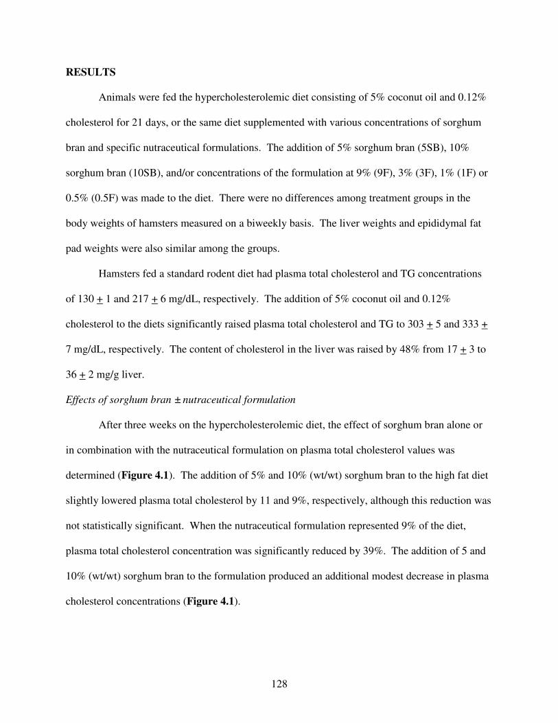
128
RESULTS
Animals were fed the hypercholesterolemic diet consisting of 5% coconut oil and 0.12%
cholesterol for 21 days, or the same diet supplemented with various concentrations of sorghum
bran and specific nutraceutical formulations. The addition of 5% sorghum bran (5SB), 10%
sorghum bran (10SB), and/or concentrations of the formulation at 9% (9F), 3% (3F), 1% (1F) or
0.5% (0.5F) was made to the diet. There were no differences among treatment groups in the
body weights of hamsters measured on a biweekly basis. The liver weights and epididymal fat
pad weights were also similar among the groups.
Hamsters fed a standard rodent diet had plasma total cholesterol and TG concentrations
of 130 + 1 and 217 + 6 mg/dL, respectively. The addition of 5% coconut oil and 0.12%
cholesterol to the diets significantly raised plasma total cholesterol and TG to 303 + 5 and 333 +
7 mg/dL, respectively. The content of cholesterol in the liver was raised by 48% from 17 + 3 to
36 + 2 mg/g liver.
Effects of sorghum bran ± nutraceutical formulation
After three weeks on the hypercholesterolemic diet, the effect of sorghum bran alone or
in combination with the nutraceutical formulation on plasma total cholesterol values was
determined (Figure 4.1). The addition of 5% and 10% (wt/wt) sorghum bran to the high fat diet
slightly lowered plasma total cholesterol by 11 and 9%, respectively, although this reduction was
not statistically significant. When the nutraceutical formulation represented 9% of the diet,
plasma total cholesterol concentration was significantly reduced by 39%. The addition of 5 and
10% (wt/wt) sorghum bran to the formulation produced an additional modest decrease in plasma
cholesterol concentrations (Figure 4.1).

129
The effect of sorghum bran and the nutraceutical formulation on plasma TG
concentrations was examined (Figure 4.2). Supplementation of the diet with the formulation
alone and in combination with 5 and 10% sorghum bran significantly lowered plasma TG
concentrations by 30, 34, and 32%, respectively. Sorghum bran at both 5% and 10% of the diet,
in the absence of the nutraceutical formulation, did not significantly decrease plasma TG
concentrations.
Dose-response effect of sorghum bran and nutraceutical formulation
To determine the dose-response effect of the nutraceutical formulation, animals were fed
a high fat diet and formulation doses ranging from 0.5 to 9% (wt/wt) in combination with 5%
(wt/wt) sorghum bran. Sorghum bran alone and supplemented with the formulation at 0.5% of
the diet slightly decreased plasma cholesterol concentrations, although these reduction were not
statistically significant. Doses of the formulation as low as 1% in combination with sorghum
bran significantly lowered plasma total cholesterol concentrations (Figure 4.3) The plasma
cholesterol concentrations of animals fed the 1% formulation and 5% sorghum bran was similar
to that observed when the diet was supplemented with 9% of the diet in the absence or presence
of sorghum bran..
The addition of 5% sorghum bran and 0.5% formulation to the high fat diet significantly
lowered plasma TG concentrations by 38%; however, a significant effect was not observed by
sorghum bran alone (Figure 4.4). Further increases of the formulation into the diet did not result
in a further reduction in plasma TG concentrations.
The liver cholesterol content was decreased in animals fed diets containing 1 to 3% of the
nutraceutical formulation in the presence of 5% sorghum bran. Diets supplemented with the
sorghum bran alone and sorghum bran and 0.5% formulation slightly, but not significantly,

130
increased liver cholesterol content (Figure 4.5). There were no statistically significant
differences observed in liver TG content among the groups (data not shown).
Effects of different nutraceutical combinations
In another experiment, the four component nutraceutical formulation was split into two
new formulations and their effect on plasma cholesterol concentrations in the
hypercholesterolemic hamster examined. One formulation contained 0.5% pantethine and 4%
turmeric while the other contained 0.5% phytosterols and 4% Spirulina. The combination of
sorghum bran, pantethine and turmeric significantly lowered plasma and plasma total cholesterol
by 22% while sorghum bran, phytosterols and Spirulina produced a 32% reduction (Figure 4.6).
While there was no significant difference between these two formulations in reducing plasma
cholesterol concentrations, the extent of inhibition by each was significantly less than the 39%
reduction observed with the four-ingredient nutraceutical formulation (Figure 4.1).
DISCUSSION
The purpose of the present study was two-fold. First, to develop a combination of natural
ingredients that produces a significant lipid-lowering effect in hypercholesterolemic hamsters.
Second, to investigate whether sorghum bran can further lower plasma lipid concentrations when
combined with this nutraceutical formulation. The combination of four natural ingredients:
Spirulina, turmeric, pantethine and phytosterols significantly lowered cholesterol plasma
cholesterol and TG concentrations in the hypercholesterolemic hamster
Sorghum bran was included in this study for two reasons. Previous studies in our
laboratory revealed a consistent, though non-significant, 10% lowering of total plasma
cholesterol concentrations with the consumption of sorghum bran at 5% of the diet [21]. In
agreement with this past observation, sorghum bran also lowered plasma cholesterol

131
approximately 10% in these studies (Figure 4.1). When added to the formulation, sorghum bran
produced a further, though non-significant lowering of plasma cholesterol concentrations
(Figure 4.1). Thus, the inclusion of sorghum bran into the diet produced, at best, only a modest
incremental decrease in plasma cholesterol concentrations. More importantly than its putative
hypocholesterolemic effect, sorghum bran is an excellent source of phytochemicals [18, 25-27].
The antioxidants present in sorghum bran are composed of condensed tannins (epicatechin and
catechin) and phenolics acids (ferulic, caffeic and coumaric). The total amount of phenolic
compounds and its antioxidant capacity is higher than any other major grain bran [22]. While
these phytochemicals present in sorghum appear to lack significant hypocholesterolemic effects
in this animal model, sorghum bran is an inexpensive source of phytochemicals and is emerging
as important dietary constituent for health conscious individuals
Natural products are becoming increasingly popular in the treatment of various disease
states [28-32]. The ingredients in the formulation employed in these studies were chosen
because they have unique mechanisms of actions when compared to the other constituents.
These effects range from inhibition of cholesterol absorption and synthesis to increasing
cholesterol and lipoprotein catabolism. While turmeric (Curcuma longa) has been widely used
as a spice and in curry powder, recent evidence has shown that turmeric has many beneficial
pharmacological effects [10, 33]. Curcumin, the active component of turmeric, can effectively
lower plasma cholesterol concentrations in experimental animal models [6, 8]. The
hypocholesterolemic effect may be attributed to the upregulation of hepatic cholesterol-7alpha-
hydroxylase, thereby increasing the rate of cholesterol catabolism [6]. Peschel et al. [34]
reported a concentration-dependent increase in LDL-receptor mRNA expression in HepG2 cells,
thereby, increasing the uptake of LDL-cholesterol from plasma. Additionally, curcumin has

132
been reported to increase HDL-cholesterol concentrations in hypercholesterolemic rats [35].
This may be attributed to the significant induction of LXRα mRNA and subsequently its direct
target, the ATP-binding cassette transporter, ABCG1 [36].
The cholesterol lowering property of phytosterols has been well documented since 1953
[37, 38]. It is believed that structural similarity to cholesterol plays a major role in its cholesterol
lowering activity. Phytosterols compete with dietary cholesterol for intestinal incorporation into
micelles, resulting in a reduction of cholesterol absorption [9]. The phytosterols in this study
were from Basikol®, a mixture of phytosterols from soy, corn and canola oils.
Spirulina is well known for its high levels of chlorophyll [39]. Chlorophyll is
metabolized to phytanic (3,7,11,15-tetramethylhexadecanoic) acid, an isoprenoid-derived fatty
acid. Phytanic acid binds and activates peroxisome proliferator-activated receptor (PPAR)-α, a
nuclear transcription factor [40, 41]. The stimulation of PPAR-α up-regulates lipoprotein lipase,
an enzyme that plays a key role in the hydrolysis of VLDL triglycerides [42]. Thus, it is
hypothesized that phytanic acid may lower blood lipid levels via its ligand-binding interactions
with PPAR-α [43].
Pantethine is a stable disulfide form of pantetheine; a metabolite, cystamine appears to be
responsible for its ability to inhibit acetyl-CoA carboxylase [44]. Pantethine has been shown to
beneficially alter plasma cholesterol concentrations in experimental animals and humans [12, 45,
46] by inhibiting cholesterol and fatty acid synthesis; interestingly, protein , DNA and
phospholipid synthesis are also inhibited in dose-dependent manners [47, 48].
At the highest dose employed in these studies, the formulation represented 9% of the diet.
This dosage was chosen because it approximates the minimum dose that has been reported to be
efficacious in lowering plasma cholesterol concentrations in a variety of experimental animals

133
[10-13]. Thus, the significant lowering of plasma cholesterol with the formulation set at 1% of
the diet, in the presence of sorghum bran, demonstrates that significant effects can be seen when
each ingredient is administered at a much lower dosage than previously reported. This was also
observed in the capability of the formulation to lower plasma TG concentrations. In this case,
the formulation at approximately 0.5% of the produced a significantly lowering of plasma TG.
The formulation appears to be effective based on incremental, additive effects of these
constitutive agents. While we hypothesized that sorghum bran would significantly augment the
plasma cholesterol-lowering actions of the formulation, we observed only a modest lowering of
cholesterol. When an experiment was performed splitting the four ingredient formulation into
two different, two-ingredient formulations, both significantly lowered plasma cholesterol
concentrations. A significant difference in their hypocholesterolemic effects between these
combination products was not observed. Based on the data presented in Figures 1 and 6, the
addition of both formulations together to form the four ingredient nutraceutical formulation
yielded a further incremental lowering in plasma cholesterol concentrations. These nutraceutical
agents are apparently not acting in a synergistic manner, but each one is contributing to the
overall hypocholesterolemic effect of the four ingredient formulation.
This is not the first study to demonstrate that a combination of natural products can be an
effective strategy to lower plasma cholesterol concentrations; such a paradigm has been found to
be successful in both experimental animals [49] and humans [29]. This report demonstrates that
the dosages of the individual natural products present in a formulation can be sharply lowered
without a loss of its hypolipidemic actions. This finding should eventually allow for
optimization of an efficacious nutraceutical formulation for use in humans.

134
ACKNOWLEDGMENTS
Support for this research was provided by grants from the United States Department of
Agriculture and the American Foundation for Pharmaceutical Education.

135
REFERENCES
1. Chisolm, G.M. and D. Steinberg, The oxidative modification hypothesis of atherogenesis:
An overview. Free Radical Biology and Medicine, 2000. 28(12): p. 1815-1826.
2. LaRosa, J.C., Pleiotropic effects of statins and their clinical significance. American
Journal of Cardiology, 2001. 88(3): p. 291-+.
3. Jacobson, T.A., Statin safety: Lessons from new drug applications for marketed statins.
American Journal of Cardiology, 2006. 97(8A): p. 44C-51C.
4. Bays, H. and E.A. Stein, Pharmacotherapy for dyslipidaemia - current therapies and
future agents. Expert Opinion on Pharmacotherapy, 2003. 4(11): p. 1901-1938.
5. Ramamoorthy, A. and S. Premakumari, Effect of supplementation of Spirulina on
hypercholesterolemic patients. Journal of Food Science and Technology-Mysore, 1996.
33(2): p. 124-127.
6. Babu, P.S. and K. Srinivasan, Hypolipidemic action of curcumin, the active principle of
turmeric (Curcuma longa) in streptozotocin induced diabetic rats. Molecular and
Cellular Biochemistry, 1997. 166(1-2): p. 169-175.
7. McCarty, M.F., Inhibition of acetyl-CoA carboxylase by cystamine may mediate the
hypotriglyceridemic activity of pantethine. Medical Hypotheses, 2001. 56(3): p. 314-317.
8. Ramirez-Tortosa, M.C., et al., Oral administration of a turmeric extract inhibits LDL
oxidation and has hypocholesterolemic effects in rabbits with experimental
atherosclerosis. Atherosclerosis, 1999. 147(2): p. 371-378.
9. Trautwein, E.A., et al., Proposed mechanisms of cholesterol-lowering action of plant
sterols. European Journal of Lipid Science and Technology, 2003. 105(3-4): p. 171-185.

136
10. Srinivasan, K., Spices as influencers of body metabolism: an overview of three decades of
research. Food Research International, 2005. 38(1): p. 77-86.
11. Derivera, C.G., et al., Preventive Effect of Spirulina-Maxima on the Fatty Liver Induced
by a Fructose-Rich Diet in the Rat, a Preliminary-Report. Life Sciences, 1993. 53(1): p.
57-61.
12. Shinomiya, M., et al., Effect of Pantethine on Cholesterol Ester Metabolism in Rat
Arterial-Wall. Atherosclerosis, 1980. 36(1): p. 75-80.
13. Ntanios, F.Y., D.E. MacDougall, and P.J.H. Jones, Gender effects of tall oil versus
soybean phytosterols as cholesterol-lowering agents in hamsters. Canadian Journal of
Physiology and Pharmacology, 1998. 76(7-8): p. 780-787.
14. Deters, M., et al., Effects of curcumin on cyclosporine-induced cholestasis and
hypercholesterolemia and on cyclosporine metabolism in the rat. Planta Med, 2003.
69(4): p. 337-43.
15. Awika, J.M. and L.W. Rooney, Sorghum phytochemicals and their potential impact on
human health. Phytochemistry, 2004. 65(9): p. 1199-1221.
16. Awika, J.M., L.W. Rooney, and R.D. Waniska, Properties of 3-deoxyanthocyanins from
sorghum. Journal of Agricultural and Food Chemistry, 2004. 52(14): p. 4388-4394.
17. Awika, J.M., et al., Screening methods to measure antioxidant activity of sorghum
(Sorghum bicolor) and sorghum products. Journal of Agricultural and Food Chemistry,
2003. 51(23): p. 6657-6662.
18. Dykes, L., et al., Phenolic compounds and antioxidant activity of sorghum grains of
varying genotypes. Journal of Agricultural and Food Chemistry, 2005. 53(17): p. 6813-
6818.

137
19. Rooney, L.W. and J.M. Awika, Overview of products and helalth benefits of specialty
sorghums. Cereal Foods World, 2005. 50(3): p. 109-+.
20. Carr, T.P., et al., Grain sorghum lipid extract reduces cholesterol absorption and plasma
non-HDL cholesterol concentration in hamsters. Journal of Nutrition, 2005. 135(9): p.
2236-2240.
21. Burdette, A.L., et al., Hypolipidemic effect of a sorghum fraction in combination with
other natural nutraceutical ingredients in the hyperlipidemic Syrian hamster. Faseb
Journal, 2006. 20(5): p. A1027-A1027.
22. Burdette, A., et al., Development of sorghum bran as an anti-inflammatory nutraceutical.
Faseb Journal, 2007. 21(5): p. A364-A365.
23. Cai, G. and T.P. Carr, Biliary cholesterol and bile acid excretion do not increase in
hamsters fed cereal-based diets containing cholesterol. Metabolism-Clinical and
Experimental, 1999. 48(3): p. 400-405.
24. Folch, J., M. Lees, and G.H.S. Stanley, A Simple Method for the Isolation and
Purification of Total Lipides from Animal Tissues. Journal of Biological Chemistry, 1957.
226(1): p. 497-509.
25. Awika, J.M., C.M. McDonough, and L.W. Rooney, Decorticating sorghum to
concentrate healthy phytochemicals. Journal of Agricultural and Food Chemistry, 2005.
53(16): p. 6230-6234.
26. Awika, J.M., et al., Screening methods to measure antioxidant activity of sorghum
(sorghum bicolor) and sorghum products. J Agric Food Chem, 2003. 51(23): p. 6657-62.
27. Awika, J.M. and L.W. Rooney, Sorghum phytochemicals and their potential impact on
human health. Phytochemistry, 2004. 65(9): p. 1199-221.

138
28. Bickford, P.C., et al., Nutraceuticals synergistically promote proliferation of human stem
cells. Stem Cells Dev, 2006. 15(1): p. 118-23.
29. Cicero, A.F., L.C. Rovati, and I. Setnikar, Eulipidemic effects of berberine administered
alone or in combination with other natural cholesterol-lowering agents. A single-blind
clinical investigation. Arzneimittelforschung, 2007. 57(1): p. 26-30.
30. Pero, R.W., et al., Formulation and in vitro/in vivo evaluation of combining DNA repair
and immune enhancing nutritional supplements. Phytomedicine, 2005. 12(4): p. 255-63.
31. Dechant, J.E., et al., Effects of glucosamine hydrochloride and chondroitin sulphate,
alone and in combination, on normal and interleukin-1 conditioned equine articular
cartilage explant metabolism. Equine Veterinary Journal, 2005. 37(3): p. 227-231.
32. Vouldoukis, I., et al., Antioxidant and anti-inflammatory properties of a Cucumis melo
LC. extract rich in superoxide dismutase activity. Journal of Ethnopharmacology, 2004.
94(1): p. 67-75.
33. Srinivasan, K., Role of spices beyond food flavoring: Nutraceuticals with multiple health
effects. Food Reviews International, 2005. 21(2): p. 167-188.
34. Peschel, D., R. Koerting, and N. Nass, Curcumin induces changes in expression of genes
involved in cholesterol homeostasis. Journal of Nutritional Biochemistry, 2007. 18(2): p.
113-119.
35. Arafa, H.M.M., Curcumin attenuates diet-induced hypercholesterolemia in rats. Medical
Science Monitor, 2005. 11(7): p. BR228-BR234.
36. Kennedy, M.A., et al., Characterization of the human ABCG1 gene - Liver X receptor
activates an internal promoter that produces a novel transcript encoding an alternative
form of the protein. Journal of Biological Chemistry, 2001. 276(42): p. 39438-39447.

139
37. Pollak, O.J., Successful Prevention of Experimental Hypercholesteremia and Cholesterol
Atherosclerosis in the Rabbit. Circulation, 1953. 7(5): p. 696-701.
38. Lin, Y.G., et al., Soy protein enhances the cholesterol-lowering effect of plant sterol
esters in cholesterol-fed hamsters. Journal of Nutrition, 2004. 134(1): p. 143-148.
39. Khan, Z., P. Bhadouria, and P.S. Bisen, Nutritional and therapeutic potential of
Spirulina. Curr Pharm Biotechnol, 2005. 6(5): p. 373-9.
40. Wolfrum, C., et al., Phytanic acid is ligand and transcriptional activator of murine liver
fatty acid binding protein. Journal of Lipid Research, 1999. 40(4): p. 708-714.
41. Heim, M., et al., Phytanic acid, a natural peroxisome proliferator-activated receptor
(PPAR) agonist, regulates glucose metabolism in rat primary hepatocytes. Faseb J, 2002.
16(7): p. 718-20.
42. Neve, B.P., J.C. Fruchart, and B. Staels, Role of the peroxisome proliferator-activated
receptors (PPAR) in atherosclerosis. Biochemical Pharmacology, 2000. 60(8): p. 1245-
1250.
43. Heim, M., et al., Phytanic acid, a natural peroxisome proliferator-activated receptor
agonist, regulates glucose metabolism in rat primary hepatocytes. Faseb Journal, 2002.
16(3).
44. McCarty, M.F., Inhibition of acetyl-CoA carboxylase by cystamine may mediate the
hypotriglyceridemic activity of pantethine. Med Hypotheses, 2001. 56(3): p. 314-7.
45. Gaddi, A., et al., Controlled Evaluation of Pantethine, a Natural Hypolipidemic
Compound, in Patients with Different Forms of Hyperlipoproteinemia. Atherosclerosis,
1984. 50(1): p. 73-83.

140
46. Carrara, P., et al., Pantethine Reduces Plasma-Cholesterol and the Severity of Arterial
Lesions in Experimental Hypercholesterolemic Rabbits. Atherosclerosis, 1984. 53(3): p.
255-264.
47. Ranganathan, S., R.L. Jackson, and J.A.K. Harmony, Effect of Pantethine on the
Biosynthesis of Cholesterol in Human-Skin Fibroblasts. Atherosclerosis, 1982. 44(3): p.
261-273.
48. Cighetti, G., et al., Effects of Pantethine on Cholesterol-Synthesis from Mevalonate in
Isolated Rat Hepatocytes. Atherosclerosis, 1986. 60(1): p. 67-77.
49. Tas, S., et al., Vanadyl sulfate, taurine, and combined vanadyl sulfate and taurine
treatments in diabetic rats: Effects on the oxidative and antioxidative systems. Archives
of Medical Research, 2007. 38(3): p. 276-283.

141
TABLES AND FIGURES
Diet Supplementation
5SB 10SB 9F 5SB + 9F 10SB + 9F
Pla
sma
Ch
ole
ster
ol
(Per
cen
t o
f H
igh
Fa
t G
rou
p)
0
20
40
60
80
100
**
*
Figure 4.1. Effect of sorghum bran and the nutraceutical formulation on plasma total
cholesterol concentration in the hypercholesterolemic hamster. Plasma total cholesterol
concentrations in hypercholesterolemic hamsters fed 5 (5SB) or 10% sumac sorghum bran
(10SB) alone or in combination with 9% (wt/wt) nutraceutical formulation (9F) Values are
expressed as percentage (mean + SEM) of plasma cholesterol concentrations of animals fed 5%
coconut oil and 0.12% cholesterol. *Significantly different from the group receiving 5%
coconut oil and 0.12% cholesterol, P < 0.05.

142
Diet Supplementation
5SB 10SB 9F 5SB + 9F 10SB + 9F
Pla
sma T
rig
lyce
rid
es
(Per
cen
t o
f H
igh
Fat
Gro
up
)
0
20
40
60
80
100
** *
Figure 4.2. Effect of sorghum bran and the nutraceutical formulation on plasma
triglyceride concentration in the hypercholesterolemic hamster. Plasma triglyceride
concentrations in hypercholesterolemic hamsters fed 5 (5SB) or 10% sumac sorghum bran
(10SB) alone or in combination with the 9% (wt/wt) nutraceutical formulation (9F). Values are
expressed as percentage (mean + SEM) of plasma triglyceride concentrations of animals fed the
high fat diet. *Significantly different from the group receiving 5% coconut oil and 0.12%
cholesterol, P < 0.05.

143
Diet Supplementation
5SB 1/2F + 5SB 1F + 5SB 3F + 5SB
Pla
sma
Ch
ole
ster
ol
(Per
cen
t o
f H
igh
Fat
Gro
up
)
0
20
40
60
80
100
**
Figure 4.3. Effect of sorghum bran and the nutraceutical formulation on plasma total
cholesterol concentration in the hypercholesterolemic hamster. Plasma cholesterol
concentrations in hypercholesterolemic hamsters fed 5% sumac sorghum bran (5SB) alone or in
combination with 0.5, 1 or 3% of the nutraceutical formulation (0.5F, 1F or 3F, respectively).
Values are expressed as percentage (mean + SEM) of plasma cholesterol concentrations of
animals fed the high fat diet. *Significantly different from the group receiving 5% coconut oil
and 0.12% cholesterol, P < 0.05.

144
Diet Supplementation
5SB 1/2F + 5SB 1F + 5SB 3F + 5SB
Pla
sma T
rig
lyce
rid
es
(Per
cen
t o
f H
igh
Fat
Gro
up
)
0
20
40
60
80
100
* **
Figure 4.4. Effect of sorghum bran and the nutraceutical formulation on plasma
triglyceride concentration in the hypercholesterolemic hamster. Plasma triglyceride
concentrations in hypercholesterolemic hamsters fed 5% sumac sorghum bran (5SB) alone or in
combination with 0.5, 1 or 3% of the nutraceutical formulation (0.5F, 1F or 3F, respectively).
Values are expressed as percentage (mean + SEM) of plasma triglyceride concentrations of
animals fed the high fat diet. *Significantly different from the group receiving 5% coconut oil
and 0.12% cholesterol, P < 0.05.

145
Diet Supplementation
5SB 1/2F + 5SB 1F + 5SB 3F + 5SB
Liv
er C
ho
lest
ero
l
(Per
cen
t o
f H
igh
Fat
Gro
up
)
0
20
40
60
80
100
120
**
Figure 4.5. Effect of sorghum bran and the nutraceutical formulation on liver total
cholesterol content in the hypercholesterolemic hamster. Liver total cholesterol content in
hypercholesterolemic hamsters fed 5% sumac sorghum bran (5SB) alone or in combination with
0.5, 1 or 3% of the nutraceutical formulation (0.5F, 1F or 3F, respectively). Values are
expressed as percentage (mean + SEM) of liver cholesterol content of animals fed a high fat diet.
*Significantly different from the group receiving 5% coconut oil and 0.12% cholesterol, P <
0.05.

146
Diet Supplementation
5SB
5SB + Pantethine + Turmeric
5SB + Sterols + Spirulina
Pla
sm
a C
ho
leste
rol
(Perc
en
t o
f C
ho
leste
rol F
ed
Gro
up
)
0
20
40
60
80
100
*
*
Figure 4.6. Effect of sorghum bran and different nutraceutical combinations on plasma
total cholesterol concentration in the hypercholesterolemic hamster. Plasma total
cholesterol concentration in hypercholesterolemic hamsters fed 5% (wt/wt) sumac sorghum bran
(5SB) alone and two-ingredient nutraceutical formulations comprising 0.5% and 4% of the diet.
Values are expressed as percentage (mean + SEM) of plasma cholesterol concentrations of
animals fed a high fat diet. *Significantly different from the group receiving 5% coconut oil and
0.12% cholesterol, P < 0.05.

147
CHAPTER FIVE
CONCLUSIONS
The results presented in this dissertation demonstrate that varieties of sorghum bran can
inhibit inflammatory processes and significantly lower plasma cholesterol in
hypercholesterolemic hamsters. The anti-inflammatory effects of sorghum bran were
investigated in LPS-stimulated mononuclear cells and in the TPA model of ear inflammation.
Black sorghum bran extract significantly reduced the secretion of cytokines TNF-α and IL-1
from LPS-stimulated human peripheral mononuclear cells. Ethanolic extracts of black and
sumac sorghum bran produced reductions in ear edema, measured by ear weight and ear
thickness, and MPO activity, an indication of neutrophil involvement. No effect was observed
with white or mycogen sorghum bran varieties or with extracts of rice, oat and wheat brans. The
anti-inflammatory activity positively correlated with the high phenolic and antioxidant activity of
black and sumac sorghum brans. Black and sumac sorghum bran extract did not affect COX-2
protein expression, an enzyme involved in prostaglandin biosynthesis.
We tested the cholesterol lowering capabilities of sorghum wax in hypercholesterolemic
hamsters. Very long chain fatty acids and aldehydes, the major components of the wax fraction,
have been shown by Cuban research institutions to lower cholesterol in experimental animals
and humans. Sorghum wax failed to lower plasma and liver cholesterol in hamsters. These
results are in agreement with numerous studies conducted by non-Cuban research facilities and
we question the efficacy of the very long chain fatty acids and aldehydes. This finding is quite

148
timely because policosanol, a mixture of very long chain fatty alcohols, has recently been added
to One-A-Day® Cholesterol Plus multivitamin.
The hypocholesterolemic action of sumac sorghum bran was evaluated in hamsters.
Sorghum bran, a rich source of tannins and anthocyanins, significantly reduced plasma
cholesterol and triglycerides in hyperlipidemic hamsters when incorporated at 20% of the diet.
The addition of sorghum bran at 5 and 10% in the diets produced non-significant decreases of 9
and 11%, respectively. This lack of significant cholesterol-lowering effect is a result, to some
degree, of high animal variability.
The effects of four nutraceuticals on plasma and liver cholesterol concentrations in
hamsters were investigated. Significant reductions in plasma and liver total cholesterol
concentrations and plasma triglycerides were observed with the combination of four natural
components (blend of phytosterols, turmeric, Spirulina, and pantethine) in the hyperlipidemic
hamster. The addition of sorghum bran to the formulation did not produce further reductions in
plasma and liver cholesterol. Therefore, it was determined that the bulk of the cholesterol-
lowering efficacy was attributed, not to sorghum bran, but to the four natural ingredients. These
ingredients alone have all been previously reported to lower cholesterol, however, in
combination, their additive pharmacological effects allowed for the administration of smaller
doses than previously documented. These results should allow for the optimization of an
inexpensive and efficacious cholesterol-lowering nutraceutical formulation.
The outcome of this research approach is the incorporation of select sorghum brans into
the American diet or as a functional food, nutraceutical or cosmeceutical. These results suggest
that consumption of rather small amounts of specific natural products may lower cholesterol and
triglycerides and possibly reduce the incidence of cardiovascular disease. The bran fraction of

149
select sorghum grains, which are among the top five grains produced worldwide, may prove
useful in lowering plasma cholesterol and ameliorating the inflammatory process in humans.