nursing management of the adult patient with neurological alterations prepared by: hikmet qubeilat....
TRANSCRIPT

Nursing Management of the Adult Patient with Neurological AlterationsPrepared by:
Hikmet Qubeilat. RN,MSC.

Brain Needs… Blood flow Glucose Oxygen

Diagnostic Studies Skull and Spinal Radiology CT (Computerized Tomography) MRI (Magnetic Resonance Imaging) PET (Positron Emission Tomography) EEG (Electroencephalogram) EMG (Electromyelogram) Cerebral Blood Flow Studies

Neurological Assessment Level of Consciousness (LOC) Pupils Vital Signs (VS) Neuromuscular status Response to stimuli Posturing Glasgow Coma Scale (GCS)


I. Neurological Disorders The normal functioning of the CNS can be
affected by a number of disorders, the most common of which are headaches, tumors, vascular problems, infections, epilepsy, head trauma, demyelinating diseases, and metabolic & nutritional diseases.

Headaches
Classified based on characteristics of the headache
Functional vs. Organic type May have more than one type of headache History & neurologic exam diagnostic keys
Not always chronic…be careful

Pattern Tension Migraine Cluster
Site Bilateral, basilar, band-like
Unilateral, anterior
Unilateral, occular
Quality Squeezing, constant
Throbbing Severe
Pattern Cycles, years Periodic, years Remitting, relapsing
Duration Days, weeks, months
Hours, days 30-90 min
Onset Anytime Prodrome, starts in AM
Nocturnal
Assoc. S&S
Stiff neck N&V, photo/phono-phobia
Horner syndrome
ONSET: Not reliable or diagnostic

HA: Essential History Onset this particular headache Character of pain, severity and duration Associated symptoms Prior history, pattern Original onset: prior testing, treatment Other therapeutic regimens
Physical Exam Neurologic examination Inspect for local infections, nuchal rigidity Palpation for tenderness, bony swellings Auscultation for bruits over major arteries

Organic vs. Traumatic vs. Functional: Diagnostics CBC: underlying illness, anemia Chem panel: if associated vomiting, dehydrated CT scan: for focal neurological signs, sinus No LP for suspected ICP; ↑ association with brain
herniation

Don’t Miss It
1. Caused by subarachnoid hemorrhage from an aneurysm or head injury
2.“Worse headache of my life”
3. Changes in LOC, focal neurological signs
4. Highly correlated with CVA
5. Untreated, 50 % mortality

Headache Teaching Guide Keep a calendar/diary Avoid triggers Medications (purpose, side effects) Stress reduction
Dark quiet room, exercise, relaxation Regular exercise
Intracranial Pressure (ICP)Brain Components Skull is a rigid vault that does not expand
It contains 3 volume components: Brain tissue: (80%) or 2% of TBW Intravascualr blood: (10%) CSF: (10%)
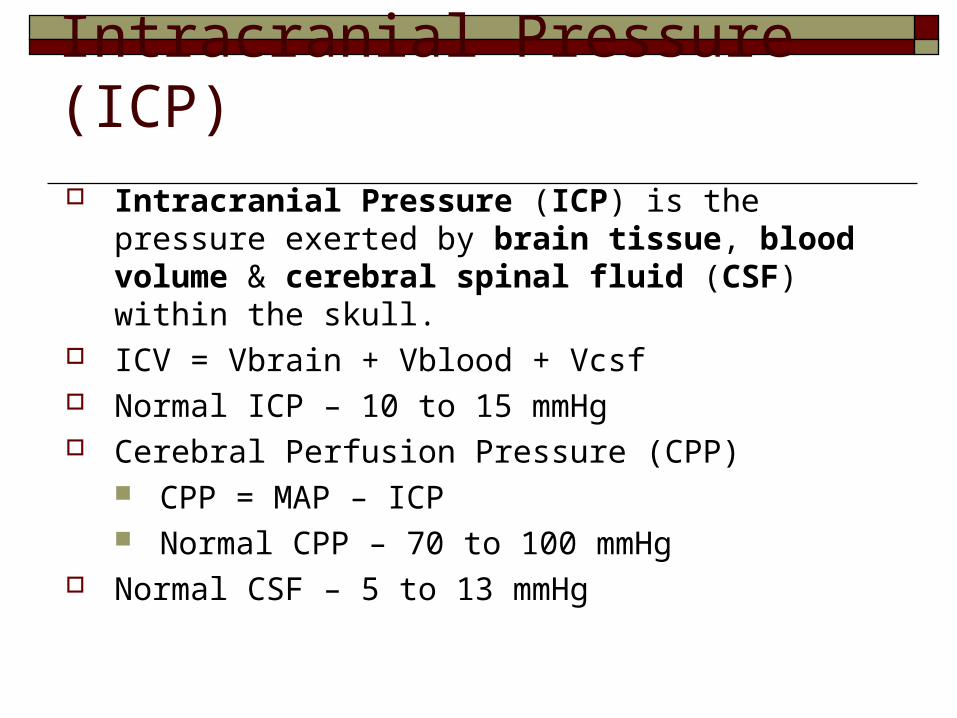
Intracranial Pressure (ICP) is the pressure exerted by brain tissue, blood volume & cerebral spinal fluid (CSF) within the skull.
ICV = Vbrain + Vblood + Vcsf Normal ICP – 10 to 15 mmHg Cerebral Perfusion Pressure (CPP)
CPP = MAP – ICP Normal CPP – 70 to 100 mmHg
Normal CSF – 5 to 13 mmHg
Intracranial Pressure (ICP)

Increased Intracranial Pressure (IICP) fluid pressure > 15 mm Hg
IICP is a life threatening situation that results from an in any or all 3 components within the skull > volume of brain tissue, blood, and / or CSF Cerebral edema: > H2O content of tissue as a result
of trauma, hemorrhage, tumor, abscess, or ischemia


Acute Coma Levels of consciousness diminish in stages:
• Confusion: can’t think rapidly and clearly التشويش• Disorientation: begin to loose consciousness
• Time, place, self• Lethargy: spontaneous speech and movement limited• Obtundation: arousal (awakeness) is reduced• Stupor: deep sleep or unresponsiveness
• Open eyes to vigorous or repeated stimuli• Coma: respond to noxious stimuli only
• Light (purposeful), full coma (non-purposeful), deep coma (no response)

19
Multiple Sclerosis
is a chronic autoimmune disorder affecting movement, sensation, and bodily functions. It is caused by destruction of the myelin sheath covering nerve fibres in the central nervous system (brain and spinal cord).
Causes:1. Autoimmune destruction. 2. Heredity. 3. Viruses. 4. Environmental factors.

20
Diagnostic Test:
1. MRI.
2. Physical examination.

21
* Early:
1. Muscle weakness causing difficulty walking
2. loss of coordination or balance
3. numbness or other abnormal sensations
4. visual disturbances, including blurred or double vision
Clinical Manifestations:

22
* Late: 1. Fatigue . 2. Muscle spasticity and stiffness 3. Tremors. 4. Paralysis . 5. pain . 6. Vertigo. 7. Speech or swallowing difficulty . 8. Loss of bowel and bladder control. 9. Sexual dysfunction . 10. Changes in cognitive ability

23
Treatment:
1. Immunosuppressant drugs . These drugs include corticosteroids such as prednisone and methylprednisolone, the hormone adrenocorticotropic hormone (ACTH), and azathioprine.
2. Physiotherapy.
3. Occupational therapy.

24
Parkinson's Disease
is a progressive movement disorder marked by tremors, rigidity, slow movements (bradykinesia), and postural instability. It occurs when, for unknown reasons, cells in one of the movement-control centers of the brain begin to die.
Causes: 1. Degeneration of brain cells in the area known as the
substantia nigra, one of the movement control centers of the brain.
2. Drugs given for psychosis, such as haloperidol (Haldol) or chlorpromazine (Thorazine), may cause parkinsonism.
25
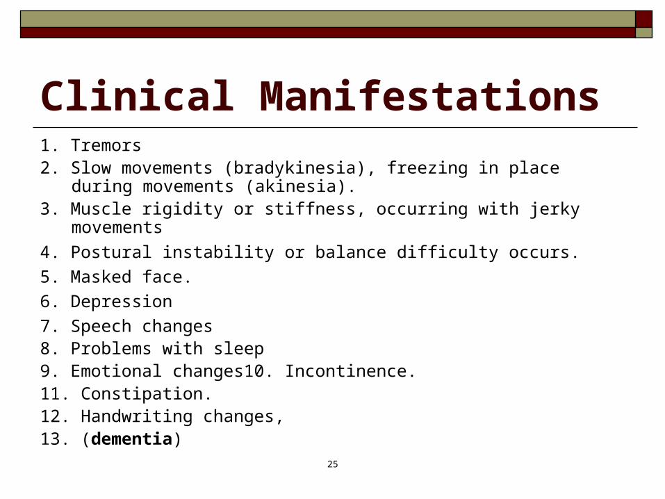
Clinical Manifestations1. Tremors2. Slow movements (bradykinesia), freezing in place during movements
(akinesia). 3. Muscle rigidity or stiffness, occurring with jerky movements
4. Postural instability or balance difficulty occurs.
5. Masked face.
6. Depression
7. Speech changes8. Problems with sleep9. Emotional changes10. Incontinence. 11. Constipation. 12. Handwriting changes, 13. (dementia)

26
Treatment:
1. Maintain regular exercise (physical therapy, occupational therapy)
2. Provide good nutrition to maintain health.
3. Drugs that replace dopamine (levodopa)
4. If the patient is unresponsive or intolerant to pharmacotherapy, Electro convulsive therapy is indicated.
Nursing Management
* Observe the patient's mood, cognition; organization and general well being
* Observe for features of depression,
*Suicidal precautions to be followed, if the patient exhibits any suicidal ideas
*Instruct the patients to speak slowly and clearly, and to pause and take a deep breath at appropriate levels.
27
Parkinson's Disease (cont’d) *In dementia, environmental modification is followed
*Avoid frequent change in the environment to minimise confusion if the memory deficit is very severe, name boards and signboards by the side of the rooms and things will be very helpful.
*Sedatives are used if sleep related problems are noticed, when sleep hygiene is unsuccessfully.
* Patients should not be forced into situations in which they feel ashamed of their appearance.
*Encourage the patient to participate in moderate exercises, free-moving
sports like swimming. *Advise the patient to organize thoughts before speaking and encourage the
client to use facial expression and gestures if possible to assist with communication.

Seizure Disorders & EpilepsySeizure:
paroxysmal, uncontrolled electrical discharge of neurons in the brain that interrupts normal function
Epilepsy: spontaneously recurring seizures caused by a chronic
underlying condition
Two major classes: Generalized Partial

Depending on type, phases may include: Prodromal phase- signs & activity preceeding seizure Aural phase- sensory warning Ictal phase- full seizure Postictal phase- recovery
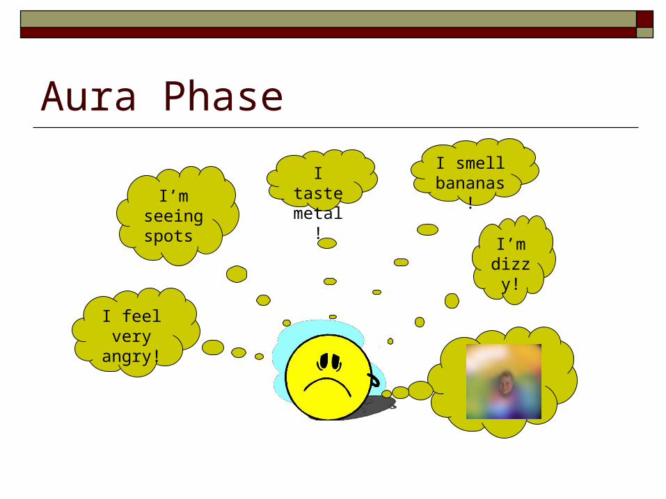
Aura Phase
I’m dizzy
!
I smell bananas!
I taste metal!I’m
seeing spots
I feel very angry!


Seizure Disorders & EpilepsyDrug Therapy for Tonic-Clonic and Partial Seizures
Carbamezepine/ Tegretol Divalproex/ Depakote Gabapentin/ Neurontin Lamotrigine/ Lamictal Levetiracetam/ Keppra
Phenytoin/ Dilantin Tiagabine/ Gabitril Topiramate/ Topamax Valproic Acid/ Depakene Felbamate/ Felbatol * Phenobarbitol**
*Felbatol has been associated with aplastic anemia**Phenobarbitol is a barbituate

Seizure Disorders & Epilepsy:Nursing Care Assure oxygen and suction equipment at bedside Safety precautions in active stage
Support/ protect head Turn to side Lossen constricted clothing Ease to floor
Time seizure, record details of seizure and post-ictal phase

Seizure Disorders & Epilepsy:Nursing Care Patient teaching:
importance of good seizure control using medication as ordered
Medical alert bracelet Avoid decreased sleep, increased fatigue Regular meals/ snacks

Seizure Disorders & Epilepsy: Status Epilepticus Medical emergency Seizure repeated continuously
Tonic clonic: hypoxia could develop if muscle contraction is lengthened. Also: hypoglycemia, acidosis, hypothermia, brain damage, death IV administration of antiepileptics Maintain airway patency

Intracranial surgery Craniotomy:
Opening the skull surgically to gain access to intracranial structures

Intracranial surgery Burr hole
Circular opening made in the skull by a drill

Intracranial surgery Craniectomy
An excision of a portion of the skull

Intracranial surgery Cranioplasty
Repair of a cranial defect by means of a plastic or metal plate

Intracranial surgery Transsphenoidal
Through the nasal sinuses to gain access to the pituitary gland

Types of Stroke
Ischemic: embolic or thrombotic blocked blood flow to the brain
Hemorrhagic: ICH, SAH, ruptured cerebral aneurysm
TIA: This is a stroke, although symptoms resolve within an hour

Signs and Symptoms of Stroke Sudden numbness or weakness of the face, arm or leg,
especially on one side of the body Sudden confusion, trouble speaking or understanding Sudden trouble seeing in one or both eyes Sudden dizziness, loss of balance or coordination or
trouble walking Sudden severe headache with no known cause

Risk Factors
High blood pressure Carotid artery disease Physical inactivity Excess alcohol intake Atrial fibrillation Diabetes Heart disease Smoking Family history Prior stroke/TIA High cholesterol Obesity

Treatment for Ischemic Stroke tPA=Thrombolytic agent Document time of symptom
onset. (If awoke with symptoms, must go by time when last seen normal)
Immediate head CT (check for blood)
Evaluate for tPA administration (review exclusion/inclusion criteria)

Treatment Cont… If not a tPA candidate, ASA in ED. Rectal ASA
if fails swallow eval. or if swallow eval. not complete.
Keep NPO, until a formal swallow eval. is done. Admit as Inpatient and perform diagnostic
testing: Carotid US, Echo, TEE, ECG monitoring for a-fib, MRI, fasting Lipid, Clotting disorder blood work (Antiphospholipid, Factor V, Antithrombin III)
Rehabilitation

Hemorrhagic Stroke Treatment Do not give antithrombotics or
anticoagulants Monitor and treat blood
pressure greater than 150/105 (Table 6, 2005 Guidelines update)
NPO, until swallow eval is completed
Anticipate Neurosurgical consult
Possible administration of blood products

Meningitis
An inflammation of the meninges of the brain and spinal cord Bacterial
Causes:Meningococcus and pneumococcus ,Haemophilus-influenza
Organisms enter brain by: Blood stream Respiratory tract Pentrating wonds of skullIt is secondary to another infections such as otitismedia, upper
respiratory infection,pneumonia Viral (aseptic): less severe than bacterial

Clinical Presentations High fever, tachycardia, chills, petechial rash headache, photophobia, stiff neck Nausea, vomiting papilledema (> ICP),confusion, altered LOC Restlessness and irritability Seizures Brudzinski’s: passive flexion of the neck produces pain &
increased rigidity Kernig’s: Flex hip and knee and then straighten the knee…
pain or resistance?

complication of MeningitisSeizuresSepsisCranial nerve dysfunctionsCerebral infarctionComaDeath

Collaborative care Bacterial menigitis is a medical emergency Treatment focus on rapid diagnosis and starting IV antibiotic
therapy immediately(7-21 days) Isolation Antipyretics Analgesics Anticonvulsants Osmotic diuretics IV fluids

Diagnosis
lumbar puncture :collect samples of CSF Bacterial:
Cloudy csf Elevated protein level Increased WBC Decreased glucose level Elevated CSF pressure
C&S OF CSF CBC Cultures from Blood, urine, throat, nose