nursing care of the pediatric neurosurgery patient || chiari malformation and syringomyelia
TRANSCRIPT

141C.C. Cartwright, D.C. Wallace (eds.), Nursing Care of the Pediatric Neurosurgery Patient, DOI 10.1007/978-3-642-32554-0_5, © Springer-Verlag Berlin Heidelberg 2013
5
5.1 Introduction
Chiari malformations are a group of abnormalities of the hindbrain which were originally described by John Cleland in 1883 and then classi fi ed in 1891 by Hans Chiari, a German professor. His work, based on autopsy results, created the classic de fi nitions of hindbrain herniation now described as Chiari I (CIM), Chiari II (CIIM), and Chiari III (CIIIM). Although named simi-larly to the other Chiari malformations, Chiari IV malformation (CIVM) is now recognized as cerebellar hypoplasia and unrelated to the oth-ers (Greenberg 2010 ; Khoury 2011 ; Oakes et al. 2011 ; Weprin and Oakes 2001 ) . Syringomyelia refers to the development of a cyst or cavity fi lled with cerebrospinal fl uid (CSF) in the spinal cord. The cyst is known as a syrinx. Despite advances in neuroimaging and embryological work, the natural history of Chiari malformation and syrin-gomyelia remains incompletely understood.
CIM consists of displacement of the cerebel-lar tonsils below the foramen magnum and is often associated with syringomyelia. CIIM, also known as the Arnold-Chiari malformation, is
associated with myelomeningocele (MM) and includes caudal displacement of the inferior cer-ebellar vermis, the fourth ventricle, and the medulla into the cervical canal. CIIIM, a rare and severe form, includes a low occipital or high cer-vical encephalocele in combination with down-ward displacement of most of the cerebellum, the fourth ventricle, and possibly portions of the brainstem. The Chiari IV represents cerebellar hypoplasia—lack of development of the cerebel-lum—that is now considered unrelated to the other Chiari malformations (Greenberg 2010 ; Khoury 2011 ; Oakes et al. 2011 ; Weprin and Oakes 2001 ) . The posterior fossa is usually nor-mal in size with an absence of hindbrain hernia-tion (Oakes et al. 2011 ) .
Two other subtypes have been described. The Chiari 0 malformation exhibits normally located cerebellar tonsils in the presence of syringomy-elia, abnormal posterior fossa anatomy, and altered CSF dynamics, analogous to Chiari I mal-formation. The Chiari 1.5 malformation repre-sents a malformation that mimics the Chiari II in the absence of spina bi fi da (Khoury 2011 ) .
5.1.1 Chiari I Malformation
Historically, Chiari malformations were described as developmental anomalies. However, currently, there is evidence to indicate that some CIMs are acquired (Oakes et al. 2011 ) . In addition, debate exists about whether the term malformation, implying faulty formation and supporting the
S. McGee , MSN, CNP, RN (*) • D. Baudendistel , MS, CNP, RN Developmental and Behavioral Pediatrics, Pediatric Neurosurgery , 6769 Mt Pleasant Rd , Ewing , 41039 KY , USA e-mail: [email protected] ; [email protected]
Chiari Malformation and Syringomyelia
Susan McGee and Diane Baudendistel

142 S. McGee and D. Baudendistel
etiology as a developmental process, accurately describes the range of the Chiari phenomena (Novegno et al. 2008 ; Rekate 2008 ) . Although the true incidence of CIM is unknown, studies have reported approximately 0.1–0.05 % since the availability of magnetic resonance imaging for diagnosis.
CIM has historically been considered to occur sporadically. However, familial clustering sug-gests inheritable genetic factors may be present in a small number of cases. Other genetic syn-dromes have been associated with CIM, such as achondroplasia and Williams syndrome. To iden-tify potential inheritable cases, it is important to obtain a thorough family history.
5.1.1.1 Developmental Anomaly CIM is anatomically the simplest of the Chiari malformations. Magnetic resonance imaging dem-onstrates descent of the cerebellar tonsils 5 mm or more below the foramen magnum (FM) and occa-sionally found below the second cervical (C2) level (Figs. 5.1 and 5.2 ). Cerebellar tonsils that enter the cervical canal but descend less than 5 mm are considered cerebellar ectopia, not meeting the
criteria for the diagnosis of Chiari malformation. Hydrocephalus is uncommon in patients with CIM. Fibrous adhesions or scarring may develop between the dura, the arachnoid, and the cerebellar tonsils. This in turn may cause obstruction of the fl ow of CSF from the fourth ventricle. Skull-based deformities, such as a small posterior fossa and steep incline of the tentorium, may be present. Basilar impression or invagination, concavity of the clivus, and atlantoaxial assimilation have been associated with CIM (Weprin and Oakes 2001 ) . Although this historically was considered a condi-tion of adulthood, CIMs have been identi fi ed in all age groups, including the neonatal population (Lazareff et al. 2002 ; Menezes 1995 ; Nohria and Oakes 1991 ; Yundt et al. 1996 ) .
5.1.1.2 Acquired Anomaly Chiari I malformations may develop in patients treated for hydrocephalus or pseudotumor with a lumboperitoneal shunt or ventriculoperitoneal shunt (Payner et al. 1994 ; Weprin and Oakes 2001 ) . Chronic shunting of CSF from the lumbar subarachnoid space to the peritoneal cavity may cause the cerebellar tonsils to move caudally
Fig. 5.1 Normal T1 sagittal MRI of a 5-year-old

1435 Chiari Malformation and Syringomyelia
below the foramen magnum. This descent of the cerebellar tonsils may be reversed by removal of the shunt. In patients with ventriculoperitoneal shunts, it has been reported that the overdrainage of the ventricles caused increased CSF in the subarachnoid space, theoretically changing the pressure gradient and contributing to the down-ward movement of the cerebellar tonsils. Other authors report that with speci fi c patients and techniques to prevent overshunting, this phenom-enon can be avoided (Rekate and Wallace 2003 ) .
5.1.2 Chiari II Malformation
The Chiari II malformation is present in nearly all children with myelomeningocele (MM) (Dias 1999 ) . CIIM is probably a primary dysgenesis of the brainstem associated with the neural tube defect and multiple other developmental anoma-lies present in these MM patients (Greenberg 2010 ) . However, there is evidence that patients undergoing intrauterine repair of myelomeningocele
may not have the typical low-lying tonsils of the CIIM (Adzick et al. 2011 ; Sutton et al. 1999 ; Tulipan et al. 1998, 1999 ) , placing into question the theory that this is a primary dysgenesis, and giving support to the hydrodynamic theories of Chiari malformations. Indeed, the Management of Myelomeningocele Study (MOMS) (Adzick et al. 2011 ) demonstrated that 36 % of the prenatal sur-gery group had no evidence of hindbrain hernia-tion at the age of 12 months compared to 4 % of the postnatal surgery group. Up to 90 % of MM patients also develop symptomatic hydrocephalus, with 50 % of infants showing evidence of hydro-cephalus at birth (Detwiler et al. 1999 ) . In the MOMS study, fewer CSF shunts were placed in infants in the prenatal surgery group by 12 months (40 %) compared to the postnatal group (82 %) ( p < 0.001) (Adzick et al. 2011 ) . For these patients, the Chiari malformation is more than hindbrain herniation but includes anatomic changes in the supratentorial structures and the skull as well. The posterior fossa abnormalities include caudal descent of the pons, medulla, cerebellar vermis
Fig. 5.2 Chiari I

144 S. McGee and D. Baudendistel
and fourth ventricle, “kinking” of the brainstem, “beaking” of the tectum, and aqueductal stenosis (Fig. 5.3 ). Some associated anomalies of the cere-bral hemispheres include polymicrogyria, cortical heterotopias, dysgenesis of the corpus callosum, and a large massa intermedia. Skull deformities include “luckenschadel” or craniolacunia shorten-ing of bony clivus, and enlargement of the fore-man magnum (Greenberg 2010 ) . Hindbrain and lower cranial nerve dysfunction is the leading cause of death in children with myelodysplasia (Oakes et al. 2011 ) .
5.1.3 Chiari III Malformation
The Chiari III malformation involves descent of most of the cerebellum and brainstem below the foramen magnum and may be associated with a cervical or occipital encephalocele. The encephal-ocele may contain cerebellum, occipital lobes, and brainstem. Herniation of the fourth and lateral
ventricles may occur. Hydrocephalus is often pres-ent (Oakes et al. 2011 ; Weprin and Oakes 2001 ) .
5.1.3.1 Etiology Despite being identi fi ed in the 1,800 s, a debate still continues about the spectrum of Chiari mal-formations. Although these malformations have abnormalities of the cerebellum and the cranio-cervical junction in common, they are thought to be distinct conditions with differing etiologic fac-tors (Greenberg 2010 ; Strayer 2001 ) . Many theo-ries about the etiology have been proposed. Ongoing research brings hope for information that will help in determining best treatment options for this challenging spectrum of disorders (Table 5.1 ).
5.1.4 Syringomyelia
Syringomyelia (or syrinx) refers to a cavitation or cyst within the substance of the spinal cord
Fig. 5.3 Chiari II

1455 Chiari Malformation and Syringomyelia
extending over many spinal levels (Figs. 5.4 and 5.5 ). Hydromyelia is a term that describes a distended central canal and is therefore lined by ependymal tissue. The technical difference between these two terms has little clinical signi fi cance because the hydrodynamics of both types of cavitations are identical as evalu-ated by magnetic resonance imaging (MRI). Therefore, medical literature currently uses the term syringomyelia to describe all intramedul-
lary cysts with cerebrospinal fl uidlike content (Oakes et al. 2011 ) . Although syringomyelia most often occurs in association with posterior fossa abnormality, a syrinx can also be associ-ated with tumors, injury, and in fl ammatory processes or may be idiopathic.
Syringomyelia is often associated with Chiari I and II malformations. Syringomyelia is present in 30–85 % of patients with Chiari I malforma-tion (Schijman 2004 ) . The medical literature has
Table 5.1 Etiology of Chiari malformations
Theory Mechanism
Hydrodynamic Hydrocephalus the primary cause Mechanical (a) Spinal cord tethering causing abnormal
development (b) Abnormal bony structures not providing enough space in the posterior fossa (cephalocranial disproportion)
Variation in pressure gradient
Pressure gradient between the intracranial and spinal compartments forcing the cerebellar tonsils to migrate caudally (craniospinal pressure gradient)
Traumatic birth Birth trauma causing tonsillar edema and arachnoid scarring
Nohria and Oakes ( 1991 ) ; Oakes et al. ( 2011 )
Fig. 5.4 T1 sagittal MRI showing ( arrow ) cervical syrinx

146 S. McGee and D. Baudendistel
posed a variety of mechanisms for the develop-ment of the syrinx in patients with Chiari malfor-mations. In general, there is agreement that the abnormal CSF dynamic associated with Chiari malformations produces a net bulk fl ow of CSF into the central canal (rather than a balanced bidi-rectional fl ow through the parenchyma) that can create the syrinx. The presence of syringomyelia will have an impact on symptom presentation, treatment options, and long-term outcomes (Dias 1999 ; Oakes et al. 2011 ; Weprin and Oakes 2001 ) . Also, it is important to realize that a new syrinx in patients with CIIM with a ventriculoperitoneal shunt may represent a shunt malfunction causing altered spinal cord CSF dynamics.
Presyrinx, fi rst described in 1999, is a revers-ible state of spinal cord edema caused by altera-tions in CSF fl ow, typically in the cervical region. Ongoing clinical examination and serial MRI
imaging are used to monitor progression. The presyrinx may advance to a syrinx if untreated (Goh et al. 2008 ; Khoury 2011 ) .
5.2 Presentation
5.2.1 Chiari I Malformation
Before the use of MRI, Chiari I malformations were thought to be a condition that presented in late childhood or adolescence. Occipital and upper cervical headache is the most common presenting symptom in this age group, occurring in 63–69 % of patients (Dias 1999 ; Hida et al. 1995 ) . The headache may be triggered or exacerbated by Valsalva maneuver, extreme neck movement, or during exertional activity such as sports. Headaches may progress over time and, primarily in younger children, may cause nighttime awak-ening. Weakness or numbness of one or both arms may be present. Some patients report gait unsteadi-ness, sensory changes, and dysphagia.
On physical exam, nystagmus, facial hyperes-thesia, dysarthria, palatal weakness, or tongue atrophy may be present. Vocal cord paralysis may be present in rare cases. Other possible fi ndings include hyperactive upper extremity re fl exes, pos-itive Babinski, weakness of upper and lower extremities, scoliosis, spasticity, and ataxia (Oakes et al. 2011 ; Weprin and Oakes 2001 ) . The litera-ture indicates, however, that 10 % of all patients with CIM present with headache only and have a normal neurological examination. This percent-age may be higher in the pediatric population. A recent study reported fi ndings about 130 children with CIM; 21 % of patients presented with head-ache only and a normal neurological examination (Yeh et al. 2006 ) . Diagnoses of CIM in children and adolescents are often based on history, symp-toms, and radiographic studies, in the absence of focal neurological fi ndings.
The availability of the MRI assisted in the identi fi cation of Chiari malformations in the younger child. Infants and the nonverbal child may present with persistent crying and irritabil-ity as well as arching of the neck. Respiratory irregularities and recurrent aspirations may, in
Fig. 5.5 T1 sagittal MRI showing cervical and thoracic syrinx
1475 Chiari Malformation and Syringomyelia
addition to the brainstem signs noted above, mark the presentation of the youngest patients (Benglis et al. 2011 ; Oakes et al. 2011 ) . One recent study identi fi ed signi fi cant differences in the presentation of children 2 years and younger compared to those 3–5 years of age. The younger age group was more likely to present with oropharyngeal symptoms (77.8–38.1 %, p = 0.01), while the 3–5-year-old subjects were more likely to present with scoliosis (38.1–16.7 %, p = 0.03) or with syrinx (85.7–27.8 %, p = 0.002). Although more of their older subjects (3–5-year-olds) presented with headache, this difference was not statistically signi fi cant (Albert et al. 2010 ) (Table 5.2 ).
5.2.2 Chiari II Malformation
Chiari II malformations are present at birth in patients with an open neural tube defect. The lit-erature reports that 18–33 % of these patients will demonstrate Chiari II symptoms (Dias 1999 ; Weprin and Oakes 2001 ) . The MOMS study pro-vides evidence that prenatal repair of open neural tube defects may lessen the incidence of CIIMs (Adzick et al. 2011 ) . Infants with a symptomatic CIIM have a more rapid and severe onset of symptoms than those who present later in child-hood. Symptom presentation early in life is related to higher morbidity and mortality.
Most patients are asymptomatic at birth, but a small group of neonates have respiratory distress.
These patients demonstrate a poor respiratory drive, probably related to brainstem dysfunction. In infancy, respiratory distress including cyanotic spells, central or obstructive apnea, inspiratory stridor, and hoarse or high-pitched cries are the most common presenting signs. New or worsen-ing stridor, accompanied by oxygen desaturation, in an infant with CIIM is considered a medical emergency.
Swallowing dysfunction is the second most common sign of a symptomatic CIIM (Dias 1999 ) . Infants demonstrate poor suck and swal-low coordination, nasal regurgitation, projectile emesis, choking, drooling, or pooling of food in the posterior pharynx. As a result, these children may suffer from failure to thrive, repeated epi-sodes of aspiration pneumonia, and chronic gas-troesophageal re fl ux. Nystagmus and vocal cord paralysis may also be present. This combination of symptoms re fl ects brainstem and lower cranial nerve dysfunction (Dias 1999 ; Weprin and Oakes 2001 ) . Decreased upper extremity tone is another common sign in young children with a CIIM.
The clinical presentation of symptomatic CIIM in the older child is usually more gradual, with milder symptoms that are often responsive to surgical intervention. Symptoms in this age group include upper extremity weakness, spastic-ity, decreased function of the lower extremities, headache, neck pain, nystagmus, ataxia, and sco-liosis. This group of symptoms is related to dys-function of the cerebellum and spinal cord. Because these symptoms may progress very
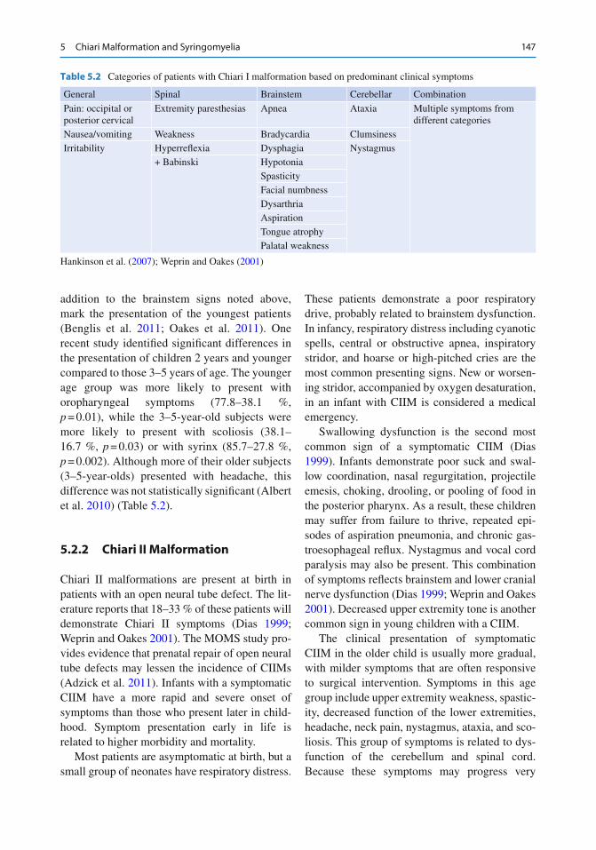
Table 5.2 Categories of patients with Chiari I malformation based on predominant clinical symptoms
General Spinal Brainstem Cerebellar Combination
Pain: occipital or posterior cervical
Extremity paresthesias Apnea Ataxia Multiple symptoms from different categories
Nausea/vomiting Weakness Bradycardia Clumsiness Irritability Hyperre fl exia Dysphagia Nystagmus
+ Babinski Hypotonia Spasticity Facial numbness Dysarthria Aspiration Tongue atrophy Palatal weakness
Hankinson et al. ( 2007 ) ; Weprin and Oakes ( 2001 )

148 S. McGee and D. Baudendistel
slowly, a complete history to identify subtle and gradual changes is vital. Presentation in adult-hood is rare but would mimic the progression of symptoms of the older child (Table 5.3 ).
5.2.3 Chiari III Malformation
Chiari III malformations are present at birth and are identi fi ed by the occipital or high cervical encephalocele. Multiple anomalies of the cere-bellum and brainstem accompany the encephalo-cele, which contains varying amounts of brain tissue. This anomaly is associated with poor prognosis due to the severity of the cranial nerve de fi cits and developmental and neurological impact. Even with supportive treatment, patients have a short life expectancy (Oakes et al. 2011 ; Weprin and Oakes 2001 ) .
5.2.4 Syringomyelia
The neurological examination should include a thorough sensory evaluation and testing of the
re fl exes, in addition to strength testing. Syringomyelia should be suspected in patients that present with scoliosis, leg or foot asymme-tries, or abnormal sensory examination. Dysesthetic pain of the trunk or extremities may be present. New or progressive spasticity is another symptom of concern for syrinx. Clumsiness, weakness, and atrophy of the upper extremities also may occur. In myelomeningo-cele patients, a worsening of urodynamics or changes in baseline motor function should be noted. In patients with Chiari I malformation, urinary incontinence may be a late sign of syrin-gomyelia (Nohria and Oakes 1991 ; Oakes et al. 2011 ; Weprin and Oakes 2001 ) .
5.3 Diagnostic Tests
Magnetic resonance imaging (MRI) of the brain, craniocervical junction, and spine is the best tool to diagnose Chiari malformations and syringo-myelia, as well as to rule out tethered cord. MRI provided a breakthrough in the diagnosis of Chiari malformations, which often present with
Table 5.3 Comparison of Chiari I and II malformations
Chiari I malformation Chiari II malformation
Brain Caudal descent of cerebellar tonsils > than 5–7 mm below foramen magnum
Caudal descent of cerebellar vermis, brainstem, and fourth ventricle below the foramen magnum
Peg like or pointed Often asymmetric
Common associated radiological fi ndings Skull Underdeveloped occiput Craniolacunia luckenschadel
Small posterior fossa Lemon sign on fetal ultrasound +/− Enlarged foramen magnum Small posterior fossa Basilar impression Enlarged foramen magnum
+/− Basilar impression Spine Assimilation of the atlas +/− Assimilation of the atlas
Progressive scoliosis (10 % in those who also have syringomyelia)
Enlarged cervical canal
Klippel-Feil deformity Klippel-Feil deformity Scoliosis
Ventricles and cisterns
Hydrocephalus (3–10 %) Hydrocephalus (90 %) Intrinsic malformation of ventricles including asymmetry, pointed frontal horns, and colpocephaly (enlarged occipital horns)
Spinal cord Syrinx (40–75 %) Syrinx (20–95 %)
Khoury ( 2011 ) ; Menezes ( 1999 ) ; Nohria and Oakes ( 1991 )

1495 Chiari Malformation and Syringomyelia
vague and nonspeci fi c signs and symptoms. Identifying the compression of the hindbrain and cervical spine as the possible cause of discomfort in these patients aided clinicians in providing useful treatment options. Recognition of Chiari I malformation in the very young child provided them with an opportunity to bene fi t from advances made in the surgical approach to this condition. Cine MRI may be used to assess CSF fl ow around the cerebellar tonsils. The location and extent of syringomyelia is best de fi ned by spinal MRI (Sherman et al. 1999 ) .
CT is of limited value in diagnosing Chiari malformations but provides information about the presence of hydrocephalus. In addition, cere-bellar tonsillar ectopia may be noted as an inci-dental fi nding on CT scan obtained for new symptoms such as headache or head injury. Sleep and swallow studies may be indicated prior to surgery to further evaluate the signs of brainstem or cranial nerve compression. Vocal cord motility may be evaluated if indicated.
Cervical radiographs can identify potential bony instability of the neck. Ultrasonography may provide identi fi cation of Chiari malforma-tions and syringomyelia in the neonate and infant, but decisions about surgical intervention are based on MRI fi ndings. Intraoperative ultrasound is used to identify whether bony decompression establishes adequate CSF fl ow. If CSF fl ow remains impaired with bony decompression, the surgery may proceed to include duraplasty and fourth ventricular stent (Sherman et al. 1999 ) .
5.4 Treatment Options for Chiari I Malformation
5.4.1 Medical
A child diagnosed with CIM presents a variety of challenges related to developmental consider-ations and the nonspeci fi c symptoms often asso-ciated with this condition. Because the CIM may present with only headache, care must be taken to con fi rm that the malformation itself is causing the headaches. Children, as well as adults, are subject to a variety of types of headaches. Taking
a thorough history of the type, pattern, and loca-tion of the headache and evaluating the effect of conservative treatment are key components of the medical management of these patients. If the headaches can be managed medically, the child may avoid a major surgical procedure. One recent review concluded that children with Chiari I malformation who are not clearly symptomatic and do not have scoliosis or syrinx can be fol-lowed conservatively. The development of symp-toms and new neurological de fi cits were extremely uncommon in a group of 124 children followed retrospectively for 1.0–8.6 years (mean 2.83 years) without surgery (Benglis et al. 2011 ) .
Children with known CIM should be followed annually for evaluation of symptom development or progression. MRI imaging with cine of the craniocervical junction to assess CSF fl ow may be indicated. The parents and child should be advised that the child should avoid contact sports and lumbar punctures.
5.4.2 Surgical
Early surgery is recommended for symptomatic patients (Hida et al. 1995 ) . Patients who have CIM identi fi ed on MRI, and have occipital head-aches unrelieved by medical management and/or other signs/symptoms associated with Chiari I malformation, are candidates for surgery. MRI evidence of a syrinx is an additional reason for surgical intervention. Common goals are improve-ment of presenting symptoms, radiographic reduction of syringomyelia, and arrest or remis-sion of associated scoliosis (Greenberg 2010 ) . If the patient also has hydrocephalus, treatment with a CSF diversionary shunt should precede surgery to treat the CIM.
The surgical procedure is planned to decom-press the posterior fossa suf fi ciently to allow room for CSF to circulate around the cerebellum and the cervical spinal cord (Boxes 5.1 and 5.2 ). Electrophysiologic neuromonitoring is generally employed. A vertical occipital incision is made to allow for bony decompression of the foramen magnum. Initial suboccipital craniectomy may
150 S. McGee and D. Baudendistel
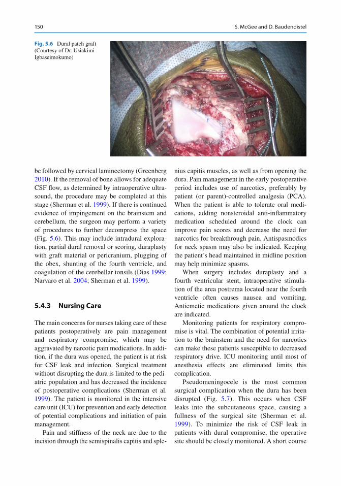
be followed by cervical laminectomy (Greenberg 2010 ) . If the removal of bone allows for adequate CSF fl ow, as determined by intraoperative ultra-sound, the procedure may be completed at this stage (Sherman et al. 1999 ) . If there is continued evidence of impingement on the brainstem and cerebellum, the surgeon may perform a variety of procedures to further decompress the space (Fig. 5.6 ). This may include intradural explora-tion, partial dural removal or scoring, duraplasty with graft material or pericranium, plugging of the obex, shunting of the fourth ventricle, and coagulation of the cerebellar tonsils (Dias 1999 ; Narvaro et al. 2004 ; Sherman et al. 1999 ) .
5.4.3 Nursing Care
The main concerns for nurses taking care of these patients postoperatively are pain management and respiratory compromise, which may be aggravated by narcotic pain medications. In addi-tion, if the dura was opened, the patient is at risk for CSF leak and infection. Surgical treatment without disrupting the dura is limited to the pedi-atric population and has decreased the incidence of postoperative complications (Sherman et al. 1999 ) . The patient is monitored in the intensive care unit (ICU) for prevention and early detection of potential complications and initiation of pain management.
Pain and stiffness of the neck are due to the incision through the semispinalis capitis and sple-
nius capitis muscles, as well as from opening the dura. Pain management in the early postoperative period includes use of narcotics, preferably by patient (or parent)-controlled analgesia (PCA). When the patient is able to tolerate oral medi-cations, adding nonsteroidal anti-in fl ammatory medication scheduled around the clock can improve pain scores and decrease the need for narcotics for breakthrough pain. Antispasmodics for neck spasm may also be indicated. Keeping the patient’s head maintained in midline position may help minimize spasms.
When surgery includes duraplasty and a fourth ventricular stent, intraoperative stimula-tion of the area postrema located near the fourth ventricle often causes nausea and vomiting. Antiemetic medications given around the clock are indicated.
Monitoring patients for respiratory compro-mise is vital. The combination of potential irrita-tion to the brainstem and the need for narcotics can make these patients susceptible to decreased respiratory drive. ICU monitoring until most of anesthesia effects are eliminated limits this complication.
Pseudomeningocele is the most common surgical complication when the dura has been disrupted (Fig. 5.7 ). This occurs when CSF leaks into the subcutaneous space, causing a fullness of the surgical site (Sherman et al. 1999 ) . To minimize the risk of CSF leak in patients with dural compromise, the operative site should be closely monitored. A short course
Fig. 5.6 Dural patch graft (Courtesy of Dr. Usiakimi Igbaseimokumo)

1515 Chiari Malformation and Syringomyelia
of dexamethasone may minimize symptoms from postoperative edema. Another possible complication is chemical meningitis (or aseptic meningitis). The symptoms include nuchal rigidity, low-grade fever, and headache. If bac-terial meningitis has been ruled out, a short course of dexamethasone is the treatment of choice. Chemical meningitis after surgery for Chiari I malformation may be related to the use of dural graft material and/or tissue sealants (Parker et al. 2011 ) .
The usual hospital length of stay is 3–5 days. Discharge criteria include normothermia, ade-quate oral fl uid intake, and pain controlled with oral medications. In addition, it is particularly important for patients who have undergone dura-plasty to have a bowel regime that keeps their
movements soft and regular to prevent disruption of the surgical site by straining.
Resolution of symptoms such as headache may be immediate, but other symptoms may take up to 3 months to begin to resolve. Symptoms resulting from long-standing brainstem compres-sion do not always completely resolve (Oakes et al. 2011 ) .
5.5 Treatment Options for Chiari II Malformation
5.5.1 Medical
Imaging for the Chiari II malformation is indi-cated only when new symptoms occur or when
Box 5.1. Chiari I Malformation: Case Study
DS is a 3-year-old girl who had a 1 month history of headache, clumsiness, dif fi culty eating, snoring, and nighttime awakening with crying. She was evaluated by her pediatrician for com-plaints of polydipsia in addition to the above symptoms. An MRI was ordered that showed low, pointed cerebellar tonsils projecting 7 mm below the foramen magnum. Cine images demon-strated restricted CSF fl ow posterior to the tonsils and anterior to the spinal cord. No syrinx was seen in the cervical region. She also had mild enlargement of the ventricles; her head circumfer-ence was at the 25th percentile for her age. Further laboratory studies did not con fi rm a cause for the polydipsia Surgery was performed that included removal of the sub occipital bone and ring of C1. Intraoperative ultrasound showed improved decompression with no pistoning of the cerebellar tonsils. The dura was then thinned, and repeat ultrasound showed marked improvement in the appearance of the craniocervical junction (CCJ). Spinal monitoring was done during the entire procedure DS was monitored in the pediatric intensive care unit over night. Continuous patient/parent-controlled analgesia (PCA) was initiated and managed by the hospital pain team. When she was taking adequate oral fl uids, DS was transitioned to oral pain medications such as nonsteroidal anti-in fl ammatory agents and narcotics for severe pain. In addition, oral muscle relaxants and stool softeners were started. The surgical dressing was removed on postoperative day #2, and incision care consisting of daily washing with soap and water was initiated. DS was discharged to home when the intravenous line was discontinued, and she was taking oral pain medication and getting out of bed with minimal assistance At her fi rst postoperative appointment (2 weeks after surgery), her parents reported improved sleep, balance, and behavior. DS was sleeping through the night without snoring. Her surgical site was healing well. The absorbable sutures were beginning to fall out spontaneously. Four months after surgery, a follow-up MRI revealed “marked improvement in the appearance of the CCJ with cerebellar tonsils no longer low lying and adequate CSF at the CCJ”. DS will follow up again with the neurosurgeon in 18 months.

152 S. McGee and D. Baudendistel
Box 5.2. One Family’s Chiari Malformation Story
It was October 31, 2001, when we fi rst got the diagnosis of Chiari malformation for our 1-year-old son. When I heard the words “brain surgery,” I felt like the air was sucked right out of my lungs. I can honestly say I remember nothing else that was told to us that day at the doctor’s of fi ce. Looking back though I guess I knew all along something was wrong but was not sure what to do because at that time Jacob was our only child and I had never been a mother before. As a baby, Jacob never really slept well and there would be periods of crying with his eyes closed or banging his head on things that would last for hours over night. After numerous visits to our pediatrician and being told that our son was just a bad sleeper, I began to assume I was maybe not the best mother. One afternoon after a morning of crying, I decided to put Jacob down for nap. I went downstairs and heard a very loud noise. Upon entering his room, I realized Jacob had fallen out of his bed and knocked himself unconscious. In the emergency room, we were told that his CT scan looked fi ne from the fall but that there was a malformation at the base of his brain. Further testing was necessary, and we were told to follow up with our pediatrician to get those things scheduled. As a parent, you believe that you can protect your child from anything, but in this circumstance, that is not true. I found myself totally helpless and lost. I would be holding my son as he was put to sleep for an MRI and having no knowledge as to what they were looking at. All I needed was someone to show me a little compassion and knowledge about what was coming next, to take the time to answer my questions and put some of my fears to rest. Surgery was scheduled, and I was introduced to an angel that will always be a part of my life. A nurse at the neurosurgeon’s of fi ce who was our surgeon’s right hand lady began to take the time and explain in so much detail about the steps we were beginning to take. She spent hours (it felt like) listening and answering questions. Whenever we called scared, nervous, or lost, she made us feel like no one was more important at that moment than our family. If we had not been pre-pared for surgery and the days after, I do not honestly think we could have survived it. For Jacob, it took two surgeries to create excellent CSF fl ow, and today he is just like any other 11-year-old boy. Both surgeries were different though; in the second one, we had an idea of what to expect but still spent every night watching the monitors on Jacob to make sure that he was breathing. The fear after the second surgery was not about hoping we got him home. It was about wondering if we were going to have to do this again in the future. Two and a half years after Jacob had his last surgery, our second son, Dylan, was beginning his Chiari journey. Dylan’s symptoms were totally different than Jacob’s, but this time I knew in my gut without a doubt what was going on. Dylan never really spoke or made sounds as an infant, had extreme dif fi culty drinking his bottles, and, when began moving, always dragged his left leg. As soon as I saw the leg dragging, I called this nurse who I trusted as much as our neuro-surgeon. Instead of telling me I was just seeing things or that I was jumping to a conclusion too quickly, she listened to me and offered me resources to fi nd out what was going on. Once we had an MRI showing his Chiari, we scheduled surgery. Dylan’s case has always been much worse than Jacob’s. To date, he has had four decompressions, and it looks like things have fi nally resolved. Every night though in the hospital, I would sit by his bed and cry because I felt so guilty that I could not make things better with a kiss (like moms are supposed to) and the following day that same nurse would check in and remind me that things will get better. Chiari malformation is a frightening diagnosis to any parent and is not an easy recovery the fi rst couple of days after surgery. At that moment in a family’s life, the only thing they focus on is their child and getting him or her healed. Compassion, knowledge, and recognition are things that can assist every family during their journey.

1535 Chiari Malformation and Syringomyelia
baseline status deteriorates. Symptoms of con-cern may include swallowing dif fi culties, weak-ness or increased weakness of the upper extremities, new spasticity, or occipital head-aches. If the child has shunted hydrocephalus, the shunt should be evaluated fi rst and revised if it is malfunctioning. In the presence of a functioning CSF shunt, evaluation of the CIIM by an MRI of the brain and craniocervical junc-tion is the next step. If the symptoms persist and the MRI shows brainstem compression or obstruction of CSF fl ow, surgical treatment is indicated. In CIIM, early surgical intervention in the child with symptoms may prevent signi fi cant morbidity and mortality (Oakes et al. 2011 ) .
5.5.2 Surgical
The surgical intervention for the CIIM parallels that for the Chiari I malformation. Based on the
need for extensive dissection of the brainstem and cranial nerve structures, electrophysiologic neuromonitoring is generally employed. A sub-occipital incision is made to allow for removal of the posterior arch of C1 and excision of any extradural constrictive band. Laminectomy is performed to the level of descent of the cerebel-lar tonsils which may require a 1-, 2-, or 3-level laminectomy. Myelomeningocele patients, unlike the Chiari I malformation patients, have an elongated foramen magnum and, thus, do not require further expansion. The dura should be opened to create CSF fl ow around the CIIM. Commonly, extremely dense arachnoidal adhe-sions require lysis. The herniated tonsils may require fulguration (cauterization). A stent spanning the obex, lying within the fourth ven-tricle proximally and the cervical subarachnoid space distally, is frequently placed. Finally, a dural augmentation graft (allograft or autograft pericrania) is typically sutured into the opened dural margins.
Fig. 5.7 T2 sagittal MRI of ( arrow ) pseudomeningocele

154 S. McGee and D. Baudendistel
5.5.3 Nursing Care
As with the CIM, these patients are monitored after surgery in the ICU or the neonatal intensive care unit. Extensive microsurgical manipulation, involv-ing multiple lower cranial nerves and brainstem structures, places the patient at risk for postopera-tive neurological deterioration, especially regarding swallowing and ventilation. These patients must be monitored for late extubation, apnea, swallowing dysfunction, and feeding problems after surgery. The risk for CSF leak and infection exists when the dura has been opened. Neck movement limitation and steroids may be indicated to minimize symp-toms related to dural opening and postoperative edema. Neck pain and stiffness occur in these patients and must be managed carefully in light of the presence of respiratory compromise preopera-tively, especially in the very young patients.
Like CIM patients, discharge criteria include normothermia, adequate oral fl uid intake, and pain controlled with oral medications. A bowel regime is needed to keep stool soft and regular to prevent disruption of the surgical site by straining.
5.6 Treatment Options for Syringomyelia
Left untreated, a syrinx can enlarge or elongate over time causing damage to the spinal cord. When syringomyelia is associated with a Chiari malformation, treatment by posterior fossa decompression of the hindbrain malformation may result in resolution of the syrinx. Primarily in CIIM, symptomatic syringomyelia may persist despite decompressive surgery. In the absence of Chiari malformation, asymptomatic syringomy-elia may be observed clinically with yearly clini-cal examinations and intermittent MRI.
Direct shunting of the syrinx may improve symptoms in those patients who have persistent symptoms after successful posterior fossa decom-pression or in those patients who have a symp-tomatic syrinx without a Chiari malformation. Options include syringoperitoneal, syringopleu-ral, and syringosubarachnoid shunts. The shunt
acts to decompress the fl uid buildup within the spinal cord, diverting the fl uid to another space for reabsorption (Menezes 1999 ) . Similarly, stenting across the obstructed fourth ventricular obex can prevent “water hammering” of CSF into the proximal cervical central canal.
5.6.1 Nursing Care
Postoperative care includes incision care, pain management, and evaluation of shunt function. With syringoperitoneal shunting, there will be an incision over the spine at the level of the syrinx and an incision over the abdomen for insertion of the distal catheter. Abdominal pain and bowel function are key areas for nursing assessment.
The syringopleural shunt will have a similar back incision with the distal catheter incision in the lateral chest. Observation of respiratory status is important with this treatment option. Decreased breath sounds and oxygen desaturation may indi-cate a symptomatic pleural effusion. Indeed, small pleural effusions are typical and generally well tolerated. The patient may have mild tachyp-nea and low oxygen saturations and may require nasal cannula oxygen supplementation for up to 1 week. If tachypnea or desaturations worsen, the patient may need more intervention including thoracentesis or removal of shunt from the pleu-ral space. Serial chest radiographs may be used to evaluate the patient’s ability to accommodate the pleural fl uid being diverted by the shunt.
The syringosubarachnoid shunt requires only one incision to accommodate both the proximal and distal catheters and may be effective in symp-tom relief.
The use of a shunt to treat syringomyelia requires ongoing follow-up to observe for signs of shunt failure.
5.7 Patient and Family Education
1. Informed consent: major risks of surgery include bleeding, CSF leak, infection, persis-tence of symptoms, neurological de fi cit, and anesthesia complications.

1555 Chiari Malformation and Syringomyelia
2. Preoperative history and physical examination.
3. Preoperative diagnostic tests that may include swallow evaluation, sleep study, MRI, and developmental assessment.
4. Educational handouts about Chiari malformations and website information recommendations.
5. Incision care after dressing removed. 6. Sutures either dissolvable or removed in
about 2 weeks. 7. Activity restrictions: no driving while on
narcotics or while neck is stiff; return to school or work in 4–6 weeks.
8. Follow-up imaging: MRI in 4–6 weeks then annually for 5 years (more frequently if syr-inx present).
9. Signs and symptoms of shunt failure, for patients requiring shunting of the syrinx.
10. Discharge instructions: incision care with obser-vation for infection or pseudomeningocele; call surgeon’s of fi ce for headache not responsive to medication and fever greater than 101°F.
5.7.1 Outcomes: Short and Long Term
CIM: Successful decompression can provide relief of headache. Symptoms due to cranial nerve or brainstem dysfunction can show improvement over several weeks to months. Follow-up swallow studies are useful to evaluate the effects of treat-ment when done six or more weeks postopera-tively. Ataxia or weakness may also gradually improve. Patients with symptoms other than iso-lated headaches on presentation bene fi t from appropriate therapies postoperatively, such as occupational, physical, and/or speech therapy.
MRI imaging should demonstrate improve-ment in CSF fl ow around the craniocervical junc-tion by approximately 6 weeks after surgery. A syrinx should radiographically resolve or decrease in size within 3–6 months of posterior fossa decompression. Symptoms may persist in spite of the radiographic improvement (Dias 1999 ) .
CIIM: Better outcomes occur with older chil-dren who present with cerebellar dysfunction, spasticity, and weakness. Results in the neonatal
and infant population have been varied, but in general, their outcomes are poorer. CIIM may cause death by respiratory failure (Dias 1999 ) . The rapidity of neurological decline and imme-diate preoperative neurological status are the most important factors affecting prognosis.
The spectrum of Chiari malformations and syringomyelia present a continuum of challenges to the pediatric patient. The range of effect on quality of life varies from mild, with effective treatments available, to very severe, with mini-mal or no bene fi t from medical intervention. Advances in radiographic imaging and surgical techniques have provided opportunities to improve the health status of many of these patients. Advances in nursing research provide the opportunity for nurses and the allied health professionals to further enhance functional level and optimal development of children with this varied spectrum of disorders. Incorporating best practice for the pediatric neurosurgical patient in the areas of wound healing, pain management, prevention of postoperative complications, and effects of hospitalization on development and psychosocial wellness will further enhance the quality of life of this young population.
Pediatric Practice Pearls
1. For pain management after posterior fossa decompression, use patient/parent-controlled analgesia; start nonsteroidal anti-in fl ammatory medications 24 h after surgery and when the patient is tak-ing fl uids orally. May require oral nar-cotics, antispasmodics, and nonsteroidal anti-in fl ammatory medication.
2. Straining with constipation can disrupt the surgical site and is particularly a risk when the dura has been disrupted. Start a bowel regime when the patient is tak-ing fl uids orally to avoid constipation.
3. Relaxation techniques and gentle mas-sage can be helpful during recovery from posterior fossa decompression. Muscle spasms often complicate the pain cycle.

156 S. McGee and D. Baudendistel
References
Adzick NS, Thom EA, Spong CY, Brock JW 3rd, Burrows PK, Johnson MP (2011) A randomized trial of prena-tal versus postnatal repair of myelomeningocele. N Engl J Med 364(11):993–1004
Albert GW, Menezes AH, Hansen DR, Greenlee JD, Weinstein SL (2010) Chiari malformation type I in children younger than age 6 years: presentation and surgical outcome. J Neurosurg Pediatr 5(6):554–561
Benglis D, Covington D, Bhatia R, Bhatia S, Elhammady MS, Ragheb J et al (2011) Outcomes in pediatric patients with chiari malformation type I followed up without surgery. J Neurosurg Pediatr 7(4):375–379
Detwiler PW, Porter RW, Rekate HL (1999) Hydrocephalus-clinical features and management. In: Choux M, DiRocco C, Hockley AD, Walker ML (eds) Pediatric neurosurgery. Churchill Livingstone, London, pp 253–271
Dias MS (1999) Myelomeningocele. In: Choux M, DiRocco C, Hockley AD, Walker ML (eds) Pediatric neurosurgery. Churchill Livingstone, London, pp 33–59
Goh S, Bottrell CL, Aiken AH, Dillon WP, Wu YW (2008) Presyrinx in children with chiari malformations. Neurology 71(5):351–356
Greenberg MS (2010) Handbook of neurosurgery, 7th edn. Thieme Medical Publishers, New York
Hankinson TC, Klimo P Jr, Feldstein NA, Anderson RC, Brockmeyer D (2007) Chiari malformations, syringo-hydromyelia and scoliosis. Neurosurg Clin N Am 18(3):549–568
Hida K, Iwasaki Y, Koyanagi I, Sawamura Y, Abe H (1995) Surgical indication & results of foremen mag-num decompression versus syringosubarachnoid shunting for syringomyelia associated with Chiari I malformation. Neurosurgery 37(4):637–639
Khoury C (2011) Chiari malformation. In: Patterson MC (ed) UpToDate. Wolters Kluwer Health, Philadelphia
Lazareff JA, Galarza M, Gravori T, Spinks TJ (2002) Tonsillectomy without craniectomy for the manage-ment of infantile Chiari I malformation. J Neurosurg 97:1018–1022
Menezes AH (1995) Primary craniovertebral anomalies and the hindbrain syndrome (Chiari I): data base anal-ysis. Pediatr Neurosurg 23:260–269
Menezes AH (1999) Craniovertebral anomalies and syrin-gomyelia. In: Choux M, DiRocco C, Hockley AD, Walker ML (eds) Pediatric neurosurgery. Churchillw Livingstone, London, pp 151–184
Narvaro R, Olavarria G, Seshadri R, Gonzales-Portillo G, McLone DG, Tomita T (2004) Surgical results of pow-sterior fossa decompression for patients with Chiari I malformation. Childs Nerv Syst 20:349–356
Nohria V, Oakes WJ (1991) Chiari I malformation: a review of 43 patients. Pediatr Neurosurg 16:222–227
Novegno F, Caldarelli M, Massa A, Chieffo D, Massimi L, Pettorini B et al (2008) The natural history of the chiari type I anomaly. J Neurosurg Pediatr 2(3):179–187
Oakes WJ, Pugh JA, Tubbe RS (2011) Chiari malforma-tions. In: Winn HR (ed) Youmans neurological sur-gery, 6th edn. Saunders, Philadelphia, pp 1918–1927
Parker SR, Harris P, Cummings TJ, George T, Fuchs H, Grand G (2011) Complications following decom-pression of Chiari malformation Type I in children: dural graft or sealant? J Neurosurg Pediatr 8:177–183
Payner TD, Prenger E, Berger TB, Crone KR (1994) Acquired Chiari malformations: incidence, diagnosis, and management. Neurosurgery 34(3):429–434
Rekate HL (2008) Natural history of the chiari type I anomaly. J Neurosurg Pediatr 2(3):177–178
Rekate HL, Wallace D (2003) Lumboperitoneal shunts in children. Pediatr Neurosurg 38:41–46
Schijman E (2004) History, anatomic forms, pathogen-esis of Chiari I malformation. Childs Nerv Syst 20:323–328
Sherman J, Larson JJ, Crone KR (1999) Posterior fossa decompression without dural opening for the treat-ment of Chiari I malformation. In: Rensachary SS, Wilkins RH (eds) Neurosurgical operative atlas, vol.8 edn. American Association of Neurological Surgeons, Park Ridge, pp 179–183
Strayer A (2001) Chiari I malformation: clinical presenta-tion & management. J Neurosci Nurs 33(2):90–104
Sutton LN, Adzick NS, Bilaniuk LT, Johnson MP, Crombleholme TM, Flake AW (1999) Improvement in hindbrain herniation demonstrated by serial fetal mag-netic resonance imaging following fetal surgery for myelomeningocele. JAMA 282(19):1826–1831
Tulipan N, Hernanz-Schulman M, Bruner JP (1998) Reduced hindbrain herniation after intrauterine myelomeningocele repair: a report of four cases. Pediatr Neurosurg 29(5):274–278
Tulipan N, Hernanz-Schulman M, Lowe LH, Bruner JP (1999) Intrauterine myelomeningocele repair reverses preexisting hindbrain herniation. Pediatr Neurosurg 31(3):137–142
Weprin BE, Oakes WJ (2001) The Chiari malformations and associated syringohydromyelia. In: McClone DG (ed) Pediatric neurosurgery. WB Saunders Co, Philadelphia, pp 214–235
Yeh DD, Koch B, Crone KR (2006) Intraoperative ultra-sonography used to determine the extent of surgery necessary during posterior fossa decompression in children with chiari malformation Type I. J. Neurosurgery ( 1 Suppl Pediatrics) 105:26–32
Yundt KD, Park TS, Tantuwaya VS, Kaufman BA (1996) Posterior fossa decompression without duraplasty in infants and young children for treatment of Chiari malformation and achondroplasia. Pediatr Neurosurg 25:221–226