number 167 20 may 1975 a pacific tabulate sponge, living...
TRANSCRIPT

POSTILLA PEABODY MUSEUM
YALE UNIVERSITY NUMBER 167 20 MAY 1975
A PACIFIC TABULATE SPONGE, LIVING REPRESENTATIVE OF A NEW ORDER OF SCLEROSPONGES
WILLARD D. HARTMAN
THOMAS F. GOREAU

A PACIFIC TABULATE SPONGE, LIVING REPRESENTATIVE OF A NEW ORDER OF SCLEROSPONGES
WILLARD D. HARTMAN
Department of Biology and Peabody Museum of Natural History, Yale University, New Haven, Connecticut 06520
THOMAS F. GOREAU1
Discovery Bay Marine Laboratory, University of the West Indies and State University of New York at Stony Brook, New York 11790
Received 1 April 1975
ABSTRACT
A new sclerosponge, Acanthochaetetes wellsi, with a calcitic skeleton made up of contiguous vertical tabulate calicles ornamented within by vertical rows or irregular clumps of spines is described from cryptic habitats on reefs in the western Pacific region. A lamellar microstructure characterizes the calicle walls and spines. Increase in number of calicles occurs as intramural offsets. An epitheca with growth lines surrounds the entire sponge.
Living tissue is restricted to the space in the calicles above the outermost tabulae and a thin layer lying above the calcareous skeleton. Siliceous spicules of two kinds, tylostyles and modified spirasters, are distributed in the living tissue but are not incorporated into the calcitic skeleton. Star-shaped groups of exhalant canals converge upon central oscules on the sponge surface and leave astrorhizal patterns impressed into the calcareous skeleton below.
A new order, the Tabulospongida, of the class Sclerospongiae is proposed to receive the new Pacific species together with its Jurassic and Cretaceous forebears. Despite suggestive similarities between acantho-chaetetids and favositids, a phylogenetic relationship between these two groups is considered unlikely on the basis of present evidence.
POSTILLA 167: 21 p. 20 MAY 1975
lrThomas F. Goreau, who was Professor of Marine Sciences at the University of the West Indies, Mona, Kingston, Jamaica, Professor of Biology at the State University of New York at Stony Brook and Director of the Discovery Bay Marine Laboratory, Discovery Bay, Jamaica, died unexpectedly in New York on 22 April 1970. This paper, based largely on specimens collected by Professor Goreau and enriched by his observations of the organism in life, was prepared by the first-named author.

POSTILLA
Published by the Peabody Museum of Natural History, Yale University
Postilla includes results of original research on systematic, evolutionary, morphological, and ecological biology, including paleontology. Syntheses and other theoretical papers based on research are also welcomed. Postilla is intended primarily for papers by the staff of the Peabody Museum or on research using material in this Museum.
Editor: Zelda Edelson
Postilla is published at frequent but irregular intervals. Manuscripts, orders for publications, and all correspondence concerning publications should be directed to:
Publications Office Peabody Museum of Natural History New Haven, Conn. 06520, USA
Lists of the publications of the Museum are available from the above office. These include Postilla, Bulletin, Discovery, and special publications. Postilla and the Bulletin are available in exchange for relevant publications of other scientific institutions anywhere in the world.
Inquiries regarding back numbers of the discontinued journal, Bulletin of the Bingham Oceanographic Collection, should be directed to:
Kraus Reprint Co. Route 100 Millwood, New York 10546

2 POSTILLA 167
INTRODUCTION
When we proposed transferring the order Chaetetida from the Phylum Cnidaria to the Phylum Porifera (Hartman and Goreau, 1972) we noted that species assigned to the family Acanthochaetetidae exhibit some basic differences from the remaining families of the order. We excluded the acan-thochaetetids from the order Chaetetida and mentioned the similarity of a Recent tabulate sponge (Hartman and Goreau, 1970b) to them. It is the purpose of this paper to name and describe more fully the living acanthochaetetid and to explore its relationships within the Class Sclerospongiae.
To our knowledge, this sponge was first collected by Professor John W. Wells of Cornell University as a beach cast specimen on Rongerik Atoll, Marshall Islands, in 1947. Living populations were discovered in 1968 by Mr. Richard H. Randall and Dr. R. H. Chesher during explorations of underwater caves on the reefs of Guam. Their specimens came to the attention of one of us (T. F. G.) who undertook further studies of it on the reefs of both Guam and Saipan. The species is now known to have a wide distribution in the western Pacific region where it occupies cryptic reef habitats comparable to those populated by ceratoporellids in the Caribbean area (Hartman and Goreau, 1970a).
METHODS
Specimens were collected by SCUBA diving and were fixed in neutral formalin as soon as possible after being brought to the surface. After a few days the sponges were transferred to 75% ethyl alcohol for long term storage. Tissue-free calcareous skeleton preparations were obtained by treatment with 5.25% sodium hypochlorite solution (commercial bleach). Scanning electron microscope studies were undertaken with gold-coated specimens on one of the following instruments: JEOL JSM-U3 and ETEC Autoscan U-1. Material for histological studies was formalin fixed, decalcified in 2% formic acid, and stained in Heidenhain's iron hematoxylin and Alcian blue.
DESCRIPTION
Acanthochaetetes wellsi sp. no v.
DIAGNOSIS. Basal skeleton calcitic and massive, formed of contiguous, vertical tabulate tubes or calicles, adjacent ones of which share common walls. Walls of calicles provided with spines arranged in vertical rows or clumped irregularly. Microstructure of calicle walls and spines composed of stacked lamellae. Astrorhizoid patterns impressed in the surface of the calcareous skeleton mark the position of the exhalant canal systems converging upon

PACIFIC TABULATE SPONGE 3
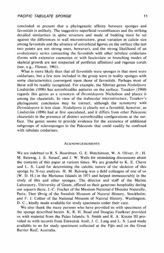
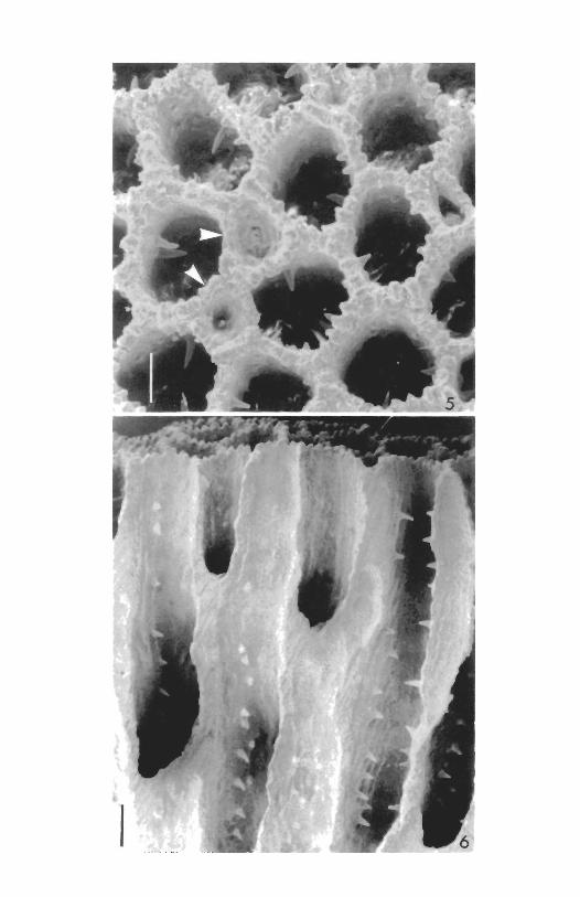
oscules. A well developed epitheca provided with concentric growth lines covers the lower surface of the sponge. Living tissue confined to spaces 1.2 to 2.0 mm deep, external to the outermost tabula of each calicle. Siliceous spicules of two types: 1) tylostyles with rounded distal ends and 2) modified spirasters, neither type incorporated into the calcareous skeleton. THE CALCAREOUS SKELETON. The basal skeleton of the sponge is composed of calcite and is formed (Fig. 1) of contiguous, vertical tabulate calicles. The calicles are elliptical, pentagonal or hexagonal in surface outline and vary in internal diameter from 315 by 300 ^m to 615 by 395 txm. The width of the calicle walls usually varies from 65 to 75 /xm but may reach up to 140 /mm. The edges of the calicles are crenulate (Fig. 5), each crenation corresponding to the upwardly directed undulations of the lamellar crystalline units of calcite that make up the calicular walls. A basally directed ridge continues from each crenation; successive ridges are separated by furrows (Fig. 6); thus cross sections through the calicles at any level have a scalloped appearance. Low, rounded knobs often arise from the ridges and ornament the walls of the calicles.
Spines are formed of successive lamellae of calcite peaked axially. The spines (Fig. 6), although horizontal in overall orientation, are actually gently arched, curving first upward and then downward and sometimes ending in short horizontal tips. Spines vary in length from 30 to 180 /im and in width at base from 30 to 50 /xm.
Tabulae (Figs. 7, 12) are slightly convex, horizontal or slightly concave and are formed of lamellar calcite. Sometimes a concave and a convex tabula form in close juxtaposition and fuse to form a single thick tabula. The tabulae are irregularly spaced and do not necessarily occur at the same level in adjacent calicles although this is not infrequently so over limited regions of the skeleton. Often a series of closely spaced tabulae, ranging from 90 to 165 /xm apart over 9 or 10 intertabular spaces, will then give way to a series of more widely spaced ones, ranging from 200 to 500 /xm apart. Even more closely spaced series range from 50 to 120 /xm apart. The thickness of tabulae is also highly variable, ranging from 20 to 135 /xm, the thinner ones occurring in closely spaced series and the thicker ones in more distantly spaced series.
Scanning electron micrographs (Fig. 14) reveal that the surface of the interior of the calicles is made up of irregularly arranged needlelike crystalline units of calcite that must compose the successive lamellae seen at lower magnifications under the light microscope.
The entire lower surface of the sponge is covered with a thin epitheca (Figs. 2, 8) that is marked with fine concentric growth lines. At times the sponge grows up to a height that is three or more times the diameter of the living surface, and a stalked condition results. At other times the living surface spreads out rapidly, and its diameter is many times the height of the sponge. It is characteristic of this sponge to die back at unknown intervals of time, perhaps erratically, and to put forth new groups of calicles at a level above the previous living surface. As a result three or more "generations" of dead, flattened masses of skeleton may overlie one another, the topmost alive

4 POSTILLA 167
and often irregular in outline and sometimes subdivided into numerous small "islands" of living tissue. The largest specimen measured has greatest diameters of 33 by 26 cm but the largest continuous living portion covers an irregular area roughly 18 by 18 cm in greatest diameters. Other specimens remain small with diameters of <1 cm to 2 or 3 cm; such individuals are characteristic of populations living on the Great Barrier Reef, Australia. SILICEOUS AND ORGANIC SKELETAL ELEMENTS. The siliceous megascleres are
thin tylostyles (Fig. 9A) with ellipsoidal heads and rounded distal ends. Dimensions, based on measurement of 25 spicules from a Guam specimen are:
Length: range, 201 to 336 ̂ m; mean (with standard error), 286±7 jiim. Width: range, 2.6 to 3.9 /x,m; mean (with SE), 3.4±0.1 ^m. Head Width: range, 6.2 to 8.5 fjum; mean (with SE), 7.4±0.4 fxm.
The tylostyles are localized, with heads directed toward the base of the sponge, in the living tissue of the outer third of the calicles.
The siliceous microscleres (Fig. 9B-H) are variable in form but are basically spirasters. In some a spiral axial rod, ornamented with branched spines that follow a spiral course along the rod, is clearly apparent (Fig. 9F, G). In others the spines are closely spaced, multibranched and robust and therefore tend to obscure the axial rod (Fig. 9C). All intermediates in size and spination occur between the extremes described (Fig. 9D, E). Our observations of a spicule preparation indicated a range of greatest length by greatest width of 6 by 5 fim to 28 by 20 /mm. The microscleres are localized in the outer layer of the living tissue (Fig. 13). The siliceous spicules of this sclerosponge are not incorporated into the calcareous basal skeleton in a manner comparable to that found in the ceratoporellids (Hartman, 1969).
Organic fibrils (probably proteinaceous) of two size ranges, 11 to 14 nm and 5 to 6 nm, respectively, as revealed in transmission electron micrographs, occur as a matrix for the calcitic skeleton and are also present between cells of the living tissue. LIVING TISSUES OF THE SPONGE. The living tissue of A. wellsi is cream to yellowish cream in color at the surface; fractured specimens reveal that the tissue in the calicles is bright yellow. It seems likely that the abundance of microscleres in the surface layer of the sponge causes a lightening of the color of that region.
Ostia open into the unit of sponge tissue enclosed in each calicle. Ostia are located in the floor of shallow crescent-shaped or sinuous grooves (Fig. 4) that usually lie very close to but above the calcareous walls of the calicles. The chord length of these grooves varies from 200 to 260 /xm and the width, from 25 to 50 fxm. The diameters of the opened ostia are unknown. These observations are based on transverse histological sections and close-up photographs of the living animal in situ.
Photographic evidence (Fig. 4) suggests that there is in most instances an ostial groove for each calicle. Some calicles seem to lack grooves, however, and it is probable that the inhalant channels of two or perhaps more calicles can be supplied with sea water from the ostia of a single groove.
Each ostium opens into a vestibular cavity (Fig. 13) from which arises an

PACIFIC TABULATE SPONGE 5
inhalant channel that subdivides, with the branches opening into choanocyte chambers. The choanocyte chambers (Fig. 13) are small, measuring from 21 by 18 fim to 24 by 20 /mm in histological preparations. The chambers open into narrow exhalant channels that fuse and increase in diameter as they make their way out of the calicle to join similar channels from adjacent calicles. At the surface of the sponge exhalant channels draining areas that vary from 20 to 50 mm2 converge upon oscules, thus forming stellate patterns (Figs. 3, 4). Exhalant channels from adjacent drainage systems characteristically fuse (Hartman and Reiswig, 1973). The diameters of the exhalant channels where they join the oscules vary from 220 to 290 /xm. Radii of the exhalant systems from the points of fusion with neighboring systems to the central oscules vary from 4.0 to 1.8 mm. One to three oscules drain each exhalant system; oscule diameter varies from 100 to 130 /xm. Measurements of the exhalant system are based on close-up photographs of living animals in situ.
As is true among the ceratoporellids (Hartman, 1969; Hartman and Goreau, 1970a) the positions of the exhalant channels that run in the thin surface layer of sponge tissue above the calcitic calicles are impressed into the calcareous skeleton to a greater or lesser extent (Fig. 1) as a result of the inhibition of upward growth of calcite beneath the channels that carry flowing water. There remain, therefore, more or less clearly marked astrorhizalike patterns on the surface of the skeleton. The oscule or oscules that open in the center of exhalant systems often lie on an elevated mound or mamelon that varies from 5 mm to less than 1 mm in height in different specimens. Mamelons are absent in some specimens and are most pronounced in specimens from the Palau populations. SYMBIONTS. Neither unicellular algal nor bacterial symbionts have been observed in the cells or tissues of this sponge. An unidentified zoanthidean is not infrequently associated with A. wellsi. RANGE AND HABITAT. Widely distributed in the western Pacific region. Populations of the sponge are known from the Marianas Islands (Guam, 3 to 30 m; Saipan, 1 to 12 m), Caroline Islands (Palaus, 10 to 30 m), Marshall Islands (Rongerik and Eniwetok atolls, 2 to 12 m), Fiji Islands (25 to 30 m), Australia (Great Barrier Reef, 6 to 10 m). A. wellsi lives in crevices and caves and on the lower surfaces of overhangs on reefs. The known overall depth range of 1 to 30 m is less at both extremes than the depth range of Caribbean sclero-sponges (Lang, Hartman and Land, 1975). HOLOTYPE. Yale Peabody Museum No. 9077. Cave, Anae Island, Guam. 7.5 to 9 m. Collected by T. F. Goreau, 19 July 1969. REPOSITORIES OF PARATYPICAL MATERIAL. National Museum of Natural History, Washington, D. C ; British Museum (Natural History), London; Museum National d'Histoire Naturelle, Paris; Peabody Museum of Natural History, Yale University. REMARKS. The species is named in honor of Professor John W. Wells, Cornell University, who first collected a skeleton of the sponge on a beach on Rongerik Atoll, Marshall Islands, in 1947. He brought the specimen to our attention subsequent to the rediscovery of Ceratoporella in Jamaica.

6 POSTILLA 167
RELATIONSHIPS
GENERIC AFFINITY. The calcareous skeleton of the Recent sponge described here is directly comparable to that of the type-species of the genus Acanthochaetetes Fischer (1970), known from Jurassic and Cretaceous strata. Fischer's figure (1970, fig. 32) of the microstructure of A. seunesi Fischer can serve as well as an illustration of the microstructure of A. wellsi. Furthermore, figures of the astrorhizal patterns on the surface of the Cretaceous species A. ramulosus (Michelin) are in every way similar to the stellate patterns on A. wellsi described herein (compare Cuif et al., 1973, especially fig. 5, with fig. 1 herein). The discussion of the astrorhizae of the Cretaceous species of Acanthochaetetes presented by these authors can now be altered in the light of studies of the Recent species. Cuif et al. note two types of astrorhizal patterns in A. ramulosus. That pictured in their figure 5 is identical to that of the Recent forms. The second type (their figure 4), in which the center is composed of a group of enlarged vertical(?) tubes, is unlike anything to be observed in the living species. It is suggested that the second pattern either represents another species or has resulted from diagenetic processes. A further observation of Cuif and his coauthors (1973) about the astrorhizae of Acanthochaetetes is thrown in doubt by studies of the Recent species. This is that the astrorhizae of A. ramulosus and A. seunesi are somehow formed of tubes, identical in structure to the tissue-bearing calicles, that ascend from the depths of the skeleton and converge at the surface. It is very doubtful that these configurations (see Cuif et al., fig. 6) have anything to do with the astrorhizal patterns at the surface; it is much more likely that the figure records several developmental abnormalities that occurred during the upgrowth of the calcareous skeleton. It should be recalled that the astrorhizal patterns in A. wellsi result from local inhibition of upgrowth of the calcareous calicles just below the exhalant channels that are converging upon an oscule or oscules located in the center of the system. The channel linings are, of course, composed entirely of living tissue and would not be preserved in fossilized specimens. If it is also recalled that all the living tissue of the sponge lies above the last tabulae to be deposited in the calicles, it is realized that there is no reason for this sponge to communicate with deeper regions of the skeleton, which is dead below a depth of no more than 2 mm from the surface.
The arrangement of the calicular spines, the occurrence of both thin and thickened tabulae at irregular intervals, and the formation of intramural offsets in asexual reproduction are additional characters that point to acan-thochaetetid affinities for the Recent sponge under discussion. COMPARISON WITH OTHER SPECIES. Assignment of the Recent species wellsi to the genus Acanthochaetetes is based on the close homology between the macroscopic and microscopic features of the calcareous skeleton. Is it warranted to set up a new species for the Recent form or is this form identical to one of the Mesozoic species? Working with characters of the calcareous skeleton for A. wellsi, it can be seen in Table 1 that the Recent species over-

PACIFIC TABULATE SPONGE 7
laps A. ramulosus in two characters and A. seunesi in a third while it differs from both fossil forms in a fourth character. Thus it may be concluded that wellsi is best considered a distinct species belonging to the genus Acantho-chaetetes, the age of which is now known to extend from Jurassic to Recent times.
TABLE 1. Characteristics of calcareous skeleton of Acanthochaetetes species.
A. seunesi* A. ramulosus* A. wellsi Diameter of calicles
(center to center)
Distance between astrorhizal centers
Wall thickness
Spacing of tabulae
* Data from Fischer, 1970.
0.60-1.20 m m 0.35-0.70 m m 0.35-0.55 m m
? 8-12 m m 4-14 m m
0.05-0.16 m m
0.20-2.00 m m
0.45-1.20 m m
0.20-2.00 m m
0.07-0.14 m m
0.05-0.50 m m
RELATIONSHIP OF ACANTHOCHAETETIDS TO OTHER SCLEROSPONGES. F i s c h e r
(1970) placed his new genus Acanthochaetetes in the new family Acan-thochaetetidae within the order Chaetetida. The members of this family have a number of characters that set them apart from the Chaetetida and all other known sclerosponges, however. The chief distinction is the calcitic nature and lamellar microstructure of the calcareous skeleton as opposed to the aragonitic composition and trabecular microstructure of all other known sclerosponges. A second distinction is the presence of spinose processes on the walls of the calicles, and a third is the presence of siliceous spicules in the form of smooth tylostyles and modified spirasters, spicule types that are quite different from those found in the ceratoporellids. These distinctive characters lead us to propose the new order TABULOSPONGIDA for the members of the family Acanthochaetetidae. DIAGNOSIS OF THE NEW ORDER. Sclerosponges with a calcitic basal skeleton having a lamellar microstructure. Walls of calicles may be provided with spines. Tabulae may be secondarily thickened and of irregular arrangement. Asexual reproduction by intramural budding. Living tissue of Recent species secretes siliceous spicules in the form of tylostyles and modified spirasters. Astrorhizal patterns impressed into the surface of the calcareous skeleton. Known range: Jurassic to Recent. TABULOSPONGIDA AND FAVOSITIDA COMPARED. The Tabulospongida share certain morphological features with members of the Favositida, an order of animals that are generally regarded as being tabulate corals. The similarities

8 POSTILLA 167
are sufficiently striking to lead us to raise the question of whether favositids were in fact sponges instead of cnidarians. The characters shared by species of the two orders are: the presence of spines on the walls of the calicles; the presence of tabulae and epithecae; and asexual reproduction by means of budding at the edge of the entire skeletal complex or in walls between calicles when space becomes available as the skeletal surface grows upward. Concerning the first character, the spines of favositids, when present, may occur in vertical rows usually numbering 6 to 12 according to the species (Hill and Stumm, 1956) or, as is true more frequently, the spines are irregularly arranged (Schouppe and Oekentorp, 1974). The spines of acanthochaetetid sponges may be lined up in vertical rows (Fig. 6) or may occur in irregular configurations and are highly variable in number per calicle even within a single sponge skeleton. The presence of tabulae and epithecae (thecae surrounding the entire skeleton) in both groups is not noteworthy in itself since such structures are of wide occurrence among sclerosponges, cnidarians and bryozoans. Further comparative studies of the microstructure of these elements would be of interest, however. Schouppe and Oekentorp (1974) regard the outer wall of the favositid corallum (called "holotheca" by them and "epitheca" by us) as the sum of the free parts of the epithecal layer of each calicle where the peripheral calicles border the outer surface of the corallum. There is no evidence of an epithecal layer surrounding each calicle in Acanthochaetetes where the microstructure of the calicular wall and epitheca differ markedly.
The methods of budding in acanthochaetetids resemble types of asexual increase in many species of Favosites. Holbrook and Bambach (1970), for example, described budding in F. helderbergiae Hall as occurring at the edges of the skeletal mass or, more usually, injunctions between calicles. Swann (1947), however, studying F. alpenensis Winchell, described peripheral budding in which a new wall is formed to cut off a corner of an existing calicle. In Striatopora flexuosa Hall, Oliver (1966) described a close relationship between each newly budded corallite and its parent. Buds formed in only two positions, at the branch axis and near the boundary between an inner, thin-walled and an outer, thick-walled zone in this dimorphic species. This mode of budding is quite unlike that of acanthochaetetid sponges as well as of many species of Favosites where buds form in walls between calicles but are not obviously given off by a particular parent calicle.
Some important differences between favositids and tabulospongids are apparent, the most striking being the occurrence of mural pores aligned in vertical rows in the calicular walls of favositids and the absence of directly comparable structures in acanthochaetetids. Submicroscopic pores do occur in the walls of the calicles (Fig. 14) of the latter group, but they are not arranged with regularity and measure only 2 to 3 /xm in diameter compared to the pores of a form like Favosites gothlandicus Lamarck where pore diameter is 200 to 300 ^m (Oekentorp and Sorauf, 1970).
The nature of the primary microstructure of the calicular walls and spines of favositids is disputed. Lafuste (1962) described a microlamellar microstruc-

PACIFIC TABULATE SPONGE 9
ture in the walls of Favosites gothlandicus Lamarck (type-species of the genus) and considered this structure to be primary. He proposed that only species with a lamellar microstructure (mostly of Silurian age) should be retained in the genus Favosites, while those species with a fibrous microstructure (especially characteristic of the Devonian) should be transferred to another genus as yet unnamed. The fact that the spines of F. gothlandicus have a fibrous microstructure suggested to Schouppe and Oekentorp (1974), however, that the lamellar nature of the calicular walls is a result of postmortem recrystallization while the microstructure of the spines has remained unchanged. Indeed, these latter authors, who have presented an excellent summary of the factual and theoretical results of previous workers on the nature of the wall structure of favositids, have concluded that lamellar types of microstructure always result from secondary recrystallization, thus agreeing with the views of Kato (1968). Schouppe and Oekentorp maintain that the primary microstructure of the walls of favositid calicles was identical to that of Recent scleractinian corals, that is, either trabecular or fibrolamellar. They argue that the latter crystal orientation is easily transformed into a lamellar formation through diagenetic processes. If this is true, then the original mineral that made up the skeleton of favositids was most probably aragonite (see Sorauf, 1971, for a discussion of this question), and both the primary micro-structure and mineral form of the calcareous skeletons of favositids and acan-thochaetetids are markedly different.
However, the difficult question of the effect of diagenesis on calcareous minerals has yet to be settled in full. An important aspect of the discovery of an organism like A. wellsi, with a nonspicular, wavy lamellar, calcitic skeleton is that it will allow an experimental approach to the problem of the diagenetic alteration of the form and microstructure of this mineral. Is it possible that a primary lamellar microstructure can transform secondarily into a fibrolamellar or fibronormal one? It is of interest to note that Richter (1972), studying carbonate inclusions within quartz crystals where it is believed that the calcium carbonate escaped diagenetic alteration, concluded that the skeletons of Devonian Tabulata (genera are regrettably unspecified) had an original Mg-calcite composition.
Two additional arguments may be brought to bear upon the question of whether the favositids have affinities to the Porifera. First, it is of significance to note that in two (Ceratoporella and Acanthochaetetes) of the three Recent genera of sclerosponges in which the surface of the basal calcareous skeleton is organized into calicular units, the surface area and volume of the calicles have overlapping values (Table 2). Since in both of these examples the tissue unit enclosed in each calicle is an amount supplied by a single ostium, one might expect on physiological grounds that there would be an optimal and maximal number of choanocyte chambers (and hence of tissue volume) that can be supplied by a single inhalant pore. The fact that these two examples belong to quite different evolutionary lines separable at the ordinal level tends to confirm the significance of the size limitations on calicle form in sclerosponges. Of further interest is the fact that both of these forms bear as-

10 POSTILLA 167
TABLE 2. Dimensions of calicles of Recent sclerosponges.
Ceratoporella nicholsoni
Acanthochaetetes wellsi
Media normani
Diameter of opening
0.2-0.5 mm
0.3-0.6 mm
0.12-0.15 mm
Depth of calicles
1.0-1.2 mm
1.2-2.0 mm
0.14-0.16 mm (to first tabula)
Volume of calicles
0.03-0.24 mm3
0.09-0.57 mm3
0.0015-0.0028 mm3
trorhizal depressions on the surface of the skeleton. These patterns exist because the tissue above the skeleton is sufficiently thin that exhalant canals when distended with water not only rise above the general surface of the sponge as varicosities but also touch the tops of the skeletal cups and inhibit their growth locally.
Media normani Kirkpatrick is exceptional among those living sponges that have both siliceous spicules and a calcareous basal skeleton in having irregularly spaced ostia that do not correspond in number or placement to the regularly arranged calicles beneath (Kirkpatrick, 1911). Further, the exhalant canals are of small diameter, and the flow of water through them is cushioned by the tissue lying above the calcareous skeleton so that the calicles are not inhibited as they grow upward.
Thus, although the examples of Ceratoporella and Acanthochaetetes would lead us to expect that related fossil sponges must have calicle sizes of a characteristic and restricted area as well as astrorhizal impressions on the skeletal surface, Media provides us with an example of a sclerosponge in which the calicles are freed from size constraints associated with the distribution of ostia and in which the surface of the skeleton fails to manifest astrorhizal patterns. We have no model among living sclerosponges for those favositid skeletons with large calicles of 1 mm or more in diameter, however. But the structure of Media suggests that there is no a priori reason why an organism with such an arrangement of calicles could not have been a sponge. Studies of both Media and Acanthochaetetes indicate that the absence of siliceous spicules (or their pseudomorphs) embedded in the calcareous skeleton does not gainsay poriferan affinities for fossils with otherwise suggestive characteristics. Thus the discovery of a Recent species of Acanthochaetetes and proof of its poriferan nature further complicate the problem of differentiating between sponges and cnidarians in the fossil record.
When the evidence for and against a phylogenetic relationship between the acanthochaetetid sponges and favositids is weighed in the balance, it must be
PACIFIC TABULATE SPONGE 11
concluded at present that a phylogenetic affinity between sponges and favositids is unlikely. The suggestive superficial resemblances and the striking detailed similarities in spine structure and mode of budding must be set against the differences in wall microstructure, great variation in calicle size among favositids and the absence of astrorhizal figures on the surface (the last two points are not strong ones, however), and the strong likelihood of an evolutionary series connecting the favositids with other tabulate cnidarians (forms with extensive coenostea or with fasciculate or branching modes of skeletal growth are not suspected of poriferan affinities) and rugosan corals (see, e.g., Flower, 1961).
What is more likely than that all favositids were sponges is that most were cnidarians, but a few now included in the group were in reality sponges with some characteristics convergent upon those of favositids. Perhaps most of these will be readily recognized. For example, the Silurian genus Nodulipora Lindstrom (1896) has astrorhizalike patterns on the surface. Tesakov (1960) regards this genus as a synonym of Desmidopora Nicholson and places it among the chaetetids. In view of the trabecular microstructure, Tesakov's phylogenetic conclusion may be correct, although the synonymy with Desmidopora is less clear. Nodulipora is clearly not a favositid, however, as Lindstrom (1896) had at first speculated, and it differs from other Paleozoic chaetetids in the presence of distinct astrorhizalike configurations at the surface. The genus seems to provide evidence for the existence of additional subgroups of sclerosponges in the Paleozoic that could readily be confused with tabulate cnidarians.
ACKNOWLEDGMENTS
We are indebted to R. S. Boardman, G. E. Hutchinson, W. A. Oliver, Jr., H. M. Reiswig, J. E. Sorauf, and J. W. Wells for stimulating discussions about the contents of this paper at various times. We are grateful to K. E. Chave and L. S. Land for determining the calcitic nature of the skeleton of this sponge by X-ray analysis. H. M. Reiswig was a field colleague of one of us (W. D. H.) in the Marianas Islands in 1971 and helped immeasurably in the study of this and other sponges. The director and staff of the Marine Laboratory, University of Guam, offered us their generous hospitality during our sojourn there. J.-C. Fischer of the Museum National d'Histoire Naturelle, Paris; Thor 0rvig of the Swedish Museum of Natural History, Stockholm; and F. J. Collier of the National Museum of Natural History, Washington, D. C , kindly made available for study specimens under their care.
We also thank the many persons who have provided us with specimens of the sponge described herein. K. R. H. Read and Douglas Faulkner provided us with material from the Palau Islands; S. Smith and R. A. Kinzie III provided us with records from Eniwetok Atoll; J. C. Lang and L. S. Land made available to us for study specimens collected at the Fijis and on the Great Barrier Reef, Australia.

12 POSTILLA 167
The scanning electron micrographs were prepared with the skilled collaboration of V. Peters and A. S. Pooley. D. Holbrook and W. Phelps helped in technical and photographic work , and C. Dean typed the manuscr ip t . To all these persons we express our grat i tude.
The firstnamed author owes a special debt of grat i tude to Mrs . Nora I. Goreau who generously made available to him for study the collections, photographs , and notes assembled by her husband before his untimely death .
This work was supported in part by a grant from the National Geographic Society.
LITERATURE CITED
Cuif, J.-P., Pierre Feuillee, J.-C. Fischer and Andre Pascal. 1973. Presence d'astrorhizes chez les Chaetetida mesozo'iques. C. R. Acad. Sci. Paris 277: 2473-2476.
Fischer. J.-C. 1970. Revision et essai de classification des Chaetetida (Cnidaria) post-paleozoiques. Ann. Paleontol. Invertebr. 56: 149-233.
Flower, R. H. 1961. Montoya and related colonial corals. Mem. Inst. Min. Technol. New Mex. 7: 3-97, pi. 1-52.
Hartman, W. D. 1969. New genera and species of coralline sponges (Porifera) from Jamaica. Postilla (Peabody Mus., Yale Univ.) 137: 1-39.
Hartman, W. D. and T. F. Goreau. 1970a. Jamaican coralline sponges: their morphology, ecology and fossil relatives. Symp. Zool. Soc. London 25: 205-243.
1970b. A new Pacific sponge: homeomorph or descendant of the tabulate "corals"? Geol. Soc. Amer. Abstr. with Program 2 (7): 570.
1972. Ceratoporella (Porifera: Sclerospongiae) and the chaetetid "corals". Trans. Conn. Acad. Arts Sci. 44: 133-148.
Hartman, W. D. and H. M. Reiswig. 1973. The individuality of sponges. Pages 567-584 in R. S. Boardman, A. H. Cheetham and W. A. Oliver, Jr. [eds.] Animal Colonies. Dowden, Hutchinson and Ross, Inc., Stroudsburg, Pa.
Hill, D. and E. C. Stumm. 1956. Tabulata. Pages F444-F477 in R. C. Moore [ed.] Treatise on Invertebrate Paleontology, Part F. University of Kansas Press, Lawrence.
Holbrook, S. J. and R. K. Bambach. 1970. Budding, growth and environmental response in Favosites helderbergiae Hall (Tabulata). Geol. Soc. Amer. Abstr. with Program 2 (7): 579.
Kato, M. 1968. Note on the fine skeletal structures in Scleractinia and in Tabulata. J. Fac. Sci. Hokkaido Univ., Ser. 4, Geol. Miner. 14: 51-56.
Kirkpatrick, R. 1911. On Media normani, a sponge with a siliceous and calcareous skeleton. Quart. J. Microsc. Sci. 56: 657-702.
Lafuste, Jean. 1962. Note preliminaire sur la microstructure de la muraille chez Favosites Lamarck (Coelenterata, Tabulata). C. R. Soc. Geol. France 1962 (2): 105-106.
Lang, J. C , W. D. Hartman, and L. S. Land. 1975. Sclerosponges: primary framework constructors on the Jamaican deep fore-reef. J. Mar. Res., in press.
Lindstrom, G. 1896. Beschreibung einiger Obersilurischer Korallen aus der Insel Gotland. Bih. K. Sven. Vetenskapsakad. Handl. 21: 1-50, pi. 1-8.
Oekentorp, K. and J. E. Sorauf. 1970. Ueber Wandporen bei Favosites (Fav.) gothlan-dicus Lamarck, 1816 (Coelenterata, Tabulata). N. Jb. Geol. Palaont. Abh. 134: 283-298.
Oliver, W. A., Jr. 1966. Description of dimorphism in Striatopora flexuosa Hall. Paleontology 9: 448-454, pi. 68-71.

PACIFIC TABULATE SPONGE 13
Richter, D. K. 1972. Authigenic quartz preserving skeletal material. Sedimentology 19: 211-218.
Schouppe, A. V. and K. Oekentorp. 1974. Morphogenese and Bau der Tabulata. Unter besonderer Berucksichtigung der Favositida. Paleontographica 145: 79-194, pi. 9-18.
Sorauf, J. E. 1971. Microstructure in the exoskeleton of some Rugosa (Coelenterata). J. Paleont. 45: 23-32, pi. 5-11.
Swann, D. H. 1947. The Favosites alpenensis lineage in the Middle Devonian Traverse group of Michigan. Contrib. Mus. Paleontol. Univ. Mich. 6: 235-318, 17 pi.
Tesakov, Yu. I. 1960. O sistematicheskom polozhenii roda Desmidopora Nicholson. Paleontol. Zhurn. 1960(4): 48-53. [In Russian.]

14 POSTILLA 167
FIGURE LEGENDS
FIG. 1. View of upper surface of calcareous skeleton of Acanthochaetetes wellsi sp. nov. showing astrorhizal patterns. Paratype, Yale Peabody Museum No. 9078. Scale = 1 cm.
FIG. 2. View of lower surface of same specimen showing epitheca, Scale = 1cm.
FIG. 3. Surface view of living A. wellsi showing exhalant canal systems and ostia. Photographed in situ by H. M. Reiswig in cave at Anae Island, Guam. Scale = 1 cm.
FIG. 4. Enlargement of portion of upper surface of A. wellsi showing patterns of exhalant canals and open ostial grooves (numerous dark spots). Photographed in situ by H. M. Reiswig in cave at Anae Island, Guam. Scale = 2.5 mm.
FIG. 5. Calicles of A. wellsi viewed from above. Two newly developing asexual buds are marked by arrows. Scanning electron microscope. Scale = 0.2 mm.
FIG 6. Longitudinal* fracture of basal skeleton of A. wellsi showing rows of spines in calicles. SEM. Scale = 0.25 mm.
FIG. 7. Looking down on tabula at base of calicle. Cut surfaces of the calicular wall may be seen around the periphery of photograph. A single .spine projects from the wall. SEM. Scale =0.05'mm.
FIG. 8. Lateral view of A. wellsi showing epitheca. SEM. Scale = 0.2 mm.
FIG. 9. Siliceous spicules of A. wellsi. A) Tylostyles. B) Spirasters. Scale of A, B = 0.025 mm. C-H) Spirasters of varying form. Scale of C-H = 0.004 mm. SEM.
FIG. 10. Longitudinal thin-section of skeleton of A. ramulosus (Cretaceous, Le Mans, France) showing microstructure of calicle walls, spines and tabulae. U. S. National Museum No. 32196. Scale = 0.2 mm.
FIG. 11. Longitudinal thin-section of outer end of calicle of A. wellsi showing micro-structure. Scale = 0 . 2 mm.
FIG. 12. Longitudinal thin-section of calicles of A. wellsi 5 mm below the surface, showing calicle walls and tabulae. Note wavy lamellar microstructure. Scale = 0.2 mm.
FIG. 13. Longitudinal section of cellular tissue within a calicle of A. wellsi. A portion of the photograph equivalent to 0.675 mm has been omitted at the arrows, c, choanocyte chamber; h, inhalant canal; m, microsclere; s, aggregation of sperm; ta, decalcified tabula; t, tylostyle; v, vestibule; w, decalcified wall of calicle showing fibers of matrix. Scale = 0.1 mm.
FIG. 14. Crystalline units of internal surface of wall of a calicle of A. wellsi. Note pores in wall. Scale = 0.001 mm.
FIG. 15. Nodulipora acuminata Lindstrom. Silurian of Gotland, Sweden. Swedish Museum of Natural History No. Cn 1048. Scale = 1 cm.

* V / 4
.••i&3r

f-i % %
8

9


m
• : *
v>
-.-*.
• * > •
i *
Jfe; ,
•:< 1 is I T *
1o** -- ' «.;; w I;
<
J ;v -
1 ..
•
* . * . " . • - . #
**%*.- * * * , » . ^ V « 4P * * . • ,
- l l ^ V . ^h
/
/:
4
c^o-
^
3S
w ''-:* jfr,:* 2$M&f

**^ :;*r>:
. V-
^*%r^:§f^
}MJ 0

INFORMATION FOR AUTHORS
REVIEW
STYLE
FORM
TITLE
ABSTRACT
NOMENCLATURE
ILLUSTRATIONS
FOOTNOTES
TABLES
REFERENCES
AUTHORS COPIES
PROOF
COPYRIGHT
The Publications Committee of the Peabody Museum of Natural History reviews and approves manuscripts for publication. Papers will be published in approximately the order in which they are accepted; delays may result if manuscript or illustrations are not in proper form. To facilitate review, the original and one carbon or xerox copy of the typescript and figures should be submitted. The author should keep a copy.
Authors of biological papers should follow the CBE Style Manual Third Edition (Amer. Inst. Biol. Sci.). Authors of paleontological manuscripts may choose to follow the Suggestions to Authors of the Reports of the U.S. Geological Survey, Fifth Edition (U.S. Govt. Printing Office).
Maximum size is 80 printed pages including illustrations (= about 100 manuscript pages including illustrations). Manuscripts must be typewritten, with wide margins, on one side of good quality SV2 x 11" paper. Double space everything. Do not underline anything except genera and species. The editors reserve the right to adjust style and form for conformity.
Should be precise and short. Title should include pertinent key words which will facilitate computerized listings. Names of new taxa are not to be given in the title. The paper must begin with an abstract. Authors must submit completed Bio Abstract forms; these can be obtained from the Postilla editors in advance of submission of the manuscripts.
Follow the International Codes of Zoological and Botanical Nomenclature. Must be planned for reduction to AVA X 1" (to allow for running head and two-line caption). If illustration must go sideways on page, reduction should be to 4 x llA". All illustrations should be called "Figures" and numbered in arabic, with letters for parts within one page. It is the author's responsibility to see that illustrations are properly lettered and mounted. Captions should be typed double-spaced on a separate page. Should not be used, with rare exceptions. If unavoidable, type double-spaced on a separate page. Should be numbered in arabic. Each must be typed on a separate page. Horizontal rules should be drawn lightly in pencil; vertical rules must not be used. Tables are expensive to set and correct; cost may be lowered and errors prevented if author submits tables typed with electric typewriter for photographic reproduction. The style manuals mentioned above must be followed for form and for abbreviations of periodicals. Double space. Each author receives 50 free copies of his Postilla. Additional copies may be ordered at cost by author when he returns galley proof. All copies have covers.
Author receives galley proof and manuscript for checking printer's errors, but extensive revision cannot be made on the galley proof. Corrected galley proof and manuscript must be returned to editors within seven days. Any issue of Postilla will be copyrighted by Peabody Museum of Natural History only if its author specifically requests it.