nudel - bisusa.com
TRANSCRIPT

NuDEL
STENT DELIVERY SYSTEM
COARCTATION OF THE AORTA AND
RIGHT VENTRICULAR OUTFLOW TRACT
INSTRUCTIONS FOR USE
General Instructions………..…………Page 2
Coarctation Indication…………………..Page 5 Right Ventricular Outflow Tract Indication.…..…………….Page 24
Stent Sizing Charts and Information…..……………..Page 35
CAUTION: FEDERAL (USA) LAW RESTRICTS THIS DEVICE TO
SALE BY OR ON THE ORDER OF A PHYSICIAN.
Read all instructions prior to use.
Distributed by: B. Braun Interventional Systems Inc. 824 Twelfth Avenue Bethlehem, PA 18018 Customer Service: TEL: (877) 836-2228 FAX: (610) 849-1334 Technical Support TEL: (800) 443-8362 Made in U.S.A.
Manufactured by: NuMED, Inc. 2880 Main Street Hopkinton, NY USA 12965 Telephone: (315) 328-4491 Facsimile: (315) 328-4941 Email: [email protected] Internet: www.numedforchildren.com

2
DESCRIPTION The NuDEL System includes a triaxial balloon in balloon designed catheter with a Covered CP Stent mounted on it; which is then covered by a sheath, as an all-in-one system. The catheter tip is tapered permitting safe entry of the access vessel.
The Covered CP Stent is balloon expandable and intended for permanent implant. The Covered CP Stent is composed of heat treated 90% platinum/10% iridium wire that is arranged in laser welded rows with a "zig" pattern. The number of zigs in a row can be varied and will impact the strength of the stent as well as the eventual expanded diameter and percent stent shortening, while the number of rows will determine the unexpanded length of the stent. The Covered CP Stent has an ePTFE covering attached to the stent framework. This covering acts as a fluid barrier creating a fluid tight conduit through the stent length.
The catheter portion of this system is a balloon in balloon triaxial catheter. Two lumens are used to inflate the balloons while one lumen is for tracking over a guidewire. The radiopaque markers are placed beneath the “working area” of the outer balloon. The inner balloon is ½ of the outer balloon diameter and 1 cm shorter. Each balloon inflates to the stated diameter and length at a specific pressure. The balloon size is ± 10% at rated burst pressure (RBP). The RBP is different for each size. Check the package label for RBP. It is important that the balloon not be inflated beyond the RBP. The inner balloon provides an initial expansion of the stent and acts as a tool to hold the stent in place, and if necessary reposition the stent, before outer balloon is inflated to deploy the stent, securing the stent against the vessel wall.
HOW SUPPLIED Supplied sterilized by ethylene oxide gas. Sterile and non-pyrogenic if package is unopened or undamaged. Do not use the product if there is doubt as to whether the product is sterile. Avoid extended exposure to light. Upon removal from package, inspect the product to ensure no damage has occurred.
MRI SAFETY INFORMATION Nonclinical testing and modeling has demonstrated that the CP Stent is MR Conditional. A patient with this device can be safely scanned in an MR system meeting the following conditions: • Static magnetic field of 1.5 T and 3 T • Maximum spatial gradient magnetic field of 2500 gauss/cm (25 T/m) • Maximum MR system reported, whole body averaged specific absorption rate (SAR) of 2.0 W/kg for 15 minutes of scanning (Normal Operating Mode)
Based on nonclinical testing and modeling, under the scan conditions defined above, the CP Stent is expected to produce a maximum in vivo temperature rise of less than 2°C after 15 minutes of continuous scanning.
MR image quality may be compromised if the area of interest is in the same area, or relatively close to the position of the device. In nonclinical testing, the image artifact caused by the device extends approximately 3 mm from the CP Stent when imaged with a spin echo pulse sequence and 6 mm when imaged with a gradient echo pulse sequence and a 3 T MRI System. The lumen of the device was obscured.

3
The presence of other implants or medical circumstances of the patient may require lower limits on some or all of the above parameters.
INSPECTION AND PREPARATION 1. Using proper sterile technique, open the catheter package and remove the NuDEL
system. Inspect the sheath and catheter for kinks prior to use. There will be four hubs on the system, and they are: catheter lumen (green hub), the inner balloon (indigo hub), the outer balloon (orange hub), and the outer sheath (white hub). Flush the catheter lumen (green hub) with heparinized saline and insert 0.035” guidewire. DO NOT ATTEMPT TO PURGE BALLOONS WITHOUT A GUIDEWIRE THROUGH THE CATHETER LUMEN.
2. Prepare an inflation solution of 30-50% by volume of contrast medium and flush solution.
3. Advance the catheter out of the sheath to expose the balloons without fully exposing the covered stent. Fill the inflation device with inflation solution. Attach one inflation device to each port of the of the NuDEL system using a three-way stopcock with rotating adapter. Purge air from the inflation devices (2 needed for the NuDEL system). Attach a syringe capable of maintaining negative pressure to the other port of the stopcock. ALWAYS START WITH THE INNER BALLOON (INDIGO HUB). Open the stopcock to the balloon and apply negative pressure with the inflation device to remove air from the balloon. Tap the catheter shaft and the inflation device repeatedly to facilitate movement of bubbles out of the balloon and inflation device. Close the stopcock to the balloon, and expel the air into the syringe. Tipping the inflation device downward, release the negative pressure, and open the stopcock to the balloon allowing the inflation solution to flow into balloon and its lumen. Repeat, alternating negative pressure and filling the balloon and its lumen with inflation solution until bubbles are no longer visible in the inflation device. It is necessary to repeat this process several times. Repeat the process with the outer balloon. Flushing the sheath (white hub) continuously with heparinized saline, the sheath is advanced to fully cover the mounted stent ensuring the delivering sheath is devoid of air.
4. Purging of the two balloons is done with negative pressure only. DO NOT INFLATE EITHER BALLOON PRIOR TO USE.
5. Leave both balloons on continuous negative pressure for insertion. Remove the 0.035” guidewire. Flush the catheter lumen with heparinized saline.
INSTRUCTIONS FOR USE Select Stent Size 1. Measure the length of the target stricture to determine the length of stent required.
Size the stent length to extend slightly proximal and distal to the stricture. 2. The appropriate stent length should be selected based on covering the entire
obstructed segment with a single stent. Note: Should more than one stent be required, place the stent most distal from the puncture site first, followed by placement of the proximal stent in tandem.
3. Measure the diameter of the reference stricture and vessel proximal and distal to the target lesion to determine the appropriate size stent and delivery system.

4
4. Consideration should be given to the stent shortening during dilation. See Stent Foreshortening chart.
Stent Deployment 1. When preparing and reclosing the NuDEL, flush the sheath (white hub) continuously
with heparinized saline, the sheath is advanced to fully cover the mounted stent ensuring the delivering sheath is devoid of air.
2. Sterile preparation and draping of the access site should be performed according to standard practice or hospital protocol.
3. The entire system is advanced over the stiff guidewire into the desired location for implant. When advancing the NuDEL system over the wire, the tip of the wire must be controlled at all times.
4. After correct positioning of the stent, the sheath is pulled back to expose the stent; the large white hub assembly on the sheath should be pulled back to the double lines on the catheter shaft. The balloon / stent assembly may move distally a little during the process. Adjustment to achieve proper stent position is confirmed by a small injection of contrast through the sidearm of the sheath.
5. During inflation of the balloons, stability of the system versus the target lesion is determined by many factors such as guidewire stiffness, NuDEL stabilization, and blood flow. Consider using adequately stiff guidewires, and stabilize the outer sheath of the NuDEL to the groin.
6. Expand the stent initially by inflating the inner balloon using the inflation device until the inner balloon is fully expanded. The stent may be “repositioned” at this point by moving the NuDEL system. The unexpanded outer balloon and the expanded inner balloon hold the stent tightly against the NuDEL system. DO NOT deflate the inner balloon before expansion of the outer balloon. This could cause the stent to slip off the balloon catheter.
7. Confirming stent target position, inflate the outer balloon to the rated pressure. Do not exceed the manufacturer’s balloon rated burst pressure.
8. Once the stent is expanded, deflate both balloons completely and rotate to ensure the stent is free and properly deployed. If there is a residual waist in the stent, expand only the outer balloon again, making sure not to exceed the rated burst pressure. A small contrast injection may be made through the sidearm of the sheath.
9. The deflated balloons are gently pulled back into the sheath, and the system removed. The result is confirmed by angiography.
10. Apply pressure to the insertion site according to standard practice or hospital protocol.
NOTE: The diameter of the stent may be increased after placement by re-expansion with a larger diameter balloon. Do not exceed the maximum recommended expanded stent diameter (24mm). The stent will shorten during expansion (see Stent Foreshortening table).

5
COARCTATION INDICATION
The NuDEL is indicated for use in the treatment of native and/or recurrent coarctation of the aorta involving the aortic isthmus or first segment of the descending aorta where there is adequate size and patency of at least one femoral artery associated with one or more of the following:
acute or chronic aortic wall injury nearly atretic descending aorta of 3 mm or less in diameter a non-compliant stenotic aortic segment found on pre-stent balloon dilation a genetic or congenital syndrome associated with aortic wall weakening or
ascending aortic aneurysm
CONTRAINDICATIONS
Patients too small to allow safe delivery of the stent without compromise to the systemic artery used for delivery;
Unfavorable aortic anatomy that does not dilate with high pressure balloon angioplasty;
Curved vasculature;
Occlusion or obstruction of systemic artery precluding delivery of the stent;
Clinical or biological signs of infection;
Active endocarditis;
Known allergy to aspirin, other antiplatelet agents, or heparin;
Pregnancy.
WARNINGS
Administer appropriate anticoagulation therapy to reduce potential thrombosis. If the patient is not appropriately anticoagulated, thrombus formation may occur.
Coarctation of the aorta involving the aortic isthmus or first segment of the descending aorta should be confirmed by diagnostic imaging.
The NuMED CP stent has not been evaluated in patients weighing less than 20 kg.
The sheath must be flushed with heparinized saline via the proximal side port prior to introducing the delivery system into the body.
The platinum/iridium stent may migrate from the site of the implant.
As with any type of implant, infection secondary to contamination of the stent may lead to aortitis, or abscess.
Over-stretching of the artery may result in rupture or aneurysm formation.
The inflated diameter of the stent should at least equal the diameter of the intended implant site.
Excessive force while crimping may weaken welds of the stent.
Crimping the 8 zig stent on a balloon catheter smaller than 12 mm may cause damage to the stent.
Excessive handling and manipulation of the covering while crimping the stent may cause the covering to tear off of the stent.

6
Crimping the device in the opposite direction of the folds in the covering may cause the covering to catch while inserting into the hemostasis valve tool and introducer. This could cause the covering to tear off of the stent.
Retracting the covered stent back into the sheath may cause the covering to catch and tear off of the stent.
Do not exceed the RBP. An inflation device with pressure gauge is recommended to monitor pressure. Pressure in excess of the RBP can cause balloon rupture and potential inability to withdraw the catheter into the sheath.
Confirm that the distal end of the introducer sheath is at least 2.5cm back from the most proximal image band before inflating the outer balloon. Failure to do so may stretch the outer tubing and severely hinder balloon deflation.
Use two appropriate size inflation devices with pressure gauges for inflation.
Do not advance the guidewire, balloon catheter, or any other component if resistance is met, without first determining the cause and taking remedial action.
This catheter is not recommended for pressure measurement or fluid injection.
Do not remove the guidewire from the catheter at any time during the procedure.
This device is intended for single use only. Do not resterilize and/or reuse it, as this can potentially result in compromised device performance and increased risk of cross-contamination.
PRECAUTIONS
Use of an inflation device with pressure gauge is highly recommended during this procedure.
Sheath-catheter manipulation and stent deployment must be conducted under fluoroscopic guidance with appropriate radiographic equipment.
Stents are delicate devices. Exercise caution when handling the stent to prevent breakage.
Guidewires should be handled with care to avoid kinking or breaking. When advancing the NuDEL system over the wire, the tip of the wire must be controlled at all times.
The NuDEL system, especially at the stent, is rigid and may make negotiation through vessels difficult.
Maintain tight catheter connections at all times. De-air the sheath with heparinized saline prior to insertion in the patient. Apply
repeated negative pressure cycles to the balloons to replace the air in the balloon with fluid: this enhances the efficiency of inflation and avoids air introduction into the circulation in the unusual case of balloon rupture.
The inflation diameter of the balloon used during stent delivery should approximate the diameter of the obstructive vessel and the intended implant site.
Under no circumstances should any portion of the sheath-catheter system be advanced or removed against resistance. Use fluoroscopy to identify and resolve the resistance.
If resistance is encountered upon removal, the whole system (balloon, guidewire and sheath) should be removed as a single unit, particularly if balloon rupture or leakage is known or suspected. This may be accomplished by firmly grasping and

7
withdrawing the sheath and catheter, using a gentle anticlockwise twisting motion with traction.
The balloons must be completely deflated before retracting into sheath. Proper functioning of the sheath-catheter depends on its integrity. Exercise caution
when handling the catheter. Damage may result from kinking, stretching, or forceful wiping of the catheter.
POTENTIAL COMPLICATIONS / ADVERSE EFFECTS NOTE: Circumferential tear of the delivery balloon catheter prior to complete expansion of the stent may cause the balloon to become tethered to the stent, requiring surgical removal. In case of rupture of an adequately sized balloon after stent expansion, it can be withdrawn and a new balloon catheter exchanged over a guidewire to complete expansion of the stent.
Cardiac catheterization carries certain risks. In addition, potential complications, and related adverse effects associated with implants include, but are not limited to:
Femoral Artery injury, thrombosis or pseudoaneurysm Stent Migration Stent Stenosis Stent Fracture Aortic Aneurysm/Pseudoaneurysm Aortic Rupture/Tear Stent Malposition Hematoma Sepsis/infection Thrombosis/Thromboembolism AV fistula formation Death Transitory arrhythmia Endocarditis Bleeding Cell necrosis at the site of implant Cerebrovascular Incident COAST II CLINICAL STUDY INFORMATION COAST II was performed to establish a reasonable assurance of safety and effectiveness of implantation of the Covered CP stent in the native and/or recurrent coarctation of the aorta in the US. The study was a prospective, multi-center, single-arm clinical study that evaluates the Covered CP Stent for treatment of coarctation of the aorta. For effectiveness, each patient serves as his or her own control. For safety, a performance goal was derived from surgical literature. 82 patients were included in the COAST II cohort at 19 investigational sites. 1. Clinical Inclusion and Exclusion Criteria
Enrollment in the COAST II study was limited to patients who met the following inclusion criteria:
Native or recurrent aortic coarctation*associated with one or more of the following: 1. Acute or chronic aortic wall injury** 2. Nearly atretic descending aorta to 3 mm or less in diameter. 3. Genetic Syndromes associated with aortic wall weakening.
Individuals with genetic syndromes such as Marfan Syndrome, Turner’s Syndrome or familial bicuspid aortic valve and ascending aortic aneurysm.
4. Advanced age. Men and woman aged 60 years or older.
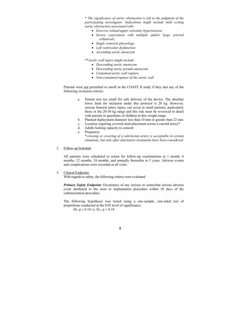
8
* The significance of aortic obstruction is left to the judgment of the participating investigator. Indications might include mild resting aortic obstruction associated with:
Exercise related upper extremity hypertension; Severe coarctation with multiple and/or large arterial
collaterals; Single ventricle physiology Left ventricular dysfunction Ascending aortic aneurysm
**Aortic wall injury might include:
Descending aortic aneurysm Descending aortic pseudo-aneurysm Contained aortic wall rupture Non-contained rupture of the aortic wall
Patients were not permitted to enroll in the COAST II study if they met any of the following exclusion criteria:
a. Patient size too small for safe delivery of the device. The absolute
lower limit for inclusion under this protocol is 20 kg. However, serious femoral artery injury can occur in small patients, particularly those in the 20-30 kg range and this risk must be reviewed in detail with parents or guardians of children in this weight range.
b. Planned deployment diameter less than 10 mm or greater than 22 mm c. Location requiring covered stent placement across a carotid artery* d. Adults lacking capacity to consent e. Pregnancy
*crossing or covering of a subclavian artery is acceptable in certain situations, but only after alternative treatments have been considered.
2. Follow-up Schedule
All patients were scheduled to return for follow-up examinations at 1 month, 6 months, 12 months, 24 months, and annually thereafter to 5 years. Adverse events and complications were recorded at all visits.
3. Clinical Endpoints
With regards to safety, the following criteria were evaluated. Primary Safety Endpoint: Occurrence of any serious or somewhat serious adverse event attributed to the stent or implantation procedure within 30 days of the catheterization procedure. The following hypothesis was tested using a one-sample, one-sided test of proportions conducted at the 0.05 level of significance: H0: p ≥ 0.18 vs. HA: p < 0.18

9
Secondary Safety Endpoint: Proportion of patients experiencing any of the following adverse events related to the device or implant procedure post 1 year
Underlying cardiac or non-cardiac disease, aortic wall injury, new aortic aneurysm formation within region of device, stent misplacement, malposition, stent fracture, aortic wall aneurysms, or restenosis requiring reintervention.
With regards to effectiveness, the following criteria were evaluated. Primary Effectiveness Endpoint #1: Improvement of aortic wall injury and/or aortic arch obstruction by a median increase of at least one grade from pre-implantation baseline to 12 month follow-up using the Severity of Illness Scale (based on upper extremity (UE) systolic BP, UE to lower extremity (LE) systolic BP, and aortic wall injury). The following hypothesis was tested using a one-sided Wilcoxon signed-rank test conducted at the 0.025 level of significance: H0: median change in grade ≤ 0 vs. HA: median change in grade > 0 Primary Effectiveness Endpoint #2: Aortic wall injury and aortic arch obstruction at Grade 4 or above at the 12 month follow-up, based on the Severity of Illness Scale, with no clinical worsening. The following hypothesis was tested using a one-sample, one-sided test of proportions conducted at the 0.05 level of significance: H0: p ≤ 0.70 vs. HA: p > 0.70 Secondary Effectiveness Endpoints:
Reduction of arm-leg systolic blood pressure gradients to less than 20mmHg and less than 15 mmHg.
Reduction of upper extremity blood pressure at 1 year compared to baseline
Repair of wall defect with <10% residual endoleak on MRI or CT in patients with aortic wall injury
Hospital length of stay compared to length of stay for surgical repair of aortic coarctation.
82 patients were enrolled in COAST II and study accountability is detailed in Table 1.
Table 1: COAST II Accountability
COAST II
Patients
Possible n (100%)
1 Month Visit n (%)
12 Month Visit n (%)
24 Month Visit n (%)
3 years Visit n (%)
4 years Visit n (%)
5 years Visit n (%)
Safety Cohort
82 82 (100%)
69 (84%)
67 (81.7%)
55 (67.1%)
38 (46.3%)
22 (26.8%)
Effectiveness Cohort
82 82 (100%)
68 (83%)
66 (80.5%)
54 (65.8%)
37 (45.1%)
21 (25.6%)

10
Subject Demographics Table 2 presents subject demographics and baseline characteristics analyzed for the enrolled subjects. The study population consisted of 52 male and 30 female subjects with a mean age of 18 (range 6 to 67 years).
Table 2: COAST II Pivotal Cohort – Patient Characteristics
Assessment Number (Percent) or
Median (Range)
Prospective
(n=29)
Legacy
(n=53)
Total
(n=82)
Gender
Male 21 (72%) 31 (58%) 52 (63%)
Female 8 (28%) 22 (42%) 30 (37%)
Age, years 20 (6 to 67) 17 (6 to 66) 18 (6 to 67)
Primary Indication
Repair of aortic wall injury 15 (52%) 34 (64%) 49 (60%)
Prevention of aortic wall injury1 14 (48%) 19 (36%) 33 (40%) 1 Includes 1 patient classified as not having pre-existing aortic wall injury, who was noted to have a small, localized intimal tear with a diameter of < ¼ the aortic diameter.
The analysis of safety was based on the implanted cohort of 82 COAST II patients completing their implant procedures. The primary safety outcomes are presented in Table 3.
Table 3. Summary of COAST II Outcomes and Pre-Specified Safety Endpoints
COAST II Safety Endpoint Event Rate (CI)
P Value
Primary Serious or Somewhat Serious Adverse Events Attributed to the Stent, Implantation or Catheterization within 30 days of the procedure (includes data from COAST combined with COAST II)
8.2% (5.2%, 12.3%)*
<0.001
Secondary
Proportion of patients experiencing any AEs related to the device or implant procedure post 1 year (among 74 patients followed for at least 1 year)
6.8% (2.2%, 15.1%)#
N/A
*90% Confidence interval # Confidence interval is not adjusted for multiplicity and is provided to illustrate the variability only. No statistical conclusion should be drawn from this confidence interval.

11
The COAST II primary safety endpoints were met with the occurrence of any serious or somewhat serious adverse event within 30 days post procedure being less than the predefined 18%. The overall incidence and types of adverse events were within expected ranges. Aortic wall injuries were rare and treated appropriately without the need for emergency surgery. The results are durable out to 60 months for each study and re-coarctation was treated by transcatheter means when it occurred. Table 4 provides a summary of the adverse events reported under COAST II.
Table 4: Summary of Adverse Events (AEs) for COAST II Stent
Related Events1 (Rates)
Stent, Implantation, or Catheterization
Related Events2
(Rates)
All Events (Rates)
Patients with adverse events at 30 days
2 (2.4%)
27 (32.9%)
42 (51.2%)
Serious or somewhat serious events at 30 days
1 (1.2%)
6 (7.3%)
7 (8.5%)
Serious or somewhat serious events at 30 days, excluding stent fracture
1 (1.2%)
6 (7.3%)
7 (8.5%)
Serious event at 30 days 0 (0.0%)
1 (1.2%)
1 (1.2%)
Patients with adverse events at 12 Months
4 (4.9%)
30 (36.6%)
58 (70.7%)
Serious or somewhat serious events at 12 months
3 (3.7%)
7 (8.5%)
14 (17.1%)
Serious or somewhat serious events at 12 months, excluding stent fracture
3 (3.7%)
7 (8.5%)
14 (17.1%)
Serious event at 12 months 1 (1.2%)
1 (1.2%)
4 (4.9%)
Patients with adverse event at 24 Months
5 (6.1%)
31 (37.8%)
60 (73.2%)
1Includes events that are due to or possible due to stent, and stent fractures. 2Includes events that are due to or possible due to stent, implantation, or catheterization, and stent fractures Table 5 presents incidence of stent fractures. There were no stent fractures detected at 12 month fluoroscopy. One stent fracture was detected at 24 month fluoroscopy. There was no loss of structural integrity in the detected fracture.
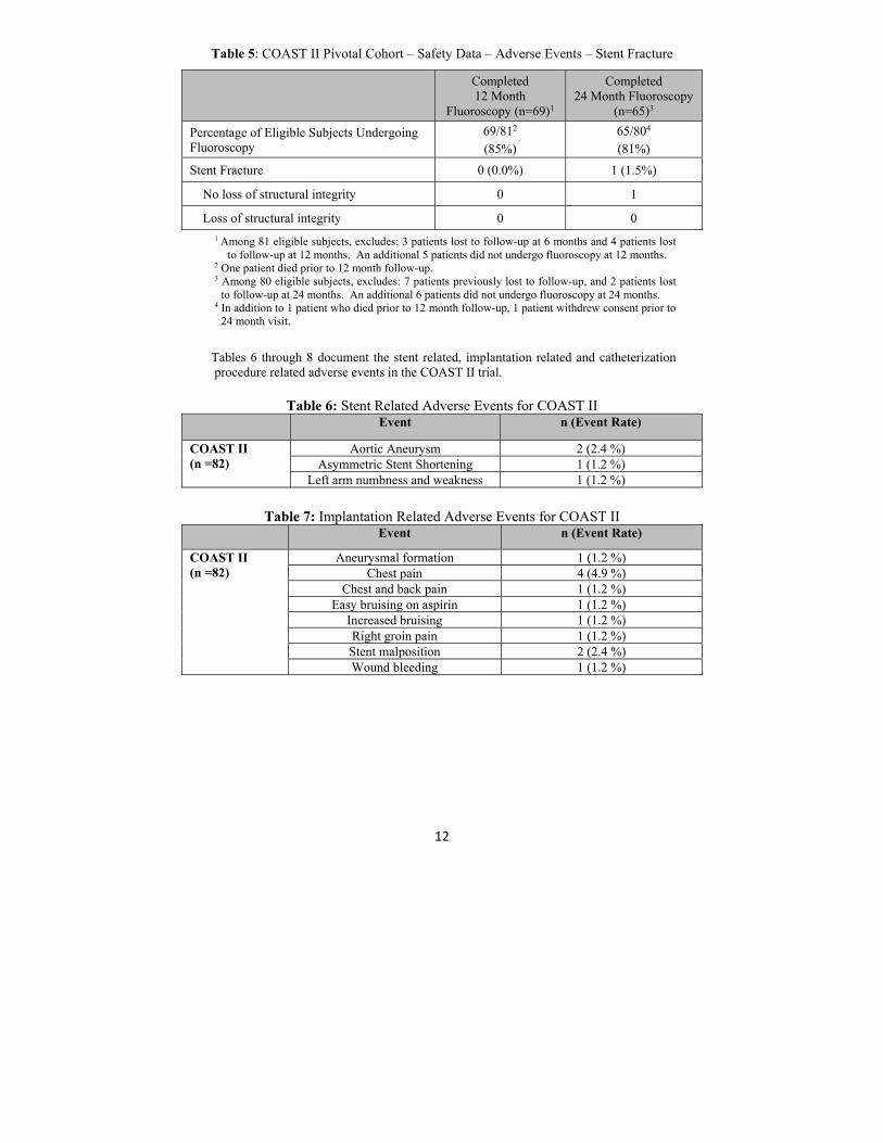
12
Table 5: COAST II Pivotal Cohort – Safety Data – Adverse Events – Stent Fracture
Completed 12 Month
Fluoroscopy (n=69)1
Completed 24 Month Fluoroscopy
(n=65)3
Percentage of Eligible Subjects Undergoing Fluoroscopy
69/812
(85%) 65/804
(81%)
Stent Fracture 0 (0.0%) 1 (1.5%)
No loss of structural integrity 0 1
Loss of structural integrity 0 0
1 Among 81 eligible subjects, excludes: 3 patients lost to follow-up at 6 months and 4 patients lost to follow-up at 12 months. An additional 5 patients did not undergo fluoroscopy at 12 months.
2 One patient died prior to 12 month follow-up. 3 Among 80 eligible subjects, excludes: 7 patients previously lost to follow-up, and 2 patients lost
to follow-up at 24 months. An additional 6 patients did not undergo fluoroscopy at 24 months. 4 In addition to 1 patient who died prior to 12 month follow-up, 1 patient withdrew consent prior to
24 month visit.
Tables 6 through 8 document the stent related, implantation related and catheterization procedure related adverse events in the COAST II trial.
Table 6: Stent Related Adverse Events for COAST II Event n (Event Rate)
COAST II (n =82)
Aortic Aneurysm 2 (2.4 %) Asymmetric Stent Shortening 1 (1.2 %)
Left arm numbness and weakness 1 (1.2 %)
Table 7: Implantation Related Adverse Events for COAST II
Event n (Event Rate)
COAST II (n =82)
Aneurysmal formation 1 (1.2 %) Chest pain 4 (4.9 %)
Chest and back pain 1 (1.2 %) Easy bruising on aspirin 1 (1.2 %)
Increased bruising 1 (1.2 %) Right groin pain 1 (1.2 %) Stent malposition 2 (2.4 %) Wound bleeding 1 (1.2 %)

13
Table 8: Catheterization Related Adverse Events for COAST II
Event n (Event Rate)
COAST II (n =82)
Aneurysm 1 (1.2 %) Atrial arrhythmia 1 (1.2 %)
Brachial plexus injury 1 (1.2 %) Contact skin rash 1 (1.2 %) Corneal abrasion 1 (1.2 %)
Discomfort right eye 1 (1.2 %) Dissection of iliac artery 1 (1.2 %)
Ecchymosis/groin tenderness 1 (1.2 %) Femoral artery occlusion 1 (1.2 %) Local hematoma groin 2 (2.4 %)
Localized groin bruising 1 (1.2 %) Minimal bleeding/cough 1 (1.2 %)
Neck swelling 1 (1.2 %) Pulsatile bleeding 1 (1.2 %)
Right iliac dissection/pulse loss 1 (1.2 %) Superficial infection of groin 1 (1.2 %) Wide complex non-sustained
tachycardia 1 (1.2 %)
There were five patients that crossed over from COAST to COAST II. One patient crossed over due to a small aneurysm after dilation, two patients due to a near atretic aorta, one patient due to localized intimal tear after dilation and one patient due to an acute, rapidly expanding aneurysm after dilation. In the COAST II trial two patients experienced two events, representing 2.4% of patients, and both events were resolved using a second Covered CP stent to fully occlude the aneurysm developed with no permanent damage. In the COAST II trial, three patients experienced aortic wall injuries by the 24 month follow-up. These injuries are detailed in Table 9, below.
Table 9: COAST II Aortic Wall Injuries by 24 Month Follow-up
Date of Catheterization
Date of MRI Injury Detected Intervention
11/11/2009 11/16/20111 Neo-intimal proliferation
Therapy for new aortic wall injury –
implantation of Covered CP Stent
2/23/2010 3/31/20111 Small aneurysm at 12 m visit
Therapy for new aortic wall injury –
implantation of Covered CP Stent
4/15/2010 5/12/2011 Small aneurysm at 12 m visit
New Covered CP Stent implanted to occlude
aneurysm 1Aortic wall injury not confirmed by MRI at follow-up visit. Confirmed during reintervention.

14
In COAST II, six patients experienced coarctation-related events, representing 7.3% of patients with events. These patients underwent catheter reinterventions. No surgical interventions were completed. Table 10 provides a summary of these interventions.
Table 10: COAST II Coarctation-Related Reintervention by 24 months Approximate
Time to Intervention
Post-procedure (Months)
Indication for Reintervention Procedure Performed
7 Persistent hypertension and gradient across aortic arch
Radiation of stent
7 Planned re-expansion of stent Radiation of stent 9 Planned re-expansion of stent Radiation of stent 25 Increased gradient with somatic
growth; neo-intimal proliferation detected in cath lab
Therapy for new aortic wall injury - implantation of Covered CP Stent
14 Aneurysm detected by MRI at 12 m visit1
Therapy for new aortic wall injury - implantation of Covered CP Stent
13 Aneurysm detected by MRI at 12 m visit1
Therapy for new aortic wall injury - implantation of Covered CP Stent
1. Presence of aneurysm in this patient was not confirmed by core laboratory review of the MRI
In COAST II, two patients experienced non-coarctation related reinterventions that were documented by the 24 month follow-up, representing 2.4% of the patients with events. These patients underwent catheter reinterventions. The time to intervention for one patient was four months, when the patient received a coronary angiogram and graft angiogram to address symptoms of angina. The time to intervention for the second patient was 26 months when the Melody valve was implanted to address a high right ventricle to pulmonary artery conduit gradient. The incidence of stent fracture for covered CP stents (COAST II) was much lower and was not observed to substantially increase over time. Also, relief from blood pressure gradient was maintained through 60 month follow-up and re-intervention was rare. When needed, this was accomplished using transcatheter interventions.

15
The key effectiveness outcomes of COAST II are shown in Table 11 through 12.
Table 11. Summary of Late Outcomes and Major Pre-Specified Effectiveness Study Endpoints
Effectiveness Endpoint Event Rate (CI)
P Value
COAST II Primary
Severity of Illness Scale Grade 4 or 5 with No Clinical Worsening at 12 Month Follow-up
80% (70.1%, 87%)*
0.048
COAST II Secondary
Proportion of patients with arm-leg systolic blood pressure differences less than 20mmHg and less than 15 mmHg at 12 month follow-up, compared to baseline
87% (up from 46% at
baseline) (76%, 94%)+
79%
(up from 38% at baseline)
(68%, 88%)+
N/A
Reduction of upper extremity blood pressure at 1 year compared to baseline
12 ± 20mmHg§
(7mmHg, 17mmHg)+#
N/A
Complete repair of aortic wall defect with first Covered CP Stent (no residual endoleak during the catheterization procedure)
47 of 49 (96%) of patients treated for
an aortic wall injury
N/A
Proportion of patients with effective treatment of AWI with no residual aneurysm seen on MRI scanning
37 of 39 (95%) patients treated for
an aortic wall injury
1/39 (2.5%) with a small aneurysm
and one patient’s MRI could not be evaluated by core
lab
N/A
Hospital length of stay compared to length of stay for surgical repair of aortic coarctation
1.2 ± 0.9 days§ (1.0 days, 1.4
days)+
<0.001
*90% Confidence interval + 95% Confidence Interval § Mean + standard deviation # Confidence interval is not adjusted for multiplicity and is provided to illustrate the variability only. No statistical conclusion should be drawn from this confidence interval.
16
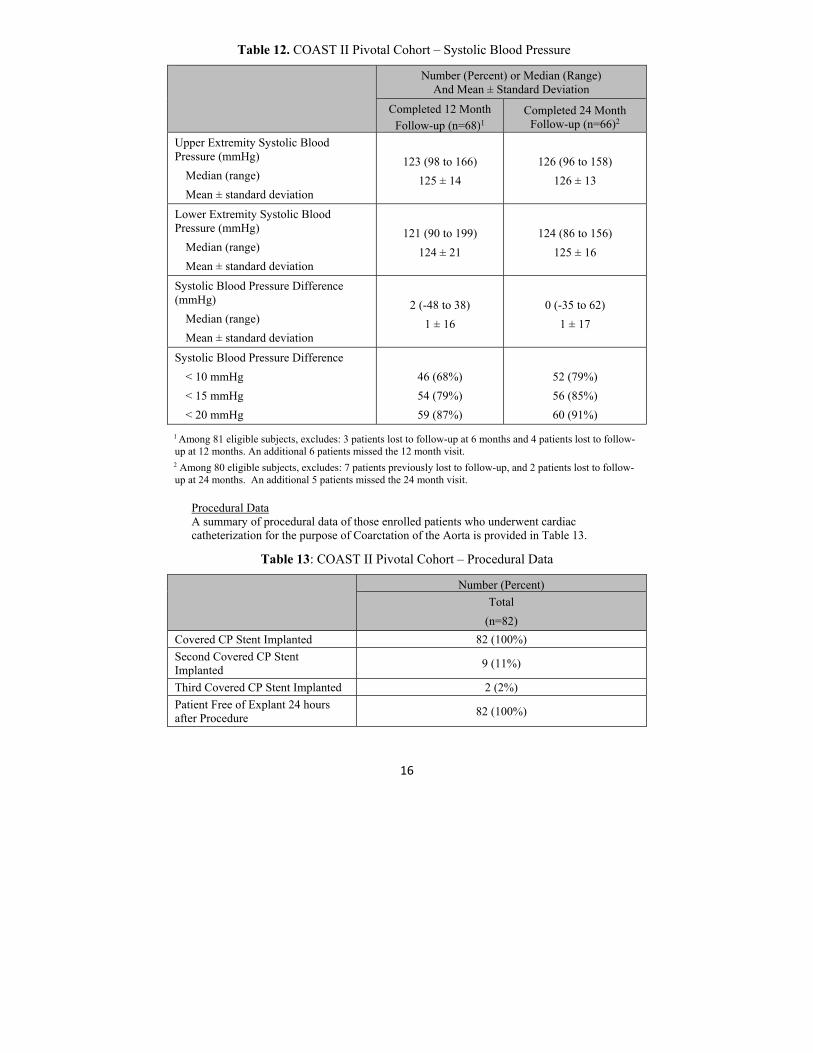
Table 12. COAST II Pivotal Cohort – Systolic Blood Pressure
Number (Percent) or Median (Range) And Mean ± Standard Deviation
Completed 12 Month Follow-up (n=68)1
Completed 24 Month Follow-up (n=66)2
Upper Extremity Systolic Blood Pressure (mmHg)
Median (range)
Mean ± standard deviation
123 (98 to 166)
125 ± 14
126 (96 to 158)
126 ± 13
Lower Extremity Systolic Blood Pressure (mmHg)
Median (range)
Mean ± standard deviation
121 (90 to 199)
124 ± 21
124 (86 to 156)
125 ± 16
Systolic Blood Pressure Difference (mmHg)
Median (range)
Mean ± standard deviation
2 (-48 to 38)
1 ± 16
0 (-35 to 62)
1 ± 17
Systolic Blood Pressure Difference
< 10 mmHg
< 15 mmHg
< 20 mmHg
46 (68%)
54 (79%)
59 (87%)
52 (79%)
56 (85%)
60 (91%)
1 Among 81 eligible subjects, excludes: 3 patients lost to follow-up at 6 months and 4 patients lost to follow-up at 12 months. An additional 6 patients missed the 12 month visit. 2 Among 80 eligible subjects, excludes: 7 patients previously lost to follow-up, and 2 patients lost to follow-up at 24 months. An additional 5 patients missed the 24 month visit.
Procedural Data A summary of procedural data of those enrolled patients who underwent cardiac catheterization for the purpose of Coarctation of the Aorta is provided in Table 13.
Table 13: COAST II Pivotal Cohort – Procedural Data
Number (Percent)
Total
(n=82)
Covered CP Stent Implanted 82 (100%)
Second Covered CP Stent Implanted
9 (11%)
Third Covered CP Stent Implanted 2 (2%)
Patient Free of Explant 24 hours after Procedure
82 (100%)

17
COAST/COAST II Follow-up Data
A.1. Percent of Patients with New Stent Fracture
% Stent Fracture (new at each interval) Interval COAST I COAST II
12 months 4/121 (3.3%)
1/127 (0.8%)
24 months 12/121 (9.9%)
5/127 (3.9%)
36 months 2/121 (1.6%)
1/127 (0.8%)
48 months 10/121 (8.2%)
4/127 (3.1%)
60 months 9/121 (7.4%)
2/127 (1.6%)
A.2. Percent of Patients with Stent Fracture (Cumulative)
% Stent Fracture (cumulative) Interval COAST I COAST II
12 months 4/121 (3%)
1/127 (0.8%)
24 months 16/121 (13%)
6/127 (4.7%)
36 months 18/121 (15%)
7/127 (5.5%)
48 months 28/121 (23%)
11/127 (8.6%)
60 months 37/121 (31%)
13/127 (10%)
B.1. Percent of Patients with Aortic Wall Injury (New or Progressive)*
% patients with AWI Interval COAST I COAST II
12 months 1/121 (0.8%)
1/127 (0.8%)
24 months 0/121 0/127
36 months 3/121 (2.5%)
0/127
48 months 1/121 (0.8%)
0/127
60 months 1/121 (0.8%)
2/127 (1.6%)
*All recorded injuries were “new”, none designated as progressive.

18
B.2. Cumulative Percent of Patients with Aortic Wall Injury (New or Progressive)*
% patients with AWI (cumulative) Interval COAST I COAST II
12 months 1/121 (0.8%)
1/127 (0.8%)
24 months 1/121 (0.8%)
1/127 (0.8%)
36 months 4/121 (3.3%)
1/127 (0.8%)
48 months 5/121 (4.1%)
1/127 (0.8%)
60 months 6/121 (5%)
3/127 (2.4%)
*All recorded injuries were “new”, none designated as progressive.
C.1. Percent of Patients with Coarctation Related Re-intervention or Surgery*
% patients with reintervention or surgery Interval COAST I COAST II
12 months 4/121 (3.3%)
9/128 (7%)
24 months 3/121 (2.5%)
3/128 (2.3%)
36 months 6/121 (5%)
3/128 (2.3%)
48 months 6/121 (5%)
3/128 (2.3%)
60 months 2/121 (1.7%)
3/128 (2.3%)
*All patients listed had catheter reinterventions, none had surgery.
C.2. Percent of Patients with Coarctation Related Re-intervention or Surgery (Cumulative)
% patients with catheter-based re-intervention or surgery (cumulative)
Interval Bare Metal Covered Stent
12 months 3/121 (2.5%)
9/128 (7%)
24 months 6/121 (5%)
12/128 (9.3%)
36 months 12/121 (9.9%)
15/128 (11.8%)
48 months 18/121 (14.9%)
18/128 (14%)
60 months 20/121 (16.5%)
21/128 (16.4%)

19
C.3. Percent of Bare Metal Patients with Catheter-Based or Surgical Coarctation-Related Re-intervention
% Bare Metal patients with catheter-based re-intervention or surgery
Interval Catheterization Surgery
12 months 3/121 (2.5%)
0 (0%)
24 months 3/121 (2.5%)
0 (0%)
36 months 6/121 (5%)
0 (0%)
48 months 6/121 (5%)
0 (0%)
60 months 2/121 (1.7%)
0 (0%)
C.4. Percent of Bare Metal Patients with Catheter-Based or Surgical Coarctation-Related Re-intervention (Cumulative)
% Bare Metal patients with catheter-based re-intervention or surgery (cumulative)
Interval Catheterization Surgery
12 months 3/121 (2.5%)
0 (0%)
24 months 6/121 (5%)
0 (0%)
36 months 12/121 (9.9%)
0 (0%)
48 months 18/121 (14.9%)
0 (0%)
60 months 20/121 (16.5%)
0 (0%)
C.5. Percent of Covered Stent Patients with Catheter-Based or Surgical Coarctation-Related Re-intervention
% Covered Stent patients with re-intervention or surgery
Interval Catheterization Surgery
12 months 9/128 (7%)
0 (0%)
24 months 3/128 (2.3%)
0 (0%)
36 months 3/128 (2.3%)
0 (0%)
48 months 3/128 (2.3%)
0 (0%)
60 months 3/128 (2.3%)
0 (0%)

20
C.6. Percent of Covered Stent Patients with Catheter-Based or Surgical Coarctation-Related Re-intervention (Cumulative)
% Covered Stent patients with re-intervention or surgery (cumulative)
Interval Catheterization Surgery
12 months 9/128 (7%)
0 (0%)
24 months 12/128 (9.3%)
0 (0%)
36 months 15/128 (11.8%)
0 (0%)
48 months 18/128 (14%)
0 (0%)
60 months 21/128 (16.4%)
0 (0%)
D.1 Percent of Patients with Upper to Lower Extremity Pressure Gradients > 15mmHg
% patients with Residual UE to LE pressure gradient > 15mmHg
Interval COAST I COAST II
12 months 16/104 (15.4%)
16/107 (15%)
24 months 8/95
(8.4%) 13/81 (16%)
36 months 6/81 (7%)
12/72 (14%)
48 months 6/67 (9%)
6/53 (10%)
60 months 7/65
(11%) 14/47 (30%)
**Note, n is different at each time interval and based on the number of patients with pressures measured at each interval**.
E.1. Percent of Patients with Proximal SBP >140
0.00%
5.00%
10.00%
15.00%
20.00%
25.00%
30.00%
COAST I
COAST II
% patients with UESBP > 140mmHg Interval COAST I COAST II
12 months 18/104 (17%)
26/104 (25%)
24 months 16/95 (17%)
20/81 (25%)
36 months 6/86 (7%)
12/88 (14%)
48 months 8/74
(11%) 10/64 (16%)
60 months 11/74 (15%)
12/52 (23%)
**Note, n is different at each time interval and based on the number of patients with pressures measured at each interval**.

21
F.1 Baseline Characteristics
Baseline Characteristics – COAST/COAST II Median (Range)
Bare Metal Patients (n=128) Bare Metal Re-intervention
Patients (n= 20)
Age (yrs) 16 (8 to 52) 13 (9 to 20)
Weight (kg) 65 (35 to 157) 51.7 (35 to 104)
Covered Stent Patients
(n=127) Covered Stent Re-
intervention Patients (n=21)
Age (yrs) 19 (6 to 67) 15 (6 to 35)
Weight (kg) 68 (17 to 122) 50 (17 to 95)
G.1 Systolic Blood Pressure Differences after Initial CoA Stenting
Systolic Blood Pressure Differences after Initial CoA Stenting Median (Range)
Bare Metal Non-Re-intervention Patients
(n=108)
Bare Metal Re-intervention Patients (n=20)
SBP difference (AAo-Dao) (mmHg)
0 (-12 to 15) 1.5 (-5 to 12)
Covered Stent Non-Re-
intervention Patients (n=106)
Covered Stent Re-intervention Patients
(n=21) SBP difference (AAo-Dao) (mmHg)
2 (-12 to 38) 7 (-3 to 20)
22
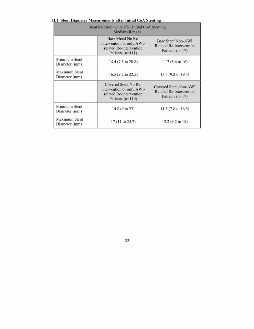
H.1 Stent Diameter Measurements after Initial CoA Stenting
Stent Measurements after Initial CoA Stenting Median (Range)
Bare Metal No Re-intervention or only AWI-
related Re-intervention Patients (n=111)
Bare Stent Non-AWI Related Re-intervention
Patients (n=17)
Minimum Stent Diameter (mm)
14.4 (7.8 to 20.8) 11.7 (8.6 to 16)
Maximum Stent Diameter (mm)
16.5 (9.2 to 22.5) 13.5 (9.2 to 19.4)
Covered Stent No Re-intervention or only AWI-
related Re-intervention Patients (n=110)
Covered Stent Non-AWI Related Re-intervention
Patients (n=17)
Minimum Stent Diameter (mm)
14.8 (9 to 23) 11.5 (7.8 to 16.5)
Maximum Stent Diameter (mm)
17 (11 to 25.7) 13.2 (9.2 to 18)
23
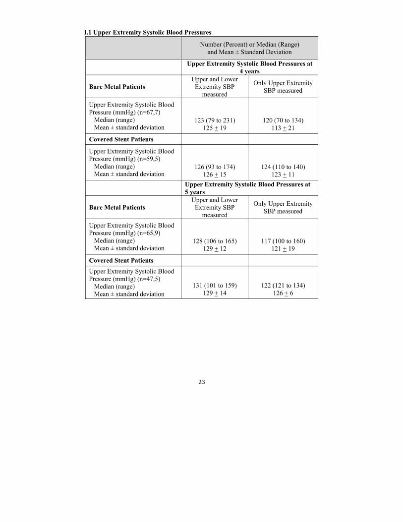
I.1 Upper Extremity Systolic Blood Pressures
Number (Percent) or Median (Range) and Mean ± Standard Deviation
Upper Extremity Systolic Blood Pressures at
4 years
Bare Metal Patients Upper and Lower
Extremity SBP measured
Only Upper Extremity SBP measured
Upper Extremity Systolic Blood Pressure (mmHg) (n=67,7) Median (range) Mean ± standard deviation
123 (79 to 231) 125 + 19
120 (70 to 134) 113 + 21
Covered Stent Patients
Upper Extremity Systolic Blood Pressure (mmHg) (n=59,5) Median (range) Mean ± standard deviation
126 (93 to 174)
126 + 15
124 (110 to 140) 123 + 11
Upper Extremity Systolic Blood Pressures at 5 years
Bare Metal Patients Upper and Lower
Extremity SBP measured
Only Upper Extremity SBP measured
Upper Extremity Systolic Blood Pressure (mmHg) (n=65,9) Median (range) Mean ± standard deviation
128 (106 to 165) 129 + 12
117 (100 to 160) 121 + 19
Covered Stent Patients
Upper Extremity Systolic Blood Pressure (mmHg) (n=47,5) Median (range) Mean ± standard deviation
131 (101 to 159) 129 + 14
122 (121 to 134) 126 + 6

24
RIGHT VENTRICULAR OUTFLOW TRACT INDICATION
Treatment of right ventricle to pulmonary artery (right ventricular outflow tract) conduit disruptions that are identified during conduit pre-dilatation procedures performed in preparation for transcatheter pulmonary valve replacement (TPVR).
CONTRAINDICATIONS
Patients too small to allow safe delivery of the stent without injury to a systemic vein or to the right side of the heart;
Clinical or biological signs of infection;
Active endocarditis;
Pregnancy.
WARNINGS
Administer appropriate anticoagulation therapy to reduce potential thrombosis. If the patient is not appropriately anticoagulated, thrombus formation may occur.
During the Premarket Approval study the Medtronic Melody Valve was used for valve restoration.
The safety and effectiveness of the Covered CP Stent for pre-stenting of the right ventricular outflow tract (RVOT) landing zone (i.e., prophylaxis or prevention of either RVOT conduit rupture or TPVR fracture; used as a primary RVOT conduit) in preparation of a transcatheter pulmonary valve replacement (TPVR) has not been evaluated.
The sheath must be flushed with heparinized saline via the proximal side port prior to introducing the delivery system into the body.
As with any type of implant, infection secondary to contamination of the stent might lead to endocarditis, or abscess formation.
The Covered Stent can migrate from the site of implant potentially causing obstruction to pulmonary artery flow.
Over-stretching of the RVOT may result in rupture or aneurysm of the RV-PA conduit or the native pulmonary artery.
The inflated diameter of the stent should at least equal the diameter of the intended implant site.
Excessive force while crimping may weaken welds of the stent.
Crimping the 8 zig stent on a balloon catheter smaller than 12mm may cause damage to the stent.
Excessive handling and manipulation of the covering while crimping the stent may cause the covering to tear off of the stent.
Crimping the device in the opposite direction of the folds in the covering may cause the covering to catch while inserting into the hemostasis valve tool and introducer. This could cause the covering to tear off of the stent.
Retracting the covered stent back in to the sheath may cause the covering to catch and tear off of the stent.

25
Do not exceed the RBP. An inflation device with pressure gauge is recommended to monitor pressure. Pressure in excess of the RBP can cause balloon rupture and potential inability to withdraw the catheter into the sheath.
Confirm that the distal end of the introducer sheath is at least 2.5cm back from the most proximal image band before inflating the outer balloon. Failure to do so may stretch the outer tubing and severely hinder balloon deflation.
Use two appropriate size inflation devices with pressure gauges for inflation.
Do not advance the guidewire, balloon catheter, or any other component if resistance is met, without first determining the cause and taking remedial action.
This catheter is not recommended for pressure measurement or fluid injection.
Do not remove the guidewire from the catheter at any time during the procedure.
This device is intended for single use only. Do not resterilize and/or reuse it, as this can potentially result in compromised device performance and increased risk of cross-contamination.
PRECAUTIONS
Use of an inflation device with pressure gauge is highly recommended during this procedure.
Sheath-catheter manipulation and stent deployment must be conducted under fluoroscopic guidance with appropriate radiographic equipment.
Stents are delicate devices. Exercise caution when handling the stent to prevent breakage.
Guidewires should be handled with care to avoid kinking or breaking. When advancing the NuDEL system over the wire, the tip of the wire must be controlled at all times.
The NuDEL system, especially at the stent, is rigid and may make negotiation through vessels difficult.
Maintain tight catheter connections at all times. De-air the sheath with heparinized saline prior to insertion in the patient. Apply
repeated negative pressure cycles to the balloons to replace the air in the balloon with fluid: this enhances the efficiency of inflation and avoids air introduction into the circulation in the unusual case of balloon rupture.
The inflation diameter of the balloon used during stent delivery should approximate the diameter of the obstructive vessel and the intended implant site.
Under no circumstances should any portion of the sheath-catheter system be advanced or removed against resistance. Use fluoroscopy to identify and resolve the resistance.
If resistance is encountered upon removal, the whole system (balloon, guidewire and sheath) should be removed as a single unit, particularly if balloon rupture or leakage is known or suspected. This may be accomplished by firmly grasping and withdrawing the sheath and catheter, using a gentle anticlockwise twisting motion with traction.
The balloons must be completely deflated before retracting into sheath. Proper functioning of the sheath-catheter depends on its integrity. Exercise caution
when handling the catheter. Damage may result from kinking, stretching, or forceful wiping of the catheter.

26
POTENTIAL COMPLICATIONS / ADVERSE EFFECTS NOTE: Circumferential tear of the delivery balloon catheter prior to complete expansion of the stent may cause the balloon to become tethered to the stent, requiring surgical removal. In case of rupture of an adequately sized balloon after stent expansion, it can be withdrawn and a new balloon catheter exchanged over a guidewire to complete expansion of the stent.
Cardiac catheterization carries certain risks. In addition, potential complications, and related adverse effects associated with implants include, but are not limited to: Stent Migration Stent Stenosis Stent Fracture Pseudoaneurysm Vessel Ruptures Stent Malposition Hematoma Sepsis/infection Thrombosis/Thromboembolism AV fistula formation Death Transitory arrhythmia Endocarditis Bleeding Cell necrosis at the site of implant Cerebrovascular Incident
PARCS CLINICAL STUDY INFORMATION PARCS was performed to provide information that will support labeling of the Covered CP Stent for the treatment of RV-PA conduit disruption, occurring during cardiac catheterizations aimed at re-dilating the conduit up to 110% of its original size, and then making the conduit competent by the implantation of a transcatheter pulmonary valve. The study was a prospective, multi-center, single arm, non-randomized clinical study trial. 50 patients were included in the pivotal cohort, and 70 patients were included in the continued access cohort. There were 39 investigational sites. 1. Clinical Inclusion and Exclusion Criteria a. Precatheterization Inclusion Criteria: i. Patient meets institutional criterion for placement of Melody® TPV ii. Patient size adequate to receive Melody® TPV implantation via venous access using the Ensemble® Transcatheter Delivery System iii. RV-PA conduit original size ≥ 16mm diameter iv. Patient age between 7 and 75 years b. Catheterization Inclusion Criteria: i. Angiographic evidence for RV-PA conduit disruption including: dissection, aneurysm, pseudo-aneurysm, tears or rupture
Recognition and treatment of conduit disruption may occur before, during or after implantation of the Melody® TPV
Conduit disruption related to prior intervention, identified angiographically before conduit dilation is performed during the Melody® implant procedure, can be eligible for CCPS implantation and study inclusion.

27
c. Precatheterization Exclusion Criteria: i. Patient size too small for transvenous placement of the Melody® TPV ii. Bloodstream infection, including endocarditis iii. Pregnancy iv. Prisoners and adults lacking the capacity of give consent d. Catheterization Exclusion Criteria: i. Conduit size is not suitable (too small or too large) for a Melody® TPV ii. Risk of coronary compression has been identified iii. Lack of angiographic evidence for RV-PA conduit disruption – Prophylactic use of study CCPS is prohibited iv. Vessel injury occurring in either the right or left branch pulmonary arteries
If injury to branch pulmonary arteries occurs during the catheterization and covered stent usage is indicated, Emergency Use guidelines must be employed.
2. Follow-up Schedule All patients are scheduled to return for follow-up examinations at 6 months. 3. Clinical Endpoints
With regards to safety, the following criteria were evaluated. Primary Safety Endpoint #1: Patient must have successful coverage of conduit disruption defined as either no residual disruption or contained disruption, followed by successful implantation of the Melody® valve. Hypothesis: At least 80% of patients will have successful implantation of the Melody® TPV following repair of the RV-PA conduit to a post- procedure SIS level of 0 or 1, using the Covered CP Stent.
Table 14: Primary Safety Endpoint #1
Number (Percent)
Pivotal
Implanted (n=502)
CAP
Implanted (n=703)
Patients with Successful Coverage of Conduit Disruption Followed by Successful Implantation of Melody Valve1 95% Confidence Interval
46 (93.9%)
(83.1, 98.7)
62 (89.9%)
(80.2, 95.8)
1 Successful coverage defined as no residual disruption or contained disruption 2 Not reported for 1 pivotal patient 3 Not reported for 1 CAP patient

28
Conclusion: Based on the pivotal cohort, successful implantation of the Melody® valve is achieved in at least 83% of patients, which exceeds the hypothesized value of 80%. Primary Safety Endpoint #2: Patient must not have any adverse event attributed to the Covered CP Stent within 30 days of the catheterization procedure, as adjudicated by the Data and Safety Monitoring Board.
Hypothesis: At least 80% of patients will be free of an adverse event attributed to the Covered CP Stent within 30 days of the catheterization procedure.
Table 15: Primary Safety Endpoint #2
Number (Percent)
Pivotal
Implanted (n=50)
CAP
Implanted (n=70)
Patients with Any Adverse Event Attributed to the Covered CP Stent within 30 Days1 95% Confidence Interval
1 (2.0%)
(0.1, 10.7)
2 (2.9%)
(0.4, 9.9) 1 Two adverse events attributed to CCPS in 1 pivotal patient (stent malposition, embolization) Conclusion: Based on the pivotal cohort, at least 80% of patients were free of an adverse event attributed to the CCPS within 30 days of the catheterization procedure. With regards to effectiveness, the following criteria were evaluated. Primary Effectiveness Endpoint #1: Improvement in the Severity of Illness Score (SIS) is defined as pre-implantation SIS minus post-procedure SIS assessed after Melody Valve insertion (e.g., a change from level 3 at baseline to level 2 post- procedure would represent an improvement of 1 level in SIS). The scale used for the clinical study was developed by the Principal Investigator.

29
Table 16: PARCS Severity of Illness Scale (SIS) – Baseline Assessment Level of Security
Level 0 Level 1 Level 2 Level 3 Level 4
No injury or conduit wall disruption: No contrast seen extending more than 2mm outside of or extravasating (leaking) outside of the longitudinal plane of the vascular lumen. (does not indicate the need for CCPS implantation)
Contained disruption: Small collection of contrast seen extending outside of the longitudinal plane of the vascular lumen ≤ ½ the diameter of the adjacent conduit, indicating the occurrence of an aneurysm, pseudo-aneurysm or well contained tear. This category can also be used to describe the unlikely occurrence of a dissection with contrast held in a contained space within the conduit lumen.
Partially Contained disruption: Large collection of contrast seen outside the wall of the RV-PA conduit ≥ ½ the diameter of the adjacent conduit.
Uncontained conduit disruption: Extravasation of contrast into the mediastinum or pleural cavity.
N/A
30
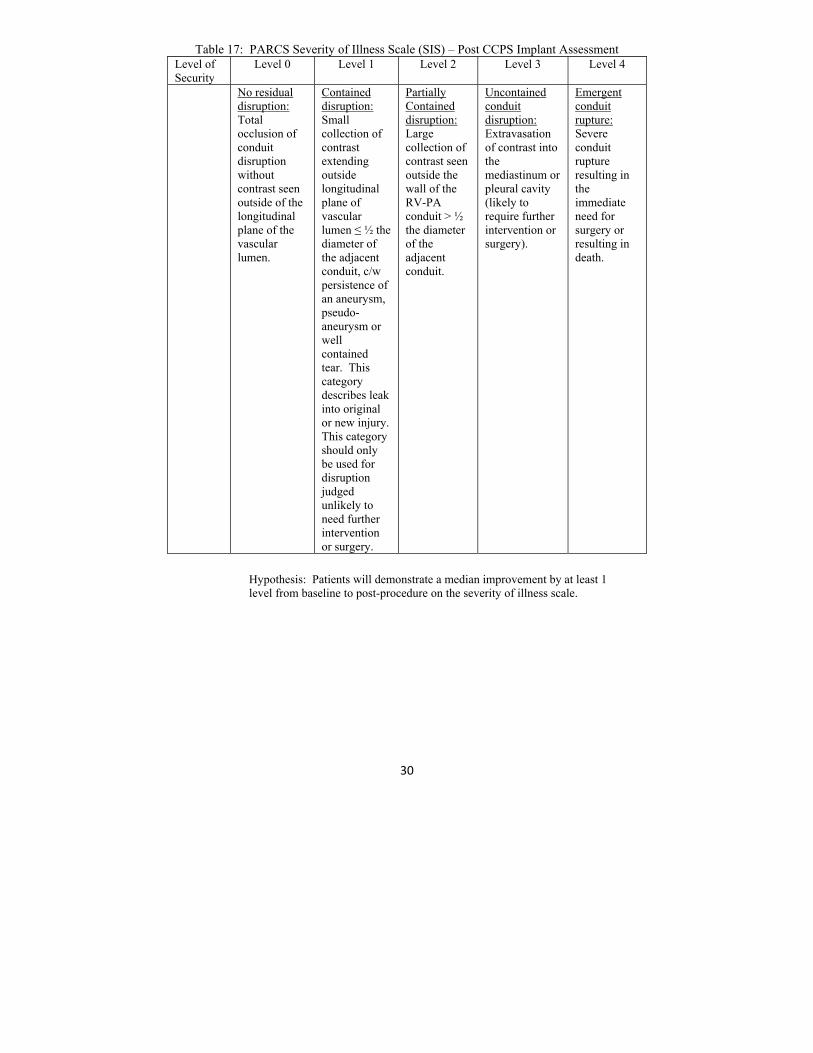
Table 17: PARCS Severity of Illness Scale (SIS) – Post CCPS Implant Assessment Level of Security
Level 0 Level 1 Level 2 Level 3 Level 4
No residual disruption: Total occlusion of conduit disruption without contrast seen outside of the longitudinal plane of the vascular lumen.
Contained disruption: Small collection of contrast extending outside longitudinal plane of vascular lumen ≤ ½ the diameter of the adjacent conduit, c/w persistence of an aneurysm, pseudo-aneurysm or well contained tear. This category describes leak into original or new injury. This category should only be used for disruption judged unlikely to need further intervention or surgery.
Partially Contained disruption: Large collection of contrast seen outside the wall of the RV-PA conduit > ½ the diameter of the adjacent conduit.
Uncontained conduit disruption: Extravasation of contrast into the mediastinum or pleural cavity (likely to require further intervention or surgery).
Emergent conduit rupture: Severe conduit rupture resulting in the immediate need for surgery or resulting in death.
Hypothesis: Patients will demonstrate a median improvement by at least 1 level from baseline to post-procedure on the severity of illness scale.
31
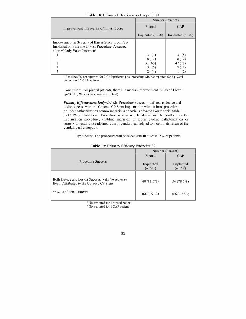
Table 18: Primary Effectiveness Endpoint #1
Improvement in Severity of Illness Score
Number (Percent)
Pivotal
Implanted (n=50)
CAP
Implanted (n=70)
Improvement in Severity of Illness Score, from Pre-Implantation Baseline to Post-Procedure, Assessed after Melody Valve Insertion1 -1 0 1 2 3
3 (6) 8 (17) 31 (66) 3 (6) 2 (4)
3 (5) 8 (12) 47 (71) 7 (11) 1 (2)
1 Baseline SIS not reported for 2 CAP patients; post-procedure SIS not reported for 3 pivotal patients and 2 CAP patients
Conclusion: For pivotal patients, there is a median improvement in SIS of 1 level (p<0.001, Wilcoxon signed-rank test). Primary Effectiveness Endpoint #2: Procedure Success – defined as device and lesion success with the Covered CP Stent implantation without intra-procedural or post-catheterization somewhat serious or serious adverse events attributable to CCPS implantation. Procedure success will be determined 6 months after the implantation procedure, enabling inclusion of repeat cardiac catheterization or surgery to repair a pseudoaneurysm or conduit tear related to incomplete repair of the conduit wall disruption. Hypothesis: The procedure will be successful in at least 75% of patients.
Table 19: Primary Efficacy Endpoint #2
Procedure Success
Number (Percent) Pivotal
Implanted (n=501)
CAP
Implanted (n=702)
Both Device and Lesion Success, with No Adverse Event Attributed to the Covered CP Stent 95% Confidence Interval
40 (81.6%)
(68.0, 91.2)
54 (78.3%)
(66.7, 87.3)
1 Not reported for 1 pivotal patient 2 Not reported for 1 CAP patient

32
Conclusion: Based on the pivotal cohort, procedure success is achieved in at least 68% of patients. This falls short of the hypothesis that procedure success would be achieved in at least 75% of patients. Secondary Effectiveness Endpoint #1: Device Success – defined as successful implantation of a Covered CP Stent, either providing complete repair of a conduit disruption or placement in preparation for a second Covered stent overlapping in tandem without adverse event.
Table 20: Secondary Effectiveness Endpoint #1
Device Success
Number (Percent) Pivotal
Implanted (n=501)
CAP Implanted (n=702)
Successful Implantation of First CCPS, Either Providing Complete Repair of Conduit Disruption or Placement in Preparation for Overlapping Tandem Stents, with No Adverse Event Attributed to the Covered CP Stent 95% Confidence Interval
46 (93.9%)
(83.1, 98.7)
66 (95.7%)
(87.8, 99.1)
1 Not reported for 1 pivotal patient 2 Not reported for 1 CAP patient
Secondary Effectiveness Endpoint #2: Lesion Success – defined as complete repair of a conduit disruption with a single Covered CP Stent or via planned, tandem covered stent implantations.
Table 21: Secondary Effectiveness Endpoint #2
Lesion Success
Number (Percent)
Pivotal Implanted (n=501)
CAP Implanted (n=702)
Successful Coverage of Conduit Disruption with First CCPS or with Subsequent Planned CCPS 95% Confidence Interval
42 (85.7%)
(72.8, 94.1)
56 (81.2%)
(69.9, 89.6) 1 Not reported for 1 pivotal patient 2 Not reported for 1 CAP patient
33
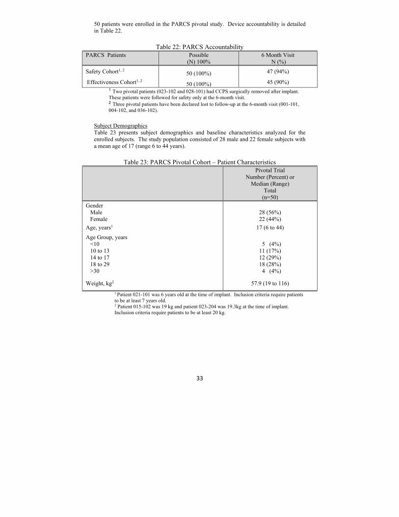
50 patients were enrolled in the PARCS pivotal study. Device accountability is detailed in Table 22.
Table 22: PARCS Accountability
PARCS Patients
Possible (N) 100%
6 Month Visit N (%)
Safety Cohort1, 2 50 (100%) 47 (94%)
Effectiveness Cohort1, 2 50 (100%) 45 (90%) 1 Two pivotal patients (023-102 and 028-101) had CCPS surgically removed after implant.
These patients were followed for safety only at the 6-month visit. 2 Three pivotal patients have been declared lost to follow-up at the 6-month visit (001-101,
004-102, and 036-102).
Subject Demographics Table 23 presents subject demographics and baseline characteristics analyzed for the enrolled subjects. The study population consisted of 28 male and 22 female subjects with a mean age of 17 (range 6 to 44 years).
Table 23: PARCS Pivotal Cohort – Patient Characteristics
Pivotal Trial Number (Percent) or
Median (Range) Total
(n=50)
Gender Male Female
28 (56%) 22 (44%)
Age, years1 17 (6 to 44)
Age Group, years <10 10 to 13 14 to 17 18 to 29 >30
5 (4%) 11 (17%) 12 (29%) 18 (28%) 4 (4%)
Weight, kg2 57.9 (19 to 116)
1 Patient 021-101 was 6 years old at the time of implant. Inclusion criteria require patients to be at least 7 years old.
2 Patient 015-102 was 19 kg and patient 023-204 was 19.3kg at the time of implant. Inclusion criteria require patients to be at least 20 kg.
34
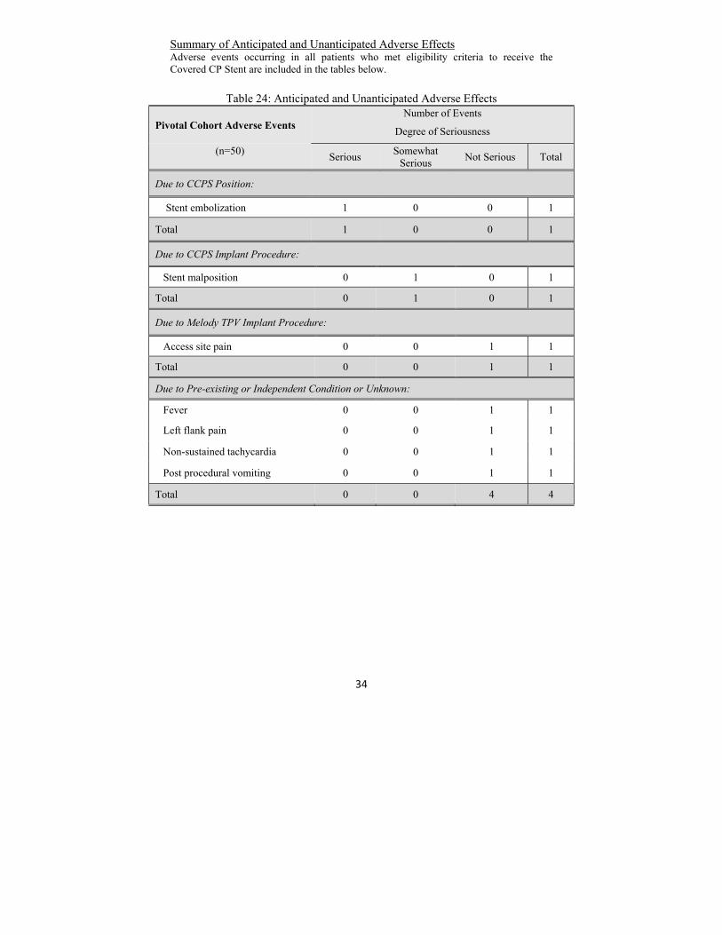
Summary of Anticipated and Unanticipated Adverse Effects Adverse events occurring in all patients who met eligibility criteria to receive the Covered CP Stent are included in the tables below.
Table 24: Anticipated and Unanticipated Adverse Effects
Pivotal Cohort Adverse Events Number of Events
Degree of Seriousness
(n=50)
Serious Somewhat
Serious Not Serious Total
Due to CCPS Position:
Stent embolization 1 0 0 1
Total 1 0 0 1
Due to CCPS Implant Procedure:
Stent malposition 0 1 0 1
Total 0 1 0 1
Due to Melody TPV Implant Procedure:
Access site pain 0 0 1 1
Total 0 0 1 1
Due to Pre-existing or Independent Condition or Unknown:
Fever 0 0 1 1
Left flank pain 0 0 1 1
Non-sustained tachycardia 0 0 1 1
Post procedural vomiting 0 0 1 1
Total 0 0 4 4

35
CP Stent® Foreshortening Chart
Inflated Balloon
Diameter
CP8Z16 (Stent length
after expansion) Percentage Shortening
CP8Z22 (Stent length
after expansion) Percentage Shortening
CP8Z28 (Stent length
after expansion) Percentage Shortening
CP8Z34 (Stent length
after expansion) Percentage Shortening
CP8Z39 (Stent length
after expansion) Percentage Shortening
CP8Z45 (Stent length
after expansion) Percentage Shortening
12mm (1.61) cm
2.8% (2.18) cm
0.8% (2.62) cm
4.4% (3.23) cm
3.1% (3.72) cm
1.9% (4.17) cm
3.8%
14mm (1.54) cm
6.5% (2.08) cm
5.4% (2.56) cm
6.8% (3.15) cm
5.4% (3.66) cm
3.6% (3.97) cm
8.4%
15mm (1.51) cm
8.5% (2.02) cm
7.9% (2.51) cm
8.6% (3.10) cm
7.0% (3.54) cm
6.6% (3.94) cm
9.2%
16mm (1.48) cm
10.6% (1.98) cm
10.1% (2.45) cm
10.7% (3.00) cm
9.8% (3.48) cm
8.2% (3.84) cm
11.4%
18mm (1.43) cm
13.7% (1.89) cm
14.0% (2.38) cm
13.3% (2.88) cm
13.5% (3.20) cm
15.6% (3.71) cm
14.5%
20mm (1.32) cm
20.0% (1.80) cm
17.9% (2.30) cm
16.3% (2.63) cm
20.9% (2.96) cm
21.9% (3.27) cm
24.7%
22mm (1.23) cm
25.4% (1.67) cm
23.9% (2.09) cm
24.0% (2.46) cm
26.0% (2.85) cm
25.0% (3.15) cm
27.3%
24mm (1.05) cm
36.4% (1.46) cm
33.8% (1.91) cm
30.3% (2.07) cm
37.9% (2.27) cm
40.1% (2.83) cm
34.9%
CP Stent® Balloon Sizing Chart Stent ID (mm)
Inner Balloon
Pressure (atm)
12mm Diameter RBP = 7.0
14mm Diameter RBP = 6.0
16mm Diameter RBP = 5.0
18mm Diameter RBP = 4.0
20mm Diameter RBP = 4.0
22mm Diameter RBP = 3.0
24mm Diameter RBP = 3.0
1.0 2.75 3.22 3.75 3.94 4.02 4.20 4.28 2.0 2.85 3.32 3.85 4.36 4.13 4.33 4.50 3.0 5.85 6.91 7.79 8.54 9.20 10.16 10.57 4.0 6.12 7.00 7.95 8.71 9.63 10.40 11.08 4.5 10.84 11.94 5.0 6.20 7.08 8.04 8.91 10.00
Outer Balloon
Pressure (atm)
1.0 10.73 13.08 14.87 16.85 17.91 20.52 22.79 2.0 10.86 13.27 15.10 17.06 18.38 21.46 23.95 3.0 11.15 13.50 15.68 17.64 19.42 21.98 24.68 4.0 11.33 13.68 15.93 18.06 20.07 5.0 11.62 13.87 16.19 6.0 11.80 13.98 7.0 12.04
*This data is based on testing performed using the NuMED BIB® Stent Placement Catheter.
The figures in bold face represent the stent ID @ Rated Burst Pressure.
FOR ALL NUMED CATHETERS AN INFLATION DEVICE WITH PRESSURE GAUGE SHOULD BE USED.
36
RETURN OF EXPLANTED DEVICE: NuMED, Inc. is interested in obtaining recovered CP Stents. Place the explanted device in a container or vial immediately after excision. For further instructions on the return of an explanted device, contact the RA Manager, NuMED, Inc. 2880 Main Street, Hopkinton, New York, 12965. Phone number: 315-328-4491.
WARNING: NuMED stents are placed in the extremely hostile environment of the human body. Stents may fail to function for a variety of causes including, but not limited to, medical complications or failure of stent by fracture and embolization. In addition, despite the exercise of all due care in design, component selection, manufacture, and testing prior to sale, stents may be easily damaged before, during, or after insertion by improper handling, crimping or other intervening acts. Metal stents placed where there are extrinsic forces of compression, i.e. right ventricular outflow tract, are especially prone to fatigue fracture and embolization and should be avoided.
WARRANTY AND LIMITATIONS
Stents and accessories are sold in an 'as is' condition. The entire risk as to the quality and performance of the stent is with the buyer. NuMED disclaims all warranties, expressed or implied, with respect to catheters and accessories, including but not limited to, any implied warranty of merchantability or fitness for a particular purpose. NuMED shall not be liable to any person for any medical expenses or any direct or consequential damages resulting from the use of any catheter or accessory or caused by any defect, failure, or malfunction of any catheter or accessory, whether a claim for such damages is based upon warranty, contract, tort, or otherwise. No person has any authority to bind NuMED to any representation or warranty with respect to catheters and accessories.
Description of Graphical Symbols
IFU-423US Rev: 00 07 December 2018
Use By
Consult Instructions
For Use
Catalog Number
Manufacturer
MR Conditional
Model Number
Batch Code Sterilized Using Ethylene Oxide
Keep Away From
Sunlight
Temperature Limitation
Do Not Reuse
MODEL NUMBER