nucleoside analogs as anti-hiv compounds and
TRANSCRIPT

University of Tennessee, Knoxville University of Tennessee, Knoxville
TRACE: Tennessee Research and Creative TRACE: Tennessee Research and Creative
Exchange Exchange
Senior Thesis Projects, 2007 College Scholars
2007
Nucleoside Analogs as Anti-HIV Compounds and Phosphorylation Nucleoside Analogs as Anti-HIV Compounds and Phosphorylation
of the Prodrug with NDP Kinase: A Molecular Modeling Study and of the Prodrug with NDP Kinase: A Molecular Modeling Study and
a Synthetic Route a Synthetic Route
Shawn Robertson
Follow this and additional works at: https://trace.tennessee.edu/utk_interstp4
Recommended Citation Recommended Citation Robertson, Shawn, "Nucleoside Analogs as Anti-HIV Compounds and Phosphorylation of the Prodrug with NDP Kinase: A Molecular Modeling Study and a Synthetic Route" (2007). Senior Thesis Projects, 2007. https://trace.tennessee.edu/utk_interstp4/10
This Project is brought to you for free and open access by the College Scholars at TRACE: Tennessee Research and Creative Exchange. It has been accepted for inclusion in Senior Thesis Projects, 2007 by an authorized administrator of TRACE: Tennessee Research and Creative Exchange. For more information, please contact [email protected].

FORMC COLLEGE SCHOIARS PROJECT APPROVAL
Scholar Mentor ~c.{.e.fdL ~(.,.f ~ ~ 4.t.·-~~v ~,~~~ t:t"'; MClf~.ry(~~·b"" 0.( I-lc. ;". / """ "",;~ N"I.>'-g« "\AC<' : A A...(. /?1./' ffku(~ a..-t K r~!L f6.<. 6· c. 1<.0'" D.
Project Title
:; .~.. .
; '\" ~>i" ~- -:-~-
COMMITTEE MEMBERS ::t:i?:·'~:~'.:~~;·_~.: .,> ~ .
PLEASE ATTACH A COpy OF THE SENIOR PROJECT TO THIS SHEET AND RETURN BOTH TO THE PROGRAM DIRECTOR. THIS PAGE SHOULD BE DATED AND COMPLETED ON THE DATE THAT YOUR DEFENSE IS HELD.
DATE COMPLETED L.f((~'1-
Nucleoside Analogs as Anti-HIV Compounds and Phosphorylation of the Prodrug with NDP Kinase: A Molecular Modeling Study and a Synthetic Route
College Scholars Senior Project
Presented for the
Bachelor of A.t1s
Dco-rec o
The University of Tennessee, Knoxville
Dr. Baker Dr. Turner
Dr. Schweitzer
Shawn Robertson
April 2007

ABSTRACT
Undergoing anabolic phosphorylation by intracellular kinases to form nucleoside triphosphates, nucleoside reverse transcriptase inhibitors (NRTIs), are the leading antiretroviral agents for the treatment of human immunodeficiency virus. Phosphorylation rates of the endogenous nucleosides and their corresponding analogs are governed by intracellular and extracellular factors. A total synthesis and molecular dynamic study of didanosine is presented to gain insight into the differences in phosphorylation between endogenous nucleosides and their analogs.
2

Introduction
Hunlan immunodeficiency virus (HIV) is a retrovirus and the formative cause of acquired
immunodeficiency syndrome (AIDS). Reverse transcriptase is an RNA-dependent DNA
polymerase that converts viral RNA (+) to a fonn, DNA, which can be incorporated into
the host's genome. The two leading therapeutic families of inhibitors of reverse
transciptase are the non-nucleoside inhibitors (NNRTIs) and the nucleoside inhibitors
(NRTIs). NNRTIs work by binding to RT and bring about a change in the binding
pocket. They are targeted at allosteric sites of reverse transcriptase, i.e, at nonsubstrate
binding sites. NRTls, as the name indicates, are biochemical variations of the "normal"
or endogenous nucleosides produced inside the cell. They must be phosphorylated by a
combination of intracellular enzymes to their active triphosphate (TP) forms in order to
be used as a substrate in DNA synthesis. Once they are converted to their active TP
form, they compete with the proviral DNA substrates for binding sites on reverse
transcriptase and act as chain terminators if incorporated into nascent viral DNA. 1 The
nucleoside analogs are specific to reverse transcriptase inhibiton and have mininlum
inhibitory effects on other DNA polymerases. There are some toxic effects that may
arise due to inhibition ofy-DNA polymerase in mitochondria. Approved NRTIs are
orally bioavailable and have a short serum half-life, yet their intracellular half-life is
relatively longer. 1 FDA-approved compounds that resemble both endogenous purine and
pyrimidine nucleosides include abacavir (ABC), tenofovir disoproxil (PMP A),
zidovudine (ZDV), stavudine (d4T), lamivudine (3TC), salcitabine (ddC), and didanosine
(ddI).2 All of these compounds act as chain tenninators due to the lack ofa 3'·hydroxyl t
group on the ribose ring.
3

Nucleoside Analogs are Prodrugs
Kinases are enzymes that facilitate in the transfer of a phosphoryl group from adenosine
triphosphate (ATP) to an acceptor. There are numerous types of acceptors because
phosphorylation is used by many biological processes. Nucleoside diphosphate kinase
(NDPK) is a very important enzyme to take into consideration when dealing with the
development of alltiretroviral treatments (NRTls) due to their lack of 3' ... OH groups on
the ribose rings of nucleoside analogs.3 Antiviral analogs are generally considered to be
phosphorylated by the same series of cellular kinases as those that act on the natural
nucleotides. Activation of analogs into the mono- and diphosphate forms is orchestrated
by nucleoside kinases and nucleoside monophosphate kinases. This is accompolished
with various degrees of specificity, i.e, purine analogs are phosphorylated under a
different set of enzymes and conditions than pyrimidines.4 In contrast though, activation
to the triphosphate form is achieved by NDP kinase, which is nonspecific for the
nucleobase moiety of the donor and acceptor nucleotide. It has previously been
mentioned that the 3' -OH on the ribose ring plays an important role in the
phosphorylation mechanism ofNDP kinase. Reasons for this could be due to hydrogen
bond formation between the gamma phosphate and the 3' ~OH or donation of a proton to
the phosphate group.s If considered a vital role in the mechanism, it could account for
the reason why dideoxy nucleotide analogs like ddI-DP are phosphorylated at such a low
rate. The phosphorylation mechanism for purine analogs and the associated enzytnes
necessary for purine nletabolism are discussed in this paper.
4

Steps for Phosphorylation
Just like endogenous nucleosides, NRTls make use of the nucleotide synthesis
and phosphorylation pathways in the host cell. The route to the diphosphate fonn varies
between the two different groups ofnucleosides, purine and pyrimidine, yet the last step
in the pathway, shared by most endogenous nucleosides and NRTIs, is catalyzed by
nucleoside diphosphate kinase. Purine metabolism is more complicated and complex
than pyrimidine metabolism due to the fact that it must go through numerous extra steps
involving adenyl ate kinase and other enzymes. The deoxyadenosine analog didanosine is
an analog of the purine nucleoside inosine. For the case of the prodrug 6-Cl-ddP, it is
transformed to the active drug, ddI, by the enzyme adenosine deaminase (Scheme 1).
CI
N~N lL_~ ')
N N
HO~
Adenosine Deaminase
Scheme 1. Transformation of 6-ClddP to 2'-3' .. ddI.
Once the monophosphate ddI has been formed, the adenylate phosphorylation pathway
may continue.6 There are many enzymes used to convert ddI to dideoxyadenosine
monophosphate and all have been indentified. 7, 8 However all the necessary enzymes
required in the phosphorylation of ddA-MP to ddA-TP have not yet been fully
characterized but assumed to be adenylate kinase and nucleoside diphosphate kinase.6
5

Structure of Nucleoside Diphosphate Kinase
The structure of nucleoside diphosphate kinase was realized through the crystal structure
obtained by Janin et a1. 9 The cartoon of the structure follows in Figure 1.
Figure 1. Crystal Structure of subunit of nucleoside diphosphate kinase.
There are approximately 150 amino acid residues that make up the enzyme nucleoside
diphosphate kinase. As with most proteins, NDP kinases are oligomeric, and in
eukaryotes the enzymes are homohexamers. There is an a/~ domain, consisting of about
90 residues that form the core of the subunit. The core represents a four-stranded anti
parallel ~-sheet and two connecting a-helixes. The strand order comprising the ~-sheet is
~2B3~I~4. ~I is connected to ~2 by the helix aI, while helix a3 connects strands ~3 to ~4.
The ~-sheet, along with its two connecting a-helixes, characterize a structural
configuration often times known as an a~ sandwich, or ~a~~a~ fold, or simply the
ferrodoxin fold. The ferrodoxin fold is very common to many proteins. This structural
element derived its name from its first observation in Pseudomonas aerogenes
6

ferrodoxin. As the a-helixes pack onto the ~-sheets, a well-maintained hydrophobic core
is created. Strands ~1 and P3 are located in the middle of the sheet and are very
hydrophibic, as evident by the amino acid residues. In order to extend the hydrophobic
core to other subunits, strand ~2 has a leucine-rich sequence which fonns a "~-strand
leucine zipper". This effectively promotes dimerization and burries the ~2 strand in the
hydrophobic core even though it is an edge strand in the individual subunit. The
majority of these proteins have one side of the beta-sheet covered by the two connecting
a helixes, while the other side remains open. This is not the case for nucleoside
diphosphate kinase. In NDP kinase both sides of the ~-sheet are covered. As mentioned
previously, the bottom side is covered with the a-helixes 1 and 3, while the top is
enshrouded by a pair of helixes, a-A-a-2, which is a hairpin centrally located betweens
strands ~2' ~3 and helix a4. The active site ofNDP kinase is a histidine residue located at
position 122 on the ~4 strand. It is positioned on the perilneter of the beta sheet and
allows it to remain exposed for phosphorylation. Finalization of the subunit fold is
acconlpolished by the addition of the Kpn loop and C-tenninal segment. The Kpn loop is
22-residues in length, 96-117, and separates a3 and ~4' Consisting of the last 20
residues, (134-152) in NDP Kinase B, the C-terminal segment is the final part of the
subunit occurring after helix a4. The quaternary structure of the enzyme is very different
between classes of organisms. In eukaryotes NDP kinase occurs as a hexamer, while in
prokaryotes it is a tetramer (as in Myxoccus). The hexamer has a 70-A diameter and a
50-A thickness. Its dihedral D3 symmetry can be viewed as three trimers related by
symmetry. Two subunits can dimerize by beta-sheet extension to essentially fonn an
eight-stranded antiparallel beta-sheet. The residues that make contact are located on
7

helix aI, strand ~2' and between residues 140 and 145 of the C-tenninal segment. At each
dimer interface, there are approximately 10-12 hydrogen bonds. The Kpn loop and C
tenninal residues 149-152 are the greatest donors to the trimer interface. The reason for
this is because the tip of the loop is in close proximity to the threefold axis of the
hexamer. In its entirety, six Kpn loops come together, three on the top face and three on
the bottom, to confine a cavity composed of roughly 100 water molecules. A fissure
fonns on the protein surface at the binding site about 20 A long, 6 A wide, and loA
deep. At the bottom of the fissure, His118 can be resolved. The mechanisms of binding
ofNDP kinase is all together different from that of a "classic" convention as seen in
protein kinases such as adenylate kinase, ATPase, GTPase , and nucleoside
monophosphate kinase. NDP kinase appears to have an anomalous binding mechanism.
P4 is a short four-residue strand that contains the active-site residue, His 118, and is found
immediately following the Kpn loop. A requisite for the loop to be connected to the p
strand is a positive psi angle of residue 116. This residue is very often an isoleucine in
most eukaryotes. The side chain of isoleucine should seem disinclined to fonn a positive
psi angle due to its branching, yet this is what is seen experimentally. This pattern is
essential to the function of the enzyme because had Ile116 a p-strand confonnation, the
branched side chain would act as a hindrance and block the approach of the incoming
nucleotide substrate from the imidazole group. A hydrogen bond from the NH of lIe 116
to the side chain of Asp 14 conserves this unfavorable conformation. The N atom of the
catalytic imidazole group of histidine can interact with the carboxylate of Glu129. This
is not the only residue that Glu129 can interact with. Ser120, a residue in close proximity
to the catalytic histidine on the p-strand, is involved in a His-Glu-Ser triad. The His 118
8

residue acts as a nucleophile, and the Glu129 residue acts as a base. The job ofGlu129 is
to keep the imidazole group in the proper position as well as to keep the N protonated.
The binding site on the enzyme will acknowledge ribose and 2'-deoxyribose, as well as
other known nucleobases. The bound nucleotide is found between the aA-a2 hairpin and
the Kpn loop. The base is located in proximity to the surface of the protein while the
phosphate groups are contained within the enzyme and are angled towards the active-site
residue. Comparing different protein structures with bound substrates shows that the
base can move by 3--4 A depending on which base is used. This proves that the job of the
binding site is to fit an array of groups. The sugar is located inside the bound enzyme
complex away from the protein surface and has many polar interactions. The 2' -OH and
3'-OH of ribose can hydrogen-bond to the amino group ofLys 12 and to the amide group
of Asn 115. If the 2' -OH is missing, as in deoxyribose, a \vater tnolecule can take its
place and have a minimal effect on the overall binding energy. The sugar assumes a C3' -
endo ring puckering, which directs the base into the anti-position. This is not the only
confonnation that the binding pocket accepts. In order for dideoxyribose to bind, the
sugar must assume a different ring pucker. The ~-phosphate folds back towards the sugar
and fonns a hydrogen bond with the sugar 3'-OH. The oxygen is a leaving group when
the active-site residue is phosphorylated by a nucleotide triphosphate, and by
nlicroreversibility, an attacking group when the active site is dephosphorylated by a
nucleotide diphosphate. This bond makes the connecting oxygen of the ~ and 'Y
phosphate Inuch more reactive. It is shown to be very important due to the low activity
of dideoxynucleotides. These analogs are phosphorylated 104-1 OS times less efficiently
than other substrates.9
9

Cellular Factors Affecting Phosphorylation
A large l'lumber of nucleoside diphosphate kinases are presented in the cell. to The
turnover of the enzyme is very high, greater than 1000 fl, so therefore even a poor
substrate will eventually be phosphorylated. I I, There are some cellular factors that can
affect the rate of phosphorylation and have nothing to do with the substituent on or
tnissing from the 3' -posi tion of the ribose ring. Metabolism may vary by cell type
because different cell types are associated \vith different efficiencies of cellular kinases.
Phosphorylation rates may be affected by the ddNTP:dNTP ratio present in the host cell.
Common sense would predict a higher ratio of ddNTP to have a greater antiviral effect
and result in greater inhibition of reverse transcriptase.7 Another factor affecting the
phosphorylation rate of the nucleoside is the activation state of the cell. The
phosphorylating activity varies in resting vs. stimulated cells for the different
nucleosides. The final factor which could affect the rate of phosphorylation is the
infection status of the cell. Whether or not the phosphorylation ability of an infected cell
is altered from that of a non-infected cell is unknown. It is speculated that there would be
higher dNTP levels in HIVMinfected individuals rather than uninfected ones.6
Effective Treatment for Aids Dementia
In order to treat AIDS delnentia there must be increased concentrations of
dideoxynucleoside analogs delivered to the brain where the AIDS virus has penetrated
the central nervous system (CNS). Didanosine (2' ,3' -dideoxyinosine, ddI) was the
second NR TI approved for treatment by the FDA. As promising as the drug appeared, it
had several delivery-related disadvantages.12 It had a highly variable bioavailability, btlt
10

more importantly, a low central nervous system uptake. The ability to construct antiviral
agents that cross the central nervous system has become a daunting task due to the
prevalence of the AIDS dementia complex, a progressive deterioration in mental capacity
accompanying AIDS patients. I3 Since dideoxynucleosides are not very lipophilic, they
penetrate the central nervous system very poorly. 14 There was a need for a prodrug that
was inactive, yet capable of being altered back to the original active fonn by some
enzynle-catalyzed reaction. The compound should address the lipophilicity problems and
problenls associated with crossing the CNS which plagued the original NR TIs. The
optimallipophilicity will correlate to the ideal intestinal absorption and membrane
permeability. While achieving a high CNS penetration rate is advantageous, ones does
not \vant to have too high of a rate to avoid passive diffusion across the blood-brain
barrier. IS A solution to crossing the blood-brain barrier and increasing the concentration
of the ddN reaching the CNS came about by going through a series of adenosine
deaminase-actived 6-halo-dideoxypurine prodrugs. 16 Adenosine deaminase is present in a
lO-fold higher concentration in the brain than in blood plasmal6. 6-Chloro-9-(2,3-
dideoxy-p-D-glycero-pentofuranosyl)purine (6-ClddP) provided a lO-fold increase in
concentration of delivery of ddI to the brain parenchyma compared to ddI controls l6.
This effective increase in concentration can be attributed to the increased lipophlicity of
6-ClddP and to its conversion to ddI inside the brain. As stated earlier, this conversion is
realized \vith the use of the enzyme, adenosine deaminase (Scheme 1 ).12 The mechanism
of conversion is nucleophilic aromatic substitution where a water molecule in the active
site of the enzyme is the nucleophile. 6-ClddP crosses the blood-brain barrier with nluch
more efficiency than ddI because it is roughly 30 times more lipophilic.
11

Modeling Approach
Since the 2' and 3' -OHs are missing from the nucleotides, there is going to be a change in
conformation upon binding. There have been several crystal structures obtained for the
enzyme, including itself in the free state, the phosphorylated intermediate, and the
enzyme complexed with endogenous nucleosides such as ADP and GDP .17
Unfortunately there have been no crystal structures obtained for the enzyme complexed
with dideoxypurine nucleosides. The 'Y-phosphate of a nucleoside triphosphate is
transferred by NDP kinase through a phosphohisitdine intermediate onto a nucleoside
diphosphate.I 8 Human nucleoside diphosphate kinase complexed with ADP was
modeled using SYBYL 7.2 on a Silicon Graphics workstation using the Tripos forcefield.
Utilizing the crystallographic data from protein data bank number 2HVD, and
incorporating the methods of Hutter et al,3 a model of the active site was set up for the
computational investigation of the enzyme NDP kinase. The residues considered in
vicinity of the active His 118 and ADP included: Glu 129, Lys 12, Asn 115, Val 112 and
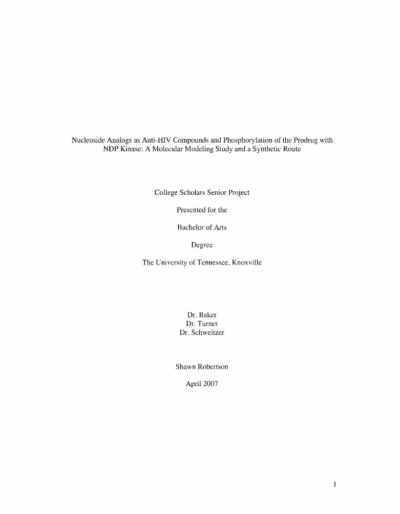
Phe 60 (Figure 2).
12
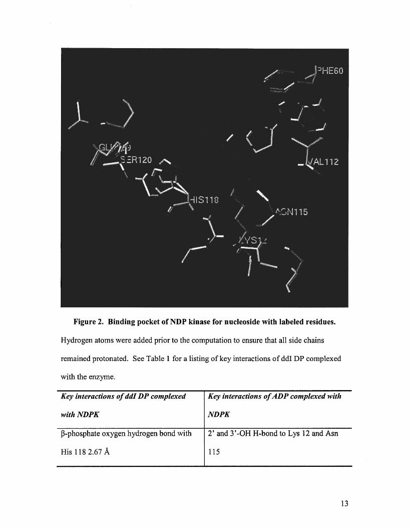
Figure 2. Binding pocket of NDP kinase for nucleoside with labeled residues.
Hydrogen atoms \vere added prior to the computation to ensure that all side chains
remained protonated. See Table 1 for a listing of key interactions of ddI DP complexed
with the enzyme.
Key interactions of ddI DP complexed
withNDPK
~-phosphate oxygen hydrogen bond with
His 118 2.67 A
Key interactions of ADP complexed with
NDPK
2' and 3'-OH H .. bond to Lys 12 and Asn
115
13
Hydrogen bond between Glu 129 and His-Glu-Ser triad (as described earlier)
Ser 120
Lys 12 and Asp 115 hydrogen bond This interaction is not seen
Intramolecular bonding between phosphate No intramolecular bonding between
atoms phosphate
1t stacking between Phe 60 and nitrogenous Val 112 and Phe 60 encompase the base as
base 3.98 A it sits in the binding pocket
Table 1. Key interactions of ddI DP complexed with NDP kinase
Discussion of Modeling
SYBYL predicted a hydrogen bond from the beta phosphate oxygen to the catalytic His
118, which was not seen on the original ADP enzyme complex (Figure 3).
Figure 3. SYBYL predicted H-bonds with bound ddI DP.
14

Intramolecular bonding of the phosphates of ddI was also shown as a possibility after
acquisition using Tripos forcefield. Other important interactions analyzed were the
hydrophobic ones that involved 1C stacking between the nitrogenous bases, Phe60, and
Va1112. In the ADP enzyme complex Val112 and Phe60 incompases the base, and the
distance behveen the center of the base and the center ofPhe 60 is less than 3.5 A. In
contrast, Val112 has no interaction between ddI, and Phe60 is shifted to produce a 3.98 A
1t stacking interaction between the two ring systenls.
Synthesis of 6 .. chloro .. 9 .. (2,3 .. dideoxy .. p-D .. glycero .. pentofuranosyl)purine
The D~glycero.pentofuranose ring system is synthesized from commercially
available L-glutanlic acid. The reaction can be considered to involve a diazotization by
interaction ofN203 (HN02) with the amino group on L-glutamic acid. This leads to the
fonnation of a diazonium ion, a good leaving group. (Scheme 2).
o II
(6H @~ _He R-NH2 ------•• R-N-N=O ------~. R-N-N=O
~ ~ R-N=N-OH
I I H H
tautom. =-===~"', R-N=N-OH
---.... R-N=N (f)
...
Scheme 2. Carbocation intermediate of N .. nitrosation.
A sensible mechanism that would produce (S)-(+)-carboxy .. ,,(-butyrolactone very rapidly
would be one where the ,,(-carboxyl group acts as a lluc1eophile. (Scheme 3).
15

Configuration will be preserved due to the neighboring-group participation effect of the
adjacent a-carboxyl group. Once the cyclic carbonium iOll is fonned, simple
deprotonation would yield the lactone (Scheme 3).
NH2
HO~OH o 0
----,...
Scheme 3. Synthesis of (S)-(+) .. Carboxy-y .. butyrolactone.
BH3, a strong Lewis acid, is used as the reducing reagent for the carboxylic acid
on the above lactone. It is an electron acceptor and can interact with lone pairs of
electrons or nucleophiles. Diborane does not exist as free BI-I3 but rather as the diborane
dimer B2H6 or complexed with a Lewis base. Diborane shows nUlnerous differences in
its reducing power than those portrayed by the alkali metal borohydrides. During the
early 1960s carboxylic acids were normally considered to be relatively resistant to
reducing agents, but this all changed with the introduction and commercial availability of
diboranc, which converts carboxylic acids to the corresponding alcohols very rapidly.2o
It is a very versatile reducing agent because it produces high yields with ease of isolation
of products over a variety of functional groups. Methylsulfide'borane (DMSB) is
advantageous in this case over the use ofborane'THF (BTHF) complex for two reasons.
Firstly DMSB is not as reactive as BTHF due to the stronger coordination ofborane to
Me2S, This assures that DMSB will not over-reduce the ring. Secondly DMSB is very
16

stable and can be produced and used in high concentrations. The first step in the
reduction is the formation of triacylborane. Since the electron deficiency of the boron
atom exerts a powerful demand on the lone pairs of the acyl oxygen, the resonance that
occurs is between the boron atom and the oxygen atom rather than the carbonyl group as
proposed by Brown et al. 20 (Schen1e 4).
Scheme 4. Resonance between boron and oxygen atoms
that the carbonyl groups of the triacylboranes now resemble those of aldehydes or
ketones more closely than those of esters (Scheme 5).20
Scheme 5. Synthesis of (S)-(+)-Hydroxymethyl-y-butyrolactone.
Protection of the hydroxyl groups via sHyl ethers is very important to synthetic
organic chemistry. The general order of stability of the protection groups towards acidic
media are TMS (1) < TES (64) < TBS (20,000) < TIPS (700,000) < TBDPS (5,000,000)
17
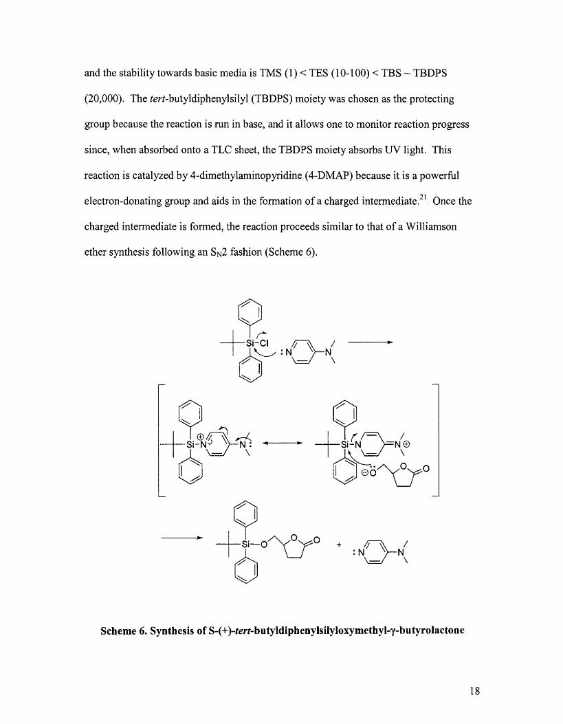
and the stability towards basic media is TMS (1) < TES (10-100) < TBS""" TBDPS
(20,000). The tert-butyldiphenylsilyl (TBDPS) moiety was chosen as the protecting
group because the reaction is run in base, and it allows one to monitor reaction progress
since, \vhen absorbed onto a TLC sheet, the TBDPS moiety absorbs UV light. This
reaction is catalyzed by 4-dimethylaminopyridine (4-DMAP) because it is a powerful
electron-donating group and aids in the formation of a charged intermediate.21. Once the
charged intennediate is formed, the reaction proceeds sitnilar to that of a Williamson
ether synthesis following an SN2 fashion (Scheme 6).
..
+ J-\\ / : N \ N - \
Scheme 6. Synthesis of S-(+)-tert-butyldiphenylsilyloxymethyl-y-butyrolactone
18

During the next stage in the synthesis, the lactone, a cyclic ester, is reduced to a
lactol. Over-reduction is prevented by the hemiacetal functionality that essentially
"masks" the aldehyde. Diisobutylaluminumhydride (DffiAL-H) is the reducing reagent
and used at -78 °C to prevent over-reduction. A proposed mechanism follows in Scheme
7.
n (-+y """v-0yO---AI 2) ° U~H )-
OH-
Scheme 7. Synthesis of 5-0-tert-butyldiphenylsilyl-2,3-dideoxy-n-glycero-
pentofuranose.
During the coupling step of the synthesis, the anomeric hydroxyl group is
converted to a trifluoroacetyl ester. This group is a very good leaving one-much better
than the hydroxyl group. The trifluoroacetyl group is very reactive, and thus the reaction
must take place at -40 °C and under strictly anhydrous conditions. The trifluoroacetyl
ester and 6-CI purine group are safe in basic media, so pyridine is a logical choice as the
catalyst. An oxonium ion is formed, and the coupling reaction can proceed by a
mechanislTI that is somewhere in between an SN 1 and SN2 reaction 15 (Scheme 8).
19

@
-----..... TBDPSIO~
..
CI
N~N lL .. J- ~
N N
• TBDPSIO~
Scheme 8. Synthesis of 6-chloro-9-[5-0-(tert-butyldiphenylsilyl)-2,3-dideoxy-p-D-
glycero-pentofuranosyl]purine.
The final step in the synthesis is the deprotection of the tert-butyldiphenylsilyl
group. This is achieved by tetrabutylammonium fluoride (TBAF) (Scheme 9).
20

Q -too. +SI-F + [R-?F 1 o R-OH ..
CI WhereR;;: ~ .. N
N ~ \\ II ~ I
~ Scheme 9. Synthesis of 6-chloro-9-(2,3-dideoxy-Jl-n-glycero .. pentofuranosyl)purine.
21

NH2
HO~OH o 0
o
HO~O 3
TBDPSiO~OH
5
Experimental
A. General Methods
o
HO~O 2
.. THF 0 °C
TBOPSCI/Pyridine OIBALH 4 .. DMAP 0 PhCH3 ... TBDPSiO~O~O --_____ ..
CH2CI2 U -78°C
.. ): N ~ N
ll" ~ N N TBDPSIO~
K2COy'BTEAC/6-ch loropuri ne
6
CI
TBAFITHF ~~N~ N N
HO~ TEAlTHF IHI-
7
All solvents and reagents used for this synthetic approach were reagent grade.
The solvents were anhydrous and dried as follows: dichloromethane (CH2Ch), pyridine,
toluene, and acetonitrile (CH3CN) were distilled from calcium hydride; tetrahydrofuran
(THF) was dtied over sodium-bel1zophenone ketyl and distilled. All solvents were
prepared fresh and stored in clean, dry glassware in a desiccator prior to use. After
reactions were complete, excess solvents were evaporated using a Bilchi rotary
evaporator and a wann water bath with temperatures no higher than 40 °C. There were
22

some evaporations carried out at very low pressures, and these nlade use of a
high-vacuum rotary evaporator. Reaction progress was monitored by TLC on aluminum
backed plates. Products were separated using column chromatography, which was
achieved using coarse 70-230 Mesh ASTM silica gel. The TLC plate was visualized
under UV light of 254-nm and also by dipping in anisaldehyde-sulfuric acid in ethanol,
followed by a short blast of heat from a heat gun.
B. Preparation of Compounds
Synthesis of (S)-(+)-Carboxy...y-butyrolactone (2)
A suspension ofL-glutamic acid (1, 50 g, 0.34 mol) in concentrated HCL (72 mL) and
H20 (130 mL) was cooled to -5 °C in a round-bottolll flask. To this suspension was
added dropwise over a period of 3 h a solution of sodium nitrite (NaN02, 35 g, 0.50 mol)
in H20 (75 mL). It is very important that the temperature be kept at -5 °C during the
entire addition. The reaction was slowly warmed to room temperature and allowed to stir
for 48 h. The water was evaporated with the use of a high-vacuum rotary evaporation
apparatus and a wann water bath of 50 °C. The product was freeze-dried using a
bench-top lyophilizer which converted the yellowish syrup into a white-yellow solid.
The solid was extracted \vith EtOAc (200 mL), suction filtered, and dried over anhyd
MgS04. After the MgS04 was filtered from the product, the EtOAc was rotary
evaporated and the product dried under high-vacuum, resulting in a yellowish syrup that
was further dried in a drying pistol (CH2Ch reflux) for 48 h to produce the product 2
(30.6 g, 235 mmol, 61.2%) as a pale-yellow solid.
23

Physicochemical data: IH NMR (CDCh, 300 MHz): 02.3-2.73 (m, 4H, H-2 and
H-3), 4.97 (m, 1H, H-4), 9.8 (bs, 1H, H-5), [Lit.22 CD30D, 100 MHz): 1.8-2.3 (m, 4H,
H-2 and H-3), 4.2 (m, 1H, H-4)]
Synthesis of (S)-(+)-Hydroxymethyl-y-butyrolactone (3)
To a solution of (S)-(+)-carboxy-y-butyrolactone (2, 5.0 g, 38 mmol) in anhyd THF (20
mL) at -78°C (dry-ice-acetone bath) was added a solution of2 M borane·methyl sulfide
complex in THF (22 mL, 44.2 mmol) over the period of 1 h under a nitrogen atmosphere.
The temperature of the addition was kept at -78°C. The mixture was stirred for 5 hat -78
°C with constant monitoring of the temperature and cooling when required. The reaction
mixture was quenched by cautiously adding dry MeOH (20 mL) at -78°C, and the
product was co evaporated with methanol three times. After high-vacuum rotary
evaporation, the product was placed on the high-vacuum pump overnight and dried to
produce 3 (3.95 g, 34 mmol, 79%) as a pale-yellow oil.
Physicochemical data: bp 125-135 °Clhigh-vacuum, (Lit.22 131-147 °C/7mm);
IH NMR data (CDCh, 300 MHz): 02.05-2.75 (m, 4H, H-2 and H-3), 3.6-3.95 (m, 3H,
H-5 and H-6), 4.58--4.68 (m, 1H, H-4), [Lit.22 (100 MHz) 2.0-2.8 (m, 4H, H-2 and H-3)
3.6--4.0 (m, 3H, H-5 and H-6), 4.65 (m, IH, H-4)].
Synthesis of (S)-(+)-tert-Butyldiphenylsilyloxymethyl-'Y-butyrolactone (4)
A solution of (S)-(+)-hydroxymethyl-y-butyrolactone (3, 3.9 g, 34 mmol), 4-
dimethylaminopyridine 4-DMAP (0.1 g, 0.8 mmol), and pyridine (2 mL, 25 mmol) was
dissolved in dry CH2Ch (40 mL) at room temperature under anhydrous conditions and
under a nitrogen atmosphere. To this mixture was added tert-butyldiphenylsilyl chloride
TBDPS-CI (10.6 mL, 40.8 mmol) dropwise over the course of 1 h. The reaction mixture
24

was allowed to stir for 24 h at room temperature and was further refluxed for 3 h;
reaction progress \vas monitored by TLC. The reaction mixture was allowed to cool to
room temperature where it was washed with H20 (2 x 25 mL). After the organic layer
was separated it was dried over anhyd MgS04 to remove any residual water, producing a
yellow-brown syrup. Column chromatography, with an eluent consisting of hexane
CH2Ch. followed by an increasing ratio of dichloromethane, was used to purify the syrup.
The desired fractions were collected, and the product was recrystallized from hexane to
yield 4 (8.2 g, 23.12 mnlol, 68%).
Physicochemical data: Rf 0.35 (4:1 CH2Ch-hexane); IH NMR data (CDCh, 300
MHz): 80.98-1.05 (s, 9H, tert-butyl), 2.1-2.75 (m, 4H, H-2 and H-3), 3.63-3.89 (2dd,
2H, H-5), 4.55-4.65 (m, 1H, H-4), 7.35~7.7 (m, 10H, phenyl), [Lit.23 (250 MHz) 8 LO
Ll (s, 9H, tert-butyl), 2.1-2.8 (m, 4H, H-2 and H-3), 3.6-3.9 (2dd, 2H, H-5), 4.5-4.7 (m,
1H, H-4), 7.3-7.8 (m, lOH, phenyl).
Synthesis of 5-0-tert-butyldiphenylsilyl-2,3-dideoxY-D-glycero-pentofuranose
(5)
(S)-(+)-tert-butyldiphenylsilyloxymethyl-y-butyrolactone (4, 5.0 g, 14 mmol) was
dissolved in dry PhCH3 (50 mL) in a dry round-bottom flask with a dry stir bar under a
nitrogen atmosphere. The flask and its contents were cooled to -78°C (dry-ice-acetone
bath) and was added cautiously diisobutylaluminumhydride DIBAL-H (15.2 mL, 1.2 M
PhCH3, 18.4 mmol) over the course of 30 min. The reaction was allowed to stir for 4 h
while the temperature was maintained at -78°C, cooling when necessary. To quench the
reaction, several crystals ofNa2S04'10 H20 were added at -78°C. The cold bath was
removed, and the reaction mixture was allowed to warm to room temperature.
25

Dichloromethane (20 mL) was added, followed by anhyd MgS04 (1 g), and the reaction
mixture was allowed to stir for 30 mins. The mixture was vacuum filtered and the
precipitate was washed with warm 1: 1 CHCh-EtOAc in order to dissolve the aluminum
complex formed and until the filtrate \vas no longer UV active. The solvent was
evaporated, and the product was purified by column chromatography eluting with
hexane, 3:1 hexane-CH2Ch, and 2:1:1 hexane-CH2Cb-EtOAc. After collection of the
desired fractions and after recrystallization from hexane, product 5 (4.12 g, 11.57 mmol,
82.1 %) was realized as white crystals.
Physicochemical data: RfO.6 (2:1:1 hexane-CH2Cb-EtOAc); IH NMR data
(CDCb, 300 MHz): 8 .95-1.05 and 1.5 (s, 9H, tert-butyl), 1.75-2.2 (m, 4H, H-2 and 1-I-
3), 1.7-2.1 and 2.5 (s, I-H), 3.2-3.85 (2dd, 2H, H-5, 4.15-4.4 (m, IH, H-4), 5.35-5.55
(M-1H, H-1), 7.35-7.75 (m, 10H, phenyl), [Lit.23 (250 MHz) 1.0 and 1.5 (s, 9H, tert
butyl), 1.7-2.2 (m, 4H, H-2 and H-3), 2.0 and 2.4-2.5 (s, 1H, -OH), 3,3-3.9 (2dd, 2H, H-
5),4.1-4.4 (m, 1H, H-4), 5.4-5.6 (m, H, H-1), 7.3, 7.8 (m, lOR, phenyl).
Synthesis of 6 .. Chloro-9-[5-0-(tert-butyldiphenylsilyl)-2,3-dideoxy-p .. D-glycerQ
pentofuranosyl]purine (6)
5-0-tert-butyldiphenylsilyl-2,3-dideoxY-D-glycero-pento furanose (5, 0.115 g,
0.322 mmol) was massed into a dry round-bottom flask (flask 1) with a stir bar and
placed on the pump overnight to insure complete dryness. In a separate flask (flask 2)
anhyd K2C03 (0.014 g, 0.10 mmol), benzyltriethylammonium chloride (BTEAC) (0.06 g,
0.26 mmol) and 6-chloropurine (0.06 g, 0.39 mmol) were also placed on the pump
overnight. Freshly distilled THF (10 mL) was added to flask 1 and cooled to -78°C,
followed by addition oftrifluoroacetic anhydride (TF AA), (0.06 mL, 0.42 mmol) and
26

pyridine (0.03 mL, 0.4 mmol). The reaction mixture was warmed to .. 40°C to .. 20 °C and
allowed to stir for 2 h, cooling when necessary. Anhyd CH3CN (10 mL) was added to
flask 2, and the mixture was cooled to 0 °C using an ice bath. The cold bath was removed
fronl flask 1, and with the aid of a cannula, the reaction mixture was transferred from
flask 1 to flask 2. The resulting mixture was slowly wanned to room temperature where
it was stirred for an additional 10 h. After evaporation of the solvent~ a brown
sludge/syrup "vas left in the flask. After addition ofCH2Ch (25 roL) and stirring for 10
min, vacuum filtration proceeded by high vacuum rotary evaporation, produced a brown
residue. Silica gel chromatography was used to purify the residue; a concentration
graded eluent was used. Initially 95% petroleum ether to 5% EtOAc was used as the
eluent. The concentration of EtOAc "vas increased by 5% every 100 mL of eluent until
the concentration reached 65% pet. ether to 35% EtOAc. Recrystallization from hexane
afforded the product 6 (0.136 g, 0.2763 Inmol) from which the ~-anomer was separated to
yield 0.09 g, 0.182 mmol, 56.7%.
Physicochemical data: Rf 0.52 (1: 1 hexane-EtOAc); IH NMR data (CDCh, 300
MHz): 8 1.05-1.1 (5, 9H, tert-butyl), 2.05-2.3 (m, 2H, H .. 3'), 2.53-2.63 (m, 2H, H .. 2'),
3.7-4.0 (2dd, 2H, H'5), 4.25-4.35 (m, 1H, H .. 8), 6.35-6.4 (t, IH, H.,l '), 7.32-7.68 (In,
lOR, phenyl), 8.48 (s, IH, H-8), 8.7 (5, 1H, R-2), [Lit.23,24 1.00-1.1 (8, 9H, tert-butyl),
2.05-2.25 (m, 2H, H-3'), 2.5-2.65 (m, 2H, H .. 2'), 3.7-4.0 (2dd, 2H, H-5'), 4.25-4.4 (m,
1H, H-4'), 6.35-6.42 (t, IH, H-l '), 7.3-7.70 (mt 10H, phenyl), 8.45 (8, 1H, H-8), 8.7 (5,
IH! H-2)]
27

Synthesis of 6 .. Chloro-9-(2,3 .. dideoxy .. p-D-glycero .. pentofuranosyl)purine (7)
6-Chloro-9-[5-0-(tert-butyldiphenylsi1yl)-2,3-dideoxy-~ .. D-glycero
pentofuranosyl]purine (6, 0.2 g, 0.4 mmol) was dissolved in anhyd THF (10 mL), and
tetrabutylammonium fluoride TBAF (0,9 mL, 1M THF, 0.89mmol) was added, Next was
added triethylamine (TEA, 0.06 g, 0.55 mmol) and HOAc (0.03 g, 0.5mmol) in THF (5
mL) dropwise. This mixture was stirred for 1 h, the solvent was evaporated, and the
product was purified on silica gel. The eluent consisted of58:1 CHCh-MeOH.
Collection and evaporation of the desired fractions yielded (8, 0.09 g , 88%) of a white
crystalline product after recrystallization from hexane.
Physicochemical data: Rr 0.62 (9: 1 CHzCh~MeOH); IH NMR data (CDCI3, 300
MHz) 2.15-2.75 (m, 4H, H-2' and H-3'), 3.68-4.08 (2dd, 2H, H-5'), 4.3-4.45 (m, 1H, H-
4'), 6.28 (t, IH), 8.46 (s, 1 H), 8.75 (s, IH), [Lit.24 1.95-2.8 (m, 4H), 3.88 (m, 2H), 4.38
(m, 1H), 6.31 (t, IH), 8.5 (8, IH), 8.74 (s, IH)]
28

List of References
1. Thomas C. Merigan, J.; Bartlett, J. G.; Bolognesi, D. The Textbook of AIDS Medicine. 2 ed.; Williams and Wilkins: Baltimore, 1999. 2. Sharma, P. L.; Nurpeisov, V.; Hemandez~Santiago, B.; Beltran, T.; Schinazi, R, F. Nucleoside inhibitors of human inlmunodeficiency virus type 1 reverse transcriptase. Curro Top. Med. Chem. 2004,4, 895·919. 3. Hutter, M. C.; Helms, V. The mechanism of phosphorylation of natural nucleosides and anti-HIV analogues by nucleoside diphosphate kinase is independent of their sugar substituents. Chem. Bio. Chem. 2002, 3, 643-651. 4. Schneider, B.; Sarfati, R.; Deville-Bonne, D.; Veron, M. Role of nucleoside diphosphate kinase in the activation of anti-HIV nucleoside analogs. J. Bioenerg, Biomembr. 2000,32, 317-324. 5. Lascu, I.; Gonin, P. The catalytic mechanism of nucleoside diphosphate kinases. J. Bioenerg, Biomembr. 2000, 32, 237-46. 6. Stein, D. S.; Moore, K. H. P. Phosphorylation of nucleoside analog antiretrovirals: a review for clinicians. Pharmacotherapy 2001, 21, 11-34. 7. Gao, W. Y.; Shirasaka, T.; Johns, D. G.; Broder, S.; Mitsuya, H. Differential phosphorylation and azidothymidine, dideoxycytidine, and dideoxyinosine in resting and activated peripheral blood mononuclear cells. J. Clin. Invest 1993, 91, 2326~33. 8. Nave, J. F.; Eschbach, A.; Wolff-Kugel, D.; Halazy, S.; Balzarini, J. Enzymatic phosphorylation and pyrophosphorylation of 2',3 '-dideoxyadenosine-5'-monophosphate, a key metabolite in the pathway for activation of the anti-HIV (human immunodeficiency virus) agent 2',3'-dideoxyinosine. Biochem. Pharmacol. 1994,48, 1105-12. 9. Janin, J.; Dutnas, C.; Morera, S.; Xu, Y.; Meyer, P.; Chiadmi, M.; Cherfils, J. Three-dimensional stnLcture of nucleoside diphosphate kinase. J. Bioenerg, Biomembr. 2000,32,215-225. 10. Kreimeyer, A.; Schneider, B.; Sarfati, R.; Faraj, A.; Somlnadossi, J. P.; Veron, M.; Deville-Bonne, D. NDP kinase reactivity towards 3TC nucleotides. Antivir. Res. 2001,50,147-156. 11. Bourdais, J.; Biondi, R.; Sarfati, S.; Guerreiro, C.; Lascu, I.; Janin, J.; Veron, M. Cellular phosphorylation of anti-HIV nuc1eosides. Role of nucleoside diphosphate kinase. J. BioI. Chem. 1996,271, 7887-90. 12. Anderson, B. D.; 1vlorgan, M. E.; Singhal, D. Enhanced oral bioavailability of DDI after administration of 6-CI-ddP, an adenosine deaminase-activated prodrug, to chronically catheterized rats. Pharma. Res. 1995, 12, 1126-33. 13. Price, R. W.; Brew, B. J. The AIDS dementia complex. J. Infect. Dis. 1988, 158, 1079-83.
29

14. Anderson, B. D.; Hoesterey, B. L.; Baker, D. C.; Galinsky, R. E. Uptake kinetics of2',3'-dideoxyinosine into brain and cerebrospinal fluid of rats: intravenous infusion studies. J. Pharmacal. Exper. Therapeuti. 1990,253, 113-18. 15. Azizbigloo, F. Synthesis of Sterically Hindered 5'Esters if 6-Chloro-9-(2,3-dideoxy-beta-D-Glycero-pentofuranosyl)purine as Anti-HIV Agents. The University of Tennessee, Knoxville, 1997. 16. Morgan, M. E.; Chi, S. C.; Murakami, K.; Mitsuya, H.; Anderson, B. D. Central nervous system targeting of2',3'-dideoxyinosine via adenosine deaminase-activated 6-halo-dideoxypurine prodrugs. Antimicrob. Agents. Chemother. 1992,36, 2156-65. 17. Cervoni, L.; Lascu, I.; Xu, Y.; Gonin, P.; Morr, M.; Merouani, M.; Janin, J.; Giartosio, A. Binding of nucleotides to nucleoside diphosphate kinase: A calorimetric study. Biochemistry 2001,40, 4583 .. 4589. 18. Chen, Y.; Gallois-Montbrun, S.; Schneider, B.; Veron, M.; Morera, S.; Deville-Bonne, D.; Janin, J. Nucleotide Binding to Nucleoside Diphosphate Kinases: X-ray Structure of Human NDPK-A in Complex with ADP and Comparison to Protein Kinases. J. Mol. BioI. 2003,332, 915-926. 19. Austin, A. T.; Howard, J. Reaction ofHN02 with glutamine and glutamic acid. J. Chern. Soc. 1961, 3593-3603. 20. Brown, H. C.; Rao, B. C. S. Hydroboration. III. The reduction of organic compounds by diborane, an acid-type reducing agent. J. Am. Chem. Soc. 1960, 82, 681-6. 21. Chaudhary, S. K.; Hernandez, O. 4-Dimethylaminopyridine: an efficient and selective catalyst for the silylation of alcohols. Tetrahedron Lett. 1979, 99-102. 22. Uzi Ravid, R. S., Leverett Smith. Synthesis of the Enantiomers of 4-substituted Gamma Lactones with Known Absolute Configuration. Tetrahedrom 1977,34, 1448-1452. 23. Azizbigloo, F. Synthesis of Sterically Hindered 5'-Esters of 6-Chloro-9-(2,3-Dideoxy-beta-D-Glycero-Pentofuranosyl Purine as Anti-HIV Agents. Tennessee, Knoxville, 1997. 24. Chu, C. K.; Ullas, G.; Lak, J. Synthesis and Structure-Activity Relationships of 6-Substituted 2',3'-Dideoxypurine Nuc1eosides as Potential Anti-Human Immunodeficiency Virus Agents. J. Med. Chem. 1990,33, 1553 .. 1561.
APPENDIX
1 H NMR Spectra
30

r· [ f
10 ::l
~
~
~
}§ I" .s ~ -~ ...... =' ..Q
~ ::: I
~
~ ~ l><
::l e R -. ~
I ~
+ '-' I ~ r:Jj '-' ~ 0
~ :z ~ }[5J p.
V'.1 ~
N
.::: ~ 8 M
~ -u Q
r 8 ~
~ ""
::x::: '"
I ] ij
.....~ ." ".. ..~.. ..... ' .................... "'"""'.......",.,.... ... ..--
I ~l
! I
\.~)PJ ...,..," '.I~'" r,,"-y l..,--J
L~~ trlY ~
L..,--J y l..,--J L,--.J , ' Y ~
~ o.s/i 1.2 .~ 0$-] (I-il em
~l i, i • i I ! ' 1 \ I' \'--r ---r-T--'-'-l---r 1 1 -'-1- ' '--1"'",--< , 'I ' '--"-1---'--'--'--' 1 • , ', \ 1 &.5 I ,D 7.5 71) 6.5 6,0 f.5 s.o 4.5 ~.o l .S 3/J 1.5 loU \ :5 110 il.5
IH NMR(CDCI3, 300 MHz) Spectrum of5-O-tert-butyldiphenylsilyl-2,3-dideoxy-D-glycero~pentofumoose (5)
34
I~ y v Y L,J y
I 0.9110.9 I I 4.1 II 6.4 I CID y y y
@J [IO]J!:o] y L,-J
QIJ LUI y y ~F'.(l-- 'j
r I T ,-r-TT J T I I I I I I 1 I I ' 1)' I ! { I I l'-T •. > 7. ~ ~.o 6.~ 6.0 S.5 ~.5 J.5 3,0 loll U ~.5
IH NMR (CDCh, 300 MHz) Spectrum of6-Chloro-9-[5-0-(tert-butyldiphenylsilyl)-2,3-dideoxy-f}-D-glycero-pentofuranosyIJpurine (6)
35

9.0 8.~ 8J} 7.5 7.0 65 6.0 ~.5 5.0 4,S 4.0 J.5 3.Q 2.5 l,O L5 to
IH NMR (CDCh, 300 MHz) Spectrum of Synthesis of 6-Chloro-9-(2,3-dideoxy-p-D-glycero-pentofuranosyl)purine (7)
36