nucleic acid sensing by the immune system: roles for the receptor
TRANSCRIPT

University of Massachusetts Medical SchooleScholarship@UMMS
GSBS Dissertations and Theses Graduate School of Biomedical Sciences
2011-07-14
Nucleic Acid Sensing by the Immune System:Roles For the Receptor For Advanced GlycationEnd Products (RAGE) and Intracellular ReceptorProteins: A DissertationCherilyn M. SiroisUniversity of Massachusetts Medical School
Follow this and additional works at: https://escholarship.umassmed.edu/gsbs_diss
Part of the Cells Commons, Hemic and Immune Systems Commons, Immune System DiseasesCommons, Immunology and Infectious Disease Commons, Nucleic Acids, Nucleotides, andNucleosides Commons, Skin and Connective Tissue Diseases Commons, and the TherapeuticsCommons
This material is brought to you by eScholarship@UMMS. It has been accepted for inclusion in GSBS Dissertations and Theses by an authorizedadministrator of eScholarship@UMMS. For more information, please contact [email protected].
Repository CitationSirois, CM. Nucleic Acid Sensing by the Immune System: Roles For the Receptor For Advanced Glycation End Products (RAGE) andIntracellular Receptor Proteins: A Dissertation. (2011). University of Massachusetts Medical School. GSBS Dissertations and Theses.Paper 551. DOI: 10.13028/91rf-3167. https://escholarship.umassmed.edu/gsbs_diss/551

NUCLEIC ACID SENSING BY THE IMMUNE SYSTEM:
ROLES FOR THE RECEPTOR FOR ADVANCED GLYCATION END
PRODUCTS (RAGE) AND INTRACELLULAR
RECEPTOR PROTEINS
A Dissertation Presented By
CHERILYN M. SIROIS
Submitted to the Faculty of the
University of Massachusetts Graduate School of Biomedical Sciences,
Worcester, Massachusetts, United States
in partial fulfillment of the requirements for the degree of
DOCTOR OF PHILOSOPHY
14 JULY, 2011
Interdisciplinary Graduate Program

NUCLEIC ACID SENSING BY THE IMMUNE SYSTEM: ROLES FOR THE RECEPTOR FOR ADVANCED GLYCATION END PRODUCTS (RAGE) AND INTRACELLULAR
RECEPTOR PROTEINSA Dissertation Presented By
CHERILYN M. SIROIS
The signatures of the Dissertation Defense Committee signifycompletion and approval as to style and content of the dissertation.
___________________________________________________Eicke Latz, M.D., Ph.D., Thesis Advisor
___________________________________________________Katherine Fitzgerald, Ph.D., Member of Committee
___________________________________________________Elizabeth Luna, Ph.D., Member of Committee
___________________________________________________Hardy Kornfeld, M.D., Member of Committee
___________________________________________________Ian Rifkin, M.D., Ph.D., Member of Committee
The signature of the Chair of the Committee signifies that the written dissertation meets the requirements of the Dissertation Defense Committee.
___________________________________________________Egil Lien, Ph.D., Chair of Committee
The signature of the Dean of the Graduate School of Biomedical Sciences signifies that the student has met all graduation requirements of the school.
___________________________________________________Anthony Carruthers, Ph.D.,
Dean of the Graduate School of Biomedical Sciences
Interdisciplinary Graduate Program14 July, 2011

Desde Quitohasta el fin de la tierrayo caminé tu piel,en ella me interné,me la aprendí al dedillo.En ella caí en éxtasis,de nuevo volvi en mí,me di las vueltas,apaciguado a veces;a veces desbocado,lúcido hasta más no poder,sonámbulo, enviciado,ensimismado, entimismado,con el tacto borracho,viviendo en la candela.Un día cuando salía el solpor donde nunca,con estos ojosque han de hacerse tierrayo vi como en tu pielpastaban las palabras.
-Euler Granda“Entre la gente y el humo de los carros” de la colecciónRelincha el sol, 1997
From Quitoto the end of the earthI walked your skin,I wrapped myself in it,I learned it down to the last finger.I fell into the ecstasy of itcame to myself again,made the rounds,sometimes appeased;sometimes out of control,lucid to the max,a sleepwalker, an addict,stuck inside myself, stuck with you,with a drunken touch,living in the fire.One dayas the sun rosesomewhere new,with these eyesthat will surely turn to dustI saw that on your skin,words were grazing.
-Euler Granda, Ecuadorian poet(Translation is mine. -CMS)
iv

We know very little, and yet it is astonishing that we know so much, and still more astonishing that so little knowledge can give us so much power.
-Bertrand Russell in his book, ABC of Relativity
in the right light, study becomes insight
-Rage Against the Machine"Take the Power Back"
from their self-titled album
v

DEDICATION
For my mother.
Because it was she who repeated, “I really think you’d like biology,”
when I was convinced that I should go into international relations. Perhaps we were both right.
vi

ACKNOWLEDGEMENTS
It has been said that it takes a whole village to raise a child. In my case, I feel that it has taken a worldwide community to move me toward this doctorate. The appreciation I show here is a mere gesture, and almost certainly an inadequate one, to recognize the many gifts of time and effort I have received while working toward this dissertation.I am grateful to my Thesis Research Advisory Committee: Drs. Egil Lien, Kate Fitzgerald, Elizabeth Luna and Hardy Kornfeld, whose guidance has played a large part in the completion of this work. I also extend many thanks to Dr. Ian Rifkin for reviewing my dissertation. Perhaps the greatest determinant of a PhD student’s success and happiness is his or her advisor, and I have had the best one imaginable. Eicke Latz has shared his vision, his inspiration, and his time with an unmatchable intensity and generosity. It is amazing to recall all the terrain, both intellectual and terrestrial, that we have covered in these six years. A tireless scientist and an intrepid professional, Eicke is also a truly kindhearted person. My apprenticeship to him has been an honor and a privilege. I have been fortunate to belong to the diverse and worldly community of colleagues and friends who constitute the Division of Infectious Diseases and Immunology, particularly those of the Latz, Golenbock and Fitzgerald laboratories. Many brilliant postdoctoral fellows have given me a kind hand through the years: Alberto Visintin (my first ‘surrogate PI’), Simon Rothenfusser, Martina Severa, Marie Charrell-Dennis, Yolanda Corbett, Gabor Horvath, Veit Hornung, Franz Bauernfeind, Shruti Sharma, and (a relative newcomer I don’t know how I ever did without) Susan Carpenter. I am also grateful to Brian Monks, Susann Paul and MJ Lindem, who were always willing to help me out, though it wasn’t in their job description to do so. There are certain beloved allies it would be an injustice to call simply ‘colleagues’ or ‘friends,’ for they are both, in varying measure, as the circumstances dictate. Annett Halle, Cathrine Knetter and Kamalpreet Nagpal, in different eras and in their own unique ways, have gotten me through the daily challenges and joys of laboratory work. Friends equally at ease inside and out of the lab, they are the ones with whom I could share anything and always be sure of complicity. Mariane Bandeira de Melo, the woman who can do it all and never break a sweat; Luiz Godoy, a friend who needs no cultivating; Pia Kasperkovitz, the astute observer; Therese Vallerskog, a master of self-reinvention; and Ryan Nistler, a roommate long beyond his tenure, have all been treasured sources of inspiration and friendship. And many thanks to Kristen Halmen and Lisa Waggoner for sharing the adventure that is lab management, as well as daily lunch.
vii

I am grateful to course coordinators at UMass who have given me an opportunity to teach during my time as a PhD student: Mary Munson, Kate Fitzgerald, Phil Zamore and Rich Konz. For what is the point of acquiring all of this knowledge, if we are unable to share it? I am also indebted to teachers from previous eras of my life: Elizabeth McCain, a tiny lady, dedicated mother and flexible teacher who was not afraid to apply a screwdriver to an electron microscope; Luis Antonio Aguilar Monsalve, who treated teaching as a matter of utmost style and grace; and Manuel Baldeón, who first introduced me to the fascinating field of Toll-like receptors and taught me to read a flow cytometry dot plot, and whose example reminds me that dedication and humility are the best ways to achieve great things. Thank you to the team in the UMass FACS Core for teaching the most useful course I took as a PhD student, and for serving as reassurance that it’s ok to get a bit excited about lasers. Special thanks to Rich and to Ted Giehl, my partner in confocal microscopy maintenance, for their faith in my abilities. Scientific work at its best is collaborative, and I am thankful for the time and efforts of the many co-authors noted throughout this dissertation. Wonderful collaborators at MedImmune, LLC, deserve special mention: Alison Humbles and Allison Miller, simply the nicest pair of Alisons one could ever hope to work with; Jane Tian, who was always engaged and ready to share; and Tony Coyle, who believed in and enabled the RAGE project from its inception to the present. It is impossible to imagine the completion of this work without their involvement. Lastly, I wish to acknowledge the support from family and friends that has encompassed this whole process. I give my most sincere appreciation to: my parents, who allowed me to be a black sheep that left the herd and have seldom questioned my decisions; my mother, whose attitude toward her profession I am proud to find myself emulating; my grandmother, Mémère, whose kindred spirit is a source of comfort; my brother, for leading a ‘real adult life’ and taking the pressure off of me to do so; my extended family, who offer their support, even if they’re not precisely sure what it is I do; dear friends, notably Álvaro and Siena but also many others, for reminding me that there is a vibrant world outside the lab; and the inexplicable and indefensible Alfredo Galarza, who is neither family nor friend, but is as indispensable to me as both. To all those mentioned here, and to those who read this work, I say,vielen Dank, tusen takk, go raibh mile maith agat, baie dankie, tack så mycket, grazie mille, merci beaucoup, muito obrigada, köszönöm szépen, dhanyawaad, dank je wel, xie xie, gracias de todo corazón, and thank you very much!
viii

ABSTRACT
As humans, we inhabit an environment shared with many microorganisms, some
of which are harmless or beneficial, and others which represent a threat to our
health. A complex network of organs, cells and their protein products form our
bodies’ immune system, tasked with detecting these potentially harmful agents
and eliminating them. This same system also serves to detect changes in the
healthy balance of normal functions in the body, and for repairing tissue damage
caused by injury. Immune recognition of nucleic acids, DNA and RNA, is one
way that the body detects invading pathogens and initiates tissue repair. A
number of specialized receptor proteins have evolved to distinguish nucleic acids
that represent “threats” from those involved in normal physiology. These proteins
include members of the Toll-like receptor family and diverse types of cytosolic
proteins, all of which reside within the confines of the cell. Few proteins on the
cell surface have been clearly characterized to interact with nucleic acids in the
extracellular environment. In this dissertation, I present collaborative work that
identifies the receptor for advanced glycation end products (RAGE) as a cell
surface receptor for nucleic acids and positions it as an important modulator of
immune responses. Molecular dimers of RAGE interact with the sugar-
phosphate backbones of nucleic acid ligands, allowing this receptor to recognize
a variety of DNA and RNA molecules regardless of their nucleotide sequence.
Expression of RAGE on cells promotes uptake of DNA and enhances
ix

subsequent responses that are dependent on the nucleic acid sensor Toll-like
receptor 9. When mice deficient in RAGE are exposed to DNA in the lung, the
predominant site of RAGE expression, they do not mount a typical early
inflammatory response, suggesting that RAGE is important in generating immune
responses to DNA in mammalian organisms. Further evidence suggests that
RAGE interacts preferentially with multimolecular complexes that contain nucleic
acids, and that these complexes may induce clustering of receptor dimers into
larger multimeric structures. Taken together, the data reported here identify
RAGE as an important cell surface receptor protein for nucleic acids, which is
capable of modulating the intensity of immune responses to DNA and RNA.
Understanding of and intervention in this recognition pathway hold therapeutic
promise for diseases characterized by excessive responses to self nucleic acids,
such as systemic lupus erythematosus, and for the pathology caused by chronic
inflammatory responses to self and foreign nucleic acids.
x

Table of Contents
..................................................................................................List of Figures xiii
...................................................................................................List of Tables xiv
.....................................................................................Copyright Information xv
.......................................................................................List of Abbreviations xvi
................................................................................CHAPTER I: Introduction 1..................................Molecular patterns initiate innate immune responses 3
...............................................Immunostimulatory nucleic acids as molecular patterns 4.........................................................................................Toll-like receptors 6
.......................................................................................................................Discovery 6............................................................................................................Protein structure 8...........................................................................................................Endosomal TLRs 8
........................................................................Endosomal localization in TLR function 9..................................Biology of endosomal TLRs: insights from autoimmune disease 13
.......................................................................................................................Signaling 14.....................................................................Cytosolic nucleic acid sensors 17
..................................................................................................................RNA sensors 17
..................................................................................................................DNA sensors 18.........................Accessory molecules for intracellular nucleic acid sensors 21
........................................................Proteins that promote proper receptor localization 22.......................................................Cofactors that promote ligand-receptor interaction 24
...........................................................................................................RAGE 26..........................................................................................................Gene and protein 27
................................................................................................................RAGE ligands 30.......................................................................................................................Signaling 32
...................................Expression in tissues and cells: complexity and ties to disease 37..................................................Thesis rationale, objectives, and summary 38
...................................................................................PREFACE to Chapter II 41
CHAPTER II: RAGE recognizes nucleic acids and promotes inflammatory ...........................................................................................responses to DNA 42
........................................................................................................Abstract 42..................................................................................................Introduction 43
.........................................................................................................Results 46RAGE concentrates DNA on cells and interacts with DNA in a sequence-independent
..........................................................................................................................manner. 46.........................RAGE binds DNA through electrostatic attractions with the backbone 48
xi

......................................................................Dimerization of the RAGE V-C1 domains 58.........................................DNA can induce formation of higher-order RAGE oligomers 62
.................RAGE expression promotes DNA uptake by cells via the endosomal route 65....................RAGE increases TLR9-dependent responses to suboptimal DNA stimuli 69
........RAGE-deficient mice have impaired inflammatory responses to DNA in the lung 72....................................................................................................Discussion 77
................................................................................Materials and Methods 82........................................................................Additional Detailed Methods 91
..................................................................................PREFACE to Chapter III 95
.............CHAPTER III: Nucleic acid complexes are optimal RAGE ligands 96........................................................................................................Abstract 96
..................................................................................................Introduction 97.........................................................................................................Results 100
..............................................DNA-containing immune complexes interact with RAGE 100unimolecular CpG-B bound less favorably, suggests that complexes interact preferentially
...................................................................................with RAGE in this binding assay. 102.........................................Complexes of HMGB1 and DNA are optimal RAGE ligands 102
................................................Parylation of HMGB1 may contribute to RAGE binding 105....................................................................................................Discussion 108
................................................................................Materials and Methods 110
...................................................................................Chapter IV: Discussion 113........................................................The panorama of nucleic acid sensing 114
..................................................RAGE, nucleic acids, and inflammasomes 115...........................DNA, PARP, HMGB1 and RAGE: regulatory integration? 117
.................................................................RAGE: a multifaceted modulator 121..........................................................................................Future directions 126
.......................................................................................................References 131
xii

List of Figures
Figure 1.1 Toll-like receptors, their ligands, and simplified signaling pathways. 15
Figure 1.2 Cytosolic nucleic acid receptors and simplified signaling pathways. 19
............................................................Figure 1.3 RAGE isoforms and ligands. 29
.........................................Figure 1.4 Signaling pathways activated by RAGE. 34
.....Figure 2.1 RAGE binds directly to nucleic acid ligands on the cell surface. 47
Figure 2.2 Additional binding data: RAGE binds directly to oligodeoxy-.....................................................nucleotides of distinct sequences and sizes. 49
Figure 2.3a-b DNA interacts with a region of positive charges at the RAGE dimer ............................................................................................................interface. 50
Figure 2.3c-d DNA interacts with a region of positive charges at the RAGE dimer ............................................................................................................interface. 52
...........Figure 2.4 Additional data: RAGE binds dsDNA at the dimer interface. 56
Figure 2.5 Constitutive RAGE dimers form higher order oligomers in the ....................................................................presence of complex DNA ligands. 61
...............Figure 2.6 Sequence alignments of selected RAGE V-C1 domains. 64
............................................Figure 2.7 RAGE promotes cellular DNA uptake. 67
Figure 2.8 RAGE increases TLR9-dependent NF-κB activation in response to ......................................................................................................DNA ligands. 71
Figure 2.9a-b RAGE mediates DNA-induced pulmonary inflammation in vivo. 75
...Figure 2.9c RAGE mediates DNA-induced pulmonary inflammation in vivo. 76
................................................Figure 2.10 pRP retroviral transduction vector. 84
........................................Figure 3.1 DNA:antibody complexes bind to RAGE. 101
Figure 3.2 HMGB1:DNA complexes interact with RAGE and induce type-I ..........................................................................................................interferon. 103
....................................................................Figure 3.3 PAR is a RAGE ligand. 107
Figure 4.1 Model of the hypothetical interregulatory network formed by RAGE, ............................................................................................HMGB1 and PARP. 120
xiii

List of Tables
.......Table 2.1 X-ray crystallography data collection and refinement statistics. 54
..........................................Table 2.2 Oligonucleotide sequences and sources. 83
xiv

Copyright Information
Material subject to copyright by others is listed in the Prefaces to Chapters II and
III. The poem by Euler Granda on page iv is reprinted here with kind permission
from the management of Editorial Libresa, Quito, Ecuador. All other material
should be considered the intellectual property of Cherilyn M. Sirois, unless
attributed to others by referencing of the source.
xv

List of Abbreviations
AGE advanced glycation end products AIM2 absent in melanoma 2Akt V-AKT murine thymoma viral oncogene homologAP-1 activator protein 1 ATPase adenosine triphosphataseBCR B cell antigen receptorBS3 bis(sulfosuccinimidyl) suberateCARD caspase activation and recruitment domain CARDIF CARD adaptor inducing interferon beta CD11b cluster of differentiation 11b CD14 monocyte differentiation antigen CD14 Cdc42 cell division cycle 42CFP cyan fluorescent protein CpG cytosine-phosphate-guanine dinucleotide DAI DNA-dependent activator of interferon regulatory factorsDAMP damage-associated molecular pattern DExD/H-box aspartate-glutamate-any amino acid-aspartate/histidine-boxDHX aspartate-glutamate-any amino acid-aspartate/histidine-boxDNA deoxyribonucleic acid ds preceding “DNA” or “RNA,” indicates “double stranded”ER endoplasmic reticulum ERK extracellular signal-regulated kinaseFRET Förster (or fluorescence) resonance energy transfergp96 stress-inducible tumor rejection antigen gp96 GRP94 glucose-regulated protein, 94-kDGTPase guanosine triphosphataseHEK293 human embryonic kidney 293 cell line HIN hemopoietic IFN-inducible nuclear proteinHLA human leukocyte antigen HMGB1 high mobility group box 1i.e. id est [Latin: that is (to say)]IFI16 interferon-gamma-inducible protein 16IFN interferonIPS-1 interferon beta promoter stimulator 1IRAK interleukin 1 receptor-associated kinaseIRF interferon regulatory factor IκB I kappa B inhibitor of NF-κBJAK Janus kinaseLGP2 laboratory of genetics and physiology 2 LPS lipopolysaccharide
xvi

LRR leucine-rich repeatLRRfip1 leucine-rich repeat in Flightless-I interacting protein-1MAL MyD88 adaptor like MAMP microbial-associated molecular patternMAPK mitogen-activated protein kinase MAVS mitochondrial antiviral signalingMDA-5 melanoma differentiation-associated gene-5 MEK1 MAPK ERK kinase 1 MHC major histocompatibility complexMyD88 myeloid differentiation primary response gene 88NF-κB nuclear factor kappa B NLRP3 NOD-like receptor family, pyrin domain containing 3ODN oligodeoxynucleotide ORN oligoribonucleotidePAMP pathogen-associated molecular patternPAR poly-adenosine triphosphate ribose parylation poly-adenosine triphosphate ribosylationpDC plasmacytoid dendritic cellPEI polyethyleneiminiepH potential hydrogen PO phosphodiesterPoly I:C polyriboinosinic:polyribocytidilic acidPS phosphorothioateRab Ras-associated protein Rac-1 Ras-related C3 botulinum toxin substrate 1RAGE receptor for advanced glycation end products Ras Harvey rat sarcoma viral oncogene homologRho Ras homolog gene family memberRIG-I retinoic acid-inducible gene IRIP1 receptor interacting protein 1 RLH RIG-I-like helicaseRNA ribonucleic acid RNA pol III ribonucleic acid polymerase IIISLE systemic lupus erythematosisSrc V-SRC avian sarcoma viral oncogeness preceding “DNA” or “RNA,” indicates “single stranded”STAT signal transducer and activator of transcription STING stimulator of interferon genes TBK1 Tank-binding kinase 1TICAM1 TIR domain-containing adaptor molecule 1TICAM2 TIR domain-containing adaptor molecule 1TIR Toll/interleukin-1 receptorTIRAP TIR domain-containing adaptor protein
xvii

TLR Toll-like receptorTRAF3 TNF receptor-associated factor 3TRAF6 TNF receptor-associated factor 6TRAM TIR-containing TRIF-related adaptor moleculeTRIF TIR domain-containing adaptor inducing interferon-betaUNC93B1 homolog B1 of C. elegans UNC93VISA virus-induced signaling adaptorYFP yellow fluorescent protein
xviii

CHAPTER I: Introduction
1

The mammalian immune system is a complex network of specialized
organs and cells that perform surveillance of the body’s physical integrity and
enact mechanisms to eliminate agents that pose a threat to normal physiological
functions. The ability to effectively distinguish “self” from “foreign” and control
interactions between the two is what allows us, as humans, to persist for long
periods in an environment full of microbes and parasites that wish to benefit from
the resources afforded by our bodies. At the same time, we derive great benefit
from a large number of commensal microorganisms whose presence, while not
strictly “self,” does not pose a direct threat to our health and integrity. Given the
large number of microorganisms and parasites in our environment, as well as the
variety of threats and benefits these organisms pose, the immune system
requires sophisticated mechanisms for distinguishing and controlling the
interactions of these “foreign” agents with our “self” environment. In recent
years, it has become clear that the same immune system that recognizes foreign
agents also plays a role in detecting self-derived signals that indicate
homeostatic perturbations. Thus, the ability to discern potential danger from
normalcy is both complex and essential for survival. Internal errors in the
functioning of the immune system can have serious consequences: exaggerated
responses to harmless agents, as seen in allergy and hypersensitivities, the
mistaken recognition of self as foreign, leading to self-directed attacks known as
autoimmunity, and collateral damage to self tissues, termed pathology.
Understanding the ways the immune system functions at the molecular level not
2

only provides insight into the fascinating way that our bodies interact with their
environment, but also helps us to devise strategies to correct immune “errors”
and create more effective therapies for diseases.
Molecular patterns initiate innate immune responses
As might be expected for a system that must discriminate between a large variety
of potential activators, responses of the immune system occur in several
interconnected phases. In the broadest of terms, these phases are grouped into
those of the early, “innate” immune response and a subsequent “adaptive”
response. Both early and late responses are mediated by specialized cells and
the effector molecules they produce, but the specificity of these cells and
associated proteins differ in important ways. The innate immune response is
characterized predominantly by the recognition of conserved “molecular patterns”
that are common among certain classes of pathogens (so-called pathogen-
associated molecular patterns, PAMPs), microbes in general (microbial-
associated molecular patterns, MAMPs) or substances that present themselves
during conditions of injury or infection (danger-associated molecular patterns,
DAMPs). The chemical nature of these molecular patterns allows them to
interact with germline-encoded receptor proteins expressed on the surface of
cells or within them, predominantly effector cells of the immune system.
Engagement of such innate immune receptor proteins initiates signaling
3

cascades that result in gene transcription and the production of effector
molecules, as well as downstream molecular signals. Many of these signals then
converge on additional cell types and receptors with more restricted specificities,
which compose the “adaptive” phase of the immune response. While the innate
phase rapidly detects signs of infection or damage and begins to control the
sources, the adaptive phase completes this process and generates long-lasting
immunological memory.
Molecular patterns take a wide variety of chemical forms, including
proteins and lipids of bacterial cell walls and membranes, fungal structural
proteins, certain carbohydrate conformations, and electrostatically-charged
molecules. While many of these “patterns” are highly conserved among certain
types of microorganisms, the most highly conserved molecules of all, that is, the
basic genetic materials of life, are also a means the immune system uses to
sense danger. Deoxyribonucleic acid (DNA) and ribonucleic acid (RNA)
molecules are potent activators of immune responses and tissue repair
processes.
Immunostimulatory nucleic acids as molecular patterns
The notion that nucleic acids could stimulate an immune response emerged in
the 1960’s, with the realization that viral genetic material induced the production
of interferons, molecules that promote changes in cells that “interfere” with viral
proliferation.1 The essential conundrum of distinguishing one’s own nucleic acids
4

from those of an invading virus was recognized during this early work and is
articulated by Rotem and colleagues in a 1963 paper in Nature:
“...if viral nucleic acid is the stimulus to make interferon this poses an awkward
problem, since both DNA and RNA viruses are able to induce its production. This
suggested a hypothesis -- that the essential stimulus to make interferon might be
nucleic acid that was ‘foreign‘ to the cell.”1
The characteristics that defined RNA and DNA molecules as “foreign” emerged
over the next several decades and continue to be refined by recent work.
Interferon stimulation was first tied to the presence of long stretches of double-
stranded RNA2, which commonly occur during viral replication but are not native
to mammalian cells, where RNA does not persist in a double-stranded state. By
the 1970’s both natural and synthetic double-stranded RNAs had been identified
as stimulators of interferon and the therapeutic potential of synthetic
polyriboinosinic:polyribocytidilic acid (poly I:C) against viral infections had been
noted3. Additional immunostimulatory characteristics of RNA unrelated to
double-strandedness emerged more than 30 years after these initial
observations, and include the presence of a 3’ triphosphate, certain sequence
motifs, and particular secondary structures4. Recognition of DNA as an inducer
of immune responses gained force in the early 1990s, with work that defined
DNA-rich mycobacterial extracts as potent instigators of inflammation5. These
and similar microbial DNA extracts, as well as synthetic oligonucleotides based
5

on microbial genetic sequences, were shown to have antitumor activity6 and
adjuvant effects, and activate specific subsets of immune cells7,8. Many of these
studies pointed to specific sequence motifs that appeared to be required for
stimulating immune effects, the most elemental of which was an unmethylated
cytosine-phosphate-guanine (CpG) dinucleotide contained within palindromic
sequences5,8. These studies grew into a subfield of research around the
therapeutic effects of “antisense” DNA molecules, and the subsequent creation of
many synthetic oligonucleotides that have proven useful for activating nucleic
acid sensing pathways in experimental and therapeutic contexts.
Toll-like receptors
Discovery
While the RNA and DNA immune activators and their biological effects became
more and more defined, an essential link was missing: what molecule or
molecules sensed these nucleic acids and instigated cytokine production? In this
context, the description by Hemmi and colleagues of a Toll-like receptor protein
that mediates responses to bacterial DNA9 began a period of revelation in the
understanding of nucleic acid sensing. This DNA-binding Toll-like receptor,
TLR9, recognizes double-stranded (ds) and single-stranded (ss) DNA by means
of interaction with unmethylated cytosine-phosphate-guanine (CpG) dinucleotide
motifs and surrounding nucleotide bases. Optimal CpG motif sequences
6

generally consist of a cytosine followed directly in the 3’ direction by a guanine
and surrounded by particular combinations of purine and pyrimidine bases. Such
motifs are common in bacterial and viral genomes, but are infrequent and
methylated in the human genome. A wide range of synthetic oligonucleotides
incorporating CpG motifs and stabilizing structural elements have been
developed that are able to selectively induce and block TLR9 signaling10-13.
At the time that TLR9 was described, the Toll-like receptor field was
blossoming. The description in 1997 of the first human homolog of the
Drosophila receptor Toll, TLR4, and the realization that it mediated mammalian
responses to the potent bacterial PAMP, lipopolysaccharide (LPS)14-16,
revolutionized the study of innate immunity. Seemingly in a heartbeat, genes for
five structurally-related TLRs were defined and named TLR1 through TLR517,
and TLR6 was described soon after18. All of these new TLRs were assumed to
play roles in immune pattern recognition19, but identification of their specific
ligands lagged behind their molecular cloning. On the heels of TLR4, TLR2 was
found to recognize bacterial membrane lipids distinct from LPS20-24 and TLRs 1
and 6 were subsequently shown to heterodimerize with TLR2 in response to
subsets of these PAMPs25,26. A flurry of research surrounding TLRs 2 and 4 had
firmly established a key role for this receptor family in sensing of bacterial
components. The description of TLR9 as a receptor for bacterial DNA thus not
only contributed another member to the panel of bacteria-detecting receptors, but
also indicated that the first clear DNA-sensing receptor protein was a TLR. Soon
7

thereafter, dsRNA was defined as a ligand for TLR327, thus consolidating a role
for TLRs in nucleic acid sensing and serving as a link between
immunostimulatory nucleic acids and cytokine production.
Protein structure
Toll-like receptors are a family of structurally-related transmembrane proteins,
possessing extracellular (or simply, “ecto-”) domains that contain a series of
repeating leucine-rich sequences (“leucine-rich repeats” or LRRs), a single
hydrophobic transmembrane region, and a cytosolic domain with homology to
members of the interleukin-1 receptor family, termed the Toll IL-1 receptor (TIR)
domain. Variations in the number and length of LRRs have led to classification of
the TLRs into structural subfamilies28, while homology of the TIR domains allows
for homotypic association with TIR domains of downstream adaptor molecules
that facilitate signal transduction29. A total of 13 TLRs have been defined in
humans (TLR1-10) and mice (TLR1-9, 11-13), while other vertebrate and
invertebrate animals possess diverse and less-studied repertoires30-32.
Endosomal TLRs
TLR9 was cloned and described together with two other novel Toll-like receptors,
dubbed TLR7 and 833,34 and these three receptors were shown to share a longer
ectodomain structure, distinguishing them as a structurally-distinct subfamily from
the six previously described TLRs. TLRs 7-9 were subsequently recognized to
8

differ in another key way: they were expressed on intracellular membranes,
rather than at the cell surface. TLRs 1, 2, 4, 5, 6 and 10 are all expressed on the
plasma membrane, while TLRs 7, 8, 9, along with TLR3*1, are expressed in the
endoplasmic reticulum of resting cells. Upon appearance of ligands, these
intracellular TLRs relocate to endosomal compartments, and are thus sometimes
referred to collectively as endosomal TLRs. Interestingly, as ligands for the
remaining endosomal TLRs emerged, it became clear that these receptors had
something else in common: they all recognize nucleic acids. TLR3 was found to
sense viral dsRNA27 and this recognition extended to synthetic dsRNA molecules
like poly I:C. The first defined ligands for TLRs 7 and 8 were synthetic
ribonucleoside analog drugs35, but the ability of these receptors to recognize
specific types of viral or synthetic ssRNA eventually became clear36.
Endosomal localization in TLR function
The fact that nucleic acid sensing TLRs are all grouped into the same
intracellular membrane-bound compartments would seem to suggest that this
localization is vital for effective sensing of foreign nucleic acids. As the
environment in which all TLR:nucleic acid functional interactions appear to occur
is within endosomes, it helps to consider some generalities of endosome
biology37 when contemplating nucleic acid sensing in this environment. The main
function of the endosomal network is to transport materials -- macromolecules,
9
1 TLRs 11257 and 13258 are also expressed on intracellular membranes

whole or fragmented microorganisms, crystalline materials, etc.-- from the
extracellular environment into the cell. Formation of an endosome generally
requires a signal from proteins on the cell surface that recognize potential
endosome “cargo,” processes known as receptor-mediated endocytosis and
phagocytosis. Passive uptake of some receptor proteins and soluble materials in
extracellular fluid also occur constantly as a result of pinocytosis. Endocytosis
initiates when a region of plasma membrane buds inward, enclosing membrane-
bound proteins and cargo components in a vesicle, which soon fuses with a
larger endosome near the cell surface. As a result, the endosome lumen is,
essentially, a continuation of the “extracellular” environment, though no longer in
direct contact with it. Hence, the lumen-exposed “ectodomains” of TLRs 3, 7-9
are functionally similar to the “extracellular” domains of TLRs 1, 2, 4-6.
Endosomes that fuse directly with vesicles from the plasma membrane are called
“early” endosomes and are characterized by an acidic pH of around 6. This pH is
maintained by hydrogen-transporting ATPases and often facilitates release of
cargo from receptor proteins. Some of these proteins will be transferred to
vesicles that fuse with distinct, “recycling” endosomes for return to the cell
surface. Other receptors, and nearly all endosome cargo, remain in the acidified
compartment, which through a number of fusion events, matures into a “late”
endosome. Endosomal maturation is a dynamic process characterized by
vesicle movement along the cytoskeleton and iterative changes in membrane
protein composition. Certain proteins, most notably those of the Rab GTPase
10
family, are thought to form “labels” that promote recognition of different types of
endosomes by other vesicular bodies38. These endosomal “markers” can be
exploited to identify specific endosomal compartments under experimental
conditions. Fusion of late endosomes with vesicles containing acid-dependent
proteases and lipases forms a degradative compartment known as the lysosome
or endolysosome, in which microbes and macromolecules are broken down.
The acidification of the endosomal compartment has been shown to be
essential for activation of TLR9 by DNA ligands39,40, and similar requirements
appear to exist for activation of TLR7 and 8 by their ligands41. Several
hypotheses have been put forth to explain why nucleic acids must meet TLR9 in
an acidified endosome for activation (refer to 42 and 43):
1. Low pH and activation of acid-dependent degradative enzymes modify ligands
in a manner necessary for their recognition by the receptor. While this may be
true for DNA molecules themselves, it would seem to be of greater importance
for extracting nucleic acids from viral particles, bacterial cells or parasites.
Similarly, these chemical changes might help to physically separate DNA from
delivery vectors such as malarial hemozoin44, artificial transfection agents, or
co-receptor proteins.
2. Acid-dependent proteases directly modify TLR9 to enable receptor
engagement by ligands. This idea, initially put forth by Ploegh and
colleagues45, has been a controversial one. It now appears that, although
11

TLR9 cleavage is not required for ligand binding, it is important for downstream
signaling of endogenous TLR946. Some indirect evidence indicates that a
similar cleavage mechanism could also affect TLR7 activation47.
3. The restricted dimensions and specific chemistry of the endosome serve to
concentrate TLR 9 ligands, thus surpassing an activation threshold.
4. Its ability to enter an endosomal compartment serves as an indicator that a
given nucleic acid molecule requires the attention of the immune system.
Since a healthy cell’s own genomic DNA is confined to the nucleus and its
RNA functions in either the nucleus or the cytosol, sequestering nucleic acid-
sensing TLRs in endosomes restricts their access solely to nucleic acids
originating from extracellular sources.
Support for this final hypothesis was convincingly developed in work by Barton
and colleagues42. In an elegant series of experiments, they showed that
swapping the transmembrane and intracellular domains of TLR9 with TLR4
directed the chimeric TLR9 protein to the plasma membrane and enabled it to
recognize self-derived DNA in the extracellular milieu. This strongly supported
the idea that sequestration of TLRs in endosomal compartments was the key
determinant in preventing their activation by self nucleic acids. At the same time,
the surface-expressed TLR9 was no longer capable of recognizing genomic DNA
from the virus HSV-2, presumably because this DNA was protected by the viral
capsid when in the extracellular environment. Thus, endosomal localization of
12

TLR9 not only prevented its interaction with extracellular self DNA but also
promoted its access to infectious viral DNA. This latter point lends support for
the role of degradation in the lysosome (hypothesis 1, above). Moreover, the fact
that surface-exposed TLR9 could effectively respond to low concentrations of
DNA in the extracellular environment suggests that the ability to concentrate DNA
(hypothesis 3) is not an essential role of the endosome for TLR9 function.
Biology of endosomal TLRs: insights from autoimmune disease
Although sequestration of TLRs in endosomes appears to largely prevent self-
recognition, involvement of intracellular TLRs has been described in certain
autoimmune pathologies. Antibodies that can bind chromatin, RNA and
associated nucleoproteins form “immune complexes” that are characteristic of
the autoimmune disease systemic lupus erythematosus (SLE). Such immune
complexes can be endocytosed via interaction with cell surface receptors that
recognize the Fc region of the antibodies, thus facilitating the uptake of
complexed nucleic acids into endosomes and activation of innate antigen
presenting cells48-50. B lymphocytes express a specialized membrane-bound
antibody known as the B-cell antigen receptor (BCR) and are able to recognize
and endocytose immune complexes via BCR engagement. Synergy between
BCR-derived and TLR7/9-derived signals are required for B-cell activation by
nucleic acids51,52. Immune complex diseases involve multiple types of immune
cells and engage several signaling pathways. Interestingly, there is evidence
13

that interplay between nucleic acid sensing TLRs modulates SLE-like pathology
in mice, with TLR7 generally exacerbating disease and TLR9 partially mitigating
TLR7-mediated effects53. Thus, understanding the interconnections between
nucleic acid receptors and their modulation is a key step in preventing and
treating certain types of autoimmunity.
Signaling
Signaling pathways initiated by endosomal nucleic acid-sensing TLRs are
common to all studied TLRs and are divided into two branches: the MyD88-
dependent and MyD88-independent pathways (Figure 1.1) The MyD88-
dependent pathway is used by all studied TLRs with the exception of TLR354,55.
The pathway takes its name from the essential role of the TIR-containing adaptor
protein myeloid differentiation primary response gene 88 (MyD88), which
associates via its TIR domain with the TIR domains of TLRs. In the case of TLRs
2 and 4, this interaction is bridged by another TIR-containing adaptor, MyD88
adaptor like (MAL)56-58 *2 , while TLRs 5, 7-9 appear to associate with MyD88
directly59-61. Association of MyD88 with TLRs is followed by the recruitment of
IL-1 receptor associated kinases (IRAK family members) to form a receptor
complex known as the “Myddosome”61,62. IRAKs are then able to recruit the
ubiquitin ligase TNF-associated factor 6 (TRAF6)59, which can activate the
14
*2 MAL is also called TIR domain-containing adaptor protein (TIRAP)

!"#
$%"&'
()*
$%"&+
,,%-"
.,%-"
!/..0,01
2
!/344
$%"!
567 3-"
(19.:;0<=9>0?9>2,
@#"A!"!B!*%"-B-3B CAC!B
$#%D$#%E
$#%+$#%F
$#%G$#%'
$#%H$#%4$#%I
39:J/?:K2.#960626K9.2,
$L9:J/?:K2.#960626K9.2,
&?:M2??9>
#@A
$%(&
$*NF
%(@F
-&)*
-&)*
6L09>O?:11:K0L/ J/K0)9>2,
$/62P( (&-
(%&,
(%&,
G H+
Figure 1.1 Toll-like receptors, their ligands, and simplified signaling pathways. TLRs 1-2, 4-6 are expressed on the cell surface, while TLRs 3, 7-9 are expressed in intracellular compartments (TLRs 10-13 are not shown). Upon ligand binding, TLRs initiate signaling cascades involving the adaptor protein MyD88 (magenta and green lines) or the adaptor protein TRIF (orange lines). Dotted lines represent simplified pathways with known intermediates. “Myddosome” refers to a multimolecular complex of MyD88 with varying combinations of IRAK1, IRAK2 and IRAK4. For explanation of abbreviations please refer to the text and the List of Abbreviations.
15

transcription factor nuclear factor κB (NF-κB), mitogen-activated protein (MAP)
kinases63 and interferon regulatory factor 5 (IRF5)64,65, leading to transcriptional
upregulation of proinflammatory cytokines. IRAK 1 activation can also lead to
production of type-I interferon (IFN) via activation of TRAF666 and the related
protein TRAF367, which activate other members of the IRF family of transcription
factors.
The MyD88-independent signaling pathway used by TLR3 involves
interaction of the TLR TIR domain with that of TIR domain-containing adaptor
inducing interferon-beta (TRIF)68,69 *3 . This leads to activation of TRAF3 and the
Tank binding kinase 1 (TBK1) complex, which phosphorylates IRFs 3 and 7,
resulting in upregulation of type-I IFN67. TRIF can also activate the kinase
receptor interacting protein 1 (RIP1), which leads to activation of NF-κB and
transcriptional activation of proinflammatory cytokines70. TLR4 also activates
these TRIF-dependent signaling pathways, and associates with TRIF with the
help of TIR-containing TRIF-related adaptor molecule (TRAM)71,72 *4 . Hence,
TLR4 is the only TLR which signals through both MyD88 and TRIF, and requires
the adaptors MAL and TRAM in order to do so. Because all known human and
murine TLRs signal via MyD88 and/or TRIF, animals doubly deficient in these two
adaptor proteins55 are considered null for all TLR-based signals and these
16
*3 TRIF is also called TIR domain-containing adaptor molecule 1 (TICAM1)
*4 TRAM is also known as TIR domain-containing adaptor molecule 2 (TICAM2)

animals are frequently used to distinguish TLR-mediated versus TLR-
independent responses to nucleic acids.
Cytosolic nucleic acid sensors
Targeted delivery of nucleic acids to endosomes by immune complexes, viruses
or cationic transfection agents is an important safeguard in restricting the
activation of nucleic acid-sensing TLRs. However, many RNA and DNA viruses
as well as certain bacterial pathogens penetrate directly into the cytosol of the
cell. It was soon recognized that the interferon-stimulating properties of nucleic
acids described in the 1960’s could not be entirely explained by the action of Toll-
like receptors alone. The identification of a cytosolic helicase protein that could
activate interferon in response to RNA73 started off a second “boom” in nucleic
acid sensing research, this time in discovery of soluble receptor proteins.
RNA sensors
This first recognized cytosolic sensor, retinoic acid inducible gene I (RIG-I) and
the structurally homologous melanoma differentiation-associated gene-5 (MDA-5)
are proteins containing an RNA-binding aspartate-glutamate-any amino acid-
aspartate/histidine-box (DExD/H-box, or simply DHX) helicase domain and two
caspase activation and recruitment domains (CARDs)73,74, which were later
found to interact with the mitochondrially-localized adaptor protein, mitochondrial
17
antiviral signaling (MAVS)75-78 *5 (Figure 1.2). Both the helicase domain73 and the
ability to interact with MAVS75 have been shown to be essential for type-I
interferon activation. A third member of this RIG-I like helicase (RLH) family,
laboratory of genetics and physiology 2 (LGP2), can bind RNA by virtue of a
helicase domain, but lacks CARDs to initiate downstream signaling74. Thus, is
not considered a true RNA sensor, but it may serve to facilitate recognition of
RNA by other RLH family members79. RLH helicase domains can bind both ds
and ssRNA and additional shared and distinguishing features of RLH ligands
have been an active area of research. While a number of distinct ligands have
been described, current evidence suggests that the optimal RIG-I ligand is blunt-
end 5‘ triphosphate-containing dsRNA (refer to 80 and references therein). The
ligands for MDA-5 and LGP2 remain largely undefined.
DNA sensors
Similar to RLH, cytosolic receptors mediate responses to DNA in a manner
independent of endosomal TLRs. Several cytosolic DNA sensors have been
described, and they can be grouped by the signaling pathways they activate
(Figure 1.2):
18
*5 MAVS has also been named virus-induced signaling adaptor (VISA), interferon-beta promoter stimulator 1 (IPS-1) and CARD adaptor inducing interferon-beta (CARDIF).

!"#$%"#
&'()
)*!$+
&,'-
&.,//
,012345 67789:;
7!+<!
,'!
'!&=!>8?@AA@BCAD
'EF9G@FDHI@B:@BD<!
JKC<!6<;
!6<;
7$'JC? !!!
!7%B
*#M;
- 4N
!%!;5
$%"#
:KC9>8?@[email protected]"9>DB
*.:D<! !%$
!7%B
Figure 1.2 Cytosolic nucleic acid receptors and simplified signaling pathways. Soluble receptor proteins recognize RNA (blue helices) or DNA (black helices) in the cytosol and initiate signaling cascades that converge upon activation of NF-κB and IRF transcription factors to promote the production of proinflammatory cytokines and type-I interferon. For explanation of abbreviations please refer to the text and the List of Abbreviations.
19

1. Receptors such as DAI81 *6 and IFI1682 induce expression of proinflammatory
cytokines and type-I interferon upon DNA ligation through signaling pathways
involving STING and either NF-κB (for proinflammatory cytokines) or IRFs (for
interferon).
2. Helicases such as DHX9 and DHX36 interact directly with DNA and MyD88
(see discussion of TLR signaling, above) to activate NF-κB and IRF783. Thus,
these cytosolic helicases appear to activate production of both
proinflammatory cytokines and type-I IFN through MyD88-dependent but TLR-
independent pathways.
3. RNA polymerase III (RNA pol III) reverse-transcribes certain types of dsDNA,
such as AT-rich DNA, to an RNA ligand that activates RIG-I to induce
expression of type-I IFN84,85.
4. Interferon induction in response to viral and bacterial DNA is induced by
leucine-rich repeat in Flightless I interacting protein-1 (LRRfip1) via activation
of beta-catenin, which enhances IRF3 activation via a coactivator pathway
involving CBP/p30086 *7.
5. Upon binding of DNA, the pyrin and HIN domain-containing receptor absent in
melanoma 2 (AIM2), forms an inflammasome complex capable of activating
the pro-inflammatory cytokine interleukin (IL)-1β87.
20
*6 The role of DAI as a DNA sensor is somewhat controversial. Although it was the first identified cytosolic DNA receptor, deficiencies in DNA sensing in DAI-knockout animals have not been clear.
7 It is also worth noting that LRRfip1 may sense dsRNA (refer to 259).

DNA ligands for these receptors are primarily double-stranded and rich in
adenine and thymine bases. Certain viral genomes and synthetic molecules
have also been used to stimulate particular receptors. However, the essential
chemical characteristics of these ligands are yet to be clearly defined.
Accessory molecules for intracellular nucleic acid sensors
A key task in understanding immunity is defining which proteins are “essential”
for a given recognition or signaling process and which ones exert enhancing or
limiting effects. The TLRs and most *8 cytosolic receptors mentioned heretofore
are all “essential” in the response to particular ligands, though the consequences
of their activation are sometimes redundant. Reductionist approaches have
supplied us with a good understanding of the mode of action of these essential
receptors. However, as the naturalist John Muir has noted, “when we try to pick
out anything by itself, we find it hitched to everything else in the universe”88.
Nucleic acid receptors are no exception to this axiom and several
macromolecules have emerged as accessory factors that contribute to proper
receptor function.
21
8 To date, a complete functional deficiency in the absence of DAI, LRRfip1 or the DHX helicases has not been clearly demonstrated.

Though negative regulation is certainly important for controlling
inflammatory reactions, such downregulation of nucleic acid sensors happens
primarily at the level of receptor gene transcription or regulation of downstream
signaling intermediates. Here, I will focus on factors that promote receptor
activation via direct (or potentially direct) molecular interactions.
Proteins that promote proper receptor localization
All TLRs are transmembrane proteins. As such, they are synthesized on the
membrane of the endoplasmic reticulum (ER) and then travel to other
membrane-bound compartments or the plasma membrane via the cell’s vesicular
transport network. The proper folding of TLRs as they are synthesized in the ER
has been shown to depend on the chaperone protein gp96 *9 . Loss of gp96
function ablates the ability of cells to respond to TLR ligands89, suggesting that
creation of a TLR “stock” in the ER requires the help of this chaperone. Similarly,
at least two ER-resident proteins appear to be important for nucleic acid sensing
TLRs to translocate from ER stores to endosomes. Absence of the ER lumen
protein associated with TLR4, A (PRAT4A) *10 appears to impede the ability of
TLR9 to effectively translocate to the endosome90. Cells deficient in PRAT4A
also showed decreased ability to respond to a TLR7 ligand, but not to a TLR3
ligand90, suggesting that this protein may be key for transport of endosomal TLRs
22
9 gp96 is also known as glucose regulated protein-94 kD (GRP94)
*10 PRAT4A is also called trinucleotide repeat-containing gene 5 (TNRC5)

that ultimately interact with MyD88, but is not essential for endosomal TLR
trafficking that activates the TRIF-mediated pathway. A membrane-embedded
ER protein, UNC93B1, however, seems to be a master regulator of all ER-to-
endosome translocation. A single point mutation in this protein keeps it from
interacting with TLRs 3, 7 and 991, which confers a “triple deficient” (“3d”)
phenotype in mice92. Subsequent work has confirmed that UNC93B1 travels with
TLRs from the ER to endosomes93 and that this protein may also exert other
regulatory effects on endosomal TLR function94. Beyond the fundamental ER-to-
endosome transport event, at least one protein may be necessary for mediating
the translocation of TLR9 from a strictly endosomal compartment to a more
mature endolysosomal compartment. Adaptor protein 3 (AP3) is required for the
production of type-I IFN but not for activation of NF-κB downstream of TLR9 in
plasmacytoid dendritic cells (pDC), suggesting that distinct signals emanate from
TLR9 depending on the maturity of the endosomal compartment in which it
resides95. However, further work has clarified that the peptide transport protein
Slc15a4 may work upstream of AP-3 to maintain essential characteristics of
acidfied compartments in pDC96, and thus these two proteins may play distinct or
overlapping roles in regulation of type-I IFN induction by endosomal TLRs.
Regulators of protein production and trafficking have not been clearly
identified for cytosolic nucleic acid receptors and the roles of such accessory
molecules are likely to be forthcoming.
23

Cofactors that promote ligand-receptor interaction
TLRs expressed on the cell surface have a number of well-defined co-receptor
molecules which facilitate their interactions with ligands97, and many of these co-
receptors are essential for ligand recognition. In contrast, no required co-
receptors have been recognized for endosomal TLRs, though several accessory
molecules appear to enhance ligand:receptor interactions and/or signaling.
CD14 is a required co-receptor for LPS and lipoprotein recognition by
surface TLRs 2 and 498,99. It exists in both membrane-anchored and soluble
forms and both of these forms seem to be able to exert co-receptor function for
surface TLRs. Recent work has shown that CD14, while not strictly required,
also serves as an important cofactor for endosomal TLRs100,101, where it appears
to play roles in ligand uptake and TLR recognition, as well as in enhancing
downstream signaling101.
CD14 itself is not a signaling receptor; however, bona fide plasma
membrane signaling receptors have also been proposed to be important for the
uptake of nucleic acid ligands into endosomal compartments. These include
several proteins common on the surface of phagocytes, such as integrins and
scavenger receptors102-104. It thus appears clear that a variety of cell-surface
proteins can bind nucleic acids and may promote their access to endosomal
TLRs. However, a lack of rigorous study of these nucleic acid:receptor
interactions at the biochemical level, as well as functional redundancy for nucleic
acid uptake that keeps any single receptor from being considered “essential,” has
24

impeded these receptors from being considered an important component of
nucleic acid sensing. Thus, key roles of an “uptake receptor” for nucleic acids
still remain to be identified.
In addition to membrane associated co-receptor molecules, regulatable
soluble factors that bind nucleic acids have also been suggested to promote
immune activation. A cathelicidin antimicrobial peptide with an alpha-helical
structure, LL37, has been shown to bind DNA and induce formation of
multimolecular complexes105. This complexation seems to promote DNA
endocytosis, thus enhancing DNA recognition by TLR9105. A similar
complexation effect is seen with the chromatin binding protein high mobility group
box 1 (HMGB1)106 *11. When released from cells under conditions of necrosis or
cell stress, HMGB1-DNA complexes appear to interact with cell surface receptors
including RAGE (this receptor is the focus of the next section). The DNA-TLR9
co-receptor effects of both HMGB1 and LL37 have been shown primarily in
plasmacytoid dendritic cells (pDC)105,106, the key cell type producing type-I IFN in
both mice and humans. Interestingly, both LL37 and HMGB1 have been
implicated in the enhancement of autoimmune syndromes (ref 107), suggesting
that they are capable of making self DNA more immunogenic. Natural cofactors
that promote interaction of extracellular nucleic acids with cytosolic RNA and
DNA receptors have not been described. However, the fact that complexation of
nucleic acids with synthetic transfection agents is an effective mechanism for
25
11 HMGB1 is also called amphoterin.

delivering immunogenic nucleic acids to cytosolic receptors suggests that natural
factors with similar capabilities could play important roles in promoting activation
of these receptors. Conversely, avoiding uptake of extracellular nucleic acids
into the cytosol is likely an important mechanism for ensuring activation of these
signaling pathways exclusively by viruses and intracellular pathogens. This idea
is reinforced by studies showing that a deficiency of nucleases that degrade
endogenous excesses of nucleic acids leads to the strong immunopathology
seen in SLE108, Aicardi-Goutières syndrome and chilblain lupus109.
All together, our current understanding of the modulators of nucleic acid
sensing suggest that the balance between limiting access of nucleic acids to
intracellular sensors and facilitating this access is central to recognition of DNA
and RNA by the immune system.
RAGE
The term “pattern recognition receptor,” while often bringing to mind the well-
studied TLRs and RLRs, encompasses many receptor proteins that recognize
ligands by means of conserved molecular patterns. One lesser known PRR is
the receptor for advanced glycation end-products (RAGE). This plasma
membrane protein was initially thought to serve as a scavenger receptor that
aided in the clearance of non-enzymatically glycated proteins (advanced
glycation end products, AGE) from blood serum (refer to 110). While its role in
26

responding to AGE is an important one, this protein does not function specifically
to clear these products from the circulation. Instead, RAGE appears to aid in the
uptake and initiation of inflammation in response to the presence of AGE and a
variety of other endogenously-derived DAMPs.
Gene and protein
The location of the gene encoding RAGE, ager, in the human genome is
suggestive of a function in immunity. ager is located in the human leukocyte
antigen (HLA) locus on chromosome 6, near the gene encoding major
histocompatibility complex three (MHC III). Structurally, RAGE belongs to the
immunoglobulin receptor superfamily and has a conformation similar to that of an
antibody heavy chain. RAGE has three extracellular domains named according
to their homology with immunoglobulin variable and constant regions, called V,
C1 and C2 (Figure 1.3). The RAGE “variable” domain does not vary in
sequence, however, and recent work has suggested that this domain is more
similar to those of adhesion molecules (also immunoglobulin superfamily
members)111. Following a hydrophobic transmembrane domain is a short
cytosolic tail consisting of just 41 amino acids and no clear signaling domains.
This full-length membrane-bound RAGE is just one of several naturally-occurring
isoforms, which seem to number approximately 20 in humans112 and in mice113,
though not all isoforms are shared between the two species113. Defining a clear
27

Figure 1.3 RAGE isoforms and ligands. Established RAGE ligands are shown in cartoon form with the secreted, membrane-cleaved and membrane-bound isoforms of RAGE protein (green). V- and C-type immunoglobulin-like RAGE domains are indicated on the cartoon and on the corresponding amino-acid sequence for membrane-bound RAGE. Exon arrangement in cDNA of soluble and membrane-bound isoforms (based on 112) shows an area of alternative splicing resulting in an altered version of exon 9 (gray box) and an alternative C-terminal amino acid sequence in the secreted protein.
28

!"#$%&'()*+(,-%*+'./
01$"+'$)$'(/
23#"%'(4'5-'"/ 67!89 :9;;
,-%*+'./ <.*+=-'./
!
"#
"$
!
"#
"$
!
"#
"$
"# $# %# &# '# (#)**+,*-+*. -/-/0/.+*- -+*123,*43 +56/-/787+ *6776614/5 .7/2,+4,5*
9# :# ;# "## ""# "$#.7-/061+++ 6.<0-*4-/6 2+0/=/6*-+ 31<5+3=481 *)242+75,7 02>4-4->13
"%# "&# "'# "(# "9# ":#6+7653-<0* 05/,*+-627 -+,8-05+0> 6*+,/0.?/< +76/-6257+ -0-751,44?
";# $## $"# $$# $%# $A,+/=,/10 5/)-,6*4++ <646,=080= 06+/64?4*/ 4,*63164-. 56-6/55-1/
$'# $(# $9# $:# $;# %##--565++*-* 6++,-,/,85 -6*160613? .)7<+-6/6/ 6606-/3/65 3+61<1+,>0
%"# %$# %%# %&# %'# %(#8-*,?00?+6 1504*-0303 356+55+6,* +0-++0+/+, /*/*/+3/++ /+,**//3+-
%9# %:# %;# &##3/.144144+ 5547*65215 55554*5/21 05565*+500 ,++6
23'.% )$'(/+?1+.$+@
$A02 +&%.)--).=+3+.*@ 7+35-).+B5%1.(
:+$-+*+(
C
D9
DE
F
9 E G H I J K L F 9; 99
999 E G H I J K L
,-%*+)/+
Figure 1.3 RAGE isoforms and ligands.
29

count of isoforms has been complicated by evidence that post-transcriptional
control of RAGE varies across tissues and cell types and that several potential
mRNA variants are candidates for the nonsense-mediated decay pathway, and
thus may never translate into functional proteins112,113. That said, the two
predominant forms of RAGE protein detectable in vivo are the full-length
membrane-bound receptor described above, and soluble forms consisting of only
the V-C1-C2 extracellular domains. These soluble isoforms originate from two
post-transcriptional processes: alternative mRNA splicing114-116 and cleavage of
the membrane-bound protein by extracellular proteases117-119. Because soluble
RAGE (sRAGE) has the same ligand binding regions as the membrane-bound
form, it is thought to function as an endogenous decoy receptor and this function
has been exploited in experimental contexts to block effects mediated by the
transmembrane receptor.
RAGE ligands
RAGE:ligand interactions are thought to occur primarily with the outermost V
receptor domain (this topic is addressed in detail in Chapter II), and six families
of ligands have been clearly established. As previously mentioned, non-
enzymatically glycoxidated adducts on proteins were the first recognized RAGE
ligands and were the basis for the receptor’s name120. Many proteins can
become covalently decorated with AGE moieties such as (carboxymethyl)
30

lysine121 and pronyl-glycine122 when in the presence of aldose reducing sugars
(i.e., glucose) in the bloodstream. Though not technically glycation products,
oxidation products such as oxidized low-density lipoprotein (oxLDL)123 and
advanced oxidation protein products (AOPP)124 form under similar conditions as
AGES and are also RAGE ligands. Thus, AGEs are a heterogeneous class of
endogenously-formed ligands.
The importance of RAGE as a true pattern-recognition receptor became
more clearly defined as additional ligands were identified. Fibrillar forms of
amyloid-β were shown to interact with RAGE, which is expressed on neurons
and microglial cells in the brain125. Both AGEs and amyloid plaques are
materials that can accumulate endogenously, and that require removal to
maintain normal homeostasis. Hence, RAGE seemed to play a role in detecting
accumulations of toxic metabolic products. This role broadened further with the
identification of two other ligand classes: proteins of the high mobility group box
(most notably HMGB1)110 and the S100 calcium binding proteins (specifically,
S100A12 and S100b)126. S100s and HMGB1 are not accumulated metabolites,
but rather endogenous molecules released from activated cells during
inflammatory processes. RAGE now appeared to be a key sensor of several
homeostatic perturbations. A specific role in inflammation was further
strengthened by the realization that RAGE could serve as a counterreceptor for
β2 integrins, such as CD11b, through homotypic interactions, thus participating in
the process of leukocyte recruitment127. The most recently defined RAGE ligand,
31

extracellular DNA128 (also refer to 129 and data presented in chapter II), bolsters
the case that RAGE is integral to inflammation and tissue maintenance, as
sensing of extracellular DNA is a key mechanism for inducing immunity and
tissue repair processes. The interaction of RAGE with many of these ligands has
been studied at the biochemical level and the available evidence indicates that
RAGE interacts with these ligands directly. However, potential roles of co-
receptors (refer to 130 and 131) have not been strictly interrogated and additional
complexity may emerge to enrich our understanding of RAGE:ligand interactions.
Signaling
The functional consequences of ligand interactions with RAGE have been a topic
of intense interest. Early in its identification as the AGE receptor, RAGE was
found to induce cellular changes consistent with a role as a signaling
receptor132,133. Subsequent work has begun to elucidate a complex network of
signals that can be initiated by RAGE ligands and the majority of these pathways
seem to converge, ultimately, on the activation of the transcription factor NF-κB
(Figure 1.4). As opposed to the relatively transient nature of NF-κB activation by
other PRRs, RAGE-mediated signals seem to lead to a prolonged upregulation of
NF-κB over periods of days or weeks, due in part to enhanced de novo synthesis
of the transcription factor itself134, as well as increased receptor expression due
to NF-κB activation of the RAGE promoter135. The receptor-proximal factors that
connect RAGE to activation of NF-κB include elements of several distinct
32

Figure 1.4 Signaling pathways activated by RAGE. Upon ligand binding, RAGE initiates distinct and intersecting signaling cascades to activate NF-κB and other transcription factors, as well as cytoskeletal remodeling. Initiation of signaling may involve caveolin-1 (Cav-1) and diaphanous-1 (Dia1) as adaptors, or direct interaction of RAGE with downstream kinases and GTPases. For complete explanation of abbreviations, please refer to the text or the List of Abbreviations.
33

!"#$
%&'()
*#+ ,-./
01/2
345
13/6.70*,/
58'
39'2
:;92
%9<=2
3*>1 ?;@9A&
39B
, ,
,
,,
13/ C*/
5D*D
*'+;A3EFG&EH;A@
5D*D
, ,
,
,
,
*,=2
,
,
I
I
Figure 1.4 Signaling pathways activated by RAGE.
34

signaling pathways and the pathway activated may depend to some extent on
the type of cell expressing RAGE, as well as the ligand activating the receptor. In
several cell types, Rho-family GTPase proteins Rac-1 and Cdc42 become
activated by RAGE136-138. Once converted to their GTP-bound active state, these
proteins have been shown to activate several different downstream kinases
including the non-receptor tyrosine kinase Src and the serine/threonine kinase
Akt138, to activate NF-κB. These GTPases have also been shown to activate the
transcription factor activator protein 1 (AP-1) downstream of RAGE138, and to
activate phosphatidylinositol 3-kinase (PI3K)138, which cause NF-κB independent
effects. Because activation of these GTPases incites rearrangements of the
actin cytoskeleton, RAGE stimulation has been implicated in cellular remodeling
events such as migration137,139, neurite outgrowth136, and adhesion140, which play
important roles in both homeostasis and immunity. Outside of the Rho family, the
related GTPase Ras is also activated downstream of RAGE141. It is not entirely
clear if this activation results directly from RAGE or from an increase in oxidant
stress caused by other pathways activated by RAGE. Ras downstream of RAGE
leads to NF-κB p65 activation via the intermediate kinases MEK1 and p42/44
ERK141,142. There is also evidence that RAGE may be able to activate ERK and
other kinases such as p38 MAPK142,143 and JAK2144 directly, without intervening
G proteins. The mechanisms by which this could occur are not entirely clear, but
ERK has been reported to induce NF-κB activation via phosphorylation of IκB
kinase α/β (IKKα/β)145 and to interact directly with the cytosolic tail region of
35

RAGE146. A cytosolic formin-homology domain-containing adaptor molecule,
Diaphanous-1, also appears to directly interact with the RAGE cytosolic tail, and
this interaction is essential for RAGE-dependent activation of Rho family
GTPases137. Because Diaphanous-1 is known to interact with proteins that bind
to the actin cytoskeleton137, this lends further evidence for a direct role of RAGE
signaling in cellular remodeling. Much remains to be clarified regarding how
these pathways intersect functionally in distinct RAGE-expressing cell types.
What is thus far clear is that both overtly immune effects such as cytokine
production as well as more general effects such as cell growth and motility are
outcomes of RAGE:ligand interactions.
While a close association between RAGE signaling cascades and the
cytoskeleton suggest that RAGE is likely to traffic upon ligation of activating
signals, the precise cell biology of this process is incompletely described. It has
been shown that receptor internalization is an essential event for ERK activation
downstream of RAGE in a neuroblastoma cell line147. Additional insight comes
from the observation that RAGE may interact closely with the structural
component of membrane caveolae, caveolin-1, and that integrity of caveolae is
required for both NF-κB and JAK-STAT activation by RAGE ligands in vascular
smooth muscle cells148. Collectively, these observations suggest that RAGE
engages with extracellular ligands on the cell surface and mediates their uptake,
via caveolae, into intracellular compartments, from which downstream signals
can occur.
36

Expression in tissues and cells: complexity and ties to disease
The variety of RAGE signals and downstream functions take on an additional
level of complexity when one considers the diversity of cell types in which RAGE
is expressed. Studies of RAGE expression patterns in mammals suggest that
there is very low, but constitutive, expression in most organs. This seems to be
attributable to expression by vascular endothelial and smooth muscle cells, which
infiltrate all body tissues. Unlike other adult tissues that have very little RAGE,
the mammalian lung shows strong constitutive levels of the receptor protein,
which has been attributed to its expression primarily on type-I alveolar epithelial
cells140,149. RAGE has been reported to be expressed on a number of
leukocytes150-153, though it is not clear if this expression is constitutive.
Nevertheless, the signaling pathways shown to be activated by RAGE are
common in many immune cell types, and such cells are often present at sites of
accumulation of RAGE ligands, particularly HMGB1 and S100 proteins. Because
leukocyte functions are closely associated with the endothelia and epithelia
through which such cells extravasate, distinguishing between RAGE-mediated
effects from leukocytes versus ones originating from structural cells in vivo is an
interesting, important, and largely unexplored question. Reductionist studies of
cell lines in vitro have yielded important insights into signaling pathways but stop
short of assessing interactions between multiple cell types. Conversely, disease
models in vivo have indicated important RAGE-dependent phenotypes, but often
yield little insight into the full interplay of mechanisms that lead to them. Hence,
37

the tissue site of RAGE:ligand interaction is an open and intriguing question. The
immune/structural cell interplay is particularly interesting in light of the implication
of RAGE in many pathologies that involve inflammation resulting in alteration of
tissue structure, or vice versa. These include lung diseases such as fibrosis,
cancer, acute respiratory distress syndrome and certain infections149. RAGE has
also been associated with diseases of the vasculature that have an inflammatory
component, including atherosclerosis, diabetic nephropathy and Alzheimer’s
disease. Animal models have implicated RAGE in a range of other inflammatory
conditions including colitis, arthritis, microbial sepsis, and autoimmune
syndromes such as experimental autoimmune encephalitis and multiple
sclerosis154,155. These studies provide strong evidence for the importance of
RAGE in inflammatory syndromes and the need to identify and untangle the web
of signals across cell types and tissues that RAGE may effect upon recognizing
its array of ligands.
Thesis rationale, objectives, and summary
Nucleic acid sensing is currently a vibrant subfield of innate immunity research.
Our laboratory has previously focused on the molecular and cellular biology of
TLR9 and its oligonucleotide ligands. The mechanisms by which nucleic acid
ligands placed into cell culture medium or injected into animals (i.e., extracellular
nucleic acids) are taken up into TLR-containing intracellular compartments have
38

been a long-standing and largely unsolved point of interest. In this context, and
in the course of collaborative work on HMGB1, we discovered that the cell
surface receptor RAGE could bind directly to TLR9-stimulatory oligonucleotides *.
The research presented in this thesis was designed to evaluate the role of RAGE
in uptake and presentation of nucleic acids to endosomal receptors, as well as
potential independent roles for RAGE as a nucleic acid sensor. Chapter II
presents extensive research into the chemical nature of nucleic acid ligands that
bind to RAGE and the oligomeric nature of the receptor complex, the biology of
RAGE:ligand uptake, and the manner in which RAGE enhances TLR9-mediated
inflammatory responses to CpG DNA both in vitro and in vivo. In physiological
contexts, DNA is closely associated with another RAGE ligand, HMGB1, and the
ways in which these two ligands and other multimolecular complexes interact
with RAGE is explored in Chapter III. Chapter IV considers how the data
presented herein relate to previous knowledge about RAGE and other immune
receptors, as well as implications for understanding larger questions of immunity
and disease, and questions yet to be addressed.
This work contributes to our understanding of the role of RAGE in
modulating immune responses, particularly those initiated by nucleic acids. As
such, it provides an important link in conceptualizing how RAGE contributes to
immune complex-forming autoimmune diseases and inflammatory pathologies of
39
* At the time, DNA as a RAGE ligand was not yet mentioned in the scientific literature.

the lung, as well as designing precise biopharmaceutical therapeutic agents for
their treatment and prevention.
40

PREFACE to Chapter II
A modified version of this chapter is under review for publication:
Sirois CM, Jin T, Miller AL, Nakamura H, Horvath GL, Mian A, Jiang J, Schrum J, Brewah Y, Tian J, Chang C-S, Chowdhury PS, Sims GP, Kolbeck R, Coyle AJ, Humbles AA, Xiao TS, Latz E. RAGE is a nucleic acid receptor that promotes inflammatory responses to DNA.
This chapter represents the main thesis project of C.M.S, who generated all data
except that indicated below. C.M.S. created all of the figures from primary data
with the exception of Figure 2.6 (by T.S.X.) and Figure 2.8 (by E.L.), and was the
principal writer and coordinator of the manuscript text.
Co-authors contributed the following data:
T.J., A.M., J.J., T.S.X.: Crystallography data in Figure 2.3b-c, Figure 2.4, Figure
2.5a, Figure 2.6, and Table 2.1.
E.L., Figure 2.3e (based on preliminary studies by C.M.S.).
H.N, J.S., E.L., Figure 2.5 (2.5a based on preliminary studies by C.M.S.)
A.L.M, Y.B., A.A.H.: In vivo data in Figure 2.9 (experiments designed in
collaboration with C.M.S. and E.L.; statistical analysis by C.M.S.).
G.L.H. aided in raw data analysis for Figure 2.5d.
Figure 2.10 was designed by Mariane Bandeira de Melo.
41

CHAPTER II: RAGE recognizes nucleic acids and promotes
inflammatory responses to DNA
Abstract
Nucleic acids are sensed by the immune system during infections and tissue
damage. Activation of the endosome-resident signaling receptors is critically
limited by restricting the access of nucleic acids from the extracellular space.
Here we show that the receptor for advanced glycation end-products (RAGE)
promoted DNA uptake into the endosomal network and lowered the threshold for
activation by DNA. Structural analysis indicated that DNA interacted with dimers
of the outermost RAGE extracellular domains, and could induce formation of
higher-order RAGE complexes. Mice deficient in RAGE were unable to mount a
typical inflammatory response to CpG DNA in the lung, indicating that RAGE is a
key component in fine-tuning interaction of nucleic acids with their sensors.
42

Introduction
Infection and tissue damage cause an immediate inflammatory response that is
characterized by the activation of local structural cells and innate immune cells
and by rapid recruitment of additional immune cells to the affected site. This
response serves to control the invading pathogen and to initiate reparative
processes that restore tissue function. However, the inflammatory process must
be well balanced, as an exaggerated immune response may result in collateral
tissue damage.
In the last decade, much has been learned about the molecular
mechanisms that orchestrate inflammatory responses. The immune system
becomes activated when any of a diverse array of receptor proteins engages its
respective ligand molecule(s). These ligands may originate either from an
infectious microbial source or from the host itself, under conditions of cell stress
and tissue damage. Under normal homeostatic conditions, such immune triggers
are absent or scarce in the host or are sequestered so as to prevent access to
compartments containing signaling receptors. Immunity-triggering molecules
take a number of distinct chemical forms, which are recognized by several
families of germline-encoded innate immune signaling receptors expressed either
on membranes156 or in the cytosol157. Certain surface-exposed immune
receptors recognize a number of lipidated or proteinaceous activators that are
foreign to the host, such as lipopeptides (Toll-like receptor (TLR)2),
43

lipopolysaccharides (TLR4), and flagellin (TLR5). In contrast, many signaling
receptors do not have immediate access to the extracellular space, as they are
expressed in the endoplasmic reticulum and endosomal membranes (TLR3, 7, 8
and 9) or in the cytosol (RIG-I like receptors, AIM-2, and as-yet undefined DNA
sensors). Indeed, all described immune sensors devoted to the recognition of
nucleic acids are expressed in compartments that are sequestered away from
the extracellular milieu.
Nucleic acids present a unique challenge to the immune system: on one
hand, sensing of microbial genetic materials is an efficient way to detect viruses
and other types of invading pathogens. However, mechanisms must exist for
distinguishing such exogenous nucleic acids from the host’s own DNA and RNA.
Sequestration of nucleic acid sensing receptors has been proposed to limit
receptor triggering by self nucleic acids that are present in the extracellular space
under homeostatic conditions, presumably due to normal cell turnover. Indeed,
experimental mis-localization of TLR9, an endosomal receptor for DNA, to the
plasma membrane led to the recognition of self-DNA from the extracellular
environment42. At the same time, endosomal localization of TLR9 was required
for efficient recognition of viral DNA42, suggesting that nucleic acids become
concentrated in endosomal compartments or that additional mechanisms of
receptor processing are required for nucleic acid recognition in the
endosome45,158,159.
44

Along with compartmentalization, other safeguards typically prevent self
nucleic acid recognition by innate immune receptors. For example, nucleases
present in the extracellular space (DNAse I), in endosomal compartments
(DNAse II) and in the cytosol (DNAse III) digest DNA and ensure that DNA
released under normal conditions escapes detection by nucleic acid
sensors160-164. However, if nucleic acid concentrations exceed the nuclease
capacity, such as during infections or in situations of increased cell damage,
signaling receptors and their downstream inflammatory effects can be triggered.
Chronic activation of inflammatory responses by nucleic acids can result in
undesirable autoimmune syndromes and dramatic pathologies164,165, yet the
recognition of DNA during tissue destruction is an integral part of the host
immune and repair responses166. Hence, effective management of self-tolerance
and damage sensing appears to require the coordinated delivery of extracellular
DNA to intracellular sites of recognition.
Here, we identify the receptor for advanced glycation end-products
(RAGE) as a cell surface receptor for nucleic acids. RAGE binds directly to DNA
and RNA and promotes their uptake into endosomal compartments, thereby
sensitizing cells to extracellular nucleic acids. A co-crystal structure of RAGE with
DNA supports the concept that RAGE binds to nucleic acids via interaction with
the charged sugar-phosphate backbones in a sequence-independent manner.
RAGE deficiency largely prevents an inflammatory response towards TLR9
45

stimulatory DNA in the lungs, suggesting that RAGE plays an important role for
the recognition of DNA in vivo.
Results
RAGE concentrates DNA on cells and interacts with DNA in a sequence-
independent manner.
To address the role of RAGE in cellular responses to DNA, we generated cell
lines expressing chimeric fluorescent RAGE driven by a tetracycline-inducible
promoter. Uninduced cells did not show significant RAGE expression and we
observed only a low amount of DNA binding to the cells (Figure 2.1a). RAGE-
mCitrine expression was approximately 100-fold higher after promoter induction
and RAGE-expressing cells bound 10- to 100-fold more fluorescently labeled
DNA compared to uninduced cells (Figure 2.1a). In addition, the amount of DNA
binding correlated directly with the level of RAGE expression (Figure 2.1a).
Notably, when incubated with fluorescently labeled DNA and visualized by
confocal microscopy, cells expressing RAGE had visibly more DNA bound at the
cell surface than cells not expressing RAGE (Figure 2.1b). These data
suggested that RAGE interacts with and concentrates DNA on cell surfaces.
We next sought to identify the characteristics of RAGE-binding nucleic
acids. We found that several structural classes of CpG oligonucleotides, which
are known to activate TLR9167, bound to purified RAGE protein with low
46

!"# %"
!&'(
)*+ + +) +)))
,
+)
+,
-)
)*)+ )*+ + +) +)) )*+ + +) +)) +))))
-)
%)
.)
"
/ 0 1
#2123455 212346&789":/;116
<6=2>
?@+)(%A
&789">/;116
<6=2>
?@+)(%A
&789">/;116
<6=2>
?@+)(%A
BCD ?6EA BCD ?6EA B!D ?6EA
F8'3G HB 014@I;=#4>1#"/J#461>H:F8'3F
F8'3&
F8'3& ?&71@".%KA F8'3& ?&71@".%KA
!&'
(?LF=2;=61A
)
)
+)-
+)-
+),
+),
+)%
+)%
+)M ) +)- +),+)%+)M
+)M
)+)-
+),
+)%
+)M
!D&N*-> HB!D&N*-> H:
-,-)+,+)
,
)
F8'3&
Figure 2.1 RAGE binds directly to nucleic acid ligands on the cell surface. (a) 293T cells expressing tetracycline-inducible RAGE-mCitrine were left uninduced (left panel, black population) or treated to induce RAGE expression (right panel, black population) and then incubated on ice with 1 µM AlexaFluor 647-labeled ODN 2336 (CpG-A, red populations), washed, and analyzed by flow cytometry. (b) HeLa cells expressing Rab 4a-YFP (red) to visualize the cytoplasm, with or without coexpression of RAGE-YFP (green), were incubated with 1 µM AlexaFluor 647-labeled ODN 2336 (CpG-A, blue; white in small panel for better viewing) for 5 minutes. Excess DNA was washed away and live cells were imaged by confocal microscopy. White arrows indicate RAGE-positive cells exhibiting DNA binding at the cell surface. (c) Ability of purified, his-tagged human RAGE extracellular domain to bind to biotinylated oligonucleotide ligands in solution was assessed by AlphaScreen homogenous binding assay. Indicated concentrations of oligodeoxynucleotides (ODN) or oligoribonucleotides (ORN) were incubated with 40 nM RAGE (V-C1-C2)-his. PO, phosphodiester-linked deoxyribose backbone; PS, phosphorothioate-linked deoxyribose backbone.
47

nanomolar affinities (Figure 2.1c). However, in contrast with previous findings128,
oligodeoxynucleotides (ODN) bound RAGE in a sequence-independent manner
(Figure 2.2a) and even phosphodiester and phosphorothioate sugar-phosphate
backbones without nucleotide bases bound to RAGE with apparent high affinities
(Figure 2.1d). We observed similar binding affinities for single-stranded (ss) and
double-stranded (ds) forms of DNA (Figure 2.2b) and RAGE was able to bind
ODN as short as 15 bases in length (Figure 2.2c). In addition, we found that
RNA could interact with RAGE at apparent high affinity (Figure 2.1e). These
studies indicated that nucleotide bases play a minimal role in DNA binding and
that RAGE may bind to nucleic acids by interaction with the sugar-phosphate
backbones.
RAGE binds DNA through electrostatic attractions with the backbone
The RAGE extracellular region is composed of three structural domains named
according to their homology to other members of the immunoglobulin
superfamily: an N-terminal variable-type (V) (or I-type, refer to 111) domain,
followed by two membrane-proximal constant-type domains (C1 and C2). The V
and C1 domains are thought to form a single structural unit, which is separated
from the C2 domain by a flexible linker region168. To assess which part of RAGE
interacts with DNA, we investigated DNA binding to recombinant V domain, V-C1
domains or C1-C2 domains, respectively. DNA binding was mediated primarily by
the outermost V domain of RAGE (Figure 2.3a), similar to what has been
48

!"# # #! #!! #!!!!
#!!
$!!
%!! $!!&'()'$*+,-$!!&'()'$!+,-$!!&'()'#.+,-$!!&'()'#!+,-$!!&'()'/+,-
)01234 67
809:;<=-,,6>61?@4
A:3B@;6C@
D
!"!!#!"!# !"# # #! #!!!
$!!
*!!
&!!
E!!<1620,'@?-;6C,C $*+,-F3BD0,'@?-;6C,C $*+,-
)01234 67
809:;<=-,,6>61?@4
A:3B@;6C@
=
C
;
809:;<=-,,6>61?@4
A:3B@;6C@ !"# %&
!"' %&!"( %&!") %&
!
#!!
$!!
%!!
!"# # #! #!! #!!!)01234 67
!
#!!
$!!
%!!
*!!
!"# # #! #!! #!!!)01234 67
809:;<=-,,6>61?@4
A:3B@;6C@ $!!&'()'$*+,-
/.$G/.%'()'$$+,-
Figure 2.2 Additional binding data: RAGE binds directly to oligodeoxy-nucleotides of distinct sequences and sizes. Indicated concentrations of ODN were incubated with 40 nM his-tagged RAGE V-C1-C2 and binding was assessed by AlphaScreen. Binding was tested for (a) homopolymers consisting of 24 nucleotides of adenosine (A), thymine (T), cytosine (C) or guanine (G) with a phosphodiester backbone (PO); (b) single-stranded 24-mer ODN 2006 with a phosphodiester backbone (black circles) and the same oligo annealed to its complementary strand (blue squares); (c) full length ODN 2006 with a phosphodiester backbone (2006-PO-24mer) and with forms of the same ODN truncated from the 3’ end; (d) the double-stranded phosphodiester-backbone vaccinia virus-derived DNA sequence (752/753-PO-22mer) used in the co-crystal. Single stranded 2006-PO-24mer is shown for reference.
49
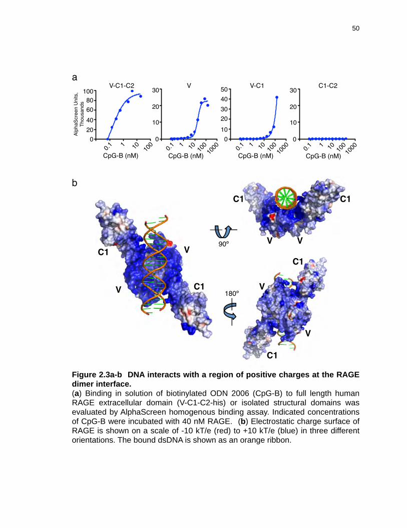
!
"#$%&"'()**+-+./01
2&340"+50
6
76
86
96
:
66;9 9 96 96
69666
768696
<6=6
:>?9
6
76
86
96
?9>?8
86=6@6A6966
6
:>?9>?8
?%B>C D+EF6;9 9 96 96
69666
?%B>C D+EF6;9 9 96 96
69666
?%B>C D+EF6;9 9 96 96
6?%B>C D+EF
!
!"#
"#
"#
"#
"# "#
! !
!
!
9A6G
H6G
Figure 2.3a-b DNA interacts with a region of positive charges at the RAGE dimer interface. (a) Binding in solution of biotinylated ODN 2006 (CpG-B) to full length human RAGE extracellular domain (V-C1-C2-his) or isolated structural domains was evaluated by AlphaScreen homogenous binding assay. Indicated concentrations of CpG-B were incubated with 40 nM RAGE. (b) Electrostatic charge surface of RAGE is shown on a scale of -10 kT/e (red) to +10 kT/e (blue) in three different orientations. The bound dsDNA is shown as an orange ribbon.
50

Figure 2.3c-d DNA interacts with a region of positive charges at the RAGE dimer interface. (c) Surface representation (center) showing positively charged residues lining the RAGE dimer interface where DNA binding occurs. DNA-binding residues are colored green on one RAGE molecule (“molecule A” in the text) and cyan on the other (“molecule B” in the text). Side panels show the molecular details of the two discrete binding sites, indicated by red boxes, for each RAGE V-C1 domain colored green and cyan. Hydrogen bonds are indicated with dotted lines, and the two strands of the dsDNA are colored silver and yellow, respectively. (d) Polyethyleneimine (PEI) competes with RAGE for binding to CpG-B (PO and PS backbones), CpG-A and CpG-C.
51

! "
#%&'()*'+',*-"*-./0-1%2*.%340-15678/0
0 90 :0 40 ;0 0 0</
/
/0/0
0/0
00
0</
/
/0/0
0/0
00
0</
/
/0/0
0/0
00
0</
/
/0/0
0/0
00
/00 90 :0 40 ;0 0
/00 90 :0 40 ;0 0
/00 90 :0 40 ;0 0
=8>[email protected]'BC
D@7
3E=F
D@7
3E=G
D@7
36D@7
3D
=8>[email protected]'BC
=8>[email protected]'BC
=8>[email protected]'BC
G*HI/
G*HI/
G*HI;
G*HI;
G*HI; G*HI/
G*HI;G*HI/
!"#!"%
!&"
!'(#
!"# %"& %"'(
!)(
)*
+
,
-
*)+
./'0 !'/"./'12''0
./%
!&"
!,
%,!"-(
*)
*
,
%")(
."/(
3+
*0#
!'(#
!"% !"#
!&"
./%
*)+
.)'(
%& %"'1#
)*
+
,
-3
./%
2''0
./'1
./'0 !'/"
%#( %&(
.)"
*)
* 3
Figure 2.3c-d DNA interacts with a region of positive charges at the RAGE dimer interface. (Please rotate the page 90º for proper viewing.)
52

reported for binding of other RAGE ligands129,168,169. The C2 domain did not
directly bind DNA in our assays (Figure 2.3a and data not shown), but may
enhance ligand binding by promoting proper folding and charge relationships
throughout the extracellular domain, as DNA binding to V-C1-C2 appears to be
more favorable than to V-C1 alone.
To understand the molecular details of the RAGE:DNA interaction, we
determined the crystal structures of the RAGE V-C1 domain in complex with a 22
nucleotide dsDNA molecule derived from vaccinia virus genomic repeat
sequences and a 22mer CpG motif-containing dsDNA molecule, at resolutions of
2.8 Å and 3.1 Å, respectively (Table 2.1 and Figure 2.4a). As the structures
were essentially the same except for the two dsDNA sequences, the following
discussion will focus on the higher resolution 2.8 Å structure. The structures were
solved by molecular replacement using two previously published RAGE V-C1
domain structures (PDB accession numbers 3CJJ111 and 3O3U129) as the search
models. The structures revealed that the RAGE V-C1 domains formed a
homodimer with the dsDNA bound in a concave cradle near the dimer interface
(Figure 2.3b). The two V-C1 monomers interacted with each other in a dyad
configuration with their V domains located at the dimer interface (see below),
creating an extensive positively charged pocket where dsDNA binds. Excellent
charge and shape complementarity were observed between the RAGE interface
and the bound dsDNA. Close examination of the protein:DNA interaction surface
53

Table 2.1 X-ray crystallography data collection and refinement statistics.
I (752-753, VV) II (981-982, CpG)Data Collection Spacegroup P61 P61
Unit cell (a, b, c) (Å) 79.12, 79.12, 224.04 77.92, 77.92, 224.39(α, β, γ) (°) 90, 90, 120 90, 90, 120
Wavelength (Å) 1.00 1.00Resolution (last shell) (Å) 50-2.80 (2.85-2.80) 50-3.10 (3.15-3.10)No of reflections (total/
unique)154198/19330 138765/13817
Completeness (last shell) (%)
99.4 (96.6) * 99.0 (85.6) *
I/s(I) (last shell) 14.49 (1.97) * 16.10 (1.97) *
Rmerge (last shell) (%)¶ 10.7 (60.7) * 14.0 (65.3) *
RefinementNumber of protein atoms 3267 3274No. of DNA base pairs 22 22No. of solvent/hetero-atoms 75 61Rmsd bond lengths (Å) 0.007 0.008Rmsd bond angles (°) 0.982 0.975Rwork (%)† 19.6 19.1Rfree (%)‡ 23.8 23.1
Ramachandran plot favored/disallowed (%) **
95.7/0 95.7/0
PDB accession code 3S59 3S58
*Asterisked numbers correspond to the last resolution shell.¶ Rmerge = ∑h ∑i |Ii(h) -<I(h)> | / ∑h∑i Ii(h), where Ii(h) and <I(h)> are the ith and mean measurement of the intensity of reflection h.† Rwork = ∑h||Fobs (h)|-|Fcalc (h)|| / ∑h|Fobs (h)|, where Fobs (h) and F calc (h) are the observed and calculated structure factors, respectively. No I/σ cutoff was applied. ‡Rfree is the R value obtained for a test set of reflections consisting of a randomly selected 5% subset of the data set excluded from refinement.**Values from Molprobity server (http://molprobity.biochem.duke.edu/)
54

Figure 2.4 Additional data: RAGE binds dsDNA at the dimer interface. (a) On the left, refined 2Fo-Fc map for the dsDNA is shown as magenta mesh superimposed on the refined model. On the right, the top 10 molecular replacement solutions for the dsDNA are shown as orange ribbons, with the refined dsDNA in red ribbon. (b) Footprint of the RAGE dimer on the dsDNA colored cyan and green for each of the V-C1 domains. (c) The two RAGE V-C1 molecules (cyan and green) in the crystal are superimposed with their bound dsDNA (blue and green), showing the switched positions for the major and minor grooves of the dsDNA in reference to the V-C1 domains. (d) The RAGE dimer interface centered at the V domain. Residues involved in RAGE dimerization are shown as sticks and colored cyan and green as the respective molecules. Water molecules are shown as red spheres and hydrogen bonds as dotted lines. The crystallographic dimer from 3CJJ is superimposed in our DNA-bound RAGE dimer structure in wheat and orange.
55

!
" #
$
$
&'#()
*')&'+
&'#$&)$&+$
!"'
*,&+
!"#%&
*#+
*#+,--
,(.
,(.
,--
,(#
,(#/-0
1""+
2("
344
5)-
5))
5)0
500
344
/-0
2("
1""+
'()
6((
76
78
79 7/
7'
7:
-".;77
76
78
79
7'
7:
7<
7<
-".;77
Figure 2.4 Additional data: RAGE binds dsDNA at the dimer interface.
56

indicated that ~7 positively charged residues on each V-C1 unit were in close
proximity to the negatively charged DNA backbone, spanning ~17 bp (Figure
2.3c and Figure 2.4b), in agreement with our finding that the minimal length
requirement for DNA binding was approximately 15 bases (Figure 2.2c). Each of
the RAGE V-C1 domains engage both strands of the dsDNA through basic or
hydrophilic residues from RAGE interacting with oxygen atoms of the DNA
backbone and, in some cases, through coordinating water molecules.
A total of 1300 Å2 of solvent accessible surface area was buried at the
RAGE:DNA interface. There are two DNA binding patches on each RAGE
molecule, hereafter referred to as “site 1” and “site 2” (Figure 2.3c). Site 1 is
located entirely in the V domain near β-strand B (βB) and the connecting loops
containing residues K37, K39 and K43, as well as K107 from β-strand G.
Notably, residues K43 from molecule A and R29 from molecule B coordinate the
same water molecule (w26) that forms a hydrogen bond with an oxygen of the
DNA phosphate backbone (Figure 2.3c upper right panel). Site 2 is located at
the juncture of the V and C1 domains, and includes residues R29 at the C-
terminus of βA, Y118 and K123 of the V-C1 linker, and R216 and R218 of βG in
the C1 domain. The amide group of R218 forms a direct hydrogen bond with a
backbone oxygen of DNA (Figure 2.3c bottom right panel). The footprints of the
two RAGE molecules on the dsDNA are very similar but not identical (Figure
2.4c), with the dsDNA switching its location of the major and minor grooves
relative to the RAGE molecules, suggesting a rather flexible surface of the RAGE
57

dimer that may be able to accommodate various forms of nucleic acids such as
ds or ss DNA and RNA. This is consistent with the dominant presence of flexible
lysine and arginine residues at the DNA binding surface.
In contrast to sequence-specific DNA recognition by proteins such as
transcription factors, the current structures showed no contact between RAGE
and the nucleotide bases. Because the overall negative charge on the DNA
molecule is determined by its phosphate backbone, we tested whether cationic
polymer polyethyleneimine (PEI) could compete with RAGE for DNA binding.
Our results showed that PEI could “mask” the negative charge of DNA and was
sufficient to negate DNA:RAGE binding in vitro (Figure 2.3d), further highlighting
the electrostatic nature of the recognition.
No major conformational changes were observed in the RAGE V-C1
domains between the DNA-bound structures and those of the non-DNA bound
forms (3CJJ129 and 3O3U111; Figure 2.5a), consistent with the observation that
the linkage between the V and C1 domains is fairly rigid168 and is not modified by
the binding of DNA ligands.
Dimerization of the RAGE V-C1 domains
A hydrophobic surface on the V domain is located at the center of the RAGE
dimer interface in the current configuration we call “trans.” It is composed of
residues L79, P80, F85, P87, and A88 from the βE strand and the connecting
loops, plus P33 and V35 from the βB strand, and P45 and P46 from the βB-βC
58

loop (Figure 2.4d and Figure 2.5a). The hydrophobic patch is surrounded by
hydrophilic residues decorating the circumference of the dimer interface. A
previous structure of RAGE V-C1 in the absence of bound DNA showed a
crystallographic C1-C1 dimer that we call “cis,” which is centered at a bound zinc
ion111. In comparison with the V domain-centered trans dimer interface, the cis
dimer interface is hydrophilic and smaller in size. Unexpectedly, examination of
the 3CJJ crystal lattice packing revealed that the trans dimers are also present in
this crystal (Figure 2.5a), though the DNA binding site is occluded by
crystallographic symmetry mates. It is thus clear that the trans dimer formation is
independent of DNA binding, and both the trans and the cis dimers can co-exist
as in the 3CJJ crystal. This is consistent with our observation that the most
intense fluorescence of RAGE on the cell surface is located at the juncture of two
adjacent cells (Figure 2.5e, Figure 2.8c), indicating that both trans and cis
dimerization may be involved in the formation of the RAGE oligomers, with the
cis and trans configurations mediating RAGE interaction on the same cell and
adjacent cells, respectively.
The chemical nature of the trans dimerization interface, including a typical
buried solvent-accessible surface area of over 1000 Å2, together with the
observations that RAGE V170, V-C119, or V-C1-C2171 domains spontaneously
dimerize in solution, suggests that the V-domain mediated dimers are likely to be
present under physiological conditions. As RAGE molecules are highly
59

Figure 2.5 Constitutive RAGE dimers form higher order oligomers in the presence of complex DNA ligands. (a) Ribbon diagrams of the RAGE V-C1 dimerization interface. Left, RAGE V-C1 crystal packing in the non-DNA bound form (3CJJ)1, with V-C1 monomers colored yellow, orange, and wheat. The orange sphere indicates a zinc ion. Middle, wheat and orange V-C1 domains from the left superimposed onto the V-C1 domains (cyan and green) in the current DNA-bound crystal structure. The dimer interface at the V domain (red circle) is represented in gray surface (right), with the hydrophobic and hydrophilic residues colored orange and green, respectively. (b) HEK293 cells expressing RAGE-mCitrine were incubated with or without the amine-reactive crosslinking agent BS3 prior to immunoblot for the mCitrine tag. (c) HEK293 cells co-expressing RAGE-CFP and RAGE-YFP were analyzed for FRET by flow cytometry. Cells show detectable baseline FRET (black histogram) in the absence of ligand. Cells expressing only RAGE-CFP (FRET-negative, gray histogram) are shown for reference. (d) Cells from (c) were incubated with the indicated concentrations of oligonucleotides and analyzed for FRET by flow cytometry. Graphs show the percent increase in FRET signal over baseline FRET. “x2” indicates biotinylated ODN complexed with streptavidin in a 2:1 ratio. (e) HEK293 cells co-expressing RAGE-mCerulean and RAGE-mCitrine were incubated for 30 minutes at 37 ºC with AlexaFluor 647-labeled CpG-A or CpG-B and analyzed for FRET by confocal microscopy. FRET efficiency in individual cells is plotted, with lines indicating the mean value for each condition.
60

! "
#
$
%
!"$!%
&'()*
+,-.
-//0102314
&'()%
&&'()$!%%(,%(-)106
& ()*+, $../%/$0%1
!" $!% &'()* &'()%& &
& 2
& 3
& 4
& 5
& (
!"#$%
!"#$& ()$*+,!"#$& ()$*+, -)
!"#$%
!"#$& ).$*+,!"#$& ).*+, -)
7809": 3; 7809": 3;
3(&
24&
2&&
6&
78!
9:4 <=> & &'?2'3(
3'(
(
,28<60=212883>?@25
23&
2&&
@&
?&
5&
3&
&&A& 5 A& 3 & 3
-32594 .5<3A/25 -//0102314& 5 & ? & @
*BC+AD)E
*BC+AD)E*BC+AF)E
B&C<39203
-32594
.5<3A/25-//0102314
5&
4&
3&
2&
&23( (&& 2&&& 3&&& 23( (&& 2&&& 3&&&
5&
4&
3&
2&
&
-./0+1
0+.2++
34,
5467,.
7.. 84.
894899
:;.
<.9
=.4
76/:>>/
09,
09;
044&D
&D
E E
E
&D
E
&D
G&H
G&H
?+>
Figure 2.5 Constitutive RAGE dimers form higher order oligomers in the presence of complex DNA ligands.
61

conserved in both their DNA binding sites and dimerization sites (Figure 2.6),
RAGE dimer binding to nucleic acids through electrostatic attraction may be a
common mode of its function in different species.
DNA can induce formation of higher-order RAGE oligomers
To confirm RAGE dimer and oligomer formation in live cells, we subjected
293T cells expressing full-length human RAGE-mCitrine to treatment with the
cell-impermeable crosslinking agent BS3, followed by cell lysis, protein
denaturation and gel electrophoresis. In the presence of crosslinker, higher
molecular weight bands corresponding to dimers and higher-order oligomers can
be detected in unstimulated cells (Figure 2.5b). To further explore the
relationship of native RAGE dimers with DNA ligands, we generated dually-
labeled cell lines appropriate for Förster resonance energy transfer (FRET)
studies. HEK293 cell lines stably expressing RAGE tagged with FRET donor
(CFP or mCerulean) and acceptor (YFP or mCitrine) fluorophores were analyzed
by both flow cytometry and confocal microscopy for energy transfer between
fluorophore tags. A constitutive baseline energy transfer was observed in
unstimulated cells (Figure 2.5c, Fig 2.5e, top panel), supporting the idea that
RAGE exists as a dimer in the absence of ligand. Addition of a CpG-B 24-mer
oligonucleotide did not alter energy transfer efficiency (Figure 2.5d, left panel,
Figure 2.5e, middle panel). Interestingly, however, the addition of a CpG-A 21-
mer induced a dose-dependent increase in FRET efficiency (Figure 2.5d, left
62
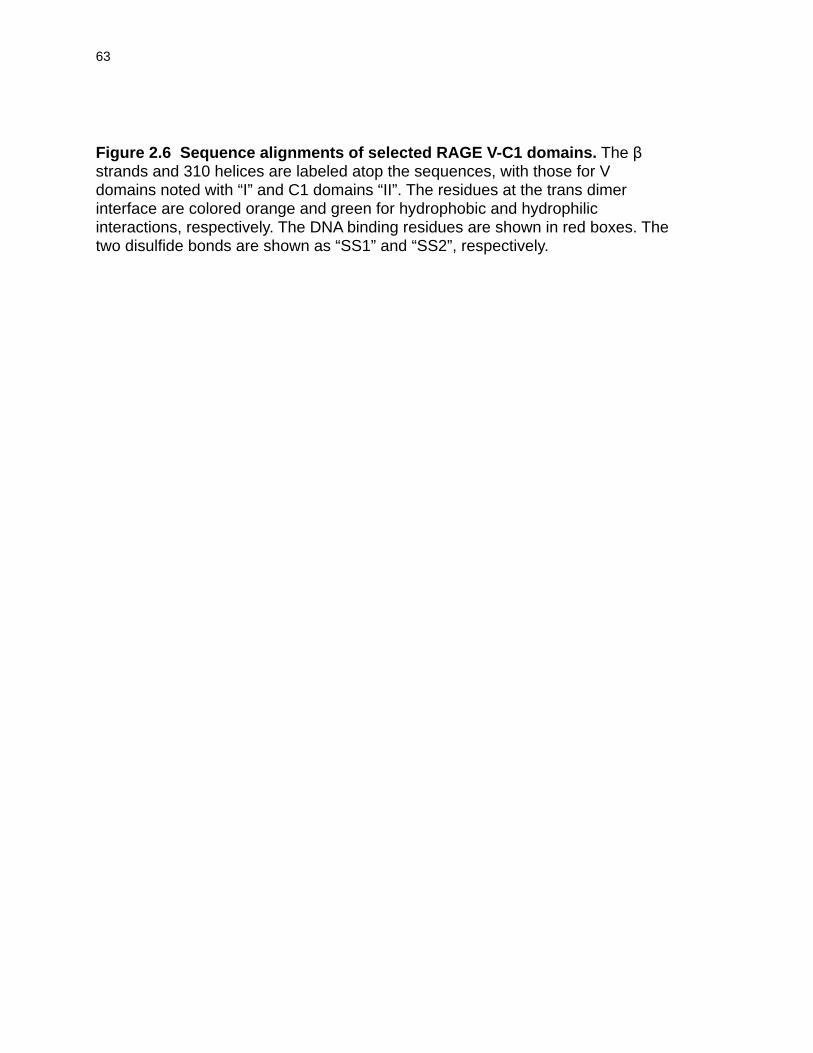
Figure 2.6 Sequence alignments of selected RAGE V-C1 domains. The β strands and 310 helices are labeled atop the sequences, with those for V domains noted with “I” and C1 domains “II”. The residues at the trans dimer interface are colored orange and green for hydrophobic and hydrophilic interactions, respectively. The DNA binding residues are shown in red boxes. The two disulfide bonds are shown as “SS1” and “SS2”, respectively.
63

!!!!"!!!!"!!!!"!!!!"!!!!"!!!!"!!!!"!!!!"!!!!"!!!!"!!!!"!!!!"
!"
##$
%$&'!
()*
+,-"./!0"1!-23454676-"36633.142864/0-102"8654#3.---389#5"1543/
:$
9;<
-91/!0"1!-63454/71-"36633..42864/0-102"8654#3.'--389#5"1543/
:=
(;>?@
-#./!0"1!-63454/76-"36633..42864/0-102"8654#3.'--389#5"1!43/
:&
1+AABC
--./!0"1!-23454676-"36633..42864/0-102"8654#3.'--#89#5"(543/
:&
D;)?@
--./!0"1!-23454#76-"36633..42864/0-102"8654#3.'--389#5"1!43/
:&
EF;??)*
-#./!##1!-23454/76-"31633..42864/01102"8654#32'-9382#5"1543/
G:
3H+CIF)?
--./!#"1!-23444/76-"31633..42864/0-102"8655"-.'--349#5"1543/
G:
JJJJ
JJJJ
JJJJ
JJJJ
JJJJ
JJJJ
JJJJ
JJJJ
JJJJ
JJJJ
JJJJ
JJJJ
%$&'!!
!L##$
!!"M
()*
+,-#4L43"5-!.92-!L17."D/1/-6206#/N1515N.!3-632!59#"#240"-53/65
$O$
9;<
-#4443"5-!.92-0L171"0#1/-6201#0N.515N.!3-632!!/3"#24D"-53#65
$O=
(;>?@
-#4443"5-!.92-0L171"0#1/-6206#/N.515N.!3-632!593"#240"-53/65
$O&
1+AABC
-#4443"5-!.92-0L17100/1/-6206#/N1515N.!3-632!493"#240"-!3#65
$O&
D;)?@
-#4443"0-!592-0L171"0/11-6256#/N1515N.!3-632!593"#240"#53/65
$O&
EF;??)*
-#4443"5-!.92-0L171"0/1(-62!6#/N1415N.!3-632!523"#24!"-!3/65
$%:
3H+CIF)?
-#4443#5-!292-!L171"5/1"-1206#/N1515N1!31632!493"#240"-!##65
$%:
JJJJ
JJJJ
JJJJ
JJJJ
JJJJ
JJJJ
JJJJ
JJJJ
JJJJ
JJJJ
JJJJ
JJJJ
##=
!!7M
!!9
()*
+,-075#2-#N3"-04#8(49-63453/26-5#562.011(320-4L04.#24D503"1--9
=&$
9;<
-075#2--N3"-04#8(89-634!39-6-5#5.22011(320-4L04.#24D503".--"
=&=
(;>?@
-075#2-#N3"-04#8(49-634!39-6-"#5622011(320-4L04.#24D503"1--"
=&&
1+AABC
-075#9--N34-04#8(D9-644539-6-5#562.011(390-4L04.#24D503"----
=&&
D;)?@
-075#2-#N3"-04#8(49-644!39-62045622011(320-4L041#2405!30.--'
=&&
EF;??)*
-074#2--N3"-04#8(49-6#4539-6-5#5122011(390-4L0!.#24050353--3
$P:
3H+CIF)?
-07559--N3"-"578(4/-654039-6-0#51220119419-35.#-593D0-#349"3
$P:
JJJJ
JJJJ
JJJJ
JJJJ
JJJJ
JJJJ
JJJJ
JJJJ
JJJJ
JJJJ
JJJJ
JJJJ
!!L##=
()*
+,3130L#7#L#3-''431(1"410"3!.315823534225.455232--"5"3--050407
=QP
9;<
3(30L#7#L#3-''43111"4(""3!.4/582#53'425.5D523'--D4"3--050407
=Q:
(;>?@
3130L#7#L#3#''43111"4(0"3L.405823534625.4D5232--"5"3-#050407
=OO
1+AABC
3330L#7#L#3-''43111"#N0"3!.3#5823-3'4251445232--"5"3-2050407
=QG
D;)?@
0(30L#7#L#4-''4311134/0"3!.415123-332-!.445232--!5"3--050407
=Q:
EF;??)*
4/30L#7#4#4-31##-.104(0"3!.4#58232342251455232--"5"3--"!0407
=Q:
3H+CIF)?
313-"3"#71-"'''1"-1/4('''41-(-3-3236-2.113.133(-4513''''.433
=O:
!R
!2
!-
!!R
!!7
!!-
!!-M
!9
!7
!7M
!!2
!!"
!!R
Figure 2.6 Sequence alignments of selected RAGE V-C1 domains. Rotate page 90º for proper viewing.
64

panel). CpG-A oligonucleotides are able to adopt large complexes in solution by
virtue of G-tetrad formation between poly-G tails on the ends of the ODN172. The
induced increase in FRET is consistent with higher-order RAGE oligomer
formation. In keeping with this observation, CpG-A DNA can be seen to induce
aggregation of RAGE into clusters on the cell surface (Figure 2.5e, bottom
panel), which is not observed in unstimulated cells or cells incubated with CpG-B
(Figure 2.5e, top and middle panels). Simply increasing the length of the DNA
ligand did not promote increased energy transfer (Figure 2.5d, right panel), nor
did complexation of short, biotinylated ODN sequences with streptavidin (Figure
2.5d, left panel). However, biotin-streptavidin complexation of a relatively long
ODN increased FRET efficiency (Figure 2.5d, right panel). These findings
suggest that while RAGE may bind DNA indiscriminately, only large ligand
complexes induce receptor reorganization on the cell surface.
RAGE expression promotes DNA uptake by cells via the endosomal route
Having established that RAGE and DNA interact at the cell surface, we
sought to understand the functional consequences of this interaction. Cells
expressing RAGE not only showed increased binding of both CpG-A and CpG-B
DNA to the cell surface, but also a notable increase in total DNA uptake over a 30
minute time period (Figure 2.7a), compared to cells not expressing RAGE. This
effect was DNA-specific, as binding and uptake of transferrin to its respective
receptor (Figure 2.7a) or the uptake of TLR2 ligand (not shown) was not
65

Figure 2.7 RAGE promotes cellular DNA uptake. (a) 293T cells expressing tetracycline-inducible RAGE-mCitrine were induced overnight (black bars) or left untreated (white bars) and then incubated with 1µM AlexaFluor 647-labeled transferrin, CpG-A or CpG-B at the indicated temperatures for 30 minutes, washed to remove unbound DNA, and analyzed by flow cytometry. Error bars indicate standard error of the mean. (*) Indicates p <0.05 as assessed by two-tailed t-test. (b-c) HeLa cells expressing RAGE-CFP (green) and the early endosome protein Rab 5a-RFP ((b), red) or the late endosome protein Rab 9a-YFP ((c), red) were incubated with 1 µM AlexaFluor 647-labeled CpG-A or CpG-B for 5 minutes, washed to remove unbound DNA, then incubated for the indicated times at 37 ºC prior to analysis by confocal microscopy. Bar graphs show the percentage of Rab-positive endosomes colocalizing with both the RAGE and DNA tags at each time point. Error bars indicate standard error of the mean.
66

!" $%&'()*+" $%&'()*
!" $%&'()*+" $%&'()*
,-./
/0123/&45
67.8-
67.89
,-./
:0()/&45
,-./
:0()/&45
67.8-
!#$%&'()&*+#%,((-.*+*/(+
!#$%&'()&*+#%,((-.*+*/(+
!#$%&'()&*+#%,((-.*+*/(+
!#$%&'()&*+#%,((-.*+*/(+
!
"
,-./
/0123/&45
67.89
#
$%%%
%
&%%%%
&$%%%
'%%%%
'$%%%
()(*
+-.
/0-.
+-.
/0-.
+-.
/0-.
+-.
/0-.
12*3!
425)678+09:4;<
.=>71 .=>7? @6!(AB*66C(
D1>E )(
D1>E )BB
.=>71
'%FC(
G%FC(
0
10
20
30
40
.=>7?
'%FC(
G%FC(
0
10
20
30
40
.=>71
'%FC(
G%FC(
0
10
20
30
40
.=>7?
'%FC(
G%FC(
0
10
20
30
40
H
H
Figure 2.7 RAGE promotes cellular DNA uptake.
67

influenced by RAGE expression. CpG DNA in cell culture medium gains access
to endosomal compartments, where it is able to interact with and stimulate Toll-
like receptor 9 (TLR9), although the mechanism by which this uptake occurs is
unknown. While cellular uptake of DNA is not strictly RAGE-dependent (Figure
2.7a), we sought to evaluate the nature of the receptor-ligand association after
surface binding. We stably expressed fluorescently tagged RAGE together with
fluorescently tagged endosomal markers and assessed the binding and
trafficking of DNA in live cells. Fluorescently labeled DNA was seen to associate
with RAGE at the cell surface and traveled together with the receptor into both
early and late endosomal compartments (Figure 2.7b-c). Notably, the kinetics of
trafficking differed depending on DNA ligand structure. While CpG-B entered the
cell quickly and could be found in late endosomes 20 minutes after exposure,
RAGE:CpG-A clusters persisted longer at the cell surface and remained primarily
in early endosomes at 20 minutes (Figure 2.7b-c). By 90 minutes, both CpG-A
and CpG-B could be seen colocalized with RAGE in late endosomes (Figure
2.7c), the compartment from which TLR9 is thought to signal39,173. Hence, RAGE
and DNA appear to remain associated during uptake and progression through
the endosomal network, and the trafficking kinetics is influenced by the DNA
structure and, potentially, the ability of these ligands to multimerize RAGE.
68

RAGE increases TLR9-dependent responses to suboptimal DNA stimuli
Both RAGE and TLR9 have been shown to activate the NF-κB family of
transcription factors upon ligation of stimulatory ligands34,133. To assess the
ability of CpG DNA to activate RAGE signaling, we employed an HEK cell line
expressing a luciferase reporter gene under the control an NF-κB-inducible
promoter. No significant reporter activation was observed upon DNA stimulation
when these cells overexpressed RAGE alone (data not shown). A stable cell line
expressing TLR9 together with TLR2 and tetracyline-inducible RAGE was
established to evaluate the ability of RAGE to contribute to TLR9-dependent
CpG-B responses. Two different length CpG-B oligonucleotides, which are
optimal for stimulation of NF-κB via TLR9, activated NF-κB in a dose-responsive
manner in cells expressing TLR9/2 without RAGE, and this activation was
notably enhanced when RAGE expression was induced (Figure 2.8a, left panel).
This RAGE-dependent enhancement was specific for DNA, as co-expression of
RAGE did not alter NF-κB activation in response to the TLR2 ligand Pam3CysK4
(Figure 2.8a, right panel). We next sought to understand whether RAGE could
similarly affect responses to mammalian DNA. For this, the luciferase reporter
cell line was co-cultured with immortalized murine bone marrow-derived
macrophages and treated with silica to induce macrophage cell death. Silica
treatment of luciferase reporter cells alone did not induce significant NF-κB
activation (data not shown). The presence of silica-killed macrophages in co-
culture activated NF-κB and this activation increased in cells expressing RAGE
69

Figure 2.8 RAGE increases TLR9-dependent NF-κB activation in response to DNA ligands. (a) HEK293 cells expressing TLR9, TLR2, tetracycline-inducible RAGE and firefly luciferase under the control of the NF-κB-inducible ELAM promoter were treated with doxycycline to induce RAGE expression and then stimulated with CpG-B or Pam3CysK4 at the indicated concentrations for 12 hours prior to measurement of luciferase protein. (b) HEK293 cells expressing TLR9 and tetracycline-inducible RAGE-mCerulean ELAM-luc reporter were cultured with or without doxycyline to induce RAGE expression, together with murine bone marrow-derived macrophages. Co-cultures were left unstimulated or stimulated with silica crystals to induce macrophage cell death. Degradation of the phosphodiester (PO) CpG-B DNA stimulus served as a positive control for benzonase activity. Error bars indicate standard deviation. (c) Co-cultured cells from (b) were incubated with silica for 6 hours, stained with propidium iodide to label DNA from dead cells, and imaged by confocal microscopy. Silica was visualized using a reflection microscopy technique1.
70

!""#$%&' )*+ ,-./
!""""
0"
1"
2"
3+%4 5663+%4 5-
3+%4 5663+%4 5-
01&7891:&789
!""!"!;<7=#>?@1 ,-./
"
A"
!""
!A"
*BC'<DEFG<EF5-,HIDF689<?8
I-FE?
J!"""/
*BC'<DEFG<EF5-,HIDF689<?8
I-FE?
J!"""/<
K
D
LFHFD<*5 ?EF7M)5NO & P & P & P & P P P
#$%&' ;Q
.8RFI7 5-H>
*BC'<DEFG<EF5-,HIDF689<?8
I-FE?
J!"""/
*BC'<DEFG<EF5-,HIDF689<?8
I-FE?
J!"""/
'8-S5-<?8.8RFI7 5-H>'8-S5-<?8
T4@&UV3WXU8E3+%4P #AY'V2 7<D95$Z<[8?
T4@&UV3WXU8E3+%4
" "
AA
!"
!"!A
!A0"
0"
0A
="
LFHFD<3+%4
;95$FRFI7 F5RFR8
Figure 2.8 RAGE increases TLR9-dependent NF-κB activation in response to DNA ligands.
71

(Figure 2.8b). Treatment of co-cultures with benzonase to degrade extracellular
DNA and RNA decreased the ability of dead macrophages to stimulate reporter
cells, suggesting that enhancement of activation by RAGE is due largely to
recognition of nucleic acids (Figure 2.8b). In effect, DNA from dying
macrophages was seen interacting with RAGE when cocultured cells were
imaged by confocal microscopy (Figure 2.8c). Taken together, these data
suggest that RAGE effectively delivers DNA into the cellular compartments in
which TLR9 is activated and that expression of RAGE sensitizes cells to both
synthetic and genomic nucleic acids in the extracellular environment.
RAGE-deficient mice have impaired inflammatory responses to DNA in the
lung
Given its ability to potentiate NF-κB activation in cultured cells, we sought
to assess the role of RAGE in proinflammatory responses to DNA in vivo. RAGE
is highly expressed in mammalian lung epithelia140; we therefore hypothesized
that RAGE might participate in immune recognition of DNA in the airways. To
address this possibility, we intranasally administered a low dose of CpG-B DNA
to C57BL/6 wild-type or RAGE deficient (ager-/-) mice and evaluated
inflammatory parameters in the lung at 24 hours after DNA exposure. Wild type
mice mounted a strong inflammatory response to CpG-B DNA, characterized by
a significant increase in neutrophils and the neutrophil granule product,
myeloperoxidase, in bronchoalveolar lavage (BAL) fluid, and increased
72

expression of the neutrophil chemoattractant LIX (Figure 2.9a). Strikingly,
RAGE-deficient animals showed significantly lower neutrophil influx, with MPO
and LIX levels indistinguishable from PBS-treated control animals (Figure 2.9a).
Histological analysis of lung tissue revealed marked perivascular and
peribronchiolar leukocyte influx in wild type mice (Figure 2.9b, left panel) that
was notably reduced in RAGE-deficient mice (Figure 2.9b, right panel). Analysis
of cytokines in the BAL fluid revealed significantly reduced production of IFN-γ,
IL-6, TNFα, and IL-12p70 in RAGE-deficient animals as compared to the wild
type (Figure 2.9c). Of note, IL-1β and IFN-α, cytokines whose production is not
directly activated by NF-κB, were also seen to be upregulated in wild type but not
RAGE-deficient mice following CpG-B exposure, suggesting a broader role of
RAGE in systemic inflammation. Taken together, these data indicate that RAGE
expression plays an important role in the formation of an inflammatory response
to immunostimulatory DNA in vivo, and particularly, in the lung.
73

Figure 2.9a-b RAGE mediates DNA-induced pulmonary inflammation in vivo. C57BL/6 wild-type or RAGE-deficient mice were administered PBS with or without 5 µg CpG-B via the intranasal route and analyzed 24 hours later for signs of pulmonary inflammation. n=6 mice per group. (a) Neutrophil cell number (left panel), myeloperoxidase concentration (MPO, middle panel) and LIX concentration (right panel) were determined in bronchoalveolar lavage fluid. Data shown are representative of two separate experiments (n=6 per group, per experiment) with similar results. Error bars indicate standard error of the mean and p values assessed by the nonparametric Mann Whitney U test are noted. ns = differences are not significant. (b) Representative hematoxylin and eosin stained paraffin sections of lung tissue from wild-type and RAGE-deficient (ager -/-) mice 24 hours after administration of 5 ug CpG-B. Black arrowheads indicate areas of apparent leukocyte infiltration in the wild type tissue and comparable areas in the ager -/- tissue.
74

!
" #$%& ()*+
!"#$ &'&
,-.
/-,
/-./-.
/-,
,-.
, ,0123*4 0123*4#$%& 5)*+ !"#$ &'&
0123*4 0123*4#$%& 5)*+ !"#$ &'&
0123*4 0123*4#$%& 5)*+ !"#$ &'&
.
6
7
8
/
,
90:
;<=>?@A
@BC;D=>?@A
E+<(FG*H$%I;J/,
A.DI DI
DI
* K ,-,,88* K ,-,,88
* K ,-,,88* K ,-,,88
* K ,-,,88* K ,-,,88
Figure 2.9a-b RAGE mediates DNA-induced pulmonary inflammation in vivo.
75

!
"
"#$
"#%
"#&
"#'
"#(
)#"
)#(
$#"
"
"#)
"#$
"#*
"#%
"#(
"
)
$
*
%
"
(
)"
)(
$"
"
("
)""
)("
"+,-./0 +,-./01234 67/8 !"#$ &'&
+,-./0 +,-./01234 67/8 !"#$ &'&
+,-./0 +,-./01234 67/8 !"#$ &'&
+,-./0 +,-./01234 67/8 !"#$ &'&
+,-./0 +,-./01234 67/8 !"#$ &'&
+,-./0 +,-./01234 67/8 !"#$ &'&
9:;)$/<"=/>?@:A
9:;)
=/>?@:A
9BC;=/>?@:A
6CB;
=D>?@:A
9:;&=D>?@:A
9BC;=D>?@:A
/ E "#""'</ E "#""%*
/ E "#""("/ E "#""%*
/ E "#""*</ E "#""$$
/ E "#""*</ E "#"%))
/ E "#""%*/ E "#""%*
/ E "#""$$/ E "#""$$
DF
DF DF
DF DFDF
Figure 2.9c RAGE mediates DNA-induced pulmonary inflammation in vivo. C57BL/6 wild-type or RAGE-deficient mice were administered PBS with or without 5 µg CpG-B via the intranasal route and analyzed 24 hours later for signs of pulmonary inflammation. n=6 mice per group. (c) Cytokine concentrations in bronchoalveolar lavage fluid were determined by ELISA. Data shown are representative of two separate experiments (n=6 per group, per experiment) with similar results. Error bars indicate standard error of the mean and p values assessed by the nonparametric Mann Whitney U test are noted. ns = differences are not significant.
76

Discussion
The mechanisms of nucleic acid uptake from the extracellular milieu have long
been a point of interest. Because nucleic acid-recognizing TLRs are expressed
in endosomal compartments, early work on these receptors assumed the
likelihood of a cell surface “uptake” receptor39. Discovery of such a receptor has
been elusive, however, and the mechanism by which extracellular DNA gains
access to intracellular receptors remains largely unsolved. A role in ligand
uptake has been described for the TLR co-receptor protein CD14101, integrins102
and scavenger receptors103,104. However, few studies have shown direct
biochemical evidence of DNA-receptor binding and an apparent functional
redundancy complicates assigning an essential role to any single receptor.
Our work indicates that surface-expressed RAGE protein binds directly to
nucleic acids and promotes their uptake into endosomal compartments,
presumably facilitating their access to endosomal TLRs 3, 7, 8 and 9. While
RAGE does not appear to be essential for DNA to access TLR9 in cultured cells,
its expression notably enhances the ability of TLR9-expressing cells to activate
the transcription factor NF-κB. The effect of RAGE is more dramatic in vivo, as
mice lacking RAGE show a clear deficiency in their ability to mount a typical
inflammatory response to TLR9-stimulatory DNA. These findings shed new light
on the role of RAGE in inflammation.
77

It has been suggested that direct signaling of RAGE upon ligation of
glycated proteins, amyloid-beta fibrils, S100 proteins and HMGB1 serves to
initiate and perpetuate inflammatory responses126,136,137,141. Our work
demonstrates a clear role for RAGE in uptake and intracellular trafficking of
nucleic acid ligands; however, the occurrence or importance of a direct signal
downstream of receptor ligation is, as yet, unclear.
Whether by simply modulating DNA uptake or by generating a regulatory
signal, RAGE appears to be important in influencing the concentration threshold
at which activation of inflammatory responses is achieved. We gave mice a
suboptimal dose of a TLR9 stimulatory ligand, which in the absence of RAGE,
was not able to induce inflammation. However, expression of RAGE at normal
physiological levels permitted the animals to mount a potent inflammatory
response to low concentrations of DNA. We speculate that such “thresholding”
may serve to sensitize RAGE-expressing cells to the presence of DNA while
allowing other cells to remain unactivated. The multi-ligand nature of RAGE may
also contribute to thresholding of responses. It has been proposed that
complexes containing multiple RAGE ligands and/or complexes capable of
simultaneously ligating RAGE and other receptors (such as DNA-HMGB1-
containing immune complexes seen in systemic lupus erythematosus) may
provide a symphony of signals that allow the immune system to distinguish
between distinct types of danger175. Such synergy between multiple RAGE
ligands may help to reconcile our observation of a direct RAGE:DNA interaction
78

with published data indicating that HMGB proteins, also RAGE ligands, are
required for TLR activation by nucleic acids176.
The roles of co-receptors, such as HMGB1, and nucleic acid sequence in
RAGE-mediated DNA responses is still somewhat unresolved. While our
previous work suggested that a DNA:HMGB1 complex was more effective at
activating plasmacytoid dendritic cells than HMGB1 alone106, the binding studies
presented here, which were carried out in the absence of HMGB1, indicate that
HMGB1 is not essential for the RAGE-DNA binding event. However, HMGB1 is
commonly present in mammalian serum and induced during tissue damage;
thus, possible synergy between DNA and HMGB1 in the initiation of inflammation
remains to be elucidated. With or without the help of co-receptors, specificity for
distinct nucleic acid sequences or structures is a hallmark of many known nucleic
acid sensors. Under normal physiological conditions, an unmethylated CpG
dinucleotide motif9 is required for activation of TLR9. Such a CpG sequence has
also been reported as necessary for DNA binding to RAGE128. Our data,
however, strongly suggest that the DNA-RAGE interaction occurs irrespective of
nucleotide sequence. Co-crystalization of RAGE with bound DNA indicates that
the binding is mediated entirely by interactions of the negatively charged
phosphates of the DNA backbone with charged and hydrophilic residues at the
RAGE:ligand binding interface. Additional binding studies using non-CpG
oligonucleotides and base-free sugar-phosphate backbones, which also bound
RAGE with no notable loss of affinity, further support a sequence-independent
79

binding modality. This lack of specificity seems to extend to RNA ligands, as both
ssRNA oligonucleotides and base-free RNA backbones interacted with RAGE in
a manner similar to DNA.
Charge-based binding is emerging as a generalized mechanism for
interaction of RAGE with its ligands. It has previously been demonstrated that
glycated BSA, which is highly acidic, may engage in electrostatic interactions
with RAGE in a manner similar to that of dsDNA shown here.129 Additionally,
NMR spectroscopic analysis of calcium-bound S100b interaction with RAGE
suggested that their interface resides adjacent to the area we designate “site 1”
of the dsDNA binding surface111. Truncation of the acidic C-terminal tail region of
HMGB1 has also been shown to significantly diminish its association with
RAGE177, suggesting that electrostatic interactions are key for any direct HMGB1
binding. It thus appears that the large positively charged surface of the RAGE V-
C1 domains, of which only a portion is involved in dsDNA binding, may be able to
engage distinct negatively-charged ligands through similar electrostatic
attractions. Interestingly, charge-based interaction has also been suggested as
the mechanism of DNA binding to other cell surface receptors178,179 and may help
to explain the apparent functional redundancy of receptors that interact with
negatively charged ligands in the extracellular space.
While the interaction mechanisms described here are ostensibly
applicable to any environment in which DNA and RAGE are present,
physiological expression patterns of RAGE suggest that it may be particularly
80

important for inflammatory responses raised in the lungs. The predominant site of
RAGE expression in adult mammals is in lung tissue150, where the receptor is
highly expressed on alveolar epithelial cells140,180. Several types of immune cells
have also been described to express RAGE150-153,181,182, though whether such
expression is constitutive or inducible is not well defined. Various conditions can
expose such cells to free DNA and RNA in the lung. For example, acute
infections such as pneumococcal pneumonia, which is characterized by bacterial
autolysis and release of foreign DNA into the lung183, present a likely scenario in
which nucleic acids would be accessible to a cell-surface receptor. Chronic
pathological conditions, such as the accumulation of DNA-containing bacterial
biofilms in cystic fibrosis160 and the deposition of nucleic-acid containing immune
complexes in the interstitial space of lupus patients184, also expose lung cells to
free extracellular DNA and RNA. Moreover, inflammation in the lung is often
characterized by recruitment of neutrophils and formation of neutrophil
extracellular traps (NETS), which involves extrusion of the cell’s own DNA185.
These and other situations suggest that recognizing extracellular nucleic acids of
foreign or host origin may serve a broad function in detecting infection and tissue
damage in the lung. The surface receptor protein RAGE, through its ability to
concentrate DNA and RNA on cells and promote their uptake into specific
receptor-containing intracellular compartments, may play a key role in regulating
the initiation of immune responses to nucleic acids.
81

Materials and Methods
Oligonucleotides
Oligonucleotide (ODN) sequences and sources are listed in Table 2.2. Base-free
phosphodiester and phosphorothioate backbones were a kind gift from Dr.
Herman Wagner (Technical University Munich, Germany).
Cell Lines
Cell lines in the HEK293, 293T and HeLa backgrounds were generated using
retro- or lentiviral transduction with human RAGE C-terminally fused to
fluorescent protein tags. cDNA encoding the human RAGE 404 amino acid
transmembrane protein isoform (GenBank accession number AY755619) was
amplified by PCR using upstream primer
5’-AAAACTCGAGATGGCAGCCGGAACAG-3ʼ and downstream primer 5’-
AAAAAGATCTAGGCCCTCCAGTACTACTCTCG-3’. RAGE cDNA and cDNA
encoding CFP or YFP were sequentially subcloned into the murine moloney
leukemia virus-based retroviral packaging vector pRP (Figure 2.10, produced by
Brian Monks). To produce transducing retroviruses, 293T cells (220,000 per well
in 96-well plates) were simultaneously transfected with pRP (100 ng) and
plasmids encoding HIV gag-pol (100 ng) and VSV-G (10 ng) using TransIt
reagent (Mirus Bio). Culture supernatants containing retrovirus were added to
pre-existing target cell lines of interest and RAGE-positive cell lines were
selected based on resistance to puromycin. Limiting dilution was used to
82

!"#$"%&"
()*+,*-
./0"1
!/$2&"(1-
34.5)6
7789:87
9:98::88
8797
98:
;1"<=%6>?@&2A10BCD1"%1"102B%<
E49
34.5),
:798:8:
9997
79878:
9789:
;1"<=%6>?@&2A10BCDB%0=1"%1"
102B%<
E49
34.F?G
79:
788
7:89:7987:88
7:9:
;1"<=%,>G@&2A10BCD1"%1"102B%<
E49
34.F?6
787
:997:98:7897:99:78:
;1"<=%,>G@&2A10BCDB%0=1"%1"
102B%<
E49
34.G?6H
9I7I7I8I9I:I8I7I:I9I9I7I7I9I:I8I7I:I9I9
7J:
+KD/J0=LBCL/$1"+10=L
$CB0/2A
92=M=%N
34.6OOH
P!9I7I:I9I7I:I9I9I9I9I:I9I7I:I9I9I9I9I:I9I7I:I9I9
7J:
+KD/J0=LBCQ$LB%+10=L
$CB0/2A
E49D3J"2/%
34.6OOH
P397
:97
:9999:97
:9999:97
:99
7J:
+KDQ$L
B%10=L$CB0/2ADJQ/1JQ/<="10"2
E49D3J"2/%
34.6,,H
:I:I::87
:87
:97
:9:
:I:I:I:I:I:
7J:
+87/C"ADE49D3J"2/%
34.6,F)
9I7I:I9I7I:I9I9I9I9I7I:I:I7I:I7I:I7I:I7I7I:
7J:
+73J"2/%
3R.F>61
P3282:272;2;282827272;2:2;27272;2;2728282;2;
:SP
1=R.8+<"2=T"<
1"#$"%&"D1"%1"
102B%<
E49
3R.F>61
P!28I2:I27I2;I2;I28I28I27I27I2;I2:I2;I27I27I2;I2;I27I28I28I2;I2;
:SP
1=R.8+<"2=T"<
1"#$"%&"D1"%1"
102B%<
E49
M=%NBU"1
B2"JQ/1JQ/<="10"2V/%<1V"0W""%<"/XA%$&C"/0=<"1
$%C"11
=%<=&B0"<YIDJQ/1JQ/2/0Q=/B0"C=%NBU"Z2D2=V/%$&C"/0=<"
MBV"C"<34.QB<V=/0=%(9[:
C=%NBU"-/28C"XBSC$/2H\5
(8L75C=%NBU"-B00B&Q"<
0/0Q",*"%<>
!/$2&"1Y92=C=%NK=/9"&Q%/C/U="1DE%&>D!B%
4="U/D7
8ZE%0"U2B0"<
4.89
8M>
A>
!"#$%'(')$*+,-./$%,0*1%2%3.%-/%2"-12,.4/%2
Table 2.2 Oligonucleotide sequences and sources.
83

!"# %&'(&)%*+ ,(-%- ./.0122"3"4 5# 670589+ ,(-%- :;;0/:6<=4*3>?)@& *%-@-A(&)% B%&%+ ,(-%- /:C;0.D62!"# E*3>3A%*+ ,(-%- .D1;0<.1<"4FA@EF% )F3&@&B -@A%+ ,(-%- <.1C0<<<1"3"4 5# <7G89+ ,(-%- <<D/0D;D6H#D; E*3>3A%*+ D;660D<:<H#D; 3*@B@&+ D.2D0D<125() E*3>3A%*+ DDC20DD2:!3FI/ 3*@B@&+ ,(-%- D:;106D.CJ>E@)@FF@& *%-@-A(&)% B%&%+ ,(-%- 66:/0CDD/J>E E*3>3A%*+ ,(-%- CD:<0C6//
!"
670 !!K!KK!!!! JJK !88 !!K! K8J !K! !!K JJ8 8!! KK! 8!K JKJ KKJ !K! K8K !JJ !!K K8K K8K !KK !!K ! 0 <7L@&M NNN O-@P N I)3 9N Q'3 N "F4 N JB% N R3A N
!"# E*3>3A%*
Figure 2.10 pRP retroviral transduction vector.RAGE-fluorescent protein chimeras were cloned into the vector at the BglII restriction site.
84

produce clonal cell lines.
For the tetracycline-inducible RAGE cell line, cDNA encoding human RAGE
and the fluorescent protein mCitrine or mCerulean were cloned into the
pEN_TTmcs entry vector (Addgene 25755) and transferred to the pSLIK-hygro
vector (Addgene 25737) by recombination (both plasmids were the gift of Dr. Iain
Fraser and are commercially available through Addgene). pSLIK plasmids were
delivered using a lentiviral transduction system186.
Binding Studies
Recombinant protein production is described in Additional Detailed Methods. In-
vitro binding of biotinylated oligonucleotides to purified, his-tagged, human RAGE
extracellular domain (V-C1-C2) was assayed using the AlphaScreen amplified
luminescent proximity assay (Perkin Elmer)186. Titrations of oligonucleotides
were incubated with 40 nM RAGE-his in buffer (50 mM Tris-HCl pH 7.4, 100 mM
NaCl, 1% ultrapure BSA, 0.01% Tween 20) at 22 ºC for 30 minutes, followed by
addition of streptavidin-conjugated donor beads and nickel-chelate acceptor
beads for 10 minutes or longer. Fluorescence was analyzed with an Envision
plate reader (Perkin Elmer). For confocal imaging studies of DNA binding to the
cell surface, HeLa cells expressing RAGE-CFP and Rab4a-YFP or only Rab4a-
YFP, were cultured on glass-bottom dishes (MatTek) overnight, then incubated
with 1 µM ODN 2336-AlexaFluor 647 at 37 ºC for 5 minutes, washed with
medium, and imaged by confocal microscopy. For flow cytometry analysis of
85

DNA binding to RAGE-positive and RAGE-negative cells, 293T cells expressing
tetracycline inducible RAGE-mCitrine were cultured overnight in medium with or
without 10 µg/mL tetracycline, then incubated with 1 µM Alexa 647-labeled ODN
2336 on ice for 30 minutes. Cells were washed with PBS and analyzed on a BD
LSR II instrument (Becton Dickinson) using BD FACS Diva (Becton Dickinson)
and FlowJo software (Treestar, Inc.).
DNA Uptake Studies
Uptake of DNA by RAGE-positive versus RAGE-negative cells was assessed
using a tetracycline-inducible RAGE-mCitrine cell line. Cells in multiwell plates
were cultured overnight in medium with or without 10 µg/mL tetracycline, then
incubated with the indicated concentrations of AlexaFluor 647-labeled ODN
2006, 2336 or transferrin for 30 minutes on ice or at 37 ºC. Cells were washed
with PBS and analyzed on a BD LSR II instrument (Becton Dickinson) using BD
FACS Diva (Becton Dickinson) and FlowJo software (Treestar). For confocal
microscopy analysis of DNA uptake and trafficking, HeLa cells stably expressing
RAGE-CFP and either Rab5a-RFP or Rab9a-YFP were cultured on glass-bottom
dishes (MatTek) overnight. Cells were “pulsed” with 500 nM Alexa 647-labeled
ODN in medium for 5 minutes at 37 ºC, washed with PBS, and then incubated in
medium for the indicated time periods prior to imaging. For quantification of
RAGE and DNA colocalization with early and late endosomes, total labeled
endosomes were manually counted in 3-6 cell images. Endosome spots
86
fluorescing in all three channels (endosomal marker, RAGE, ODN), were
considered “triple-positive.”
Confocal Microscopy
Live cells were seeded at 20,000 - 30,000 cells in 100 µL medium on glass-
bottom culture dishes (MatTek) and cultured overnight prior to treatment and
imaging. Images shown represent a single z-plane though the approximate
center of the cells of interest. Images were obtained with Leica SP2 AOBS or
SP5 SMD confocal microscopes. Sequential scanning was used to avoid cross
excitation between fluorescence channels.
Förster Resonance Energy Transfer (FRET) Measurements
HEK293 cells coexpressing RAGE-CFP and RAGE-YFP were brought into
suspension and incubated in PBS with the indicated CpG ODN on ice for 20
minutes. After further incubation at 37 ºC for 5 minutes, the cells were washed
with ice-cold PBS and kept on ice until flow cytometry FRET analysis was
performed as previously described187. RAGE-CFP or RAGE-YFP single
transfectants were used as controls. Microscopy-based sensitized emission
FRET was performed using a Leica SP5 SMD confocal microscope. HEK293
cells were cultured on glass-bottom dishes and incubated with 10 µg/mL
doxycycline for 24 h to express tetracycline-inducible RAGE-mCerulean and
RAGE-mCitrine alone (as controls) or together. Cells were then treated with 0.25
87

µM AlexaFluor 647-labeled ODN 2006 or 2336 for 15-30 minutes at 37 ºC and
imaged. FRET efficiency in areas of DNA:RAGE interaction was assessed by
Leica TCS software, color coded as indicated, and representative cells were
graphed.
RAGE Crosslinking
The amine-reactive crosslinking agent bis(sulfosuccinimidyl) suberate (Pierce)
was prepared as a stock solution in water and diluted in PBS to working
concentrations. Adherent HEK293 cells expressing human RAGE-YFP were
detached from culture flasks and washed 3 times with PBS to remove amine-
containing serum components. 300,000 cells per sample were incubated in PBS
with or without crosslinker for 30 minutes on ice. Cells were then gently pelleted
by centrifugation, BS3-containing supernatant was removed, cells were lysed in
Laemmli buffer (63 mM Tris-HCl, 10% glycerol, 2% sodium dodecyl sulfate,
0.0025% bromophenol blue), and samples were analyzed by denaturing gel
elctrophoresis and immunoblotting with antibody directed against the YFP tag
(anti-GFP mAb, Clontech). Identical results were obtained in similar experiments
with cells expressing tetracycline-inducible RAGE labeled with a monomeric
Citrine tag, indicating that the weak dimerization potential of YFP was not the
sole driver of chimeric RAGE dimerization in these cell lines.
88

Luciferase Reporter Assays
HEK293 cells stably expressing human TLRs 2 and 9, tetracycline-inducible
human RAGE-mCerulean and a firefly luciferase gene under the control of the
NF-κB inducible ELAM promoter were incubated overnight with 10 µg/mL
doxycycline to induce RAGE expression, or left uninduced. Cells were stimulated
as indicated for 8-16 hours and luciferase activity was assessed using SteadyGlo
substrate (Promega) and an Envision multiwell plate reader (Perkin Elmer).
HEK293 cells stably expressing human TLR9 and tetracycline-inducible human
RAGE-mCerulean were co-cultured with immortalized mouse C57BL6
macrophages alone or with silica (Min-U-Sil 15, Western Reserve Chemical) for 6
hours prior to luciferase analysis or confocal microscopy.
Mice and In Vivo Studies
RAGE deficient (ager-/-) mice were produced by Taconic Artemis Pharmaceuticals
(Cologne, Germany). Experiments were approved by the MedImmune, LLC
internal Institutional Animal Care and Use Committee (IACUC), protocol
MI-09-007. Mice were anesthetized with isofluorane prior to intranasal inoculation
of 5 µg of CpG-B ODN 1826 in a total volume of 50 µl of PBS. 24 hours after
DNA administration, bronchoalveolar lavage fluid (BALF) was collected by 3 x 0.6
ml washes with PBS/10mM EDTA/20mM HEPES, and lungs were harvested for
histology. Lung tissue was inflated with 10% formalin prior to paraffin embedding,
sectioning and staining with hematoxylin and eosin. Cytokines in BALF were
89

assessed by an MSD platform (MesoScale Diagnostics). IFNα (PBL Interferon
Source), and LIX (R&D Systems) were measured by ELISA. Myeloperoxidase
was quantified with a kit from Cell Sciences. Data shown are from one of two
independent experiments with similar results.
Structural Determination
Human RAGE V-C1 domain (residues 23-237) was expressed in bacteria and
purified using metal-ion affinity, hydrophobic interaction, and size-exclusion
chromatography (see Additional Detailed Methods). V-C1:DNA complexes were
crystallized and the structures determined by molecular replacement with ideal
dsDNA and the V-C1 domain structure as search models 111,129. Model building
and refinement were carried out as detailed in Additional Detailed Methods and
in Table 2.1. The structures and X-ray diffraction data were deposited at the
RCSB protein data bank with accession numbers 3S58 and 3S59.
Statistical Analyses
Error bars in figures represent standard error of the mean for multiple samples,
unless otherwise noted. Data sets were assessed for normality and significance
of differences (p value) in cell culture-based assays was evaluated using
unpaired t-tests, with alpha defined as 0.05. Significance of differences in animal
studies was evaluated using nonparametric Mann-Whitney U tests, with alpha
defined as 0.05. U Tests compared the untreated and DNA-treated group of the
90
same genotype, and the DNA-treated group of each genotype, as indicated.
Prism software (GraphPad Software, Inc.) was used for all statistical analyses.
Additional Detailed Methods
Protein expression and purification
Protein for AlphaScreen binding studies: His-tagged human RAGE extracellular
domain and deletion mutants containing the V domain (amino acids 1-121), V-C1
domains (amino acids 1-258) or C1-C2 domains (amino acids 122-342) were
amplified by PCR and cloned into a his-tag containing plasmid. For the C1-C2
deletion mutant, the RAGE leader sequence was added to the constructs by
overlapping extension PCR. To generate recombinant RAGE deletion mutant
proteins, expression vectors were transfected into Freestyle 293F cells
(Invitrogen) and his-tagged proteins were subsequently purified from the cell
culture supernatants using a HisTrap column (GE Healthcare) and a DuoFlow
FPLC system (BioRad). Proteins were eluted from the column in high salt buffer
containing 500 mM sodium chloride and 25 mM imidazole, and dialyzed against
PBS.
Protein for structural analysis: The V-C1 domain (Ala23 to Glu237) of human
RAGE was cloned into a pET30a vector (EMD Biosciences) with a TEV cleavable
N-terminal GB1 tag. Transformed BL21 (DE3) Codon Plus RIPL cells
91

(Stratagene) were grown at 37 °C until OD600 reached 1.2. Cells were then
induced with 0.2 mM IPTG at 18 °C for 4 hours, harvested and resuspended in a
buffer containing 100 mM NaCl, 5 mM imidazole and 20 mM Tris-HCl pH 8.0,
supplemented with DNase- (Biomatik) and protease inhibitors (Roche Applied
Science). Cells were lysed by sonication, and soluble protein was purified from
cleared cell lysate by HisPrep IMAC column (GE Healthcare Bio-Sciences). Non-
specific DNA contaminants were removed by adding ammonium sulfate powder
to the IMAC elution fractions to a final concentration of 3 M. The protein pellet
was dissolved in a buffer containing 100 mM NaCl and 20 mM Tris-HCl, pH 8.0,
before TEV protease cleavage of the expression tag. The RAGE V-C1 domain
was further purified by a second IMAC column, followed by phenyl-Sepharose
hydrophobic interaction chromatography and size exclusion chromatography.
Crystallization
DNA oligos were synthesized by Integrated DNA Technologies without a 5’-
phosphate. dsDNA with different sequences and lengths were tested in co-
crystallization with the RAGE V-C1 domain. The two 22mer dsDNA oligos used in
the current crystal structures were derived from vaccinia virus genomic repeat
sequences (annealed from 5’-CCATGACTGTAGGAAACTCTAG-3’ and 5’-
GCTAGAGTTTCCTACAGTCATG-3’) and CpG sequences (annealed from 5’-
CTGCAACGATGCTACGAACGTG-3’ and
92

5’-CACGTTCGTAGCATCGTTGCAG-3’). Oligos were dissolved in a buffer
containing 100 mM KCl, 5 mM DTT, 20 mM HEPES, pH 7.4. Complementary
oligos were mixed at a 1:1 molar ratio, heated to 95 °C, and annealed by slow
cooling to room temperature. Annealed dsDNAs were added to diluted protein
solutions (1 mg/ml) and the protein-DNA complexes were concentrated with
centrifugal concentrators (Millipore) to 10-20 mg/ml before setting up
crystallization using the hanging drop vapor diffusion method. The RAGE V-
C1:DNA complex was crystallized with a well solution containing 12% PEG6000,
and 0.1 M Tris-HCl, pH 7.4. A solution containing 12% PEG6000, 10% ethylene
glycol, 10% glycol, and 0.1 M Tris-HCl, pH 7.4, was used as a cryoprotectant to
freeze crystals in liquid nitrogen for data collection.
X-ray diffraction, structure determination and refinement
X-ray diffraction data were collected at GM/CA-CAT at the Advanced Photon
Source (APS, Argonne, IL). Data were processed with the HKL200 program
suite188 and XDS189. The RAGE V-C1-DNA structures were determined by
molecular replacement with Phaser190 from the CCP4 program suite191.
Previously reported RAGE structures111,129, and ideal dsDNAs from Coot192 or
make_na server193 were used as the initial search models. Structure building and
refinement were carried out with Coot and Phenix194. The final refined models
show no gross structural changes for RAGE upon DNA binding. DNA base pair
hydrogen bonding restraints were generated by the PDB to 3D restraints
93
server195 and employed during refinement. TLS parameters were generated by
TLSMD server196 and Phenix194 and applied throughout the refinement, as well
as twinned refinement protocols as suggested by the Xtriage program from
Phenix194. Final structural models were validated by the Molprobity server197 and
the RCSB ADIT validation server198. Electrostatic charge surfaces were
calculated with Delphi software199 and displayed in Pymol (Delano Scientific LLC,
San Carlos, CA).
94
PREFACE to Chapter III
Figure 3.2 in this chapter contains data excerpts from an article previously
published in the journal Nature Immunology:
Tian J, Avalos AM, Mao S-Y, Chen B, Senthil K, Wu H, Parroche P, Drabic S, Golenbock D, Sirois C, Hua J, An LL, Audoly L, La Rosa G, Bierhaus A, Nawroth P, Marshak-Rothstein A, Crow MK, Fitzgerald KA, Latz E, Kiener PA, Coyle AJ. Toll-like receptor 9-dependent activation by DNA-containing immune complexes is mediated by HMGB1 and RAGE. 2007. Nature Immunology 8 (5):487-496.
Data in Figure 3.2a were generated by E.L. using cells produced by C.M.S.
Data in Figure 3.2b-c were generated by J.T.
Chemical structure diagrams in Figure 3.3 were obtained from WikiMedia
Commons (http://commons.wikimedia.org) and are not subject to copyright.
All other figure components in this chapter represent unpublished data generated
by C.M.S.
95

CHAPTER III: Nucleic acid complexes are optimal RAGE ligands
Abstract
Complexation with specific antibodies and/or DNA- and RNA-binding proteins are
common presentations of immunogenic nucleic acids in vivo. We sought to
address the ability of such complexes to interact with the receptor for advanced
glycation end products (RAGE) and the functional implications of these
interactions. Here we show that DNA-containing immune complexes as well as
DNA-HMGB1 complexes interact directly with RAGE. Complexation of HMGB1
with certain types of DNA modifies its binding to RAGE and its ability to activate
production of type-I interferon by plasmacytoid dendritic cells. Additionally,
nucleic acid-like posttranslational modification (poly-ADP ribosylation) of HMGB1
may similarly influence the ability of this ligand to interact with RAGE. These
findings suggest that nucleic acid-containing multimolecular complexes may be a
functionally significant format for RAGE ligands in vivo.
96

Introduction
Aside from experimental situations, extracellular nucleic acids are unlikely to
persist in the body in “naked” forms. A plethora of chromatin-associated
structural proteins, polymerases and transcription factors associate with DNA and
RNA in the intracellular environment. Events that liberate self nucleic acids are
likely to also release many of these associated proteins. The same can be said
of microbial nucleic acids released by autolysis and other forms of bacterial death
in tissues. Thus, uncomplexed nucleic acids are likely to be truly rare in
physiological contexts of immunity. While reductionist studies of purified nucleic
acid ligands have lent important insights into the essential nature of DNA or RNA
for activation of innate immune receptor proteins, a complete understanding of
immune responses to nucleic acid-containing ligands requires consideration of
the forms these ligands acquire in vivo.
The importance of nucleic-acid containing complexes in activating the
immune system has become clear in studies of certain autoimmune diseases,
particularly systemic lupus erythematosus (SLE) and Sjögren’s syndrome. These
diseases are characterized by the production of antibodies with specific
paratopes for self components such as chromatin, RNA and associated
proteins200. Once in the circulation, these so-called “autoantibodies” can bind to
nucleic acid debris associated with normal cell turnover, forming antibody-ligand
aggregates known as immune complexes. In the absence of autoantibodies,
97

such debris is effectively degraded and disposed of by nucleases, proteases and
the body’s scavenger functions. However, the formation of immune complexes
appears to promote recognition of self nucleic acids and inflammation201. Many
nucleic acid receptors preferentially activate signaling pathways that produce
type-I interferons, and these cytokines play an important role in the pathogenesis
of autoimmune diseases characterized by immune complexes202.
While the importance of antibody-based immune complexes in
autoimmune diseases is quite clear, less attention has been paid to the
complexing of nucleic acids by non-antibody proteins. Recently, the mammalian
antimicrobial peptide LL37 has been shown to associate with and complex
extracellular DNA, and promote interferon production through activation of
TLR9203. Complexation of endogenous nucleic acids by LL37 appears to be a
key factor in breaking normal immune tolerance to self DNA and promoting the
skin pathology of the autoimmune disease psoriasis. LL37 is found in psoriatic
skin lesions, and has been shown to convert non-stimulatory “naked” genomic
DNA into a stimulatory ligand for plasmacytoid dendritic cells, the key type-I IFN
producing cell type203. Interestingly, LL37 associated with self DNA extruded
from neutrophils in extracellular traps (NETs) has been shown to also play a key
role in interferon production in SLE204, suggesting that complexation of nucleic
acids with this antimicrobial peptide can cause similar effects as antibody-nucleic
acid complexes.
98

Somewhat more famous than LL37, another intracellular protein that has
gained recognition as an extracellular effector molecule is HMGB1. This small 30
kDa protein was identified in studies of calf thymus chromatin proteins in the
1970s, as one of a “high mobilility group” (HMG) that moved rapidly in gel
electrophoresis205. A subset of these proteins are characterized by one or more
DNA-binding “box” domains, and are thus termed HMGB. HMGB1, the most
studied of the mammalian HMG proteins, is a non-histone structural protein that
binds to chromatin in the nucleus to facilitate DNA bending and transcription
factor binding206. However, under conditions of cell stress or cell death, HMGB1
can be released into the extracellular environment, where it serves as a potent
proinflammatory cytokine207,208. Many of the proinflammatory effects of HMGB1
have been traced to its interaction with RAGE on the cell surface209. Thus,
HMGB1 is both a RAGE binding protein and a DNA binding protein, as well as an
inflammatory mediator. This combination of attributes suggests that association
of HMGB1 and DNA may affect RAGE in a synergistic manner.
Given the importance of nucleic acid complexes in the initiation of
inflammation and the perpetuation of autoimmunity, we investigated the ability of
RAGE to interact with nucleic-acid containing complexes.
99

Results
DNA-containing immune complexes interact with RAGE
Because naked nucleic acid ligands can effectively bind to RAGE in-vitro (see
Chapter II), we sought to assess the relative binding of DNA-containing immune
complexes to this receptor. Synthetic immune complexes can be generated by
mixing immunogens with specific antibodies in vitro. We digested bacterial
plasmid DNA with a multi-cutting restriction enzyme and incubated the fragments
with a DNA-specific monoclonal antibody to generate dsDNA immune
complexes. Cut DNA incubated with anti-dsDNA antibody showed retarded
migration in agarose gel electrophoresis, consistent with immune complex
formation (Figure 3.1a). Complexes generated in this manner were tested for
their ability to bind directly to purified human RAGE extracellular domain in a
physiological buffer solution. Complexes were titrated over a range to give DNA
concentrations roughly equivalent to that of CpG-A and CpG-B oligonucleotide
controls (i.e., CpG oligos were used at a starting concentration of 200 nM,
roughly equivalent to 2 µg, the amount of DNA used in each complex). Binding
of immune complexes closely mimicked that of CpG-A DNA, with affinity in the
low nanomolar range (Figure 3.1b). The CpG-A oligonucleotide is thought to
aggregate into DNA-only complexes by virtue of G-tetrad formation between
molecules. The fact that both this DNA-only complex and the DNA-antibody
complex showed similar binding signal, while the unimolecular CpG-B bound less
100

!
"!!
#!!
$!! %&' # )*+,- '. "/#!!'. 01234567'45678
!"#$%&'())*,*-./01$23/%*4/
!"#$ &''()*+ ,-./0123+34
3+35
3+36
3+75
3+48 3+
8 7 4
833
7333
5333
73333
9'
"#$:&(;<(
=>?,@'A&B/CD
E,@'A&B/CD
7248
72733
72F33
4 ,- "#$G /$9H DCA2
&
9
Figure 3.1 DNA:antibody complexes bind to RAGE. (a) Characterization of DNA:antibody complexes by agarose gel electrophoresis. Electrophoretic mobility of complexes made from the indicated concentrations of cut plasmid DNA and anti-DNA monoclonal antibody were compared to plasmid DNA alone. Retardation of DNA complexed to antibody is seen as upward smearing of bands. (b) The ability of DNA:antibody complexes (open blue circles) to bind to 10 nM purified, his-tagged human RAGE extracellular region (V-C1-C2) was assessed by AlphaScreen binding assay. CpG oligonucleotides and antibody alone served, respectively, as positive and negative RAGE binding controls.
101

favorably, suggests that complexes interact preferentially with RAGE in this
binding assay.
Complexes of HMGB1 and DNA are optimal RAGE ligands
Both HMGB1 and DNA have been reported to be direct RAGE ligands. However,
because HMGB1 is a DNA binding protein, it is likely that these two ligands
frequently exist as a complex in the extracellular environment. We first sought to
visualize the relationship of an HMGB1:DNA complex with RAGE. In confocal
microscopy studies, a complex of fluorescent CpG-A with HMGB1 was seen to
associate with fluorescently-tagged RAGE on the cell surface (Figure 3.2a, left
panel). As early as 10 minutes after addition, the complex and RAGE could be
seen to colocalize in intracellular compartments resembling endosomes. By 60
minutes after addition, nearly all RAGE had been internalized from the cell
surface and localized with HMGB1:DNA complexes within the cell (Figure 3.2a,
middle panel). To confirm that these compartments were endosomes, we
assessed the colocalization of fluorescently-tagged RAGE with fluorescently-
labeled transferrin after a 60-minute incubation. Transferrin in complex with its
specific receptor is known to enter early endosomes, where it releases its cargo
of iron ions prior to transfer to recycling endosomes and return of the transferrin
receptor complex to the cell surface37. Many RAGE-containing compartments
were transferrin-positive after 60 minutes of incubation (Figure 3.2a, right panel)
suggesting that RAGE-associated uptake of HMGB1:DNA complexes occurs via
102

a
b
c
Figure 3.2 HMGB1:DNA complexes interact with RAGE and induce type-I interferon. (a) HEK293 cells expressing RAGE protein tagged with YFP were stimulated with fluorescently-labeled CpG-A oligonucleotide or fluorescently-labeled transferrin for the indicated times and imaged by confocal microscopy. Cell nuclei are stained with Hoechst dye (blue). (b) Binding to immobilized RAGE of HMGB1 alone or complexed with CpG-A or CpG-B oligonucleotides was assessed in a plate-based binding assay. (c) Murine plasmacytoid dendritic cells were stimulated with HMGB1 or S100b alone, or with these ligands pre-incubated with CpG-A. Interferon-α in culture supernatants after 24 hours of stimulation was measured by ELISA.
103

an endosomal route, similar to that observed for DNA alone (see Chapter II).
Because both DNA alone and DNA complexed with HMGB1 seem to
interact in a similar manner with RAGE on cells, we sought to identify differences
in ligand:receptor interactions at the molecular level. We assessed the relative
affinity of RAGE for HMGB1 alone versus DNA-complexed HMGB1 with a plate-
based binding assay. A CpG-A:HMGB1 complex bound RAGE at a
concentration more than 100 times lower than HMGB1 alone, or HMGB1
complexed to CpG-B oligonucleotide (Figure 3.2b). The large difference in
signal between HMGB1 complexes containing aggregated CpG-A versus short,
unimolecular CpG-B suggests that increased binding is not due to mere synergy
between two RAGE ligands (HMGB1 and DNA), but rather to attainment of a
large complex structure.
To evaluate this possibility on a functional level, we stimulated murine
plasmacytoid dendritic cells (pDC) with HMGB1 or another RAGE ligand, S100b,
either alone or combined with CpG-A. HMGB1 and S100b alone induced little
interferon production by pDC in culture (Figure 3.2c). Complexation of HMGB1
with CpG-A, however, stimulated cells to produce IFN-α in a dose-dependent
manner (Figure 3.2c). S100b is not thought to bind to DNA and is unlikely to
form complexes. In keeping with this, a mixture of S100b and CpG-A was unable
to induce strong IFN-a production from cultured pDC (Figure 3.2c). Taken
together, these data suggest that increased binding to RAGE and downstream
104

cytokine production are preferentially induced not just by combinations of RAGE
ligands, but by the multimolecular complex structures that they form.
Parylation of HMGB1 may contribute to RAGE binding
In addition to complexing with DNA, a post-translational modification, poly-ADP
ribosylation (“parylation”), on HMGB1 may also enhance its interaction with
RAGE. Poly ADP-ribose (PAR) is a polymer of ADP-ribose moieties derived from
nicotine-adenine dinucleotide (NAD), and is covalently added to multiple sites on
HMGB1 by poly ADP-ribose polymerases (PARP family members) in the cell
nucleus210,211. Parylation of HMGB1 facilitates its dissociation from chromatin
and subsequent passive diffusion out of the nucleus211. Once in the cytosol,
HMGB1 can be released from the cell upon cell death or, in the case of
monocytes and macrophages, through an active secretion process212-215. NAD
monomers have a chemical structure similar to that of an RNA adenine
dinucleotide (Figure 3.3a), and thus, PAR is structurally similar to polyadenosine
RNA. Because RNA binds directly to RAGE (see Chapter II), we hypothesized
that PAR might also be a RAGE ligand. We tested the ability of RAGE to bind to
isolated PAR polymer using several variations of a plate-based binding assay.
PAR and the RAGE extracellular domain showed detectable binding to each
other, regardless of which of the two binding partners was immobilized on the
plate (Figure 3.3b). Activated PARP parylates itself as well as other proteins;
RAGE also bound to immobilized parylated PARP (Figure 3.3b). Taken together,
105

!
! #
!"$
!"%
! &
'
' #
()*)+*,-./ 0.*,1234 637 43891:,; 43891:,;
<,=0.>/ 234 234 23421234
?-0*,.=/ 43891:,; 0.*,1234 @37 0.*,1234 @37
!"#"%&'()
7
0
" "
! !" " "" ""# "#
"##" "##"
#"
%
%
%
%
%#$
6-AB13(2 C,7-;)
.,+-*,.)10>).,.) >,.D+A)-*,>) 6-AB10>).-;,.) 4E3
Figure 3.3 PAR is a RAGE ligand. (a) Chemical structures of poly-ADP ribose (PAR), nicotine adenine dinucleotide (NAD) and an adenosine RNA dinucleotide. (b) Cartoon representations of plate-based binding assays (top) and signal representing relative RAGE:PAR binding (bottom). Background absorbance readings of negative controls = ~0.1.
106

these data suggest that RAGE is able to interact with parylated protein by virtue
of direct binding to PAR adducts. Hence, parylation of HMGB1 might serve to
increase its interaction with RAGE in a manner similar to complexation with DNA.
107

Discussion
The results presented here provide initial evidence that multimolecular
complexes containing nucleic acids or similar molecules such as PAR are optimal
RAGE ligands. Important future directions for this work include directly
evaluating the ability of parylated HMGB1 to interact with RAGE, and the
outcome of this interaction in vivo. Because glycohydrolases that remove PAR
moieties exist in the cytosol216, it is not yet clear whether HMGB1 is de-parylated
before its release from cells, or if parylation could play a role in inflammatory
activation by extracellular HMGB1. Parylated or not, there is strong evidence
that HMGB1 is a component of naturally-occurring immune complexes, including
those circulating in the serum of SLE patients217,218. Hence, the effects seen here
for DNA-antibody complexes and DNA-HMGB1 complexes are likely to be
similar, and perhaps further enhanced, when these components come together in
vivo.
RAGE is expressed on many cell types, and exposure to particular ligands
may vary depending on the tissue in which RAGE is expressed. Binding of
individual ligands to RAGE has been shown primarily to initiate signaling
pathways that result in activation of NF-κB. While NF-κB-induced cytokines
certainly play a role in autoimmunity219, type-I interferon has emerged as a key
“signature” of nucleic acid recognition in several autoimmune syndromes220. As
shown here, single RAGE ligands like HMGB1 and S100 alone may not be
108

sufficient to induce interferon production, but complexation of HMGB1 can attain
this activation threshold. Type-I IFN production by pDC, presumably through
TLR9, is preferentially activated by nucleic acids like CpG-A that adopt complex
structures221. The data in this chapter provide preliminary evidence that
multimolecular complexes of RAGE ligands may also activate this pathway, and
suggest that receptor “clustering” may be a key event for IFN activation. Given
the large amount of evidence that RAGE preferentially activates NF-κB upon
receptor ligation (see Chapter I), type-I IFN activation may be a smaller role that
RAGE plays specifically after binding nucleic acid-containing ligands.
Nevertheless, because type-I IFNs enhance their own production via feedback
loops, small increases in IFN activation may translate into large differences in
autoimmune pathology and thus, RAGE:ligand interactions may be a contributing
factor in the cytokine balance of autoimmune diseases.
109

Materials and Methods
Studies of DNA immune complexes
To generate immune complexes, plasmid pcDNA3 (Invitrogen) was digested with
the restriction enzyme DpnI (Fermentas) to generate double-stranded DNA
fragments. The indicated dilutions of monoclonal anti-dsDNA (MAB030,
Chemicon) were mixed with 2 µg of cut DNA in PBS and incubated for
approximately 1 hour at 4 ºC. Preparations were used immediately or frozen at
-20 ºC until use. Samples (containing 2 µg of DNA per well) were
electrophoresed on a 1% agarose gel containing trace amounts of ethidium
bromide, in Tris-borate-EDTA electrophoresis buffer, and the gel was imaged
using a Gel Dock XR System with Quantity One software (BioRad).
For the AlphaScreen binding assay, the indicated dilutions of immune
complexes were incubated with 10 nM his-tagged human RAGE extracellular
domain (V-C1-C2) and 1 µg/mL biotinylated anti-mouse IgG monoclonal antibody
in assay buffer (50 mM Tris-HCl pH 7.4, 100 mM NaCl, 1% ultrapure BSA, 0.01%
Tween 20) for 30 minutes at room temperature. Biotinylated CpG-A (ODN 2216,
Integrated DNA Technologies) and CpG-B (ODN 2006 with a phosphodiester
backbone, Integrated DNA Technologies) at the indicated concentrations were
incubated with 10 nM RAGE-his in the same way. Streptavidin-conjugated
AlphaScreen donor beads and nickel chelate acceptor beads (Perkin Elmer)
were added to the reaction and incubated for an additional 20 minutes at room
110

temperature. Induced chemiluminescent signal was measured with an EnVision
plate reader and associated software (Perkin Elmer).
Studies of HMGB1:DNA complexes
Confocal microscopy: HEK293 cells were retrovirally transduced with an
expression vector encoding full-length human RAGE with a YFP tag fused to the
C terminus (cytoplasmic domain). HEK cells stably expressing RAGE-YFP were
seeded onto glass bottom tissue culture dishes (MatTek) and cultured for 16
hours in growth medium (DMEM, 10% FBS, 10 µg/ml ciprofloxacin). Cells were
treated with Alexa647-conjugated human transferrin (Invitrogen) or a complex of
HMGB1 (calf thymus derived, 3 g/ml; gift from MedImmune) and Bodipy 630/650
conjugated CpG-A (ODN 2216, 1 µM; Eurofins MWG Operon) for the indicated
time periods. Nuclei were stained with Hoechst 33342 dye 5 minutes before the
cells were imaged. After three washes in prewarmed growth medium, the cells
were analyzed with a Leica TCS SP2 AOBS laser scanning confocal microscope.
Sequential scanning was performed to ascertain separation of fluorescence
emission with only one laser and the respective photomultiplier tubes active per
scan.
Binding assay: Recombinant Fc-tagged human RAGE extracellular
domain was coated onto 96-well plates. HMGB1 alone or complexed with CpG-A
(ODN 2216, Invivogen) or CpG-B (ODN 2006, Invivogen) was added and binding
was detected by biotin-labeled monoclonal antibody to HMGB1.
111

In vitro pDC stimulation: pDC were isolated from murine bone marrow
cells (> 60% pure) using a plasmacytoid dendritic cell isolation kit (Miltenyi
Biotec) and plated in 96-well plates at a density of 2.5 x 10⁴ cells per well. Cells
were stimulated for 24 hours with the indicated concentrations of ligands
(HMGB1, produced internally at MedImmune; S100b, Calbiochem) alone, or pre-
complexed with CpG-A (ODN 2216, Invivogen). IFN-α in cell culture supernatant
was measured by ELISA (R&D Systems).
RAGE binding to PAR and parylated protein
Plate-based binding assay: 96-well high protein binding plates (Maxisorp, Nunc)
were coated with PBS containing either 0.6 µM his-tagged RAGE extracellular
domain (gift from MedImmune) or 2 µg/mL anti-PAR monoclonal antibody
(Trevigen) at 4 ºC overnight. Wells were washed and then blocked with PBS
containing 0.01% Tween 20 and 3% bovine serum albumin at room temperature
for 1.5 hours. PAR polymer (10 ng/uL, Trevigen) or parylated PARP protein (200
nM) in blocking buffer were added for 2 hours at room temperature. After
washing, bound PAR or parylated PARP as detected by addition of anti-PAR
polyclonal antibody (1:4000 dilution, Trevigen) followed by HRP-conjugated anti-
rabbit secondary antibody (BioRad) or 0.6 µM hRAGE-his followed by nickel-
HRP (BioRad). Secondary antibodies alone and/or non-binding “ligands” plus
secondary antibodies were used as negative controls.
112

Chapter IV: Discussion
113
The panorama of nucleic acid sensing
Proper sensing of the location and origin of nucleic acids is an essential process
in maintaining structural homeostasis and in defending mammalian organisms
from microbial and parasitic pathogens. Nucleic acid-sensing receptor proteins
are situated in specific locales, i.e., the extracellular face of cell membranes and
the lumen of intracellular compartments, where the presence of DNA or RNA is
indicative of cell damage or infection. The origin of nucleic acids in these
locations, and even more importantly, in the cytosol, is further determined by their
biochemical nature; for example, presence of unmethylated CpG dinucleotide
motifs in DNA indicate bacterial or viral origin, while presence of a 5‘ triphosphate
on RNA indicates that it was produced by a virus, not the mammalian cell. The
combination of “location” and “origin” information provides a code that allows the
body to distinguish a true threat to its integrity, requiring immune and repair
responses, from a minor disturbance that requires only “clean up” of misplaced
nucleic acids. Hence, proper interpretation of the location-origin code is vital for
efficiently controlled responses to nucleic acids. Perhaps for this reason, an
ever-increasing team of proteins intervene in various aspects of nucleic acid
sensing and discrimination.
This “team” can be divided into three conceptual divisions: access control,
facilitation, and response. “Access control” includes nucleases that simply
eliminate DNA and RNA that they encounter, to prevent access of these
114

molecules to the “response” division. “Facilitator” proteins are receptors like
RAGE, and nucleic acid-complexing agents like HMGB1 and LL37 that help to
protect nucleic acids from nucleases and/or promote their uptake. Combinations
of access control and facilitation influence the relative ability of nucleic acids to
interact with the “response” division, true sensor proteins that check for
“passwords” in the biochemical structure of the nucleic acid and activate signals.
RAGE, nucleic acids, and inflammasomes
The preceding chapters have considered the manner in which a facilitator
protein, RAGE, interacts with nucleic acids in the extracellular environment and
influences the activation of endosomal TLRs (response mediators). Despite this
empirical focus, the data presented here also shed light on potential roles for
RAGE in activation of immune sensors other than TLRs. Both Chapters II and III
highlight ways in which “bulky” ligands, either multimers of CpG-A or complexes
of DNA and proteins, interact preferentially with RAGE. Another bulky RAGE
ligand, amyloid-β fibrils, has been shown to activate the NOD-like receptor family,
pyrin domain containing 3 (NLRP3) inflammasome, an IL-1β processing
complex222. The essential activating stimulus for NLRP3 is still somewhat cryptic
and several hypotheses have been put forward223. Fibrillar amyloid-β seems to
induce inflammasome activation by disrupting the integrity of endolysosomes222,
115

and the same mechanism of action has been shown for other activators, such as
large crystalline structures224. There is, then, reason to speculate that large
complexes taken up by RAGE into the endolysosomal network may also be able
to “burst” these compartments in a manner similar to that shown for amyloid-β,
leading to inflammasome activation. This might account for the finding that CpG
DNA administered into the murine lung could induce IL-1β secretion into the
airways in a RAGE-dependent manner (see Chapter II). While the CpG-B
oligonucleotide used in those experiments does not form bulky structures on its
own, complexation of the oligo with proteins in vivo (refer to Chapter III) might
serve to create an inflammasome-activating stimulus. The endolysosomal
rupture hypothesis is closely associated with another proposed mechanism of
NLRP3 activation by reactive oxygen species (ROS)223,225. Although this thesis
work did not find evidence of direct signaling of RAGE upon nucleic acid ligation,
interaction of RAGE with its other ligands has been reported to induce
ROS139,141,142,226. Hence, there are two potential mechanisms by which bulky
and/or nucleic-acid containing RAGE ligand complexes may activate IL-1 via the
NLRP3 inflammasome. It is furthermore interesting to note that both RAGE227
and NLRP3228 have been implicated in the pathology of atherosclerosis. RAGE
activity has been proposed to explain the link between diabetes (in which AGEs
are increased in the bloodstream) and vascular disease (which is exacerbated by
inflammation)229,230. The ability of nucleic-acid containing RAGE ligands to exert
effects similar to those of AGEs may thus contribute to the documented
116

connection between atherosclerosis and the immune complex disorder
SLE231-234.
Another IL-1-activating inflammasome complex is formed by the cytosolic
DNA sensor absent in melanoma 2 (AIM2)87,235. AIM2 has been reported to
interact with several kinds of double-stranded synthetic and pathogen-derived
DNA87,236-238. While the evidence presented here shows that RAGE facilitates
uptake of DNA into endosomes (see Chapter II) not cytosol, the endolysosomal
rupture model suggests a mechanism by which nucleic acid RAGE ligands in
intracellular compartments could also gain access to cytosolic AIM2. Hence,
disruption of endolysosome integrity may be capable of inducing an IL-1
response via two distinct inflammasome receptor complexes. As IL-1β is one of
the most potent inflammatory cytokines and an important mediator of both
beneficial and pathological immune responses, this potential role for RAGE and
nucleic acids in inflammasome activation warrants further experimental
interrogation.
DNA, PARP, HMGB1 and RAGE: regulatory integration?
To again recall John Muir’s words (see Chapter I), in attempting to “pick out” the
ways in which HMGB1 might interact with RAGE, we found that, though perhaps
not connected to “everything in the universe,” it was certainly connected to many
117

processes in the nucleus, cytosol and extracellular environment. As discussed in
Chapter III, HMGB1 both binds DNA and becomes poly-ADP ribosylated in the
nucleus. Upon recognizing that poly-ADP ribose (PAR) is structurally similar to
RNA, we became interested in its ability to interact with RAGE and found that it
could do so (see Chapter III). This opens the possibility that HMGB1 may ligate
RAGE via PAR and/or bound DNA. It is worth recalling that HMGB1 consists of
two DNA-binding box domains (A-box and B-box), in addition to a 30 amino acid
“tail” domain consisting solely of aspartic acid and glutamic acid residues. Direct
HMGB1 binding to RAGE has been suggested to be mediated by this acidic
tail177, and the tail also appears to fold back on the box regions to block DNA
binding sites239. It would therefore appear that acidic, negatively-charged
molecules interact both with the box regions of HMGB1 and with the ligand
binding region of RAGE (see Chapter II). It is thus conceivable that bound DNA
could serve as a “bridge” between HMGB1 and RAGE, mediating indirect
binding. Under other circumstances, parylation on HMGB1 might play a similar
role in mediating indirect binding, while a direct interaction between the HMGB1
tail and RAGE could also occur. This variety of likely binding modalities suggests
that the nature of the RAGE:HMGB1 interaction could vary depending on the
manner in which HMGB1 is released from cells (parylated or not), the absence
vs. presence of extracellular DNA, and the relative concentration of these factors
in the environment. By this logic, direct ligation of RAGE by individual ligands
(HMGB1 alone, nucleic acid alone, PAR alone) may promote different receptor
118

function than ligation of RAGE by a multimolecular complex of interacting ligands
(refer to Chapter III, Discussion).
Beyond basic ligand-receptor considerations, cell stress responses
relating to HMGB1 and parylation may also be tied to RAGE (Figure 4.1). As
noted above, RAGE signaling appears to induce the generation of reactive
oxygen species (ROS). ROS production during inflammation and cell stress is
linked to DNA damage240, and damaged chromatin is the activating trigger for
poly-ADP ribose polymerases (PARP)241. PARP activation leads to HMGB1
parylation and release from the cell211, which can then feed back on RAGE, to
promote further inflammatory signaling. Additionally, extracellular signal-
regulated kinases (ERK1/2), which are activated by RAGE, can activate PARP
even in the absence of DNA damage242. Hence, RAGE signaling may upregulate
the release of its own ligands (HMGB1 and other parylated proteins) from
intracellular stores. Furthermore, PARP has been shown to directly co-activate
transcription by NF-κB243,244, the predominant transcription factor induced by
RAGE (see Chapter I). Both the presence of PARP243 and its acetylation by
nuclear cofactors244 were shown to be important for optimal NF-κB activation.
This suggests that PARP regulation under cell stress conditions could modulate
activation of NF-κB and its role in the RAGE feedback loop (refer to 134). Taken
together, the known connections between PARP, HMGB1, DNA, RAGE, kinases,
and transcription factors suggest a complex regulatory network in which all of
119

!"#!
$#%!"#&
#"&$
$'()*+,--.-*)/01*234
!"#!5!"#
!"#!5!"#
67&89
:,+),(0;2
<,+);404
67&895!"#
#=:
#=:
! !
<>?8
Figure 4.1 Model of the hypothetical interregulatory network formed by RAGE, HMGB1 and PARP. For narration of the model and for explanation of abbreviations, please refer to the text and the List of Abbreviations.
120

these players contribute to modulation of cell stress responses and inflammation.
Clearly defining the biology of these interactions may help to elucidate
demonstrated connections between DNA damage, chronic inflammation, and
cancer, as well as develop effective therapeutics that intervene in this network.
RAGE: a multifaceted modulator
The research presented here has focused on the manner in which RAGE
interacts with nucleic acid-containing ligands, and the potential downstream
consequences of these interactions on cell biology, inflammation and
homeostasis. However, a survey of the RAGE literature indicates many distinct
functions for RAGE, beyond that of a nucleic-acid receptor. While the interaction
of RAGE with its other known ligands was discussed in Chapter I, several other
functional roles for RAGE are worth acknowledging here:
1. RAGE is important in development of the nervous system. HMGB1 was
initially identified as a RAGE ligand important in neurite outgrowth, the process
by which developing neurons in the central nervous system begin to form
axons and dendrites110. S100 proteins may induce similar effects in
developing neurons and have been tied to growth of nervous system
tumors245. RAGE also appears to aid in regeneration of damaged peripheral
121

nerves246, thus supporting a role for this receptor in multiple aspects of
neuronal growth. However, RAGE-deficient mice show no major
developmental defects, neural or otherwise (refer to 247), indicating that these
roles in nervous system development are either redundant, or consist of subtle
modulatory effects.
2. RAGE is involved in cell adherence and spreading. Expression of RAGE in
HEK293 cells has been shown to increase the ability of these cells to adhere
and spread on collagen IV-coated glass. Because collagen IV is a component
of basement membranes248 and the alveolar epithelial type-I cells that form
alveolae highly express RAGE140,249, this receptor may play an important role
in the formation of alveolae during lung development (refer to 149) or tissue
repair. Interaction of cells with extracellular matrix is by no means limited to
lung epithelia, and thus, RAGE may promote adherence in multiple cell types
that express it.
3. RAGE induces cytoskeletal rearrangement. Cell growth, adherence and
spreading mentioned in points 1 and 2 all require remodeling of the actin
cytoskeleton. It is perhaps not surprising, then, that RAGE has been shown to
associate with various proteins involved in cytoskeletal dynamics119,137.
Demonstrated defects in formation of actin rings and podosomes in RAGE-
deficient bone marrow precursor cells are a striking example, and these
defects prevent proper development of bone-resorptive osteoclast cells.250 As
signaling and vesicular trafficking in the cytoplasm are all tied to the structural
122

framework of the cell, connections between cytoskeletal changes, RAGE
ligand uptake, and RAGE signaling are likely to be an integrated and dynamic
process.
4. RAGE interacts with integrins. RAGE has been reported to be a direct
counter-receptor for the β2 integrin Mac-1 (CD11b)127, and a key factor in
leukocyte recruitment251. While interactions seem most likely to occur between
Mac-1 on macrophages and RAGE on endothelia and epithelia, same-cell
interactions of RAGE with αVβ3 integrins have also been suggested250. It is
interesting to note that the two different RAGE dimer orientations described in
Chapter II could account for these same-cell and inter-cell interaction
modalities.
How might one reconcile the multiplicity of distinct RAGE functions with its role
as a nucleic acid receptor? What is the advantage of having a single receptor
interact with so many distinct ligands, and initiate multiple, distinct (and oft
converging) signaling pathways?
I propose that RAGE serves as a clearinghouse for signal integration.
Because of its broad ability to ligate negatively-charged molecules, it is an ideal
integrator of multiple signals in a number of distinct tissue environments,
including brain, lung and vasculature. The variety of roles attributed to RAGE
may have more to do with the specializations of the cells by which it is
expressed, than differences in receptor function itself. That is to say, in the
123
presence of growth signals, a neuron should grow. When in contact with the
extracellular matrix, an epithelial or endothelial cell should adhere and spread.
And in the presence of danger signals, an immune cell should respond. In the
context of inflammation, RAGE seems to provide information about the type of
threat that is perturbing homeostasis. Because of its location on the plasma
membrane, RAGE senses that danger exists in the extracellular space.
Detection of DNA in this environment indicates that self cells have been
damaged or bacteria are present. Complexes of DNA with DAMPs like HMGB1
and LL37 indicate that a cellular response has already been initiated, thus
releasing these molecules. The cellular response that led to formation of ligand
complexes may be due to viral infection, and hence anti-viral interferon
production is warranted. Production of cytokines and chemokines by “first
responder” cells serves, in part, to recruit additional leukocytes and lymphocytes
to the affected area. Thus, the ability to promote cytoskeletal rearrangement
necessary for motility, as well as the ability of RAGE to simultaneously sense
molecular patterns and interact with integrins on migrating cells, provides an
efficient means of promoting response amplification. Interestingly, response
downregulation is also built into RAGE function. Secretion of soluble RAGE
(sRAGE) or cleavage of surface-expressed RAGE releases a decoy receptor into
the extracellular milieu that can block the multiple effects of the membrane-bound
receptor252. Upregulated release of sRAGE by epithelial cells upon cytokine
stimulation has been shown in culture119, and changes in sRAGE levels are
124

considered biomarkers in certain human diseases. Low levels of circulating
sRAGE correlate with pathology in chronic inflammatory diseases like
rheumatoid arthritis and atherosclerosis252, suggesting that lack of blocking by
sRAGE leads to greater inflammation. Paradoxically, high sRAGE levels have
been found in SLE252 and may represent an active, but inadequate, attempt to
downregulate nucleic acid recognition pathways. Thus, RAGE may serve
alternately as an amplifier and dampener of inflammatory responses, depending
on the relative expression of its isoforms.
Perhaps the most important attribute for RAGE as a signal integrator is its
regulability. Low constitutive expression in endothelia, epithelia and immune
cells likely serves to keep RAGE among the very earliest sensors of
perturbations in the environment. One could think of this as the “patrol officer”
function of RAGE. Yet, RAGE may be most effective when it becomes
upregulated in areas of ligand concentration. This could be considered the “riot
police” function; large amounts of receptor are deployed to handle an increased
disturbance in the environment. Just as having a high number of police on the
streets all the time is a waste of resources and may lead to excessive martialism,
high expression of a ligand-promiscuous, proinflammatory receptor is not
something that would be of benefit under normal conditions. But in the same
way riot police can help return a city to normalcy, so can expression of a master
integrator of inflammatory signals, when the time is right.
125

This combination of regulability, site-specific expression, and promiscuity
in both ligand binding and initiation of signaling cascades positions RAGE as an
“ombudsman” molecule in cellular responses. It can sense the presence of key
stimuli, influence their access to intracellular receptors, and either amplify or
block signal generation, as the circumstances warrant. Although it is central in
these processes, the role of RAGE is modulatory and often subtle, hence a lack
of clear insufficiencies in RAGE knockout animals, and seemingly paradoxical
disease phenotypes in both animals and humans. The ombudsman does not
carry out all functions itself, but merely instigates, informs, promotes and
suppresses according to the signals in its environment.
Future directions
In the context of healthy physiology, RAGE seems to be an effective modulator of
inflammation and cell stress. The challenge appears when prolonged
disturbances lead to chronic inflammation. Under these circumstances, RAGE
becomes overwhelmed by excesses of ligands and amplification loops, and
ceases to adequately regulate signals. Hence, specific blocking of RAGE could
contribute positively to resolution of inflammation in diseases including diabetes,
atherosclerosis, Alzheimer’s disease, some types of cancer, and SLE. Due to its
natural ability to block membrane bound RAGE, sRAGE is a clear candidate for a
126

therapeutic agent; however, no clinical trials involving sRAGE administration
have been published to date. Indirect methods of raising sRAGE levels, such as
agents that promote cleavage of surface-bound RAGE by extracellular
proteases, might be another means to achieve the desired blocking effect.
Antibodies and peptides capable of directly blocking the ligand binding site(s) of
RAGE may also be of therapeutic benefit. A RAGE-blocking compound,
presumably of this type, is currently in clinical trial in the United States for
treatment of Alzheimer’s disease253. Intervention in receptor-mediated pathways
can involve compounds that activate (agonists) as well those that block
(antagonists). An application for RAGE agonists, rather than antagonists, seems
less likely to have therapeutic potential in inflammatory disease. However, one
could speculate that RAGE agonists might be beneficial in accelerating
development, for instance in the lungs of premature infants, or in cases of
peripheral nerve damage.
General immunosuppresive agents, such as corticosteroids, and general
immune stimulators, such as IFN-α, often generate significant side effects in
patients254,255. Design of modern therapeutics focuses on chemical and
biological agents which can serve as “silver bullets” that target very specific
inflammatory signaling axes, to achieve desired immune modulation without off-
target effects. Hence, clarifying the multiplicity of roles for RAGE in ligand
uptake, trafficking, and signaling will likely aid in this sort of cutting-edge drug
design.
127

In terms of the nucleic acid - RAGE axis considered here, several key
questions remain to be addressed. Identifying the specific cell types that mediate
the phenotype of RAGE-dependent immune responses in the lung is a crucial
first step. RAGE is expressed on both pulmonary epithelium and on immune
cells. Expression of nucleic acid sensing TLRs is limited to a small subset of
leukocytes, and hence, a role for RAGE in enhancing nucleic acid-TLR
interaction is likely to be most relevant in these cell types. However, epithelial
cell activation, characterized by the release of chemokines and expression of
surface markers, plays an important role in immune cell recruitment. Defining the
relative roles of these cell types when RAGE comes into contact with
extracellular DNA is essential for dissecting how RAGE modulates immune
responses that are ultimately dependent on activation of intracellular receptor
proteins. Defining the relative role of distinct cell types in response to nucleic
acids may also shed light on how RAGE intervenes in other inflammatory
diseases that involve vascular pathology, such as atherosclerosis and diabetes.
Signals that originate directly from RAGE, in addition to those from
classical nucleic acid sensors, also require further interrogation. MAP kinase and
ERK phosphorylation, NF-κB activation, and cytokine release upon DNA
stimulation of RAGE-expressing cells were addressed but not found in my
studies; however, activation of these factors by RAGE upon ligand binding has
been established clearly by others (see Chapter I). It is likely that cell type,
relative expression level of RAGE isoforms and downstream signaling
128

intermediates, and other physiological factors influence RAGE signaling in
response to a particular ligand. Defining the cell types responsible for RAGE-
based responses to nucleic acids in vivo (as discussed above) may aid in
determining the conditions required to assay direct RAGE signaling in response
to nucleic acids in cell-based assays.
RAGE-enhanced activation of receptor axes other than TLRs are also an
interesting avenue to pursue. As discussed earlier in this chapter, there is reason
to suspect that nucleic acid-containing RAGE ligands may activate
inflammasomes that regulate maturation of IL-1 family cytokines. IL-1 blockade
is a broad therapeutic strategy employed for certain inflammatory conditions256.
Due to the subtle nature of RAGE-based inflammatory modulation, preferential
blockade of RAGE rather than IL-1 in inflammasome-related disorders may
provide an attractive “silver bullet” approach.
The manner in which RAGE directly or indirectly interacts with the actin
cytoskeleton is one of the least studied and perhaps one of the most interesting
aspects of RAGE biology. In terms of interactions with nucleic acid sensors,
understanding RAGE’s relationship with the cytoskeleton may shed light on the
trafficking processes that bring intracellular sensors in contact with extracellular
stimuli. The manner in which intracellular nucleic acid sensors “decide” to traffic
between compartments such as the ER and endosomes is still largely unknown.
It is attractive to speculate that RAGE, as a first-line receptor for nucleic acids on
the cell surface, might initiate cytoskeleton-based intracellular trafficking of
129

sensor proteins to functional compartments. Understanding the specific manner
in which RAGE integrates extracellular signals with intracellular structural
changes may also clarify how molecular patterns induce cell motility and the
tissue remodeling processes that lead to fibrosis and other forms of pathology.
In conclusion, the promiscuity and subtlety of RAGE make it both a central
factor in receptor-mediated recognition of extracellular proinflammatory signals,
and an integrator of multiple messages. As this body of work clearly defines
RAGE as a nucleic-acid binding receptor, the implications of DNA and RNA
interacting with RAGE add a novel set of pieces to the puzzle of how this protein
detects homeostatic perturbations and promotes their resolution.
130

131
References
1. Rotem, Z., Cox, R.A. & Isaacs, A. Inhibition of virus multiplication by foreign nucleic acid. Nature, 197, 564-566 (1963).
2. Tytell, A.A., Lampson, G.P., Field, A.K. & Hilleman, M.R. Inducers of interferon and host resistance. 3. Double-stranded RNA from reovirus type 3 virions (reo 3-RNA). Proc Natl Acad Sci USA 58, 1719-1722 (1967).
3. Hilleman, M.R. Prospects for the use of double-stranded ribonucleic acid (poly I:C) inducers in man. J Infect Dis 121, 196-211 (1970).
4. Schlee, M., Barchet, W., Hornung, V. & Hartmann, G. Beyond double-stranded RNA-type I IFN induction by 3pRNA and other viral nucleic acids. Curr Top Microbiol Immunol 316, 207-230 (2007).
5. Yamamoto, S., et al. Unique palindromic sequences in synthetic oligonucleotides are required to induce IFN [correction of INF] and augment IFN-mediated [correction of INF] natural killer activity. J Immunol 148, 4072-4076 (1992).
6. Tokunaga, T., et al. Antitumor activity of deoxyribonucleic acid fraction from Mycobacterium bovis BCG. I. Isolation, physicochemical characterization, and antitumor activity. J Natl Cancer Inst 72, 955-962 (1984).
7. Tokunaga, T., et al. Synthetic oligonucleotides with particular base sequences from the cDNA encoding proteins of Mycobacterium bovis BCG induce interferons and activate natural killer cells. Microbiol Immunol 36, 55-66 (1992).
8. Krieg, A.M., et al. CpG motifs in bacterial DNA trigger direct B-cell activation. Nature 374, 546-549 (1995).
9. Hemmi, H., et al. A Toll-like receptor recognizes bacterial DNA. Nature 408, 740-745 (2000).
10. Samulowitz, U., et al. A novel class of immune-stimulatory CpG oligodeoxynucleotides unifies high potency in type I interferon induction with preferred structural properties. Oligonucleotides 20, 93-101 (2010).
11. Yu, D., et al. 'Immunomers'--novel 3'-3'-linked CpG oligodeoxyribonucleotides as potent immunomodulatory agents. Nucleic Acids Res 30, 4460-4469 (2002).
12. Peng, W.-M., et al. Inhibitory oligodeoxynucleotides downregulate herpes simplex virus-induced plasmacytoid dendritic cell type I interferon production and modulate cell function. Hum Immunol 68, 879-887 (2007).
13. Li, Y., et al. A novel antagonist of TLR9 blocking all classes of immunostimulatory CpG-ODNs. Vaccine 29, 2193-2198 (2011).
14. Medzhitov, R., Preston-Hurlburt, P. & Janeway, C.A., Jr. A human homologue of the Drosophila Toll protein signals activation of adaptive immunity. Nature 388, 394-397 (1997).
15. Poltorak, A., et al. Defective LPS signaling in C3H/HeJ and C57BL/10ScCr

132
mice: mutations in Tlr4 gene. Science 282, 2085-2088 (1998). 16. Beutler, B. Tlr4: central component of the sole mammalian LPS sensor.
Curr Opin Immunol 12, 20-26 (2000). 17. Rock, F.L., Hardiman, G., Timans, J.C., Kastelein, R.A. & Bazan, J.F. A
family of human receptors structurally related to Drosophila Toll. Proc Natl Acad Sci USA 95, 588-593 (1998).
18. Takeuchi, O., et al. TLR6: A novel member of an expanding toll-like receptor family. Gene 231, 59-65 (1999).
19. Kopp, E.B. & Medzhitov, R. The Toll-receptor family and control of innate immunity. Curr Opin Immunol 11, 13-18 (1999).
20. Schwandner, R., Dziarski, R., Wesche, H., Rothe, M. & Kirschning, C.J. Peptidoglycan- and lipoteichoic acid-induced cell activation is mediated by toll-like receptor 2. J Biol Chem 274, 17406-17409 (1999).
21. Yoshimura, A., et al. Cutting edge: recognition of Gram-positive bacterial cell wall components by the innate immune system occurs via Toll-like receptor 2. J Immunol 163, 1-5 (1999).
22. Aliprantis, A.O., et al. Cell activation and apoptosis by bacterial lipoproteins through toll-like receptor-2. Science 285, 736-739 (1999).
23. Lien, E., et al. Toll-like receptor 2 functions as a pattern recognition receptor for diverse bacterial products. J Biol Chem 274, 33419-33425 (1999).
24. Hirschfeld, M., Ma, Y., Weis, J.H., Vogel, S.N. & Weis, J.J. Cutting edge: repurification of lipopolysaccharide eliminates signaling through both human and murine toll-like receptor 2. J Immunol 165, 618-622 (2000).
25. Hajjar, A.M., et al. Cutting edge: functional interactions between toll-like receptor (TLR) 2 and TLR1 or TLR6 in response to phenol-soluble modulin. J Immunol 166, 15-19 (2001).
26. Takeuchi, O., et al. Discrimination of bacterial lipoproteins by Toll-like receptor 6. Int Immunol 13, 933-940 (2001).
27. Alexopoulou, L., Holt, A.C., Medzhitov, R. & Flavell, R.A. Recognition of double-stranded RNA and activation of NF-kappaB by Toll-like receptor 3. Nature 413, 732-738 (2001).
28. Botos, I., Segal, D.M. & Davies, D.R. The structural biology of Toll-like receptors. Structure 19, 447-459 (2011).
29. O'Neill, L.A.J. & Bowie, A.G. The family of five: TIR-domain-containing adaptors in Toll-like receptor signalling. Nat Rev Immunol 7, 353-364 (2007).
30. Roach, J.C., et al. The evolution of vertebrate Toll-like receptors. Proc Natl Acad Sci USA 102, 9577-9582 (2005).
31. Sasaki, N., Ogasawara, M., Sekiguchi, T., Kusumoto, S. & Satake, H. Toll-like receptors of the ascidian Ciona intestinalis: prototypes with hybrid functionalities of vertebrate Toll-like receptors. J Biol Chem 284, 27336-27343 (2009).
32. Sodergren, E., et al. The genome of the sea urchin Strongylocentrotus purpuratus. Science 314, 941-952 (2006).

133
33. Du, X., Poltorak, A., Wei, Y. & Beutler, B. Three novel mammalian toll-like receptors: gene structure, expression, and evolution. Eur Cytokine Netw 11, 362-371 (2000).
34. Chuang, T.H. & Ulevitch, R.J. Cloning and characterization of a sub-family of human toll-like receptors: hTLR7, hTLR8 and hTLR9. Eur Cytokine Netw 11, 372-378 (2000).
35. Hemmi, H., et al. Small anti-viral compounds activate immune cells via the TLR7 MyD88–dependent signaling pathway. Nat. Immunol. 3, 196-200 (2002).
36. Diebold, S.S. Innate Antiviral Responses by Means of TLR7-Mediated Recognition of Single-Stranded RNA. Science 303, 1529-1531 (2004).
37. Alberts, B. Molecular biology of the cell, (Garland Science, New York, 2002).
38. Zerial, M. & McBride, H. Rab proteins as membrane organizers. Nat Rev Mol Cell Biol 2, 107-117 (2001).
39. Hacker, H., et al. CpG-DNA-specific activation of antigen-presenting cells requires stress kinase activity and is preceded by non-specific endocytosis and endosomal maturation. EMBO J 17, 6230-6240 (1998).
40. Rutz, M., et al. Toll-like receptor 9 binds single-stranded CpG-DNA in a sequence- and pH-dependent manner. Eur J Immunol 34, 2541-2550 (2004).
41. Heil, F., et al. The Toll-like receptor 7 (TLR7)-specific stimulus loxoribine uncovers a strong relationship within the TLR7, 8 and 9 subfamily. Eur J Immunol 33, 2987-2997 (2003).
42. Barton, G.M., Kagan, J.C. & Medzhitov, R. Intracellular localization of Toll-like receptor 9 prevents recognition of self DNA but facilitates access to viral DNA. Nat Immunol 7, 49-56 (2006).
43. Lamphier, M.S., Sirois, C.M., Verma, A., Golenbock, D.T. & Latz, E. TLR9 and the recognition of self and non-self nucleic acids. Ann N Y Acad Sci 1082, 31-43 (2006).
44. Parroche, P., et al. Malaria hemozoin is immunologically inert but radically enhances innate responses by presenting malaria DNA to Toll-like receptor 9. Proc Natl Acad Sci USA 104, 1919-1924 (2007).
45. Park, B., et al. Proteolytic cleavage in an endolysosomal compartment is required for activation of Toll-like receptor 9. Nat Immunol 9, 1407-1414 (2008).
46. Ewald, S.E., et al. Nucleic acid recognition by Toll-like receptors is coupled to stepwise processing by cathepsins and asparagine endopeptidase. J Exp Med 208, 643-651 (2011).
47. Iavarone, C., et al. A point mutation in the amino terminus of TLR7 abolishes signaling without affecting ligand binding. J Immunol 186, 4213-4222 (2011).
48. Yasuda, K., et al. Murine dendritic cell type I IFN production induced by human IgG-RNA immune complexes is IFN regulatory factor (IRF)5 and

134
IRF7 dependent and is required for IL-6 production. J Immunol 178, 6876-6885 (2007).
49. Yasuda, K., et al. Requirement for DNA CpG content in TLR9-dependent dendritic cell activation induced by DNA-containing immune complexes. J Immunol 183, 3109-3117 (2009).
50. Avalos, A.M., et al. Differential cytokine production and bystander activation of autoreactive B cells in response to CpG-A and CpG-B oligonucleotides. J Immunol 183, 6262-6268 (2009).
51. Leadbetter, E.A., et al. Chromatin-IgG complexes activate B cells by dual engagement of IgM and Toll-like receptors. Nature 416, 603-607 (2002).
52. Lau, C.M., et al. RNA-associated autoantigens activate B cells by combined B cell antigen receptor/Toll-like receptor 7 engagement. J Exp Med 202, 1171-1177 (2005).
53. Nickerson, K.M., et al. TLR9 regulates TLR7- and MyD88-dependent autoantibody production and disease in a murine model of lupus. J Immunol 184, 1840-1848 (2010).
54. Jiang, Z., et al. Poly(I-C)-induced Toll-like receptor 3 (TLR3)-mediated activation of NFkappa B and MAP kinase is through an interleukin-1 receptor-associated kinase (IRAK)-independent pathway employing the signaling components TLR3-TRAF6-TAK1-TAB2-PKR. J Biol Chem 278, 16713-16719 (2003).
55. Yamamoto, M., et al. Role of adaptor TRIF in the MyD88-independent toll-like receptor signaling pathway. Science 301, 640-643 (2003).
56. Fitzgerald, K.A., et al. Mal (MyD88-adapter-like) is required for Toll-like receptor-4 signal transduction. Nature 413, 78-83 (2001).
57. Horng, T., Barton, G.M. & Medzhitov, R. TIRAP: an adapter molecule in the Toll signaling pathway. Nat Immunol 2, 835-841 (2001).
58. Yamamoto, M., et al. Essential role for TIRAP in activation of the signalling cascade shared by TLR2 and TLR4. Nature 420, 324-329 (2002).
59. Hacker, H., et al. Immune cell activation by bacterial CpG-DNA through myeloid differentiation marker 88 and tumor necrosis factor receptor-associated factor (TRAF)6. J Exp Med 192, 595-600 (2000).
60. Hayashi, F., et al. The innate immune response to bacterial flagellin is mediated by Toll-like receptor 5. Nature 410, 1099-1103 (2001).
61. Gay, N.J., Gangloff, M. & O'Neill, L.A. What the Myddosome structure tells us about the initiation of innate immunity. Trends Immunol 32, 104-109 (2011).
62. Motshwene, P.G., et al. An oligomeric signaling platform formed by the Toll-like receptor signal transducers MyD88 and IRAK-4. J Biol Chem 284, 25404-25411 (2009).
63. Yeo, S.J., Yoon, J.G. & Yi, A.K. Myeloid differentiation factor 88-dependent post-transcriptional regulation of cyclooxygenase-2 expression by CpG DNA: tumor necrosis factor-alpha receptor-associated factor 6, a diverging point in the Toll-like receptor 9-signaling. J Biol Chem 278, 40590-40600

135
(2003). 64. Takaoka, A., et al. Integral role of IRF-5 in the gene induction programme
activated by Toll-like receptors. Nature 434, 243-249 (2005). 65. Balkhi, M.Y., Fitzgerald, K.A. & Pitha, P.M. Functional regulation of MyD88-
activated interferon regulatory factor 5 by K63-linked polyubiquitination. Mol Cell Biol 28, 7296-7308 (2008).
66. Honda, K., et al. Role of a transductional-transcriptional processor complex involving MyD88 and IRF-7 in Toll-like receptor signaling. Proc Natl Acad Sci U S A 101, 15416-15421 (2004).
67. Oganesyan, G., et al. Critical role of TRAF3 in the Toll-like receptor-dependent and -independent antiviral response. Nature 439, 208-211 (2006).
68. Yamamoto, M., et al. Cutting edge: a novel Toll/IL-1 receptor domain-containing adapter that preferentially activates the IFN-beta promoter in the Toll-like receptor signaling. J Immunol 169, 6668-6672 (2002).
69. Oshiumi, H., Matsumoto, M., Funami, K., Akazawa, T. & Seya, T. TICAM-1, an adaptor molecule that participates in Toll-like receptor 3-mediated interferon-beta induction. Nat Immunol 4, 161-167 (2003).
70. Meylan, E., et al. RIP1 is an essential mediator of Toll-like receptor 3-induced NF-kappa B activation. Nat Immunol 5, 503-507 (2004).
71. Fitzgerald, K.A., et al. LPS-TLR4 signaling to IRF-3/7 and NF-kappaB involves the toll adapters TRAM and TRIF. J Exp Med 198, 1043-1055 (2003).
72. Oshiumi, H., et al. TIR-containing adapter molecule (TICAM)-2, a bridging adapter recruiting to toll-like receptor 4 TICAM-1 that induces interferon-beta. J Biol Chem 278, 49751-49762 (2003).
73. Yoneyama, M., et al. The RNA helicase RIG-I has an essential function in double-stranded RNA-induced innate antiviral responses. Nat Immunol 5, 730-737 (2004).
74. Yoneyama, M., et al. Shared and unique functions of the DExD/H-box helicases RIG-I, MDA5, and LGP2 in antiviral innate immunity. J Immunol 175, 2851-2858 (2005).
75. Seth, R., Sun, L., Ea, C. & Chen, Z. Identification and Characterization of MAVS, a Mitochondrial Antiviral Signaling Protein that Activates NF-κB and IRF3. Cell 122, 669-682 (2005).
76. Xu, L., et al. VISA Is an Adapter Protein Required for Virus-Triggered IFN-β Signaling. Molecular Cell 19, 727-740 (2005).
77. Kawai, T., et al. IPS-1, an adaptor triggering RIG-I- and Mda5-mediated type I interferon induction. Nat Immunol 6, 981-988 (2005).
78. Meylan, E., et al. Cardif is an adaptor protein in the RIG-I antiviral pathway and is targeted by hepatitis C virus. Nature 437, 1167-1172 (2005).
79. Satoh, T., et al. LGP2 is a positive regulator of RIG-I- and MDA5-mediated antiviral responses. Proc Natl Acad Sci U S A 107, 1512-1517 (2010).
80. Schlee, M. & Hartmann, G. The chase for the RIG-I ligand--recent

136
advances. Mol Ther 18, 1254-1262 (2010). 81. Takaoka, A., et al. DAI (DLM-1/ZBP1) is a cytosolic DNA sensor and an
activator of innate immune response. Nature 448, 501-505 (2007). 82. Unterholzner, L., et al. IFI16 is an innate immune sensor for intracellular
DNA. Nat Immunol 11, 997-1004 (2010). 83. Kim, T., et al. Aspartate-glutamate-alanine-histidine box motif (DEAH)/RNA
helicase A helicases sense microbial DNA in human plasmacytoid dendritic cells. Proc Natl Acad Sci U S A 107, 15181-15186 (2010).
84. Chiu, Y.-H., Macmillan, J.B. & Chen, Z.J. RNA polymerase III detects cytosolic DNA and induces type I interferons through the RIG-I pathway. Cell 138, 576-591 (2009).
85. Chiu, Y.H., Macmillan, J.B. & Chen, Z.J. RNA polymerase III detects cytosolic DNA and induces type I interferons through the RIG-I pathway. Cell 138, 576-591 (2009).
86. Yang, P., et al. The cytosolic nucleic acid sensor LRRFIP1 mediates the production of type I interferon via a beta-catenin-dependent pathway. Nat Immunol 11, 487-494 (2010).
87. Hornung, V., et al. AIM2 recognizes cytosolic dsDNA and forms a caspase-1-activating inflammasome with ASC. Nature 458, 514-518 (2009).
88. Muir, J. My first summer in the Sierra, (Houghton Mifflin company, Boston, New York,, 1911).
89. Yang, Y., et al. Heat Shock Protein gp96 Is a Master Chaperone for Toll-like Receptors and Is Important in the Innate Function of Macrophages. Immunity 26, 215-226 (2007).
90. Takahashi, K., et al. A protein associated with Toll-like receptor (TLR) 4 (PRAT4A) is required for TLR-dependent immune responses. J Exp Med 204, 2963-2976 (2007).
91. Brinkmann, M.M., et al. The interaction between the ER membrane protein UNC93B and TLR3, 7, and 9 is crucial for TLR signaling. J Cell Biol 177, 265-275 (2007).
92. Tabeta, K., et al. The Unc93b1 mutation 3d disrupts exogenous antigen presentation and signaling via Toll-like receptors 3, 7 and 9. Nat Immunol 7, 156-164 (2006).
93. Kim, Y.-M., Brinkmann, M.M., Paquet, M.-E. & Ploegh, H.L. UNC93B1 delivers nucleotide-sensing toll-like receptors to endolysosomes. Nature 452, 234-238 (2008).
94. Fukui, R., et al. Unc93B1 biases Toll-like receptor responses to nucleic acid in dendritic cells toward DNA- but against RNA-sensing. J Exp Med 206, 1339-1350 (2009).
95. Sasai, M., Linehan, M.M. & Iwasaki, A. Bifurcation of Toll-like receptor 9 signaling by adaptor protein 3. Science 329, 1530-1534 (2010).
96. Blasius, A.L., et al. Slc15a4, AP-3, and Hermansky-Pudlak syndrome proteins are required for Toll-like receptor signaling in plasmacytoid dendritic cells. Proc Natl Acad Sci U S A 107, 19973-19978 (2010).
137
97. Fitzgerald, K.A., Rowe, D.C. & Golenbock, D.T. Endotoxin recognition and signal transduction by the TLR4/MD2-complex. Microbes Infect 6, 1361-1367 (2004).
98. Means, T.K., et al. The CD14 ligands lipoarabinomannan and lipopolysaccharide differ in their requirement for Toll-like receptors. J Immunol 163, 6748-6755 (1999).
99. Bochkov, V.N., et al. Protective role of phospholipid oxidation products in endotoxin-induced tissue damage. Nature 419, 77-81 (2002).
100. Lee, H.K., Dunzendorfer, S., Soldau, K. & Tobias, P.S. Double-stranded RNA-mediated TLR3 activation is enhanced by CD14. Immunity 24, 153-163 (2006).
101. Baumann, C.L., et al. CD14 is a coreceptor of Toll-like receptors 7 and 9. J Exp Med 207, 2689-2701 (2010).
102. Benimetskaya, L., et al. Mac-1 (CD11b/CD18) is an oligodeoxynucleotide-binding protein. Nat Med 3, 414-420 (1997).
103. Jozefowski, S., Sulahian, T.H., Arredouani, M. & Kobzik, L. Role of scavenger receptor MARCO in macrophage responses to CpG oligodeoxynucleotides. J Leukoc Biol 80, 870-879 (2006).
104. Zhu, P., Liu, X., Treml, L.S., Cancro, M.P. & Freedman, B.D. Mechanism and regulatory function of CpG signaling via scavenger receptor B1 in primary B cells. J Biol Chem 284, 22878-22887 (2009).
105. Lande, R., et al. Plasmacytoid dendritic cells sense self-DNA coupled with antimicrobial peptide. Nature 449, 564-569 (2007).
106. Tian, J., et al. Toll-like receptor 9-dependent activation by DNA-containing immune complexes is mediated by HMGB1 and RAGE. Nat Immunol 8, 487-496 (2007).
107. Gilliet, M. & Lande, R. Antimicrobial peptides and self-DNA in autoimmune skin inflammation. Curr Opin Immunol 20, 401-407 (2008).
108. Lee-Kirsch, M.A., et al. Mutations in the gene encoding the 3'-5' DNA exonuclease TREX1 are associated with systemic lupus erythematosus. Nat Genet 39, 1065-1067 (2007).
109. Stetson, D.B., Ko, J.S., Heidmann, T. & Medzhitov, R. Trex1 prevents cell-intrinsic initiation of autoimmunity. Cell 134, 587-598 (2008).
110. Hori, O., et al. The receptor for advanced glycation end products (RAGE) is a cellular binding site for amphoterin. Mediation of neurite outgrowth and co-expression of rage and amphoterin in the developing nervous system. J Biol Chem 270, 25752-25761 (1995).
111. Koch, M., et al. Structural basis for ligand recognition and activation of RAGE. Structure 18, 1342-1352 (2010).
112. Hudson, B.I., et al. Identification, classification, and expression of RAGE gene splice variants. FASEB J 22, 1572-1580 (2008).
113. Kalea, A.Z., et al. Alternative splicing of the murine receptor for advanced glycation end-products (RAGE) gene. FASEB J 23, 1766-1774 (2009).
114. Schlueter, C., Hauke, S., Flohr, A.M., Rogalla, P. & Bullerdiek, J. Tissue-

138
specific expression patterns of the RAGE receptor and its soluble forms--a result of regulated alternative splicing? Biochim Biophys Acta 1630, 1-6 (2003).
115. Hudson, B.I., et al. Identification, classification, and expression of RAGE gene splice variants. FASEB J 22, 1572-1580 (2008).
116. Kalea, A., et al. Alternative splicing of the murine receptor for advanced glycation end-products (RAGE) gene. FASEB J 23, 1766-1774 (2009).
117. Raucci, A., et al. A soluble form of the receptor for advanced glycation endproducts (RAGE) is produced by proteolytic cleavage of the membrane-bound form by the sheddase a disintegrin and metalloprotease 10 (ADAM10). FASEB J 22, 3716-3727 (2008).
118. Galichet, A., Weibel, M. & Heizmann, C.W. Calcium-regulated intramembrane proteolysis of the RAGE receptor. Biochem Biophys Res Commun 370, 1-5 (2008).
119. Buckley, S.T., Medina, C., Kasper, M. & Ehrhardt, C. Interplay between RAGE, CD44, and focal adhesion molecules in epithelial-mesenchymal transition of alveolar epithelial cells. Am J Physiol Lung Cell Mol Physiol 300, L548-559 (2011).
120. Neeper, M., et al. Cloning and expression of a cell surface receptor for advanced glycosylation end products of proteins. J Biol Chem 267, 14998-15004 (1992).
121. Kislinger, T., et al. N(epsilon)-(carboxymethyl)lysine adducts of proteins are ligands for receptor for advanced glycation end products that activate cell signaling pathways and modulate gene expression. J Biol Chem 274, 31740-31749 (1999).
122. Zill, H., et al. RAGE-mediated MAPK activation by food-derived AGE and non-AGE products. Biochem Biophys Res Commun 300, 311-315 (2003).
123. Harja, E., et al. Vascular and inflammatory stresses mediate atherosclerosis via RAGE and its ligands in apoE-/- mice. J Clin Invest 118, 183-194 (2008).
124. Guo, Z.J., et al. Advanced oxidation protein products activate vascular endothelial cells via a RAGE-mediated signaling pathway. Antioxid Redox Signal 10, 1699-1712 (2008).
125. Yan, S.D., et al. RAGE and amyloid-beta peptide neurotoxicity in Alzheimer's disease. Nature 382, 685-691 (1996).
126. Hofmann, M.A., et al. RAGE mediates a novel proinflammatory axis: a central cell surface receptor for S100/calgranulin polypeptides. Cell 97, 889-901 (1999).
127. Chavakis, T., et al. The pattern recognition receptor (RAGE) is a counterreceptor for leukocyte integrins: a novel pathway for inflammatory cell recruitment. J Exp Med 198, 1507-1515 (2003).
128. Ruan, B.H., et al. Complement C3a, CpG oligos, and DNA/C3a complex stimulate IFN-alpha production in a receptor for advanced glycation end product-dependent manner. J Immunol 185, 4213-4222 (2010).

139
129. Park, H. & Boyington, J.C. The 1.5 A crystal structure of human receptor for advanced glycation endproducts (RAGE) ectodomains reveals unique features determining ligand binding. J Biol Chem 285, 40762-40770 (2010).
130. Schmidt, A.M., et al. The endothelial cell binding site for advanced glycation end products consists of a complex: an integral membrane protein and a lactoferrin-like polypeptide. J Biol Chem 269, 9882-9888 (1994).
131. Vlassara, H., et al. Identification of galectin-3 as a high-affinity binding protein for advanced glycation end products (AGE): a new member of the AGE-receptor complex. Mol Med 1, 634-646 (1995).
132. Yui, S., Sasaki, T., Araki, N., Horiuchi, S. & Yamazaki, M. Induction of macrophage growth by advanced glycation end products of the Maillard reaction. J Immunol 152, 1943-1949 (1994).
133. Yan, S.D., et al. Enhanced cellular oxidant stress by the interaction of advanced glycation end products with their receptors/binding proteins. J Biol Chem 269, 9889-9897 (1994).
134. Bierhaus, A., et al. Diabetes-associated sustained activation of the transcription factor nuclear factor-kappaB. Diabetes 50, 2792-2808 (2001).
135. Li, J. & Schmidt, A.M. Characterization and functional analysis of the promoter of RAGE, the receptor for advanced glycation end products J Biol Chem 272, 16498-16506 (1997).
136. Huttunen, H.J., Fages, C. & Rauvala, H. Receptor for advanced glycation end products (RAGE)-mediated neurite outgrowth and activation of NF-kappaB require the cytoplasmic domain of the receptor but different downstream signaling pathways. J Biol Chem 274, 19919-19924 (1999).
137. Hudson, B.I., et al. Interaction of the RAGE cytoplasmic domain with diaphanous-1 is required for ligand-stimulated cellular migration through activation of Rac1 and Cdc42. J Biol Chem 283, 34457-34468 (2008).
138. Bianchi, R., Kastrisianaki, E., Giambanco, I. & Donato, R. S100B protein stimulates microglia migration via RAGE-dependent up-regulation of chemokine expression and release. J Biol Chem 286, 7214-7226 (2011).
139. Reddy, M.A., et al. Key role of Src kinase in S100B-induced activation of the receptor for advanced glycation end products in vascular smooth muscle cells. J Biol Chem 281, 13685-13693 (2006).
140. Demling, N., et al. Promotion of cell adherence and spreading: a novel function of RAGE, the highly selective differentiation marker of human alveolar epithelial type I cells. Cell Tissue Res 323, 475-488 (2006).
141. Lander, H.M., et al. Activation of the receptor for advanced glycation end products triggers a p21(ras)-dependent mitogen-activated protein kinase pathway regulated by oxidant stress. J Biol Chem 272, 17810-17814 (1997).
142. Yeh, C.H., et al. Requirement for p38 and p44/p42 mitogen-activated protein kinases in RAGE-mediated nuclear factor-kappaB transcriptional activation and cytokine secretion. Diabetes 50, 1495-1504 (2001).

140
143. Taguchi, A., et al. Blockade of RAGE-amphoterin signalling suppresses tumour growth and metastases. Nature 405, 354-360 (2000).
144. Huang, J.S., et al. Role of receptor for advanced glycation end-product (RAGE) and the JAK/STAT-signaling pathway in AGE-induced collagen production in NRK-49F cells. J. Cell. Biochem. 81, 102-113 (2001).
145. Chandrakesan, P., et al. Novel changes in NF-{kappa}B activity during progression and regression phases of hyperplasia: role of MEK, ERK, and p38. J Biol Chem 285, 33485-33498 (2010).
146. Ishihara, K., Tsutsumi, K., Kawane, S., Nakajima, M. & Kasaoka, T. The receptor for advanced glycation end-products (RAGE) directly binds to ERK by a D-domain-like docking site. FEBS Lett 550, 107-113 (2003).
147. Sevillano, N., et al. Internalization of The Receptor for Advanced Glycation end Products (RAGE) is Required to Mediate Intracellular Responses. J Biochem 145, 21-30 (2008).
148. Reddy, M.A., et al. Key role of Src kinase in S100B-induced activation of the receptor for advanced glycation end products in vascular smooth muscle cells. J Biol Chem 281, 13685-13693 (2006).
149. Buckley, S.T. & Ehrhardt, C. The receptor for advanced glycation end products (RAGE) and the lung. J Biomed Biotechnol 2010, 917108 (2010).
150. Brett, J., et al. Survey of the distribution of a newly characterized receptor for advanced glycation end products in tissues. Am J Pathol 143, 1699-1712 (1993).
151. Dumitriu, I.E., Bianchi, M.E., Bacci, M., Manfredi, A.A. & Rovere-Querini, P. The secretion of HMGB1 is required for the migration of maturing dendritic cells. J Leukoc Biol 81, 84-91 (2007).
152. Moser, B., et al. Receptor for advanced glycation end products expression on T cells contributes to antigen-specific cellular expansion in vivo. J Immunol 179, 8051-8058 (2007).
153. Qin, Y.H., et al. HMGB1 enhances the proinflammatory activity of lipopolysaccharide by promoting the phosphorylation of MAPK p38 through receptor for advanced glycation end products. J Immunol 183, 6244-6250 (2009).
154. Ramasamy, R., Yan, S.F. & Schmidt, A.M. RAGE: therapeutic target and biomarker of the inflammatory response--the evidence mounts. J Leukoc Biol 86, 505-512 (2009).
155. Creagh-Brown, B.C., Quinlan, G.J., Evans, T.W. & Burke-Gaffney, A. The RAGE axis in systemic inflammation, acute lung injury and myocardial dysfunction: an important therapeutic target? Intensive Care Med 36, 1644-1656 (2010).
156. Takeda, K., Kaisho, T. & Akira, S. Toll-like receptors. Annu Rev Immunol 21, 335-376 (2003).
157. Chen, G., Shaw, M.H., Kim, Y.G. & Nunez, G. NOD-like receptors: role in innate immunity and inflammatory disease. Annu Rev Pathol 4, 365-398 (2009).

141
158. Ewald, S.E., et al. The ectodomain of Toll-like receptor 9 is cleaved to generate a functional receptor. Nature 456, 658-662 (2008).
159. Ewald, S.E., et al. Nucleic acid recognition by Toll-like receptors is coupled to stepwise processing by cathepsins and asparagine endopeptidase. J Exp Med (2011).
160. Shak, S., Capon, D.J., Hellmiss, R., Marsters, S.A. & Baker, C.L. Recombinant human DNase I reduces the viscosity of cystic fibrosis sputum. Proc Natl Acad Sci U S A 87, 9188-9192 (1990).
161. Evans, C.J. & Aguilera, R.J. DNase II: genes, enzymes and function. Gene 322, 1-15 (2003).
162. Kawane, K., Tanaka, H., Kitahara, Y., Shimaoka, S. & Nagata, S. Cytokine-dependent but acquired immunity-independent arthritis caused by DNA escaped from degradation. Proc Natl Acad Sci U S A 107, 19432-19437 (2010).
163. Napirei, M., et al. Features of systemic lupus erythematosus in Dnase1-deficient mice. Nat Genet 25, 177-181 (2000).
164. Crow, Y.J. & Rehwinkel, J. Aicardi-Goutieres syndrome and related phenotypes: linking nucleic acid metabolism with autoimmunity. Hum Mol Genet 18, R130-136 (2009).
165. Horton, C.G., Pan, Z.J. & Farris, A.D. Targeting Toll-like receptors for treatment of SLE. Mediators Inflamm 2010(2010).
166. Gregorio, J., et al. Plasmacytoid dendritic cells sense skin injury and promote wound healing through type I interferons. J Exp Med 207, 2921-2930 (2010).
167. Vollmer, J., et al. Characterization of three CpG oligodeoxynucleotide classes with distinct immunostimulatory activities. Eur J Immunol 34, 251-262 (2004).
168. Dattilo, B.M., et al. The extracellular region of the receptor for advanced glycation end products is composed of two independent structural units. Biochemistry 46, 6957-6970 (2007).
169. Allmen, E.U., Koch, M., Fritz, G. & Legler, D.F. V domain of RAGE interacts with AGEs on prostate carcinoma cells. Prostate 68, 748-758 (2008).
170. Ostendorp, T., et al. Structural and functional insights into RAGE activation by multimeric S100B. EMBO J 26, 3868-3878 (2007).
171. Srikrishna, G., et al. Carboxylated N-glycans on RAGE promote S100A12 binding and signaling. J Cell Biochem 110, 645-659 (2010).
172. Dapic, V., et al. Biophysical and biological properties of quadruplex oligodeoxyribonucleotides. Nucleic Acids Res 31, 2097-2107 (2003).
173. Latz, E., et al. TLR9 signals after translocating from the ER to CpG DNA in the lysosome. Nat Immunol 5, 190-198 (2004).
174. Hornung, V., et al. Silica crystals and aluminum salts activate the NALP3 inflammasome through phagosomal destabilization. Nat Immunol 9, 847-856 (2008).
175. Krieg, A.M. TLR9 and DNA 'feel' RAGE. Nat Immunol 8, 475-477 (2007).

142
176. Yanai, H., et al. HMGB proteins function as universal sentinels for nucleic-acid-mediated innate immune responses. Nature 462, 99-103 (2009).
177. Banerjee, S., Friggeri, A., Liu, G. & Abraham, E. The C-terminal acidic tail is responsible for the inhibitory effects of HMGB1 on efferocytosis. J Leukoc Biol 88, 973-979 (2010).
178. Takakura, Y., et al. Characterization of plasmid DNA binding and uptake by peritoneal macrophages from class A scavenger receptor knockout mice. Pharm Res 16, 503-508 (1999).
179. Bijsterbosch, M.K., et al. In vivo fate of phosphorothioate antisense oligodeoxynucleotides: predominant uptake by scavenger receptors on endothelial liver cells. Nucleic Acids Res 25, 3290-3296 (1997).
180. Shirasawa, M., et al. Receptor for advanced glycation end-products is a marker of type I lung alveolar cells. Genes Cells 9, 165-174 (2004).
181. Katsuoka, F., et al. Type II alveolar epithelial cells in lung express receptor for advanced glycation end products (RAGE) gene. Biochem Biophys Res Commun 238, 512-516 (1997).
182. Yan, S.S., et al. Suppression of experimental autoimmune encephalomyelitis by selective blockade of encephalitogenic T-cell infiltration of the central nervous system. Nat Med 9, 287-293 (2003).
183. Xu, F., et al. Modulation of the inflammatory response to Streptococcus pneumoniae in a model of acute lung tissue infection. Am J Respir Cell Mol Biol 39, 522-529 (2008).
184. Ohosone, Y., et al. Spectrum and clinical significance of autoantibodies against transfer RNA. Arthritis Rheum 41, 1625-1631 (1998).
185. Marcos, V., et al. CXCR2 mediates NADPH oxidase-independent neutrophil extracellular trap formation in cystic fibrosis airway inflammation. Nat Med 16, 1018-1023 (2010).
186. Shin, K.J., et al. A single lentiviral vector platform for microRNA-based conditional RNA interference and coordinated transgene expression. Proc Natl Acad Sci U S A 103, 13759-13764 (2006).
187. Szentesi, G., et al. Computer program for determining fluorescence resonance energy transfer efficiency from flow cytometric data on a cell-by-cell basis. Comput Methods Programs Biomed 75, 201-211 (2004).
188. Otwinowski, Z. & Minor, W. Processing of X-ray diffraction data collected in oscillation mode. Methods in Enzymology 276, 307-326 (1997).
189. Kabsch, W. Xds. Acta Crystallogr D Biol Crystallogr 66, 125-132 (2010). 190. McCoy, A.J., et al. Phaser crystallographic software. J Appl Crystallogr 40,
658-674 (2007). 191. The CCP4 suite: programs for protein crystallography. Acta Crystallogr D
Biol Crystallogr 50, 760-763 (1994). 192. Emsley, P. & Cowtan, K. Coot: model-building tools for molecular graphics.
Acta Crystallogr D Biol Crystallogr 60, 2126-2132 (2004). 193. Lakshiminarayanan, A.V. & Sasisekharan, V. Stereochemistry of nucleic
acids and polynucleotides. II. Allowed conformations of the monomer unit

143
for different ribose puckerings. Biochim Biophys Acta 204, 49-59 (1970). 194. Adams, P.D., et al. PHENIX: a comprehensive Python-based system for
macromolecular structure solution. Acta Crystallogr D Biol Crystallogr 66, 213-221 (2010).
195. Laurberg, M., et al. Structural basis for translation termination on the 70S ribosome. Nature 454, 852-857 (2008).
196. Painter, J. & Merritt, E.A. Optimal description of a protein structure in terms of multiple groups undergoing TLS motion. Acta Crystallogr D Biol Crystallogr 62, 439-450 (2006).
197. Chen, V.B., et al. MolProbity: all-atom structure validation for macromolecular crystallography. Acta Crystallogr D Biol Crystallogr 66, 12-21 (2010).
198. Yang, H., et al. Automated and accurate deposition of structures solved by X-ray diffraction to the Protein Data Bank. Acta Crystallogr D Biol Crystallogr 60, 1833-1839 (2004).
199. Honig, B. & Nicholls, A. Classical electrostatics in biology and chemistry. Science 268, 1144-1149 (1995).
200. Brasington, R.D., et al. 14. Immunologic rheumatic disorders. in J Allergy Clin Immunol, Vol. 111 S593-601 (2003).
201. Avalos, A.M., Busconi, L. & Marshak-Rothstein, A. Regulation of autoreactive B cell responses to endogenous TLR ligands. Autoimmunity 43, 76-83 (2010).
202. Crow, M.K. Type I interferon in organ-targeted autoimmune and inflammatory diseases. Arthritis Res Ther 12 Suppl 1, S5 (2010).
203. Lande, R., et al. Plasmacytoid dendritic cells sense self-DNA coupled with antimicrobial peptide. Nature 449, 564-569 (2007).
204. Garcia-Romo, G.S., et al. Netting neutrophils are major inducers of type I IFN production in pediatric systemic lupus erythematosus. Sci Transl Med 3, 73ra20 (2011).
205. Javaherian, K., Liu, J.F. & Wang, J.C. Nonhistone proteins HMG1 and HMG2 change the DNA helical structure. Science 199, 1345-1346 (1978).
206. Stros, M. HMGB proteins: interactions with DNA and chromatin. Biochim Biophys Acta 1799, 101-113 (2010).
207. Scaffidi, P., Misteli, T. & Bianchi, M.E. Release of chromatin protein HMGB1 by necrotic cells triggers inflammation. Nature 418, 191-195 (2002).
208. Yang, H. & Tracey, K.J. Targeting HMGB1 in inflammation. Biochim Biophys Acta 1799, 149-156 (2010).
209. Sims, G.P., Rowe, D.C., Rietdijk, S.T., Herbst, R. & Coyle, A.J. HMGB1 and RAGE in inflammation and cancer. Annu Rev Immunol 28, 367-388 (2010).
210. Reeves, R., Chang, D. & Chung, S.C. Carbohydrate modifications of the high mobility group proteins. Proc Natl Acad Sci U S A 78, 6704-6708 (1981).
211. Ditsworth, D., Zong, W.X. & Thompson, C.B. Activation of poly(ADP)-ribose

144
polymerase (PARP-1) induces release of the pro-inflammatory mediator HMGB1 from the nucleus. J Biol Chem 282, 17845-17854 (2007).
212. Wang, H., et al. HMG-1 as a late mediator of endotoxin lethality in mice. Science 285, 248-251 (1999).
213. Gardella, S., et al. The nuclear protein HMGB1 is secreted by monocytes via a non-classical, vesicle-mediated secretory pathway. EMBO Rep 3, 995-1001 (2002).
214. Tang, D., et al. Hydrogen peroxide stimulates macrophages and monocytes to actively release HMGB1. J Leukoc Biol 81, 741-747 (2007).
215. Bonaldi, T., et al. Monocytic cells hyperacetylate chromatin protein HMGB1 to redirect it towards secretion. EMBO J 22, 5551-5560 (2003).
216. Brochu, G., et al. Mode of action of poly(ADP-ribose) glycohydrolase. Biochim Biophys Acta 1219, 342-350 (1994).
217. Tian, J., et al. Toll-like receptor 9-dependent activation by DNA-containing immune complexes is mediated by HMGB1 and RAGE. Nat Immunol 8, 487-496 (2007).
218. Urbonaviciute, V., et al. Induction of inflammatory and immune responses by HMGB1-nucleosome complexes: implications for the pathogenesis of SLE. J Exp Med 205, 3007-3018 (2008).
219. Kurylowicz, A. & Nauman, J. The role of nuclear factor-kappaB in the development of autoimmune diseases: a link between genes and environment. Acta Biochim Pol 55, 629-647 (2008).
220. Marshak-Rothstein, A. Toll-like receptors in systemic autoimmune disease. Nat Rev Immunol 6, 823-835 (2006).
221. Hartmann, G., et al. Rational design of new CpG oligonucleotides that combine B cell activation with high IFN-alpha induction in plasmacytoid dendritic cells. Eur J Immunol 33, 1633-1641 (2003).
222. Halle, A., et al. The NALP3 inflammasome is involved in the innate immune response to amyloid-beta. Nat Immunol 9, 857-865 (2008).
223. Tschopp, J. & Schroder, K. NLRP3 inflammasome activation: The convergence of multiple signalling pathways on ROS production? Nat Rev Immunol 10, 210-215 (2010).
224. Hornung, V., et al. Silica crystals and aluminum salts activate the NALP3 inflammasome through phagosomal destabilization. Nat Immunol 9, 847-856 (2008).
225. Latz, E. The inflammasomes: mechanisms of activation and function. Current Opinion in Immunology 22, 28-33 (2010).
226. Mangalmurti, N.S., et al. Advanced glycation end products on stored red blood cells increase endothelial reactive oxygen species generation through interaction with receptor for advanced glycation end products. Transfusion 50, 2353-2361 (2010).
227. Yan, S.F., Ramasamy, R. & Schmidt, A.M. The receptor for advanced glycation endproducts (RAGE) and cardiovascular disease. Expert Rev Mol Med 11, e9 (2009).

145
228. Duewell, P., et al. NLRP3 inflammasomes are required for atherogenesis and activated by cholesterol crystals. Nature 464, 1357-1361 (2010).
229. Sun, M., Yokoyama, M., Ishiwata, T. & Asano, G. Deposition of advanced glycation end products (AGE) and expression of the receptor for AGE in cardiovascular tissue of the diabetic rat. Int J Exp Pathol 79, 207-222 (1998).
230. Schmidt, A.M., Yan, S.D., Wautier, J.L. & Stern, D. Activation of receptor for advanced glycation end products: a mechanism for chronic vascular dysfunction in diabetic vasculopathy and atherosclerosis. Circ Res 84, 489-497 (1999).
231. Boros, C.A., et al. Early determinants of atherosclerosis in pediatric systemic lupus erythematosus. Clin Exp Rheumatol 29, 575-581 (2011).
232. Symmons, D.P. & Gabriel, S.E. Epidemiology of CVD in rheumatic disease, with a focus on RA and SLE. Nat Rev Rheumatol 7, 399-408 (2011).
233. Yassin, L.M., et al. Atherosclerosis development in SLE patients is not determined by monocytes ability to bind/endocytose Ox-LDL. Autoimmunity 44, 201-210 (2011).
234. Shaw, P.J., McDermott, M.F. & Kanneganti, T.D. Inflammasomes and autoimmunity. Trends Mol Med 17, 57-64 (2011).
235. Fernandes-Alnemri, T., Yu, J.W., Datta, P., Wu, J. & Alnemri, E.S. AIM2 activates the inflammasome and cell death in response to cytoplasmic DNA. Nature 458, 509-513 (2009).
236. Roberts, T.L., et al. HIN-200 proteins regulate caspase activation in response to foreign cytoplasmic DNA. Science 323, 1057-1060 (2009).
237. Muruve, D.A., et al. The inflammasome recognizes cytosolic microbial and host DNA and triggers an innate immune response. Nature 452, 103-107 (2008).
238. Rathinam, V.A., et al. The AIM2 inflammasome is essential for host defense against cytosolic bacteria and DNA viruses. Nat Immunol 11, 395-402 (2010).
239. Wang, Q., Zeng, M., Wang, W. & Tang, J. The HMGB1 acidic tail regulates HMGB1 DNA binding specificity by a unique mechanism. Biochem Biophys Res Commun 360, 14-19 (2007).
240. Richter, C., Park, J.W. & Ames, B.N. Normal oxidative damage to mitochondrial and nuclear DNA is extensive. Proc Natl Acad Sci U S A 85, 6465-6467 (1988).
241. Ikejima, M., et al. The zinc fingers of human poly(ADP-ribose) polymerase are differentially required for the recognition of DNA breaks and nicks and the consequent enzyme activation. Other structures recognize intact DNA. J Biol Chem 265, 21907-21913 (1990).
242. Cohen-Armon, M., et al. DNA-independent PARP-1 activation by phosphorylated ERK2 increases Elk1 activity: a link to histone acetylation. Mol Cell 25, 297-308 (2007).
243. Hassa, P.O. & Hottiger, M.O. A role of poly (ADP-ribose) polymerase in NF-

146
kappaB transcriptional activation. Biol Chem 380, 953-959 (1999). 244. Hassa, P.O., et al. Acetylation of poly(ADP-ribose) polymerase-1 by
p300/CREB-binding protein regulates coactivation of NF-kappaB-dependent transcription. J Biol Chem 280, 40450-40464 (2005).
245. Huttunen, H.J., et al. Coregulation of neurite outgrowth and cell survival by amphoterin and S100 proteins through receptor for advanced glycation end products (RAGE) activation. J Biol Chem 275, 40096-40105 (2000).
246. Rong, L.L., et al. Antagonism of RAGE suppresses peripheral nerve regeneration. FASEB J 18, 1812-1817 (2004).
247. Bierhaus, A., et al. Understanding RAGE, the receptor for advanced glycation end products. J Mol Med 83, 876-886 (2005).
248. Khoshnoodi, J., Pedchenko, V. & Hudson, B.G. Mammalian collagen IV. Microsc Res Tech 71, 357-370 (2008).
249. Fehrenbach, H., et al. Receptor for advanced glycation endproducts (RAGE) exhibits highly differential cellular and subcellular localisation in rat and human lung. Cell Mol Biol (Noisy-le-grand) 44, 1147-1157 (1998).
250. Zhou, Z., et al. Regulation of osteoclast function and bone mass by RAGE. J Exp Med 203, 1067-1080 (2006).
251. Frommhold, D., et al. RAGE and ICAM-1 cooperate in mediating leukocyte recruitment during acute inflammation in vivo. Blood 116, 841-849 (2010).
252. Maillard-Lefebvre, H., et al. Soluble receptor for advanced glycation end products: a new biomarker in diagnosis and prognosis of chronic inflammatory diseases. Rheumatology (Oxford) 48, 1190-1196 (2009).
253. Neugroschl, J. & Sano, M. Current treatment and recent clinical research in Alzheimer's disease. Mt Sinai J Med 77, 3-16 (2010).
254. Gogas, H. & Kirkwood, J.M. Predictors of response to interferon therapy. Curr Opin Oncol 21, 138-143 (2009).
255. Siegel, C.A. Review article: explaining risks of inflammatory bowel disease therapy to patients. Aliment Pharmacol Ther 33, 23-32 (2011).
256. Luheshi, N.M., Rothwell, N.J. & Brough, D. Dual functionality of interleukin-1 family cytokines: implications for anti-interleukin-1 therapy. Br J Pharmacol 157, 1318-1329 (2009).
257. Pifer, R., Benson, A., Sturge, C.R. & Yarovinsky, F. UNC93B1 is essential for TLR11 activation and IL-12-dependent host resistance to Toxoplasma gondii. J Biol Chem 286, 3307-3314 (2011).
258. Shi, Z., et al. A novel Toll-like receptor that recognizes vesicular stomatitis virus. J Biol Chem 286, 4517-4524 (2011).
259. Arakawa, R., Bagashev, A., Song, L., Maurer, K. & Sullivan, K.E. Characterization of LRRFIP1. Biochem Cell Biol 88, 899-906 (2010).