nucleic acid amplification protocols and applications guide
TRANSCRIPT
I. Introduction 1A. Basic PCR 1
B. RT-PCR 2
C. Hot-Start PCR 3
D. Long PCR 4
E. Quantitative Endpoint PCR 4
F. Quantitative Real-Time PCR 5
G. Rapid Amplified Polymorphic DNA Analysis 7
H. Rapid Amplification of cDNA Ends (RACE) 7
I. Differential Display PCR 8
J. In situ PCR 8
K. High-Fidelity PCR 9
L. PCR and DNA Sequencing: Cycle Sequencing 9
M. Cloning PCR Products 9
II. General Considerations for PCR Optimization 10A. Magnesium Concentration 10
B. Buffer Considerations 11
C. Enzyme Concentration 11
D. PCR Primer Design 11
E. Template Quality 11
F. Template Quantity 12
G. Cycling Parameters 12
H. PCR Enhancers and Additives 12
I. Nucleic Acid Cross-Contamination 13
III. General Considerations for RT-PCR 13A. Overview of the Access and AccessQuick™ RT-PCR
Systems 13
B. Template Considerations 13
C. Reverse Transcription Primer Design 14
D. Cycle Parameters 14
IV. Thermostable DNA Polymerases 14A. Taq DNA Polymerase 15
B. Tfl DNA Polymerase 15
C. Tth DNA Polymerase 15
D. Tli DNA Polymerase 16
E. Pfu DNA Polymerase 16
V. Reverse Transcriptases 16A. AMV Reverse Transcriptase 16
B. M-MLV Reverse Transcriptase 17
C. M-MLV Reverse Transcriptase, RNase H Minus 17
VI. Example of a PCR Protocol 17VII. Example of an RT-PCR Protocol 18
A. Access RT-PCR Protocol 18
B. ImProm-II™ Reverse Transcription System Protocol 19
VIII.Troubleshooting PCR and RT-PCR 21IX. References 22
Protocols & Applications Guidewww.promega.comrev. 3/11
| 1Nucleic Acid Amplification
CONTENTSPROTOCOLS & APPLICATIONS GUIDE

I. IntroductionThe polymerase chain reaction (PCR) is a relatively simpletechnique that amplifies a DNA template to producespecific DNA fragments in vitro. Traditional methods ofcloning a DNA sequence into a vector and replicating it ina living cell often require days or weeks of work, butamplification of DNA sequences by PCR requires onlyhours. While most biochemical analyses, including nucleicacid detection with radioisotopes, require the input ofsignificant amounts of biological material, the PCR processrequires very little. Thus, PCR can achieve more sensitivedetection and higher levels of amplification of specificsequences in less time than previously used methods. Thesefeatures make the technique extremely useful, not only inbasic research, but also in commercial uses, includinggenetic identity testing, forensics, industrial quality controland in vitro diagnostics. Basic PCR is commonplace in manymolecular biology labs where it is used to amplify DNAfragments and detect DNA or RNA sequences within a cellor environment. However, PCR has evolved far beyondsimple amplification and detection, and many extensionsof the original PCR method have been described. Thischapter provides an overview of different types of PCRmethods, applications and optimization. A detailedtreatment of these methods is beyond the scope of thispublication. However, an extensive bibliography isprovided in the References section for researchers whorequire more comprehensive information.
A. Basic PCRThe PCR process was originally developed to amplify shortsegments of a longer DNA molecule (Saiki et al. 1985). Atypical amplification reaction includes target DNA, athermostable DNA polymerase, two oligonucleotideprimers, deoxynucleotide triphosphates (dNTPs), reactionbuffer and magnesium. Once assembled, the reaction isplaced in a thermal cycler, an instrument that subjects thereaction to a series of different temperatures for set amountsof time. This series of temperature and time adjustmentsis referred to as one cycle of amplification. Each PCR cycletheoretically doubles the amount of targeted sequence(amplicon) in the reaction. Ten cycles theoretically multiplythe amplicon by a factor of about one thousand; 20 cycles,by a factor of more than a million in a matter of hours.Each cycle of PCR includes steps for template denaturation,primer annealing and primer extension (Figure 1.1). Theinitial step denatures the target DNA by heating it to 94°Cor higher for 15 seconds to 2 minutes. In the denaturationprocess, the two intertwined strands of DNA separate fromone another, producing the necessary single-stranded DNAtemplate for replication by the thermostable DNApolymerase. In the next step of a cycle, the temperature isreduced to approximately 40–60°C. At this temperature,the oligonucleotide primers can form stable associations(anneal) with the denatured target DNA and serve asprimers for the DNA polymerase. This step lastsapproximately 15–60 seconds. Finally, the synthesis of newDNA begins as the reaction temperature is raised to the
optimum for the DNA polymerase. For most thermostableDNA polymerases, this temperature is in the range of70–74°C. The extension step lasts approximately 1–2minutes. The next cycle begins with a return to 94°C fordenaturation.Each step of the cycle should be optimized for each templateand primer pair combination. If the temperature duringthe annealing and extension steps are similar, these twosteps can be combined into a single step in which bothprimer annealing and extension take place. After 20–40cycles, the amplified product may be analyzed for size,quantity, sequence, etc., or used in further experimentalprocedures.An animated presentation illustrating the PCR process isavailable.
Additional Resources for Basic PCRTechnical Bulletins and Manuals
TB254 GoTaq® PCR Core Systems Technical Bulletin9PIM750 PCR Master Mix Promega Product
Information9PIM300 GoTaq® DNA Polymerase Promega Product
Information9PIM829 GoTaq® Flexi DNA Polymerase Promega
Product InformationPromega PublicationsEnsuring successful PCR using online resourcesGoTaq® Green Master Mix: From amplification to analysisIntroducing GoTaq® DNA Polymerase: Improvedamplification with a choice of buffersPerformance advantages designed into Promega's PCRMaster MixOnline ToolsAmplification Product SelectorCitationsBermejo-Alvarez, P. et al. (2008) Can bovine invitro-matured oocytes selectively process X- or Y-sortedsperm differentially? Biol. Reprod. 79, 594–7.To determine whether oocytes are able to select X-bearingor Y-bearing spermatozoa, the authors performed in vitrofertilization of bovine oocytes with X-sorted semen,Y-sorted semen, a mixture of X- and Y-sorted semen, andunsorted semen. The gender of the resulting embryos wasdetermined by amplifying two DNA targets: a Ychromosome-specific target for gender assignment and abovine-specific satellite sequence as a control. PCRs wereperformed using GoTaq® Flexi DNA Polymerase (1 unitper 25μl reaction), and amplified products were analyzedby agarose gel electrophoresis followed by ethidiumbromide staining.PubMed Number: 18579751Staniszewska, I. et al. (2008) Integrin alpha9 beta1 is areceptor for nerve growth factor and other neurotrophins. J. Cell Sci 121, 504–13.
Protocols & Applications Guidewww.promega.comrev. 3/11
| 1Nucleic Acid Amplification PROTOCOLS & APPLICATIONS GUIDE
1-1

Cycle 1
Cycle 2
Cycle 35′
3′
3′
5′
3′
3′
3′
3′
3′3′5′
3′5′
3′5′
3′5′
5′3′
5′
5′
3′3′
5′3′
5′3′3′
3′
3′
3′
3′
3′
3′
3′
3′
3′
3′
3′
3′
3′
3′
3′
5′
5′
5′
5′3′5′3′5′3′5′
5′3′5′
5′
5′
3′
5′
5′
3′
5′
5′
5′
5′
5′
5′
5′
5′
3′
5′3′
3′
5′
5′
5′
5′
5′
3′
5′
3′5′
5′3′5′
5′
Amplification of short "target" product
Cycles 4–30
Extend primers
Extend primers
Extend primers
Denature and anneal primers
Denature and anneal primers
Denature and anneal primers
Unamplified DNA
Target Region
=Short "target" product
=Long product11
60M
A06
_5A
Figure 1.1. Schematic diagram of the PCR process.
The authors investigated the ability of α9β1 integrin to actas a neurotrophin receptor and affect cell signalingpathways. As part of the study, RT-PCR was used to detectthe presence of other neurotrophin receptors in their modelcell line SW480. Reverse transcription was performed usingthe Reverse Transcription System and 1μg of total RNAisolated using the SV Total RNA Isolation System. Theresulting cDNA (5μg) was amplified for 35 cycles (β-actinas a control) or 40 cycles (TrkA and p75NTR) using GoTaq®
Green Master Mix. RT-PCR results were confirmed byWestern blot analysis.PubMed Number: 18230652
B. RT-PCRThermostable DNA polymerases used for basic PCR requirea DNA template, and as such, the technique is limited tothe analysis of DNA samples. Yet numerous instances exist
in which amplification of RNA would be preferred. Toapply PCR to the study of RNA, the RNA sample must firstbe reverse transcribed to cDNA to provide the necessaryDNA template for the thermostable polymerase (Figure1.2). This process is called reverse transcription (RT), hencethe name RT-PCR.Avian myeloblastosis virus (AMV) or Moloney murineleukemia virus (M-MLV or MuLV) reverse transcriptasesare generally used to produce a DNA copy of the RNAtemplate using either random primers, an oligo(dT) primeror sequence-specific primers. Promega offers GoScript™Reverse Transcriptase (Cat.# A5003) and ImProm-II™Reverse Transcriptase (Cat.# A3801). GoScript™ ReverseTranscriptase is qualified for use in qPCR and is compatiblewith GoTaq® qPCR and Plexor® qPCR Systems forperforming RT-qPCR. Alternatively, some thermostableDNA polymerases (e.g., Tth DNA polymerase) possess a
Protocols & Applications Guidewww.promega.comrev. 3/11
| 1Nucleic Acid Amplification PROTOCOLS & APPLICATIONS GUIDE
1-2

reverse transcriptase activity, which can be activated byusing manganese instead of magnesium as a cofactor(Myers and Gelfand, 1991). After this initial reversetranscription step to produce the cDNA template, basicPCR is carried out to amplify the target sequence.The quality and purity of the RNA template is crucial tothe success of RT-PCR. Total RNA or poly(A)+ RNA canbe used as the starting template, but both must be intactand free of contaminating genomic DNA. Specific captureof poly(A)+ RNA will enrich a targeted message so thatless of the reverse transcription reaction is needed forsubsequent amplification. The efficiency of the first-strandsynthesis reaction, which can be related to the RNA quality,also will significantly affect amplification results.
1439
MA
04_6
A
AAAAAAAA 3′
First Strand Synthesis:
Random primer
Oligo(dT) primer
mRNAfirst-strand cDNAN6 N6 N6 N6 N6 N6
5′
AAAAAAAA 3′TTTTTTTT 5′
mRNAfirst-strand cDNA
PCR5′
Sequence-specific primer ( )
AAAAAAAA 3′5′
mRNAfirst-strand cDNA
5′
3′
3′
Figure 1.2. Schematic diagram of RT-PCR.
Additional Resources for RT-PCRTechnical Bulletins and Manuals
TB220 Access RT-PCR System Technical BulletinTM316 GoScript™ Reverse Transcriptase Technical
ManualTM236 ImProm-II™ Reverse Transcription System
Technical ManualTB099 Reverse Transcription System Technical
Bulletin9PIA170 AccessQuick™ RT-PCR System Promega
Product InformationPromega PublicationsAccessQuick™ RT-PCR System: Simple, stable and sensitiveUsing ImProm-II™ Reverse Transcription System forcoupled RT-PCRTechnically speaking: Promega RT-PCR systems explainedUsing the Access RT-PCR System: Reaction parameters thataffect efficient amplificationCitationsNanashima, N. et al. (2008) The hairless phenotype of theHirosaki hairless rat is due to the deletion of an 80-kbgenomic DNA containing five basic keratin genes. J. Biol.Chem. 283, 16868–75.The mutation responsible for the hairless phenotype waslinked to a 80kb deletion of chromosome 7q36. Becausemany basic keratin genes are located at 7q36, the authors
examined keratin gene expression in the Hirosaki rat usingRT-PCR. Expression of kb21, kb23 and kb26 was notdetected, whereas other basic keratin genes were expressed.RT-PCR was performed using the AccessQuick™ RT-PCRSystem and 0.5μg of total RNA isolated from rat skin for21–30 cycles.PubMed Number: 18420582Capozzo, A.V. et al. (2003) Development of DNA vaccinesagainst hemolytic-uremic syndrome in a murine model. Infect. Immun. 71, 3971–8.Researchers used the pGEM®-T Vector System to clone theentire 1.4kb Shiga toxin type 2 gene (Stx2) from E. coliO157-H7 C600 (933W). The resultant construct, namedpGEMTStx2, was used as a template in PCR to amplifyeach region of the gene corresponding to Shiga toxin type2 subunits A and B. Each PCR product was digested withBamHI and EcoRI, then ligated into pCDNA 3.1+ to createpStx2ΔA and pStx2B. Mice then were immunized witheither one or both of these constructs and another constructexpressing murine granulocyte-macrophagecolony-stimulating factor. Expression of each subunit inmouse tissue was verified by RT-PCR using specific primersand the AccessQuick™ RT-PCR System.PubMed Number: 12819084
C. Hot-Start PCRHot-start PCR is a common technique to reduce nonspecificamplification due to assembly of amplification reactions atroom temperature. At these lower temperatures, PCRprimers can anneal to template sequences that are notperfectly complementary. Since thermostable DNApolymerases have activity at these low temperatures(although in most cases the activity is less than 25%) thepolymerase can extend misannealed primers. This newlysynthesized region then acts as a template for primerextension and synthesis of undesired amplificationproducts. However, if the reaction is heated to temperatures>60°C before polymerization begins, the stringency ofprimer annealing is increased, and synthesis of undesiredPCR products is avoided or reduced.Hot-start PCR also can reduce the amount of primer-dimersynthesized by increasing the stringency of primerannealing. At lower temperatures, PCR primers can annealto each other via regions of complementarity, and the DNApolymerase can extend the annealed primers to produceprimer dimer, which often appears as a diffuse band ofapproximately 50–100bp on an ethidium bromide-stainedgel. The formation of nonspecific products andprimer-dimer can compete for reagent availability withamplification of the desired product. Thus, hot-start PCRcan improve the yield of specific PCR products.To perform manual hot-start PCR, reactions are assembledon ice or at room temperature, but one critical componentis omitted until the reaction is heated to 60–65°C, at whichpoint the missing reagent is added. This omission preventsthe polymerase from extending primers until the critical
Protocols & Applications Guidewww.promega.comrev. 3/11
| 1Nucleic Acid Amplification PROTOCOLS & APPLICATIONS GUIDE
1-3

component is added at the higher temperature whereprimer annealing is more stringent. However, this methodis tedious and increases the risk of contamination. A second,less labor-intensive approach involves the reversibleinactivation or physical separation of one or more criticalcomponents in the reaction. For example, the magnesiumor DNA polymerase can be sequestered in a wax bead,which melts as the reaction is heated to 94°C during thedenaturation step, releasing the component only at highertemperatures (Carothers et al. 1989; Krishnan et al. 1991;Clark, 1988). The DNA polymerase also can be kept in aninactive state by binding to an oligonucleotide, also knownas an aptamer (Lin and Jayasena, 1997; Dang and Jayasena,1996) or an antibody (Scalice et al. 1994; Sharkey et al. 1994).This bond is disrupted at the higher temperatures, releasingthe functional DNA polymerase. Finally, the DNApolymerase can be maintained in an inactive state throughchemical modification (Moretti, T. et al 1998).
Additional Resources for Hot-Start PCRTechnical Bulletins and Manuals
9PIM500 GoTaq® Hot Start Polymerase PromegaProduct Information
9PIM512 GoTaq® Hot Start Green Master Mix PromegaProduct Information
9PIM513 GoTaq® Hot Start Colorless Master MixPromega Product Information
Promega PublicationsGet the convenience of hot-start PCR with the new GoTaq®
Hot Start Polymerase
D. Long PCRAmplification of long DNA fragments is desirable fornumerous applications such as physical mappingapplications (Rose, 1991) and direct cloning from genomes.While basic PCR works well when smaller fragments areamplified, amplification efficiency (and therefore the yieldof amplified fragments) decreases significantly as theamplicon size increases over 5kb. This decrease in yieldcan be attributed to the accumulation of truncated products,which are not suitable substrates for additional cycles ofamplification. These products appear as smeared, asopposed to discrete, bands on a gel.In 1994, Wayne Barnes (Barnes, 1994) and other researchers(Cheng et al. 1994) examined factors affectingpolymerization across larger regions of DNA bythermostable DNA polymerases and identified keyvariables affecting the yield of longer PCR fragments. Theydevised an approach using a mixture of two thermostablepolymerases to synthesize longer PCR products. The firstpolymerase lacks a 3′→5′ exonuclease (proofreading)activity; the second enzyme, present at a reducedconcentration, contains a potent proofreading activity.Presumably, when the nonproofreading DNA polymerase(e.g., Taq DNA polymerase) misincorporates a dNTP,subsequent extension of the newly synthesized DNA eitherproceeds very slowly or stops completely. The proofreading
polymerase (e.g., Pfu DNA polymerase or Tli DNApolymerase) serves to remove the misincorporatednucleotide, allowing the DNA polymerases to continueextension of the new strand.Although the use of two thermostable DNA polymerasescan significantly increase yield, other conditions can havea significant impact on the yield of longer PCR products(Cheng et al. 1995). Logically, longer extension times canincrease the yield of longer PCR products because fewerpartial products are synthesized. Extension times dependon the length of the target; times of 10–20 minutes arecommon. In addition, template quality is crucial.Depurination of the template, which is promoted atelevated temperatures and lower pH, will result in morepartial products and decreased overall yield. In long PCR,denaturation time is reduced to 2–10 seconds to decreasedepurination of the template. Additives, such as glyceroland dimethyl sulfoxide (DMSO), also help lower thestrand-separation and primer-annealing temperatures,alleviating some of the depurination effects of hightemperatures. Cheng et al. also found that reducingpotassium concentrations by 10–40% increased theamplification efficiency of longer products (Cheng et al.1995).
E. Quantitative Endpoint PCRPCR and RT-PCR are generally used in a qualitative formatto evaluate biological samples. However, a wide variety ofapplications, such as determining viral load, measuringresponses to therapeutic agents and characterizing geneexpression, would be improved by quantitativedetermination of target abundance. Theoretically, thisshould be easy to achieve, given the exponential nature ofPCR, because a linear relationship exists between thenumber of amplification cycles and the logarithm of thenumber of molecules. In practice, however, amplificationefficiency is decreased because of contaminants (inhibitors),competitive reactions, substrate exhaustion, polymeraseinactivation and target reannealing. As the number of cyclesincreases, the amplification efficiency decreases, eventuallyresulting in a plateau effect.Normally, quantitative PCR requires that measurementsbe taken before the plateau phase so that the relationshipbetween the number of cycles and molecules is relativelylinear. This point must be determined empirically fordifferent reactions because of the numerous factors thatcan affect amplification efficiency. Because themeasurement is taken prior to the reaction plateau,quantitative PCR uses fewer amplification cycles than basicPCR. This can cause problems in detecting the final productbecause there is less product to detect.To monitor amplification efficiency, many applications aredesigned to include an internal standard in the PCR. Onesuch approach includes a second primer pair that is specificfor a “housekeeping” gene (i.e., a gene that has constantexpression levels among the samples compared) in thereaction (Gaudette and Crain, 1991; Murphy et al. 1990).Amplification of housekeeping genes verifies that the target
Protocols & Applications Guidewww.promega.comrev. 3/11
| 1Nucleic Acid Amplification PROTOCOLS & APPLICATIONS GUIDE
1-4
nucleic acid and reaction components were of acceptablequality but does not account for differences in amplificationefficiencies due to differences in product size or primerannealing efficiency between the internal standard andtarget being quantified.The concept of competitive PCR—a variation of quantitativePCR—is a response to this limitation. In competitive PCR,a known amount of a control template is added to thereaction. This template is amplified using the same primerpair as the experimental target molecule but yields adistinguishable product (e.g., different size, restrictiondigest pattern, etc.). The amounts of control and testproduct are compared after amplification. While theseapproaches control for the quality of the target nucleic acid,buffer components and primer annealing efficiencies, theyhave their own limitations (Siebert and Larrick, 1993;McCulloch et al. 1995), including the fact that many dependon final analysis by electrophoresis.Numerous fluorescent and solid-phase assays exist tomeasure the amount of amplification product generated ineach reaction, but they often fail to discriminate amplifiedDNA of interest from nonspecific amplification products.Some of these analyses rely on blotting techniques, whichintroduce another variable due to nucleic acid transferefficiencies, while other assays were developed to eliminatethe need for gel electrophoresis yet provide the requisitespecificity. Real-time PCR, which provides the ability toview the results of each amplification cycle, is a popularway of overcoming the need for analysis by electrophoresis.
F. Quantitative Real-Time PCRThe use of fluorescently labeled oligonucleotide probes orprimers or fluorescent DNA-binding dyes to detect andquantitate a PCR product allows quantitative PCR to beperformed in real time. Specially designed instrumentsperform both thermal cycling to amplify the target andfluorescence detection to monitor PCR productaccumulation. DNA-binding dyes are easy to use but donot differentiate between specific and nonspecific PCRproducts and are not conducive to multiplex reactions.Fluorescently labeled nucleic acid probes have theadvantage that they react with only specific PCR products,but they can be expensive and difficult to design. SomeqPCR technologies employ fluorescently labeled PCRprimers instead of probes. One example, which will bediscussed in more detail below, is the Plexor® technology,which requires only a single fluorescently labeled primer,is compatible with multiplex PCR and allows specific andnonspecific amplification products to be differentiated(Sherrill et al. 2004; Frackman et al. 2006).The use of fluorescent DNA-binding dyes is one of theeasiest qPCR approaches. The dye is simply added to thereaction, and fluorescence is measured at each PCR cycle.Because fluorescence of these dyes increases dramaticallyin the presence of double-stranded DNA, DNA synthesiscan be monitored as an increase in fluorescent signal.However, preliminary work often must be done to ensurethat the PCR conditions yield only specific product. In
subsequent reactions, specific amplification can verified bya melt curve analysis. Thermal melt curves are generatedby allowing all product to form double-stranded DNA ata lower temperature (approximately 60°C) and slowlyramping the temperature to denaturing levels(approximately 95°C). The product length and sequenceaffect melting temperature (Tm), so the melt curve is usedto characterize amplicon homogeneity. Nonspecificamplification can be identified by broad peaks in the meltcurve or peaks with unexpected Tm values. Bydistinguishing specific and nonspecific amplificationproducts, the melt curve adds a quality control aspectduring routine use. The generation of melt curves is notpossible with assays that rely on the 5′→3′ exonucleaseactivity of Taq DNA polymerase, such as the probe-basedTaqMan® technology.
The GoTaq® qPCR Master Mix (Cat.# A6001) is a qPCRreagent system that contains a proprietary fluorescentDNA-binding dye that often exhibits greater fluorescenceenhancement upon binding to double-stranded DNA andless PCR inhibition than the commonly used SYBR® GreenI dye. The dye in the GoTaq® qPCR Master Mix enablesefficient amplification, resulting in earlier quantificationcycle (Cq, formerly known as cycle threshold [Ct]) valuesand an expanded linear range using the same filters andsettings as SYBR® Green I. The GoTaq® qPCR Master Mixis provided as a simple-to-use, stabilized 2X formulationthat includes all components for qPCR except sample DNA,primers and water. For more information, view the GoTaq®
qPCR Master Mix video.Real-time PCR using labeled oligonucleotide primers orprobes employs two different fluorescent reporters andrelies on energy transfer from one reporter (the energydonor) to a second reporter (the energy acceptor) when thereporters are in close proximity. The second reporter canbe a quencher or a fluor. If the second reporter is aquencher, the energy from the first reporter is absorbedbut re-emitted as heat rather than light, leading to adecrease in fluorescent signal. Alternatively, if the secondreporter is a fluor, the energy can be absorbed andre-emitted at another wavelength through fluorescentresonance energy transfer (FRET, reviewed in Didenko,2001), and the progress of the reaction can be monitoredby the decrease in fluorescence of the energy donor or theincrease in fluorescence of the energy acceptor. During theexponential phase of PCR, the change in fluorescence isproportional to accumulation of PCR product.
Examples of a primer-based approach are the Plexor® qPCRand qRT-PCR Systems, which require two PCR primers,only one of which is fluorescently labeled. These systemstake advantage of the specific interaction between twomodified nucleotides (Sherrill et al. 2004; Johnson et al. 2004;Moser and Prudent, 2003). The two novel bases, isoguanine(iso-dG) and 5′-methylisocytosine (iso-dC), form a uniquebase pair in double-stranded DNA (Johnson et al. 2004). Toperform fluorescent quantitative PCR using this new
Protocols & Applications Guidewww.promega.comrev. 3/11
| 1Nucleic Acid Amplification PROTOCOLS & APPLICATIONS GUIDE
1-5
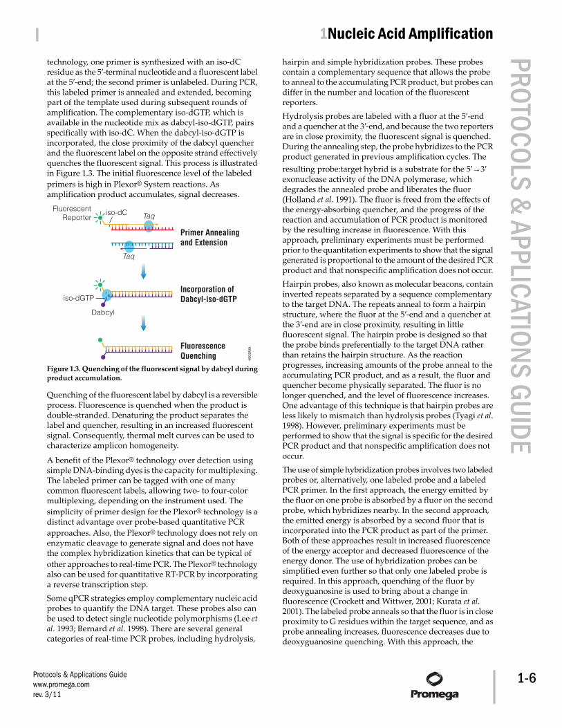
technology, one primer is synthesized with an iso-dCresidue as the 5′-terminal nucleotide and a fluorescent labelat the 5′-end; the second primer is unlabeled. During PCR,this labeled primer is annealed and extended, becomingpart of the template used during subsequent rounds ofamplification. The complementary iso-dGTP, which isavailable in the nucleotide mix as dabcyl-iso-dGTP, pairsspecifically with iso-dC. When the dabcyl-iso-dGTP isincorporated, the close proximity of the dabcyl quencherand the fluorescent label on the opposite strand effectivelyquenches the fluorescent signal. This process is illustratedin Figure 1.3. The initial fluorescence level of the labeledprimers is high in Plexor® System reactions. Asamplification product accumulates, signal decreases.
4909
MA
Primer Annealingand Extension
Incorporation ofDabcyl-iso-dGTP
FluorescenceQuenching
iso-dGTP
Dabcyl
Taq
Taq
iso-dCFluorescentReporter
Figure 1.3. Quenching of the fluorescent signal by dabcyl duringproduct accumulation.
Quenching of the fluorescent label by dabcyl is a reversibleprocess. Fluorescence is quenched when the product isdouble-stranded. Denaturing the product separates thelabel and quencher, resulting in an increased fluorescentsignal. Consequently, thermal melt curves can be used tocharacterize amplicon homogeneity.
A benefit of the Plexor® technology over detection usingsimple DNA-binding dyes is the capacity for multiplexing.The labeled primer can be tagged with one of manycommon fluorescent labels, allowing two- to four-colormultiplexing, depending on the instrument used. Thesimplicity of primer design for the Plexor® technology is adistinct advantage over probe-based quantitative PCRapproaches. Also, the Plexor® technology does not rely onenzymatic cleavage to generate signal and does not havethe complex hybridization kinetics that can be typical ofother approaches to real-time PCR. The Plexor® technologyalso can be used for quantitative RT-PCR by incorporatinga reverse transcription step.Some qPCR strategies employ complementary nucleic acidprobes to quantify the DNA target. These probes also canbe used to detect single nucleotide polymorphisms (Lee etal. 1993; Bernard et al. 1998). There are several generalcategories of real-time PCR probes, including hydrolysis,
hairpin and simple hybridization probes. These probescontain a complementary sequence that allows the probeto anneal to the accumulating PCR product, but probes candiffer in the number and location of the fluorescentreporters.Hydrolysis probes are labeled with a fluor at the 5′-endand a quencher at the 3′-end, and because the two reportersare in close proximity, the fluorescent signal is quenched.During the annealing step, the probe hybridizes to the PCRproduct generated in previous amplification cycles. Theresulting probe:target hybrid is a substrate for the 5′→3′exonuclease activity of the DNA polymerase, whichdegrades the annealed probe and liberates the fluor(Holland et al. 1991). The fluor is freed from the effects ofthe energy-absorbing quencher, and the progress of thereaction and accumulation of PCR product is monitoredby the resulting increase in fluorescence. With thisapproach, preliminary experiments must be performedprior to the quantitation experiments to show that the signalgenerated is proportional to the amount of the desired PCRproduct and that nonspecific amplification does not occur.Hairpin probes, also known as molecular beacons, containinverted repeats separated by a sequence complementaryto the target DNA. The repeats anneal to form a hairpinstructure, where the fluor at the 5′-end and a quencher atthe 3′-end are in close proximity, resulting in littlefluorescent signal. The hairpin probe is designed so thatthe probe binds preferentially to the target DNA ratherthan retains the hairpin structure. As the reactionprogresses, increasing amounts of the probe anneal to theaccumulating PCR product, and as a result, the fluor andquencher become physically separated. The fluor is nolonger quenched, and the level of fluorescence increases.One advantage of this technique is that hairpin probes areless likely to mismatch than hydrolysis probes (Tyagi et al.1998). However, preliminary experiments must beperformed to show that the signal is specific for the desiredPCR product and that nonspecific amplification does notoccur.The use of simple hybridization probes involves two labeledprobes or, alternatively, one labeled probe and a labeledPCR primer. In the first approach, the energy emitted bythe fluor on one probe is absorbed by a fluor on the secondprobe, which hybridizes nearby. In the second approach,the emitted energy is absorbed by a second fluor that isincorporated into the PCR product as part of the primer.Both of these approaches result in increased fluorescenceof the energy acceptor and decreased fluorescence of theenergy donor. The use of hybridization probes can besimplified even further so that only one labeled probe isrequired. In this approach, quenching of the fluor bydeoxyguanosine is used to bring about a change influorescence (Crockett and Wittwer, 2001; Kurata et al.2001). The labeled probe anneals so that the fluor is in closeproximity to G residues within the target sequence, and asprobe annealing increases, fluorescence decreases due todeoxyguanosine quenching. With this approach, the
Protocols & Applications Guidewww.promega.comrev. 3/11
| 1Nucleic Acid Amplification PROTOCOLS & APPLICATIONS GUIDE
1-6
location of probe is limited because the probe musthybridize so that the fluorescent dye is very near a Gresidue. The advantage of simple hybridization probes istheir ability to be multiplexed more easily than hydrolysisand hairpin probes through the use of differently coloredfluors and probes with different melting temperatures(reviewed in Wittwer et al. 2001).
Additional Resources for Real-Time PCRTechnical Bulletins and Manuals
TM318 GoTaq® qPCR Master Mix Technical ManualTM262 Plexor® qPCR System Technical ManualTM263 Plexor® One-Step qRT-PCR System Technical
ManualTM264 Plexor® Two-Step qRT-PCR System Technical
ManualPromega PublicationsIntroducing GoTaq® qPCR Master Mix: The bright choicefor dye-based qPCRThe Plexor™ Systems provide accurate quantitation inmultiplex qPCR and qRT-PCRPlexor™ technology: A new chemistry for real-time PCR
G. Rapid Amplified Polymorphic DNA AnalysisGenetic analysis of organisms at the molecular level is animportant and widely practiced scientific tool. Severaltechniques developed over more than a decade offer theopportunity to identify each individual or type ofindividual in a species unambiguously.One important PCR-based genetic analysis is randomamplified polymorphic DNA analysis (RAPD; reviewed inMcClelland and Welsh, 1994; Power, 1996; Black, 1993).RAPD uses small, nonspecific primers to amplify seeminglyrandom regions of genomic DNA. Successful primer pairsproduce different banding profiles of PCR productsbetween individuals, strains, cultivars or species whenanalyzed by gel electrophoresis.Slight modifications to the basic PCR method are made forRAPD. First, the primers are approximately 10 bases inlength compared to the 17- to 23-base primer length ofnormal PCR. Because primers are shorter, the annealingtemperature is reduced to less than 40°C.As with most PCR techniques, RAPD requires very littlematerial for analysis and is relatively insensitive to templateintegrity. No blotting techniques are required, thuseliminating the use of 32P, bypassing probe generation anddecreasing the amount of time required to obtain results.
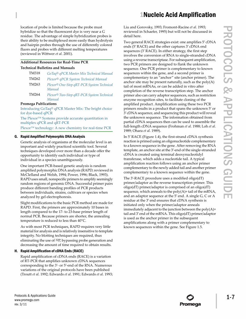
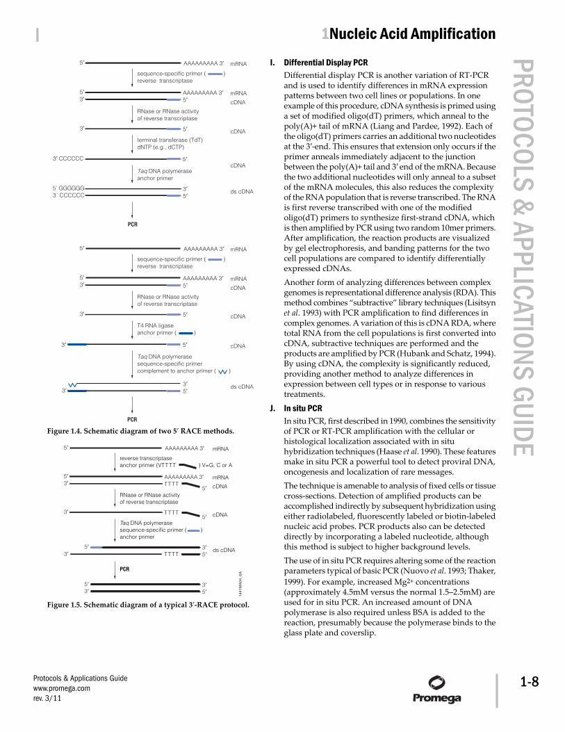
H. Rapid Amplification of cDNA Ends (RACE)Rapid amplification of cDNA ends (RACE) is a variationof RT-PCR that amplifies unknown cDNA sequencescorresponding to the 3′- or 5′-end of the RNA. Numerousvariations of the original protocols have been published(Troutt et al. 1992; Edwards et al. 1991; Edwards et al. 1993;
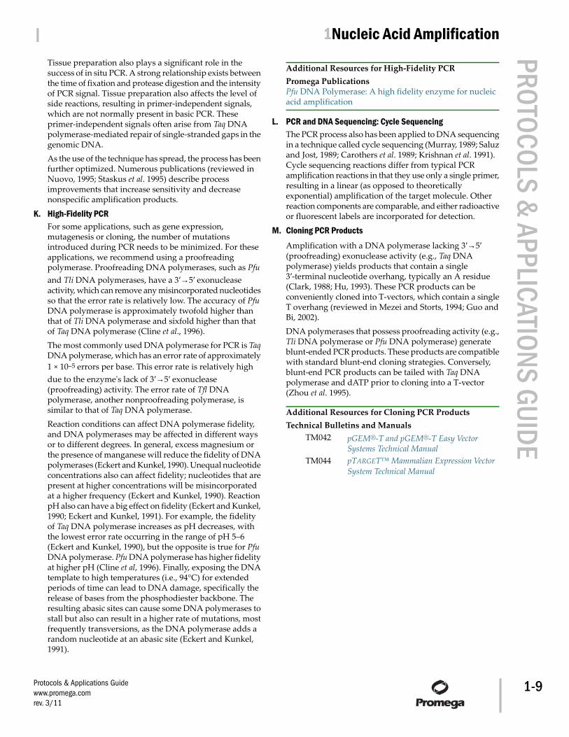
Liu and Gorovsky, 1993; Fromont-Racine et al. 1993;reviewed in Schaefer, 1995) but will not be discussed indetail here.Two general RACE strategies exist: one amplifies 5′ cDNAends (5′ RACE) and the other captures 3′ cDNA endsequences (3′ RACE). In either strategy, the first stepinvolves the conversion of RNA to single-stranded cDNAusing a reverse transcriptase. For subsequent amplification,two PCR primers are designed to flank the unknownsequence. One PCR primer is complementary to knownsequences within the gene, and a second primer iscomplementary to an “anchor” site (anchor primer). Theanchor site may be present naturally, such as the poly(A)tail of most mRNAs, or can be added in vitro aftercompletion of the reverse transcription step. The anchorprimer also can carry adaptor sequences, such as restrictionenzyme recognition sites, to facilitate cloning of theamplified product. Amplification using these two PCRprimers results in a product that spans the unknown 5′ or3′ cDNA sequence, and sequencing this product will revealthe unknown sequence. The information obtained frompartial cDNA sequences then can be used to assemble thefull-length cDNA sequence (Frohman et al. 1988; Loh et al.1989; Ohara et al. 1989).In 5′ RACE (Figure 1.4), the first-strand cDNA synthesisreaction is primed using an oligonucleotide complementaryto a known sequence in the gene. After removing the RNAtemplate, an anchor site at the 3′-end of the single-strandedcDNA is created using terminal deoxynucleotidyltransferase, which adds a nucleotide tail. A typicalamplification reaction follows using an anchor primercomplementary to the newly added tail and another primercomplementary to a known sequence within the gene.The 3′-RACE procedure uses a modified oligo(dT)primer/adaptor as the reverse transcription primer. Thisoligo(dT) primer/adaptor is comprised of an oligo(dT)sequence, which anneals to the poly(A)+ tail of the mRNA,and an adaptor sequence at the 5′ end. A single G, C or Aresidue at the 3′ end ensures that cDNA synthesis isinitiated only when the primer/adaptor annealsimmediately adjacent to the junction between the poly(A)+tail and 3′ end of the mRNA. This oligo(dT) primer/adaptoris used as the anchor primer in the subsequentamplifications along with a primer complementary toknown sequences within the gene. See Figure 1.5.
Protocols & Applications Guidewww.promega.comrev. 3/11
| 1Nucleic Acid Amplification PROTOCOLS & APPLICATIONS GUIDE
1-7
5′3′
3′ CCCCCC
cDNA
AAAAAAAAA 3′ mRNA
sequence-specific primer ( ) reverse transcriptase
5′
AAAAAAAAA 3′5′
mRNA5′3′
3′5′
5´ GGGGGG3´ CCCCCC
cDNA
terminal transferase (TdT)dNTP (e.g., dCTP)
5′cDNA
ds cDNA
PCR
RNase or RNase activity of reverse transcriptase
Taq DNA polymeraseanchor primer
5′3′
3′
cDNA
AAAAAAAAA 3′ mRNA
sequence-specific primer ( ) reverse transcriptase
5′
AAAAAAAAA 3′5′
mRNA5′3′
3′5′3′
cDNA
T4 RNA ligaseanchor primer ( )
5′ cDNA
ds cDNA
PCR
RNase or RNase activity of reverse transcriptase
Taq DNA polymerasesequence-specific primercomplement to anchor primer ( )
Figure 1.4. Schematic diagram of two 5′ RACE methods.
1441
MA
04_6
A
5′
cDNA
AAAAAAAAA 3′ mRNA
reverse transcriptaseanchor primer (VTTTT ) V=G, C or A
5′
3′
AAAAAAAAA 3′ mRNA5′
5′TTTT3′
5′
3′
5′ 3′3′ 5′
5′
TTTT
TTTT
3′
cDNA
Taq DNA polymerasesequence-specific primer ( )anchor primer
ds cDNA
PCR
RNase or RNase activity of reverse transcriptase
Figure 1.5. Schematic diagram of a typical 3′-RACE protocol.
I. Differential Display PCRDifferential display PCR is another variation of RT-PCRand is used to identify differences in mRNA expressionpatterns between two cell lines or populations. In oneexample of this procedure, cDNA synthesis is primed usinga set of modified oligo(dT) primers, which anneal to thepoly(A)+ tail of mRNA (Liang and Pardee, 1992). Each ofthe oligo(dT) primers carries an additional two nucleotidesat the 3′-end. This ensures that extension only occurs if theprimer anneals immediately adjacent to the junctionbetween the poly(A)+ tail and 3′ end of the mRNA. Becausethe two additional nucleotides will only anneal to a subsetof the mRNA molecules, this also reduces the complexityof the RNA population that is reverse transcribed. The RNAis first reverse transcribed with one of the modifiedoligo(dT) primers to synthesize first-strand cDNA, whichis then amplified by PCR using two random 10mer primers.After amplification, the reaction products are visualizedby gel electrophoresis, and banding patterns for the twocell populations are compared to identify differentiallyexpressed cDNAs.Another form of analyzing differences between complexgenomes is representational difference analysis (RDA). Thismethod combines “subtractive” library techniques (Lisitsynet al. 1993) with PCR amplification to find differences incomplex genomes. A variation of this is cDNA RDA, wheretotal RNA from the cell populations is first converted intocDNA, subtractive techniques are performed and theproducts are amplified by PCR (Hubank and Schatz, 1994).By using cDNA, the complexity is significantly reduced,providing another method to analyze differences inexpression between cell types or in response to varioustreatments.
J. In situ PCRIn situ PCR, first described in 1990, combines the sensitivityof PCR or RT-PCR amplification with the cellular orhistological localization associated with in situhybridization techniques (Haase et al. 1990). These featuresmake in situ PCR a powerful tool to detect proviral DNA,oncogenesis and localization of rare messages.The technique is amenable to analysis of fixed cells or tissuecross-sections. Detection of amplified products can beaccomplished indirectly by subsequent hybridization usingeither radiolabeled, fluorescently labeled or biotin-labelednucleic acid probes. PCR products also can be detecteddirectly by incorporating a labeled nucleotide, althoughthis method is subject to higher background levels.The use of in situ PCR requires altering some of the reactionparameters typical of basic PCR (Nuovo et al. 1993; Thaker,1999). For example, increased Mg2+ concentrations(approximately 4.5mM versus the normal 1.5–2.5mM) areused for in situ PCR. An increased amount of DNApolymerase is also required unless BSA is added to thereaction, presumably because the polymerase binds to theglass plate and coverslip.
Protocols & Applications Guidewww.promega.comrev. 3/11
| 1Nucleic Acid Amplification PROTOCOLS & APPLICATIONS GUIDE
1-8
Tissue preparation also plays a significant role in thesuccess of in situ PCR. A strong relationship exists betweenthe time of fixation and protease digestion and the intensityof PCR signal. Tissue preparation also affects the level ofside reactions, resulting in primer-independent signals,which are not normally present in basic PCR. Theseprimer-independent signals often arise from Taq DNApolymerase-mediated repair of single-stranded gaps in thegenomic DNA.As the use of the technique has spread, the process has beenfurther optimized. Numerous publications (reviewed inNuovo, 1995; Staskus et al. 1995) describe processimprovements that increase sensitivity and decreasenonspecific amplification products.
K. High-Fidelity PCRFor some applications, such as gene expression,mutagenesis or cloning, the number of mutationsintroduced during PCR needs to be minimized. For theseapplications, we recommend using a proofreadingpolymerase. Proofreading DNA polymerases, such as Pfuand Tli DNA polymerases, have a 3′→5′ exonucleaseactivity, which can remove any misincorporated nucleotidesso that the error rate is relatively low. The accuracy of PfuDNA polymerase is approximately twofold higher thanthat of Tli DNA polymerase and sixfold higher than thatof Taq DNA polymerase (Cline et al., 1996).The most commonly used DNA polymerase for PCR is TaqDNA polymerase, which has an error rate of approximately1 × 10–5 errors per base. This error rate is relatively highdue to the enzyme's lack of 3′→5′ exonuclease(proofreading) activity. The error rate of Tfl DNApolymerase, another nonproofreading polymerase, issimilar to that of Taq DNA polymerase.Reaction conditions can affect DNA polymerase fidelity,and DNA polymerases may be affected in different waysor to different degrees. In general, excess magnesium orthe presence of manganese will reduce the fidelity of DNApolymerases (Eckert and Kunkel, 1990). Unequal nucleotideconcentrations also can affect fidelity; nucleotides that arepresent at higher concentrations will be misincorporatedat a higher frequency (Eckert and Kunkel, 1990). ReactionpH also can have a big effect on fidelity (Eckert and Kunkel,1990; Eckert and Kunkel, 1991). For example, the fidelityof Taq DNA polymerase increases as pH decreases, withthe lowest error rate occurring in the range of pH 5–6(Eckert and Kunkel, 1990), but the opposite is true for PfuDNA polymerase. Pfu DNA polymerase has higher fidelityat higher pH (Cline et al, 1996). Finally, exposing the DNAtemplate to high temperatures (i.e., 94°C) for extendedperiods of time can lead to DNA damage, specifically therelease of bases from the phosphodiester backbone. Theresulting abasic sites can cause some DNA polymerases tostall but also can result in a higher rate of mutations, mostfrequently transversions, as the DNA polymerase adds arandom nucleotide at an abasic site (Eckert and Kunkel,1991).
Additional Resources for High-Fidelity PCRPromega PublicationsPfu DNA Polymerase: A high fidelity enzyme for nucleicacid amplification
L. PCR and DNA Sequencing: Cycle SequencingThe PCR process also has been applied to DNA sequencingin a technique called cycle sequencing (Murray, 1989; Saluzand Jost, 1989; Carothers et al. 1989; Krishnan et al. 1991).Cycle sequencing reactions differ from typical PCRamplification reactions in that they use only a single primer,resulting in a linear (as opposed to theoreticallyexponential) amplification of the target molecule. Otherreaction components are comparable, and either radioactiveor fluorescent labels are incorporated for detection.
M. Cloning PCR ProductsAmplification with a DNA polymerase lacking 3′→5′(proofreading) exonuclease activity (e.g., Taq DNApolymerase) yields products that contain a single3′-terminal nucleotide overhang, typically an A residue(Clark, 1988; Hu, 1993). These PCR products can beconveniently cloned into T-vectors, which contain a singleT overhang (reviewed in Mezei and Storts, 1994; Guo andBi, 2002).DNA polymerases that possess proofreading activity (e.g.,Tli DNA polymerase or Pfu DNA polymerase) generateblunt-ended PCR products. These products are compatiblewith standard blunt-end cloning strategies. Conversely,blunt-end PCR products can be tailed with Taq DNApolymerase and dATP prior to cloning into a T-vector(Zhou et al. 1995).
Additional Resources for Cloning PCR ProductsTechnical Bulletins and Manuals
TM042 pGEM®-T and pGEM®-T Easy VectorSystems Technical Manual
TM044 pTARGET™ Mammalian Expression VectorSystem Technical Manual
Protocols & Applications Guidewww.promega.comrev. 3/11
| 1Nucleic Acid Amplification PROTOCOLS & APPLICATIONS GUIDE
1-9
Promega PublicationsTechnically speaking: T-vector cloningRapid ligation for the pGEM®-T and pGEM®-T Easy VectorSystemsCloning blunt-end Pfu DNA polymerase-generated PCRfragments into pGEM®-T Vector SystemsTechnically speaking: Optimized cloning with T vectorsDigestion of PCR and RT-PCR products with restrictionendonucleases without prior purification or precipitationVector MapspGEM®-T VectorpGEM®-T Easy VectorpTARGET™ Mammalian Expression VectorCitationsKurth, E.G. et al. (2008) Involvement of BmoR and BmoGin n-alkane metabolism in Pseudomonas butanovora. Microbiology 154, 139–47.The authors characterized five open-reading framesflanking the alcohol-inducible alkane monooxygenase(BMO) structural gene of Pseudomonas butanovora. Strainswith mutated bmoR, encoding a putative transcriptionalregulator, or bmoG, encoding a putative chaperonin, werecreated by gene inactivation. The bmoR gene was amplifiedand cloned into the pGEM®-T Vector for disruption witha kanamycin cassette. The two termini of the bmoG genewere amplified separately, ligated to the kanamycin cassetteand cloned into the pGEM®-T Easy Vector. Plasmidsencoding the disrupted genes were transformed intoPseudomonas butanovora by electroporation.PubMed Number: 18174133Bröker, D. et al. (2008) The genomes of thenon-clearing-zone-forming and natural-rubber-degradingspecies Gordonia polyisoprenivorans and Gordonia westfalicaharbor genes expressing Lcp activity in Streptomyces strains. Appl. Environ. Microbiol. 74, 2288–97.Natural rubber-degrading bacteria fall into two categories:those forming clearing zones on latex overlay plates andthose that do not. To investigate this degradation process,the authors amplified latex-clearing protein (lcp) homologsfrom non-clearing-zone-forming bacteria using degeneratePCR primers based on lcp sequences from clearing-zoneforming species. The 3′ region of the lcp gene in G. westfalicawas amplified by nested PCR using biotinylated primers,and the amplified products were cloned in the pGEM®-TEasy Vector and sequenced using universal M13 forwardand reverse primers.PubMed Number: 18296529
II. General Considerations for PCR OptimizationThis discussion focuses on the use of Taq DNA polymerasein PCR, since this is the enzyme most commonly used inPCR. Many of these suggestions also apply when usingother DNA polymerases.
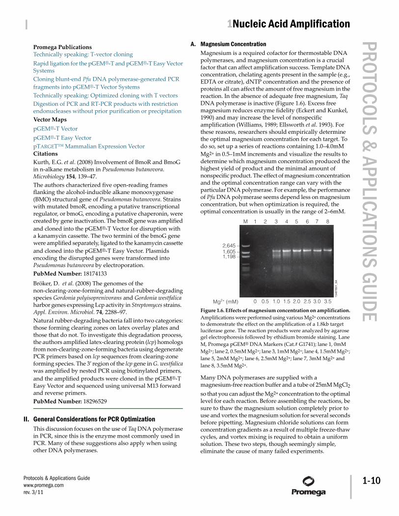
A. Magnesium ConcentrationMagnesium is a required cofactor for thermostable DNApolymerases, and magnesium concentration is a crucialfactor that can affect amplification success. Template DNAconcentration, chelating agents present in the sample (e.g.,EDTA or citrate), dNTP concentration and the presence ofproteins all can affect the amount of free magnesium in thereaction. In the absence of adequate free magnesium, TaqDNA polymerase is inactive (Figure 1.6). Excess freemagnesium reduces enzyme fidelity (Eckert and Kunkel,1990) and may increase the level of nonspecificamplification (Williams, 1989; Ellsworth et al. 1993). Forthese reasons, researchers should empirically determinethe optimal magnesium concentration for each target. Todo so, set up a series of reactions containing 1.0–4.0mMMg2+ in 0.5–1mM increments and visualize the results todetermine which magnesium concentration produced thehighest yield of product and the minimal amount ofnonspecific product. The effect of magnesium concentrationand the optimal concentration range can vary with theparticular DNA polymerase. For example, the performanceof Pfu DNA polymerase seems depend less on magnesiumconcentration, but when optimization is required, theoptimal concentration is usually in the range of 2–6mM.
1348
TB
01_6
A
M
2,645 –1,605 –1,198 –
Mg2+ (mM)
1 2 3 4 5 6 7 8
0 0.5 1.0 1.5 2.0 2.5 3.0 3.5
Figure 1.6. Effects of magnesium concentration on amplification.Amplifications were performed using various Mg2+ concentrationsto demonstrate the effect on the amplification of a 1.8kb targetluciferase gene. The reaction products were analyzed by agarosegel electrophoresis followed by ethidium bromide staining. LaneM, Promega pGEM® DNA Markers (Cat.# G1741); lane 1, 0mMMg2+; lane 2, 0.5mM Mg2+; lane 3, 1mM Mg2+; lane 4, 1.5mM Mg2+;lane 5, 2mM Mg2+; lane 6, 2.5mM Mg2+; lane 7, 3mM Mg2+ andlane 8, 3.5mM Mg2+.
Many DNA polymerases are supplied with amagnesium-free reaction buffer and a tube of 25mM MgCl2so that you can adjust the Mg2+ concentration to the optimallevel for each reaction. Before assembling the reactions, besure to thaw the magnesium solution completely prior touse and vortex the magnesium solution for several secondsbefore pipetting. Magnesium chloride solutions can formconcentration gradients as a result of multiple freeze-thawcycles, and vortex mixing is required to obtain a uniformsolution. These two steps, though seemingly simple,eliminate the cause of many failed experiments.
Protocols & Applications Guidewww.promega.comrev. 3/11
| 1Nucleic Acid Amplification PROTOCOLS & APPLICATIONS GUIDE
1-10
Some scientists prefer to use reaction buffers that alreadycontain MgCl2 at a final concentration of 1.5mM. It shouldbe noted, however, that Hu et al. reported performancevariability of reaction buffer solutions containingmagnesium (Hu et al. 1992). The free magnesium changesof 0.6mM observed in their experiments dramaticallyaffected amplification yields in an allele-specific manner.The authors found that heating the buffer at 90°C for 10minutes restored the homogeneity of the solution. Theypostulated that magnesium chloride precipitates as a resultof multiple freeze-thaw cycles.
B. Buffer ConsiderationsMost reaction buffers consist of a buffering agent, mostoften a Tris-based buffer, and salt, commonly KCl. Thebuffer regulates the pH of the reaction, which affects DNApolymerase activity and fidelity. Modest concentrations ofKCl will increase DNA polymerase activity by 50–60% overactivities in the absence of KCl; 50mM KCl is consideredoptimal (Gelfand, 1989).
GoTaq® DNA Polymerase contains native Taq DNApolymerase in a proprietary formulation. It is supplied with5X Green GoTaq® Reaction Buffer and 5X Colorless GoTaq®
Reaction Buffer. The 5X Green GoTaq® Reaction Buffercontains two dyes (blue and yellow) that separate duringelectrophoresis to monitor migration progress. The bufferalso contains a compound that increases the density of thesample so that it will sink into the well of the agarose gel,allowing reactions to be directly loaded onto an agarosegel without the need for loading dye. The blue dyecomigrates at the same rate as a 3–5kb DNA fragment in a1% agarose gel. The yellow dye migrates at a rate fasterthan primers (<50bp) in a 1% agarose gel. The 5X ColorlessGoTaq® Reaction Buffer and 5X Green GoTaq® ReactionBuffer have the same formulation, except for the dyes. The5X Colorless GoTaq® Reaction Buffer is recommended forany applications where absorbance or fluorescencemeasurements of the PCR amplimer will be taken withoutprior cleanup. Both buffers are supplied at pH 8.5 andcontain MgCl2 at a concentration of 7.5mM for a finalconcentration of 1.5mM.
GoTaq® Flexi DNA Polymerase is supplied with 5X GreenGoTaq® Flexi Reaction Buffer and 5X Colorless GoTaq®
Flexi Reaction Buffer. The compositions are identical to the5X Green GoTaq® Reaction Buffer and 5X Colorless GoTaq®
Reaction Buffer, except that the GoTaq® Flexi reactionbuffers do not contain MgCl2. Instead, the GoTaq® FlexiDNA Polymerase is supplied with a tube of 25mM MgCl2so that reactions can be supplemented with varyingconcentrations of magnesium.
C. Enzyme ConcentrationWe recommend using 1–1.25 units of Taq DNA polymerasein a 50μl amplification reaction. In most cases, this is anexcess of enzyme, and adding more enzyme will notsignificantly increase product yield. In fact, increasedamounts of enzyme increase the likelihood of generating
artifacts associated with the intrinsic 5′→3′ exonucleaseactivity of Taq DNA polymerase, resulting in smeared bandsin an agarose gel (Longley et al. 1990; Bell and DeMarini,1991).Pipetting errors are a frequent cause of excessive enzymelevels. Accurate dispensing of small volumes of enzymesolutions in 50% glycerol is difficult, so we stronglyrecommend preparing a reaction master mix, whichrequires a larger volume of each reagent, to reduce pipettingerrors.
D. PCR Primer DesignPCR primers define the target region to be amplified andgenerally range in length from 15–30 bases. Ideally primerswill have a GC-content of 40–60%. Avoid three G or Cresidues in a row near the 3′-end of the primer to minimizenonspecific primer annealing. Also, avoid primers withintra- or intermolecular complementary sequences tominimize the production of primer-dimer. Intramolecularregions of secondary structure can interfere with primerannealing to the template and should be avoided.Ideally, the melting temperature (Tm), the temperature atwhich 50% of the primer molecules are annealed to thecomplementary sequence, of the two primers will be within5°C so that the primers anneal efficiently at the sametemperature. Primers can be designed to include sequencesthat are useful for downstream applications. For example,restriction enzyme sites can be placed at the 5′-ends of PCRprimers to facilitate subsequent cloning of the PCR product,or a T7 RNA polymerase promoter can be added to allowin vitro transcription without the need to subclone the PCRproduct into a vector.
E. Template QualitySuccessful amplification depends on DNA templatequantity and quality. Reagents commonly used to purifynucleic acids (salts, guanidine, proteases, organic solventsand SDS) are potent inactivators of DNA polymerases. Forexample, 0.01% SDS will inhibit Taq DNA polymerase by90%, while 0.1% SDS will inhibit Taq DNA polymerase by99.9% (Konat et al. 1994). A few other examples of PCRinhibitors are phenol (Katcher and Schwartz, 1994), heparin(Beutler et al. 1990; Holodniy et al. 1991), xylene cyanol,bromophenol blue (Hoppe et al. 1992), plant polysaccharides(Demeke and Adams, 1992), and the polyamines spermineand spermidine (Ahokas and Erkkila, 1993). In some cases,the inhibitor is not introduced into the reaction with thenucleic acid template. A good example of this is aninhibitory substance that can be released from polystyreneor polypropylene upon exposure to ultraviolet light (Paoet al. 1993; Linquist et al. 1998).If an amplification reaction fails and you suspect the DNAtemplate is contaminated with an inhibitor, add the suspectDNA preparation to a control reaction with a DNA templateand primer pair that has amplified well in the past . Failureto amplify the control DNA usually indicates the presence
Protocols & Applications Guidewww.promega.comrev. 3/11
| 1Nucleic Acid Amplification PROTOCOLS & APPLICATIONS GUIDE
1-11
of an inhibitor. Additional steps to clean up the DNApreparation, such as phenol:chloroform extraction orethanol precipitation, may be necessary.
F. Template QuantityThe amount of template required for successfulamplification depends upon the complexity of the DNAsample. For example, of a 4kb plasmid containing a 1kbtarget sequence, 25% of the input DNA is the target ofinterest. Conversely, a 1kb target sequence in the humangenome (3.3 × 10⁹bp) represents approximately 0.00003%of the input DNA. Thus, approximately 1,000,000-fold morehuman genomic DNA is required to maintain the samenumber of target copies per reaction. Common mistakesinclude using too much plasmid DNA, too much PCRproduct or too little genomic DNA as the template.Reactions with too little DNA template will have low yields,while reactions with too much DNA template can beplagued by nonspecific amplification. If possible, start with>104 copies of the target sequence to obtain a signal in 25–30cycles, but try to keep the final DNA concentration of thereaction ≤10ng/μl. When reamplifying a PCR product, theconcentration of the specific PCR product is often notknown. We recommend diluting the previous amplificationreaction 1:10 to 1:10,000 before reamplification.
1μg of 1kb RNA = 1.77 × 10¹² molecules
1μg of 1kb dsDNA = 9.12 × 10¹¹ molecules
1μg of pGEM® Vector DNA = 2.85 × 10¹¹ molecules
1μg of lambda DNA = 1.9 × 10¹⁰ molecules
1μg of E. coli genomic DNA = 2 × 10⁸ molecules
1μg of human genomic DNA = 3.04 × 10⁵ molecules
G. Cycling ParametersThe two most commonly altered cycling parameters areannealing temperature and extension time. The lengthsand temperatures for the other steps of a PCR cycle do notusually vary significantly. However in some cases, thedenaturation cycle can be shortened or a lower denaturationtemperature used to reduce the number of depurinationevents, which can lead to mutations in the PCR products.Primer sequence is a major factor that determines theoptimal annealing temperature, which is often within 5°Cof the melting temperature of the primers. Using anannealing temperature slightly higher than the primer Tmwill increase annealing stringency and can minimizenonspecific primer annealing and decrease the amount ofundesired products synthesized. Using an annealingtemperature lower than the primer Tm can result in higheryields, as the primers anneal more efficiently at the lowertemperature. We recommend testing several annealingtemperatures, starting approximately 5°C below the Tm, todetermine the best annealing conditions. In many cases,nonspecific amplification and primer-dimer formation can
be reduced through optimization of annealing temperature,but if undesirable PCR products remain a problem, considerincorporating one of the many hot-start PCR methods.Oligonucleotide synthesis facilities will often provide anestimate of a primer's Tm. The Tm also can be calculatedusing the Biomath Calculators. Numerous formulas existto determine the theoretical Tm of nucleic acids (Baldino,Jr. et al. 1989; Rychlik et al. 1990). The formula below canbe used to estimate the melting temperature foroligonucleotides:
Tm = 81.5 + 16.6 × (log10[Na+]) + 0.41 × (%G+C) – 675/n
where [Na+] is the molar salt concentration and n = numberof bases in the oligonucleotideExample:To calculate the melting temperature of a 22meroligonucleotide with 60% G+C in 50mM KCl:Tm = 81.5 + 16.6 × (log10[0.05]) + 0.41 × (60) – 675/22
= 81.5 + 16.6 × (–1.30) + 24.60 – 30.68 = 54°CThe length of the extension cycle, which may need to beoptimized, depends on PCR product size and the DNApolymerase being used. In general, allow approximately 1minute for every 1kb of amplicon (minimum extensiontime = 1 minute) for nonproofreading DNA polymerasesand 2 minutes for every 1kb of amplicon for proofreadingDNA polymerases. Avoid excessively long extension times,as they can increase the likelihood of generating artifactsassociated with the intrinsic 5′→3′ exonuclease activity ofTaq DNA polymerase (Longley et al. 1990; Bell andDeMarini, 1991).PCR typically involves 25–35 cycles of amplification. Therisk of undesirable PCR products appearing in the reactionincreases as the cycle number increases, so we recommendperforming only enough cycles to synthesize the desiredamount of product. If nonspecific amplification productsaccumulate before sufficient amounts of PCR product canbe synthesized, consider diluting the products of the firstreaction and performing a second amplification with thesame primers or primers that anneal to sequences withinthe desired PCR product (nested primers).
H. PCR Enhancers and AdditivesAddition of PCR-enhancing agents can increase yield ofthe desired PCR product or decrease production ofundesired products. There are many PCR enhancers, whichcan act through a number of different mechanisms. Thesereagents will not enhance all PCRs; the beneficial effectsare often template- and primer-specific and will need to bedetermined empirically. Some of the more commonenhancing agents are discussed below.Addition of betaine, DMSO and formamide can be helpfulwhen amplifying GC-rich templates and templates thatform strong secondary structures, which can cause DNApolymerases to stall. GC-rich templates can be problematicdue to inefficient separation of the two DNA strands or the
Protocols & Applications Guidewww.promega.comrev. 3/11
| 1Nucleic Acid Amplification PROTOCOLS & APPLICATIONS GUIDE
1-12

tendency for the complementary, GC-rich primers to formintermolecular secondary structures, which will competewith primer annealing to the template. Betaine reduces theamount of energy required to separate DNA strands (Reeset al. 1993). DMSO and formamide are thought to aidamplification in a similar manner by interfering withhydrogen bond formation between two DNA strands(Geiduschek and Herskovits, 1961).Some reactions that amplify poorly in the absence ofenhancers will give a higher yield of PCR product whenbetaine (1M), DMSO (1–10%) or formamide (1–10%) areadded. Concentrations of DMSO greater than 10% andformamide greater than 5% can inhibit Taq DNApolymerase and presumably other DNA polymerases aswell (Varadaraj and Skinner, 1994).In some cases, general stabilizing agents such as BSA(0.1mg/ml), gelatin (0.1–1.0%) and nonionic detergents(0–0.5%) can overcome amplification failure. Theseadditives can increase DNA polymerase stability andreduce the loss of reagents through adsorption to tubewalls. BSA also has been shown to overcome the inhibitoryeffects of melanin on RT-PCR (Giambernardi et al. 1998).Nonionic detergents, such as Tween®-20, NP-40 and Triton®
X-100, have the added benefit of overcoming inhibitoryeffects of trace amounts of strong ionic detergents, such as0.01% SDS (Gelfand and White, 1990). Ammonium ionscan make an amplification reaction more tolerant ofnonoptimal conditions. For this reason, some PCR reagentsinclude 10–20mM (NH4)2SO4. Other PCR enhancers includeglycerol (5–20%), polyethylene glycol (5–15%) andtetramethyl ammonium chloride (60mM).
I. Nucleic Acid Cross-ContaminationIt is important to minimize cross-contamination betweensamples and prevent carryover of RNA and DNA from oneexperiment to the next. Use separate work areas andpipettors for pre- and postamplification steps. Use positivedisplacement pipettes or aerosol-resistant tips to reducecross-contamination during pipetting. Wear gloves, andchange them often.There are a number of techniques that can be used toprevent amplification of contaminating DNA. PCR reagentscan be treated with isopsoralen, a photo-activated,cross-linking reagent that intercalates into double-strandedDNA molecules and forms covalent, interstrand crosslinks,to prevent DNA denaturation and replication. Theseinterstrand crosslinks effectively render contaminatingDNA unamplifiable.Treatment of PCR reagents with uracil-N-glycosylase(UNG), a DNA repair enzyme that hydrolyzes thebase-ribose bond at uracil residues, eliminates one of themost common sources of DNA contamination: previouslyamplified PCR products. UNG treatment preventsreplication of uracil-containing DNA by causing the DNApolymerase to stall at the resulting abasic sites. For UNGto be an effective safeguard against contamination, theproducts of previous amplifications must be synthesized
in the presence of dUTP. This is easily accomplished bysubstituting dUTP for some or all of the dTTP in thereaction. Nonproofreading polymerases will readilyincorporate dUTP into a PCR product, but proofreadingpolymerases incorporate dUTP much less efficiently(Slupphaug et al. 1993; Greagg et al. 1999; Lasken et al. 1996).Since dUTP incorporation has no noticeable effect on theintensity of ethidium bromide staining or electrophoreticmobility of the PCR product, reactions can be analyzed bystandard agarose gel electrophoresis. While both methodsare effective (Rys and Persing, 1993), UNG treatment hasthe advantage that both single-stranded anddouble-stranded DNA templates will be renderedunamplifiable (Longo et al. 1990).
III. General Considerations for RT-PCRPlease also read General Considerations for PCROptimization. Many of the important parameters discussedthere also apply to RT-PCR. For a discussion of reversetranscriptases commonly used for RT-PCR, see the ReverseTranscription section.
A. Overview of the Access and AccessQuick™ RT-PCR SystemsThe Access RT-PCR System and AccessQuick™ RT-PCRSystem are designed for the reverse transcription andamplification of a specific target RNA from either totalRNA or mRNA (Miller and Storts, 1995; Knoche andDenhart, 2001). These one-tube, two-enzyme systemsprovide sensitive, quick and reproducible analysis of evenrare RNAs (Miller and Storts, 1996). The systems use AMVReverse Transcriptase for first-strand cDNA synthesis andthe thermostable Tfl DNA Polymerase from Thermus flavus(Kaledin et al. 1981) for second-strand cDNA synthesis andDNA amplification. The systems include an optimizedsingle-buffer system that permits sensitive detection ofRNA transcripts without the need for buffer additionsbetween reverse transcription and PCR amplification steps.This simplifies the procedure and reduces the potential forcontamination. The elevated reaction temperature (45°C)possible with AMV reverse transcriptase minimizesproblems encountered with RNA secondary structures(Brooks et al. 1995).
B. Template ConsiderationsFor RT-PCR, successful reverse transcription depends onRNA integrity and purity. Procedures for creating andmaintaining a ribonuclease-free (RNase-free) environmentto minimize RNA degradation are described in Blumberg,1987. The use of an RNase inhibitor (e.g., RecombinantRNasin® Ribonuclease Inhibitor) is strongly recommended.For optimal results, the RNA template, whether a totalRNA preparation, an mRNA population or a synthesizedRNA transcript, should be DNA-free to avoid amplificationof contaminating DNA. The most commonly used DNApolymerases for PCR have no reverse transcriptase activityunder standard reaction conditions, and thus, amplificationproducts will be generated only if the template containstrace amounts of DNA with similar sequences.
Protocols & Applications Guidewww.promega.comrev. 3/11
| 1Nucleic Acid Amplification PROTOCOLS & APPLICATIONS GUIDE
1-13

Successful RT-PCR also depends on RNA quantity, whichmay need to be varied to determine the optimal amount.Excellent amplification results can be obtained with theAccess and AccessQuick™ RT-PCR Systems using totalRNA template levels in the range of 1pg–1μg per reaction(Figure 1.7) or poly(A)+ RNA template levels in the rangeof 1pg–100ng.
bp
1166
TD
09_6
A
1,500 –1,000 –
– 540bp β-actin amplimer
500 –
100 –
pg total RNA per reaction
106M 105 104 103 102 101 1 0 M
Figure 1.7. Amplification of a specific message in total RNA.RT-PCR amplifications containing the indicated amounts of mouseliver total RNA were performed using the Access RT-PCR Systemas described in the Access RT-PCR protocol using oligonucleotideprimers specific to the mouse β-actin transcript. The specific 540bpamplicon is indicated. Equivalent aliquots of each amplificationreaction were separated on a 3% NuSieve®/ 1% agarose gel in 1XTAE buffer containing 0.5μg/ml ethidium bromide. Lanes M, 100bpDNA Ladder (Cat.# G2101).
C. Reverse Transcription Primer DesignSelection of an appropriate primer for reverse transcriptiondepends on target mRNA size and the presence ofsecondary structure. For example, a primer that annealsspecifically to the 3′-end of the transcript (asequence-specific primer or oligo(dT) primer) may beproblematic when reverse transcribing the 5′-ends of longmRNAs or molecules that have significant secondarystructure, which can cause the reverse transcriptase to stallduring cDNA synthesis. Random hexamers prime reversetranscription at multiple points along the transcript. Forthis reason, they are useful for either long mRNAs ortranscripts with significant secondary structure.Whenever possible, we recommend using a primer thatanneals only to defined sequences in particular RNAs(sequence-specific primers) rather than to entire RNApopulations in the sample (e.g., random hexamers oroligo(dT) primer). To differentiate between amplificationof cDNA and amplification of contaminating genomic DNA,design primers to anneal to sequences in exons on oppositesides of an intron so that any amplification product derivedfrom genomic DNA will be much larger than the productamplified from the target cDNA. This size difference notonly makes it possible to differentiate the two products bygel electrophoresis but also favors the synthesis of thesmaller cDNA-derived product (amplification of smallerfragments is often more efficient that that of longfragments).
Regardless of primer choice, the final primer concentrationin the reaction is usually within the range of 0.1–1.0μM,but this may need to be optimized. We recommend usinga final concentration of 1μM primer (50pmol in a 50μlreaction) as a starting point for optimization. Moreinformation on PCR primer design is provided in the PCRPrimer Design section.
D. Cycle ParametersEfficient first-strand cDNA synthesis can be accomplishedin a 20- to 60-minute incubation at 37–45°C using AMVreverse transcriptase or at 37–42° for M-MLV reversetranscriptase. When using AMV RT we recommend usinga sequence-specific primer and performing reversetranscription at 45°C for 45 minutes as a starting point. Thehigher reaction temperature will minimize the effects ofRNA secondary structure and encourage full-length cDNAsynthesis. First-strand cDNA synthesis with randomhexamers and oligo(dT) primer should be conducted atroom temperature (20–25°C) and 37°C, respectively.The Access and AccessQuick™ RT-PCR Systems do notrequire RNA denaturation prior to initiation of the reversetranscription reaction. If desired, however, a denaturationstep may be incorporated by incubating a separate tubecontaining the primers and RNA template at 94°C for 2minutes. Do not incubate AMV reverse transcriptase at94°C; it will be inactivated. The template/primer mixturethen can be cooled to 45°C and added to the RT-PCR mixfor the standard reverse transcription incubation at 45°C.Following the reverse transcription, we recommend a2-minute incubation at 94°C to denature the RNA/cDNAhybrid, inactivate AMV reverse transcriptase and dissociateAMV RT from the cDNA. It has been reported that AMVreverse transcriptase must be inactivated to obtain highyields of amplification product (Sellner et al. 1992;Chumakov, 1994).Most RNA samples can be detected using 30–40 cycles ofamplification. If the target RNA is rare or if only a smallamount of starting material is available, it may be necessaryto increase the number of cycles to 45 or 50 or dilute theproducts of the first reaction and reamplify.
IV. Thermostable DNA PolymerasesPrior to the use of thermostable DNA polymerases in PCR,researchers had to laboriously replenish the reaction withfresh enzyme (such as Klenow or T4 DNA polymerase)after each denaturation cycle. Thermostable DNApolymerases revolutionized and popularized PCR becauseof their ability to withstand the high denaturationtemperatures. The use of thermostable DNA polymerasesalso allowed higher annealing temperatures, whichimproved the stringency of primer annealing.Thermostable DNA polymerases can be used for eitherone-enzyme or two-enzyme RT-PCR (Myers and Gelfand,1991; Chiocchia and Smith, 1997). For example, Tth DNApolymerase can act as a reverse transcriptase in the presenceof Mn2+ for one-enzyme RT-PCR (Myers and Gelfand, 1991).
Protocols & Applications Guidewww.promega.comrev. 3/11
| 1Nucleic Acid Amplification PROTOCOLS & APPLICATIONS GUIDE
1-14
All of the DNA polymerases mentioned below can be usedto amplifye first-strand cDNA produced by a reversetranscriptase, such as AMV RT, in two-enzyme RT-PCR.Thermostable DNA polymerases can be divided into twogroups: those with a 3′→5′ exonuclease (proofreading)activity, such as Pfu DNA polymerase, and those withoutthe proofreading function, such as Taq DNA polymerase.These two groups have some important differences.Proofreading DNA polymerases are more accurate thannonproofreading polymerases due to the 3′→5′ exonucleaseactivity, which can remove a misincorporated nucleotidefrom a growing DNA chain. When the amplified productis to be cloned, expressed or used in mutation analysis, PfuDNA polymerase is a better choice due to its high fidelity.However, for routine PCR, where simple detection of anamplification product is the goal, Taq DNA polymerase isthe most commonly used enzyme because yields tend tobe higher with a nonproofreading DNA polymerase.Amplification with nonproofreading DNA polymerasesresults in the template-independent addition of a singlenucleotide to the 3′-end of the PCR product, whereas theuse of proofreading DNA polymerases results inblunt-ended PCR products (Clark, 1988; Hu, 1993). Thesingle-nucleotide overhang can simplify the cloning of PCRproducts.Proofreading DNA polymerases also are used in blendswith nonproofreading DNA polymerases, oramino-terminally truncated versions of Taq DNApolymerase, to amplify longer stretches of DNA withgreater accuracy than the nonproofreading DNApolymerase alone (Barnes, 1994; Cline et al. 1996). See LongPCR.
A. Taq DNA PolymeraseTaq DNA polymerase is isolated from Thermus aquaticusand catalyzes the primer-dependent incorporation ofnucleotides into duplex DNA in the 5′→3′ direction in thepresence of Mg2+. The enzyme does not possess 3′→5′exonuclease activity but has 5′→3′ exonuclease activity.Taq DNA polymerase is suitable for most PCR applicationsthat do not require a high-fidelity enzyme, such as detectingspecific DNA or RNA sequences. The error rate of Taq DNApolymerase is approximately 1 × 10⁻⁵ errors/base, althoughthe fidelity does depend somewhat on the reactionconditions. The fidelity is slightly higher at lower pH, lowermagnesium concentration and relatively low dNTPconcentration (Eckert and Kunkel, 1990; Eckert and Kunkel,1991). See High-Fidelity PCR.Taq DNA polymerase is commonly used to amplify PCRproducts of 5kb or less. PCR products in the range of 5–10kbcan be amplified with Taq DNA polymerase but oftenrequire more optimization than smaller PCR products. Forproducts larger than approximately 10kb, we recommendan enzyme or enzyme mix and reaction conditions that aredesigned for long PCR.
Taq DNA polymerase is a processive enzyme with anextension rate of >60 nucleotides/second at 70°C (Innis etal. 1988), so an extension step of 1 minute per 1kb to beamplified should be sufficient to generate full-length PCRproducts. The enzyme has a half-life of 40 minutes at 95°C(Lawyer et al. 1993). Because Taq DNA polymerase is anonproofreading polymerase, PCR products generatedwith Taq DNA polymerase will contain a single-nucleotide3′ overhang, usually a 3′ A overhang.
Additional Resources for Taq DNA PolymeraseTechnical Bulletins and Manuals
9PIM300 GoTaq® DNA Polymerase Promega ProductInformation
9PIM829 GoTaq® Flexi DNA Polymerase PromegaProduct Information
Promega PublicationsGoTaq® Green Master Mix for quick and easy two-stepRT-PCR
B. Tfl DNA PolymeraseTfl DNA polymerase catalyzes the primer-dependentpolymerization of nucleotides into duplex DNA in thepresence of Mg2+. In the presence of Mn2+, Tfl DNApolymerase can use RNA as a template. Tfl DNApolymerase exhibits a 5′→3′ exonuclease activity but lacksa 3′→5′ exonuclease activity. This enzyme is commonlyused in PCR (Gaensslen et al. 1992), where its activity issimilar to that of Taq DNA polymerase. Tfl DNApolymerase is used in the Access and AccessQuick™RT-PCR Systems.
C. Tth DNA PolymeraseTth DNA polymerase catalyzes polymerization ofnucleotides into duplex DNA in the 5′→3′ direction in thepresence of MgCl2. The enzyme can use an RNA templatein the presence of MnCl2 (Myers and Gelfand, 1991;
Ruttimann et al. 1985). Tth DNA polymerase exhibits a 5′→3′exonuclease activity but lacks detectable 3′→5′ exonucleaseactivity. The error rate of Tth DNA polymerase has beenmeasured at 7.7 × 10⁻⁵ errors/base (Arakawa et al. 1996).Tth DNA polymerase can amplify target DNA in thepresence of phenol-saturated buffer (Katcher and Schwartz,1994) and has been reported to be more resistant toinhibition by blood components than other thermostablepolymerases (Ehrlich et al. 1991; Bej and Mahbubani, 1992).Tth DNA polymerase is commonly used for PCR (Myersand Gelfand, 1991; Carballeira et al. 1990) and RT-PCR(Myers and Gelfand, 1991; Chiocchia and Smith, 1997). Forprimer extension, RT-PCR and cDNA synthesis using RNAtemplates with complex secondary structure, the highreaction temperature of Tth DNA polymerase may be anadvantage over more commonly used reversetranscriptases, such as AMV and M-MLV reversetranscriptases. Recombinant Tth DNA polymerase has beenshown to exhibit RNase H-like activity (Auer et al. 1995).
Protocols & Applications Guidewww.promega.comrev. 3/11
| 1Nucleic Acid Amplification PROTOCOLS & APPLICATIONS GUIDE
1-15

Additional Resources for Tth DNA PolymeraseTechnical Bulletins and Manuals
9PIM210 Tth DNA Polymerase Promega ProductInformation
D. Tli DNA PolymeraseTli DNA polymerase replicates DNA throughpolymerization of nucleotides into duplex DNA in the 5′→3′direction in the presence of Mg2+. This enzyme also containsa 3′→5′ exonuclease activity, which results in increasedfidelity of nucleotide incorporation. There is no detectablereverse transcriptase activity or 5′→3′ exonuclease activity.Tli DNA polymerase will promote strand displacement at72°C but will not displace DNA at 55°C (Kong et al. 1993).Greater than 95% of the amplified products will beblunt-ended.Tli DNA polymerase is commonly used for PCR andRT-PCR, where its proofreading activity makes it usefulfor high-fidelity and long PCR (Keohavong et al. 1993). Dueto the 3′→5′ exonuclease activity of Tli DNA polymerase,the enzyme can degrade the oligonucleotide primers usedto initiate DNA synthesis. This exonucleolytic attack canbe effectively prevented by using hot-start PCR orintroducing a single phosphorothioate bond at the 3′ terminiof the primers (Byrappa et al. 1995). Tli DNA polymerasealso can be used for primer extension, where the highoptimal temperature of the enzyme may be an advantagefor templates with complex secondary structure.
E. Pfu DNA PolymerasePfu DNA polymerase has one of the lowest error rates ofall known thermophilic DNA polymerases used foramplification due to the high 3′→5′ exonuclease activity(Cline et al. 1996; Andre et al. 1997). For cloning andexpressing DNA after PCR, Pfu DNA polymerase is oftenthe enzyme of choice. Pfu DNA polymerase can be usedalone to amplify DNA fragments up to 5kb by increasingthe extension time to 2 minutes per kilobase. It is also usedin blends with DNA polymerases lacking the proofreadingfunction, such as Taq DNA polymerase, to achieve longeramplification products than with Pfu DNA polymerasealone (Barnes, 1994). However, the proofreading activitycan shorten PCR primers, leading to decreased yield andincreased nonspecific amplification. This exonucleolyticattack can be effectively prevented by using hot-start PCRor introducing a single phosphorothioate bond at the3′-termini of the primers (Byrappa et al. 1995).
Additional Resources for Pfu DNA PolymeraseTechnical Bulletins and Manuals
9PIM774 Pfu DNA Polymerase Promega ProductInformation
Promega PublicationsPfu DNA Polymerase: A high fidelity enzyme for nucleicacid amplification
V. Reverse TranscriptasesThe discovery of reverse transcriptases, or RNA-dependentDNA polymerases, and their role in retrovirus infection(Baltimore, 1970; Temin and Mizutani, 1970) alteredmolecular biology’s central dogma ofDNA→RNA→protein. Reverse transcriptases use an RNAtemplate to synthesize DNA and require a primer forsynthesis, like other DNA polymerases. For in vitroapplications, the primer can be either oligo(dT), whichhybridizes to the poly(A)+ tails of eukaryotic mRNAs,random hexamers, which prime synthesis throughout thelength of the RNA template, or a sequence-specific primer,which hybridizes to a known sequence within the RNAtemplate. Polymerization from a primer then proceeds asfor DNA-dependent DNA polymerases. The commonlyused reverse transcriptases, AMV reverse transcriptase,M-MLV reverse transcriptase and M-MLV reversetranscriptase, RNase H minus, perform the same reactionbut at different optimum temperatures (AMV, 42°C;M-MLV, 37°C; and M-MLV RT, RNase H–, 42°C).Some reverse transcriptases also possess intrinsic 3′- or5′-exoribonuclease (RNase) activity, which is generally usedto degrade the RNA template after first strand cDNAsynthesis. Absence of the 5′-exoribonuclease (RNase H)activity may aid production of longer cDNAs (Berger et al.1983).Some DNA-dependent DNA polymerases also possess areverse transcriptase activity, which can be favored undercertain conditions. For example, the thermostable,DNA-dependent Tth DNA polymerase exhibits reversetranscriptase activity when Mn2+ is substituted for Mg2+
in a reaction.
A. AMV Reverse TranscriptaseAMV RT catalyzes DNA polymerization using templateDNA, RNA or RNA:DNA hybrids (Houts et al. 1979). AMVreverse transcriptase is the preferred reverse transcriptasefor templates with high secondary structure due to itshigher reaction temperature (up to 58°C). AMV RT is usedin a wide variety of applications including cDNA synthesis(Houts et al. 1979; Gubler and Hoffman, 1983), RT-PCR andrapid amplification of cDNA ends (RACE; Skinner et al.1994). Although the high optimal temperature (42°C) makesit the enzyme of choice for cDNA synthesis using templateswith complex secondary structure, its relatively high RNaseH activity limits its usefulness for generation of long cDNAs(>5kb). For these templates, M-MLV RT or M-MLV RT,RNase H minus, may be a better choice.
Additional Resources for AMV Reverse TranscriptaseTechnical Bulletins and Manuals
9PIM510 AMV Reverse Transcriptase Promega ProductInformation
Protocols & Applications Guidewww.promega.comrev. 3/11
| 1Nucleic Acid Amplification PROTOCOLS & APPLICATIONS GUIDE
1-16

B. M-MLV Reverse TranscriptaseM-MLV RT is a single-polypeptide, RNA-dependent DNApolymerase. The enzyme also has DNA-dependent DNApolymerase activity at high enzyme levels (Roth et al. 1985).M-MLV RT is used in a variety of applications, includingcDNA synthesis, RT-PCR and RACE (Gerard, 1983). Itsrelatively low RNase H activity compared to AMV RTmakes M-MLV RT the enzyme of choice for generating longcDNAs (>5kb) (Sambrook and Russell, 2001). However, forshort templates with complex secondary structure, AMVRT or M-MLV RT, RNase H minus, may be a better choicedue to their higher optimal temperatures. M-MLV RT isless processive than AMV RT, so more units of M-MLV RTmay be required to generate the same amount of cDNA(Schaefer, 1995).
Additional Resources for M-MLV Reverse TranscriptaseTechnical Bulletins and Manuals
9PIM170 M-MLV Reverse Transcriptase PromegaProduct Information
C. M-MLV Reverse Transcriptase, RNase H MinusM-MLV reverse transcriptase, RNase H minus, is anRNA-dependent, 5′→3′ DNA polymerase that has beengenetically altered to remove the associated ribonucleaseH activity, which causes degradation of the RNA strand ofan RNA:DNA hybrid (Tanese and Goff, 1988). The absenceof RNase H activity makes M-MLV, RNase H minus, theenzyme of choice for generating long cDNAs (>5kb).However, for shorter templates with complex secondarystructure, AMV reverse transcriptase may be a better choicebecause it can be used at higher reaction temperatures.There are two forms of M-MLV, RNase H minus: thedeletion mutant and the point mutant. As the namessuggest, the deletion mutant had a specific sequence in theRNase H domain deleted, and the point mutant has a pointmutation introduced in the RNase H domain. While thenative M-MLV RT has a recommended reactiontemperature of 37°C, the deletion and point mutants aremore stable at higher temperatures and can be used atreaction temperatures of up to 50°C and 55°C, respectively,depending upon the reverse transcription primers used.The point mutant is often preferred over the deletionmutant because the point mutant has DNA polymeraseactivity comparable to that of the wildtype M-MLV enzyme,whereas the deletion mutant has a slightly reduced DNApolymerase activity compared to that of the wildtypeenzyme (Figure 1.8).
0 200 4000
200
400
600
800
1,000
Point MutantM-MLV RT
Deletion MutantRNase H+
Units of Enzyme
cDNA
Syn
thes
ized
(ng)
2919MA03_0A
Figure 1.8. Comparison of the mass amount of total cDNAsynthesized from 2μg of a 7.5kb RNA template by increasingamounts of three Promega M-MLV reverse transcriptases. Eachfirst-strand cDNA reaction was performed using 2μg of a 7.5kbRNA template (1μl), 0.5μg of oligo(dT)15 primer (1μl) and 14μlwater. The RNA and oligo(dT)15 primer were heated at 70°C for5 minutes and cooled on ice for 5 minutes. Five microliters ofM-MLV RT 5X Buffer, 1.25μl of 10μM dNTPs, 0.5μl of α-32P dCTP(10μCi/μl, 400Ci/mmol) and either 25, 50, 100, 150, 200 or 400 unitsof M-MLV Reverse Transcriptase, RNase H Minus, Point Mutant;M-MLV Reverse Transcriptase, RNase H Minus, Deletion Mutant;or native M-MLV Reverse Transcriptase (RNase H+) was used ina final volume of 25μl. Reactions were incubated at 42°C for 60minutes. TCA precipitations were performed, and first-strandcDNA yields were calculated.
Additional Resources for M-MLV Reverse Transcriptase,RNase H MinusTechnical Bulletins and Manuals
9PIM530 M-MLV Reverse Transcriptase, RNase HMinus, Promega Product Information
9PIM368 M-MLV Reverse Transcriptase, RNase HMinus, Point Mutant, Promega ProductInformation
VI. Example of a PCR ProtocolMaterials Required:(see Composition of Solutions section)• template DNA• downstream primer• upstream primer• GoTaq® DNA Polymerase (Cat.# M8291)• MgCl2, 25mM• Nuclease-Free Water (Cat.# P1193)• nuclease-free light mineral oil (e.g., Sigma Cat.# M5904)
if using a thermal cycler without a heated lid; do notautoclave
• dNTP mix, 10mM of each dNTP
Protocols & Applications Guidewww.promega.comrev. 3/11
| 1Nucleic Acid Amplification PROTOCOLS & APPLICATIONS GUIDE
1-17
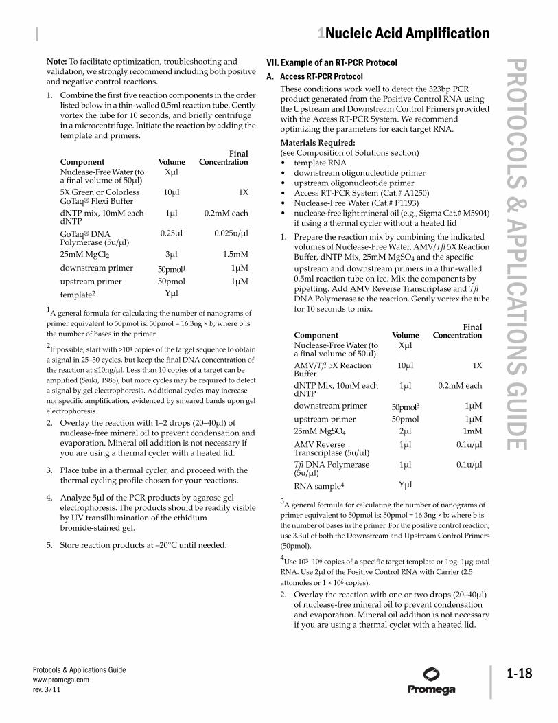
Note: To facilitate optimization, troubleshooting andvalidation, we strongly recommend including both positiveand negative control reactions.1. Combine the first five reaction components in the order
listed below in a thin-walled 0.5ml reaction tube. Gentlyvortex the tube for 10 seconds, and briefly centrifugein a microcentrifuge. Initiate the reaction by adding thetemplate and primers.
FinalConcentrationVolumeComponent
XμlNuclease-Free Water (toa final volume of 50μl)
1X10μl5X Green or ColorlessGoTaq® Flexi Buffer
0.2mM each1μldNTP mix, 10mM eachdNTP
0.025u/μl0.25μlGoTaq® DNAPolymerase (5u/μl)
1.5mM3μl25mM MgCl21μM50pmol1downstream primer1μM50pmolupstream primer
Yμltemplate2
1A general formula for calculating the number of nanograms ofprimer equivalent to 50pmol is: 50pmol = 16.3ng × b; where b isthe number of bases in the primer.2If possible, start with >104 copies of the target sequence to obtaina signal in 25–30 cycles, but keep the final DNA concentration ofthe reaction at ≤10ng/μl. Less than 10 copies of a target can beamplified (Saiki, 1988), but more cycles may be required to detecta signal by gel electrophoresis. Additional cycles may increasenonspecific amplification, evidenced by smeared bands upon gelelectrophoresis.2. Overlay the reaction with 1–2 drops (20–40μl) of
nuclease-free mineral oil to prevent condensation andevaporation. Mineral oil addition is not necessary ifyou are using a thermal cycler with a heated lid.
3. Place tube in a thermal cycler, and proceed with thethermal cycling profile chosen for your reactions.
4. Analyze 5μl of the PCR products by agarose gelelectrophoresis. The products should be readily visibleby UV transillumination of the ethidiumbromide-stained gel.
5. Store reaction products at –20°C until needed.
VII. Example of an RT-PCR ProtocolA. Access RT-PCR Protocol
These conditions work well to detect the 323bp PCRproduct generated from the Positive Control RNA usingthe Upstream and Downstream Control Primers providedwith the Access RT-PCR System. We recommendoptimizing the parameters for each target RNA.Materials Required:(see Composition of Solutions section)• template RNA• downstream oligonucleotide primer• upstream oligonucleotide primer• Access RT-PCR System (Cat.# A1250)• Nuclease-Free Water (Cat.# P1193)• nuclease-free light mineral oil (e.g., Sigma Cat.# M5904)
if using a thermal cycler without a heated lid1. Prepare the reaction mix by combining the indicated
volumes of Nuclease-Free Water, AMV/Tfl 5X ReactionBuffer, dNTP Mix, 25mM MgSO4 and the specificupstream and downstream primers in a thin-walled0.5ml reaction tube on ice. Mix the components bypipetting. Add AMV Reverse Transcriptase and TflDNA Polymerase to the reaction. Gently vortex the tubefor 10 seconds to mix.
FinalConcentrationVolumeComponent
XμlNuclease-Free Water (toa final volume of 50μl)
1X10μlAMV/Tfl 5X ReactionBuffer
0.2mM each1μldNTP Mix, 10mM eachdNTP
1μM50pmol3downstream primer1μM50pmolupstream primer1mM2μl25mM MgSO4
0.1u/μl1μlAMV ReverseTranscriptase (5u/μl)
0.1u/μl1μlTfl DNA Polymerase(5u/μl)
YμlRNA sample4
3A general formula for calculating the number of nanograms ofprimer equivalent to 50pmol is: 50pmol = 16.3ng × b; where b isthe number of bases in the primer. For the positive control reaction,use 3.3μl of both the Downstream and Upstream Control Primers(50pmol).4Use 103–106 copies of a specific target template or 1pg–1μg totalRNA. Use 2μl of the Positive Control RNA with Carrier (2.5attomoles or 1 × 10⁶ copies).2. Overlay the reaction with one or two drops (20–40μl)
of nuclease-free mineral oil to prevent condensationand evaporation. Mineral oil addition is not necessaryif you are using a thermal cycler with a heated lid.
Protocols & Applications Guidewww.promega.comrev. 3/11
| 1Nucleic Acid Amplification PROTOCOLS & APPLICATIONS GUIDE
1-18
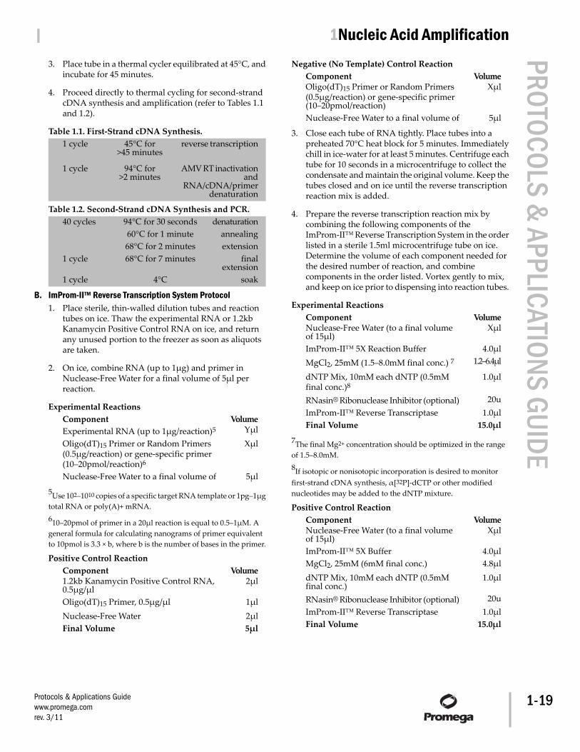
3. Place tube in a thermal cycler equilibrated at 45°C, andincubate for 45 minutes.
4. Proceed directly to thermal cycling for second-strandcDNA synthesis and amplification (refer to Tables 1.1and 1.2).
Table 1.1. First-Strand cDNA Synthesis.reverse transcription45°C for
>45 minutes1 cycle
AMV RT inactivationand
94°C for>2 minutes
1 cycle
RNA/cDNA/primerdenaturation
Table 1.2. Second-Strand cDNA Synthesis and PCR.denaturation94°C for 30 seconds40 cycles
annealing60°C for 1 minuteextension68°C for 2 minutes
finalextension
68°C for 7 minutes1 cycle
soak4°C1 cycle
B. ImProm-II™ Reverse Transcription System Protocol1. Place sterile, thin-walled dilution tubes and reaction
tubes on ice. Thaw the experimental RNA or 1.2kbKanamycin Positive Control RNA on ice, and returnany unused portion to the freezer as soon as aliquotsare taken.
2. On ice, combine RNA (up to 1μg) and primer inNuclease-Free Water for a final volume of 5μl perreaction.
Experimental ReactionsVolumeComponent
YμlExperimental RNA (up to 1μg/reaction)5
XμlOligo(dT)15 Primer or Random Primers(0.5μg/reaction) or gene-specific primer(10–20pmol/reaction)6
5μlNuclease-Free Water to a final volume of
5Use 102–1010 copies of a specific target RNA template or 1pg–1μgtotal RNA or poly(A)+ mRNA.610–20pmol of primer in a 20μl reaction is equal to 0.5–1μM. Ageneral formula for calculating nanograms of primer equivalentto 10pmol is 3.3 × b, where b is the number of bases in the primer.
Positive Control ReactionVolumeComponent
2μl1.2kb Kanamycin Positive Control RNA,0.5μg/μl
1μlOligo(dT)15 Primer, 0.5μg/μl2μlNuclease-Free Water5μlFinal Volume
Negative (No Template) Control ReactionVolumeComponent
XμlOligo(dT)15 Primer or Random Primers(0.5μg/reaction) or gene-specific primer(10–20pmol/reaction)
5μlNuclease-Free Water to a final volume of
3. Close each tube of RNA tightly. Place tubes into apreheated 70°C heat block for 5 minutes. Immediatelychill in ice-water for at least 5 minutes. Centrifuge eachtube for 10 seconds in a microcentrifuge to collect thecondensate and maintain the original volume. Keep thetubes closed and on ice until the reverse transcriptionreaction mix is added.
4. Prepare the reverse transcription reaction mix bycombining the following components of theImProm-II™ Reverse Transcription System in the orderlisted in a sterile 1.5ml microcentrifuge tube on ice.Determine the volume of each component needed forthe desired number of reaction, and combinecomponents in the order listed. Vortex gently to mix,and keep on ice prior to dispensing into reaction tubes.
Experimental ReactionsVolumeComponent
XμlNuclease-Free Water (to a final volumeof 15μl)
4.0μlImProm-II™ 5X Reaction Buffer1.2–6.4μlMgCl2, 25mM (1.5–8.0mM final conc.) 7
1.0μldNTP Mix, 10mM each dNTP (0.5mMfinal conc.)8
20uRNasin® Ribonuclease Inhibitor (optional)1.0μlImProm-II™ Reverse Transcriptase
15.0μlFinal Volume
7The final Mg2+ concentration should be optimized in the rangeof 1.5–8.0mM.8If isotopic or nonisotopic incorporation is desired to monitorfirst-strand cDNA synthesis, α[32P]-dCTP or other modifiednucleotides may be added to the dNTP mixture.
Positive Control ReactionVolumeComponent
XμlNuclease-Free Water (to a final volumeof 15μl)
4.0μlImProm-II™ 5X Buffer4.8μlMgCl2, 25mM (6mM final conc.)1.0μldNTP Mix, 10mM each dNTP (0.5mM
final conc.)20uRNasin® Ribonuclease Inhibitor (optional)
1.0μlImProm-II™ Reverse Transcriptase15.0μlFinal Volume
Protocols & Applications Guidewww.promega.comrev. 3/11
| 1Nucleic Acid Amplification PROTOCOLS & APPLICATIONS GUIDE
1-19
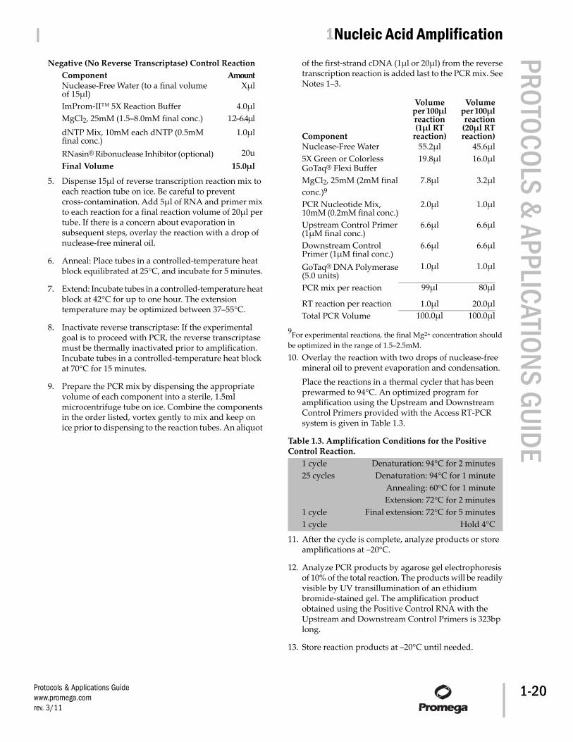
Negative (No Reverse Transcriptase) Control ReactionAmountComponent
XμlNuclease-Free Water (to a final volumeof 15μl)
4.0μlImProm-II™ 5X Reaction Buffer1.2–6.4μlMgCl2, 25mM (1.5–8.0mM final conc.)
1.0μldNTP Mix, 10mM each dNTP (0.5mMfinal conc.)
20uRNasin® Ribonuclease Inhibitor (optional)15.0μlFinal Volume
5. Dispense 15μl of reverse transcription reaction mix toeach reaction tube on ice. Be careful to preventcross-contamination. Add 5μl of RNA and primer mixto each reaction for a final reaction volume of 20μl pertube. If there is a concern about evaporation insubsequent steps, overlay the reaction with a drop ofnuclease-free mineral oil.
6. Anneal: Place tubes in a controlled-temperature heatblock equilibrated at 25°C, and incubate for 5 minutes.
7. Extend: Incubate tubes in a controlled-temperature heatblock at 42°C for up to one hour. The extensiontemperature may be optimized between 37–55°C.
8. Inactivate reverse transcriptase: If the experimentalgoal is to proceed with PCR, the reverse transcriptasemust be thermally inactivated prior to amplification.Incubate tubes in a controlled-temperature heat blockat 70°C for 15 minutes.
9. Prepare the PCR mix by dispensing the appropriatevolume of each component into a sterile, 1.5mlmicrocentrifuge tube on ice. Combine the componentsin the order listed, vortex gently to mix and keep onice prior to dispensing to the reaction tubes. An aliquot
of the first-strand cDNA (1μl or 20μl) from the reversetranscription reaction is added last to the PCR mix. SeeNotes 1–3.
Volumeper 100μlreaction
(20μl RTreaction)
Volumeper 100μlreaction(1μl RT
reaction)Component45.6μl55.2μlNuclease-Free Water16.0μl19.8μl5X Green or Colorless
GoTaq® Flexi Buffer3.2μl7.8μlMgCl2, 25mM (2mM final
conc.)9
1.0μl2.0μlPCR Nucleotide Mix,10mM (0.2mM final conc.)
6.6μl6.6μlUpstream Control Primer(1μM final conc.)
6.6μl6.6μlDownstream ControlPrimer (1μM final conc.)
1.0μl1.0μlGoTaq® DNA Polymerase(5.0 units)
80μl99μlPCR mix per reaction
20.0μl1.0μlRT reaction per reaction100.0μl100.0μlTotal PCR Volume
9For experimental reactions, the final Mg2+ concentration shouldbe optimized in the range of 1.5–2.5mM.10. Overlay the reaction with two drops of nuclease-free
mineral oil to prevent evaporation and condensation.Place the reactions in a thermal cycler that has beenprewarmed to 94°C. An optimized program foramplification using the Upstream and DownstreamControl Primers provided with the Access RT-PCRsystem is given in Table 1.3.
Table 1.3. Amplification Conditions for the PositiveControl Reaction.
Denaturation: 94°C for 2 minutes1 cycleDenaturation: 94°C for 1 minute25 cycles
Annealing: 60°C for 1 minuteExtension: 72°C for 2 minutes
Final extension: 72°C for 5 minutes1 cycleHold 4°C1 cycle
11. After the cycle is complete, analyze products or storeamplifications at –20°C.
12. Analyze PCR products by agarose gel electrophoresisof 10% of the total reaction. The products will be readilyvisible by UV transillumination of an ethidiumbromide-stained gel. The amplification productobtained using the Positive Control RNA with theUpstream and Downstream Control Primers is 323bplong.
13. Store reaction products at –20°C until needed.
Protocols & Applications Guidewww.promega.comrev. 3/11
| 1Nucleic Acid Amplification PROTOCOLS & APPLICATIONS GUIDE
1-20
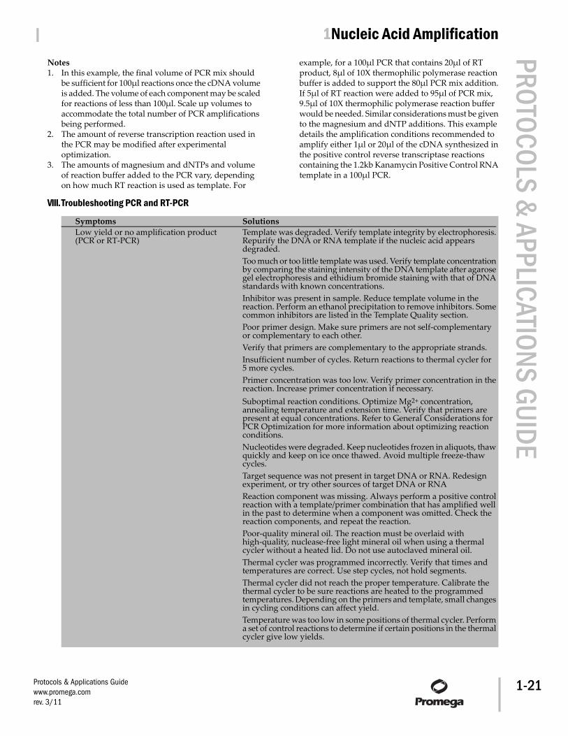
Notes1. In this example, the final volume of PCR mix should
be sufficient for 100μl reactions once the cDNA volumeis added. The volume of each component may be scaledfor reactions of less than 100μl. Scale up volumes toaccommodate the total number of PCR amplificationsbeing performed.
2. The amount of reverse transcription reaction used inthe PCR may be modified after experimentaloptimization.
3. The amounts of magnesium and dNTPs and volumeof reaction buffer added to the PCR vary, dependingon how much RT reaction is used as template. For
example, for a 100μl PCR that contains 20μl of RTproduct, 8μl of 10X thermophilic polymerase reactionbuffer is added to support the 80μl PCR mix addition.If 5μl of RT reaction were added to 95μl of PCR mix,9.5μl of 10X thermophilic polymerase reaction bufferwould be needed. Similar considerations must be givento the magnesium and dNTP additions. This exampledetails the amplification conditions recommended toamplify either 1μl or 20μl of the cDNA synthesized inthe positive control reverse transcriptase reactionscontaining the 1.2kb Kanamycin Positive Control RNAtemplate in a 100μl PCR.
VIII.Troubleshooting PCR and RT-PCR
SolutionsSymptomsTemplate was degraded. Verify template integrity by electrophoresis.Repurify the DNA or RNA template if the nucleic acid appearsdegraded.
Low yield or no amplification product(PCR or RT-PCR)
Too much or too little template was used. Verify template concentrationby comparing the staining intensity of the DNA template after agarosegel electrophoresis and ethidium bromide staining with that of DNAstandards with known concentrations.Inhibitor was present in sample. Reduce template volume in thereaction. Perform an ethanol precipitation to remove inhibitors. Somecommon inhibitors are listed in the Template Quality section.Poor primer design. Make sure primers are not self-complementaryor complementary to each other.Verify that primers are complementary to the appropriate strands.Insufficient number of cycles. Return reactions to thermal cycler for5 more cycles.Primer concentration was too low. Verify primer concentration in thereaction. Increase primer concentration if necessary.Suboptimal reaction conditions. Optimize Mg2+ concentration,annealing temperature and extension time. Verify that primers arepresent at equal concentrations. Refer to General Considerations forPCR Optimization for more information about optimizing reactionconditions.Nucleotides were degraded. Keep nucleotides frozen in aliquots, thawquickly and keep on ice once thawed. Avoid multiple freeze-thawcycles.Target sequence was not present in target DNA or RNA. Redesignexperiment, or try other sources of target DNA or RNAReaction component was missing. Always perform a positive controlreaction with a template/primer combination that has amplified wellin the past to determine when a component was omitted. Check thereaction components, and repeat the reaction.Poor-quality mineral oil. The reaction must be overlaid withhigh-quality, nuclease-free light mineral oil when using a thermalcycler without a heated lid. Do not use autoclaved mineral oil.Thermal cycler was programmed incorrectly. Verify that times andtemperatures are correct. Use step cycles, not hold segments.Thermal cycler did not reach the proper temperature. Calibrate thethermal cycler to be sure reactions are heated to the programmedtemperatures. Depending on the primers and template, small changesin cycling conditions can affect yield.Temperature was too low in some positions of thermal cycler. Performa set of control reactions to determine if certain positions in the thermalcycler give low yields.
Protocols & Applications Guidewww.promega.comrev. 3/11
| 1Nucleic Acid Amplification PROTOCOLS & APPLICATIONS GUIDE
1-21
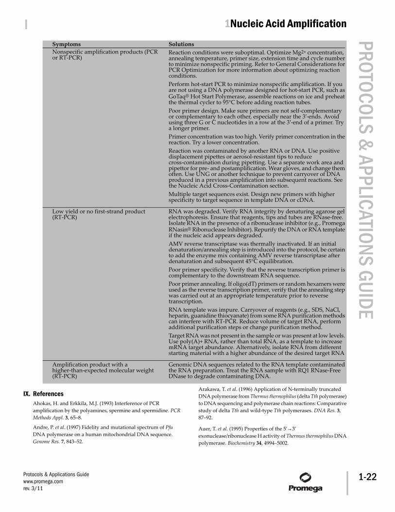
SolutionsSymptomsReaction conditions were suboptimal. Optimize Mg2+ concentration,annealing temperature, primer size, extension time and cycle numberto minimize nonspecific priming. Refer to General Considerations forPCR Optimization for more information about optimizing reactionconditions.
Nonspecific amplification products (PCRor RT-PCR)
Perform hot-start PCR to minimize nonspecific amplification. If youare not using a DNA polymerase designed for hot-start PCR, such asGoTaq® Hot Start Polymerase, assemble reactions on ice and preheatthe thermal cycler to 95°C before adding reaction tubes.Poor primer design. Make sure primers are not self-complementaryor complementary to each other, especially near the 3′-ends. Avoidusing three G or C nucleotides in a row at the 3′-end of a primer. Trya longer primer.Primer concentration was too high. Verify primer concentration in thereaction. Try a lower concentration.Reaction was contaminated by another RNA or DNA. Use positivedisplacement pipettes or aerosol-resistant tips to reducecross-contamination during pipetting. Use a separate work area andpipettor for pre- and postamplification. Wear gloves, and change themoften. Use UNG or another technique to prevent carryover of DNAproduced in a previous amplification into subsequent reactions. Seethe Nucleic Acid Cross-Contamination section.Multiple target sequences exist. Design new primers with higherspecificity to target sequence in template DNA or cDNA.
RNA was degraded. Verify RNA integrity by denaturing agarose gelelectrophoresis. Ensure that reagents, tips and tubes are RNase-free.Isolate RNA in the presence of a ribonuclease inhibitor (e.g., PromegaRNasin® Ribonuclease Inhibitor). Repurify the DNA or RNA templateif the nucleic acid appears degraded.
Low yield or no first-strand product(RT-PCR)
AMV reverse transcriptase was thermally inactivated. If an initialdenaturation/annealing step is introduced into the protocol, be certainto add the enzyme mix containing AMV reverse transcriptase afterdenaturation and subsequent 45°C equilibration.Poor primer specificity. Verify that the reverse transcription primer iscomplementary to the downstream RNA sequence.Poor primer annealing. If oligo(dT) primers or random hexamers wereused as the reverse transcription primer, verify that the annealing stepwas carried out at an appropriate temperature prior to reversetranscription.RNA template was impure. Carryover of reagents (e.g., SDS, NaCl,heparin, guanidine thiocyanate) from some RNA purification methodscan interfere with RT-PCR. Reduce volume of target RNA, performadditional purification steps or change purification method.Target RNA was not present in the sample or was present at low levels.Use poly(A)+ RNA, rather than total RNA, as a template to increasemRNA target abundance. Alternatively, isolate RNA from differentstarting material with a higher abundance of the desired target RNA
Genomic DNA sequences related to the RNA template contaminatedthe RNA preparation. Treat the RNA sample with RQ1 RNase-FreeDNase to degrade contaminating DNA.
Amplification product with ahigher-than-expected molecular weight(RT-PCR)
IX. ReferencesAhokas, H. and Erkkila, M.J. (1993) Interference of PCRamplification by the polyamines, spermine and spermidine. PCRMethods Appl. 3, 65–8.
Andre, P. et al. (1997) Fidelity and mutational spectrum of PfuDNA polymerase on a human mitochondrial DNA sequence.Genome Res. 7, 843–52.
Arakawa, T. et al. (1996) Application of N-terminally truncatedDNA polymerase from Thermus thermophilus (delta Tth polymerase)to DNA sequencing and polymerase chain reactions: Comparativestudy of delta Tth and wild-type Tth polymerases. DNA Res. 3,87–92.
Auer, T. et al. (1995) Properties of the 5′→3′exonuclease/ribonuclease H activity of Thermus thermophilus DNApolymerase. Biochemistry 34, 4994–5002.
Protocols & Applications Guidewww.promega.comrev. 3/11
| 1Nucleic Acid Amplification PROTOCOLS & APPLICATIONS GUIDE
1-22
Baldino, F., Jr. et al. (1989) High-resolution in situ hybridizationhistochemistry. Meth. Enzymol. 168, 761–77.
Baltimore, D. (1970) RNA-dependent DNA polymerase in virionsof RNA tumour viruses. Nature 226, 1209–11.
Barnes, W.M. (1994) PCR amplification of up to 35-kb DNA withhigh fidelity and high yield from lambda bacteriophage templates.Proc. Natl. Acad. Sci. USA 91, 2216–20.
Bej, A.K. and Mahbubani, M.H. (1992) Applications of thepolymerase chain reaction in environmental microbiology. PCRMethods Appl. 1, 151–9.
Bell, D.A. and DeMarini, D.M. (1991) Excessive cycling convertsPCR products to random-length higher molecular weightfragments. Nucl. Acids Res. 19, 5079.
Berger, S.L. et al. (1983) Reverse transcriptase and its associatedribonuclease H: Interplay of two enzyme activities controls theyield of single-stranded complementary deoxyribonucleic acid.Biochemistry 22, 2365–72.
Bernard, P.S. et al. (1998) Integrated amplification and detectionof the C677T point mutation in the methylenetetrahydrofolatereductase gene by fluorescence resonance energy transfer andprobe melting curves. Anal. Biochem. 255, 101–7.
Beutler, E. et al. (1990) Interference of heparin with the polymerasechain reaction. Biotechniques 9, 166.
Black, W.C. (1993) PCR with arbitrary primers: Approach withcare. Insect Mol. Biol. 2, 1–6.
Blumberg, D.D. (1987) Creating a ribonuclease-free environment.Meth. Enzymol. 152, 20–4.
Brooks, E.M. et al. (1995) Secondary structure in the 3′ UTR of EGFand the choice of reverse transcriptases affect the detection ofmessage diversity by RT-PCR. Biotechniques 19, 806–15.
Byrappa, S. et al. (1995) A highly efficient procedure for site-specificmutagenesis of full-length plasmids using Vent DNA polymerase.Genome Res. 5, 404–7.
Carballeira, N. et al. (1990) Purification of a thermostable DNApolymerase from Thermus thermophilus HB8, useful in thepolymerase chain reaction. Biotechniques 9, 276–81.
Carothers, A.M. et al. (1989) Point mutation analysis in amammalian gene: Rapid preparation of total RNA, PCRamplification of cDNA, and Taq sequencing by a novel method.Biotechniques 7, 494–9.
Cheng, S. et al. (1994) Effective amplification of long targets fromcloned inserts and human genomic DNA. Proc. Natl. Acad. Sci. USA91, 5695–9.
Cheng, S. et al. (1995) Template integrity is essential for PCRamplification of 20- to 30-kb sequences from genomic DNA. PCRMethods Appl. 4, 294–8.
Chiocchia, G. and Smith, K.A. (1997) Highly sensitive method todetect mRNAs in individual cells by direct RT-PCR using Tth DNApolymerase. Biotechniques 22, 312–8.
Chumakov, K.M. (1994) Reverse transcriptase can inhibit PCR andstimulate primer-dimer formation. PCR Methods Appl. 4, 62–4.
Clark, J.M. (1988) Novel non-templated nucleotide additionreactions catalyzed by procaryotic and eucaryotic DNApolymerases. Nucl. Acids Res. 16, 9677–86.
Cline, J. et al. (1996) PCR fidelity of Pfu DNA polymerase and otherthermostable DNA polymerases. Nucl. Acids Res. 24, 3546–51.
Crockett, A.O. and Wittwer, C.T. (2001) Fluorescein-labeledoligonucleotides for real-time PCR: Using the inherent quenchingof deoxyguanosine nucleotides. Anal. Biochem. 290, 89–97.
Dang, C. and Jayasena, S.D. (1996) Oligonucleotide inhibitors ofTaq DNA polymerase facilitate detection of low copy numbertargets by PCR. J. Mol. Biol. 264, 268–78.
Demeke, T. and Adams, R. (1992) The effects of plantpolysaccharides and buffer additives on PCR. Biotechniques 12,332–4.
Didenko, V. (2001) DNA probes using fluorescence resonanceenergy transfer (FRET): Designs and applications. Biotechniques31, 1106–21.
Eckert, K.A. and Kunkel, T.A. (1990) High fidelity DNA synthesisby the Thermus aquaticus DNA polymerase. Nucl. Acids Res. 18,3739–44.
Eckert, K.A. and Kunkel, T.A. (1991) DNA polymerase fidelity andthe polymerase chain reaction. PCR Methods Appl. 1, 17–24.
Edwards, J.B. et al. (1991) Oligodeoxyribonucleotide ligation tosingle-stranded cDNAs: A new tool for cloning 5′ ends of mRNAsand for constructing cDNA libraries by in vitro amplification. Nucl.Acids Res. 19, 5227–32.
Edwards, J.B. et al. (1993) Methods in Molecular Biology (Vol. 15),White, B.A., ed., Humana Press, Totowa, NJ.
Ehrlich, H.A. et al. (1991) Recent advances in the polymerase chainreaction. Science 252, 1643–51.
Ellsworth, D.L. et al. (1993) Artifactual variation in randomlyamplified polymorphic DNA banding patterns. Biotechniques 14,214–7.
Frackman, S. et al. (2006) The Plexor™ Systems provide accuratequantitation in multiplex qPCR and qRT-PCR. Promega Notes 92,10–13.
Frohman, M.A. et al. (1988) Rapid production of full-length cDNAsfrom rare transcripts: Amplification using a single gene-specificoligonucleotide primer. Proc. Natl. Acad. Sci. USA 85, 8998–9002.
Fromont-Racine, M. et al. (1993) A highly sensitive method formapping the 5′ termini of mRNAs. Nucl. Acids Res. 21, 1683–4.
Gaensslen, R.E. et al. (1992) A polymerase chain reaction (PCR)method for sex and species determination with novel controls fordeoxyribonucleic acid (DNA) template length. J. Forensic Sci. 37,6–20.
Gaudette, M.F. and Crain, W.R. (1991) A simple method forquantifying specific mRNAs in small numbers of early mouseembryos. Nucl. Acids Res. 19, 1879–84.
Geiduschek, E.P. and Herskovits, T.T. (1961) Nonaqueous solutionsof DNA. Reversible and irreversible denaturation in methanol.Arch. Biochem. Biophy. 95, 114–29.
Protocols & Applications Guidewww.promega.comrev. 3/11
| 1Nucleic Acid Amplification PROTOCOLS & APPLICATIONS GUIDE
1-23

Gelfand, D.H. (1989) Taq DNA polymerase. In: PCR Techology:Principles and applications of DNA amplifications, Erlich, H.A., ed.,Stockton Press, New York, 17–22.
Gelfand, D.H. and White, T.J. (1990) In: PCR Protocols: A Guide toMethods and Applications, Innis, M.A., Gelfand, D.H., Sninsky, J.J.and White, T.J., eds, Academic Press, San Diego, CA, 129–41.
Gerard, G.F. (1983) Enzymes of Nucleic Acid Synthesis andModification: DNA Enzymes, CRC Press, Boca Raton, FL.
Giambernardi, T.A. et al. (1998) Bovine serum albumin reversesinhibition of RT-PCR by melanin. Biotechniques 25, 564–6.
Greagg, M.A. et al. (1999) A read-ahead function in archaeal DNApolymerases detects promutagenic template-strand uracil. Proc.Natl. Acad. Sci. USA 96, 9045–50.
Gubler, U. and Hoffman, B.J. (1983) A simple and very efficientmethod for generating cDNA libraries. Gene 25, 263–9.
Guo, B. and Bi, Y. (2002) Cloning PCR products: An overview.Methods Mol. Biol. 192, 111–9.
Haase, A.T. et al. (1990) Amplification and detection of lentiviralDNA inside cells. Proc. Natl. Acad. Sci. USA 87, 4971–5.
Holland, P.M. et al. (1991) Detection of specific polymerase chainreaction product by utilizing the 5′→3′ exonuclease activity ofThermus aquaticus DNA polymerase. Proc. Natl. Acad. Sci. USA 88,7276–80.
Holodniy, M. et al. (1991) Inhibition of human immunodeficiencyvirus gene amplification by heparin. J. Clin. Microbiol. 29, 676–9.
Hoppe, B.L. et al. (1992) Gel-loading dyes compatible with PCR.Biotechniques 12, 679–80.
Houts, G.E. et al. (1979) Reverse transcriptase from avianmyeloblastosis virus. J. Virol. 29, 517–22.
Hu, C.Y. et al. (1992) Effect of freezing of the PCR buffer on theamplification specificity: Allelic exclusion and preferentialamplification of contaminating molecules. PCR Methods Appl. 2,182–3.
Hu, G. (1993) DNA polymerase-catalyzed addition of nontemplatedextra nucleotides to the 3′ end of a DNA fragment. DNA Cell Biol.12, 763–70.
Hubank, M. and Schatz, D.G. (1994) Identifying differences inmRNA expression by representational difference analysis of cDNA.Nucl. Acids Res. 22, 5640–8.
Innis, M.A. et al. (1988) DNA sequencing with Thermus aquaticusDNA polymerase and direct sequencing of polymerase chainreaction-amplified DNA. Proc. Natl. Acad. Sci. USA 85, 9436–40.
Johnson, S.C. et al. (2004) A third base pair for the polymerasechain reaction: Inserting isoC and isoG. Nucl. Acids Res. 32, 1937–41.
Kaledin, A.S. et al. (1981) Isolation and properties ofDNA-polymerase from the extreme thermophilic bacteriumThermus flavus. Biokhimiia 46, 1576–84.
Katcher, H.L. and Schwartz, I. (1994) A distinctive property of TthDNA polymerase: Enzymatic amplification in the presence ofphenol. Biotechniques 16, 84–92.
Keohavong, P. et al. (1993) Predominant mutations induced by theThermococcus litoralis, Vent DNA polymerase during DNAamplification in vitro. PCR Methods Appl. 2, 288–92.
Knoche, K. and Denhart, M. (2001) AccessQuick™ RT-PCR System:Simple, stable and sensitive. Promega Notes 79, 12–4.
Konat, G.W. et al. (1994) PCR Technology: Current Innovations,Griffin, H.G. and Griffin, A.M., eds, CRC Press, Inc. Boca RatonFlorida.
Kong, H.M. et al. (1993) Characterization of a DNA polymerasefrom the hyperthermophile archaea Thermococcus litoralis. VentDNA polymerase, steady state kinetics, thermal stability,processivity, strand displacement, and exonuclease activities. J.Biol. Chem. 268, 1965–75.
Krishnan, B.R. et al. (1991) Linear amplification DNA sequencingdirectly from single phage plaques and bacterial colonies. Nucl.Acids Res. 19, 1153.
Kurata, S. et al. (2001) Fluorescent quenching-based quantitativedetection of specific DNA/RNA using a BODIPY® FL-labeled probeor primer. Nucl. Acids Res. 29, e34.
Lasken, R.S. et al. (1996) Archaebacterial DNA polymerases tightlybind uracil-containing DNA. J. Biol. Chem. 271, 17692–6.
Lawyer, F.C. et al. (1993) High-level expression, purification, andenzymatic characterization of full-length Thermus aquaticus DNApolymerase and a truncated form deficient in 5′ to 3′ exonucleaseactivity. PCR Methods Appl. 2, 275–87.
Lee, L.G. et al. (1993) Allelic discrimination by nick-translationPCR with fluorogenic probes. Nucl. Acids Res. 21, 3761–6.
Liang, P. and Pardee, A.B. (1992) Differential display of eukaryoticmessenger RNA by means of the polymerase chain reaction. Science257, 967–71.
Lin, Y. and Jayasena, S.D. (1997) Inhibition of multiple thermostableDNA polymerases by a heterodimeric aptamer. J. Mol. Biol. 271,100–11.
Linquist, V. et al. (1998) UV irradiation of polystyrene pipetsreleases PCR inhibitors. Biotechniques 24, 50–2.
Lisitsyn, N. et al. (1993) Cloning the differences between twocomplex genomes. Science 259, 946–51.
Liu, X. and Gorovsky, M.A. (1993) Mapping the 5′ and 3′ ends ofTetrahymena thermophila mRNAs using RNA ligase mediatedamplification of cDNA ends (RLM-RACE). Nucl. Acids Res. 21,4954–60.
Loh, E.Y. et al. (1989) Polymerase chain reaction with single-sidedspecificity: Analysis of T cell receptor delta chain. Science 243,217–20.
Longley, M.J. et al. (1990) Characterization of the 5′ to 3′ exonucleaseassociated with Thermus aquaticus DNA polymerase. Nucl. AcidsRes. 18, 7317–22.
Longo, M.C. et al. (1990) Use of uracil DNA glycosylase to controlcarry-over contamination in polymerase chain reactions. Gene 93,125–8.
McClelland, M. and Welsh, J. (1994) DNA fingerprinting byarbitrarily primed PCR. PCR Methods Appl. 4, S59–65.
Protocols & Applications Guidewww.promega.comrev. 3/11
| 1Nucleic Acid Amplification PROTOCOLS & APPLICATIONS GUIDE
1-24
McCulloch, R.K. et al. (1995) An evaluation of competitor type andsize for use in the determination of mRNA by competitive PCR.PCR Methods Appl. 4, 219–26.
Mezei, L.M. and Storts, D.R. (1994) Cloning PCR products. In: PCRTechnology: Current Innovations, Griffin, H.G. and Griffin, A.M.,eds., CRC Press, Boca Raton, FL, 21–7.
Miller, K. and Storts, D. (1995) PCR ACCESS! A sensitivesingle-tube, two-enzyme system for RT-PCR. Promega Notes 53,2–5.
Miller, K. and Storts, D. (1996) An improved single buffer, twoenzyme system for RT-PCR. J. NIH Res. 8(2), 48.
Moretti, T. et al. (1998) Enhancement of PCR amplification yieldand specificity using AmpliTaq Gold DNA polymerase.BioTechniques 25, 716–22.
Moser, M.J. and Prudent, J.R. (2003) Enzymatic repair of anexpanded genetic information system. Nucl. Acids Res. 31, 5048–53.
Murphy, L.D. et al. (1990) Use of the polymerase chain reaction inthe quantitation of mdr-1 gene expression. Biochemistry 29, 10351–6.
Murray, V. (1989) Improved double-stranded DNA sequencingusing the linear polymerase chain reaction. Nucl. Acids Res. 17,8889.
Myers, T.W. and Gelfand, D.H. (1991) Reverse transcription andDNA amplification by a Thermus thermophilus DNA polymerase.Biochemistry 30, 7661–6.
Nuovo, G.J. (1995) In situ PCR: Protocols and applications. PCRMethods Appl. 4, S151–67.
Nuovo, G.J. et al. (1993) Importance of different variables forenhancing in situ detection of PCR-amplified DNA. PCR MethodsAppl. 2, 305–12.
Ohara, O. et al. (1989) One-sided polymerase chain reaction: Theamplification of cDNA. Proc. Natl. Acad. Sci. USA 86, 5673–7.
Pao, C.C. et al. (1993) Inhibition of in vitro enzymatic DNAamplification reaction by ultra-violet light irradiation. Mol. CellProbes. 7, 217–9.
Power, E.G.M. (1996) RAPD typing in microbiologyema technicalreview. J. Hosp. Infect. 34, 247–65.
Rees, W.A. et al. (1993) Betaine can eliminate the base paircomposition dependence of DNA melting. Biochemistry 32, 137–44.
Rose, E.A. (1991) Applications of the polymerase chain reaction togenome analysis. FASEB J. 5, 46–54.
Roth, M.J. et al. (1985) Purification and characterization of murineretroviral reverse transcriptase expressed in Escherichia coli. J. Biol.Chem. 260, 9326–35.
Ruttimann, C. et al. (1985) DNA polymerases from the extremelythermophilic bacterium Thermus thermophilus HB-8. Eur. J. Biochem.149, 41–6.
Rychlik, W. et al. (1990) Optimization of the annealing temperaturefor DNA amplification in vitro. Nucl. Acids Res. 18, 6409–12.
Rys, P.N. and Persing, D.H. (1993) Preventing false positives:Quantitative evaluation of three protocols for inactivation of
polymerase chain reaction amplification products. J. Clin. Microbiol.31, 2356–60.
Saiki , R.K. (1988) Primer-directed enzymatic amplification of DNAwith a thermostable DNA polymerase. Science 239, 487–91.
Saiki, R. et al. (1985) Enzymatic amplification of beta-globingenomic sequences and restriction site analysis for diagnosis ofsickle cell anemia. Science 230, 1350–4.
Saluz, H. and Jost, J.P. (1989) A simple high-resolution procedureto study DNA methylation and in vivo DNA-protein interactionson a single-copy gene level in higher eukaryotes. Proc. Natl. Acad.Sci. USA 86, 2602–6.
Sambrook, J. and Russell, D.W. (2001) Molecular Cloning: ALaboratory Manual, 3rd edition. Cold Spring Harbor LaboratoryPress, Cold Spring Harbor, NY.
Scalice, E. et al. (1994) Monoclonal antibodies prepared against theDNA polymerase from Thermus aquaticus are potent inhibitors ofenzyme activity. J. Immunol. Methods 172, 147–63.
Schaefer, B.C. (1995) Revolutions in rapid amplification of cDNAends: New strategies for polymerase chain reaction cloning offull-length cDNA ends. Anal. Biochem. 227, 255–73.
Sellner, L.N. et al. (1992) Reverse transcriptase inhibits Taqpolymerase activity. Nucl. Acids Res. 20, 1487–90.
Sharkey, D. et al. (1994) Antibodies as thermolabile switches: Hightemperature triggering for the polymerase chain reaction.Biotechnology 12, 506–9.
Sherrill, C.B. et al. (2004) Nucleic acid analysis using an expandedgenetic alphabet to quench fluorescence. J. Am. Chem. Soc. 126,4550–6.
Siebert, P.D. and Larrick, J.W. (1993) PCR MIMICS: CompetitiveDNA fragments for use as internal standards in quantitative PCR.Biotechniques 14, 244–9.
Skinner, T.L. et al. (1994) Three different calmodulin-encodingcDNAs isolated by a modified 5′-RACE using degenerateoligodeoxyribonucleotides. Gene 151, 247–51.
Slupphaug, G. et al. (1993) Low incorporation of dUMP by somethermostable DNA polymerases may limit their use in PCRamplifications. Anal. Biochem. 211, 164–9.
Staskus, K.A. et al. (1995) PCR in situ: New technologies withsingle-cell resolution for the detection and investigation of virallatency and persistence. In: The Polymerase Chain Reaction (PCR)for Human Viral Diagnosis. Clewley, J.P., ed., CRC Press, Boca Raton,FL, 21–40
Tanese, N. and Goff, S.P. (1988) Domain structure of the Moloneymurine leukemia virus reverse transcriptase: Mutational analysisand separate expression of the DNA polymerase and RNase Hactivities. Proc. Natl. Acad. Sci. USA 85, 1777–81.
Temin, H. and Mizutani, S. (1970) RNA-dependent DNApolymerase in virions of Rous sarcoma virus. Nature 226, 1211–3.
Thaker, V. (1999) In situ RT-PCR and hybridization techniques.Methods. Mol. Biol. 115, 379–402.
Protocols & Applications Guidewww.promega.comrev. 3/11
| 1Nucleic Acid Amplification PROTOCOLS & APPLICATIONS GUIDE
1-25

Troutt, A.B. et al. (1992) Ligation-anchored PCR: A simpleamplification technique with single-sided specificity. Proc. Natl.Acad. Sci. USA 89, 9823–5.
Tyagi, S. et al. (1998) Multicolor molecular beacons for allelediscrimination. Nat. Biotechnol. 16, 49–53.
Varadaraj, K. and Skinner, D.M. (1994) Denaturants or cosolventsimprove the specificity of PCR amplification of a G + C-rich DNAusing genetically engineered DNA polymerases. Gene 140, 1–5.
Williams, J.F. (1989) Optimization strategies for the polymerasechain reaction. Biotechniques 7, 762–9.
Wittwer, C.T. et al. (2001) Real-time multiplex PCR assays. Methods25, 430–42.
Zhou, M.Y. et al. (1995) Universal cloning method by TA strategy.Biotechniques 19, 34–5.
Patents for the foundational PCR process, European Pat. Nos. 201,184 and200,362, expired on March 28, 2006. In the U.S., the patents covering thefoundational PCR process expired on March 29, 2005.RT-PCR reactions at temperatures above 45°C are covered by U.S. Pat. Nos.5,817,465 and 5,654,143 and European Pat. No. 0 568 272.GoTaq, pGEM, Plexor and RNasin are registered trademarks of PromegaCorporation. AccessQuick, GoScript, ImProm-II and pTARGET aretrademarks of Promega Corporation.BODIPY is a registered trademark of Molecular Probes, Inc. NuSieve is aregistered trademark of Lonza Sales AG. SYBR is a registered trademark ofMolecular Probes, Inc. TaqMan is a registered trademark of Roche MolecularSystems, Inc. Triton is a registered trademark of Union Carbide Chemicals& Plastics Technology Corporation. Tween is a registered trademark of ICIAmericas, Inc.Products may be covered by pending or issued patents or may have certainlimitations. Please visit our Web site for more information.All prices and specifications are subject to change without prior notice.Product claims are subject to change. Please contact Promega TechnicalServices or access the Promega online catalog for the most up-to-dateinformation on Promega products.© 2004–2011 Promega Corporation. All Rights Reserved.
Protocols & Applications Guidewww.promega.comrev. 3/11
| 1Nucleic Acid Amplification PROTOCOLS & APPLICATIONS GUIDE
1-26