nuclear medicine for newbies - university of north
TRANSCRIPT

Nuclear Medicine for Newbies
Jorge Oldan MDUNC Department of RadiologyDivision of Nuclear Medicine
March 2016

Ed Module Outline
• Commonly-performed NM examinations at UNC
• Radiopharmaceutical, Technique, Indications with normal exams illustrated
• Need to Knows highlighted
• Case-based Q&A of six ‘Aunt Minnies’

What’s a radiopharmaceutical?
• A radionuclide (which is radioactive and emits something we can detect, usually gamma rays) is bound to a pharmaceutical (which gives the physiologic function) to make a radiopharmaceuticalor radiotracer
– For example, we attach technetium-99m to DTPA, which is filtered by the kidney, to calculate glomerular filtration rate.
– Or we can attach it to MDP, which is taken up by the bone, to do a bone scan.


Normal Whole Body Bone ScanClinical Indication: 44yoF new diagnosis breast cancer
• Search Pattern Tips: Look for focal uptake outside the joints, especially in the axial and proximal appendicular skeleton
• Critical Organ: Renal

Whole Body Bone Scan
• Radiopharmaceutical: 99Technetium Methylene Diphosphonate (MDP) IV
• Technique: planar
• Indications: bone neoplasm to include primary and metastatic disease, trauma
• Need to Knows: which tumors do we get bone scans for? (breast, prostate, most others use PET scan)
– Breast cancer often sends solitary metastases to the sternum, whereas prostate cancer metastases often start in the spine
– Any history of trauma? Inflammatory or degenerative arthritis?

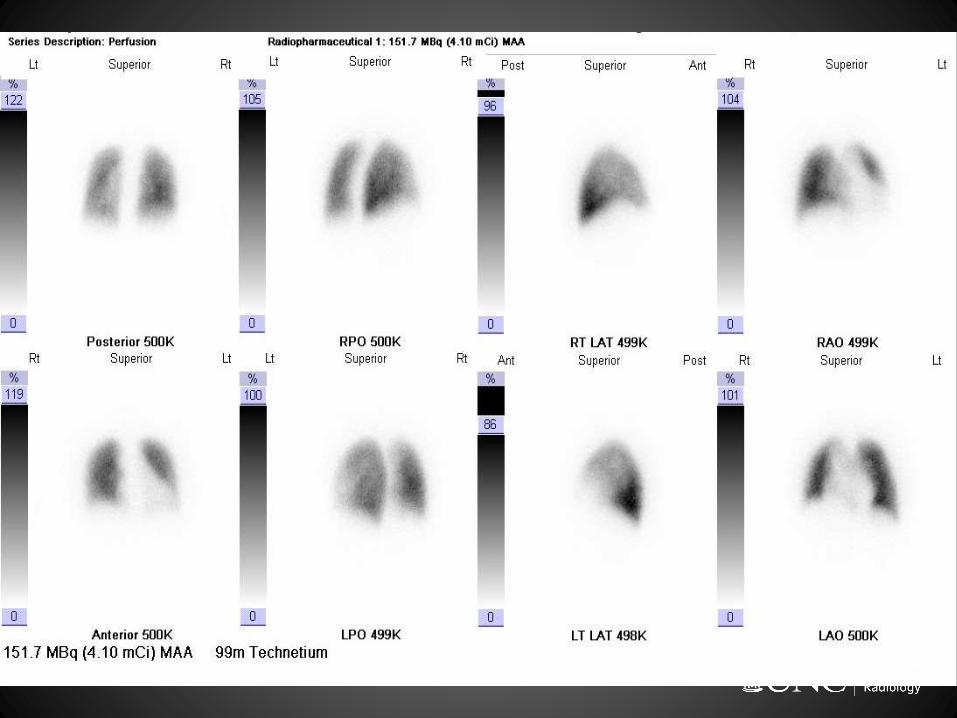

Normal V/Q ScanClinical Indication: 46yoF acute shortness of breath
• Dual planar exam, Ventilation (‘V’) followed by Perfusion (‘Q’)
• Mismatch of ventilated and perfused lung segments in PE ie segments are ventilated but not perfused due to pulmonary arterial emboli
• Search Pattern Tips: – Look for wedge-shaped
peripheral defects
– Make sure they are not also absent on ventilation images

Ventilation Perfusion Scan
• Radiopharmaceutical: 133Xenon inhalation gas followed by 99Technetium Macro-Aggregated Albumin (MAA) IV
• Technique: planar
• Indications: acute or chronic pulmonary embolism and pulmonary hypertension
• Need to Knows:
– Should be done with a chest X-ray• Other lung illnesses such as COPD, pneumonia, or a pleural effusion will
produce matched defects
– Often done when there is bad renal function, a contrast allergy, or there is a strong desire to decrease radiation dose.

131I 24 hour Thyroid uptake = 27.4% (Normal range = 15-35% at 24 hrs)

Normal Thyroid Uptake and ScanClinical Indication: 20yoF with subclinical hyperthyroidism
• Search Pattern Tips: Lumpy or smooth? Background? Salivary glands?
• Critical Organ: Thyroid
131I 24 hour Thyroid uptake = 27.4%. (Normal range = 15-35% at 24 hrs)
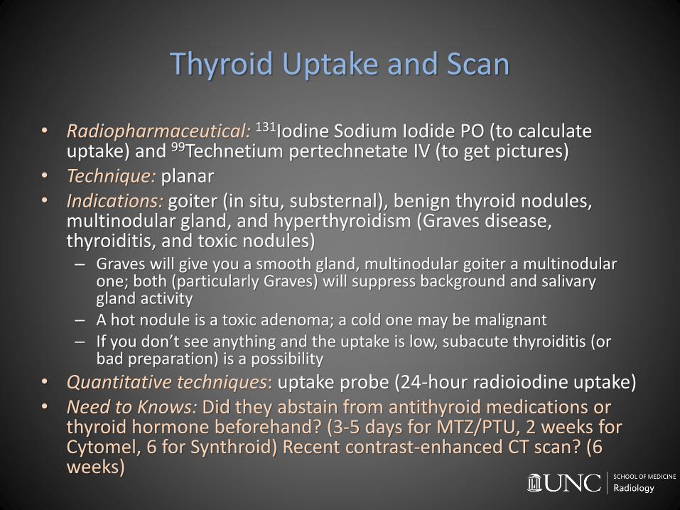
Thyroid Uptake and Scan
• Radiopharmaceutical: 131Iodine Sodium Iodide PO (to calculate uptake) and 99Technetium pertechnetate IV (to get pictures)
• Technique: planar• Indications: goiter (in situ, substernal), benign thyroid nodules,
multinodular gland, and hyperthyroidism (Graves disease, thyroiditis, and toxic nodules)– Graves will give you a smooth gland, multinodular goiter a multinodular
one; both (particularly Graves) will suppress background and salivary gland activity
– A hot nodule is a toxic adenoma; a cold one may be malignant– If you don’t see anything and the uptake is low, subacute thyroiditis (or
bad preparation) is a possibility
• Quantitative techniques: uptake probe (24-hour radioiodine uptake)• Need to Knows: Did they abstain from antithyroid medications or
thyroid hormone beforehand? (3-5 days for MTZ/PTU, 2 weeks for Cytomel, 6 for Synthroid) Recent contrast-enhanced CT scan? (6 weeks)


Normal Whole Body Thyroid ScanClinical Indication: 37yoF status post Rx follicular thyroid CA
• Search Pattern Tips: Uptake outside usual organs (nose, mouth, thyroid bed, liver, bladder)? Particularly lung or bone?
• Critical Organ: Bladder

Whole Body Thyroid Scan
• Radiopharmaceutical: 131Iodine or 123Iodine Sodium Iodide PO (or IV)
• Technique: planar, SPECT and/or SPECT-CT
• Indications: post thyroidectomy or thyroid ablation, thyroid bed remnant, and staging locoregionaldisease/distant metastases for papillary and follicular and medullary cancers
• Need to Knows:
– Should be off thyroid hormone until TSH is above 30 (usu. 6 weeks for Synthroid, 2 weeks for Cytomel), or be given Thyrogen (recombinant TSH) for two days before test
– Negative pregnancy test

Normal PET ScanClinical Indication: 47yoF history of treated recurrent breast cancer
• Search Pattern Tips: – Look for hot spots in lymph nodes,
lung, liver, and bone• You can’t see brain mets, the brain is too
hot
– Adrenals for lung cancer– Peritoneum for GI and GYN tumors– Is the spleen hot in a lymphoma?– In a melanoma, look everywhere– Since there’s a CT, go back and check
for lung nodules
• Preparation– Patient should be NPO for 4 hours
and have received no insulin for 6; no exercise for 24
– With diabetic patients we do the best we can since we don’t want patients to be hypoglycemic; however, insulin does drive glucose into muscle
• Critical Organ: Bladder

PET Scan
• Radiopharmaceutical:18Fluorine Fluorodeoxyglucose(FDG) IV
• Technique: PET/CT
• Indications: Primary and metastatic malignancy
– Occasionally, infection or epilepsy
• Need to Knows: Recent chemotherapy, surgery, or radiation
– How recent? EANM guidelines suggest 10 days after chemo, 2 weeks after growth factors, 6 weeks after local surgery, and 2-3 months after radiation.


LymphoscintigraphyClinical Indication: 39yoM melanoma mid-back
• Note activity in skin around the injection site and in LN
• Most commonly requested for melanoma to identify the sentinel lymph node
• NB: SNI injection ipsilateral breast periareolar for invasive breast cancer patients, no NM imaging (breast surgeons use gamma probe to identify LN)
• Search Pattern Tips: Nodes elsewhere? (axillae, inguinal, other)
• Critical Organ: Spleen

Lymphoscintigraphy
• Radiopharmaceutical: 99Technetium Sulfur Colloid injection
• Injection Techniques: intradermal, peritumoral, and periareolar
– Here, we will be doing intradermal injections for melanoma only; breast injections (which use the other two techniques) are done in mammo
• Imaging and Localization Techniques: planar and intraoperative gamma probe
• Indications: identification and localization of sentinel lymph node (SLN) for intraoperative gammaprobe-directed sampling
• Need to Knows: Injection site (verify this before injecting! Some surgeons have measurements from landmarks)– Occasionally, prior radiation or chemotherapy will change drainage
sites


Normal Hepatobiliary ScanClinical Indication: 37yoM RUQ abdominal pain
• Search Pattern Tips: Is there a gallbladder? Is it really a gallbladder?
• Critical Organ: Gallbladder

Hepatobiliary Scan
• Radiopharmaceutical: 99Technetium Mebrofenin IV
• Technique: planar
• Indications: acute cholecystitis, chronic acalculous cholecystitis, common bile duct obstruction, biliary ectasia, bile leak, and postoperative complications
• Pharmacologic protocols: morphine sulfate and sincalide (CCK)
• Quantitative analysis: gallbladder ejection fraction (GBEF)
• Need to Knows: – NPO for 4-24 hours, off opioids for 8
– Don’t give morphine until you see the bowel
– Don’t give CCK after giving morphine
– If you can’t give morphine, you can wait 4 hours


Normal Gastric Emptying ExamClinical Indication: 25yoF early satiety and vomiting
• Search Pattern Tips: did they draw the ROIs correctly?
• Critical Organ: Colon

ROIs

Gastric Emptying Study
• Radiopharmaceutical: 99Technetium sulfur colloid as solid meal (e.g. eggs)
• Technique: planar
• Indications: gastroparesis, gastroesophageal reflux, and aspiration
• Quantitative analysis: Quantitative analysis: T ½, 4-hour retained activity, and geometric mean methodology
• Need to Knows: Less than 40% emptying at 2 hours and 90% emptying at 4 hours means delayed emptying.
– More than 70% emptying at 1 hour means rapid emptying.


Parathyroid ExamClinical Indication: 58yoF with hypercalcemia
• Search Pattern Tips: Hot spot on 2-hour images in thyroid or mediastinum?
• Critical Organ: Gallbladder

Parathyroid Scan
• Radiopharmaceutical: 99Technetium sestamibi
• Techniques: dual-phase, dual-tracer, planar, and SPECT-CT
• Indications: hyperparathyroidism (adenoma, hyperplasia, and ectopic)
• Need to Knows:
– Other thyroid lesions? (thyroid nodules will also have persistent uptake on delayed images)
– This is the same tracer used in cardiac (and uncommonly performed breast) scans.

131Iodine Therapy
• Radiopharmaceutical: 131Iodine Sodium Iodide PO, requires calculation of administered activity for benign vs malignant conditions
• Patient Issues: selection, preparation, informed consent, understanding and calculation of administered activity, counseling of patients and families on radiation safety issues, release criteria, and follow-up; pregnancy
• Indications: benign (hyperthyroidism), malignant (thyroid cancer remnant vs metastases)
• Need to Knows: Thyroid hormone withdrawal or thyrogen?– Did they go on a low-salt diet?
– Make sure they are not pregnant!
– Over 33 mCimust talk to radiation safety


Diuretic renal scanClinical indication: 46F with hydroureter
• Search Pattern Tips: Do the kidneys clear on their own? How about after Lasix?
– Unlike most radiology studies, right and left are not reversed; the scan is obtained posteriorly to decrease attenuation
• Curve is useful, but should first look at source images (can be mis-drawn, or patient may have motion)
• Critical Organ: Bladder

Renal Nuclear Medicine Studies
• Radiopharmaceuticals: 99mTc MAG-3, 99mTc DTPA
• Technique: planar
• Pharmacologic protocols: diuretics (e.g. furosemide)
• Quantitative analysis: relative renal function, and response to diuretic challenge
• Indications: obstructive vs nonobstructive hydronephrosis, and stent function
• Need to knows: This one is a little more of a process. • The patient is injected with MAG-3 (here), and imaged as the tracer passes through
the collecting system.
• If the patient excretes normally, the exam (can) end. The study is normal.
• If the patient fails to excrete (the tracer stays in the collecting system), we give Lasix.• If the patient still fails to excrete, we call obstruction. Otherwise, the system is merely dilated.
Diuretic Challenge

Renal Nuclear Medicine Studies
Cortical
* Radiopharmaceutical: 99mTc dimercaptosuccinic acid (DMSA)
* Techniques: planar, SPECT, and/or SPECT-CT
* Quantitative analysis: relative renal function
* Indications: relative function, scarring, and prenephrectomyassessment
Perfusion and Function
* Radiopharmaceuticals: 99mTc Mercaptoacetyltriglycine (MAG-3), 99mTc DTPA
* Technique: planar
* Quantitative analysis: relative renal function, renogram, and glomerular filtration rate (GFR)
* Indications: renal dysfunction/failure, renal artery occlusion, and renal vein thrombosis

Infection Nuclear Medicine Studies
67Gallium Citrate
* 67Ga cyclotron produced
* Administer IV
* T1/2 is 78 hours
* Used in infection and inflammation imaging
* Infrequently performed; still useful in some chronic infections
* At UNC, still used for skull base osteomyelitis and otitis externa(due to proximity of the brain)
111In-WBC or 99mTc-WBC
* 111Indium Oxine Leukocytes IV* 99mTechnetium hexamethylpro-
pyleneamine oxime (HMPAO = Ceretec) Leukocytes IV
* Used in abscess, osteomyelitis, cellulitis, synovitis/arthritis, septic joint, hardware infection
* 99mTc-WBC more economical, allow for higher administered dose, better visualization of small parts. BUT physiologic excretion of 99mTc may reduce sensitivity
* Classically, 99mTc is used in the extremities and 111In in the trunk.


Pediatric Nuclear Medicine Studies
* Bone scan
* Similar to bone scans in adults, the main application being neuroblastoma
* Often done with MIBG scan
* Be aware of growth plates
* Gastric emptying study
* Similar to adults, sometimes a liquid study is done
* DMSA
* This is usually a pediatric study
* Looks for scarring; hydronephrosis often visible
* Diuretic renal (MAG-3) scan
* Interpreted similarly (normal/dilation/obstruction)
* However, patient usually has a Foley and is hydrated beforehand
• Most common at UNC

CNS Nuclear Medicine Studies
BRAIN PERFUSION
* 99mTechnetium Ceretec IV
* Techniques: planar, SPECT, and/or SPECT-CT
* Indication: confirmation of clinical brain death, epilepsy
* Prompt activity in the brain and sagittal sinus effectively rules out brain death
* In epilepsy, uptake is increased ictally and decreased interictallyat the seizure focus
CSF / SHUNT
* CSF FLOW 111Indium Diethylenetriamine Pentaacetic(DTPA) Intrathecal Injetion
* Techniques: planar, SPECT, and/or SPECT-CT
* Indication: normal pressure hydrocephalus
* SHUNTOGRAM 99mTechnetium DTPA Injection into shunt reservoir
* Techniques: planar, SPECT, and/or SPECT-CT
* Indication: V-P shunt patency

More Nuclear Imaging Intel . . .
• Most studies use Tc-99m as radionuclide, with 140 keV photon and 6-hour half-life.• PET scans use F-18, with 511 keV photons (produced from positron annihilation) and
a 108-minute half-life.• Some other common nuclides used at UNC are: • In-111, 171 keV and 2.8 days• Xe-133, 81 keV and 5.24 days
• Xenon’s half-life is actually much less in biological terms because it is exhaled.
• I-123, 159 keV and 13.3 hours• I-131, 364 keV and 8.02 days
• Radioactive decays are exponential, so after two half-lives, one-quarter of the radioactivity is left, and after three, one-eighth.
• If you are doing two scans, the lower-energy photon is done first. The reason is that photons will lose energy on their track throughout the body (attenuation) and a higher-energy radionuclide may be confused with a lower-energy radionuclide.• This is why we do ventilation before perfusion in a V/Q scan. (We would prefer to do
the perfusion scan first and cancel the ventilation scan for a normal patient, but persistent Tc-99m would ‘downscatter’ into the Xe-133 window).

Case-based Q&A of six ‘Aunt Minnies’

Case #1 Clinical Indication: 41yoF hyperthyroidism
131I 24 hour Thyroid uptake = 68%. (Normal range = 15-35% at 24 hrs)

Case #1 Graves Disease
• Often treated with I-131 therapy– Note that this destroys the
thyroid, so check with clinician beforehand or make sure the patient is definitely scheduled for therapy beforehand• Why not give just enough to make
them neither hypothyroid nor hyperthyroid? Eventually they become hyperthyroid again
• Why not just use thyroid-suppressive drugs like methimazole or propylthiouracil? More side effects over the long term
• Often empirically chosen at 10-15 mCi

Q. If the thyroid uptake is verified at 3%, how much radioiodine therapy would you administer?
1. 0 mCi
2. 10 mCi
3. 50 mCi
4. 200 mCi

A. If the thyroid uptake is verified at 3%, how much radioiodine therapy would you administer?
1. 0 mCi
A 3% uptake suggests subacute thyroiditis, which is a self-limiting thyroiditis, thought to be viral, and is not treated with radioactive iodine. In practice the images would be different as well.
2. 10 mCi
3. 50 mCi
4. 200 mCi

Case #2 Clinical Indication: 74yoM history of prostate cancer
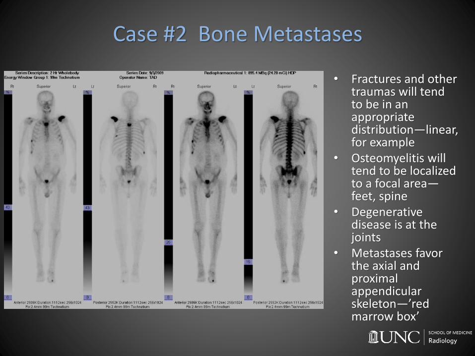
Case #2 Bone Metastases
• Fractures and other traumas will tend to be in an appropriate distribution—linear, for example
• Osteomyelitis will tend to be localized to a focal area—feet, spine
• Degenerative disease is at the joints
• Metastases favor the axial and proximal appendicular skeleton—’red marrow box’

Q. Which of these bone scan lesions is most likely to be benign?
1. Scapula
2. Spine
3. Rib
4. Toe

A. Which of these bone scan lesions is most likely to be benign?
1. Scapula
2. Spine
3. Rib
4. Toe
While metastases can occur anywhere in the skeleton, we tend to be somewhat more suspicious of metastases outside the axial and proximal appendicular skeleton. The exceptions are lung cancer and melanoma—for which bone scans are rarely obtained.

Case #3 Clinical Indication: 21yoM recently biopsied mediastinal mass

Case #3 Positive PET
• The ‘hot’ supraclavicular and paraspinal areas actually likely represent brown fat, a heat-generating tissue that uses a lot of glucose
• The brain and heart and bladder (and to a lesser extent, kidneys, liver, and bone marrow) have a low level of physiologic uptake.
• The hot masses in the chest don’t have a physiologic explanation, so…

Q. The most likely diagnosis is
1. Brown fat
2. Infection
3. Lymphoma
4. Normal scan

A. The most likely diagnosis is
1. Brown fat
2. Infection
3. Lymphoma
While the bilaterally symmetric uptake in the supraclavicular region is consistent with brown fat, the enlarged, FDG-avid masses in the chest are consistent with lymphoma.
4. Normal scan

Case #4 Clinical Indication: 53yoF breast cancer pt with SOB
VENTILATION PERFUSION

Case #4 Pulmonary Embolism
• Multiple defects in the lingula and basilar segments.
• These are NOT matched to ventilation defects.

Q. Critical organs for the Ventilation and Perfusion scan radiopharmaceuticals are respectively
1. trachea and lungs.
2. lungs and liver.
3. trachea and liver.
4. lungs and lungs.

A. Critical organs for the Ventilation and Perfusion scan radiopharmaceuticals are respectively
1. trachea and lungs.
2. lungs and liver.
3. trachea and liver.
4. lungs and lungs.

Case #5 Clinical Indication: 74yoM RUQ abdominal pain, normal GB US

Case #5 Cholecystitis
• The gallbladder is not seen.• Once the bowel is visible, we can try
giving morphine to contract the sphincter of Oddi– If the gallbladder shows up after morphine,
we may have chronic cholecystitis, but acute cholecystitis has been ruled out.
– What if we don’t see the bowel (or the patient can’t get morphine)? We can also wait 4 hours
• If you don’t see the bowel, you can try a fatty meal or CCK (if morphine not given, and you have no suspicion of CBD obstruction—see below).
• If you still don’t see the bowel (but you do see the bile duct), you might think about partial CBD obstruction. If you don’t see the bowel or the bile duct, you might think about complete CBD obstruction or poor liver function.

Q. During a Hepatobiliary Scan, the Morphine Sulfate is administered intravenously
1. at any time during the exam.
2. immediately following visualization of the GB.
3. at 60 minutes if GB is nonvisualized.
4. at 60 minutes if GB is nonvisualized, with tracer in bowel.

A. During a Hepatobiliary Scan, the Morphine Sulfate is administered intravenously
1. at any time during the exam.
2. immediately following visualization of the GB.
3. at 60 minutes if GB is nonvisualized.
4. at 60 minutes if GB is nonvisualized, with tracer in bowel.

Case #635yoF with ureteropelvic junction obstruction

Q. What is your next step?
1. Nothing, the study is normal.
2. Nothing, there is high-grade obstruction.
3. Give Lasix to differentiate between high-grade obstruction and a dilated collecting system on the right
4. Give Lasix to differentiate between high-grade obstruction and a dilated collecting system on the left

Q. What is your next step?
1. Nothing, the study is normal.
2. Nothing, there is high-grade obstruction.
3. Give Lasix to differentiate between high-grade obstruction and a dilated collecting system on the right
– The kidney isn’t clearing; if it clears with Lasix, this is just dilation, whereas if it doesn’t, this is a high-grade obstruction. Right is right on a renal scan!
4. Give Lasix to differentiate between high-grade obstruction and a dilated collecting system on the left.

• Post Lasix administration, the right kidney clears; this is not a high-grade obstruction

Think Back
• Common NM examinations at UNC
WB Bone V/Q PET Thyroid WB Thyroid Hepatobiliary
Gastric Emptying Parathyroid 131Iodine Therapy Diuretic
• Radiopharmaceutical, Technique, Indications to include normal exams and Need To Knows
• Case-based Q&A of six Aunt MinniesGraves Disease Bone Mets M1 Disease PE Cholecystitis
Hydronephrosis without obstruction