novel engineered cationic antimicrobial...
TRANSCRIPT

i
NOVEL ENGINEERED CATIONIC ANTIMICROBIAL PEPTIDES HAVE A BROAD-
SPECTRUM ACTIVITY AGAINST: FRANCISELLA TULARENSIS, BURKHOLDERIA
PSEUDOMALLI AND YERSINIA PESTIS
by
Suha Abdelbaqi
PharmD, Jordan University of Science and Technology, Jordan, 2010
Submitted to the Graduate Faculty of
the Department of Infectious Diseases and Microbiology
Graduate School of Public Health in partial fulfillment
of the requirements for the degree of
Master of Science
University of Pittsburgh
2014

ii
UNIVERSITY OF PITTSBURGH
Graduate School of Public Health
This thesis was presented
by
Suha Abdelbaqi
It was defended on
June 13th 2014
and approved by
Amy L. Hartman, Ph.D., Research Assistant Professor, Department of Infectious Diseases and
Microbiology, Graduate School of Public Health, University of Pittsburgh
Ronald C. Montelaro, PhD., Professor Microbiology & Molecular Genetics, School of Medicine,
University of Pittsburgh
Todd A. Reinhart, ScD., Professor Department of Infectious Diseases and Microbiology, Graduate
School of Public Health, University of Pittsburgh
Thesis director: Douglas S. Reed, Ph.D., Associate Professor, Department of Immunology, School of
Medicine, University of Pittsburgh

iii
Copyright © by Suha Abdelbaqi
2014

iv
ABSTRACT
Broad spectrum antimicrobial activity against biodefense pathogens is highly desirable as
a delay in treatment of infection with these pathogens can be fatal. Engineered cationic
antimicrobial peptides (eCAPs) are de novo synthesized amphipathic agents with broad spectrum
activities against gram-positive and gram-negative bacteria including Pseudomonas aeruginosa
and methicillin-resistant Staphylococcus aureus (MRSA). We evaluated eCAPs designated as
WLBU2 and WR12 against three bacterial pathogens that are considered potential biological
weapons and a great public health concern. Using both bacterial killing and growth inhibition
assays, different concentrations of WLBU2 and WR12 were tested in vitro against Francisella
tularensis, Burkholderia pseudomallei, and Yersinia pestis. LL37, a natural mammalian
antimicrobial peptide was used for comparison. WLBU2 proved to be the best candidate against
F. tularensis with a single dose of 3μM needed to achieve a 90% reduction in bacterial count and
a minimum inhibitory concentration (MIC) of 25 μM. LL37 failed to achieve more than 8%
reduction in the bacterial count of F. tularensis with concentrations up to 100 µM. Both WLBU2
and WR12 achieved significantly better results against Y. pestis compared to LL37, but only WR12
was able to attain a MBC of 50 μM. Results for B. pseudomallei varied depending on the testing
environment; a substantial reduction in bacterial count was seen with WLBU2 in potassium
phosphate buffer but not in phosphate buffered saline. WLBU3 and WLBU4 were also tested
NOVEL ENGINEERED CATIONIC ANTIMICROBIAL PEPTIDES HAVE A
BROAD-SPECTRUM ACTIVITY AGAINST: FRANCISELLA TULARENSIS,
BURKHOLDERIA PSEUDOMALLI AND YERSINIA PESTIS
Suha Abdelbaqi, M.S.
University of Pittsburgh, 2014

v
against B. pseudomallei and results indicate that increasing the length of the peptides did not
enhance their activity against B. pseudomallei. Moreover, WLBU2 was tested against Francisella
tularensis (SchuS4) infected macrophage cells (J774) and found to decrease intracellular bacterial
count by more than 90% at a concentration of 12.5µM. This data demonstrates the potential
usefulness of eCAPs against the three pathogens that were evaluated and the broad applicability
of eCAPs, particularly WLBU2, against bacterial biodefense pathogens.

vi
PREFACE
First, have a definite, clear practical ideal; a goal,
an objective.
Second, have the necessary means to achieve your
ends; wisdom, money, materials, and methods.
Third, adjust all your means to that end.
– Aristotle

vii
TABLE OF CONTENTS
1.0 INTRODUCTION …………………………………………………………………………....1
1.1 Francisella tularensis ………………………………………………………………...2
1.2 Burkholderia pseudomallei ……………………………………………..............................6
1.3 Yersinia pestis …………………………………………………………………….......8
1.4 Engineered cationic antimicrobial peptides ……………………………………….....13
2.0 SPECIFIC AIMS ……………………………………………………………….…………...16
2.1 AIM 1: Evaluate the antimicrobial activity of WLBU2 and WR12 in vitro against
virulent isolates of all three bacterial pathogens (F.tularensis, B. pseudomallei, Y. pestis)
to establish their ability to kill or inhibit growth of the pathogens……………………....16
2.2 AIM 2: Assess the antimicrobial activity of WLBU2 and WR12 ex vivo with F.
tularensis infected macrophages ………………………………………………………...16
3.0 METHODS..………………………………………………………………………………....17
3.1 Biosafety……………………………………………………………………………..17
3.2 Bacteria ……………………………………………………………….......................17
3.3 Peptides ……………………………………………………………………………...18
3.4 Assays …………………………………………………………………………….…20
3.4.1 In vitro assays ……………………………………………………………..20
3.4.2 MTT assay…………………………………………………………………21
3.4.3 Ex vivo assay ………………………………………………………………21
3.5 Statistical analysis……………………………………………………………………22
4.0 RESULTS …………………………………………………………………………………...23

viii
4.1 Defining a calibration curve …………………………………………………………23
4.2 In vitro antimicrobial activity of eCAPs……………………………………………..25
4.2.1 Bacterial killing assay ………………………………………………...…25
4.2.2 Growth inhibition assay …………………………………………………33
4.3 Antimicrobial activity of WLBU2 against F. tularensis infected macrophages ....…40
5.0 DISCUSSION ……………………………………………………………………………….43
BIBLIOGRAPHY ……………………………………………………………………………….51

ix
LIST OF TABLES
TABLE 1 EQUATIONS GENERATED FROM THE CALIBRATION CURVE OF EACH BACTERIA. ............. 25
TABLE 2 PERCENTAGE KILLING OF F. TULARENSIS BY WLBU2, WR12 OR LL37...................................... 28
TABLE 3 PERCENTAGE KILLING OF B. PSEUDOMALLEI BY WLBU2, WR12 OR LL37. .............................. 31
TABLE 4 PERCENTAGE KILLING OF Y. PESTIS BY WLBU2, WR12 OR LL37. ............................................... 32
TABLE 5 PERCENTAGE KILLING OF F. TULARENSIS BY ONE OR TWO CONSECUTIVE DOSES OF
WLBU2.. ............................................................................................................................................................ 33
TABLE 6 PERCENTAGE INHIBITION OF F. TULARENSIS BY WLBU2, WR12 OR LL37. ............................... 35
TABLE 7 PERCENTAGE INHIBITION OF B. PSEUDOMALLEI BY WLBU2, WR12 OR LL37. ........................ 37
TABLE 8 PERCENTAGE INHIBITION OF Y. PESTIS BY WLBU2, WR12 OR LL37. ......................................... 39

x
LIST OF FIGURES
FIGURE 1 SCHEMATICS OF WLBU2 AND WR12 .. ............................................................................................. 22
FIGURE 2 CALIBRATION CURVE TO CALCULATE THE CONVERSION FACTOR BETWEEN OD
READING AT 600NM AND THE NUMBER OF COLONY FORMING UNITS GROWING PER ML
(C.F.U/ML). ....................................................................................................................................................... 24
FIGURE 3 DOSE-DEPENDENT KILLING OF F. TULARENSIS BY WLBU2, WR12 OR LL37. .......................... 27
FIGURE 4 DOSE-DEPENDENT KILLING OF B. PSEUDOMALLEI BY WLBU2, WR12 OR LL37. ................... 30
FIGURE 5 DOSE-DEPENDENT KILLING OF Y. PESTIS BY WLBU2, WR12 OR LL37. .................................... 31
FIGURE 6 DOSE-DEPENDENT KILLING OF F. TULARENSIS BY ONE DOSE AND TWO DOSES OF
WLBU2. ............................................................................................................................................................. 32
FIGURE 7 DOSE – DEPENDENT INHIBITION OF F. TULARENSIS GROWTH BY WLBU2, WR12 OR LL37.
........................................................................................................................................................................... 35
FIGURE 8 DOSE – DEPENDENT INHIBITION OF B. PSEUDOMALLEI GROWTH BY WLBU2, WR12 OR
LL37. .................................................................................................................................................................. 36
FIGURE 9 DOSE – DEPENDENT INHIBITION OF B. PSEUDOMALLEI GROWTH BY WLBU2, WLBU3,
WLBU4 OR LL37. ............................................................................................................................................ 38
FIGURE 10 DOSE – DEPENDENT INHIBITION OF Y. PESTIS GROWTH BY WLBU2, WR12 OR LL37. ....... 39
FIGURE 12 DOSE-DEPENDENT KILLING OF INTRACELLULAR F. TULARENSIS BY WLBU2. ................... 42
FIGURE 13 A MODEL OF ANTIMICROBIAL PEPTIDES MECHANISM OF ACTION...................................... 46
FIGURE 11 J774 MACROPHAGE CELLS TREATED FOR 1 HOUR WITH A RANGE OF CONCENTRATIONS OF WLBU2 ...................................................................................................................................................................41

1
1.0 INTRODUCTION
Biodefense agents are pathogenic organisms or toxins that pose a severe threat to public
health and can be used against humans, animals, or plants for terrorist purposes. These agents are
typically found in nature, but they could be also modified to enhance their virulence, ability to
disseminate or make them resistant to current antibiotics or vaccines. Such biological agents are
attractive to terrorists as an alternative to conventional weapons due to their low costs, their relative
ease of production, ability to aerosolize, and difficulty of detection. Biodefense agents are
classified by the National Institute of Allergy and Infectious Diseases (NIAID) according to the
risk for human health into 3 categories of A, B and C [1]. In 2012, a new designation, "Tier 1",
has been developed for those select agents and toxins considered to be of the greatest risk for
deliberate misuse and the most significant potential for mass casualties or devastating effects on
the economy or critical infrastructure [2].
Francisella tularensis, Burkholderia pseudomallei and Yersinia pestis are all gram
negative facultative intracellular tier 1 select agents that are able to cause fatal diseases if not
treated appropriately. The use or even threatened use of such organism can produce a widespread
social disruption. Therefore, establishing effective treatment strategies against such pathogens is
of the utmost importance. Unfortunately, some of these agents have already developed resistance
against current available antibiotics emphasizing the need for new therapeutic agents [3][4]. In this

2
study, de novo engineered cationic antimicrobial peptides, WLBU & WR12, are investigated as a
potential alternative for treatment of these organisms.
1.1 Francisella tularensis
Francisella tularensis is a small, pleomorphic, gram-negative coccobacillus and the
causative agent of the bacterial zoonotic disease, tularemia [5]. F. tularensis was first isolated by
McCoy in 1912 following an outbreak of a plague-like disease in rodents in Tulare County,
California [6]. Since then four different subspecies of F. tularensis have been identified: tularensis,
holarctica, mediasiatica and novicida [7]. Most human cases are caused by either subspecies
holarctica or tularensis, with the latter being the more pathogenic subspecies. With the exception
of a presumptive laboratory release, subspecies tularensis is only found in North America.
Since its identification, tularemia cases have been documented throughout the Northern
hemisphere including Europe, China, Korea, Japan, the former Soviet Union and North America.
Up until the 1950s, the United States had thousands of tularemia cases reported annually with a
peak number of 2,291 in 1939 [8]. With the discovery of antibiotics these numbers declined
drastically reaching less than 150 cases per year between 1990 and 2000. Except for Hawaii, all
American states have reported at least one case of tularemia with the majority of them residing in
the south-central and western region (Arkansas, Missouri, South Dakota, and Oklahoma) [9].
Francisella tularensis is thought to be maintained in soil, water and through its extensive
host distribution including humans, domesticated and wild animals such as lagomorphs,
carnivores, rodents, birds and fish [10, 11]. It is primarily transmitted to humans and other animals
through arthropod vectors such as biting flies and ticks. Ninety percent of tularemia cases in the
United States occur via an arthropod bite, more specifically via the American dog tick, the Lone

3
Star tick, and the Rocky Mountain wood tick [10, 12]. It can also occur directly through
contaminated food, water, infected animals and their carcasses [13]. Aerosolization of F. tularensis
can occur during handling of contaminated material which may lead to the inhalation of the
bacteria. For instance, 11 cases of primary pneumonic tularemia occurred in the summer of 2000
on Martha’s Vineyard Massachusetts due to the aerosolization of the bacteria from infected animal
carcasses and their dens during lawn mowing [14]. Further, laboratory transmission of F.
tularensis can occur readily due to the ease of its aerosolization; opening a culture plate is enough
to create an aerosol [15]. To date, there has been no supportive evidence of person to person
transmission.
The onset of tularemia is usually abrupt with flu-like symptoms, fever and generalized
weakness. Chest tightness, sub-sternal pain and dry cough have also been noted [16]. Following
an incubation period between 1 to 21 days and depending on the route of inoculation, tularemia
manifests in one of six forms: oropharyngeal, pneumonic, typhoidal, ulceroglandular, glandular or
oculoglandular [17]. The most common form of the disease is ulceroglandular tularemia following
an infected arthropod bite or handling contaminated animals [18]. Pneumonic tularemia results
from inhaling contaminated aerosols or through the dissemination of the bacteria from another site.
It is characterized by an acute illness with pharyngitis, pleuropneumonitis, bronchiolitis and/or
hilar lymphadenitis [19]. Patients may progress into respiratory failure and death. Typhoidal
tularemia has no specific site of inoculation and is characterized with systemic illness including
fever, abdominal pain, and diarrhea and vomiting. If not treated promptly, both typhoidal and
pneumonic tularemia may lead to death [13].
In situations of contained tularemia causalities, where individual patient management is
possible, a parenteral route of treatment with antibiotics is recommended. The aminoglycosides

4
streptomycin and gentamicin are both bactericidal against F. tularensis and are considered the gold
standard for treatment in these situations. Streptomycin (1 g IM twice daily) or gentamicin (5
mg/kg IM or IV once daily) should be continued for 10 days. Tetracycline or chloramphenicol are
bacteriostatic against F. tularensis and can be used as alternative treatment. Due to their
bacteriostatic profile, these drugs should be continued up to 14 days to reduce the possibility of
relapse [13](23).
Fluoroquinolones, such as ciprofloxacin and doxycycline, have a bactericidal effect against
intracellular organisms such as F. tularensis. During a human outbreak in Spain (between
December 1997 and February 1998) ciprofloxacin treatment resulted in the lowest therapeutic
failure and side effects compared to streptomycin and doxycycline [20]. Currently, ciprofloxacin
can be regarded as the drug of choice for the treatment of uncomplicated tularemia and as a second-
line treatment for severe infections; although relapse may occur due to incomplete clearance of F.
tularensis. In case of massive causalities, e.g. during a bioterrorism attack, oral doxycycline and
ciprofloxacin are the preferred choices for both adults and children. Achieving the appropriate
treatment decreases the mortality rate of tularemia from 35% to 1% [21]. Post-exposure
prophylactic treatment would include streptomycin, gentamicin, doxycycline, or ciprofloxacin for
14 days [13]. Even with antibiotics available for treatment of F. tularensis infection, pneumonic
tularemia requires intensive care and the recovery rate is extremely slow so that even a moderate
outbreak can overwhelm medical facilities. Further, F. tularensis can be genetically modified to
become antibiotic resistant.
The F. tularensis live vaccine strain (LVS) is the only vaccine available to protect against
tularemia. LVS was generated in the former Soviet Union as a classically passaged live attenuated
strain derived from a virulent F. tularensis subsp. holarctica strain. Studies conducted in the

5
1960’s demonstrated that LVS was safe when given to humans and could protect against aerosol
challenge with SchuS4, a virulent strain of F. tularensis subsp. tularensis although this protection
failed at higher challenge doses. Nevertheless, LVS is only available as an Investigational New
Drug (IND) and unlikely to be approved by the FDA due to concerns regarding the potential for
reversion and incomplete protection against aerosolized F. tularensis. As an IND, LVS is currently
given to at-risk laboratory personnel who work with tularemia. Consequently, there is no actual
way of controlling this disease in nature. Public awareness of this organism and its potential harm
to humans should be a priority. In regions where tularemia is endemic, people should avoid
handling sick or dead animals and insect populations should be reduced [22].
F. tularensis is considered one of the most infectious bacterial pathogens. In a bioterrorist
attack F. tularensis would most likely be used as an aerosol resulting in pneumonic tularemia
represented as respiratory symptoms, sub-sternal discomfort, and cough. In 1970, a World Health
Organization (WHO) committee stated that a 50-kg aerosol dispersion of a virulent F. tularensis
strain in a population area of five million people would cause 19,000 deaths and 250,000
hospitalizations. Without the appropriate antibiotics, most of the hospitalized cases would progress
into respiratory failure and death [23]. F. tularensis progression is slower than anthrax or plague,
but the disease caused by this organism persists for several weeks with a high relapse rate. The
WHO estimated that such an attack with tularemia would cost around $5.4 billion per 100,000
persons infected [24].

6
1.2 Burkholderia pseudomallei
Burkholderia pseudomallei is a gram-negative, motile rod-shaped, soil saprophyte that can
be easily collected from water and wet soil in endemic areas. B. pseudomallei is the causative
agent of melioidosis, a tropical infectious disease affecting humans, animals and plants. Previously
known as Pseudomonas pseudomallei, B. pseudomallei is able to infect various organs causing a
range of clinical symptoms that might be fatal to the infected patient. Despite its rare presence in
the western countries, concern about the potential use of B. pseudomallei in bioterrorism fuels a
great deal of the B. pseudomallei research.
Melioidosis is endemic in various areas of Southeast Asian and northern Australia.
Thailand has the highest annual incidence of melioidosis; 4.4 cases per 100,000. In Ubon
Ratchathani – Thailand, 20% of community acquired bacterial infections are due to B.
pseudomallei [25]. Further, severe melioidosis has a case fatality rate of 50% in that region [26].
Northern Australia has about the same incidence rate as Thailand, and B. pseudomallei is
considered the most common case for fatal community acquired bacterial pneumonia; the mortality
rate is about 20% among patients with melioidosis [27]. Sporadic cases were observed worldwide,
particularly in the Caribbean region, Africa and the Middle East [28]. Cases that occurred in the
western countries usually involved returning travelers and military veterans [29].
Humans and animals can be infected through ingestion, inhalation or inoculation of a skin
wound. In humans, most cases occur directly from the environment through skin abrasions.
Inhalation of B. pseudomallei is usually related to heavy rainfalls and strong wind that might carry
it from the soil or water into our respiratory system [30]. Person to person transmission is rare, but
has been reported between family members in close contact [31]. Vertical transmission is also

7
considered rare with only a few newborn cases reported [32]. Transmission by mosquitoes and rat
fleas has been reported, but the role of vector born transmission remains unclear [28, 33].
Melioidosis is often referred to as “the great imitator” due to its wide spectrum of clinical
diseases. Melioidosis can present as an acute or chronic, localized or generalized disease. Other
cases may result in an inapparent infection with no noticeable signs or symptoms [34]. Following
an incubation period between 1 and 21 days, the lung is mostly affected by this organism. The
ensuing pulmonary disease can range from mild respiratory illness to acute or chronic pneumonia
that may rapidly progress to septic shock and death. Pneumonia is the most common manifestation
between all patients with melioidosis [25] and may progress to septicemia if left untreated.
Septicemic melioidosis is the most serious form of the disease with a mortality rate as high as 70%
[35][36].
Despite being intrinsically resistant to many drugs, B. pseudomallei is susceptible to some
antibiotics. Treatment is divided into 2 phases: intensive and eradication. As a general rule,
intravenous bactericidal drugs are used in the intensive phase, while oral bacteriostatic drugs are
used in the eradication phase. In life threatening disease, the current regimen is comprised of
intravenous Ceftazidime for at least 2 weeks followed by oral combination of ciprofloxacin and
azithromycin or doxycycline and co-trimoxazole for 20 weeks to prevent relapse. For mild to
moderate disease, IV amoxycillin-clavulanate is used for 2 weeks followed by co-trimoxazole and
doxycycline for 20 weeks. Localized melioidosis should be treated with incision and drainage with
co- trimoxazole for 6 – 8 weeks [37].
Relapse and recurrence of infection is common in melioidosis despite receiving appropriate
therapy. Relapse is defined as “reappearance of signs and symptoms after clinical response while
still on antimicrobial therapy” while recurrent infection is “a new episode of melioidosis caused

8
by the same organism after full clinical recovery” [38]. Both infections are thought to be due to
failure of the host to eliminate the offending pathogen during initial attack. Many factors aid B.
pseudomallei evasion of the host system including hiding in phagocytic cells and sealed abscesses.
In endemic areas such as Malaysia and northeast Thailand the overall relapse rate is higher than
15% per year despite prolonged treatment [39].
With no effective vaccine available, precautionary measures should be taken in endemic
areas. These measures include covering all open wounds and wearing the appropriate gear for
outdoor activities. Infected animals are usually euthanized rather than treated due to the high risk
of relapse and expensive medication needed. If treated, animals should be under lifelong
monitoring for relapse [38].
CDC assigned B. pseudomallei as a category B select agent and a possible bioweapon
leading to an increased research interest around the world. Even in literature, this agent’s potential
as a bioweapon was suggested by Sir Arthur Conan-Doyle in his Sherlock Holmes story “The
Dying Detective” [40]. It is believed that B. pseudomallei was being weaponized by the former
USSR with the potential of creating an engineered antibiotic resistant strain. Although the
prospective of weaponizing this agent is uncertain, it has a great potential due to its high mortality
rates, wide range of hosts, ability to be aerosolized and intrinsic antibiotic resistance
1.3 Yersinia pestis
Yersinia pestis is a gram-negative, non-motile, non-spore forming coccobacillus belonging
to the family Enterobacteriaceae. Y. pestis is believed to be a clone of Y. pseudotuberculosis that
emerged within the last 1,500 to 20,000 years by acquiring different genetic elements making it
able to be transmitted through fleas [41]. Y. pestis had numerous nomenclature changes from

9
Bacterium pestis to Bacillus pestis to Pasteurella pestis and finally Y. pestis in 1970 [42]. Plague
(a.k.a the Black Death) is the zoonotic disease caused by this organism. It can be transmitted to
humans through flea bites causing a rapidly progressive severe illness that is usually deadly if not
appropriately treated.
Plague was the cause of three major pandemics in which nearly 200 million people were
killed. The first pandemic, the Justinian plague, started in Egypt around A.D. 541 and then swiftly
transmitted through the Middle East and Mediterranean Europe leading to the loss of 50 – 60% of
the population. The second pandemic started with an epidemic in Europe during the 14th century.
This epidemic killed 17 to 28 million Europeans (30 – 40% of the population) and more
importantly resulted in relentless epidemic cycles that comprise the second pandemic. The third
pandemic started in China around 1855 and disseminated to Africa, Australia, Europe, Middle
East, North America, and South America by 1900. It is believed that plague was introduced to the
United States from China in the 20th through ship rats and their fleas. Fortunately, mortality rates
were significantly reduced compared to previous pandemics which is thought to be due to effective
antibiotics and active public health measures [43]. Currently, it is believed that around 4,000
human plague cases occur annually worldwide [44].
Plague is enzootic in rodents, but it can be transmitted to humans by infected fleas. When
the rat fleas, the classical vector for plague, feed on an infected animal the bacteria infects and
multiplies in the midgut of the flea. Ultimately the flea dies out of starvation, but not before
infecting several hosts. In the infected mammal, Y. pestis spreads from the bite site to the regional
lymph node and multiply quickly causing the lymph node to swell and form a bubo. Further, the
bacteria spread to the bloodstream and colonize in different organs such as liver, spleen and lung
[45].

10
Y. pestis can rarely transmit to humans through direct contact with infected animals
especially rodents. Person-to-person transmission through respiratory droplets may occur; in 1994
an outbreak of pneumonic plague occurred in Surat, India. A total of 460 plague cases were
compiled between September 20 and September 25 in Surat. When news of the plague became
known to the public about 700,000 people fled the city. It is estimated that this small outbreak
cost India US$3 billion and the global economy US$5 to $6 billion [46]. Fortunately pneumonic
plague is rare, but bubonic plague may spread to the lungs triggering a secondary pneumonic
plague that can be transmitted to other people and cause considerable mortality if untreated [43].
Plague manifests as one of three primary forms: Bubonic, septicemic, or pneumonic.
Human epidemics commonly start as bubonic plague following a flea’s bite. If not treated promptly
infected patients usually develop secondary plague septicemia or pneumonia increasing the risk of
person to person transmission. Untreated bubonic plague may lead to 40 – 60% case fatalities
while untreated septicemic and pneumonic plague is always fatal; death is usually due to a septic
shock [47, 48].
Pneumonic plague is very rare, nonetheless it is the most dangerous and deadly form of the
disease. Disease spreads via respiratory droplets from infected animals or humans. It initially
presents as a febrile like illness but progresses rapidly with 1 to 3 days into an overwhelming
pneumonia with productive bloody cough, dyspnea and cyanosis. Cervical lymph nodes may be
affected creating buboes in the neck region. The disease quickly overwhelms the lungs and
hemorrhages develop resulting in hemorrhagic pneumonia. The terminal events may include
respiratory failure and circulatory collapse. Septicemic plague patients may develop secondary
plague pneumonia through the hematogenous dissemination of the bacteria to the lung (68)[49].

11
Rapid diagnosis and prompt administration of appropriate antibiotics are essential for the
success of treatment and reduction of complications. Streptomycin, tetracycline, and sulfonamides
are considered the standard treatment, with streptomycin being the treatment of choice for plague.
Using one or a combination of antibiotics depends on the severity of the infection. Gentamicin
alone or with tetracycline can be used as a substitute for streptomycin. Antibiotic treatment
continues for 10 days or 2 days after the fever subsides. Patients suspected of having pneumonic
plague or secondary pneumonia following bubonic plague should be isolated for at least 4 days
after starting treatment to prevent human-to-human transmission [50, 51]. A recent study by
Layton et al demonstrated successful treatment of established pneumonic plague using
Levofloxacin in African green monkeys. The monkeys were given a respiratory dose of 100 lethal
doses for 50% of animals (LD50) and were observed for fever and vital signs by telemetry.
Intravenous Levofloxacin treatment was initiated when the animal spiked a fever in a fashion that
mimics antibiotic levels achieved in humans. All animals treated with a placebo died, while all
animals treated for 10 days with Levofloxacin survived. In the treated group fever resolved within
24–48 hours of initiating treatment. This study indicates that Levofloxacin may be a suitable broad-
spectrum antibiotic for therapy in an aerosolized bioweapons attack and should be further studied
and evaluated for bubonic plague [52].
Prophylactic therapy consists of sulfonamide, trimethoprim-sulfamethoxazole, or
tetracycline administered to the patient and their close contacts. In the case of a bioterrorist attack
with aerosolized plague, streptomycin or gentamicin are the preferred choice in a contained
casualty setting, while oral therapy with doxycycline, tetracycline, Levofloxacin or ciprofloxacin
are preferred for mass casualty situations. There are two plague vaccines currently available
around the world: a live vaccine made from an attenuated strain of Y. pestis and a killed vaccine

12
derived from a formalin-fixed virulent strain. Vaccines are administered intramuscularly as a series
of three primary shots over a period of a year then booster shots should be given every 1 to 2 years.
Due to their side effects, need for repeated boosters and ineffectiveness against pneumonic plague,
these vaccines are only given to people at high risk of contracting Y. pestis such as military
personnel serving in endemic areas [53, 54].
In 1995, two multi-drug resistant isolates of Y. pestis were observed in Madagascar. One
strain was isolated from a 16 year old male and the other from a 14 year old patient with both of
them presenting symptoms of bubonic plague. Both strains were resistant to streptomycin and
several other antibiotics recommended for plague treatment. Both patients were successfully
treated with a combination of intramuscular injections of streptomycin (2 g per day for 4 days) and
oral administration of trimethoprim-sulfamethoxazole (2 g per day for 10 days) [50]. Thus there is
cause for concern that antibiotic resistant strains of Y. pestis could occur naturally or be made in a
laboratory.
Due to its high fatality and rapid progression, Y. pestis is considered a potential biological
weapon. During the World War II, the Japanese army released plague-infected fleas over several
areas in China leading to many plague outbreaks [55]. Offensive biological weapons programs in
both the USA and the Soviet Union researched and successfully developed techniques to aerosolize
plague in place of using infected fleas. It was estimated by the WHO that releasing an aerosol of
50kg of Y. pestis over a city of 5 million would lead to development of pneumonic plague in
150,000 people, of which 36,000 are expected to die [56]. Research in weaponizing Y. pestis was
terminated in the USA around 1970 (when all offensive biological warfare research was
terminated) but it continued in the Soviet Union. In the year 1995 an individual in Ohio was
arrested after illegally acquiring Y. pestis by mail. This incident lead to establishment of new

13
antiterrorism regulations which included regulations guiding tracking shipments (and eventually
inventories) of pathogens designated as ‘select agents’, those for which there was concern they
could be used as biological weapons. Today Y. pestis is classified as a tier 1 select agent by the
United States Department of Health and Human Services [57].
1.4 Engineered Cationic Antimicrobial Peptides (eCAPs)
Cationic antimicrobial peptides (CAPs) are amphipathic peptides of 12–50 amino acids
with a net positive charge. They are ubiquitous peptides naturally found in all living species and
are known to be active components of the innate immunity against infectious pathogens [58]. In
response to infectious pathogens, CAPs can be released from macrophages, granules of
neutrophils, or with mucosal and skin secretions from epithelial cells. CAPs can act against a wide
range of targets including gram-positive and gram-negative bacteria, fungi and parasites - the only
essential component in these targets is a negatively charged plasma membrane. Therefore, normal
cells have a relative resistance due to the peptides preference for negatively charged, cholesterol
free membranes [59]. The mechanism of action of CAPs is not fully understood, although studies
have implicated the electrostatic interaction between the peptides and the lipid molecules on the
bacterial membrane. When compared to other antibiotics, CAPs are able to kill bacteria rapidly,
within 30 to 180 seconds, limiting the bacterium’s ability to develop resistance against these
peptides. Therefore, CAPs are considered a good candidate for use against multi-drug resistance
(MDR) bacteria.
Although these cationic peptides have a wide spectrum of action against pathogens, their
activity strongly depends on the environment they are released in. The optimal potency of each
peptide is only achieved in specific environmental conditions and readily lost when subjected to a

14
different setting. For instance, human β-defensin loses its antimicrobial activity when subjected to
the abnormally high NaCl concentration in a cystic fibrosis lung [60]. Therefore, to be able to
utilize their antimicrobial activity to the fullest, development of CAPs has shifted towards
modifying host-derived peptides or choosing specific amino acids for de novo synthesis. Studies
of an HIV-1 transmembrane protein (referred to as lentivirus lytic peptide 1 (LLP1)) demonstrated
that it had remarkable antimicrobial properties [61]. As a result of that work, it was concluded that
de novo synthesis of an α-helical amphipathic cationic antimicrobial peptide, using arginine
residues on the polar side and valine residues on the non-polar side, would greatly improve the
potency and selectivity of the peptide [62]. Further studies of the influence of length and helical
structure on activity lead to development of a series of de novo synthesized peptide designated
WLBU [63]. WLBU peptides are composed of arginine in the hydrophilic face, valine and
tryptophan residues in the hydrophobic face separated from each other by at least seven amino
acids (Figure 1). Moreover, WLBU studies demonstrated that tryptophan substitution significantly
increased antimicrobial activity in challenging environments such as serum and blood. Therefore,
WR12 was designed using only arginine in the hydrophilic face and tryptophan in the hydrophobic
face [63, 64] (Figure 1).
Several studies tested the potency of engineered peptide derivatives against various agents
such as Pseudomonas aeruginosa, methicillin resistant Staphylococcus aureus, Streptococcus
gordonii, Fusobacterium nucleatum, and Porphyromonas gingivalis [65-68]. Unlike the natural
peptide LL37, WLBU2 was able to eliminate P. aeruginosa and S. aureus in physiological NaCL
concentrations and kill P. aeruginosa in human serum, whole blood and skin fibroblasts without
evident adverse effects to the host cells. In vivo toxicity and efficacy of WLBU2 against P.
aeruginosa were also tested using a mouse model. An intravenous dose of 3mg/kg of WLBU2 was

15
able to clear the bacteria and save all the 14 mice from the P. aeruginosa infection. WLBU2 had
no apparent toxic effect on the mice at up to 12 mg per kg of their body weight [64]. Further,
WR12 and WLBU2 demonstrated an optimal antibacterial selectivity against a panel of MDR
clinical isolates such as Acinetobacter baumannii and Klebsiella pneumoniae [66]. Taking all this
into consideration, this study aims to evaluate the in vitro and ex vivo antimicrobial activity of
WLBU series and WR12 against F. tularensis, B. pseudomallei and Y. pestis.

16
2.0 SPECIFIC AIMS
This study aims to investigate the potential role of WLBU and WR12 as a antimicrobial
agents against F. tularensis, B. pseudomallei and Y. pestis. The potency of WLBU and WR12 is
compared to that of LL37 under defined test condition to demonstrate the ability of de novo
synthesized peptides with optimized amphipathic structures to overcome certain limitations of host
antimicrobial.
2.1 AIM 1: Evaluate the antimicrobial activity of WLBU and WR12 in vitro against virulent
isolates of all three bacterial pathogens (F.tularensis, B. pseudomallei, and Y. pestis) to
establish their ability to kill or inhibit growth of the pathogens. All procedures will be first
evaluated with the attenuated LVS strain of F. tularensis prior to use in BSL-3.
a. Determine the growth of each pathogen in specified media and the exponential phase by
establishing a growth curve for each of them. This will further help in determining the
equations needed for bacterial concentration calculations.
b. Determine the effectiveness of our peptides in reducing the bacterial count of the tested
pathogens and the minimum bactericidal concentration (MBC), if applicable.
c. Determine the effectiveness of our peptides in inhibiting the growth of the tested pathogens
and the minimum inhibitory concentration (MIC), if applicable.
2.2 AIM 2: Assess the antimicrobial activity of WLBU2 ex vivo with F. tularensis infected
macrophages.
a. Determine ability of WLBU2 to modulate macrophage cells (J774) cell viability.
b. Determine the reduction in bacterial counts after using WLBU2 against F. tularensis
(SchuS4) infected J774 cells.

17
3.0 METHODS AND MATERIALS
3.1 Biosafety
All experiments using F. tularensis, B. pseudomallei or Y. pestis were performed in the
biosafety level 3+ Regional Biocontainment Laboratory (RBL) at the University of Pittsburgh.
Powered air purifying respirators (PAPRs) were worn for respiratory protection, and all work
was conducted in a class II biosafety cabinet using Vesphene IIse (diluted 1:128, Steris
Corporation, cat. #646101) as a disinfectant.
3.2 Bacteria
All the following bacteria were obtained from Biodefense and Emerging Infections
Research Resources Repository, Manassas, VA. Francisella tularensis live attenuated vaccine
strain (LVS) and SchuS4 strain (Catalog No. NR-643) were grown on cysteine heart agar (CHA)
for 2 days at 37°C and cultured in brain heart infusion broth (BHI) or Chamberlain’s chemically
defined medium (CCDM) for 17 to 19 hours [69]. Burkholderia pseudomallei strain 1026b
(Catalog No. NR-9910) was grown on Luria Broth (LB) Agar for one day at 37°C and then cultured
in BHI for 11 hours. Yersinia pestis strain CO92 (Catalog No. NR-641) was grown on tryptic soy
agar (TSA) for 2 days at 37°C and then cultured in BHI or Bacto nutrient broth for 24 to 27 hours.
The overnight culture of bacteria was prepared by streaking an agar plate with a loopful of the
frozen bacterial stock (-80 freezer). The plate was incubate for 24hrs at 37°C for B. pseudomallei,
48 hrs at 37°C for F. tularensis and 48hrs at 30°C for Y. pestis. Next, several colonies were taken
from the plate and re-suspend in 3ml of growth media e.g. BHI. Optical density (OD) was adjusted
to 0.1 then 0.5ml of the previous solution was added to 24.5ml of the growth media in a 125ml

18
filtered top flask. Bacterial culture was incubated at 37°C in a shacking incubator (250 rpm) for
the number of hours needed for the bacteria to reach the exponential phase; B. pseudomallei
(11hours), Y. pestis (24 – 27 hours), and F. tularensis (17 – 19 hours). After incubation, 10ml of
the culture were spun down at 2800 rpm for 10 minutes then re-suspend in PBS or Growth media
depending on the assay done.
3.3 Peptides
Peptides were synthesized using standard Fmoc (9-fluorenylmethoxycarbonyl chloride)
synthesis protocols (Genscript, Piscataway, NJ). Synthetic peptides were characterized and
purified by reverse-phase high pressure liquid chromatography on Vydac C18 or C4 columns (The
Separations Group, Hesperia, Cal.). The identification of each was established by mass
spectrometry (Electrospray Quatro II triple quadruple mass spectrometer; Mi-cromass, Inc.,
Manchester, United Kingdom). Peptide concentrations were determined using a quantitative
ninhydrin assay. The peptides and their sequences were as follows.
WLBU2: 24-mer peptide composed of 13 arginine (R) in the hydrophilic face and 8 valine
(V) and 3 tryptophan (W) residues in the hydrophobic (Figure 1)
(RRWVR RVRRV WRRVV RVVRR WVRR)
WLBU3: 36-mer peptide composed of 18 arginine in the hydrophilic face and 14 valine
and 4 tryptophan residues in the hydrophobic
(VRRVW RRVVR VVRR WVRR VRRV WRRV VRVV RRWVRR)

19
WLBU4: 48-mer peptide composed of 23 arginine in the hydrophilic face and 20 valine
and 5 tryptophan residues in the hydrophobic
(RVVRV VRRWV RRVRR VWRRV VRVVR RWVRR VRRVW RRVVR
VVRRW RVV)
WR12: 12-mer peptide composed of 6 arginine in the hydrophilic face and 6 tryptophan
residues in the hydrophobic; requires mannitol to prevent precipitation (Figure 1)
(RWWRWW RRWWRR)
LL37: 37-mer peptide (Leu,Gly,Asp,Phe,Arg,Lys,Ser,Glu,Ile,Val,Gln,Pro,Thr); first
natural amphipathic alpha helical peptide isolated from human
( Leu - Leu - Gly - Asp - Phe - Phe - Arg - Lys - Ser - Lys - Glu - Lys - Ile - Gly - Lys -
Glu - Phe - Lys - Arg - Ile - Val - Gln - Arg - Ile - Lys - Asp - Phe - Leu - Arg - Asn - Leu
- Val - Pro - Arg - Thr - Glu – Ser)

20
3.4 Assays
3.4.1 In vitro assays:
Prior to any experiments, growth curves were established for each pathogen to determine
log-phase and stationary phase. Also, mathematical equations were derived from calibration curves
(optical density/colony-forming unit (c.f.u) curves) to achieve the needed bacterial concentration.
Bacterial killing assay (BKA): A of bacterial suspensions (concentration of 1×10⁶ c.f.u/mL) in
phosphate buffer saline or potassium phosphate buffer were incubated with two-fold dilutions of
peptides (concentration of 1.5µM to 100µM) in a 96-well plate (Falcon No. 3072, Becton-
Dickinson, Oxnard, CA) for 60 min at 37°C. Serial peptide dilutions were performed and plated
on agar plates. Colonies were counted following an appropriate incubation time to determine the
reduction in bacterial count. The minimum bactericidal concentration (MBC) is determined as
peptide concentration reducing bacterial count by at least 99%.
Growth inhibition assay (GIA): Bacterial suspensions (concentration of 1×10⁶ c.f.u/mL) in growth
media were incubated with two-fold dilutions of peptides (concentration of 1.5µM to 100µM) in
a 96 well plate for 24 to 48 hours at 37°C. After incubation the optical density of each well was
measured at 600nm using a spectrophotometer. Percent inhibition was calculated from the
difference in optical density measured. The minimum inhibitory concentration (MIC) was
determined as peptide concentration reducing bacterial growth by at least 90%.

21
3.4.2 MTS assay:
Macrophage J774 cells (ATCC TIB – 67) were seeded in Dulbecco's Modification of
Eagle's Medium (DMEM, Cellgro) supplemented with 10% fetal bovine serum at 37°C on 96-
well cell culture plates at a density of 3 × 10⁸ cells/well. A set of wells is prepared with medium
only for background subtraction and another with untreated cells for 100% viability reference.
Seeded Plate was incubated overnight at 37 °C, 5% CO2, 95% humidity. WLBU2 was added at
concentration ranging from 3 µM to 100 µM and incubated for an hour. Following incubation
cells were washed using PBS and 100 µL of media were added to each well. Then 20 µl of MTS
solution, 3-(4, 5-dimethylthiazol-2-yl)-5-(3-carboxymethoxyphenyl)-2-(4-sulfophenyl)-2H-
tetrazolium (Sigma, Lakehood, N.J.), were added to each well (final concentration of MTS will
be 0.33 mg/ml) and incubated for 2hr at 37°C. Absorbance was recorded at 490 nm to determine
the viability of cells with different peptide concentrations compared to untreated cells.
3.4.3 Ex vivo assay:
For ex vivo infection with F. tularensis, a standardized assay using a mouse macrophage
cell line (J774) was used. J774 cells were seeded in Dulbecco's Modification of Eagle's Medium
(DMEM, Cellgro) supplemented with 10% fetal bovine serum at 37°C on 12-well cell culture
plates at a density of 3 × 10⁷ cells/well. Seeded plates were incubated overnight at 37 °C, 5% CO2,
95% humidity. Cells were infected with F. tularensis SchuS4 at an MOI of 1:100 and incubated
further for 2 hr. Cells were washed three times with 2 ml of potassium buffer saline(PBS)
containing 50µg/ml gentamicin followed by addition of 1ml of media with 50µg/ml gentamicin to
each well and incubated for 1 hr. Subsequently, cells were washed again three times with PBS
and different concentrations of WLBU2 (3µM to 25µM) was added to each well and incubated for

22
another 1hr. Cells were then lysed using 0.02% sodium dodecyl sulfate (SDS) and 100µl of the
lysate was further diluted in PBS and plated on CHA. Colonies were counted to determine bacterial
count reduction with WLBU2.
3.5 Statistical Analysis:
Analysis was performed using percentages. Significance of differences in bacterial killing
or inhibition between different peptides was conducted using two way analysis of variance
(ANOVA) in Graphpad prism 6. P < 0.0001 was considered to be statistically significant.
WLBU2 WR12
The cationic amphipathic peptides were designed as demonstrated in helical wheel diagrams. Arginine,
valine, and tryptophan residues were arranged to form α helices, with the hydrophilic and hydrophobic faces
indicated in blue and yellow circles, respectively. The 12- and 48-mers LBU1 and WLBU4 are shown as
representatives of the LBU and WLBU series, respectively.
Figure 1 Schematics of WLBU2 and WR12 .[66]

23
4.0 RESULTS
4.1 Defining bacterial concentration by a standard equation for each pathogen
Light absorbance at different dilutions of bacterial cultures were measured to produce a
calibration curve which enabled conversion of the optical density (OD) measured into cell counts
and provided a standard equation for calculating the required concentration. This equation was
obtained by growing bacteria to the exponential phase as described above, taking OD readings of
several dilutions, and plating them. Concentration (measured as c.f.u/ml) was plotted against the
corresponding OD (Figure 2); by performing a linear regression analysis we were able to obtain
the specific equation for each bacteria (Table 1).

24
0 .0 0 .1 0 .2 0 .3
0
5 .01 0 8
1 .01 0 9
1 .51 0 9
F . tu la r e n s is
O D 6 0 0
Co
nc
en
tra
tio
n (
cfu
/ml)
A
0 .0 0 .1 0 .2 0 .3 0 .4 0 .5
0
2 .01 0 8
4 .01 0 8
6 .01 0 8
8 .01 0 8
1 .01 0 9
B . p s e u d o m a lle i
O D 6 0 0
Co
nc
en
tra
tio
n (
cfu
/ml)
B
0 .0 0 .1 0 .2 0 .3 0 .4
0
1 .01 0 7
2 .01 0 7
3 .01 0 7
4 .01 0 7
5 .01 0 7
Y . p e s tis
O D 6 0 0
Co
nc
en
tra
tio
n (
cfu
/ml)
C
(a) F. tularensis (b) B. pseudomallei (c) Y. pestis. Each OD reading was done in triplicate. A linear regression
analysis was done using data points collected for each pathogen.
Figure 2 Calibration curve to calculate the conversion factor between OD reading at 600nm and the
number of colony forming units growing per ml (c.f.u/ml).

25
Table 1 Equations generated from the calibration curve of each bacteria.
Bacteria Equation
F. tularensis Y = 3.774e+009*X - 8.304e+007
B. Pseudomallei Y = 2.303e+009*X - 3.535e+008
Y. pestis Y = 8.391e+007*X - 4.056e+006
Y= bacterial concentration (c.f.u/ml), X = OD
4.2 In vitro antimicrobial activity of eCAPs
In order to investigate the antimicrobial activity of WLBU2 and WR12 compared to LL37,
pathogens were treated with each peptide and evaluated using both a growth inhibition assay and
a bacterial killing assay.
4.2.1 Bacterial Killing Assay (BKA)
The bacterial killing assay, as the name indicates, assesses the potency of each peptide to
kill the treated bacteria. Pathogens are incubated with 2-fold dilutions of each peptide for 1 hour
then plated on suitable agar. CFUs are counted to determine the concentration remaining after
peptide treatment. Figure 3 shows the dose-dependent bacterial reduction of F. tularensis SchuS4
in percentage after peptide treatment. Both WLBU2 and WR12 outperformed LL37 against
SchuS4. WLBU2 attained 95% killing at a concentration of 3μM and 85% killing was achieved
using 100 μM of WR12 (Table 2). The minimum bactericidal concentration (MBC), defined as the
peptide concentration needed to kill 99% of the bacteria, was not achieved with any of the peptides.
Statistical analysis revealed significant differences between WLBU2 and LL37 at concentrations

26
≥1.5μM and between WR12 and LL37 at concentrations ≥25μM (P < 0.0001). Moreover, WLBU2
had significantly better antimicrobial activity against SchuS4 than WR12 at concentrations ≤50μM
(P <0.0001). Thus, WLBU2 proved to be the best candidate between the tested peptides against
SchuS4.
Next, I used the BKA to assess the antimicrobial activity of WLBU2 and WR12 against B.
pseudomallei. Figure 4 shows the dose-dependent bacterial reduction in percentage after peptide
treatment. Results show that WR12 and LL37 were unable to reduce bacterial counts of B.
pseudomallei, while WLBU2 achieved a mean of 45% killing at 100µM (Figure 4A, Table3).
Although statistical analysis indicates a significant increase in antimicrobial activity for WLBU2
compared to LL37, a 45% reduction in bacterial count is considered a modest activity.
In a study done by Kanthawong et al (2009) using potassium phosphate buffer (PPB) rather
than PBS in the BKA, LL37 achieved 75% - 100% killing of 22 out of 24 different isolates of B.
pseudomallei [70]. Therefore, I repeated the BKA of B. pseudomallei using PPB as my media. As
Figure 4B indicates, LL37 and WR12 failed to reduce bacterial counts as before but WLBU2
activity increased to 60% killing at 25µM.

27
0
2 0
4 0
6 0
8 0
1 0 0
P e p tid e M
%re
du
cti
on
in b
ac
teria
l c
ou
nt
W L B U 2
W R 1 2
L L 3 7
0 1 .5 3 6 .25 1 2 .5 25 50 100
Bacterial cultures (2x10⁶ c.f.u/ml) grown in BHI were treated with two-fold dilutions of the peptide in
PBS. Percent reduction in bacterial count (y axis) upon treatment is plotted as a function of peptide
concentration (x axis). Two-way analysis of variance (ANOVA) indicated a (*) significant difference
at concentration ≥ 1.5μM, p <0.0001 between WLBU and LL37, a (**) significant difference at
concentration ≥ 25μM, p < 0.0001 between WR12 and LL37, and a (***) significant difference at
concentration ≤ 50μM, p < 0.0001 between WLBU2 and WR12. The data shown is a result of two
independent experimental trials.
Figure 3 Dose-dependent killing of F. tularensis by WLBU2, WR12 or LL37.

28
Table 2 Percentage killing of F. tularensis by WLBU2, WR12 or LL37.
Peptide concentrations are summarized according to their ability to achieve 85, 95 and 99% killing of the bacteria.
Having demonstrated the activity of WLBU2 and WR12 against F. tularensis and B.
pseudomallei, activity against Y. pestis was assessed. Figure 5 shows a dose-dependent reduction
of Y. pestis after peptide treatment. Both WLBU2 & WR12 were significantly more effective at
killing Y. pestis than LL37 with a 95% reduction in bacterial count at a concentration of 25µM of
WR12 and 100µM of WLBU2 (Table 4). Statistical analysis revealed significant difference
between WLBU2 and LL37 at concentrations ≥3μM and between WR12 and LL37 at
concentrations ≥1.5μM (P < 0.0001). No significant difference between WLBU2 and WR12 was
found against Y. pestis although, WR12 was able to achieve an MBC at a concentration of 50 μM.
In agreement with the results shown here, a study done by O’Loughlin et al (2010) found that
LL37 had minimal antimicrobial effect on Y. pestis due to bacterial modification of the outer
membrane lipids decreasing its net negative charge [71].
For the following experiments with F. tularensis BHI medium was substituted with
Chamberlain’s Chemically Defined Medium (CCDM) due to unexpected difficulties with bacterial
overnight growth in BHI. Previously, incubation of bacteria for 17 – 19 hours in BHI puts the
bacterial growth in the exponential phase confirmed by an OD reading of 0.9 – 1 at 600mm. But
since December 2013, F. tularensis stopped growing at the previous rate reaching a maximum OD
Francisella tularensis
%killing WLBU2 (µM) WR12 (µM) LL37 (µM)
85% < 1.5 100 > 100
90% 3 > 100 >100
95% >3 > 100 > 100

29
of 0.5 after 19 hours incubation. Several attempts to restore the previous growth seen in BHI were
made but were unsuccessful. These included: BHI from different vendors, confirming pH using
different pH meters and test papers, alternative sources of distilled water, adding cysteine, ferric
pyrophosphate, or Isovitalex (supplements typically used in other media that supports Francisella
growth). Growth in CCDM was not altered. Therefore, CCDM replaced the BHI as the media used
for the overnight culture of F. tularensis.
Looking back at Figure 3, within the range of WLBU2 doses tested against F. tularensis it
did not achieve the MBC. A likely explanation was saturation of the peptide required additional
peptide to achieve the MBC. Figure 6 shows the comparison between the dose-dependent bacterial
reduction in percentage after WLBU2 treatment with one and two doses. Rather than plating after
1 hour of incubating the bacteria with the peptide, a second dose of peptide at the same
concentration tested was added and the plate was incubated for another hour. Clearly, multiple
dosing of WLBU2 achieved significantly better antimicrobial activity with an MBC of 50 µM
(Table 5).

30
0
2 0
4 0
6 0
8 0
1 0 0
P e p tid e [M]
% r
ed
uc
tio
n i
n
ba
cte
ria
l c
ou
nt
W L B U 2 (P B S )
W R 1 2 (P B S )
L L 3 7 (P B S )
10050251 2 .56 .2531 .51
A
0
2 0
4 0
6 0
8 0
1 0 0
P e p tid e M
% re
du
ctio
n in
b
ac
te
ria
l c
ou
nt
W L B U 2 (P P B )
W R 1 2 (P P B )
L L 3 7 (P P B )
0 1 .5 3 6 .25 1 2 .5 25 50 100
B
Bacterial culture (1.2x10⁶ c.f.u/ml) grown in BHI were treated with two-fold dilutions of the peptide in A.
PBS or B. PPB. Percent reduction in bacterial count (y axis) upon treatment is plotted as a function of
peptide concentration (x axis). Two-way analysis of variance (ANOVA) of WLBU2 (PBS) versus LL37 (*)
and WLBU2 (PPB) versus LL37 (**) was performed. (*) significant difference at concentration 25, 50 and
100μM, p <0.0001. (**) significant difference at concentration ≥ 3μM, p < 0.0001. The data shown is a
result of three independent experimental trials.
Figure 4 Dose-dependent killing of B. pseudomallei by WLBU2, WR12 or LL37.

31
Table 3 Percentage killing of B. pseudomallei by WLBU2, WR12 or LL37.
Peptide concentrations are summarized according to their ability to achieve 85, 95 and 99% killing of the
bacteria
0
2 0
4 0
6 0
8 0
1 0 0
P e p tid e M
% r
ed
uc
tio
n i
n
ba
cte
ria
l c
ou
nt
W L B U 2
L L 3 7
W R 1 2
1.5
3
6.2
5
12.5 2
550
1000
Bacterial cultures (2x10⁶ c.f.u/ml) grown in BHI were treated with two-fold dilutions of the peptide in PBS.
Percent reduction in bacterial count (y-axis) upon treatment is plotted as a function of peptide concentration
(x axis). Two-way analysis of variance (ANOVA) of WR12 versus LL37 (*) and WLBU2 versus LL37 (**)
was performed. (*) significant difference at concentration ≥ 3μM, P < 0.0001. (**) significant difference at
concentration ≥ 1.5μM, P < 0.0001. The data shown is a result of three independent experimental trials.
Figure 5 Dose-dependent killing of Y. pestis by WLBU2, WR12 or LL37.
B. pseudomallei
%killing WLBU2 (µM) WR12 (µM) LL37 (µM)
85% >100 >100 >100
90% >100 >100 >100
95% >100 >100 >100

32
Table 4 Percentage killing of Y. pestis by WLBU2, WR12 or LL37.
Peptide concentrations are summarized according to their ability to achieve 85, 95 and 99% killing of the bacteria
0
2 0
4 0
6 0
8 0
1 0 0
W L B U 2 M
% r
ed
uc
tio
n
in b
ac
te
ria
l c
ou
nt
W L B U 2 (o n e d o s e )
W L B U 2 (tw o d o s e )
10050251 2 .56 .2531 .50
Bacterial cultures (1x10⁶ c.f.u/ml) grown in CCDM were treated with two-fold dilutions of the peptide in
PBS. Percent reduction in bacterial count (y-axis) upon treatment is plotted as a function of peptide
concentration (x axis). Two-way analysis of variance (ANOVA) of WLBU2 (one dose) versus WLBU2
(two doses) was performed. (*) significant difference at concentrations 3, 6.25 and 12.5μM, P < 0.0001.
The data shown is a result of three independent experimental trials.
Figure 6 Dose-dependent killing of F. tularensis by one dose and two doses of WLBU2.
Y. pestis
%killing WLBU2 (µM) WR12 (µM) LL37 (µM)
85% 6.25 < 12.5 > 100
90% <100 < 25 >100
95% 100 25 > 100

33
Table 5 Percentage killing of F. tularensis by one or two consecutive doses of WLBU2.
Francisella tularensis
%killing WLBU2 (µM)
One Doses
WLBU2 (µM)
Two Doses
85% 25 < 3
90% 50 6
100% > 100 50
Peptide concentrations are summarized according to their ability to achieve 85, 90 and 99% killing of the
bacteria
4.2.2 Growth inhibition assay (GIA)
The growth inhibition assay is an in vitro test that depends on the measurement of turbidity
as an indicator of microorganism population. Deslouches et al demonstrated the usefulness of this
assay in studying the antimicrobial effects of several peptides on P. aeruginosa and MRSA [66].
Using this assay, bacterial growth is monitored after addition of the peptides in growth media with
the bacteria for 24hr giving us an indication as to their antimicrobial activity. Therefore, in the
effort to properly evaluate the antimicrobial effect of WLBU2 and WR12 on all three pathogens,
a growth inhibition assay was performed.
Figure 7 shows the dose dependent inhibition of F. tularensis growth by WLBU2, WR12
and LL37. As predicted, WLBU2 had the highest growth inhibition with a minimum inhibitory
concentration (MIC) of 25 μM (Table 6). WR12 had moderate activity at concentrations ≥12.5µM
but failed to reach MIC at the highest dose tested (100 μM). WLBU2 had significantly higher
activity than WR12 between 3 μM and 50μM (P<0.0001). On the other hand, LL37 performed
better compared to the results obtained in the BKA with a MIC of 50 µM. The difference between

34
the BKA and GIA might be a result of the difference in the liquid media (PBS for the BKA; BHI
for the GIA) between the two in vitro assays. As mentioned before, in a GIA bacteria are incubated
with the peptides in a growth medium rather than PBS. Certain components in the BHI might
enhance the activity of LL37 against F. tularensis. Another explanation could be that the bacteria
in this assay are incubated with the peptides for 24hr compared to 1 hour in a BKA. Although
studies have shown that antimicrobial peptides rapidly kill P. aeruginosa within 30 – 180 seconds
[65], a kinetic analysis of WLBU2, WR12 and LL37 specifically against F. tularensis is needed.
Further, in the BKA assay the peptide is interacting with static bacteria in comparison to the
metabolically active bacteria in culture media in the GIA assay. The difference in bacterial
membrane dynamics and its stability between the two assays could also justify the variable LL37
results.
Figure 8 shows the dose – dependent inhibition of B. pseudomallei growth by WLBU2,
WR12 or LL37. Similar to the BKA results, none of the peptides were able to achieve their MIC
within the concentrations tested (Table 7). LL37 activity is enhanced, as seen before with F.
tularensis, and a moderate increase in WR12 activity is seen as well. Statistical analysis indicates
no difference in activity between the three peptides. All were able to reach approximately a 50%
reduction in growth at the highest concentration tested.

35
0
2 0
4 0
6 0
8 0
1 0 0
P e p tid e [M ]
% i
nh
ibit
ion
W L B U 2
W R 1 2
L L 3 7
0 1 .5 3 6 .25 1 2 .5 25 50 100
Bacterial cultures (2X10⁶ c.f.u/ml) grown and treated in BHI with twofold dilutions of the peptide. Percent
inhibition in bacterial growth (y-axis) upon treatment is plotted as a function of peptide concentration (x
axis). Two-way analysis of variance (ANOVA) of WR12 versus WLBU2 (*), WLBU2 versus LL37 and
WR12 versus LL37 (**) was performed. (*)(**) significant difference at concentrations between 3 μM and
50μM, P < 0.0001. The data shown was a result of three independent experimental trials. Each trial was
done in triplicates.
Figure 7 Dose – dependent inhibition of F. tularensis growth by WLBU2, WR12 or LL37.
Table 6 Percentage inhibition of F. tularensis by WLBU2, WR12 or LL37.
Peptide concentrations are summarized according to their ability to achieve 85, 95 and 99% inhibition of
the bacterial growth.
Francisella tularensis
%Inhibition WLBU2 (µM) WR12 (µM) LL37 (µM)
85% <25 >100 25
90% 25 >100 50
95% <100 >100 >100

36
The WLBU series of peptides is composed of peptides with the same composition but in
different lengths. Deslouches et al studied the optimal length and concluded that WLBU2 with
its 24 residues was the lead compound against P. aeruginosa and MRSA [63]. Due to the
modest activity of WLBU2 against B. Pseudomallei, the longer peptides WLBU3 & WLBU4
were tested using the growth inhibition assay.
0
2 0
4 0
6 0
8 0
1 0 0
p e p tid e M
% I
nh
ibit
ion
of
Gro
wth
W R 1 2
L L 3 7
W L B U 2
0 1 .5 3 6 .25 1 2 .5 25 50 100
Bacterial cultures (1.2X10⁶ c.f.u/ml) grown and treated in BHI with two-fold dilutions of the peptide.
Percent inhibition in bacterial growth (y-axis) upon treatment is plotted as a function of peptide
concentration (x axis). Two-way analysis of variance (ANOVA) of WR12 versus WLBU2, WLBU2 versus
LL37 and WR12 versus LL37 was performed. No statistical difference was found. The data shown was
a result of three independent experimental trials. Each trial was done in triplicates.
Figure 8 Dose – dependent inhibition of B. pseudomallei growth by WLBU2, WR12 or LL37.

37
Table 7 Percentage inhibition of B. pseudomallei by WLBU2, WR12 or LL37.
Peptide concentrations are summarized according to their ability to achieve 85, 95 and 99% inhibition of the
bacterial growth.
Figure 9 shows the dose dependent growth inhibition in percentage after treatment with
each peptide. There was a slight difference between the growth inhibition of the three WLBU
peptides with WLBU2 and WLBU3 having a significant increase of activity compared to either
LL37 or WLBU4 at concentration 1.5µM. No significant difference was found between WLBU2
and WLBU3, thus increasing the length of the peptides did not improve activity against B.
pseudomallei.
As for Y. pestis, Figure 10 shows the dose-dependent inhibition of growth by WLBU2,
WR12 or LL37. Contrary to the results obtained from the BKA, none of the peptides were able to
achieve MIC, and there were no significant differences between them (Table 8). Again that might
be due to the effect of the media used for overnight growth on the peptides’ antimicrobial activity
or bacterial surface composition.
B. pseudomallei
%Inhibition WLBU2 (µM) WR12 (µM) LL37 (µM)
85% >100 >100 >100
90% >100 >100 >100
95% >100 >100 >100

38
0
2 0
4 0
6 0
8 0
1 0 0
P e p tid e [M ]
% I
nh
ibit
ion
of
Gro
wth
W L B U 2
W L B U 3
W L B U 4
L L 3 7
0 1 .5 3 6 .25 1 2 .5 25 50 100
Bacterial cultures (1.2X10⁶ c.f.u/ml) grown and treated in BHI with twofold dilutions of the peptide. Percent
inhibition in growth (y-axis) upon treatment is plotted as a function of peptide concentration (x axis). Two-
way analysis of variance (ANOVA) between WLBU2, WLBU3, WLBU4 and LL37 was performed. (*)
Indicates Statistical difference between WLBU2 and WLBU4, WLBU2 and LL37 at 1.5µM. (**) indicates
statistical difference between WLBU3 and LL37, WLBU3 and WLBU4 at 1.5µM. The data shown was a
result of three independent experimental trials. Each trial was done in triplicates.
Figure 9 Dose – dependent inhibition of B. pseudomallei growth by WLBU2, WLBU3, WLBU4 or
LL37.
39
0
2 0
4 0
6 0
8 0
1 0 0
p e p tid e M
% I
nh
ibit
ion
of
Gro
wth
L L 3 7
W R 1 2
W L B U 2
10050251 2 .56 .2531 .50
Bacterial cultures (2X10⁶ c.f.u/ml) grown and treated in Bacto nutrient media with twofold dilutions of the
peptide. Percent inhibition in bacterial growth (y-axis) upon treatment is plotted as a function of peptide
concentration (x axis). Two-way analysis of variance (ANOVA) of WR12 versus WLBU2, WLBU2 versus
LL37 and WR12 versus LL37 was performed. No statistical difference was found. The data shown was a
result of two independent experimental trials. Each trial was done in triplicates.
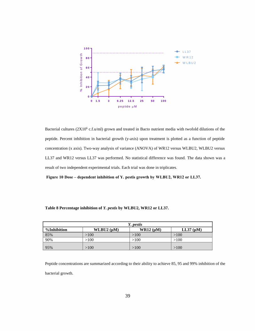
Figure 10 Dose – dependent inhibition of Y. pestis growth by WLBU2, WR12 or LL37.
Table 8 Percentage inhibition of Y. pestis by WLBU2, WR12 or LL37.
Peptide concentrations are summarized according to their ability to achieve 85, 95 and 99% inhibition of the
bacterial growth.
Y. pestis
%Inhibition WLBU2 (µM) WR12 (µM) LL37 (µM)
85% >100 >100 >100
90% >100 >100 >100
95% >100 >100 >100

40
4.3 Antimicrobial activity of WLBU2 against F. tularensis infected macrophages
Many CAPs act directly by permeabilizing the membrane of the microbial targets without
interacting with any specific receptors. Thus, their effect on mammalian cells is a concern
considering that some peptides have shown potentially harmful side effects such as lysis of red
blood cells [72]. For the ex vivo experiments with eCAPs, a transformed mouse macrophage cell
line (J774) was infected with F. tularensis. Transformed cells are more sensitive to membrane-
permeabilizing peptides due to higher phospatidylserine content[73]. Before moving on with the
ex vivo experiments the cytotoxicity of WLBU2 on J774 macrophages was determined using a
MTS test. As Figure 11 demonstrates WLBU2 treatment resulted in a dose-dependent decrease in
cell viability with 50% of the cells lost at a concentration of 25μM. Therefore, only WLBU2
concentrations of 25μM or less were used in any ex vivo experiments.
F. tularensis is a facultative intracellular organism that invades and multiplies within
macrophages [74, 75]. Thus, for a treatment to be successful it will have to act on the intracellular
pathogen. To determine the intracellular antimicrobial effect of WLBU2, ex vivo bacterial
clearance of SchuS4 from infected macrophage cells (J774) was assessed using concentrations
ranging from 3μM to 25μM. J774 cells were infected with SchuS4 at an MOI of 1:100, washed,
and then treated with different concentrations of WLBU2 (3µM to 25µM) for one hour. Cell lysate
was plated on CHA plates to determine bacterial killing and bacterial colonies were counted after
48 hours of incubation. Figure 12 illustrates the dose-dependent killing of intracellular F.
tularensis by WLBU2. WLBU2 demonstrated a high antimicrobial efficacy against intracellular
bacteria with only 20% bacteria remaining after treatment with 12.5μM for an hour. These findings
combined with the MTS test results suggest that at a dose of 12.5 μM the peptide is able to kill
most of the intracellular bacteria with minimal harm to the mammalian cells.

41
0
2 0
4 0
6 0
8 0
1 0 0
W L B U 2 M
via
bil
ity
(%
of
co
ntr
ol
)
0 1 .5 3 6 .2 5 1 2 .5 2 5 5 0 1 0 0
Cell viability was determined using a MTS assay. Treatment of cells with WLBU2 resulted in a dose-
dependent decrease in cell viability with 50% of the cells lost at a concentration of 25μM. Results of cell
growth was measured relative to a control where J774 cells were grown in 100% media. The data shown
was a result of three independent experimental trials.
Figure 11 J774 macrophage cells treated for 1 hour with a range of concentrations of WLBU2.

42
0
2 0
4 0
6 0
8 0
1 0 0
W L B U 2 M
% S
ch
uS
4 r
em
ain
ing
0 3 6 .2 5 1 2 .5 2 5
F. tularensis infected J774 cells (MOI 1:100) were treated with twofold dilutions of WLBU2 for 1 hour.
Percent remaining of the bacteria (y-axis) upon treatment is plotted as a function of peptide concentration
(x axis). The data shown was a result of three independent experimental trials.
Figure 12 Dose-dependent killing of intracellular F. tularensis by WLBU2.

43
5.0 DISCUSSION
The discovery of numerous natural cationic peptides in the last three decades and their
antimicrobial role in our bodies has sparked the interest of many scientists. The growing
understanding of the structure and function of such peptides led to the development of several
eCAPs such as WLBU and WR series. Here the antimicrobial activity of WLBU and WR12
peptides was tested against three tier 1 select agents: Francisella tularensis, Burkholderia
pseudomallei and Yersinia pestis. These bacterial agents are considered tier 1 agents due to their
potential for misuse, high fatality rate if not treated appropriately, and the development of
antibiotic resistance to their current effective treatment.
Previous studies documented the superior antimicrobial activity of WLBU2 and WR12
against several bacterial agents such as P. aeruginosa and MRSA [65-68]. In this study, in vitro
tests of WLBU2 and WR12 also demonstrated superior antimicrobial effect against F. tularensis
and Y. pestis when compared to the natural peptide (LL37). WLBU2 proved to be the best
candidate against F. tularensis with a single dose of 3μM needed to achieve a 90% reduction in
bacterial count and a MIC of 25 μM. Both WLBU2 and WR12 achieved similar results against Y.
pestis, but only WR12 was able to attain a MBC of 50 μM.
B. pseudomallei provided a greater challenge with only moderate susceptibility to WLBU2.
Nevertheless, WLBU2 antimicrobial effectiveness against B. pseudomallei surpasses that of LL37.
It was also observed that results depended on the testing environment. A substantial reduction in
bacterial count was seen with WLBU2 in potassium phosphate buffer but not in phosphate buffered
saline. The role of PPB in enhancing the peptides’ antimicrobial function is still unclear. PPB may

44
affect the bacterial membrane enhancing its disruption by the peptide or create a more suitable
environment for peptide activity. The difference in outcome between my experiments and the
Kanthawong et al study concerning LL37 might be related to the B. pseudomallei isolates used.
The isolate used in this study, 1026b, is considered a “prototypical” lab strain isolated from a
diabetic 29-year-old female patient with septicemic melioidosis. While in Kanthawong et al, 13
isolates were from patients admitted to Srinagarind Hospital (Faculty of Medicine, Khon Kaen
University, Khon Kaen, Thailand), and the other 11 were from soil collected from the northeastern
endemic region of Thailand [70]. It is well established that there are different LPS structures of B.
pseudomallei depending on the isolate: smooth type A, smooth type B, rough type A or rough type
B LPS structures [76]. Since LPS form a major part of the outer membrane of the bacteria and
shields it from antimicrobial attacks, such variation might contribute to the variation in bacterial
susceptibility to peptides [77]. Additional complexity to bacterial surfaces is generated by
production of surface polysaccharide capsules. Bacterial capsule is known to hinder complement
activation and phagocytosis of B. pseudomallei [78]. Wikraiphat et al further showed that capsule
mutants of this bacterium survived poorly in macrophages and were more susceptible to
antimicrobial killing by histatin and lactoferrin when compared to the wild type [79]. Also,
Hayden et al suggested that genetic changes that occur in subpopulations of B. pseudomallei during
acute infections and post-treatment are not found in 1026b which could contribute to the variance
seen in the antimicrobial activity of the peptides [80].
The mechanism of action of these peptides is still under investigation but the general
assumption is that their antimicrobial action stems from their ability to attach and disrupt the
microbial outer membrane leading to depolarization, metabolite leakage, and ultimately death of
the pathogen (Figure 13). The variability of surface-exposed structures on the outer membrane and

45
capsule of the three pathogens in this study may explain the difference in the antimicrobial activity
of the eCAPs. Several studies verified the influence of certain antigens found on the surface of Y.
pestis and F. tularensis in increasing their susceptibility to certain CAPs. For example, Galvan et
al demonstrated that the susceptibility of Y. pestis to CAPs such as cathelicidin is mediated by the
capsular antigen fraction 1 [81]. Also, Vonkavaara et al illustrated the importance of surface lipid
A and Kdo core for interactions of F. tularensis with CAPs. On the other hand, Madhongsa et al
demonstrated that the MIC value of several peptides such as polymyxin B for B. pseudomallei is
several magnitudes higher than those of P. aeruginosa or E. coli due to the type of LPS moiety
on its outer membrane [76]. Further, B. pseudomallei resists treatment by forming micro-colonies
using its glycol-calyx polysaccharide capsule and creating a protective environment against the
peptide. This might explain eCAPs enhanced activity against F. tularensis and Y. pestis in
comparison to the moderate antimicrobial effect of WLBU2 and failure of WR12 against B.
pseudomallei.

46
In this model the attached peptides aggregate and inser into the bacterial membrane so that hydrophobic
peptide regions align with the lipid core region and the hydrophilic peptide region form the interior
region of the pore. Hydrophilic regions of the peptide are shown coloured red, hydrophobic regions of
the peptide are shown coloure blue. Reprinted by permission from Macmillan Publishers Ltd: Nature
[82], copyright 2005.
Figure 13 A model of antimicrobial peptides mechanism of action.

47
It important to note that the antimicrobial activity of WLBU2 was affected by the media
used in overnight cultures of bacteria. F. tularensis grown in BHI was more susceptible to WLBU2
with 90% reduction at 3µM compared to 50 µM for the bacteria grown in CCDM. Hazlett et al
have shown that SDS-PAGE analysis of F. tularensis grown in CCDM or Muller Hinton Broth
(MHB) displayed different protein profiles from those grown in BHI. While F. tularensis grown
in BHI had similar mRNA profile to the bacteria isolated from macrophages, MHB and CCDM
yield bacteria that are phenotypically different with abundant outer membrane proteins that are
usually down-regulated during host adaptation [83]. This difference might be due to the high level
of free amino acids in MHB (120 mM) and CCDM (75mM) affecting the protein production in
bacteria. Further, Cherwonogrodzkey et al demonstrated that when encapsulated LVS was
passaged in CCDM it created a thicker capsule and increases in virulence [84]. Because CAPs
interact with bacterial surface, variation in the outer membrane proteins and capsule production by
different media might be responsible for the difference in WLBU2 activity against F. tularensis.
In practice, antibiotics are usually given in multiple doses to maintain a drug concentration
capable of ridding the infected body from the pathogens. When a single dose of WLBU2 was
tested against F. tularensis, it was unable to achieve a MBC and leveled off at 97% reduction in
bacterial count. We theorized that saturation of WLBU2 was accounting for the inability of
WLBU2 to achieve the MBC (defined as ≥99% killing). With that in mind, WLBU2 was tested in
vitro with 2 consecutive doses at the same concentration. Compared to a single dose, WLBU2
multiple dosing proved to be more effective with complete killing of F. tularensis at 50 μM
(MBC=50 μM). Basically, the initial dose of 50 µM WLBU2 eliminated more than 90% of the
bacteria making it easier for the second dose to achieve complete killing. Also, WLBU2 multiple
dosing performed better than the single dosing over the whole gradient of doses. This further

48
confirms the assumption that saturation of the peptide had occurred, requiring additional peptide
to achieve higher killing activity and that bacteria surviving the first dose of peptide were not
resistant to WLBU2.
During the generation of the WLBU series, peptides were tested against P. aeruginosa to
determine the length for optimal antimicrobial activity. It was thought that longer peptides would
have higher activity due to their greater membrane-spanning capacity. Nevertheless, WLBU2 with
its 24 residues had the ideal length against P. aeruginosa with no noticeable increase in activity
with WLBU3 (36-residue) and WLBU4 (48-residue) [63]. Similarly, when testing WLBU2, 3 and
4 against B. pseudomallei there were no noticeable difference in the growth inhibition of the three
peptides against this pathogen. Thus, B. pseudomallei is able to resist the antimicrobial effect of
the WLBU series regardless of their length. Madhongsa et al emphasized the structure activity
relationship of the antimicrobial peptides and its importance against B. pseudomallei. Using an
isolate with high biofilm production (H777), Madhongsa et al demonstrated the higher efficacy of
cyclic bacitracin against B. pseudomallei compared to other linear αhelix peptides [76]. Therefore,
greater membrane permeabilization ability against our tested isolate might be achieved not by
increasing the length of the our peptides but by higher order of oligomers and cyclic structures
In light of the evident antibacterial efficacy of WLBU2 in vitro, it is vital to know whether
this peptide displays any toxicity to mammalian cells or not. Deslouches et al demonstrated that
concentrations up to 30μM of WLBU2 can damage the bacterial cells while sparing leukocytes
and human skin fibroblasts [64]. This antimicrobial selectivity may be attributed to the difference
in lipid content between eukaryotic and bacterial cell membranes. Due to the presence of
cholesterol in eukaryotic membranes they tend to form weaker hydrophobic interactions with

49
eCAPs, while the negatively charged bacterial membranes forms a strong electrostatic interaction
with the them [85].
F. tularensis is an intracellular organism that invades and replicates in macrophages.
Accordingly, for the peptides to be effective in vivo they would have to exert their antimicrobial
activity on intracellular pathogens with minimal effect on the macrophages. From the MTS
staining assays results it was concluded that while WLBU2 could be toxic for J774 cells, under
the concentration of 25μM WLBU2 had minimal toxic effect on J774 macrophages. Taking that
into consideration, all ex vivo infected macrophages were treated with concentrations of less than
25μM of WLBU2. WLBU2 demonstrated a high antimicrobial efficacy against intracellular
bacteria with only 20% bacteria remaining after treatment with 12.5μM for an hour. Despite our
limited knowledge about the pharmacokinetics and dynamics of these peptides, results of the ex
vivo experiments using WLBU2 against F. tularensis infected macrophages suggest that the
peptide is able to kill the intracullar bacteria with minimal harm to the mammalian cells.
Several factors should be considered when these peptides are tested in in vivo settings,
including the concentrations of antimicrobial peptides at the sites of infection, inhibiting or
synergistic substances present in tissues and fluids (i.e. lysozyme, natural antimicrobial peptides
and physiological salt concentrations). Deslouches et al demonstrated that the WLBU2 had
prophylactic and therapeutic effects against P. aeruginosa bacteraemia in mice; 3 and 4 mg/kg
given 1 hour post-infection was able to increase survival rates compared to control [65]. Therefore,
for future directions I would suggest assessing the antimicrobial activity and toxicity of WLBU2
in a mouse model for tularemia. Balb/c mice can be infected via the respiratory tract by aerosol
delivery of SchuS4 and then treated with i.v. (intravenous) or i.n. (intranasal) WLBU2. Mice
weight and temperature should be monitored daily to evaluate the effectiveness of WLBU2 in

50
clearing the infection. Also the same model can be used to assess the benefits of prophylactic and
multiple dosing of the peptide.
In conclusion, there are three major requirements for any successful antimicrobial peptides:
potency, specificity and activity under physiological conditions. In this study we demonstrated the
potency of WLBU2 and its selectivity for F. tularensis. Further investigation is warranted, but
WLBU2 demonstrates a great potential as a broad spectrum antimicrobial agent against F.
tularensis. As for B. pseudomallei and Y. pestis, results were inconclusive about the usefulness of
WLBU and WR12 against these pathogens. Nevertheless, eCAPs could be useful as a broad-
spectrum countermeasure in situations concerning public health, such as bioterror attacks, until the
causative agent is identified.

51
BIBLIOGRAPHY
1. National Institute of Allergy and Infectious Diseases (NIAID) 04 June 2014
<http://www.niaid.nih.gov>
2. Centers for Disease Control and Prevention. Centers for Disease Control and Prevention.
04 June 2014 <http://www.cdc.gov/>.
3. "NSAR Home." NSAR Home. 03 May 2014 <http://www.selectagents.gov/>
4. Centers for Disease Control and Prevention. Centers for Disease Control and Prevention.
02 May 2014 <http://www.cdc.gov/>.
5. Feldman KA. Tularemia JAVMA 2003 222:725 - 730.
6. McCoy, G. W., and C. W. Chapin. 1912. Further observations on a plague- like disease of
rodents with a preliminary note on the causative agent, Bacterium tularense. J. Infect. Dis.
10:61–72.
7. Forsman, M., Sandström, G., and Sjöstedt, A. (1994) Analy- sis of 16S ribosomal
DNAsequences of Francisella strains and utilization for determination of the phylogeny of
the genus and for identification of strains by PCR. Internatl J System Bacteriol 44: 38–46.
8. Jellison WL. Tularemia in North America, 1930–1974. Missoula, Montana: University of
Montana, 1974.
9. CDC. Tularemia -- United states, 1990 - 2000. MMWR Morb Mortal Wkly Rep 2002,51:181
- 184.
10. Morner T. The ecology of tularaemia Rev. Sci. Tech. Off. Int. Epiz. 1992 11:1123 - 1130.
11. Berdal BP, Mehl R, Meidell NK, Lorentzen-Styr A-M, Scheel O. Field investigations of
tularemia in Norway. FEMS Immunology & Medical Microbiology 1996,13:191-195.
12. Parola P, Raoult D. Ticks and tickborne bacterial diseases in humans: an emerging
infectious threat. Clin Infect Dis 2001,32:897-928.
13. Dennis DT, Inglesby TV, Henderson DA, Bartlett JG, Ascher MS, Eitzen E, et al.
Tularemia as a biological weapon: medical and public health management. Jama
2001,285:2763-2773.

52
14. Feldman KA, Enscore RE, Lathrop SL, Matyas BT, McGuill M, Schriefer ME, et al. An
outbreak of primary pneumonic tularemia on Martha's Vineyard. N Engl J Med
2001,345:1601-1606.
15. Overholt EL, Tigertt WD, Kadull PJ, Ward MK, Charkes ND, Rene RM, et al. An analysis
of forty-two cases of laboratory-acquired tularemia. Treatment with broad spectrum
antibiotics. Am J Med 1961,30:785-806.
16. Evans ME, Gregory DW, Schaffner W, McGee ZA. Tularemia: a 30-year experience with
88 cases. Medicine (Baltimore) 1985,64:251-269.
17. Robert G. Darling CLC, Kermit D. Huebner,David G. Jarrett. Threats in bioterrorism I:
CDC category A agents Emerg Med Clin N Am 2002,20 273-309.
18. Ohara Y, Sato T, Homma M. Arthropod-borne tularemia in Japan: clinical analysis of 1,374
cases observed between 1924 and 1996. J Med Entomol 1998,35:471-473.
19. Avery FW, Barnett TB. Pulmonary tularemia. A report of five cases and consideration of
pathogenesis and terminology. Am Rev Respir Dis 1967,95:584-591.
20. Pérez-Castrillón JL, Bachiller-Luque P, Martín-Luquero M, Mena-Martín FJ, Herreros V.
Tularemia Epidemic in Northwestern Spain: Clinical Description and Therapeutic
Response. Clinical Infectious Diseases 2001,33:573-576.
21. Balali Mood, M., Balali Mood, B., Moshiri, M., 2013. Sulfur mustard. In: Wexler, P.,
Greim, H., Moser, V., Wiegand, T.J., Lafarga, J.V.T., Peyster, A., Harper, S., Abdollahi,
M., Gad, S.C., Ray, S.D. (Eds.), Encyclopedia of Toxicology, third ed. Elsevier.
22. Gilligan, P. H.1995.Pseudomonas and Burkholderia, p.509– 519. In P. R. Murray, E. J.
Baron, M. A. Pfaller, F. C. Tenover, and R. H. Yolken (ed.), Manual of clinical
microbiology, 6th ed. American Society for Microbiology Press, Washington, D.C
23. Center for Civilian Biodefense Studies. Tularemia.Retrieved January 2014 from
ww.hopkinsbiodefense.org/pages/agents/toctularemia.html.
24. Arnold F. Kaufmann MIM, and George P. Schmid. The Economic Impact of a Bioterrorist
Attack: Are Prevention and Postattack Intervention Programs Justifiable? Emerging
Infectious Disease 1997,3:83 - 94.
25. Chaowagul W, White NJ, Dance DA, Wattanagoon Y, Naigowit P, Davis TM, et al.
Melioidosis: a major cause of community-acquired septicemia in northeastern Thailand. J
Infect Dis 1989,159:890-899.
26. White NJ, Dance DA, Chaowagul W, Wattanagoon Y, Wuthiekanun V, Pitakwatchara N.
Halving of mortality of severe melioidosis by ceftazidime. Lancet 1989,2:697-701.
27. White NJ. Melioidosis. The Lancet 2003,361:1715-1722.

53
28. Cheng AC, Currie BJ. Melioidosis: epidemiology, pathophysiology, and management. Clin
Microbiol Rev 2005,18:383-416.
29. Dance DA. Melioidosis: the tip of the iceberg? Clin Microbiol Rev 1991,4:52-60.
30. Robert E. Antosia JDC. Handbook of Bioterrorism and Disaster Medicine. New York:
Springer Science and Business Media; 2006.
31. Currie BJ. Advances and remaining uncertainties in the epidemiology of Burkholderia
pseudomallei and melioidosis. Trans R Soc Trop Med Hyg 2008,102:225-227.
32. Ralph A, McBride J, Currie BJ. Transmission of Burkholderia Pseudomallei Via Breast
Milk in Northern Australia. The Pediatric Infectious Disease Journal 2004,23:1169-1171
1110.1097/1101.inf.0000145548.0000179395.da.
33. Jacob Gilad IH, Tsvika Dushnitsky, David Schwartz and Yoram Amsalem Burkholderia
mallei and Burkholderia pseudomallei as Bioterrorism Agents: National Aspects of
Emergency Preparedness. IMAJ 2007,9:499-503.
34. Currie BJ, Fisher DA, Anstey NM, Jacups SP. Melioidosis: acute and chronic disease,
relapse and re-activation. Trans R Soc Trop Med Hyg 2000,94:301-304.
35. Puthucheary SD, Vadivelu J. Human Melioidosis. Singapore University Press, 2002.
36. Currie BJ, Fisher DA, Howard DM, Burrow JN, Lo D, Selva-Nayagam S, et al. Endemic
melioidosis in tropical northern Australia: a 10-year prospective study and review of the
literature. Clin Infect Dis 2000,31:981-986.
37. S H How CKL. Melioidosis: A Potentially Life Threatening Infection. Med J Malaysia
2006,61:386 - 395.
38. Puthucheary SD. Melioidosis in Malaysia. Med J Malaysia 2009,64:266 - 274.
39. Chaowagul W, Suputtamongkol Y, Dance DA, Rajchanuvong A, Pattara-arechachai J,
White NJ. Relapse in melioidosis: incidence and risk factors. J Infect Dis 1993,168:1181-
1185.
40. Sodeman WA, Jr. Sherlock Holmes and tropical medicine: a centennial appraisal. Am J
Trop Med Hyg 1994,50:99-101.
41. Hinnebusch BJ, Rudolph AE, Cherepanov P, Dixon JE, Schwan TG, Forsberg A. Role of
Yersinia murine toxin in survival of Yersinia pestis in the midgut of the flea vector. Science
2002,296:733-735.
42. Butler, T. 1983. Plague and other Yersinia infections. Plenum Press, NewYork, N.Y.

54
43. Perry RD, Fetherston JD. Yersinia pestis--etiologic agent of plague. Clin Microbiol Rev
1997,10:35-66.
44. Stenseth NC, Atshabar BB, Begon M, Belmain SR, Bertherat E, Carniel E, et al. Plague:
past, present, and future. PLoS Med 2008,5:e3.
45. Cavanaugh DC. Specific effect of temperature upon transmission of the plague bacillus by
the oriental rat flea, Xenopsylla cheopis. Am J Trop Med Hyg 1971,20:264-273.
46. Ashok K Dutt RAaMM. Surat plague of 1994 re-examined Southeast Asian J Trop Med
Public Health 2006,37.
47. Crook LD, Tempest B. Plague. A clinical review of 27 cases. Arch Intern Med
1992,152:1253-1256.
48. Butler T. The black death past and present. 1. Plague in the 1980s. Trans R Soc Trop Med
Hyg 1989,83:458-460.
49. Anisimov AP, Amoako KK. Treatment of plague: promising alternatives to antibiotics. J
Med Microbiol 2006,55:1461-1475.
50. Galimand M, Carniel E, Courvalin P. Resistance of Yersinia pestis to antimicrobial agents.
Antimicrob Agents Chemother 2006,50:3233-3236.
51. Titball RW, Leary SE. Plague. Br Med Bull 1998,54:625-633.
52. Layton RC, Mega W, McDonald JD, Brasel TL, Barr EB, Gigliotti AP, et al. Levofloxacin
cures experimental pneumonic plague in African green monkeys. PLoS Negl Trop Dis
2011,5:e959.
53. Inglesby TV, Dennis DT, Henderson DA, et al. Plague as a biological weapon: Medical
and public health management. JAMA 2000,283:2281-2290.
54. Sun W, Roland KL, Curtiss R, 3rd. Developing live vaccines against plague. J Infect Dev
Ctries 2011,5:614-627.
55. Harris SH. Factories of Death. New York, NY: Routledge; 1994:78, 96
56. Health Aspects of Chemical and Biological Weapons . Geneva, Switzerland: World Health
Organization; 1970:98-109.
57. Carus WS. Bioterrorism and Biocrimes: The Illicit Use of biological Agents in the 20th
Century. Washington, DC: Center for Counterproliferation Research, National Defense
University; 1998
58. Hancock RE, Chapple DS. Peptide antibiotics. Antimicrob Agents Chemother
1999,43:1317-1323.

55
59. Boman HG. Peptide antibiotics and their role in innate immunity. Annu Rev Immunol
1995,13:61-92.
60. Goldman MJ, Anderson GM, Stolzenberg ED, Kari UP, Zasloff M, Wilson JM. Human
beta-defensin-1 is a salt-sensitive antibiotic in lung that is inactivated in cystic fibrosis.
Cell 1997,88:553-560.
61. Miller MA, Cloyd MW, Liebmann J, Rinaldo CR, Jr., Islam KR, Wang SZ, et al.
Alterations in cell membrane permeability by the lentivirus lytic peptide (LLP-1) of HIV-
1 transmembrane protein. Virology 1993,196:89-100.
62. Tencza SB, Creighton DJ, Yuan T, Vogel HJ, Montelaro RC, Mietzner TA. Lentivirus-
derived antimicrobial peptides: increased potency by sequence engineering and
dimerization. J Antimicrob Chemother 1999,44:33-41.
63. Deslouches B, Phadke SM, Lazarevic V, Cascio M, Islam K, Montelaro RC, et al. De novo
generation of cationic antimicrobial peptides: influence of length and tryptophan
substitution on antimicrobial activity. Antimicrob Agents Chemother 2005,49:316-322.
64. Deslouches B, Islam K, Craigo JK, Paranjape SM, Montelaro RC, Mietzner TA. Activity
of the de novo engineered antimicrobial peptide WLBU2 against Pseudomonas aeruginosa
in human serum and whole blood: implications for systemic applications. Antimicrob
Agents Chemother 2005,49:3208-3216.
65. Deslouches B, Gonzalez IA, DeAlmeida D, Islam K, Steele C, Montelaro RC, et al. De
novo-derived cationic antimicrobial peptide activity in a murine model of Pseudomonas
aeruginosa bacteraemia. J Antimicrob Chemother 2007,60:669-672.
66. Deslouches B, Steckbeck JD, Craigo JK, Doi Y, Mietzner TA, Montelaro RC. Rational
design of engineered cationic antimicrobial peptides consisting exclusively of arginine and
tryptophan, and their activity against multidrug-resistant pathogens. Antimicrob Agents
Chemother 2013,57:2511-2521.
67. Shruti M. Phadke BD, Sara E. Hileman, Ronald C. Montelaro, Harold C. Wiesenfeld,and
Timothy A. Mietzner. Antimicrobial Peptides in Mucosal Secretions: The Importance of
Local Secretions in Mitigating Infection. The Journal of Nutrition 2005,5:1289 - 1293.
68. Novak KF, Diamond WJ, Kirakodu S, Peyyala R, Anderson KW, Montelaro RC, et al.
Efficacy of the de novo-derived antimicrobial peptide WLBU2 against oral bacteria.
Antimicrob Agents Chemother 2007,51:1837-1839.
69. Mc Gann P, Rozak DA, Nikolich MP, Bowden RA, Lindler LE, Wolcott MJ, et al. A novel
brain heart infusion broth supports the study of common Francisella tularensis serotypes. J
Microbiol Methods 2010,80:164-171.

56
70. Kanthawong S, Nazmi K, Wongratanacheewin S, Bolscher JG, Wuthiekanun V,
Taweechaisupapong S. In vitro susceptibility of Burkholderia pseudomallei to
antimicrobial peptides. Int J Antimicrob Agents 2009,34:309-314.
71. O'Loughlin JL, Spinner JL, Minnich SA, Kobayashi SD. Yersinia pestis two-component
gene regulatory systems promote survival in human neutrophils. Infect Immun
2010,78:773-782.
72. Pacor S. Analysis of the cytotoxicity of synthetic antimicrobial peptides on mouse
leucocytes: implications for systemic use. Journal of Antimicrobial Chemotherapy
2002,50:339-348.
73. Palffy R, Gardlik R, Behuliak M, Kadasi L, Turna J, Celec P. On the physiology and
pathophysiology of antimicrobial peptides. Mol Med 2009,15:51-59.
74. Tarnvik A. Nature of protective immunity to Francisella tularensis. Rev Infect Dis
1989,11:440-451.
75. Fortier AH, Green SJ, Polsinelli T, Jones TR, Crawford RM, Leiby DA, et al. Life and
death of an intracellular pathogen: Francisella tularensis and the macrophage. Immunol Ser
1994,60:349-361.
76. Madhongsa K, Pasan S, Phophetleb O, Nasompag S, Thammasirirak S, Daduang S, et al.
Antimicrobial action of the cyclic peptide bactenecin on Burkholderia pseudomallei
correlates with efficient membrane permeabilization. PLoS Negl Trop Dis 2013,7:e2267.
77. Woods MNBaDE. Isolation of Polymyxin B-Susceptible Mutants of Burkholderia
pseudomallei and Molecular Characterization of Genetic Loci involved in Polymyxin B
Resistance Antimicrob Agents Chemother,43:2648 - 2656.
78. Reckseidler-Zenteno SL, DeVinney R, Woods DE. The capsular polysaccharide of
Burkholderia pseudomallei contributes to survival in serum by reducing complement factor
C3b deposition. Infect Immun 2005,73:1106-1115.
79. Wikraiphat C, Charoensap J, Utaisincharoen P, Wongratanacheewin S,
Taweechaisupapong S, Woods DE, et al. Comparative in vivo and in vitro analyses of
putative virulence factors of Burkholderia pseudomallei using lipopolysaccharide, capsule
and flagellin mutants. FEMS Immunol Med Microbiol 2009,56:253-259.
80. Hayden HS, Lim R, Brittnacher MJ, Sims EH, Ramage ER, Fong C, et al. Evolution of
Burkholderia pseudomallei in recurrent melioidosis. PLoS One 2012,7:e36507.
81. Galvan EM, Lasaro MA, Schifferli DM. Capsular antigen fraction 1 and Pla modulate the
susceptibility of Yersinia pestis to pulmonary antimicrobial peptides such as cathelicidin.
Infect Immun 2008,76:1456-1464.

57
82. Brogden KA. Antimicrobial peptides: pore formers or metabolic inhibitors in bacteria? Nat
Rev Microbiol 2005,3:238-250.
83. Hazlett KR, Caldon SD, McArthur DG, Cirillo KA, Kirimanjeswara GS, Magguilli ML, et
al. Adaptation of Francisella tularensis to the mammalian environment is governed by cues
which can be mimicked in vitro. Infect Immun 2008,76:4479-4488.
84. John W. Cherwonogrodzky MHKaMRs. Increased encapsulation and virulence of
Francisella tularensis live vaccine strain (LVS) by subculturing on synthetic medium.
Vaccine 1994,12:773 - 775.
85. Biggin PC, Sansom MSP. Interactions of α-helices with lipid bilayers: a review of
simulation studies. Biophysical Chemistry 1999,76:161-183.