novel approaches to induce apoptosis in human follicular
TRANSCRIPT
Doctoral dissertation
To be presented by permission of the Faculty of Medicine of the University of Kuopio
for public examination in Auditorium ML1, Medistudia building, University of Kuopio,
on Monday 18th June 2007, at 3 p.m.
Institute of Clinical MedicineDepartment of Clinical Microbiology
University of Kuopio
JOANNA SKOMMER
Novel Approaches to Induce Apoptosisin Human Follicular Lymphoma Cell Lines
Preclinical Assessment
JOKAKUOPIO 2007
KUOPION YLIOPISTON JULKAISUJA D. LÄÄKETIEDE 411KUOPIO UNIVERSITY PUBLICATIONS D. MEDICAL SCIENCES 411
Distributor : Kuopio University Library P.O. Box 1627 FI-70211 KUOPIO FINLAND Tel. +358 17 163 430 Fax +358 17 163 410 www.uku.fi/kirjasto/julkaisutoiminta/julkmyyn.html
Series Editors: Professor Esko Alhava, M.D., Ph.D. Institute of Clinical Medicine, Department of Surgery Professor Raimo Sulkava, M.D., Ph.D. School of Public Health and Clinical Nutrition Professor Markku Tammi, M.D., Ph.D. Institute of Biomedicine, Department of Anatomy
Author´s address: Institute of Clinical Medicine Department of Clinical Microbiology University of Kuopio P.O. Box 1627 FI-70211 KUOPIO FINLAND Tel. +358 17 162 700 Fax +358 17 162 705 E-mail : [email protected]
Supervisor : Professor Jukka Pelkonen, M.D., Ph.D. Institute of Clinical Medicine Department of Clinical Microbiology University of Kuopio
Reviewers: Professor Zbigniew Darzynkiewicz, M.D., Ph.D. Brander Cancer Research Institute New York Medical College Valhalla, New York, USA
Professor Faustino Mollinedo, Ph.D. Centro de Investigación del Cáncer Instituto de Biología Molecular y Celular del Cáncer Consejo Superior de Investigaciones Científ icas (CSIC) Universidad de Salamanca Salamanca, Spain
Opponent: Docent Marko Salmi MediCity Research Laboratory University of Turku
ISBN 978-951-27-0671-6ISBN 978-951-27-0748-5 (PDF)ISSN 1235-0303
KopijyväKuopio 2007Finland

Skommer, Joanna. Novel approaches to induce apoptosis in follicular lymphoma cell lines – preclinical assessment. Kuopio University Publications D. Medical Sciences 411. 2007. 80 p. ISBN 978-951-27-0671-6 ISBN 978-951-27-0748-5 (PDF) ISSN 1235-0303 ABSTRACT
Follicular lymphoma (FL) is one of the commonest entities of non-Hodgkin´s lymphoma (NHL) worldwide. Many patients with advanced stages of FL achieve a very good partial remission, but few, if any, can be cured. Thus, more effective treatment regimens are indispensable. As evading cell death is paramount to tumour progression, the aim of the present investigation was to characterize novel inducers of apoptosis in FL cell lines. Initially, we examined the action of a novel small molecule inhibitor of Bcl-2 oncoprotein, HA14-1, demonstrating that FL cell lines are sensitive to its single-agent pro-apoptotic action, and analysing: i) the temporal and quantitative relationship between two apoptotic events pivotal in HA14-1-evoked apoptosis (mitochondrial breach and caspase activation); ii) cell cycle-specificity of HA14-1-triggered apoptosis. FL is mostly an indolent disease, with multiple courses of chemotherapy severely impairing the quality of patients´ life. Thus, we went on to study the effects of a natural polyphenol curcumin, known to exert anti-proliferative and/or pro-apoptotic effects in several tumour cell lines. The compound has superior safety records from clinical trials, and has shown promising chemopreventive and/or anticancer properties in animal models, and in humans. FL cells were sensitive to the action of curcumin, with significant cessation of cell growth observed at reportedly attainable in vivo concentration. Employing flow cytometry and biochemical approaches, we demonstrated that curcumin-induced apoptosis is caspase-dependent, associated with ROS generation and lysosomal rupture, and can be significantly enhanced by ascorbic acid. We also investigated gene expression changes during curcumin-triggered apoptosis in FL cell line HF4.9, applying a new technology of bead chip microarrays. Complex reprogramming was associated with induction of apoptosis in curcumin-treated HF4.9 cells, including genes encoding tumour and metastasis suppressors, proteins involved in the regulation of cell adhesion and migration, or transcription/splicing factors. Importantly, CXCR4, a chemokine receptor in control of cell migration that reportedly contributes to local and distant tumour dissemination, was down-regulated in all FL cell lines tested. Finally, specificity of CXCR4 down-regulation in different cell death contexts was investigated. Overall, the thesis advances the current knowledge on stimuli to engage apoptotic response in FL cells, indicating that both Bcl-2 inhibitors and natural compounds such as curcumin may prove beneficial for the management of FL, and warranting their further investigation in vivo. National Library of Medicine Classification: QU 350, QU 375, QZ 350, WH 525 Medical Subject Headings: Apoptosis; Apoptosis Regulatory Proteins; Cell Death; Curcumin; Lymphoma, Follicular; Mitochondria; Microarray Analysis


"Life has two rules: Number One, never quit;
Number Two, always remember rule number one"
Duke Ellington


ACKNOWLEDGEMENTS This study was carried out in the Department of Clinical Microbiology, University of Kuopio, during 2005-2006. I am grateful to my principal supervisor, Professor Jukka Pelkonen, for providing working space, and making constant attempts to reveal the beauty of severe Finnish winter and virtues of cross-country skiing. I owe my sincere thanks for the excellent technical assistance and friendship from Eila Pelkonen. Many thanks also for all the self-picked forest fruits, and a taste of wonderful Chinese cuisine! I would like to thank Juhani Soini and Päivi Junni from the Microarray Centre at Turku Centre for Biotechnology for all the help and advice on design and analysis of microarray experiments, and Professor Andrzej Deptala, Department of Hematology, Oncology and Internal Medicine, CSKMSWiA Hospital, Warsaw, for friendship, support, fruitful collaboration and discussions. Professors Zbigniew Darzynkiewicz (New York Medical College, Valhalla, New York, USA) and Professor Faustino Mollinedo (Universidad de Salamanca, Salamanca, Spain), the official reviewers of this thesis, are greatly acknowledged for their feedback and support. I am deeply grateful for all valuable comments and corrections provided. I dedicate my warm thanks to Donald for the fruitful collaboration, everlasting encouragement and patience, and sharing ups and downs. I would not manage to proceed with my work without your support. I did my best to do the best I could, but all in all you are the one who got me through. I would like to express a sincere gratitude to my closest family, especially my parents Grazyna and Janusz, and sister Natalia, and also Ewa and Pawel, uncle Zdzislaw, and aunt and uncle Roza and Zdzislaw for continuous support from the distance. To all my friends spread all over the world - for encouragement and wonderful time we spent together - thank you! Magda and Krzys – our time together in Kuopio, and later on in Turku, rocked!! I hope future holds more such evenings and nights together! Finally, I am tremendously grateful for all the encouragement given to me by follicular lymphoma patients, their friends and families interested in my research work into curcumin action. With all my heart I wish you nothing but successes in the fight against the malignancy!

This work was financed from the Kuopio University Hospital grant. I was also supported by the Centre for International Mobility (CIMO) scholarship. Moreover, the generous support from L´Oréal –Poland (scholarship for Women in Science 2006/2007) is gratefully acknowledged. Kuopio,
Joanna Skommer

ABBREVIATIONS
AIF apoptosis inducing factor ANT adenine nucleotide transporter AO acridine orange AP-1 activating protein-1 Bax Bcl-2 associated X protein Bcl-2 B-cell lymphoma 2 protein Bcl-6 B-cell lymphoma 6 protein Bcl-XL Bcl-2-related gene (large variant) b-FGF basic fibroblast growth factor BH Bcl-2 homology CARD caspase-activation recruitment domain COX2 cyclooxygenase-2 CsA cyclosporine A CXCR4 CXC chemokine receptor 4 CypD cyclophilin D Da Dalton DISC death-inducing signaling complex DR death receptor Drp1 dynamin-related protein 1 Endo G endonuclease G ER endoplasmic reticulum FADD Fas Associated Death Domain FL follicular lymphoma FLICA fluorochrome-labeled inhibitor of caspases fmk fluoromethylketone Gas2 growth-arrest specific 2 protein GC germinal center HA14-1 ethyl 2-amino-6-bromo-4-(1-cyano-2-ethoxy-2-oxoethyl)-4H- chromene-3-carboxylate HIF-1 hypoxia-inducible factor-1 HtrA2/Omi high-temperature requirement protein A2 IAP inhibitor of apoptosis protein ICAD inhibitor of caspase-activated deoxyribonuclease IM inner membrane mAb monoclonal antibody MAC mitochondrial apoptosis-induced channel MMP matrix metalloproteinase MOMP mitochondrial outer membrane permeabilization NAC N-acetylcysteine NAO nonyl-acridine orange

OM outer membrane Opa1 optic atrophia 1 PARP poly (ADP-ribose) polymerase PKC protein kinase C PTP permeability transition pore RIP receptor-interacting protein ROS reactive oxygen species SDF-1 stromal cell-derived factor-1 Smac/DIABLO second mitochondria-derived activator of caspase/direct IAP- binding protein with low PI VDAC voltage-dependent anion channel VEGF vascular endothelial growth factor tBid truncated Bid TIMP-1 tissue inhibitor of metalloproteinase zVAD benzyloxycarbonyl-Val-Ala-Asp

LIST OF ORIGINAL PUBLICATIONS
The thesis is based on the following original publications, which are referred to in
the text by the Roman numerals:
I Wlodkowic D, Skommer J, Pelkonen J (2006) Multiparametric analysis of
HA14-1-triggered apoptosis in follicular lymphoma cells. Leukemia Res
30(9):1187-1192
II Skommer J, Wlodkowic D, Pelkonen J (2006) Cellular Foundation of
Curcumin-induced Apoptosis in Follicular Lymphoma Cell Lines. Exp Hematol
34(4):463-74.
III Skommer J, Wlodkowic D, Pelkonen J (2007) Gene expression profiling during
curcumin-induced apoptosis reveals down-regulation of CXCR4. Exp Hematol
35(1):84-95
IV Skommer J, Wlodkowic D, Pelkonen J (2007) CXCR4 expression during
tumour cell death. Leukemia Res Oct 23; [Epub ahead of print]
The original articles have been reproduced with permission of the copyright holders.
The thesis includes also previously unpublished data.


CONTENTS
1. INTRODUCTION .........................................................................................................................12
2. REVIEW OF THE LITERATURE .............................................................................................14
2.1. FOLICULLAR LYMPHOMA.................................................................................................14 2.2. APOPTOSIS ............................................................................................................................16
2.2.1. Apoptosis, necrosis, and others........................................................................................16 2.2.2. Caspase-controlled hydrolytic eruption in cell death: "the kiss of death" .......................19 2.2.3. Mitochondrion in cell death – the ultimate valve .............................................................22
2.3. THE BCL-2 RIDDLE...............................................................................................................30 2.3.1. Bcl-2 family members.......................................................................................................30 2.3.4. Outline of current approaches to inhibit Bcl-2 ................................................................33
2.4. CURCUMIN ............................................................................................................................35 2.4.1. Targeting of apoptotic pathways by curcumin .................................................................36 2.4.2. Successes and failures of curcumin in clinical trials........................................................40
3 AIMS OF THE STUDY .................................................................................................................42
4 MATERIALS AND METHODS ...................................................................................................43
4.1. CELL LINES AND CULTURING (I-IV) ..........................................................................................43 4.2. CELL TREATMENT EXPERIMENTS ..............................................................................................43 4.3. CELL PROLIFERATION AND VIABILITY ASSAYS..........................................................................44
4.3.1. [3H]-Thymidine incorporation assay (II) .........................................................................44 4.3.2. Trypan blue staining (II) ..................................................................................................45
4.4. APOPTOSIS DETECTION .............................................................................................................45 4.5.1. Morphological assessment (II) .........................................................................................45 4.5.2. Quantification of fractional DNA content (II)..................................................................45 4.5.3. Conformational DNA/RNA changes (II, III, IV)...............................................................45 4.5.4. Cell membrane permeabilization (II) ...............................................................................46 4.5.5. Mitochondrial membrane potential depolarisation (I, II) ................................................46 4.5.6. Cytochrome c release (II).................................................................................................47 4.5.7. Caspase activation (I, II, IV) ............................................................................................47 4.5.8. Multiparametric flow cytometry assays (I).......................................................................47
4.6. CELL CYCLE ANALYSIS .............................................................................................................48 4.7. LYSOSOMAL MEMBRANE PERMEABILIZATION (II).....................................................................48 4.8. DETECTION OF REACTIVE OXYGEN SPECIES (ROS) (II) .............................................................49 4.9. CELL SURFACE STAINING (III)...................................................................................................49 4.10. IMMUNOBLOTTING (II, III, IV) ................................................................................................49 4.10. RNA ISOLATION (III) ..............................................................................................................50 4.11. LARGE-SCALE GENE EXPRESSION PROFILING (III) ...................................................................50
4.11.1. Probe preparation ..........................................................................................................50 4.11.2. Bead chip microarrays ...................................................................................................51 4.11.3. qRT- PCR .......................................................................................................................53
4.12. DATA ANALYSIS......................................................................................................................53 4.12.1. Microarray data analysis (III) .......................................................................................53
5. RESULTS AND DISCUSSION ....................................................................................................55
5.1. HA14-1 AS AN EFFECTIVE INDUCER OF APOPTOSIS (PUBLICATION I) ........................................55 5.2. CHARACTERIZATION OF CURCUMIN-INDUCED APOPTOSIS (PUBLICATION II) ............................57 5.3. GLOBAL GENE EXPRESSION REPROGRAMMING INDUCED BY CURCUMIN (PUBLICATION III)......59
5.3.1. CXCR4..............................................................................................................................60 5.3.2. CD20 ................................................................................................................................63
5.4. CXCR4 DOWN-REGULATION IS NOT A COMMON APOPTOSIS-ASSOCIATED EVENT (PUBLICATION IV)...................................................................................................................................................65
6. CONCLUSIONS, CLINICAL IMPLICATIONS AND FUTURE DIRECTIONS ..................67
APPENDIX: ORIGINAL PUBLICATIONS I-IV

12
1. INTRODUCTION
All long-living organisms face a formidable challenge dealing with the accumulation
of genetic lesions over time, which, if amass, manifest themselves as life-threatening
conditions. One of those is cancer, described as a disease that involves excessive
proliferation of cells and abandonment of their ability to die. Normally, cells can kill
themselves in a highly balanced process known as apoptosis, and incapacitated
execution or regulation of thereof has calamitous consequences, leading to a variety
of cancers, including B cell follicular lymphoma. Intrinsically, cancer cells are
primed for death during malignant transformation, and can evade destruction and go
on to form tumours only upon development of additional protective mechanisms.
Thus, it is intellectually appealing that a restoration of derailed cell death pathways
should preferentially kill cancer cells over their non-malignant counterparts.
Additionally, the anti-tumour efficacy of anticancer drugs is mostly ascribed to their
ability to induce apoptosis, and other forms of cell death, such as autophagic cell
death, caspase-independent apoptosis-like cell death, mitotic catastrophe or necrosis.
Hence, studies aimed to delineate exact mechanisms behind cell demise as well as
efforts to find novel potent apoptotic inducers are of considerable clinical
importance and may suggest credible targets for cancer therapy.
A plethora of new apoptosis-based therapies emerges from the understanding of
mechanisms underlying apoptosis and knowledge on its core components. Both
drugs directed to inhibit apoptosis-blocking proteins and agents that stimulate or
restore pro-apoptotic pathways are being extensively studied in vitro and in vivo,
with emerging efforts towards their translation to the clinics.
Side effects of conventional chemotherapy represent one of the major obstacles to
the successful treatment of malignant disorders. In the case of follicular lymphoma,
the cyclical pattern of relapses necessitates repeated administration of cytotoxic
drugs, severely decreasing the quality of patients´ life. As the initial therapy of

13
advanced stage FL commonly comprises of a watch and wait strategy until the
disease becomes symptomatic, the natural compounds with chemopreventive and/or
anticancer properties and minimal side effects may represent an alternative strategy
to safely defeat this malignancy.
The present study was primarily focused on the investigation of the pro-apoptotic
properties of a novel small molecule Bcl-2 inhibitor, HA14-1, and a natural
compound isolated from the Asian tropical plant Curcuma longa, curcumin, in
follicular lymphoma cell lines.

14
2. REVIEW OF THE LITERATURE
2.1. FOLICULLAR LYMPHOMA
Follicular lymphoma (FL) is one of the commonest subtypes of non-Hodgkin´s
lymphoma (NHL) worldwide and its incidence is rapidly increasing in western
countries [de Jong, 2005]. Although mostly a disease of adulthood, FL has also been
reported in children, in whom it displays localized presentation and more favourable
prognosis. Typically, the disease exhibits a relatively indolent clinical course.
However, majority of patients present with disseminated disease already at
diagnosis, with lymph nodes, spleen, bone marrow, and peripheral blood being the
sites most frequently involved in FL presentation. Moreover, cyclical pattern of
induced remissions and relapses, with 25-60% of cases where transformation to a
more aggressive histology occurs, is common [Horning and Rosenberg, 1994]. The
median survival ranges from 8 to 12 years, yet after the initial relapse both response
rate and relapse-free survival time tend to decrease, resulting in a median survival of
only 4-5 years [Johnson et al., 1995]. The management options in the treatment of
follicular lymphoma include watchful observation [Advani et al., 2004], alkylating
agents, anthracyclines, purine nucleoside analogs, radiation therapy, combination
chemotherapy, interferon, radiolabeled and unlabeled monoclonal antibodies (with
revolutionizing chimeric anti-CD20 mAb rituximab), and autologous bone marrow
transplantation (BMT) or peripheral blood stem-cell transplantation (PBSCT)
[Gandhi and Marcus, 2005; Fisher et al., 2005; Coiffier 2005; Hiddemann et al.,
2005]. Although the overwhelming majority of patients with advanced stages of FL
achieve a very good partial remission, few achieve complete remission, and even
fewer, if any, can be cured. Dose intensification has the potential to eradicate disease
completely, but is limited by its treatment-related short-term and long-term toxicity.

15
Follicular lymphoma is a tumour of peripheral B lymphocytes representing
malignant counterparts of normal germinal centre B cells, and histologically it
recapitulates the architecture and cytologic features of the normal secondary
lymphoid follicle. Follicular center lymphoma B cells express surface
immunoglobulin (sIg) and are CD19+, CD20+, CD10+/-, and CD5-. FL has been
classically associated with the translocation t(14;18)(q32;q21) leading to the
rearrangement of the BCL-2 gene in 70 to 95% of reported tumours and
accumulation of follicle center cells with prolonged survival. The uniform
expression of Bcl-2 by FL cells contrasts with their exquisite sensitivity to treatment
in vivo and explicit propensity to undergo spontaneous apoptosis in vitro.
Additionally, of FL cases, 10-20% lack Bcl-2 over-expression and yet still exhibit
inhibition of apoptosis. It has been thus concluded that Bcl-2 over-expression per se
cannot explain the pathogenesis and/or clinical behaviour of this malignancy.
Indeed, FL cells display a unique pattern of antiapoptotic protein expression
compared with the balance of anti- and pro-apoptotic proteins observed in normal
germinal center (GC) B cells [Gulmann et al., 2005; de Jong, 2005; Ghia et al.,
1998]. Activation of the PI3K/AKT pathway, over-expression of MCL-1 and
survivin, have all been proposed as contributing mechanisms. Furthermore, Bcl-XL
has been suggested to play a pivotal role in the control of survival of FL cells in
vitro, and its over-expression has been associated with adverse prognosis in FL
subjects [Zhao et al., 2004; Ghia et al., 1998]. Moreover, tumour microenvironment
signals, such as CD40 receptor ligation, interleukin-4 (IL-4) receptor stimulation, or
the interaction of the integrin ligand VCAM-1 with its receptor, appear to contribute
to FL cell survival in vivo through the up-regulation of death-inhibiting proteins
[Ghia et al., 1998; Taylor et al. 1999]. Importantly, the transformed lymphomas
retain the t(14;18) translocation and usually acquire multiple, complex new
chromosomal abnormalities (at least one karyotypic abnormality in addition to
t(14;18), with an average of six alterations) [Horsman et al., 2001]. For instance, in 6
to 14% of FLs, predominantly FLs grade 3B harbouring a diffuse large B-cell

16
lymphoma (DLBL) component, the rearrangements involving the BCL6 proto-
oncogene locus (3q27) have been encountered. Genome-wide analysis of the DNA
copy-number changes in FL and transformed/relapsed biopsies revealed multiple
genomic aberrations acquired upon transformation. Some of the genes within
regions of gain/amplification or loss were differentially expressed, including CUTL1
which regulates normal B lymphopoiesis and has been correlated with lymphoid
abnormalities in mice [Martinez-Climent et al., 2003; Sinclair et al., 2001]. Finally,
based on distinct transformation-associated gene expression profiles, 2 groups of FL
patients, with an increase or decrease in the expression of MYC and its target genes,
can be discriminated [Lossos et al. 2002].
2.2. APOPTOSIS "Life is pleasant. Death is peaceful. It's the transition that's troublesome"
Isaac Asimov
2.2.1. Apoptosis, necrosis, and others
Apoptosis (also referred to as type 1 programmed cell death) is a genetically
controlled event central to the development, homeostasis and disease, with
compelling evidence that shows its evolutionarily conserved mechanism. For cell
death to be classified as apoptotic, a pattern of molecular events and morphological
changes, including prominent condensation of cytoplasm, rounding up, chromatin
condensation to compact and simple geometric figures, loss of mitochondrial
membrane potential, karyorhexis, blebbing with maintenance of membrane integrity
(zeiosis) and plasma-membrane asymmetry coupled to the display of phagocytosis
markers on the cell surface [Leist and Jaattela, 2001; Edinger and Thompson, 2004]
must be observed. Caspase activation is traditionally considered a hallmark of
apoptosis, as is the absence of autophagocytosis.

17
Several models defining the processes of caspase-independent cell death have been
recently described, and include mitotic catastrophe (a default pathway after mitotic
failure and development of aneuploid cells), autophagic cell death or necrosis.
In numerous biological systems the cell´s suicide programme involves the
autophagic/lysosomal compartment. Accordingly, the autophagic cell death (type 2
programmed cell death) describes cell demise morphologically distinct from
apoptosis, with early degradation of organelles but preservation of cytoskeletal
elements until late stages, and late (if any) caspase activation and DNA
fragmentation. It results from excessive levels of cellular macroautophagy, a
degradative strategy conserved across taxa that provides a mechanism for the
turnover of damaged and/or excessive organelles and long-lived proteins [Levine
and Yuan, 2005]. Necrosis, on the contrary, is considered as a passive form of cell
death in which cell dies as a result of bioenergetic catastrophe inflicted by external
conditions. This is characterized by swelling of organelles (odema), energy
breakdown, random DNA degradation, relative early breakdown of plasma
membrane and eventually total cell disintegration that leads to inflammation around
the dead cells, attributable to the release of the cellular contents and
proinflammatory molecules [Leist and Jaattela, 2001; Edinger and Thompson,
2004]. Still, recent discoveries suggest that certain necrotic programmes are driven
by energy-dependent mechanisms leading to what appears to be a self-determined
cell fate, shedding doubts on the long-standing tenet that necrosis is merely a passive
and unregulated cellular collapse [Chiarugi, 2005; Zong et al. 2004].
Recently, the idea that different cell death modes represent a continuum has been
gaining momentum. For many years viewed as an exact opposite to necrotic cell
death, apoptosis can be converted to necrotic phenotype, and stimuli typically
associated with apoptosis can induce necrosis (and vice versa) [Kalai et al., 2002],
arguing against the formal, clear-cut distinction. Thus, Majno and Joris denoted
early stage of primary necrosis during which cell swell as oncosis, sparing the term
necrosis for changes that occur after cell membrane disruption, which could be
18
applied to cells dying via oncosis or apoptosis [Majno and Joris, 1999]. Similarly,
there is no clear discrepancy between apoptosis and autophagy, as complex forms of
cell death with hallmarks of both apoptosis and autophagy have been observed,
apoptotic process may end with autophagy, and conversely autophagy may provide
cellular volume reduction prior to apoptosis [Gonzales-Polo et al., 2005; Martinet et
al., 2005]. Indeed, a lethal process marked by an initial autophagosome
accumulation, a hallmark of type 2 cell death, can shift to mitochondrion-dependent
caspase activation, a hallmark of type 1 cell death [Gonzales-Polo et al., 2005].
Finally, autophagy can be seen as an option for cells with dysfunctional apoptotic
machinery that allows avoiding necrosis. Depending on cellular context, autophagy
may have pro-apoptotic or anti-apoptotic functions, and the molecular mechanism
determining the switch between these two responses still remains to be elucidated.
Nevertheless, with emerging evidence for the existence of intermediate cell death
forms in addition to "classical" apoptosis and necrosis [reviewed by Leist and
Jäättelä, 2001], the recent proposal on how to define "apoptosis" is "a caspase-
mediated cell death with associated apoptotic morphology", with other forms
referred as "cell death" until underlying mechanisms are readily distinguished
[Zhivotovsky, 2004]. Importantly, in many cases features of different forms of cell
death are observed in the same cell [Levine and Yuan, 2005].
An estimated 1010 cells die every day in each of us. The killing of an errant cell
represents, however, only half of the story, and the other half is to securely get rid of
dying cells. The purpose of classical apoptosis is the efficient display of
phagocytosis markers on the cell surface well before spillage of cellular constituents,
allowing for rapid removal of corpses (engulfment) to prevent harmful events
associated with impaired clearance of dead cells, such as autoimmune disease, tissue
damage and/or inflammation. Nevertheless, the major "eat-me" signal in mammalian
cells - translocation of phosphatidylserine (PS) to the outer leaflet of the plasma
membrane where it becomes accessible to bridging proteins - is independent on

19
caspase activation and can occur during caspase-independent death pathways, and,
at least in some cell models, during oncosis [Krysko et al., 2004; Lecoeur et al.,
2001]. Therefore, non-apoptotically-dying eukaryotic cells can also be efficiently
phagocytosed.
2.2.2. Caspase-controlled hydrolytic eruption in cell death: "the kiss of death"
As mentioned above, apoptosis is traditionally recognized as a death accompanied
by hydrolysis-driven disintegration mediated by C(ysteine dependent) ASP(artate
cleaving prote)ASES - caspases. The apoptotic caspases are classified as the initiator
(or apical) caspases (caspase -2,-8,-9,-10) and the executioner caspases (caspase -3,-
6,-7), depending on their position along the apoptotic cascade [Thornberry and
Lazebnik, 1998]. To preclude unscheduled cell dismantling, each caspase is
synthesized as a latent precursor - zymogen that requires an activation event
[Boatright and Salvesen, 2003; Shi, 2002]. Initiator caspases, such as caspase 8, 9
and 10, require active dimerization by an adaptor molecule independent of cleavage
[Boatright et al., 2003; Green, 2005], whereas executioner caspases are activated by
proteolysis within their interdomain linker [Boatright and Salvesen, 2003].
In general, apoptotic stimuli engage initiator caspases either through death receptors
stimulation (extrinsic pathway) or through mitochondrial outer membrane
permeabilization (intrinsic pathway) (Fig. 1), although emerging evidence shows
that other intracellular compartments and/or organelles (nucleus, endoplasmatic
reticulum, Golgi, lysosomes) participate in this process [reviewed by Guicciardi et
al., 2004; Norbury and Zhivotovsky, 2004; Garrido and Kroemer, 2004; and
Viktorsson et al., 2005]. The receptor mediated pathway is initiated by binding of
ligands to the tumour necrosis factor (TNF) family of plasma membrane death
receptors, such as Fas, TNFR1, and the TRAIL receptors DR4 and DR5. Upon
binding of their cognate ligands, the receptors form trimeric complexes that
subsequently interact with an adaptor protein FADD, which in turn binds pro-

20
caspase-8, resulting in formation of the Death Inducing Signaling Complex (DISC)
and subsequent activation of caspase 8. Depending on cell type, an initiator caspase-
8 cleaves and activates pro-caspase-3 directly (type I cells) or via engagement of
intrinsic pathway by cleaving Bid to tBid (type II cells). Additionally, Bid cleavage
can be mediated by cathepsins, granzyme B, and c-Jun N-terminal kinase (JNK)
[Cirman et al., 2004; Waterhouse et al., 2005; Deng et al., 2003].
Figure 1. General routes to caspase activation
Subsequent translocation of tBid to the mitochondria initiates the intrinsic,
mitochondria-mediated pathway, promoting release of mitochondrial proteins
including holocytochrome c, AIF, Smac/Diablo and others. Apart from ligands of
TNF family of plasma membrane death receptors, a plethora of compounds
casp-9 activation
caspases 3/6/7
IAPs
Omi/HtrA2
apoptosome
cyt c
casp-10 activation
tBid casp- 8 activation Bid
SMAC/Diablo TYPE I cells
TYPE II cells
Intrinsic pathway mitochondrial cyc c release and apoptosome assembly
Extrinsic pathway Death receptor ligation and DISC assembly phosphatidylserine
DISCCa2+
Casp 2 activation

21
converge on mitochondria to directly induce intrinsic pathway of cell death. In the
cytosol, the normally benign holocytochrome c interacts with Apaf-1 (a mammalian
homologue of Ced-4), stretching it out into more linear molecule that polymerizes
upon binding ATP and recruits procaspase 9 via its N-terminal caspase-activation
recruitment domain (CARD), forming a ~1 MDa protein complex of wheel-like
seven-fold symmetry structure - apoptosome. According to the current model,
activation of caspase 9 within apoptosome is achieved by dimerization of its
monomers [Boatright and Salvesen, 2003], and subsequently triggers a self-
amplifying torrent proteolytic cascade that culminates in cell death (Fig. 1).
Caspase 10, a close relative of caspase-8, may also directly activate pro-caspase-3
and -7. Caspase-8 and -10 have overlapping cleavage preferences for several
substrates, such as the kinases RIP and PAK2, and selective substrate cleavage
specificities for others (e.g. Bid) [Fischer et al. 2005].
At cytosolic concentrations executioner caspases exist as preformed dimers,
activated by cleavage carried out by an initiator caspase or occasionally by other
proteases (e.g. granzyme B). Besides, these efficient cell demise effectors are
controlled by inhibitor of apoptosis proteins (IAPs), binding and ubiquitinating the
caspases for proteasomal degradation. These are additionally antagonized by two
mitochondrial proteins SMAC/Diablo and Omi/HtrA2 (Fig. 1).
Once activated, the executioner caspases orchestrate apoptosis through cleavage of a
restricted array of cellular proteins, including lamin A, actin, Rb, ICAD, PARP,
Gas2, gelsolin, PKC [Lavrik et al. 2005], leading to cell demise. Caspases 3 and 7
have recently been identified as linchpins in regulation of mitochondrial uncoupling
during apoptosis. Next, both initiator and executioner caspases can mediate cleavage
of Bcl-2 family proteins [Cheng et al., 1997; Zhu et al., 2005; Gomez-Bougie et al.
2005], not only inactivating MMP-inhibitory function of Bcl-2 or Bcl-XL, but also
producing an MMP-promoting protein fragment [Hail et al. 2006]. Additionally, the
activation of caspases may control the initial autophagic activity. Indeed, several

22
studies reported that inhibition of caspase activities induced autophagy-related
sequestration of mitochondria and cell death [Djavaheri-Mergny et al. 2006].
Besides being involved in the regulation and execution of apoptosis, caspases have
also a plethora of functions in other cell processes, such as cell differentiation, and
negative or positive cell cycle control in B cells.
2.2.3. Mitochondrion in cell death – the ultimate valve
"We have tried to provide a map of the many roads to cellular ruin through the
mitochondrial pathway; the ones most traveled in apoptotic cell death remain to be
determined"
Diana Spierings et al. 2005 In aerobic cells mitochondria are organelles essential for respiration and oxidative
energy production, forming a dynamic network that can serve as an effective power
transmitter between remote parts of the cell, and are required for multiple
biosynthetic pathways, marking them altogether as important for cell well-being and
survival. Early evidence connecting mitochondria with apoptotic cell death
conceived also that pro-apoptotic and pro-survival functions of these organelles are
relatively separated [Newmeyer and Ferguson-Miller, 2003]. Recently, multiple
mechanisms have been anticipated to explain mitochondrial function in cell death,
including both mitochondrial physiological processes as well as passive release of
apoptogenic proteins upon permeabilization of the outer mitochondrial membrane.
The mitochondrial damage is considered as the "point-of-no-return" on the road to
death programme. However, it is a stepwise, cumulative process not an abrupt
phenomenon. Respiratory dysfunctions that occur in an early stage of apoptosis
might potentially be overcome (by adding exogenous cytochrome c), but become
irreversible with progressive damage to mitochondria over the time [van Loo et al
2002]. It also appears that some cells survive cytochrome c release and without
respiration (rhoo cells), providing the metabolic functions of mitochondria (amino

23
acid, heme and steroid metabolism, the membrane potential, protein import, integrity
of inner membrane) are maintained. Additionally, the caspase-mediated
amplification of the initial damage to mitochondria caused either by low levels of
stress or in particular cell types may be required for cell death to proceed.
Mitochondrial outer membrane permeabilization (MOMP)-mediated control of cell
fate has been firmly established by several observations:
1) MOMP generally precedes the signs of advanced apoptosis or necrosis,
independent of the death-initiating pathway and irrespective of the cell type
2) MOMP has better predictive value for cell death than other events, e.g.
caspase activation
3) MOMP can trigger an increase in mitochondrion-specific autophagy, denoted
as "mitoptosis" or "mitophagy". Initially, it removes damaged and ROS-
overproducing mitochondria, but if excessive can result in cell death
[Skulachev, 2006]
4) A plethora of different apoptotic triggers (death domain receptors,
chemotherapeutics, DNA-damaging agents, growth factor withdrawal,
irradiation) converge on mitochondrial membranes. Lesions affecting distinct
organelles within a cell, such as nuclei, the endoplasmic reticulum, or
lysosomes, can trigger cell death through a final mitochondrial pathway.
5) Anti-apoptotic members of Bcl-2 family physically interact with
mitochondrial membrane proteins and inhibit cell death by virtue of MOMP
prevention
6) MOMP inhibition prevents or retards cell death; preservation of mitochondrial
functions (e.g. by over-expressed Bcl-2 or the mitochondrion-targeted
cytomegalovirus protein vMIA) during apoptosis can delay events associated
with caspase activation such as loss of plasma membrane integrity or
asymmetry
24
7) A large body of evidence suggest that MOMP constitutes a rate-limiting event
in autophagic cell death
8) cell-free systems have identified several mitochondrial proteins as rate-
limiting for the activation of catabolic hydrolases (caspases and nucleases)
MOMP manifests at the level of the outer membrane (OM; allowing for the release
of cytochrome c and other proteins), and/or inner membrane (IM) as a loss of the
mitochondrial membrane potential (∆ψm). The latter one can occur before, during or
after MOMP depending on the circumstances, and providing clues on the
mechanism involved.
If the inner membrane participates in MOMP, a phenomenon known as
permeability transition (PT) allows water and molecules up to 1.5kD to pass
through, resulting in the equilibration of ions between the matrix and the cytoplasm.
It is suggested that PT pore (PTP) consists of cyclophilin D (CypD) in the matrix,
the adenine nucleotide translocator (ANT) protein in the inner membrane, associated
with VDAC and the peripheral benzodiazepine receptor in the outer membrane, and
possibly with some other proteins (e.g. creatinine kinase), or is formed by aggregates
of misfolded and otherwise damaged integral membrane proteins [Kim et al. 2006,
He and Lemasters 2002]. Sustained opening of PTP leads to ∆ψm loss and osmotic
swelling of the matrix, often sufficient to break outer membrane and produce
MOMP [Bouchier-Hayes et al. 2005, Waterhouse et al. 2002]. Importantly, many
other events apart of PTP opening can induce ∆ψm loss, and its transient opening
(trough flickering of the pore) can still sustain ∆ψm. Thus, PT-associated MOMP is
strictly defined as a process that can be inhibited by ligands of putative PT pore
constituents. The generality of PT as a primary mechanism for MOMP has been
questioned, as swelling of mitochondrial matrix is not always observed in apoptotic
cells, ∆ψm collapse does not always precede cytochrome c release and is often
blocked by caspase inhibition, positing it is a secondary event and a consequence of,

25
not a reason for, cytochrome c release [Bouchier-Hayes et al. 2005, Von Ahsen et al.
2000]. Still, the inner mitochondrial membrane components of PTP remain to be
identified, and the absence of CypD or ANT may not preclude PTP opening [Forte
and Bernardi, 2005], undermining the conclusions that PTP participates in cell death
pathways only in response to a restricted set of stimuli.
It has also been proposed that mitochondrial hyperpolarization may cause
MOMP, yet follow up studies suggest that although hyperpolarization does occur
during apoptosis, it is not an absolute requirement for the release of cytochrome c
[Waterhouse et al. 2002].
Another mechanism does not imply involvement of PT, and considers MOMP
as a process essentially intrinsic to the outer membrane and requiring members of
the Bcl-2 family to promote or prevent the formation of pores. Briefly, according to
the basic mechanisms suggested, they form membrane-spanning pores, interact with
and regulate pre-existing channels such as PTP, and conceivably alter the membrane
structure by interactions with membrane lipids. Although the significance of
channel-forming activity of BH3-only and anti-apoptotic Bcl-2 family proteins
under physiological conditions remains controversial, it is well substantiated that
activation of Bax and Bak upon induction of apoptosis involves their
oligomerization (Bax oligomers up to hexamers have been reported), integration into
mitochondrial membrane (Bax), and formation of non-selective channels/lipidic
pores, and in the absence of Bax and Bak (double knockouts) MOMP does not occur
and cells are protected against several apoptotic but not necrotic death stimuli
[Lindsten and Thompson, 2006]. In non-apoptotic cells Bax exists as a monomer
either freely in the cytosol or loosely attached to the outer mitochondrial membrane.
The activated Bax can be distinguished from its inactive form by exposition of 6A7
epitope (an initial and reversible event that occurs prior to oligomerisation) upon
conformational change within its N-terminus [Sharpe et al., 2004]. Some studies

26
indicate, however, that conformational changes in Bax and its subsequent
translocation to mitochondria are insufficient for engaging its molecular function,
and it is the Bax channel-forming activity that is required for apoptosis [Hetz et al.,
2005]. Alternatively, both Bax and Bak have been shown to interact with some
resident mitochondrial proteins, such as previously mentioned components of the
permeability transition pore VDAC1 and VDAC2, or adenine nucleotide
translocator [Cheng et al., 2003]. Importantly, the Bax-VDAC interaction has
promoting, whereas Bak-VDAC1 inhibiting, effects on MOMP. As recently
demonstrated, Bax can also facilitate calcium-dependent PTP-opening [Schmidt-
Mende et al., 2006]. The changed conductance of existing in mitochondria channels
could lead to mitochondrial swelling and the non-specific rupture of the MOM.
Nevertheless, the action of pro-apoptotic members of Bcl-2 family on VDAC
conformation is still controversial, as many groups have failed to validate the
requirement of either VDAC or ANT for Bax killing, and no defects in recombinant
Bax- or tBid-induced cytochrome c release were observed in cyclopholin D
knockouts. Another model suggests that Bcl-2 family proteins can alter the
composition or curvature of the mitochondrial lipid bilayer. Gong et al. have
recently reported that the activity of tBid at mitochondria may be analogous to that
of antibiotic polypeptides, which promote the outflow of bacterial cell contents
through destabilization of the membrane bilayer structure [Gong et al., 2004].
Moreover, tBid-mediated lipid and cardiolipin redistribution could induce Bax to
bind, intercalate and permeabilize the mitochondrial membrane. Indeed, the outer
membrane permeabilization can be promoted by BH3/Bax interaction, and the
process reportedly requires cardiolipin [Kagan et al., 2005].
Recently, a hybrid model has been proposed [De Giorgi et al., 2002], asserting
that PTP opening may not lead to mitochondrial swelling but promotes perforation
of the outer membrane trough the recruitment and activation of Bax. Other proteins
of the OM could also modulate the function of Bcl-2 related proteins. An unresolved

27
but intriguing issue is also the involvement of mitochondrial fission and fusion-
related proteins, such as Drp1 and Mfn2 [Youle and Karbowski 2005, Sugioka et al.
2004, Karbowski et al. 2002]. Although clearly an extensive mitochondrial fission is
associated with apoptosis, it is disputable whether this phenomenon contributes to
cytochrome c efflux.
Finally, recent evidence suggests that ceramide and sphingosine are able to form
channels in mitochondrial membranes [Otera et al. 2005]. Nonetheless, sphingosine
channels, unlike ceramide ones, are not large enough to allow the passage of pro-
apoptotic proteins from the intermembrane space of mitochondria to the cytoplasm
[Siskind et al. 2005].
It is currently suggested that as much as 90% of the mitochondrial cytochrome c is
kept within the folds of inner membrane (i.e. cristae) - relatively closed
compartments with movement of molecules from within restricted by the diameter
of the openings (i.e. cristae junctions). Thus, in addition to Bax/Bak –dependent
permeabilization of the mitochondrial outer membrane, extensive remodelling of the
mitochondrial inner membrane also appears to be required for efficient translocation
of cytochrome c into cytosol. Scorrano and De Strooper have proposed a model
whereby formation of oligomers between OPA1 bound to the inner membrane and
shorter OPA1 isoforms located within intermembrane space restricts the passage of
cytochrome c, and other cristae proteins, into the mitochondrial intermembrane
space. Upon proapoptotic stimuli, OPA1 oligomers become destabilized, which
leads to remodelling of IM and release of cytochrome c contained within cristae
[Delivani and Martin, 2006].
In any case, MOMP can result in cell destruction through four, not mutually
exclusive, mechanisms [Spierings et al., 2005; Ricci et al., 2003]:

28
1) Release of death-promoting molecules involved in the activation of caspases
(holocytochrome c, Smac/DIABLO, Omi/HtrA2) and the pro-apoptotic factors
involved in caspase-independent cell death (AIF, endonuclease G).
The mitochondrial intermembrane space (IMS) contains several death-promoting
proteins. As mentioned above, the release of holocytochrome c mediates the
allosteric activation and hepta-oligomerization of Apaf-1, and subsequent
recruitment and activation of caspase-9. Importantly, recent studies by Tang and
co-workers indicate that the proapoptotic properties of cytochrome c can be
neutralized by electrostatic interactions with nucleotides, preventing its
interaction with Apaf-1. Smac/DIABLO and Omi/HtrA2 both have IAP-binding
N-termini. AIF and EndoG, once in the cytosol, are able to translocate to the
nucleus and promote DNA-fragmentation and caspase-independent cell death.
Additionally, depending on the cell type, mitochondrial intermembrane space
contains procaspases, and caspases 3 and 7 may amplify cytochrome c release
during apoptosis [Adrain and Martin, 2006].
It is still debated whether mitochondrial IMS proteins are co-released during
apoptosis. As elegantly showed by Uren and co-workers, cytochrome c,
Smac/DIABLO and Omi/HtrA2 translocate from the intermembrane space,
whereas AIF and EndoG remain tethered to the inner membrane [Uren et al.,
2005]. To add even more complexity, the pro-apoptotic proteins released on
MOMP can be further regulated following their translocation; AIF and EndoG
activity can be blocked by Hsp70 [Kalinowska et al., 2005], whereas
Apollon/BRUCE binds and ubiquitinlates SMAC/DIABLO and HtrA2, marking
them for proteasomal degradation [Sekine et al., 2005].
2) Irreparable loss of mitochondrial functions essential for cell survival

29
Cytochrome c transfers electrons between complex III (cytochrome bc1) and
complex IV (cytochrome c oxidase) of the respiratory chain. Although some
cells (rhoº cells) can survive cytochrome c release, dissipation of the
mitochondrial transmembrane potential (∆ψm) has in general lethal
consequences. Loss of ∆ψm is indicative of abnormal inner membrane
permeability, and is expected to induce a cessation of the import of most proteins
synthesized in the cytosol, release of Ca2+ and glutathione from the
mitochondrial matrix, uncoupling of oxidative phosphorylation with cessation of
ATP synthesis, oxidation of NAD(P)H2 and glutathione, and finally hyper-
production of superoxide anion by the uncoupled respiratory chain. In addition, a
decreased rate of electron transfer will result in decreased consumption of
mitochondrial pyruvate, and its conversion into lactate, which in turn leads to
cytoplasmic acidification and cell death.
3) Induction of reactive oxygen species (ROS)
Mitochondria are the major cellular source of ROS. Excess of ROS may react
with and modify cellular macromolecules and critical cellular targets (lipid
peroxidation, calcium mobilization, mitochondrial permeability transition, ATP
depletion, protein oxidation, loss of electron transport, DNA damage), promoting
cell death. Interestingly, under some conditions ROS can also stimulate
protective mechanisms, such as NF-κB activation or caspase inhibition, blocking
cell death [Ricci et al., 2003].
4) Induction of "mitoptosis"
Modest induction of MMP, below the threshold required for induction of
apoptosis, results in the selective sequestration of mitochondria in
autophagosomes. Likewise, NGF (nerve cell growth factor)-starved neurons
cultured in the presence of caspase inhibitors manifest MMP and subsequently
autophagic removal of mitochondria, resulting in cell death presumably as a sign

30
of metabolic insufficiency. This mitochondrion-specific autophagy is denoted as
mitoptosis.
2.3. THE BCL-2 RIDDLE
2.3.1. Bcl-2 family members
The Bcl-2 family of proteins is the crucial integrator of cell survival and cell death
signals. More than 30 members of the family have been identified over the past
years. In mammalian cells the pro-survival members of the family (Bcl-2, Bcl-XL,
Bcl-w, Mcl-1, and A1) are functionally opposed by two pro-apoptotic groups: Bax-
like proteins (Bax, Bak and Bok) that share a high degree of structural similarity to
pro-survival Bcl-2-like proteins, and the BH3-only proteins (e.g. Bad, Bim, Bmf,
Noxa, PUMA) that display sequence conservation only in the amphipathic α-helical
BH3 region (Fig.2). Recently, novel members of the family have been described,
including Bcl-GL, Bfk and Bcl-rambo, Bcl-B, or Bcl-2/adenovirus E1B 19 kDa-
interacting protein 3 (BNIP3) [Kataoka et al., 2001; Coultas et al., 2003; Ke et al.
2001].
Most members of the clan possess a hydrophobic C-terminal segment, facilitating
their interaction with the endoplasmatic reticulum(ER), nuclear envelope and the
outer mitochondrial membrane, where they reside or congregate during apoptosis.
Moreover, the members of the family can be found in the cytosol or being bound to
microtubules. The control over the subcellular localizations of different Bcl-2
proteins occurs through heterodimerisation, phosphorylation, proteolysis, or
interaction with FK-506-binding protein 38 (FKBP38) [Kang et al., 2005; Shirane
and Nakayama, 2003; Kaufmann et al., 2004].

31
Figure 2.
2.3.2. Cellular role of Bcl-2 family proteins
I have recently reviewed the role of Bcl-2 proteins as key regulators of
mitochondrial membrane permeability [Skommer et al., 2006], which is also shortly
summarized above. However, there is a burgeoning knowledge on ancillary
functions of Bcl-2 proteins that determine cell fate, as summarized below:
1) Bid is phosphorylated in ATM-dependent manner after DNA damage and
translocates into the nucleus. The phosporylated Bid is required for the cell cycle
BH3 BH1 BH2Bax
BH3 BH1 BH2
BH3 BH1 BH2
Bak
Bok
multi-domain proapoptotic
members
BH4 BH3 BH1 BH2Bcl-2 Bcl-xl BH4 BH3 BH1 BH2
Bcl-w BH4 BH3 BH1 BH2
BH3 BH1 BH2Mcl-1
BH1 BH2
BH4 BH1 BH2
A1
Boo
anti-apoptotic members
BH3
BH3
Bik/Nbk/Blk
Bad
BH3 Bid
BH3 Bim
BH3 Bmf
BH3 Hrk/DP5
BH3
BH3
Noxa
PUMA
the "BH3-only" pro-apoptotic
members
32
arrest in S phase and thus may play a pro-survival role [Kamer et al., 2005;
Zinkel et al., 2005; Gross 2006]
2) BNIP3 is a linchpin in ceramide- and arsenic trioxide-induced autophagic cell
death
3) Apart from regulatory influence of Bcl-2 on mitochondria, the protein can exert
protective effects also when expressed at the endoplasmic reticulum (ER),
through regulation of caspase activation, calcium homeostasis or Bax activation
[Rudner et al., 2002].
4) ER-targeted Bcl-2 inhibits autophagy and caspase-independent cell death,
conceivably through a direct interaction with the evolutionary conserved
autophagy protein Beclin 1, providing a rheostat that maintains autophagy at
attuned with cell survival levels [Pattingre et al., 2005]
5) Approximately 10-15% of Bax or Bak is also localized at the endoplasmic
reticulum, where they regulate the unfolded protein response (UPR) and steady-
state ER calcium homeostasis.
6) Bik reportedly regulates calcium release from ER upstream of Bax and Bak
7) Depending on cellular context, Bcl-2 protein may modify subcellular localisation
of Apaf-1 [Ruiz-Vela et al., 2001]
8) Bcl-2 proteins can regulate cell cycle; For instance, Bcl-2 and Bcl-XL are
antiproliferative by facilitating G0, whereas Bax accelerates S-phase progression
[Zinkel et al. 2006].

33
2.3.4. Outline of current approaches to inhibit Bcl-2
Overexpression of Bcl-2 and/or Bcl-XL or loss of Bak and/or Bax function has been
linked to acquired resistance of tumours to radiation and/or chemotherapy. The
strategies to overcome the cytoprotective effects of Bcl-2 and related anti-apoptotic
proteins in cancer and leukemia include shutting off gene transcription, inducing
mRNA degradation with antisense oligonucleotides (ASOs) or mRNA decay with
drugs down-regulating or inactivating nucleolin [Otake et al. 2004, Otake et al.
2005], directly attacking the proteins with small-molecule drugs, and bringing into
play endogenous antagonists of anti-apoptotic Bcl-2 family proteins. There are also
strategies to increase the amount of pro-apoptotic Bcl-2 members within cells,
including adenoviral administration of Bak, Bax or Bik [Lowe et al., 2001;
Naumann et al. 2003; Shinoura and Hamada, 2003].
Multiple drugs have been shown to regulate the BCL2 gene expression, including
some synthetic retinoids, histone deacetylases inhibitors, peroxisome-proliferator-
activated receptor γ (PPARγ)-modulating drugs, or curcumin (see below). The most
common drug targeting BCL-2 mRNA - G3139 (oblimersen sodium) - has shown
promising bioactivity in some, but not all studies. Among the major disadvantages
of this approach are slow degradation rate of the Bcl-2 protein (which necessitates a
prolonged suppression of mRNA accumulation) and G3139-induced inflammatory
responses.
Drugs attacking directly Bcl-2 proteins are the alternative that is currently being
extensively tested in pre- and clinical trials [Tzung et al. 2001]. Since pro-apoptotic
Bcl-2 family members dock into the BH1-BH2 groove of pro-survival members via
their BH3 domain, it has been suggested that BH3 mimetics, developed trough
rational design or functional screening, could promote apoptosis in cancer cells.
Indeed, the pro-apoptotic action of a variety of BH3 peptides has been reported in
cancer cell lines, and upon improvement of pharmacological properties (e.g.
34
hydrocarbon stapling to stabilize the alpha-helical BH3 peptide derived from BH3-
only protein BID, conjugation with N-(2-hydroxypropyl)methacrylamide (HPMA))
in mouse xenograft models [Walensky et al., 2004; Oman et al. in press].
Considering that BH3 only proteins differ with respect to their binding preferences,
the respective mimetics of BH3 domains may target multiple pro-survival members
but potentially could also be designed as more selective antagonists. Clinical
opportunities emerging from such approach are yet to be learnt.
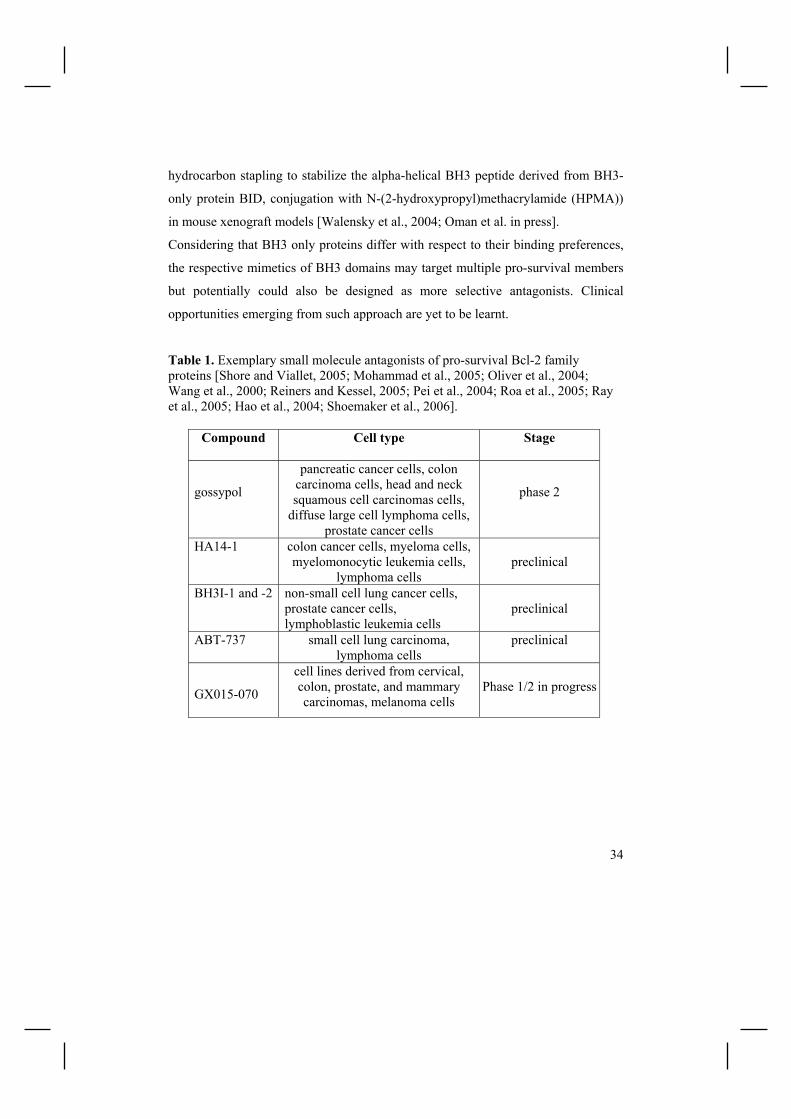
Table 1. Exemplary small molecule antagonists of pro-survival Bcl-2 family proteins [Shore and Viallet, 2005; Mohammad et al., 2005; Oliver et al., 2004; Wang et al., 2000; Reiners and Kessel, 2005; Pei et al., 2004; Roa et al., 2005; Ray et al., 2005; Hao et al., 2004; Shoemaker et al., 2006].
Compound Cell type Stage
gossypol
pancreatic cancer cells, colon carcinoma cells, head and neck squamous cell carcinomas cells,
diffuse large cell lymphoma cells, prostate cancer cells
phase 2
HA14-1 colon cancer cells, myeloma cells, myelomonocytic leukemia cells,
lymphoma cells
preclinical
BH3I-1 and -2 non-small cell lung cancer cells, prostate cancer cells, lymphoblastic leukemia cells
preclinical
ABT-737 small cell lung carcinoma, lymphoma cells
preclinical
GX015-070
cell lines derived from cervical, colon, prostate, and mammary carcinomas, melanoma cells
Phase 1/2 in progress

35
2.4. CURCUMIN “Nothing will benefit human health and increase the chances for survival of life on
Earth as much as the evolution to a vegetarian diet”
Albert Einstein
Given the long natural history of follicular lymphoma, survival benefits must be
watchfully balanced with quality of life. Indeed, severe side effects invoked by most
of anticancer agents are the important impediment to the therapy of FL and other
malignant disorders. Not surprisingly thus the idea of conquering cancer with natural
compounds exerting minimal or no toxicity in humans has been gaining momentum.
As the ancient Indian scripture Ayurveda, developed through daily life experiences
with the mutual relationship between mankind and nature, is being translated into
English, Spanish, German, French and other languages, it has become a rich source
of inspiration in search for pharmacologically safe anticancer drugs.
Curcumin (diferuloylmethane) is a major yellow pigment extracted from the spice
turmeric, which is isolated from a tropical plant native to southern and southeastern
Asia - Curcuma longa. Curcumin´s properties as a colouring and flavouring agent
have led to its widely use as a dietary additive in a variety of foods, including
mustard and other species, gelatins, puddings, sorbets, soups, margarine, and even
alcoholic and non-alcoholic beverages. The maximum dietary consumption of
turmeric (1.5g per person per day) is being noted in certain South East Asian
communities, but its usage as a colouring, food preservative and flavouring agent is
global - over 2400 metric tons of turmeric are imported annually into the USA for
consumer use [Sharma et al. 2005]. Commercial curcumin contains three major
components: curcumin I (Fig. 3), demethoxycurcumin (curcumin II), and
bisdemethoxycurcumin (curcumin III), which are together referred to as
curcuminoids.

36
Figure 3. Structure of curcumin I
Curcumin has been used in Asian medicine since the second millenium BC to treat
biliary disorders, anorexia, cough, diabetic wounds, hepatic disorders, rheumatism,
and sinusitis [Jain and DeFilipps 1991]. The chemopreventive and anticancer effects
of this plant polyphenol are widely recognized and have been a subject of several
excellent reviews [Duvoix et al. 2005, Aggarwal el al. 2003, Sharma et al. 2005]. Of
utmost importance, the outstanding safety records from phase I/II clinical trials,
supported by a long history of curcumin´s consumption as a dietary additive without
any side effects, are supportive for its clinical application.
2.4.1. Targeting of apoptotic pathways by curcumin
Cancer is a disorder of hyperproliferation and/or excessive survival that tends to
metastasize into the vital organs through invasion followed by angiogenesis and
distant metastasis. Although the order of acquisition of underlying changes may
differ depending on tumour type and even between cells within the tumour, in
general terms malignant progression can be divided into several discernible stages.
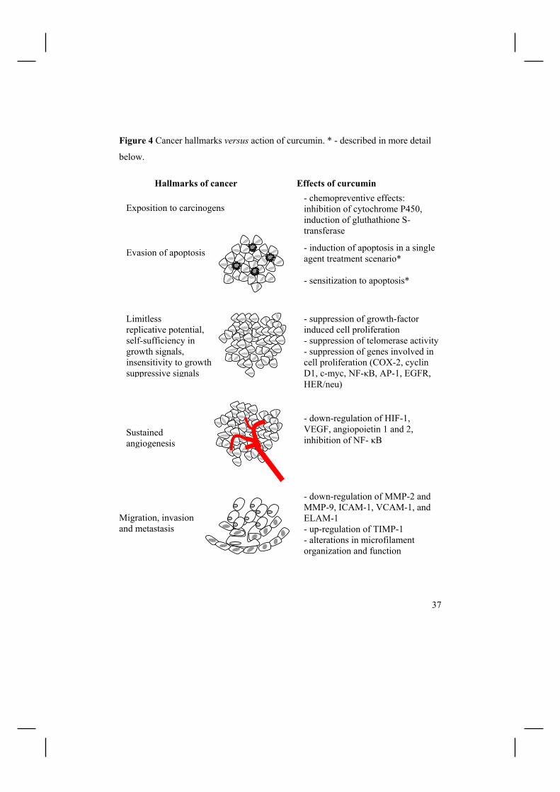
Accordingly, Figure 4 summarizes selected in vitro and/or in vivo activities of
curcumin in relation to the hallmarks of cancer in a step-wise mode. Such
pleiotropic effects of curcumin are mediated by effects on a host of cell signalling
factors, including AP-1 transcription factor, c-Myc, Egr-1, IκB kinase, NF-κB,
protein kinase C, epidermal growth factor receptor tyrosine kinase, c-Jun N terminal
kinase, protein tyrosine kinases, protein serine/threonine kinases, or bcl-2-family
proteins (see below) [Chen et al. 2004, Aggarwal et al. 2003, Aggarwal et al. 2005].
37
Figure 4 Cancer hallmarks versus action of curcumin. * - described in more detail
below.
Hallmarks of cancer Effects of curcumin
Evasion of apoptosis - induction of apoptosis in a single agent treatment scenario* - sensitization to apoptosis*
Limitless replicative potential, self-sufficiency in growth signals, insensitivity to growth suppressive signals
- suppression of growth-factor induced cell proliferation - suppression of telomerase activity- suppression of genes involved in cell proliferation (COX-2, cyclin D1, c-myc, NF-κB, AP-1, EGFR, HER/neu)
Sustained angiogenesis
- down-regulation of HIF-1, VEGF, angiopoietin 1 and 2, inhibition of NF- κB
Migration, invasion and metastasis
- down-regulation of MMP-2 and MMP-9, ICAM-1, VCAM-1, and ELAM-1 - up-regulation of TIMP-1 - alterations in microfilament organization and function
- chemopreventive effects: inhibition of cytochrome P450, induction of gluthathione S-transferase
Exposition to carcinogens

38
Ample evidence exists to show the induction of apoptosis upon curcumin treatment
in diverse tumour cells, including B-cell and T-cell leukemia, B-cell lymphoma,
colon carcinoma and breast carcinoma cells. Mostly, curcumin sequentially induces
activation of caspase-8, Bid cleavage, mitochondrial breach, PTP opening, release of
cytochrome c, activation of caspase-9 and -3, and cleavage of PARP and ICAD.
Translocation of AIF to the nucleus has also been reported. Additionally, curcumin
induced apoptosis-like pathway that lacked the involvement of mitochondrial
membrane depolarization and was not dependent on caspase activity in human
lymphoblastoid T (Jurkat) cells [Piwocka et al. 1999]. Various mechanisms account
for the pro-apoptotic effects of curcumin (Fig. 5). Inhibition of NF-κB translocation,
originally reported by Singh and Aggarwal [1995], has been observed in multiple
cell types, such as colon, gastric, squamous epithelial tumor, B-cell lymphoma,
chronic lymphocytic leukemia [Everett et al. 2006] and multiple myeloma cell lines
and/or primary cells 8 [Bharti et al. 2003], and may be due to a direct effect of
curcumin on NIK, IKKα, or IKKβ kinases [Plummer et al. 1999]. That curcumin
exerts apoptosis via p53-dependent pathway was shown in mammary epithelial
carcinoma cells [Choudhuri et al., 2005], neuroblastoma cell lines [Liontas and
Yeger, 2004], and colon cancer cells. Down-regulation of antiapoptotic proteins
(Bcl-2, Bcl-XL, IAP1, IAP2, XIAP) represents another mechanism of curcumin-
triggered apoptosis. Bax, but not Bak, has been suggested as a critical regulator of
curcumin-induced apoptosis in human colon cancer cells [Rashmi et al., 2005].
Accordingly, several approaches to augment curcumin-based therapy have been
proposed, including silencing of Hsp70, Bcl-XL, or Ku70 [Rashmi et al., 2004], or
over-expression of Smac [Rashmi et al., 2005].

39
Figure 5 Commonly reported curcumin´s targets within the apoptotic pathway of
cell death
Relatively limited data are available on the effects of curucmin on chemtotherapy-
induced killing of cancer cells. Somasundaram and co-workers reported that
curcumin inhibited camptothecin-, mechlorethamine-, and doxorubicin-induced
apoptosis both in vitro and in vivo, and suggested curcumin to generally hamper
apoptosis induced by ROS-generating and JNK-activating chemotherapeutics
[Somasundaram et al. 2002]. In another study however, pre-treatment with curcumin
was shown to enhance cytotoxicity of vinorelbine in vitro in lung squamous cell
carcinoma cell line [Sen et al. 2005]. In hepatic cancer cell line the combination of
curcumin and cisplatin resulted in synergistic antitumour activity, whereas the
DISC
APOPTOSIS
casp-9 activation
caspases 3/6/7
IAPs
Omi/HtrA2
apoptosome
SMAC/Diablo
p53
nucleus
Bax
AIF
cyt c
tBid
casp- 8 activation
Bid
Bcl2, BclXl
NF-κB inactive
NF-κB active

40
effects of curcumin and doxorubicin were additive [Notarbartolo et al. 2005].
Moreover, curcumin potentiated taxol-induced apoptosis in HeLa cells, an effect at
least partially related to curcumin-induced down-regulation of NF-κB and
serine/threonine kinase Akt pathways [Bava et al. 2005]. Both the potential of
cooperative tumour cell killing and danger of curcumin-mediated decrease in
anticancer drug efficacy have yet to be assessed in the clinic.
2.4.2. Successes and failures of curcumin in clinical trials
The side effects following administration of curcumin are extremely rare. Abnormal
liver tests and transient hypotension have been reported in rats and dogs,
respectively, but not in humans. When taken without food curcuminoids may cause
gastritis and peptic ulcer, and thus it is advisable to take curcumin supplements with
meals or postprandial.
The pharmacokinetics of curcumin in humans has been analyzed: - in patients with advanced colorectal cancer; Administration of curcumin at 36-180
mg/day for up to 4 months was well tolerated, but curcumin was recovered only
from feces, not blood or urine [Sharma et al. 2001]. In a subsequent phase I study
in 15 patients, a curcuminoid formulation was consumed orally for up to 4 months,
equating to curcumin doses between 0.45 and 3.6 g/day [Sharma et al. 2004].
Daily dose of 3.6 g has been later on shown amenable for achievement of
pharmacologically efficacious levels of curcumin in the colorectum [Garcea et al.
2005].
- in patients with one of the following: urinary bladder cancer, arsenic Bowen´s
disease of the skin, uterine cervical intraepithelial neoplasm, oral leucoplakia,
intestinal neoplasia of the stomach [Cheng et al. 2001]; Curcumin was given orally
for 3 months, at doses ranging from 0.5 to 12 g/day. No treatment-related toxicity
was noted at doses up to 8 g/day. The serum concentration peaked at 1-2 h after

41
oral intake, declining gradually over the next 12 h. The 8 g/day dose resulted in a
serum concentration of 1.77±0.87 µM
- in patients with chronic anterior uveitis [Lal et al. ] - in patients suffering from idiopathic inflammatory orbital pseudotumors [Lal et al.
2000]; Curcumin was administered orally at dose of 1125 mg/day for 6-22
months. 4 out of 5 patients that completed the study completely recovered.
- in patients with ulcerative proctitis (n= 5) and Crohn's disease (n=5) [Holt et al.
2005]; Curcumin was administered orally at dose of 1.08 g/day for 1 month
followed by 5.76 g/day for next 2 months.
- in healthy volunteers, curcumin was administered orally at dose of 50-200 mg,
without toxicity and detectable systemic bioavailability [Sharma et al. 2005]
Although encouraging, the clinical studies clearly demonstrate that orally
administered curcumin has poor bioavailability and may undergo intestinal
metabolism. Moreover, curcumin is highly hydrophobic and thus cannot be
administered systematically. Fortunately, several approaches have already been
suggested to improve curcumin´s bioavailability and to circumvent the difficulty
with intravenous dosing:
- liposome encapsulation [Li et al., 2005]
- curcumin biojonjugates [Kumar et al., 2000]
- co-administration with piperine [Shoba et al., 1998]
- prolonged release biodegradable microspheres encapsulation [Kumar et al., 2002]

42
3 AIMS OF THE STUDY
The study was undertaken to investigate cellular and molecular mechanisms
underlying apoptosis triggered by selected chemical and natural compounds in
human follicular lymphoma B cell lines.
The specific questions/aims of the study were: To elucidate the temporal and quantitative relationship between mitochondrial
membrane depolarization and caspase activation in HA14-1-induced apoptosis, and
cell cycle specificity of HA14-1-triggered apoptosis. (I)
Does the natural dietary additive curcumin trigger apoptotic response in Bcl-2 over-
expressing FL cell lines and how their sensitivity to curcumin can be enhanced?
What are the mechanisms behind curcumin-evoked apoptosis in this model? (II)
To identify gene expression reprogramming relevant for the pathophysiology and
therapy of follicular lymphoma in curcumin-treated HF4.9 cells using DNA
microarray technology. (III)
To determine whether CXCR4 is a component down-regulated specifically during
curcumin-induced cell demise, or is a general apoptosis/cell death associated
phenomenon. (IV)

43
4 MATERIALS AND METHODS
"Cytometry is easy. Biology is hard."
Howard Shapiro
4.1. Cell lines and culturing (I-IV)
The experiments have been performed on three follicular lymphoma cell lines
HF1A3, HF4.9 and HF28RA [Eray et al. 2003]. Cells were cultured in RPMI 1640
medium (Cambrex, Verviers, Belgium) supplemented with 5% heat-inactivated FCS
(EuroClone, Pero, Italy), 2 mM L-glutamine (Fluka Chemie, Buch, Switzerland),
200 µg/µl streptomycin (Sigma, St. Louis, MO, USA), 240 IU/ml penicillin (Orion,
Espoo, Finland), 10 mM HEPES buffer (Cambrex), 0.1 mM NAA (Cambrex), 1 mM
Na-pyruvate (Cambrex) and 20 µM 2-mercaptoethanol (Fluka Chemie) at 37˚C in a
5% CO2 humidified atmosphere.
4.2. Cell treatment experiments
In Publication I, HF4.9 cells were treated with 5µM HA14-1 (Alexis Biochemicals)
for 0-4h at 15 min intervals. Pan caspase inhibitor zVAD-fmk (100µM) was added
2 h before HA14-1 administration.
In Publication II, freshly dissolved curcumin was added directly to cell cultures,
and all experiments were performed in the dark due to possible degradation products
of the polyphenol. Cell death was also induced in HF1A3 cells by CD95 cross-
linking with anti-Fas antibody (100ng/ml; clone CH11; Upstate, NY, USA).
Ascorbic acid was procured from Sigma. PD150606 (inhibitor of calpains), CA-074-
Me and zFA-fmk (cathepsin B inhibitors), and ALNN (calpain I inhibitor) were
generously provided by Dr Michael Courtney (AIV Institute, Kuopio, Finland). Pan-
caspase inhibitor zVAD-fmk was added at various concentrations (0-100µM) 2h
before exposition to curcumin. All compounds were diluted in cell culture medium

44
to working stock immediately before use. No alterations in growth variables were
observed in vehicle controls.
In Publication III, HF4.9 cells were treated with 15 µM curcumin for 0-36h.
Alternatively, HF4.9 cells were pre-treated for 1h with 250 nM CsA (cyclophilin D
inhibitor), followed by administration of curcumin (10 and 15 µM) for 24h.
Additionally, HF1A3, HF4.9, and HF28RA cells were exposed to AMD3100
(Sigma), a selective CXCR4 antagonist, alone or in media devoted of serum.
In Publication IV, HF4.9 cells were left unstimulated or treated with 2.5µM
doxorubicin (a gift from the Kuopio University Hospital), 5nM vincristine (a gift
from the Kuopio University Hospital), 7.5µM HA14-1 (Alexis Biochemicals), or
100ng/ml brefeldin A (BFA). HF1A3 cells were treated with CD95 cross-linking
antibody. To assess the effect of oncotic stimuli on CXCR4 expression, HF1A3 cells
were also exposed to 3% H2O2 for 15min.
4.3. Cell proliferation and viability assays
4.3.1. [3H]-Thymidine incorporation assay (II)
The anti-proliferative effects of curcumin were monitored by thymidine
incorporation assay. Cells (1x10⁶ cells/ml) were treated in flat-bottom, 96-well
(200µl/well) plates with different concentrations of curcumin (1-50 µM) in complete
medium for 20 h before adding 1µCi/ml [methyl-3H]-thymidine for an additional 4 h
at 37˚C. The incorporated radioactive thymidine was quantified by scintillation
counting with Microbeta counter (Perkin Elmer, MA, USA). All determinations
were made in triplicate.
45
4.3.2. Trypan blue staining (II)
Cell viability/growth was also examined using the trypan blue exclusion method.
After incubation with curcumin, the cells were mixed with an equal volume of PBS
containing 0.4% (w/v) trypan blue dye, and counted manually using a
hemocytometer. Trypan blue negative cells were scored.
4.4. Apoptosis detection
4.5.1. Morphological assessment (II)
A cell that is undergoing apoptosis demonstrates nuclear condensation and DNA
fragmentation. For visualization of nuclear alterations in curcumin-treated cells,
cells were rinsed with cell culture media and stained with Hoechst 33342 for 20 min
at room temperature. Imaging was carried out with a cooled Apogee KX85 CCD
(publication I) and IX70 Olympus (publication III) microscope with appropriate
filter cubes. Images were assembled into figures using Adobe Photoshop.
4.5.2. Quantification of fractional DNA content (II)
Propidium iodide staining was used for quantification of cells with increased sub-G1
content. At the end of experiment, cells were harvested, washed twice with PBS and
fixed in 70% EtOH at +4°C overnight. Next, cells were treated with RNase
(10µg/ml; Sigma) for 2h at 56°C followed by 1h incubation at 37°C with 5µg/ml PI
(Sigma). Subsequently, cells were analyzed on FACScan flow cytometer (Becton
Dickinson) running under Cell Quest software, with 10000 events acquired per
sample. Data analysis was performed with Summit v3.1 software (Dako
Cytomation, Fort Collins, CO, USA).
4.5.3. Conformational DNA/RNA changes (II, III, IV)
The vital stain SYTO16 (Molecular Probes, Eugene, OR, USA) allows the accurate
estimation of the number of viable cells, and in combination with permeability

46
marker PI (Sigma) has been used to identify an early stage of apoptosis. The
specificity of SYTO16 to detect apoptotic changes in FL cells was confirmed by the
lack of SYTO16dim/PI- events in hyperthermia-induced (56˚C for 5 min) or H2O2-
induced (0.3% for 4h) oncosis. Upon staining, cells were analyzed on FACScan flow
cytometer (Becton Dickinson, San Jose, CA, USA) running under CellQuest
software (Becton Dickinson), with 10000 events acquired per sample. Data were
subsequently analyzed using Summit v3.1 software (Dako Cytomation, Fort Collins,
CO, USA).
4.5.4. Cell membrane permeabilization (II)
For identification of early cell membrane permeabilization, cells were stained with
YO-PRO 1 (Molecular Probes), a 374Da cation. Cells with early cell membrane
permeabilization are stained only with YO-PRO 1, whereas those in late stages of
apoptosis and necrotic cells are characterized by more pronounced loss in cell
membrane integrity, and are thus stained with both YO-PRO 1 and PI. Briefly, cells
were harvested, washed with PBS, incubated with 250nM YO-PRO 1 and 5µg PI for
20 min at RT in darkness, and analyzed on FACScan flow cytometer (Becton
Dickinson). The nuclear morphology of PI+ cells was investigated to determine
whether it is necrosis or advanced apoptosis (postapoptotic necrosis).
4.5.5. Mitochondrial membrane potential depolarisation (I, II)
Lipophilic cations, including JC-1, TMRM, CMXRos and others, accumulate in the
mitochondrial matrix, driven by the electrochemical gradient. Herein, loss of
mitochondrial membrane potential was assessed using TMRM (Molecular Probes) in
combination with FLICA and cell membrane permeability marker (publication I, for
a detailed description of the procedure see below), or in combination with
permeability marker alone (publication II).

47
4.5.6. Cytochrome c release (II)
Preparation of mitochondria or cytosolic extracts from control and curcumin-treated
cells (2x10⁷) was performed using the ApoAlert Cell Fractionation kit (BD
Biosciences) according to the manufacturer instructions. Equal loading of
mitochondria and cytosol was assessed by immunobloting with mouse anti-
cytochrome oxidase subunit IV (COX4) and by Poncaeu S staining, respectively.
4.5.7. Caspase activation (I, II, IV)
Most often, activation of caspases is assayed indirectly by testing whether their
inhibitors prevent particular apoptotic events (publication I). Commonly, the
activation of caspases is detected via immunoblotting techniques using specific
antibodies against caspases, or against cleavage products, such as poly(ADP-ribose)
polymerase (PARP), (publication II and IV). Another common approach is to
employ fluorochrome substrates which become fluorescent upon cleavage by the
caspase, or FLICA - a cell permeable and non-toxic FLuorochrome Inhibitor of
CAspases. When added to a population of cells, FLICA probe enters each cell and
covalently binds to a reactive cysteine residue that resides on the large subunit of the
active caspase heterodimer. Because FLICA becomes covalently bound to the
enzyme, it is retained within the cell, whereas uncoupled FLICA diffuses out of the
cell and is washed away. Thus, the remaining fluorescence signal can be considered
as a direct measure of the number of active caspases present in the cell (publication
I and II).
4.5.8. Multiparametric flow cytometry assays (I)
4.5.8.1. Triplicate TMRM / FLICA / 7-AAD staining
In publication I the multivariate assay analogical to one used by Pozarowski et al.
[Pozarowski et al. 2003] was applied. The approach allows simultaneous tracking of
mitochondrial membrane depolarisation (by TMRM), caspase activation (by FLICA)

48
and cell membrane permeability (by 7-AAD). After the challenge cells were
harvested, resuspended in 100µl of fresh medium, followed by staining with FLICA
(Immunochemistry Technologies, LLC) according to the manufacturer's instructions
(1h at 37°C). Next, cells were harvested, washed twice with ice-cold PBS, and
stained with 150 nM TMRM (Molecular Probes) for 15 min at 37°C. Finally, cells
were stained with 7-AAD (5µg/ml; Molecular Probes) for 3-5 minutes at RT. All
staining procedures were performed in the dark.
4.5.8.2. Detection of cell cycle specificity of apoptosis
In publication I, FL cells were exposed to HA14-1 at dose and for the time
indicated, followed by and staining with FAM-VAD-FMK for 1h at 37°C. Next,
cells were fixed in ethanol for at least 2 h at -20°C, stained with propidium iodide
(PI, 5 µg/ml; Sigma) in the presence of RNase (20µg/ml; Sigma) for 1h at 37°C, and
immediately analyzed on FACScan flow cytometer (Becton Dickinson).
4.6. Cell cycle analysis
Cell cycle alterations induced by curcumin (unpublished data) were studied by flow
cytometry analysis. Cells were plated at the density of 1 x 10⁶ and cultured for 24 h
in media alone, or treated with curcumin. Cells were harvested and washed twice
with PBS, fixed with 70% ethanol overnight, pre-treated with 10µg/ml RNase
(Sigma) for 1h, followed by staining with PI (5 µg/ml; Sigma) for 2h. The cell cycle
profile was determined with FACScan flow cytometer, running under CellQuest
software, with subsequent analysis using Summit software.
4.7. Lysosomal membrane permeabilization (II)
In publication II, cells were assessed for lysosomal stability following treatment
with curcumin using the acridine orange (AO) uptake method. Cell were stained
after the challenge with curcumin, and analyzed immediately on FACScan flow

49
cytometer. AO is a lysosomotropic base and a metachromatic fluorochrome
exhibiting red fluorescence when highly concentrated (as is the case in intact
lysosomes where AO is retained in its charged form - AOH+). When outside the
lysosomes, AO exhibits green fluorescence. The lysosomal burst was further
evaluated by staining with another acidophilic dye - LysoTracker Red (Molecular
Probes), followed by fluorescent microscopy analysis. The loss of punctuate staining
of LysoTracker red indicates disruption of acidic vacuoles.
4.8. Detection of reactive oxygen species (ROS) (II)
The intracellular ROS generation can be assessed using dihydroethidine probe
(DHE), which is oxidized by superoxide anion to become ethidium bromide (EthBr)
emitting red fluorescence, or by dihydrofluorescein diacetate (H2DCFDA). After the
challenge with HA14-1 or curcumin, cells were harvested, washed with PBS, and
stained with 2.5µM DHE for 20min at RT. For H2DCFDA staining, cells were
loaded with 1 µM H2DCFDA for 20-30 min at 37˚C prior to cell stimulation.
4.9. Cell surface staining (III)
The cell surface expression of CXCR4 in FL cell lines was assessed using FITC-
conjugated mouse monoclonal anti-human CXCR4 antibody (clone 12G5; R&D
Systems). Briefly, 0.5x10⁶ cells were stained with 20µl of anti-CXCR4 Ab, or FITC-
conjugated IgG1 isotype-control (BD Biosciences, San Diego, CA, USA), for 30min
at RT. After staining cells were analyzed by flow cytometry (FACScan, Becton
Dickinson).
4.10. Immunoblotting (II, III, IV)
Samples prepared in 1x Laemmli buffer (62.5 mM Tris-HCl, pH 6.8, 1% SDS (w/v),
5% 2-mercaptoethanol, 10% glycerol (v/v), and 0.001% bromophenol blue (w/v))
were resolved by 10-15% SDS-PAGE and transferred onto nitrocellulose.
Nitrocellulose was blocked with 5% milk in TBS-T (0.1% Tween 20) 1h at RT.
Primary antibodies were incubated at +4˚C overnight. The following primary

50
antibodies were used: rabbit anti-Bad (1:1000), rabbit anti-Bax (1:1000), rabbit anti-
Bim (1:1000), rabbit anti-Bok (1:1000), rabbit anti-Puma (1:1000) were procured
from Cell Signaling Technology (Beverly, MA, USA); rabbit anti-CXCR4 (1:500)
and goat anti-CD20 (1:250) were from Abcam; mouse anti-PARP (1:500) and anti-
Mcl-1 (1:1000) were from Santa Cruz Biotechnology (Santa Cruz, CA, USA). The
appropriate secondary antibodies were from Zymed (S.San Francisco, CA, USA).
Blots were developed with enhanced chemiluminescence detection method (ECL
kit, Amersham Pharmacia Biotech, Buckinghamshire, UK). Multiple exposures were
taken at different times, so that nonsaturated ECL film was used for quantitation.
Films were digitized by a flatbed transparency scanner and quantified with Java
domain NIH Image J software (v1.31 freely available at
http://rsb.info.nih.gov/ij/index.html).
4.10. RNA isolation (III)
HF4.9 cells (4-6x10⁶) were treated with curcumin for 0, 8, 24 or 36 h. At the end of
experiment, a sample of cells was assayed for viability using SYTO16/PI staining to
verify cells´ response to the treatment. Subsequently, cells were harvested, and total
RNA was isolated using TRI Reagent (Ambion), according to the manufacturer's
instructions. The quality and integrity of the extracted RNAs were analyzed by gel
electrophoresis and by spectrometric analysis at 260/280 nm. The experiment was
performed in quadruplicate maintaining the same conditions. For each time point,
equal quantities of total RNA were pooled from two independent experiments.
4.11. Large-scale gene expression profiling (III)
4.11.1. Probe preparation
Total RNA was processed for hybridization in Finnish DNA Microarray Centre at
Turku Centre for Biotechnology. Briefly, 300ng total RNA was amplified overnight

51
(14h) with Illumina RNA TotalPrep Amplification kit (Ambion). During IVT
reaction cRNA was biotinylated. Prior to and after the amplifications the
RNA/cRNA concentrations where checked with Nanodrop ND-1000 and
RNA/cRNA quality was controlled by BioRad´s Experion electrophoresis station. A
hybridization mixture containing 1.5µg of biotinylated cRNA was then hybridized to
Sentrix Human WG-6 BeadChips (Illumina, Inc., San Diego, CA, USA) at 55ºC
overnight (19h).
4.11.2. Bead chip microarrays
In publication III, Sentix Human-WG6 Expression BeadChips (Illumina) were used
to analyze the curcumin-triggered reprogramming. The Human-WG6 Expression
BeadChip allows profiling of six individual samples in parallel, >48,000 transcripts
each. Each sample is probed by a pair of arrays. One stripe in each array pair is
based on National Centre for Biotechnology Information (NCBI) Reference
Sequence (RefSeq) genes, with probe content that is well annotated. The second
stripe includes probes for genes with unique locations in the human genome such as
ESTs. Probes are specifically designed to avoid querying pseudogenes and SNP
sites. The arrays are separated from one another by a seal, and thus each can be
hybridized to a different sample [Steemers and Gunderson; 2005].
The array substrate contains wells in a highly ordered, pre-defined pattern.
Oligonucleotide probes (29-base address concatenated to a 50-base gene-specific
probe) are immobilized onto beads (approximately 3µm in size) that are
subsequently quantitatively pooled. Libraries of beads are self-assembled into the
etched microwell array substrates (Fig. 6).

52
Figure 6. The outline of BeadArray technology. A) The Sentrix BeadChip is created
using a MEMS-patterned slide substrate. B) Gene-specific probes are immobilized
on beads. Actual beads have hundreds of thousands of copies of the same sequence
attached. C) Beads are quantitatively pooled and subsequently self-assembled into
the patterned substrate. D) Scanning electron micrograph of an assembled array
containing 3 µm diameter silica beads. Modified from Steemers and Gunderson,
2005.
A B C D
Each bead in every array contains hundred of thousands of covalently attached
oligonucleotide probes, and each bead type has an average 30-fold representation,
providing statistical accuracy of multiple measurements. Finally, each bead on the
array is identified by virtue of hybridization-based decoding procedure,
simultaneously validating the performance of each bead type.
address probe
29b 50b
labelled cRNA Photo-resist
Silicon wafer
Plasma etching
cleaning

53
Several control categories are included into Sentrix BeadArrays: i) controls for the
biological specimen; ii) controls for sample labeling (optional); iii) controls for
hybridization (Cy3-labeled Hyb control, low stringency Hyb control, high stringency
Hyb control), iv) signal generation controls; v) negative controls
Recently, an evaluation of the reproducibility of microarray results using two
platforms: Affymetrix GeneChip and Illumina BeadChip revealed very high data
reproducibility [Barnes et al., 2005].
4.11.3. qRT- PCR
12µg total RNA from each time point was pooled from 2-4 independent
experiments, and DNase-treated (DNA-free kit, Ambion) according to
manufacturer´s instructions, followed by spectrometric analysis. Next, 5µg RNA
was reverse transcribed using High Capacity cDNA Archieve Kit (Applied
Biosystems, Warrington, UK), following manufacturer's instructions. cDNA
dilutions (1:50), and standard series (serial dilutions of pooled cDNA samples) were
prepared. The amplification of target genes was carried out using 12ng of template
(RNA equivalents), gene specific primers (QuantiTect Primer Assays, Qiagen), and
SYBR Green Master Mix PCR (Qiagen), in total volume of 25µl using a RotorGene
3000 thermocycler (Corbett Research, Sydney, Australia). Dissociation curves were
analyzed to ensure only a single product was amplified. Data were normalized to b-
actin mRNA expression, and Student´s t test was used to assess the differences in
relative gene expression.
4.12. Data analysis
4.12.1. Microarray data analysis (III)
Gene profile files from BeadStudio were saved as tab delimited .txt files. Genome
file corresponding to Sentix Human-WG6 Expression BeadChips was obtained from

54
Illumina. Genome file and gene profile data files were up-loaded to GeneSpring 7.2
software (Silicon Genetics, Redwood City, CA, USA). The outline of data analysis
is presented on Fig. 7. Briefly, raw data were normalized to baseline arrays (control),
and genes with detection precision value >0.95 in at least 2 out of 8 samples were
filtered (14719 probes passed the requirement). Next, genes with ≥2-fold change at
least 1 of the 3 comparisons (control versus 8, 24 or 36 hours) were filtered. Using
the 5% false discovery rate (FDR) 99 targets were found to be modulated by
curcumin. K-means clustering was applied to the significant genes using standard
correlation analysis as implemented in the Gene Spring 7.2 program, resulting in
groupings based on general pattern of expression changes with time following
curcumin addition. Genes were also grouped based on their molecular function, as
provided in Gene Spring software. Information on genes was obtained from NCBI.
Figure 7. Workflow of microarray data analysis applied in the present study
(publication III)
filtering interesting data
data quality control
Data from image analysis
normalization, calculate averages and log ratios
checking the normality, and quality of replicates
checking the normality, and quality of replicates
exploratory data analysis

55
5. RESULTS AND DISCUSSION “All truths are easy to understand once they are discovered; the point is to discover
them”
Galileo Galilei
5.1. HA14-1 as an effective inducer of apoptosis (Publication I)
HA14-1 is a small molecule inhibitor of Bcl-2 protein, found through a computer
screening strategy and confirmed by a competitive binding assay based on
fluorescence polarization [Wang et al., 2000] (Fig. 8).
Figure 8 Structure of HA14-1 (A) and model for the complex of HA14-1 with the
Bcl-2 surface pocket [Wang et al., 2000].
The pro-apoptotic activity of HA14-1 has been suggested to correlate positively with
the level of Bcl-2 in treated cells [Lickliter et al. 2003]. Surprisingly, the action of
the compound has not been studied in follicular lymphoma cells with physiological
over-expression of Bcl-2. We demonstrated that HA14-1 is an effective trigger of
apoptosis in the 3 FL-derived cell lines, as depicted by morphological changes
(chromatin condensation, nuclear fragmentation, and formation of apoptotic bodies),
loss of SYTO16 fluorescence, and fractional DNA content ([Skommer et al. 2006];
not included in the present thesis).
A B

56
In publication I we reported that HA14-1 triggers rapid, yet relatively transient,
mitochondrial membrane depolarisation, in a partially PT-dependent manner.
Importantly, applying multivariate flow cytometry analysis, we observed that only
fraction of cells with mitochondrial breach was characterized by concomitant
caspase activation. Based on the kinetics and pharmacological inhibitor studies,
coupled with the lack of cell population with active caspases but preserved
mitochondrial membrane potential in multiparametric flow cytometry assays, we
concluded that HA14-1- triggered caspase activation occurs solely as a consequence
of mitochondrial rupture. The outline of the findings has been depicted in Figure 9.
Figure 9 Proposed model for HA14-1 action in follicular lymphoma cells.
Often, the cell cycle specificity of anti-tumour agents, which is deduced from in
vitro studies, provides rationale for design of treatment protocols. The analysis of
cell cycle specificity of HA14-1-evoked apoptosis in vitro revealed that mostly cells
from the G1 phase undergo apoptosis. Transient exposure to a high dose of the
compound appeared to induce apoptosis mainly in G1 and S-phase cells, suggesting
that drugs arresting cells in G1 and/or S phase, or those inducing cell demise from
biochemical features of apoptosis
morphological apoptotic attributes
zVAD-fmk
caspase activation
HA14-1
Bcl-2 and possibly other family members
mitochondrial rupture caspase activation CsA

57
G2/M phase, could be successfully employed with HA14-1 in combinatorial
regimens.
In conceptual agreement to our findings, the group headed by Prof. Rosenberg
published an excellent paper showing the action of newly synthesized Bcl-2
inhibitor, ABT-737, in cancer cell lines and in vivo models [Oltersdorf et al. 2005].
Among others, it was shown that ABT-737 administered as single agent is able to
induce apoptosis in cell lines with t(14;18) and in cells derived from patients with
follicular lymphoma, strongly supporting our observations, and firmly confirming
the potential of Bcl-2 inhibitors in a fight against FL. According to a recent proposal
by Certo et al. [2006], the BH3-only proteins induce apoptosis in a single-agent
treatment scenario only in cells poised to die, i.e. cells that rely on the presence of
anti-apoptotic Bcl-2 proteins. It is possible that in these cells direct activators of Bax
and Bak, or alternatively Bax and Bak themselves, are engaged to induce cell death
and are kept in check by one or more of the anti-apoptotic Bcl-2 family members. If
this is the case, neutralization of anti-apoptotic Bcl-2 proteins causes death (so called
"death by default"), as observed in FL cells.
Importantly, certain approaches to sensitize cells to the action of small molecule
Bcl-2 proteins inhibitors have already been tested, such as pharmacological
improvement or co-administration with ursodeoxycholic acid (UDCA) - a relatively
non-toxic bile acid causing a conformational change in Bcl-2 and thus promoting
HA14-1 binding [Castelli et al. 2004]. Only recently a novel approach to limit
toxicity and improve water solubility of small molecule Bcl-2 inhibitors has been
developed [Oman et al. in press]. Certainly antagonists of Bcl-2 family proteins hold
significant promise for the future of cancer therapy.
5.2. Characterization of curcumin-induced apoptosis (Publication II)
Inferred from the observation that curcumin can decrease Bcl-2 expression and
induce apoptosis in several cell lines of hematopoetic origin, we attempted to
analyze its action in FL cell lines. Although it has been suggested that ectopic over-

58
expression of Bcl-2 may attenuate curcumin´s action, FL cell lines appeared to be
very sensitive to cytotoxic, anti-proliferative and pro-apoptotic action of this
polyphenol. Caspase-dependency of curcumin-evoked apoptosis has been
demonstrated by employing z-VAD-fmk titration study. Often applied at relatively
high concentrations (50-100µM), zVAD-fmk reportedly inhibits not only caspases,
but also other proteases involved in the execution of cell death, such as calpains and
cathepsins [Rozman-Pungercar et al. 2003]. However, curcumin-induced loss of cell
viability was restored already at 30µM dose of zVAD-fmk, underscoring the crucial
involvement of caspases. Intriguingly, increase in intracellular ROS appeared to act
as an upstream signal that commenced apoptosis. Lysosomal rupture, observed in all
cell lines tested, presents a perplexing snippet in the process of curcumin-induced
cell death. Specifically, it suggests the existence of additional cell death pathways,
apart from mitochondrial pathway, of cell death. It will be interesting to dissect
whether mitochondria and lysosomes both contribute to cell fate in this model, or
whether any of these organelles has an up-stream role in cell death induction. As the
application of pharmacological inhibitors of cathepsins and calpains exert highly
lethal effects on FL cell lines, additional studies, including genetic knock-down or
knock-out experiments, are necessary to elucidate the role of proteases present in
lysosomal lumen in the progression and phenotypic outcome of curcumin-triggered
apoptosis.
The major obstacle in the design of effective anticancer curcumin-based therapies is
a low bioavailability of the polyphenol. Accordingly, we seek to identify agents able
to sensitize FL cells to curcumin-elicited apoptosis. As co-administration with
piperine, the active ingredient of black pepper, reportedly enhances curcumin
bioavailability by 2,000% in human volunteers [Shoba et al. 1998], we first tested its
ability to enhance curcumin-triggered cell killing in vitro. However, no enhancement
in any of the growth variables was observed upon co-administration with
physiologically relevant concentrations of piperine (JS, unpublished observations).

59
Surprisingly though, ascorbic acid appeared to augment curcumin-induced
cytotoxicity and apoptosis in all FL cell lines tested, an effect associated with and
dependent on synergistic ROS generation. The pro-oxidant role of ascorbic acid has
been previously reported both in vitro and in vivo, and on-going controversies on
ascorbic acid and its pro/anti-oxidant activity exist. Considering the widespread of
data published up to date, it appears that action of ascorbic acid highly depends on
its dose, the cellular context, and presence of other compounds, e.g. iron.
Clearly, these experiments proved that curcumin is a trigger of the multilayered
response in follicular lymphoma cells, and guided the subsequent attempt to provide
further insights into molecular mechanisms underlying the action of this polyphenol
using large scale gene expression profiling.
5.3. Global gene expression reprogramming induced by curcumin
(Publication III)
In our search for delineating the molecular mechanisms behind anti-tumour
activity of curcumin, we went on to determine the large scale gene expression
reprogramming induced in response to curcumin administration in vitro. Our study
was the first to address the global gene expression changes following curcumin
treatment in follicular lymphoma cells, and the first to analyse such a wide range of
mRNA species upon curcumin treatment in general.
In line with previously observed induction of apoptosis, and inhibition of cell
growth and glucose metabolism, proliferation in FL cell lines treated with curcumin,
the polyphenol appeared to induce complex genetic reprogramming in HF4.9 cells.
Genes encoding proteins involved in the regulation of apoptosis, REDOX, cell
motility, transcription, splicing, ion homeostasis, intracellular signalling, lymphoid
development, B-cell activation, and others, were identified as curcumin-regulated. In

60
total, 97 mRNA species were recognized as curcumin-responsive, and tables
summarizing both up- and down-regulated ones can be found from publication III.
Importantly, prolonged exposition to curcumin was required to sustain many of gene
expression changes. This is in line with other reports showing, that 24-48h exposure
to curcumin is necessary for maximal induction of apoptosis [Everett et al. 2007].
Therefore, and combined with rapid inactivation by hepatic and intestinal
glucoronidation, the systemic treatment of follicular lymphoma with curcumin in a
single-agent treatment scenario would require constant intravenous infusion. Thus,
identification and development of combinatorial treatment regimens with
compounds improving curcumin´s bioavailability and/or lowering threshold for
curcumin-induced apoptosis are currently the primary and active research area.
5.3.1. CXCR4
Of great importance, among panoply of curcumin-responsive genes the
CXCR4 gene was markedly down-regulated, a finding confirmed by both qRT-PCR
and immunoblotting. The effect was observed in all FL cell lines tested. As the
major obstacle in the successful administration of curcumin is its low bioavailability
in vivo, we also asked whether low but attainable in vivo doses of curcumin are able
to down-regulate CXCR4 over longer treatment times. FL cell lines were cultured
with 2µM curcumin for 5 days (single curcumin administration in day 0), followed
by protein isolation and immunoblotting. Surprisingly, even at such a low
concentration curcumin largely reduced the expression of CXCR4 protein in all cell
lines tested. Since interfering with CXCR4 function by means of pharmacological
inhibitor AMD3100 retarded long-term cell growth and induced cell death in a cell
line dependent fashion, it is plausible to reason that down-regulation of CXCR4 may
have a correlate to the growth-suppresive effects of curcumin in some cellular
contexts.
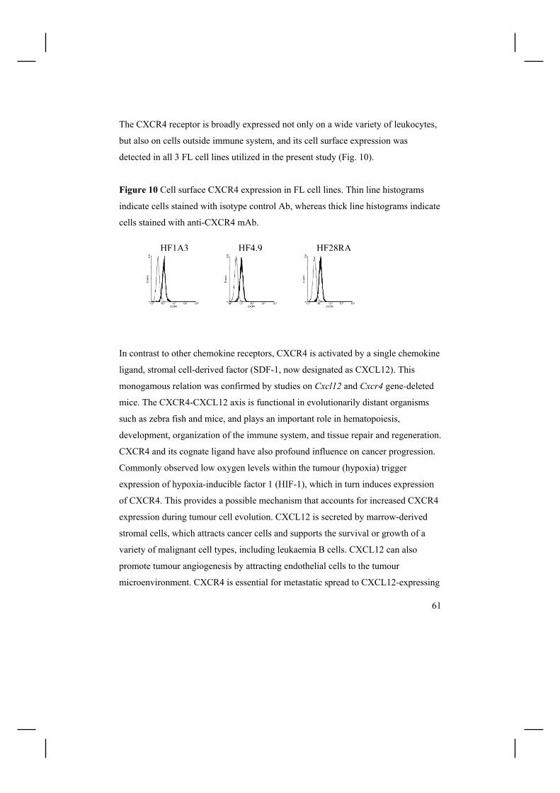
61
The CXCR4 receptor is broadly expressed not only on a wide variety of leukocytes,
but also on cells outside immune system, and its cell surface expression was
detected in all 3 FL cell lines utilized in the present study (Fig. 10).
Figure 10 Cell surface CXCR4 expression in FL cell lines. Thin line histograms
indicate cells stained with isotype control Ab, whereas thick line histograms indicate
cells stained with anti-CXCR4 mAb.
In contrast to other chemokine receptors, CXCR4 is activated by a single chemokine
ligand, stromal cell-derived factor (SDF-1, now designated as CXCL12). This
monogamous relation was confirmed by studies on Cxcl12 and Cxcr4 gene-deleted
mice. The CXCR4-CXCL12 axis is functional in evolutionarily distant organisms
such as zebra fish and mice, and plays an important role in hematopoiesis,
development, organization of the immune system, and tissue repair and regeneration.
CXCR4 and its cognate ligand have also profound influence on cancer progression.
Commonly observed low oxygen levels within the tumour (hypoxia) trigger
expression of hypoxia-inducible factor 1 (HIF-1), which in turn induces expression
of CXCR4. This provides a possible mechanism that accounts for increased CXCR4
expression during tumour cell evolution. CXCL12 is secreted by marrow-derived
stromal cells, which attracts cancer cells and supports the survival or growth of a
variety of malignant cell types, including leukaemia B cells. CXCL12 can also
promote tumour angiogenesis by attracting endothelial cells to the tumour
microenvironment. CXCR4 is essential for metastatic spread to CXCL12-expressing
HF1A3 HF4.9 HF28RA

62
organs, such as the marrow, and recirculation of cancer cells between the blood and
the marrow or lymphoid tissue, where they receive protective survival signals
[Burger and Kipps, 2006]. Indeed, ample evidence suggest that CXCR4 is crucial in
the trafficking of hematopoietic malignancies, such as B-cell chronic lymphocytic
leukaemia (CLL), multiple myeloma, non-Hodgkin lymphomas (including follicular
lymphomas), and acute leukemias (ALL, precursor B-cell acute lyphoblastic
leukaemia and AML, acute myelogenous leukaemia). It is also involved in anti-
cancer drug resistance, as many cells become resistant to spontaneous or drug-
induced apoptosis upon adhesion to marrow stromal fibronectin. As CXCR4 appears
to be a linchpin in the context of crosstalk between tumour cells and their respective
microenvironment, CXCR4-targeted therapeutic approaches may soon prove to be
clinically relevant. Therefore, the concept that well tolerated natural compound
curcumin impedes CXCR4 expression is seminal for the future clinical assessment
of curcumin as the innovative therapeutic option for follicular lymphoma patients.
Although the present study clearly indicates that curcumin down-regulates the
CXCR4 gene expression in FL cell lines, a number of questions remain to be
answered: 1) what is the mechanism underlying curcumin-mediated down-regulation
of CXCR4 and how does it contribute to the process of apoptosis?; 2) is it an effect
ascribed only to FL cells, or could it be observed in a wider range of tumour cells? ;
3) can tumour cells develop resistance to curcumin-induced CXCR4 down-
regulation? ; 4) does (and if yes, how) this in vitro effect translate to the situation in
vivo? Specifically, does curcumin treatment decrease CXCR4 expression in cancer
cells in vivo, and can curcumin inhibit homing of circulating malignant cells to the
sites rich in SDF-1 in animal models?; and 5) can curcumin mobilize normal
hematopoietic progenitor cells in a manner analogical to CXCR4 antagonists? As the
interest in function of CXCR4-SDF1 axis in tumour dissemination is mushrooming,
we are unlikely to await the answers for long.
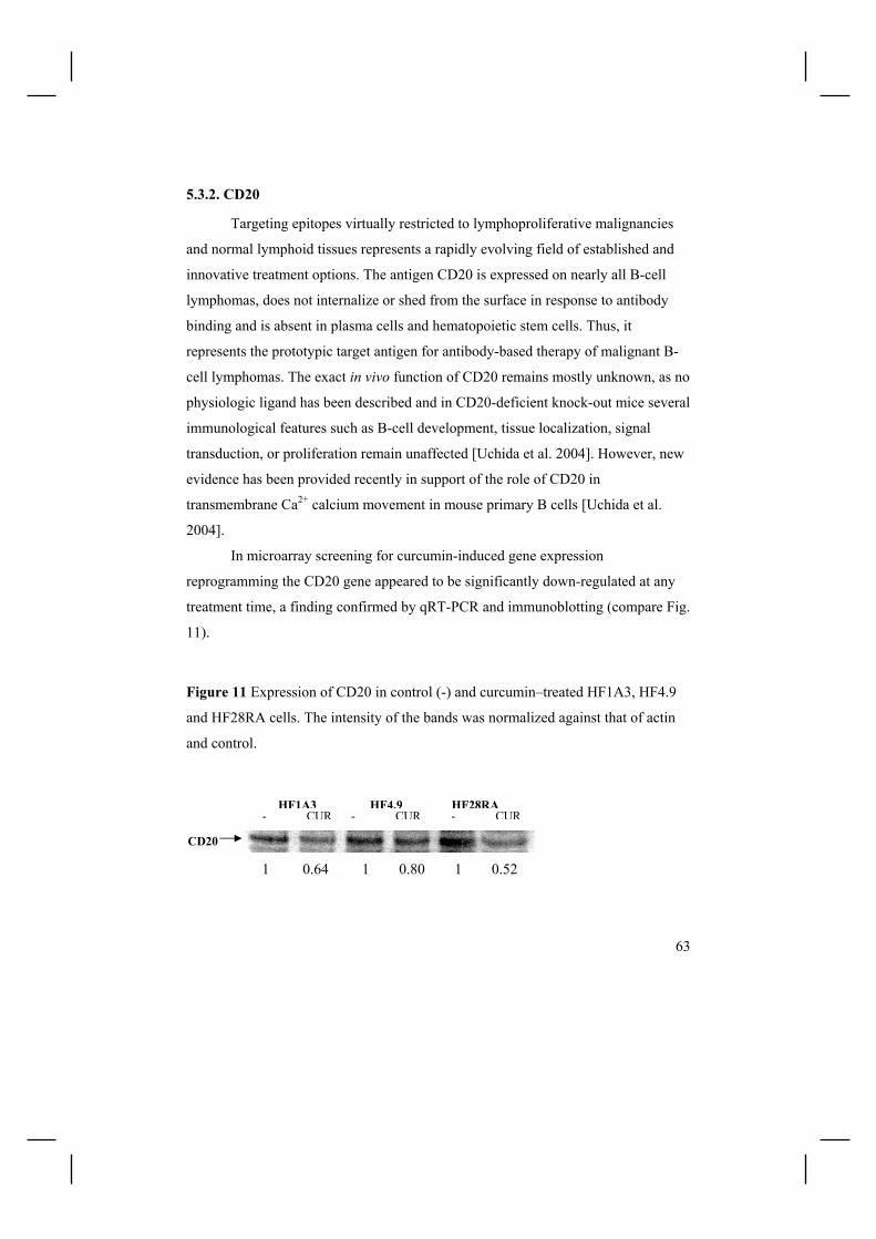
63
5.3.2. CD20
Targeting epitopes virtually restricted to lymphoproliferative malignancies
and normal lymphoid tissues represents a rapidly evolving field of established and
innovative treatment options. The antigen CD20 is expressed on nearly all B-cell
lymphomas, does not internalize or shed from the surface in response to antibody
binding and is absent in plasma cells and hematopoietic stem cells. Thus, it
represents the prototypic target antigen for antibody-based therapy of malignant B-
cell lymphomas. The exact in vivo function of CD20 remains mostly unknown, as no
physiologic ligand has been described and in CD20-deficient knock-out mice several
immunological features such as B-cell development, tissue localization, signal
transduction, or proliferation remain unaffected [Uchida et al. 2004]. However, new
evidence has been provided recently in support of the role of CD20 in
transmembrane Ca2+ calcium movement in mouse primary B cells [Uchida et al.
2004].
In microarray screening for curcumin-induced gene expression
reprogramming the CD20 gene appeared to be significantly down-regulated at any
treatment time, a finding confirmed by qRT-PCR and immunoblotting (compare Fig.
11).
Figure 11 Expression of CD20 in control (-) and curcumin–treated HF1A3, HF4.9
and HF28RA cells. The intensity of the bands was normalized against that of actin
and control.
- CUR - CUR - CURHF1A3 HF4.9 HF28RA
CD20
1 0.64 1 0.80 1 0.52

64
Although the information on the role of CD20 is still limited, the observation that
curcumin induces a marked decrease in CD20 expression hints for possible clinical
implications, brings new questions, and also suggests further experimental work to
be done: 1) Potentially, down-regulation of CD20 may have adverse effects on the
efficacy of anti-CD20 therapy in B-cell malignancies; hence the in vitro and in vivo
effects of combinatorial treatment with curcumin and anti-CD20 mAbs should be
assessed; 2) anti-CD20 mAbs exert different effects on B cells depending on their
stage of differentiation. It will be interesting to test whether effects of curcumin on
CD20 expression are also differentiation-dependent; 3) As in the case of CXCR4,
the specificity of curcumin-mediated down-regulation of CD20 in FL cell lines as
compared to normal B cells and other tumour cell lines/primary cells, as well as
effects of curcumin on CD20 expression in vivo should be examined; 4) Does CD20
down-regulation contribute to curcumin-induced apoptosis? Again, I hope for at
least some of these queries to be deciphered soon.

65
5.4. CXCR4 down-regulation is not a common apoptosis-associated event
(Publication IV)
The final and important question here was whether CXCR4 down-regulation
is commonplace in dying cells. The scarcity of data on CXCR4 expression during
tumour cell demise, coupled with our original observation that curcumin down-
regulates CXCR4 expression at both mRNA and protein levels, prompted us to
study the generality of the phenomenon on treatment with pro-apoptotic agents
targeting different organelles/pathways (Fig. 12), including anti-CD95 mAb (death
receptor pathway), HA14-1 (mitochondrial pathway), doxorubicin (DNA damage),
vincristine (microtubule depolymerization), and inducer of ER-stress brefeldin A
(BFA) .
Figure 12 Schematic representation of apoptotic pathways targeted to access the
CXCR4 expression during apoptotic cell death.
Casp2 activation
Bid
DISC
cyt c
tBid casp- 8
activation
HA14-1
anti-CD95 mAb
casp-9 activation
caspases 3/6/7
IAPs
Omi/HtrA2
apoptosome
SMAC/Diablo
nucleus
Doxorubicin
microtubules
Vincristine
BFA
Ca2+
66
Additionally, to investigate CXCR4 expression during non-apoptotic cell death,
HF1A3 cells were stimulated with 3% H2O2 for 15min, when HF1A3 cells die via
oncosis. Interestingly, we did not detect CXCR4 down-regulation in any of the
abovementioned treatment scenarios. These data indicate that i) CXCR4 protein
down-regulation is not commonly observed during tumour cell death; and ii)
curcumin is a specific inducer of CXCR4 down-regulation in our model.
Considering the role of CXCR4 in vivo, the above results suggest also the
superiority of curcumin over traditional anticancer drugs (Dox, Vin), and
compounds in clinical development as anti-cancer agents (HA14-1, BFA), in
management of local and distant tumour metastasis.

67
6. CONCLUSIONS, CLINICAL IMPLICATIONS AND FUTURE DIRECTIONS The seminal discovery that the BCL2 gene inhibits cell death rather than promotes
cell proliferation [Vaux et al. 1988, McDonnell et al. 1989] gave foundation for now
widely embraced premise that impaired apoptosis is a crucial step in tumorigenesis.
Indeed, disturbances in regulation of selective cell death underlie many diseases,
including cancer, autoimmunity and degenerative disorders, and the antitumour
effects of anticancer drugs are linked to their ability to induce apoptosis within
tumours. Nevertheless, other forms of cell demise, such as autophagic cell death or
necrosis, might also be induced by chemotherapy. Clearly, studies on molecular
mechanisms behind any type of programmed cell death should benefit our
understanding of the effects of anticancer agents and represent a research market
that will continue to grow.
Intrinsic and/or acquired resistance to autophagic cell death and apoptosis may result
in resistance to anticancer drugs. The Achilles´ heel of most of the tumour cells is
that they remain sensitive to some apoptotic triggers, and thus agents stimulating
certain proapoptotic proteins and/or signalling pathways represent promising
strategies to combat cancer. Moreover, targeting alternative cell death routes in cells
resistant to apoptosis or cells in which apoptosis is inhibited, as well as combining
both apoptosis-inducing and survival suppressing strategies, may appear clinically
advantageous.

68
The concluding remarks based on the results summarized above and reported in
Publications I-IV are as follows:
1. HA14-1, a BH3 mimetic, induces partially PT-dependent apoptosis in
follicular lymphoma cells in a single agent regimen (Publication I)
Small molecule inhibitors of Bcl-2 (e.g. HA14-1) represent an alternative strategy to
induce apoptosis in follicular lymphoma cells even in a single agent treatment
scenario, and thus FL cells can be considered as primed for death. Mechanistically,
HA14-1-evoked caspase activation occurs solely as a consequence of mitochondrial
rupture, and ensuing cell death is partially PT-dependent. Bcl-2 antagonists first
emerged as compounds that should potentially sensitize cells to the action of other
stress stimuli such as anticancer drugs or growth factor withdrawal. However, FL
cells exposed to BH3 mimetic HA14-1 readily undergo cell death without any
additional stress stimuli, suggesting that they are primed for death, and underlying
that they indeed may be susceptible to targeting by drugs developed as mimetics of
sensitizer BH3 domains.
2. Cellular basis of curcumin-induced cell death (Publication II)
Curcumin induces cessation of FL cell growth at attainable in vivo 1µM
concentration, and is an efficient inducer of apoptosis at higher concentrations. The
pro-apoptotic effects of curcumin are enhanced by physiologically relevant doses of
ascorbic acid, warranting clinical studies on curcumin-based therapies in follicular
lymphoma patients. Follicular lymphoma is an indolent disease, with a watchful
waiting strategy considered as an appropriate treatment option for patients
presenting early stages of FL. Hence, the safe and patient-friendly dietary approach,
such as administration of curcumin, may represent an alternative strategy with
chemopreventive and/or tumour suppressing potential.
Mechanistically, caspases are essential for curcumin-induced apoptosis in FL cells,
whereas the pertinence of lysosomal proteases in the propagation and phenotypic

69
outcome of cell death in this model remains to be dissected. Due to inherent cell
death associated with exposition to calpain and/or cathepsin inhibitors, this will
require genetic manipulation in a tightly regulated manner. It is indeed of great
importance, especially because cell death pathways other than classical apoptosis
(such as autophagic cell death) are increasingly recognized to contribute to overall
level of drug-induced cell killing.
3. Curcumin induces CXCR4 downregulation, which is not a cell-death
associated event (Publication III and IV)
Curcumin induces complex gene expression changes in HF4.9 cells, including
down-regulation of CXCR4 both at mRNA and protein levels. Based on the
literature data available up to date, CXCR4 down-regulation should be of supreme
importance for the therapeutic effects of curcumin in vivo. FL is recognized as a
disease of functional B cells in which the clinical behaviour is determined not only
by deregulated apoptotic-pathways within the malignant cells, but also by functional
cross-talk with the immunologic regulatory network. Hence, curcumin may interfere
with survival of B lymphoma cells in survival niche, where they receive enough
microenvironmental signals to resist chemotherapy-triggered cell death. Moreover,
as CXCR4-CXCL12 axis is thought to be involved in lymphoma cell trafficking and
dissemination, and CXCR4-deficient hybridoma cells reportedly fail to disseminate
to CXCL12-expressing target organs, curcumin may also inhibit metastasis of FL
cells. Gene expression reprogramming triggered by curcumin appears to require
sustained exposition to the compound, supporting the premise of a prolonged
curcumin administration being therapeutically beneficial over short-term treatment.
Finally, CXCR4 down-regulation, observed during curcumin-induced apoptosis, is
not generic to cell death underlying the advantage of curcumin over traditional
chemotherapeutics.

70
References
Adrain C, Martin SJ (2006) Double knockout blow for caspases. Science 311(5762):785-6
Advani R, Rosenberg SA, Horning SJ (2004) Stage I and II follicular non-Hodgkin's lymphoma: long-term follow-up of no initial therapy. J Clin Oncol. 22(8):1454-9.
Aggarwal BB, Kumar A, Bharti AC (2003) Anticancer potential of curcumin: preclinical and clinical studies. Anticancer Res 23(1A):363-98.
Aggarwal S, Ichikawa H, Takada Y, Sandur SK, Shishodia S, Aggarwal BB (2005) Curcumin (Diferuloylmethane) Downregulates Expression of Cell Proliferation, Antiapoptotic and Metastatic Gene Products Through Suppression of I{kappa}B{alpha} Kinase and AKT Activation. Mol Pharmacol
Antonsson B (2004) Mitochondria and the Bcl-2 family proteins in apoptosis signaling pathways. Mol Cell Biochem 256-257(1-2):141-55.
Azoulay-Zohar H, Israelson A, Abu-Hamad S, Shoshan-Barmatz V (2004) In self-defence: hexokinase promotes voltage-dependent anion channel closure and prevents mitochondria-mediated apoptotic cell death. Biochem J 377(Pt 2):347-55.
Baines CP, Kaiser RA, Purcell NH, Blair NS, Osinska H, Hambleton MA, Brunskill EW, Saven MR, Gottlied RA, Dorn GW, Robbins J, Molkentin JD (2005) Loss of cyclophilin D reveals a critical role for mitochondrial permeability transition in cell death. Nature 434(7033):658-62.
Barnes M, Freudenberg J, Thompson S, Aronow B, Pavlidis P (2005) Experimental comparison and cross-validation of the Affymetrix and Illumina gene expression analysis platforms. Nucleic Acids Res 33(18):5914-23.
Boatright KM, Renatus M, Scott FL, Sperandio S, Shin H, Pedersen IM, Ricci JE, Edris WA, Sutherlin DP, Green DR, Salvesen GS (2003) A unified model for apical caspase activation. Mol Cell 11(2):529-41.
Boatright KM, Salvesen GS (2003) Mechanisms of caspase activation. Curr Opin Cell Biol 15(6):725-31.
Bouchier-Hayes L, Lartigue L, Newmeyer DD (2005) Mitochondria: pharmacological manipulation of cell death. J Clin Invest 115(10):2640-7.
Burger JA, Kipps TJ (2006) CXCR4: a key receptor in the crosstalk between tumor cells and their microenvironment. Blood
Castelli M, Reiners JJ, Kessel D (2004) A mechanism for the proapoptotic activity of ursodeoxycholic acid: effects on Bcl-2 conformation. Cell Death Differ 11(8):906-14.
Certo M, Del Gaizo Moore V, Nishino M, Wei G, Korsmeyer S, Armstrong SA, Letai A (2006) Mitochondria primed by death signals determine cellular addiction to antiapoptotic BCL-2 family members. Cancer Cell 9(5):351-65
Chandra D, Choy G, Daniel PT, Tang DG (2005) Bax-dependent regulation of Bak by voltage-dependent anion channel 2. J Biol Chem 280(19):19051-61.
71
Chang AL, Hsu CH, Lin JK, et al (2001) Phase I clinical trial of curcumin, a chemopreventive agent, in patients with high-risk or pre-malignant lesions. Anticancer Res 21:2895-2900
Chen HW, Yu SL, Chen JJ, Li HN, Lin YC, Yao PL, Chou HY, Chien CT, Chen WJ, Lee YT, Yang PC (2004) Anti-invasive gene expression profile of curcumin in lung adenocarcinoma based on a high throughput microarray analysis. Mol Pharmacol 65(1):99-110.
Chen L, Willis S, Wei A, Smith B, Fletcher J, Hinds M, Colman P, Day C, Adams J, Huang D (2005) Differential Targeting of Prosurvival Bcl-2 Proteins by Their BH3-Only Ligands Allows Complementary Apoptotic Function. Molecular Cell 17(3):393-403
Cheng EH, Kirsch DG, Clem RJ, Ravi R, Kastan MB, Bedi A, Ueno K, Hardwick JM (1997) Conversion of Bcl-2 to a Bax-like death effector by caspases. Science 278(5345):1966-8.
Cheng EH, Sheiko TV, Fisher JK, Craigen WJ, Korsmeyer SJ (2003) VDAC2 inhibits BAK activation and mitochondrial apoptosis. Science 301:513-7
Chiarugi A (2005) "Simple but not simpler": toward a unified picture of energy requirements in cell death. FASEB J 19(13):1783-8.
Chou JJ, Li H, Salvesen GS, Yuan J, Wagner G (1999) Solution structure of BID, an intracellular amplifier of apoptotic signaling. Cell 96(5):615-24.
Choudhuri T, Pal S, Das T, Sa G (2005) Curcumin selectively induces apoptosis in deregulated cyclin D1-expressed cells at G2 phase of cell cycle in a p53-dependent manner. J Biol Chem 280(20):20059-68.
Cirman T, Oresic K, Mazovec GD, Turk V, Reed JC, Myers RM, Salvesen GS, Turk B (2004) Selective disruption of lysosomes in HeLa cells triggers apoptosis mediated by cleavage of Bid by multiple papain-like lysosomal cathepsins. J Biol Chem 279(5):3578-87.
Coiffier B (2005) First-line treatment of follicular lymphoma in the era of monoclonal antibodies. Clin Adv Hematol Oncol 3(6):484-505.
Coultas L, Pellegrini M, Visvader JE, Lindeman GJ, Chen L, Adams JM, Huang DC, Strasser A (2003) Bfk: a novel weakly proapoptotic member of the Bcl-2 protein family with a BH3 and a BH2 region. Cell Death Differ 10(2):185-92.
Danial NN, Korsmeyer SJ (2004) Cell death: critical control points. Cell 23;116(2):205-19
De Jong D (2005) Molecular pathogenesis of follicular lymphoma: a cross talk of genetic and immunologic factors. J Clin Oncol 23:6358-6363
Dejean LM, Martinez-Caballero S, Manon S, Kinnally KW (2005) Regulation of the mitochondrial apoptosis-induced channel, MAC, by BCL-2 family proteins. Biochim Biophys Acta
Delivani P, Martin SJ (2006) Mitochondrial membrane remodelling in apoptosis: an inside story. Cell Death Differ 13:2007-2010
Deng Y, Ren X, Yang L, Lin Y, Wu X (2003) A JNK-dependent pathway is required for TNFalpha-induced apoptosis. Cell 115(1):61-70

72
Duvoix A, Blasius R, Delhalle S, Schnekenburger M, Morceau F, Henry E, Dicato M, Diederich M (2005) Chemopreventive and therapeutic effects of curcumin. Cancer Lett 223(2):181-90.
Edinger AL, Thompson CB (2004) Death by design: apoptosis, necrosis and autophagy. Curr Opin Cell Biol 16(6):663-9
Eray M, Postila V, Eeva J, Ripatti A, Karjalainen-Lindsberg ML, Knuutila S, Andersson LC, Pelkonen J (2003) Follicular lymphoma cell lines, an in vitro model for antigenic selection and cytokine-mediated growth regulation of germinal centre B cells. Scand J Immunol 57(6):545-55.
Everett PC, Meyers JA, Makkinje A, Rabbi M, Lerner A (2006) Preclinical assessment of curcumin as a potential therapy for B-CLL. Am J Hematol 82:23-30
Fischer U, Stroh C, Schulze-Osthoff K (2005) Unique and overlapping substrate specificities of caspase-8 and caspase-10. Oncogene
Fisher RI, LeBlanc M, Press OW, Maloney DG, Unger JM, Miller TP (2005) New treatment options have changed the survival of patients with follicular lymphoma. J Clin Oncol 23(33):8447-52.
Gandhi MK, Marcus RE (2005) Follicular lymphoma: time for a re-think? Blood Rev 19(3):165-78.
Garcea G, Berry DP, Jones DJ, Singh R, Dennison AR, Farmer PB, Sharma RA, Steward WP, Gescher AJ (2005) Consumption of the putative chemopreventive agent curcumin by cancer patients: assessment of curcumin levels in the colorectum and their pharmacodynamic consequences. Cancer Epidemiol Biomarkers Prev 14(1):120-5.
Garrido C, Kroemer G (2004) Life's smile, death's grin: vital functions of apoptosis-executing proteins. Curr Opin Cell Biol 16(6):639-46.
Ghia P, Boussiotis VA, Schultze JL, Cardoso AA, Dorfman DM, Gribben JG, Freedman AS, Nadler LM (1998) Unbalanced expression of bcl-2 family proteins in follicular lymphoma: contribution of CD40 signaling in promoting survival. Blood 91(1):244-51
Gillespie DA, Vousden KH (2003) The secret life of histones. Cell 114(6):655-6. Gomez-Bougie P, Oliver L, Le Gouill S, Bataille R, Amiot M (2005) Melphalan-
induced apoptosis in multiple myeloma cells is associated with a cleavage of Mcl-1 and Bim and a decrease in the Mcl-1/Bim complex. Oncogene 24(54):8076-9
Gong XM, Choi J, Franzin CM, Zhai D, Reed JC, Marassi FM (2004) Conformation of membrane-associated proapoptotic tBid. J Biol Chem 279:28954-60.
Gonzalez-Polo RA, Boya P, Pauleau AL, Jalil A, Larochette N, Souquere S, Eskelinen EL, Pierron G, Saftig P, Kroemer G (2005) The apoptosis/autophagy paradox: autophagic vacuolization before apoptotic death. J Cell Sci 118(Pt 14):3091-102.
Green DR (2005) Apoptotic pathways: ten minutes to dead. Cell 121(5):671-4.

73
Green DR, Kroemer G (2004) The pathophysiology of mitochondrial cell death. Science 305(5684):626-9.
Grinberg M, Sarig R, Zaltsman Y, Frumkin D, Grammatikakis N, Reuveny E, Gross A (2002) tBID Homooligomerizes in the mitochondrial membrane to induce apoptosis. J Biol Chem 77(14):12237-45.
Gross A (2006) BID as a double agent in cell life and death. Cell Cycle 5(6):582-4. Guicciardi ME, Leist M, Gores GJ (2004) Lysosomes in cell death. Oncogene
23(16):2881-90. Gulmann C, Espina V, Petricoin E 3rd, Longo DL, Santi M, Knutsen T, Raffeld M,
Jaffe ES, Liotta LA, Feldman AL (2005) Proteomic analysis of apoptotic pathways reveals prognostic factors in follicular lymphoma. Clin Cancer Res 11(16):5847-55.
Hail N Jr, Carter BZ, Konopleva M, Andreeff M (2006) Apoptosis effector mechanisms: A requiem performed in different keys. Apoptosis 11(6):889-904
Hao JH, Yu M, Liu FT, Newland AC, Jia L (2004) Bcl-2 inhibitors sensitize tumor necrosis factor-related apoptosis-inducing ligand-induced apoptosis by uncoupling of mitochondrial respiration in human leukemic CEM cells. Cancer Res 64(10):3607-16.
Hetz C, Vitte PA, Bombrun A, Rostovtseva TK, Montessuit S, Hiver A, Schwarz MK, Church DJ, Korsmeyer SJ, Martinou JC, Antonsson B (2005) Bax channel inhibitors prevent mitochondrion-mediated apoptosis and protect neurons in a model of global brain ischemia. J Biol Chem 280(52):42960-70.
Hiddemann W, Buske C, Dreyling M, Weigert O, Lenz G, Forstpointner R, Nickenig C, Unterhalt M (2005) Treatment strategies in follicular lymphomas: current status and future perspectives. J Clin Oncol 23(26):6394-9
Holt PR, Katz S, Kirshoff R (2005) Curcumin therapy in inflammatory bowel disease: a pilot study. Dig Dis Sci 50(11):2191-3.
Horning SJ, Rosenberg SA (1984) The natural history of initially untreated low-grade non-Hodgkin's lymphomas. N Engl J Med 311(23):1471-5.
Horsman DE, Connors JM, Pantzar T, Gascoyne RD (2001) Analysis of secondary chromosomal alterations in 165 cases of follicular lymphoma with t(14;18). Genes Chromosomes Cancer 30(4):375-82.
Johnson PW, Rohatiner AZ, Whelan JS, Price CG, Love S, Lim J, Matthews J, Norton AJ, Amess JA, Lister TA (1995) Patterns of survival in patients with recurrent follicular lymphoma: a 20-year study from a single center. J Clin Oncol 13(1):140-7.
Kagan VE, Tyurin VA, Jiang J, Tyurina YY, Ritov VB, Amoscato AA, Osipov AN, Belikova NA, Kapralov AA, Kini V, Vlasova II, Zhao Q, Zou M, Di P, Svistunenko DA, Kurnikov IV, Borisenko GG (2005) Cytochrome c acts as a cardiolipin oxygenase required for release of proapoptotic factors. Nat Chem Biol 1:223-32
Kalai M, Van Loo G, Vanden Berghe T, Meeus A, Burm W, Saelens X, Vandenabeele P (2002) Tipping the balance between necrosis and apoptosis in

74
human and murine cells treated with interferon and dsRNA. Cell Death Differ 9(9):981-94
Kalinowska M, Garncarz W, Pietrowska M, Garrard WT, Widlak P (2005) Regulation of the human apoptotic DNase/RNase Endonuclease G: involvement of Hsp70 and ATP. Apoptosis 10(4):821-30
Kamer I, Sarig R, Zaltsman Y, Niv H, Oberkovitz G, Regev L, Haimovich G, Lerenthal Y, Marcellus RC, Gross A (2005) Proapoptotic BID is an ATM effector in the DNA-damage response. Cell 122(4):593-603.
Kang CB, Tai J, Chia J, Yoon HS (2005) The flexible loop of Bcl-2 is required for molecular interaction with immunosuppressant FK-506 binding protein 38 (FKBP38). FEBS Lett 579(6):1469-76.
Karbowski M, Lee YJ, Gaume B, Jeong SY, Frank S, Nechushtan A, Santel A, Fuller M, Smith CL, Youle RJ (2002) Spatial and temporal association of Bax with mitochondrial fission sites, Drp1, and Mfn2 during apoptosis. J Cell Biol 159(6):931-8.
Kataoka T, Holler N, Micheau O, Martinon F, Tinel A, Hofmann K, Tschopp J (2001) Bcl-rambo, a novel Bcl-2 homologue that induces apoptosis via its unique C-terminal extension. J Biol Chem 276(22):19548-54.
Kaufmann T, Schinzel A, Borner C (2004) Bcl-w(edding) with mitochondria. Trends Cell Biol 14(1):8-12
Ke N, Godzik A, Reed JC [2001] Bcl-B, a novel Bcl-2 family member that differentially binds and regulates Bax and Bak. J Biol Chem 276(16):12481-4.
Kim M, Jung SO, Park K, Jeong EJ, Joung HA, Kim TH, Seol DW, Chung BH (2005) Detection of Bax protein conformational change using a surface plasmon resonance imaging-based antibody chip. Biochem Biophys Res Commun
Krysko O, De Ridder L, Cornelissen M (2004) Phosphatidylserine exposure during early primary necrosis (oncosis) in JB6 cells as evidenced by immunogold labeling technique. Apoptosis 9(4):495-500.
Kumar S, Dubey KK, Tripathi S, Fujii M, Misra K (2000) Design and synthesis of curcumin-bioconjugates to improve systemic delivery. Nucleic Acids Symp Ser (44):75-6.
Kumar V, Lewis SA, Mutalik S, Shenoy DB, Venkatesh, Udupa N (2002) Biodegradable microspheres of curcumin for treatment of inflammation. Indian J Physiol Pharmacol 46(2):209-17.
Lal B, Kapoor AK, Agrawal PK, Asthana OP, Srimal RC (2000) Role of curcumin in idiopathic inflammatory orbital pseudotumours. Phytother Res 14(6):443-7.
Lavrik IN, Golks A, Krammer PH (2005) Caspases: pharmacological manipulation of cell death. J Clin Invest 115(10):2665-72.
Le Gouill S, Podar K, Harousseau JL, Anderson KC (2004) Mcl-1 Regulation and Its Role in Multiple Myeloma. Cell Cycle. 2004 3(10):1259-62.
Lecoeur H, Prevost MC, Gougeon ML (2001) Oncosis is associated with exposure of phosphatidylserine residues on the outside layer of the plasma membrane: a

75
reconsideration of the specificity of the annexin V/propidium iodide assay. Cytometry 1;44(1):65-72.
Leist M, Jaattela M (2001) Four deaths and a funeral: from caspases to alternative mechanisms. Nat Rev Mol Cell Biol 2(8):589-98
Levine B, Yuan J (2005) Autophagy in cell death: an innocent convict? J Clin Invest 115(10):2679-88.
Li L, Braiteh FS, Kurzrock R (2005) Liposome-encapsulated curcumin: in vitro and in vivo effects on proliferation, apoptosis, signaling, and angiogenesis. Cancer 104(6):1322-31.
Lickliter JD, Wood NJ, Johnson L, McHugh G, Tan J, Wood F et al . HA14-1 selectively induces apoptosis in Bcl-2-overexpressing leukemia/lymphoma cells, and enhances cytarabine-induced cell death. Leukemia 2003;17:2074-80.
Lin B, Kolluri SK, Lin F, Liu W, Han YH, Cao X, Dawson MI, Reed JC, Zhang XK (2004) Conversion of Bcl-2 from protector to killer by interaction with nuclear orphan receptor Nur77/TR3. Cell 116(4):527-40.
Lindsten T, Thompson CB (2006) Cell death in the absence of Bax and Bak. Cell Death Differ 2006 May 5; [Epub ahead of print]
Liontas A, Yeger H (2004) Curcumin and resveratrol induce apoptosis and nuclear translocation and activation of p53 in human neuroblastoma. Anticancer Res 24(2B):987-98
Lossos IS, Alizadeh AA, Diehn M, Warnke R, Thorstenson Y, Oefner PJ, Brown PO, Botstein D, Levy R (2002) Transformation of follicular lymphoma to diffuse large-cell lymphoma: alternative patterns with increased or decreased expression of c-myc and its regulated genes. Proc Natl Acad Sci U S A 99(13):8886-91.
Lowe SL, Rubinchik S, Honda T, McDonnell TJ, Dong JY, Norris JS (2001) Prostate-specific expression of Bax delivered by an adenoviral vector induces apoptosis in LNCaP prostate cancer cells. Gene Ther 8(18):1363-71.
Majno G, Joris I (1999) Apoptosis, oncosis, and necrosis. An overview of cell death. Am J Pathol. 146(1):3-15
Martinet W, De Meyer GR, Herman AG, Kockx MM (2005) Amino acid deprivation induces both apoptosis and autophagy in murine C2C12 muscle cells. Biotechnol Lett 27(16):1157-63.
McDonnell JM, Fushman D, Milliman CL, Korsmeyer SJ, Cowburn D (1999) Solution structure of the proapoptotic molecule BID: a structural basis for apoptotic agonists and antagonists. Cell 96(5):625-34.
McDonnell TJ, Deane N, Platt FM, Nunez G, Jaeger U, McKearn JP, Korsmeyer SJ (1989) bcl-2-immunoglobulin transgenic mice demonstrate extended B cell survival and follicular lymphoproliferation. Cell 57(1):79-88.
Minn AJ, Velez P, Schendel SL, Liang H, Muchmore SW, Fesik SW, Fill M, Thompson CB (1997) Bcl-x(L) forms an ion channel in synthetic lipid membranes. Nature 385(6614):353-7

76
Mohammad RM, Wang S, Aboukameel A, Chen B, Wu X, Chen J, Al-Katib A (2005) Preclinical studies of a nonpeptidic small-molecule inhibitor of Bcl-2 and Bcl-X(L) [(-)-gossypol] against diffuse large cell lymphoma. Mol Cancer Ther 4(1):13-21
Nakagawa T, Shimizu S, Watanabe T, Yamaguchi O, Otsu K, Yamagata H, Inohara H, Kubo T, Tsujimoto Y (2005) Cyclophilin D-dependent mitochondrial permeability transition regulates some necrotic but not apoptotic cell death. Nature 434(7033):652-8.
Naumann U, Schmidt F, Wick W, Frank B, Weit S, Gillissen B, Daniel P, Weller M (2003) Adenoviral natural born killer gene therapy for malignant glioma. Hum Gene Ther 14(13):1235-46.
Norbury CJ, Zhivotovsky B (2004) DNA damage-induced apoptosis. Oncogene 23(16):2797-808.
Nothwehr SF, Martinou JC (2003) A retention factor keeps death at bay. Nat Cell Biol 5(4):281-3.
Oliver CL, Bauer JA, Wolter KG, Ubell ML, Narayan A, O´Connell KM, Fisher SG, Wang S, Wu X, Ji M, Carey TE, Bradford CR (2004) In vitro effects of the BH3 mimetic, (-)-gossypol, on head and neck squamous cell carcinoma cells. Clin Cancer Res 10(22):7757-63.
Oltersdorf T, Elmore SW, Shoemaker AR, Armstrong RC, Augeri DJ, Belli BA, Bruncko M, Deckwerth TL, Dinges J, Hajduk PJ, Joseph MK, Kitada S, Korsmeyer SJ, Kunzer AR, Letai A, Li C, Mitten MJ, Nettesheim DG, Ng S, Nimmer PM, O'Connor JM, Oleksijew A, Petros AM, Reed JC, Shen W, Tahir SK, Thompson CB, Tomaselli KJ, Wang B, Wendt MD, Zhang H, Fesik SW, Rosenberg SH (2005) An inhibitor of Bcl-2 family proteins induces regression of solid tumours. Nature 435(7042):677-81.
Otake Y, Sengupta TK, Bandyopadhyay S, Spicer EK, Fernandes DJ (2004) Drug-induced destabilization of bcl-2 mRNA: a new approach for inducing apoptosis in tumor cells. Curr Opin Investig Drugs 5(6):616-22.
Otake Y, Sengupta TK, Bandyopadhyay S, Spicer EK, Fernandes DJ (2005) Retinoid-induced apoptosis in HL-60 cells is associated with nucleolin down-regulation and destabilization of Bcl-2 mRNA. Mol Pharmacol 67(1):319-26.
Otera H, Ohsakaya S, Nagaura Z, Ishihara N, Mihara K (2005) Export of mitochondrial AIF in response to proapoptotic stimuli depends on processing at the intermembrane space. EMBO J 24(7):1375-86.
Pastorino JG, Hoek JB, Shulga N (2005) Activation of Glycogen Synthase Kinase 3{beta} Disrupts the Binding of Hexokinase II to Mitochondria by Phosphorylating Voltage-Dependent Anion Channel and Potentiates Chemotherapy-Induced Cytotoxicity. Cancer Res 65(22):10545-10554
Pastorino JG, Shulga N, Hoek JB (2002) Mitochondrial binding of hexokinase II inhibits Bax-induced cytochrome c release and apoptosis. J Biol Chem 277(9):7610-8

77
Pattingre S, Tassa A, Qu X, Garuti R, Liang XH, Mizushima N, Packer M, Schneider MD, Levine B (2005) Bcl-2 antiapoptotic proteins inhibit Beclin 1-dependent autophagy. Cell 122(6):927-39.
Pei XY, Dai Y, Grant S (2004) The small-molecule Bcl-2 inhibitor HA14-1 interacts synergistically with flavopiridol to induce mitochondrial injury and apoptosis in human myeloma cells through a free radical-dependent and Jun NH2-terminal kinase-dependent mechanism. Mol Cancer Ther 3(12):1513-24
Piwocka K, Zablocki K, Wieckowski MR, Skierski J, Feiga I, Szopa J, Drela N, Wojtczak L, Sikora E (1999) A novel apoptosis-like pathway, independent of mitochondria and caspases, induced by curcumin in human lymphoblastoid T (Jurkat) cells. Exp Cell Res 249(2):299-307
Plummer SM, Holloway KA, Manson MM et al. (1999) Inhibition of cyclo-oxygenase 2 expression in colon cells by the chemopreventive agent curcumin involves inhibition of NF-κB activation via the NIK/IKK signalling complex. Oncogene 18:6013-6020
Pozarowski P, Huang X, Halicka DH, Lee B, Johnson G, Darzynkiewicz Z (2003) Interactions of fluorochrome-labeled caspase inhibitors with apoptotic cells: a caution in data interpretation. Cytometry A. 55(1):50-60.
Ray S, Bucur O, Almasan A (2005) Sensitization of prostate carcinoma cells to Apo2L/TRAIL by a Bcl-2 family protein inhibitor. Apoptosis 10(6):1411-8.
Reiners JJ Jr, Kessel D (2005) Susceptibility of myelomonocytic leukemia U937 cells to the induction of apoptosis by the non-peptidic Bcl-2 ligand HA14-1 is cell cycle phase-dependent. Cancer Lett 221(2):153-63
Ricci JE, Waterhouse N, Green DR (2003) Mitochondrial functions during cell death, a complex (I-V) dilemma. Cell Death Differ 10(5):488-92
Roa W, Chen H, Alexander A, Gulavita S, Thng J, Sun XJ, Petruk K, Moore R (2005) Enhancement of radiation sensitivity with BH3I-1 in non-small cell lung cancer. Clin Invest Med 28(2):55-63.
Rozman-Pungercar J, Kopitar-Jerala N, Bogyo M, Turk D, Vasiljeva O, Stefe I, Vandenabeele P, Bromme D, Puizdar V, Fonovic M, Trstenjak-Prebanda M, Dolenc I, Turk V, Turk B (2003) Inhibition of papain-like cysteine proteases and legumain by caspase-specific inhibitors: when reaction mechanism is more important than specificity. Cell Death Differ 10(8):881-8
Rudner J, Jendrossek V, Belka C (2002) New insights in the role of Bcl-2 Bcl-2 and the endoplasmic reticulum. Apoptosis 7(5):441-7.
Ruzi-Vela A, Albar JP, Martinez CA (2001) Apaf-1 localization is modulated indirectly by Bcl-2 expression. FEBS Lett 501(1):79-83.
Schmidt-Mende J, Gogvadze V, Hellstrom-Lindberg E, Zhivotovsky B (2006) Early mitochondrial alterations in ATRA-induced cell death. Cell Death Differ 13:119-28
Schuler M, Green DR (2005) Transcription, apoptosis and p53: catch-22. Trends Genet 21(3):182-7.

78
Sekine K, Hao Y, Suzuki Y, Takahashi R, Tsuruo T, Naito M (2005) HtrA2 cleaves Apollon and induces cell death by IAP-binding motif in Apollon-deficient cells. Biochem Biophys Res Commun 330(1):279-85
Sen S, Sharma H, Singh N (2005) Curcumin enhances Vinorelbine mediated apoptosis in NSCLC cells by the mitochondrial pathway. Biochem Biophys Res Commun 331(4):1245-52
Sharma RA, Gescher AJ, Steward WP (2005) Curcumin: the story so far. Eur J Cancer 41(13):1955-68.
Sharpe JC, Arnoult D, Youle RJ (2004) Control of mitochondrial permeability by Bcl-2 family members. Biochim Biophys Acta 1644(2-3):107-13.
Shinoura N, Hamada H (2003) Gene therapy using an adenovirus vector for apoptosis-related genes is a highly effective therapeutic modality for killing glioma cells. Curr Gene Ther 3(2):147-53.
Shirane M, Nakayama KI (2003) Inherent calcineurin inhibitor FKBP38 targets Bcl-2 to mitochondria and inhibits apoptosis. Nat Cell Biol 5(1):28-37
Shoba G, Joy D, Joseph T, Majeed M, Rajendran R, Srinivas PS (1998) Influence of piperine on the pharmacokinetics of curcumin in animals and human volunteers. Planta Med 64(4):353-6.
Shore GC, Viallet J (2005) Modulating the bcl-2 family of apoptosis suppressors for potential therapeutic benefit in cancer. Hematology 226-30.
Sinclair AM, Lee JA, Goldstein A, Xing D, Liu S, Ju R, Tucker PW, Neufeld EJ, Scheuermann RH (2001) Lymphoid apoptosis and myeloid hyperplasia in CCAAT displacement protein mutant mice. Blood 98(13):3658-67.
Siskind LJ, Fluss S, Bui M, Colombini M (2005) Sphingosine forms channels in membranes that differ greatly from those formed by ceramide. J Bioenerg Biomembr 37(4):227-36.
Skulachev VP (2006) Bioenergetic aspects of apoptosis, necrosis and mitoptosis. Apoptosis 11(4):473-85
Somasundaram S, Edmund NA, Moore DT, Small GW, Shi YY, Orlowski RZ (2002) Dietary curcumin inhibits chemotherapy-induced apoptosis in models of human breast cancer. Cancer Res 62(13):3868-75
Spierings D, McStay G, Saleh M, Bender C, Chipuk J, Maurer U, Green DR (2005) Connected to death: the (unexpurgated) mitochondrial pathway of apoptosis. Science 310(5745):66-7
Steemers FJ, Gunderson KL (2005) Illumina, Inc. Pharmacogenomics 6(7):777-782. Sugioka R, Shimizu S, Tsujimoto Y (2004) Fzo1, a protein involved in
mitochondrial fusion, inhibits apoptosis. J Biol Chem 279(50):52726-34. Sundararajan R, Cuconati A, Nelson D, White E (2001) Tumor necrosis factor-alpha
induces Bax-Bak interaction and apoptosis, which is inhibited by adenovirus E1B 19K. J Biol Chem 276(48):45120-7.
Sundararajan R, White E (2001) E1B 19K blocks Bax oligomerization and tumor necrosis factor alpha-mediated apoptosis. J Virol 75(16):7506-16

79
Tan KO, Fu NY, Sukumaran SK, Chan SL, Kang JH, Poon KL, Chen BS, Yu VC (2005) MAP-1 is a mitochondrial effector of Bax. Proc Natl Acad Sci U S A 102(41):14623-8
Taylor ST, Hickman JA, Dive C (1999) Survival signals within the tumour microenvironment suppress drug-induced apoptosis: lessons learned from B lymphomas. Endocr Relat Cancer 6(1):21-3
Thomenius MJ, Distelhorst CW (2003) Bcl-2 on the endoplasmic reticulum: protecting the mitochondria from a distance. J Cell Sci 116(Pt 22):4493-9
Thomenius MJ, Wang NS, Reineks EZ, Wang Z, Distelhorst CW (2003) Bcl-2 on the endoplasmic reticulum regulates Bax activity by binding to BH3-only proteins. J Biol Chem 278(8):6243-50.
Thornberry NA, Lazebnik Y (1998) Caspases: enemies within. Science 281(5381):1312-6.
Tzung SP, Kim KM, Basanez G, Giedt CD, Simon J, Zimmerberg J, Zhang KY, Hockenbery DM (2001) Antimycin A mimics a cell-death-inducing Bcl-2 homology domain 3. Nat Cell Biol 3(2):183-91
Uchida J, Lee Y, Hasegawa M, Liang Y, Bradney A, Oliver JA, Bowen K, Steeber DA, Haas KM, Poe JC, Tedder TF (2004) Mouse CD20 expression and function. Int Immunol 16(1):119-29
Uren RT, Dewson G, Bonzon C, Lithgow T, Newmeyer DD, Kluck RM (2005) Mitochondrial release of pro-apoptotic proteins: electrostatic interactions can hold cytochrome c but not Smac/DIABLO to mitochondrial membranes. J Biol Chem 280(3):2266-74.
Walensky LD, Kung AL, Escher I, Malia TJ, Barbuto S, Wright RD, Wagner G, Verdine GL, Korsmeyer SJ (2004) Activation of apoptosis in vivo by a hydrocarbon-stapled BH3 helix. Science 305(5689):1466-70.
van Loo G, Saelens X, van Gurp M, MacFarlane M, Martin SJ, Vandenabeele P (2002) The role of mitochondrial factors in apoptosis: a Russian roulette with more than one bullet. Cell Death Differ. 9(10):1031-42.
Wang JL, Liu D, Zhang ZJ, Shan S, Han X, Srinivasula SM, Croce CM, Alnemri ES, Huang Z (2000) Structure-based discovery of an organic compound that binds Bcl-2 protein and induces apoptosis of tumor cells. PNAS 97(13):7124-9.
Wang X, Wang J, Wong SC, Chow LS, Nicholls JM, Wong YC, Liu Y, Kwong DL, Sham JS, Tsa SW (2000) Cytotoxic effect of gossypol on colon carcinoma cells. Life Sci 67(22):2663-71
Waterhouse NJ, Ricci JE, Green DR (2002) And all of a sudden it's over: mitochondrial outer-membrane permeabilization in apoptosis. Biochimie 84(2-3):113-21.
Waterhouse NJ, Sedelies KA, Browne KA, Wowk ME, Newbold A, Sutton VR, Clarke CJ, Oliaro J, Lindemann RK, Bird PI, Johnstone RW, Trapani JA (2005) A central role for Bid in granzyme B-induced apoptosis. J Biol Chem 280(6):4476-82.

80
Vaux DL, Cory S, Adams JM (1988) Bcl-2 gene promotes haemopoietic cell survival and cooperates with c-myc to immortalize pre-B cells. Nature 335(6189):440-2
Wei MC, Zong WX, Cheng EH, Lindsten T, Panoutsakopoulou V, Ross AJ, Roth KA, MacGregor GR, Thompson CB, Korsmeyer SJ (2001) Proapoptotic BAX and BAK: a requisite gateway to mitochondrial dysfunction and death. Science 292(5517):727-30.
Viktorsson K, Lewensohn R, Zhivotovsky B (2005) Apoptotic pathways and therapy resistance in human malignancies. Adv Cancer Res 94:143-96.
Willis SN, Chen L, Dewson G, Wei A, Naik E, Fletcher JI, Adams JM, Huang DC (2005) Proapoptotic Bak is sequestered by Mcl-1 and Bcl-xL, but not Bcl-2, until displaced by BH3-only proteins. Genes Dev 19(11):1294-305
Von Ahsen O, Waterhouse NJ, Kuwana T, Newmeyer DD, Green DR (2000) The 'harmless' release of cytochrome c. Cell Death Differ 7(12):1192-9.
Vousden KH (2005) Apoptosis. p53 and PUMA: a deadly duo. Science 309(5741):1685-6.
Youle RJ, Karbowski M (2005) Mitochondrial fission in apoptosis. Nat Rev Mol Cell Biol 6(8):657-63.
Zhao WL, Daneshpouy ME, Mounier N, Briere J, Leboeuf C, Plassa LF, Turpin E, Cayuela JM, Ameisen JC, Gisselbrecht C, Janin A (2004) Prognostic significance of bcl-xL gene expression and apoptotic cell counts in follicular lymphoma. Blood 103(2):695-7.
Zhivotovsky B (2004) Apoptosis, necrosis and between. Cell Cycle 3(1):64-6. Zhu J, Xiong L, Yu B, Wu J (2005) Apoptosis induced by a new member of saponin
family is mediated through caspase-8-dependent cleavage of Bcl-2. Mol Pharmacol 68(6):1831-8.
Zinkel S, Gross A, Yang E (2006) BCL2 family in DNA damage and cell cycle control. Cell Death Differ 13(8):1351-9
Zinkel SS, Hurov KE, Ong C, Abtahi FM, Gross A, Korsmeyer SJ (2005) A role for proapoptotic BID in the DNA-damage response. Cell 122(4):579-91
Zong WX, Ditsworth D, Bauer DE, Wang ZQ, Thompson CB (2004) Alkylating DNA damage stimulates a regulated form of necrotic cell death. Genes Dev 18(11):1272-82.


Kuopio University Publications D. Medical Sciences D 392. Pesonen, Tuula. Trends in Suicidality in Eastern Finland, 1988–1997. 2006. 119 p. Acad. Diss. D 393. Tuhkanen, Hanna. DNA copy number changes in the stromal and epithelial cells of ovarian and breast tumours. 2006. 112 p. Acad. Diss. D 394. Koskelo, Reijo. Säädettävien kalusteiden vaikutukset tuki- ja liikuntaelimistön terveyteen lukiolaisilla. 2006. 96 p. Acad. Diss. D 395. Elo, Mika. Stress-Related Protein Synthesis in Mammalian Cells Exposed to Hydrostatic Pressure. 2006. 74 p. Acad. Diss. D 396. Remes-Pakarinen, Terhi. Influences of genetic factors and regular exercise on bone in middle-aged men. 2006. 95 p. Acad. Diss. D 397. Saarela, Tanja. Susceptibility genes of diabetes and endothelial dysfunction in preeclampsia. 2006. 103 p. Acad. Diss. D 398. Piippo-Savolainen, Eija. Wheezy babies - wheezy adults? Adulthood asthma, bronchial reactivity and lung function after hospitalization for bronchiolitis in early life. 2006. 91 p. Acad. Diss. D 399. Kauppinen, Anu. Lipocalin Allergen-Induced T Cell Response: Prospects for Peptide-Based Immunotherapy. 2006. 81 p. Acad. Diss. D 400. Vasara, Anna. Autologous chondrocyte transplantation: Properties of the repair tissue in humans and in animal models. 2007. 92 p. Acad. Diss. D 401. Andrulionyte, Laura. Transcription factors as candidate genes for type 2 diabetes: studies on peroxisome proliferator-activated receptors, hepatic nuclear factor 4α and PPARγ coactivator Iα. 2007. 112 p. Acad. Diss. D 402. Raatikainen, Kaisa. Health behavioural and social risks in obstetrics: effect on pregnancy outcome. 2007. 100 p. Acad. Diss. D 403. Kinnunen, Tuure. The role of T cell recognition in the immune response against lipocalin allergens: prospects for immunotherapy. 2007. 76 p. Acad. Diss. D 404. Gratz, Silvia. Aflatoxin binding by probiotics : experimental studies on intestinal aflatoxin transport, metabolism and toxicity. 2007. 85 p. Acad. Diss. D 405. Ming, Zhiyong. Upper limb musculoskeletal disorders with special reference to sympathetic nerve functions and tactile sensation. 2007. 91 p. Acad. Diss. D 406. Timonen, Leena. Group-based exercise training in mobility impaired older women. 2007. 91 p. Acad. Diss.