nov. in u.s.a. morphology, physiology, and serology of ...jb.asm.org/content/96/5/1606.full.pdf ·...
TRANSCRIPT
JOURNAL OF BACERIOLOGY, Nov. 1968, p. 1606-1610Copyright @ 1968 American Society for Microbiology
Vol. 96, No. 5Printed in U.S.A.
Morphology, Physiology, and Serology of a PasteurellaSpecies Pathogenic for White Perch
(Roccus americanus)W. A. JANSSEN kND M. J. SURGALLA
Biological Sciences Laboratory, Fort Detrick, Frederick, Maryland 21701
Received for publication 29 August 1968
The Pasteurella species implicated as the etiologic agent of a massive white perchmortality in the Chesapeake Bay and first described by S. F. Snieszko et al. has beencharacterized further in our laboratory. The general morphology and physiology ofthis organism is similar to that of the pasteurellae and several known fish patho-gens. There are enough dissimilarities, however, to rule out its identification withany established species. The organism is obligately halophilic and grows in a temper-ature range between 17 and 31 C on ordinary media containing 1% NaCl. It has a rel-atively narrow range of pH, temperature, and salinity tolerance, and a very shortsurvival time in spent media or brackish water, in contrast to Pasteurella pestis and P.pseudotuberculosis. Serological tests also indicate that this organism is distinct fromother species which it resembles. On the basis of classic morphological and physiolog-ical criteria, this organism fits best in the genus Pasteurella; the species namepiscicida (L. noun piscis, a fish; L.v.L.adj. suffix-cidus, to kill; M.L. noun piscicida,fish killer) is proposed.
During the summer of 1963, an epizootic oc-curred among white perch (Roccus americanus) inthe upper Chesapeake Bay and its tributarieswhich destroyed approximately 50% of thepopulation (8). A Pasteurella species (designatedPasteurella sp., white perch), which appeared tobe the etiologic agent, was isolated from theblood and organs of moribund white perch anddiseased striped bass (Roccus saxatilis) by Snieszkoand his co-workers (9). The morphology andphysiology of this organism, as well as the rapidlyfatal septicemic character of the disease, led tothe suspicion that the organism might be closelyrelated to Pasteurella pestis and, therefore,potentially dangerous to man. This suspicion wasreinforced when a preliminary serological testrevealed that rabbit antiplague serum reactedwith the organism to form gel precipitin bands.
Because of the possible hazards involved, thework begun by Snieszko et al. (9) was continuedin our laboratory at Fort Detrick in order to(i) confirm and extend the identification ofPasteurella sp. (white perch), (ii) determine itsvirulence and pathogenicity in white perch, and(iii) examine its potential danger to public health.The morphology, physiology, and serology of theorganism are the subjects of this paper; a studyof its virulence, pathogenicity, and epizootiologywill be reported in a future communication.
MATERIALS AND METHODS
Twenty-seven of S. F. Snieszko's original isolatesof the Pasteurella sp. (white perch) from white perchand striped bass were studied. These isolates weregrown on Difco Heart Infusion Broth (HIB) and BBLBlood Agar Base (BAB). Purple Broth (Difco) wasused as the basal medium in all sugar fermentationtests. Sodium chloride was added to all media toachieve a final concentration of 1%, except when salttolerance of the organisms was under study; in thiscase, Nutrient Broth (Difco) without added NaClwas used as the basal medium. Cultures were incu-bated statically at 23 C for 48 hr before use.
Cultures of P. pestis (virulent Alexander strain)and P. pseudotuberculosis (type I strain C-7) from thecollection at Fort Detrick were employed where indi-cated. The cultures of Haemophilus piscium andAeromonas salmonicida, as well as the isolates ofPasturella sp. (white perch), were supplied by S. F.Snieszko. The Vibrio parahaemolyticus culture wasobtained from L. J. Berry, and all other cultures usedin the serological studies were from the collection ofR. R. Brubaker. All cultures of organisms other thanthe Pasteurella sp. (white perch) were incubated at26 C on a reciprocating shaker for 48 hr before use.
All tests, techniques, and media used were as recom-mended in the Manual of Microbiological Methods(10). In addition, we also used the following tests:vibriostat 0/129 sensitivity, as described by Shewanet al. (7); coagulase activity with rabbit plasma, asdescribed by Beesley et al. (2); pesticin production
1606
on October 1, 2018 by guest
http://jb.asm.org/
Dow
nloaded from
VOL. 96, 1968 PASTEURELLA SPECIES PATHOGENIC FOR WHITE PERCH
and sensitivity as described by Brubaker et al. (3);and cytochrome oxidase as described by Ewing andJohnson (4).Phage sensitivity of Pasteurella sp. (white perch)
was tested by placing drops of phage cultures on agarplates having visible, confluent, surface growth of thebacteria. The phages, which lyse P. pestis but not P.pseudotuberculosis, were identified in our laboratory asPP1, PP2, PP4, PP5, PP7, PP8, and C-16.The temperature range in which growth of organ-
isms occurred on the surface of BAB in petri disheswas determined with the method described by Land-man et al. (5).
Salinity and pH tolerance ranges were determinedby preparing duplicate sets of cultures in 100-mlEhrlenmeyer flasks, each containing 50 ml of mediumadjusted to a certain pH or concentration of NaCl.For the salinity test, the medium used was NutrientBroth (Difco). In the first pair of flasks, no NaCl wasadded; in the second pair of flasks, 0.5% NaCl wasadded; and, in the remaining series of flasks, increas-ing concentrations of NaCl, by 0.5% incrementsthrough a maximum of 9%, were added. The sameprocedure was followed in the pH test, except thatHIB containing 1% NaCl was used and the pH wasadjusted, by appropriate addition of 0.1 N HCl orNaOH, to 4.5 in the first pair of flasks, to 5.0 in thesecond pair of flasks, and to pH values through pH11, in 0.5 increments, in the remaining flasks. Afterautoclaving the media, the pH in one set of flasks wasmeasured and it was assumed that thepH was identicalin the duplicate set. All flasks were inoculated with 0.1ml of a 24-hr HIB culture of the indicated organismdiluted 1:10,000 with sterile HIB just before inocula-tion. A series of control flasks were sampled immedi-ately after inoculation, and all of the test flasks weresampled after 48 hr of incubation at 26 C. The numberof viable organisms in each sample was determined bythe standard pour-plate method with BAB.
Survival of the various Pasteurella species inbrackish water was determined by inoculating culturesof a representative strain into water from the Chesa-peake Bay, at Solomons, Md., which contained 1.7%NaCl. The organisms were grown in 50 ml of HIB;then they were washed free from medium by centrifu-gation and were suspended in filter-sterilized brackishwater twice before finally being dispersed in 200 ml offilter-sterilized brackish water. These suspensions weredispensed as 10-ml portions into sterile screw-captest tubes and were incubated at 23 C. Tubes contain-ing uninoculated sterile brackish water served as acontrol. An identical test series was also preparedwith unsterilized brackish water. Immediately afterthe test systems were prepared, a single tube from eachseries of inoculated water and uninoculated controlswas sampled and discarded; this procedure was re-peated after 1, 2, 3, 4, 5, 14, 21, 28, 42, and 70 days ofincubation. The number of organisms surviving afterthe various time intervals was determined by thestandard pour-plate method with BAB.
Serological studies involved the use of the geldiffusion analysis method of Ouchterlony (6). Gelplates contained 1% Ionagar §2 (Oxoid) in phys-iological saline plus 0.1 mg of Merthiolate per ml.
Wells were 6 mm in diameter and 9 mm apart (centerto center). The plates were incubated at 23 C and wereexamined after 6, 12, and 24 hr. Bacterial culturesemployed as antigens in the serological tests weregrown in appropriate media for 48 hr at 26 C, exposedto sonic vibrations for 10 min with a Raytheon sonicoscillator, and sterilized by filtration; then 0.1 mg ofMerthiolate per ml was added to the filtrate, whichwas stored at 5 C until used. Antisera were producedwith New Zealand white rabbits by daily intravenousinjection (over a 2-week period) of undiluted 48-hrHIB cultures of Pasteurella sp. (white perch). Theinitial injection was 0.2 ml (containing 2 X 10⁸ organ-isms); then each dose was increased by 0.1 ml until adose of 1 ml (containing 109 organisms) was reachedand held constant. Two weeks after the final dose, therabbits were bled, and 0.1 of Merthiolate per ml wasadded to the serum before storage at 5 C.The immunological similarity between Pasteurella
sp. (white perch), P. pestis and P. pseudotuberculosiswas studied by actively or passively immunizingguinea pigs and mice against Pasteurella sp. (whiteperch) and testing for immunity against lethal infec-tion with P. psstis. The animals were injected intra-muscularly with 0.1-ml doses containing 5 X 10⁷organisms. Each of 30 Hartley strain guinea pigs(average weight, 500 g) was injected oace weekly for2 months. Two weeks after the final injection, all ofthe experimental animals and 10 nonimmunized con-trol animals were each injected intraperitoneally with235 virulent P. pestis cells. Each of 60 Detrick mice(average weight, 20 g) was injected twice weekly for 1month; 1 week after the final immunizing dose, theanitnals were separated into six equal groups and wereinoculated with graded doses of virulent P. pestis byintraperitoneal injection. Also injected with gradeddoses of virulent P. pestis were 60 nonimmunizedcontrol mice and 60 mice passively immunized byintraperitoneal injection of 0.5 ml of rabbit antiseraagainst Pasteurella sp. (white perch) immediatelybefore challenge. The six challenge doses ranged from8 to 558 organisms in threefold stepwise increases.In another passive protection test, mice in groups of10 were each injected intraperitoneally with 0.5 ml ofphysiological saline, normal rabbit serum, rabbit anti-serum against Pasteurella sp. (white perch), or rabbitantiserum against P. pestis. Immediately after theseinjections, each mouse was challenged intraperitone-ally with 18,000 virulent P. pestis cells. All of theactively immunized animals had detectable gel pre-cipitins against Pasteurella sp. (white perch); the rabbitantiserum used for passive immunization had a gelprecipitin titer of 1:8.
RESULTSThe 27 isolates of Pasteurella sp. (white perch)
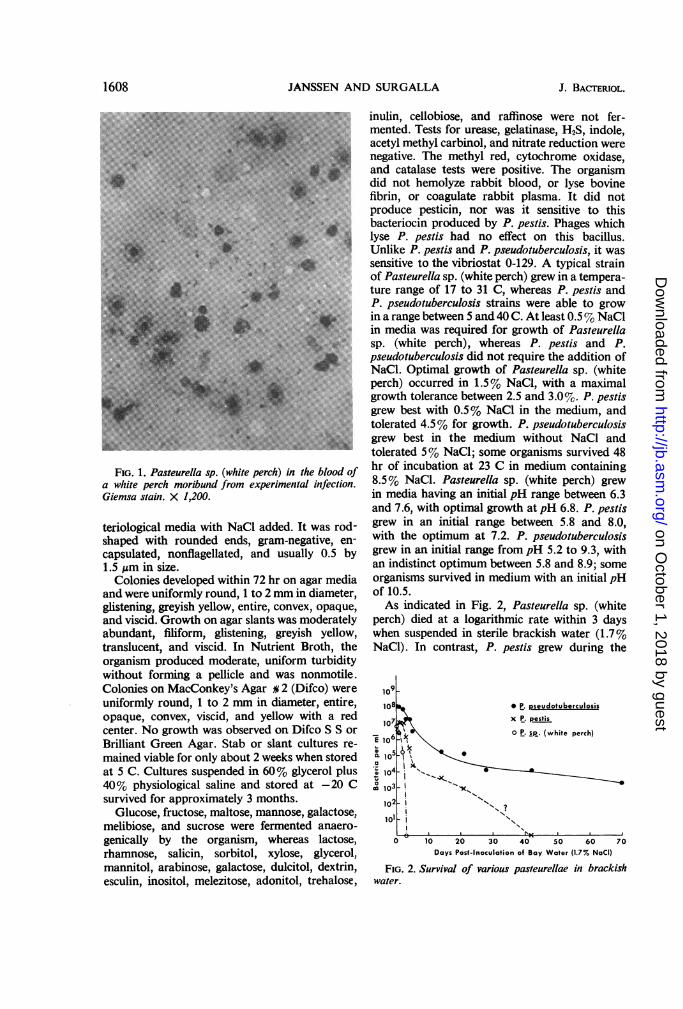
proved to be identical in every respect. Whenobserved in preparations of blood or organs,stained with Geimsa's blood stain, from mori-bund, experimentally infected white perch (Fig.1), the bacilli had the typical bipolar safety-pinappearance characteristic of P. pestis. Theorganism grew moderately well in ordinary bac-
1607
on October 1, 2018 by guest
http://jb.asm.org/
Dow
nloaded from
JANSSEN AND SURGALLA
FIG. 1. Pasteurella sp. (white perch) in the blood ofa white perch moribund from experimental infection.Giemsa stain. X 1,200.
teriological media with NaCI added. It was rod-shaped with rounded ends, gram-negative, en-capsulated, nonflagellated, and usually 0.5 by1.5 j,m in size.Colonies developed within 72 hr on agar media
and were uniformly round, 1 to 2 mm in diameter,glistening, greyish yellow, entire, convex, opaque,and viscid. Growth on agar slants was moderatelyabundant, filiform, glistening, greyish yellow,translucent, and viscid. In Nutrient Broth, theorganism produced moderate, uniform turbiditywithout forming a pellicle and was nonmotile.Colonies on MacConkey's Agar a 2 (Difco) wereuniformly round, 1 to 2 mm in diameter, entire,opaque, convex, viscid, and yellow with a redcenter. No growth was observed on Difco S S orBrilliant Green Agar. Stab or slant cultures re-mained viable for only about 2 weeks when storedat 5 C. Cultures suspended in 60% glycerol plus40% physiological saline and stored at -20 Csurvived for approximately 3 months.
Glucose, fructose, maltose, mannose, galactose,melibiose, and sucrose were fermented anaero-genically by the organism, whereas lactose,rhamnose, salicin, sorbitol, xylose, glycerol,mannitol, arabinose, galactose, dulcitol, dextrin,esculin, inositol, melezitose, adonitol, trehalose,
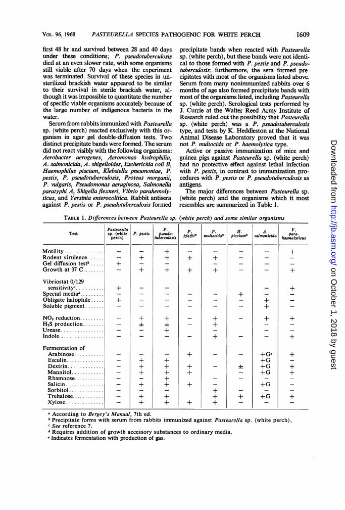
inulin, cellobiose, and raffinose were not fer-mented. Tests for urease, gelatinase, H2S, indole,acetyl methyl carbinol, and nitrate reduction werenegative. The methyl red, cytochrome oxidase,and catalase tests were positive. The organismdid not hemolyze rabbit blood, or lyse bovinefibrin, or coagulate rabbit plasma. It did notproduce pesticin, nor was it sensitive to thisbacteriocin produced by P. pestis. Phages whichlyse P. pestis had no effect on this bacillus.Unlike P. pestis and P. pseudotuberculosis, it wassensitive to the vibriostat 0-129. A typical strainof Pasteurella sp. (white perch) grew in a tempera-ture range of 17 to 31 C, whereas P. pestis andP. pseudotuberculosis strains were able to growin a range between 5 and 40 C. At least 0.5% NaClin media was required for growth of Pasteurellasp. (white perch), whereas P. pestis and P.pseudotuberculosis did not require the addition ofNaCl. Optimal growth of Pasteurella sp. (whiteperch) occurred in 1.5% NaCl, with a maximalgrowth tolerance between 2.5 and 3.0%. P. pestisgrew best with 0.5% NaCl in the medium, andtolerated 4.5% for growth. P. pseudotuberculosisgrew best in the medium without NaCl andtolerated 5% NaCl; some organisms survived 48hr of incubation at 23 C in medium containing8.5% NaCI. Pasteurella sp. (white perch) grewin media having an initial pH range between 6.3and 7.6, with optimal growth at pH 6.8. P. pestisgrew in an initial range between 5.8 and 8.0,with the optimum at 7.2. P. pseudotuberculosisgrew in an initial range from pH 5.2 to 9.3, withan indistinct optimum between 5.8 and 8.9; someorganisms survived in medium with an initial pHof 10.5.As indicated in Fig. 2, Pasteurella sp. (white
perch) died at a logarithmic rate within 3 dayswhen suspended in sterile brackish water (1.7%NaCI). In contrast, P. pestis grew during the
109108 R*. pseudotuberculosis
107 x P. pestis
l6 ;k 0 P. sp. (white perch)Eio3/10 $. \_
0 10 20 30 40 50 60 70Days Post-inoculation of Bay Water (1.7% NaCI)
FIG. 2. Survival of various pasteurellae in brackishwater.
1608 J. BACTERIOL.
on October 1, 2018 by guest
http://jb.asm.org/
Dow
nloaded from
VoL. 96, 1968 PASTEURELLA SPECIES PATHOGENIC FOR WHITE PERCH
first 48 hr and survived between 28 and 40 daysunder these conditions; P. pseudotuberculosis
died at an even slower rate, with some organismsstill viable after 70 days when the experimentwas terminated. Survival of these species in un-
sterilized brackish water appeared to be similar
to their survival in sterile brackish water, al-
though it was impossible to quantitate the number
of specific viable organisms accurately because of
the large number of indigenous bacteria in the
water.
Serum from rabbits immunized with Pastearella
sp. (white perch) reacted exclusively with this or-
ganism in agar gel double-diffusion tests. Two
distinct precipitate bands were formed. The serum
did not react visibly with the following organisms:
Aerobacter aerogenes, Aeromonas hydrophilia,A. salmonicida, A. shigelloides, Escherichia coli B,
Haemophilus piscium, Kiebsiella pneumoniae, P.
pestis, P. pseudotuberculosis, Proteus morganii,
P. vulgaris, Pseudomonas aeruginosa, Salmonella
paratyphi A, Shigella flexneri, Vibrio parahemoly-
ticus, and Yersinia enterocolitica. Rabbit antisera
against P. pestis or P. pseudotuberculosis formed
precipitate bands when reacted with Pasteurella
sp. (white perch), but these bands were not identi-
cal to those formed with P. pestis and P. pseudo-
tuberculosis; furthermore, the sera formed pre-
cipitates with most of the organisms listed above.
Serum from many nonimmunized rabbits over 6
months of age also formed precipitate bands with
most of the organisms listed, including Pasteurella
sp. (white perch). Serological tests performed byJ. Currie at the Walter Reed Army Institute of
Research ruled out the possibility that Pasteurella
sp. (white perch) was a P. pseudotuberculosis
type, and tests by K. Heddleston at the National
Animal Disease Laboratory proved that it was
not P. multocida or P. haemolytica type.
Active or passive immunization of mice and
guinea pigs against Pasteurella sp. (white perch)had no protective effect against lethal infection
with P. pestis, in contrast to immunization pro-
cedures with P. pestis or P. pseudotuberculosis as
antigens.T'he major differences between Pasteurella sp.
(white perch) and the organisms which it most
resembles are summarized in Table 1.
TABLE 1. Differences between Pasteurella sp. (white perch) and some similar organisms
Pasteurella P. P P. HA.VTest sp. (white P. pestis pseudo- ffi P.toid H.cim salok a Pa.aperch) tuberculosis pfJi0 mloia icu0 stoii ahem otlyicus
Motility.-....+........- +Rodent virulence - + + + + - - -Gel diffusion testb. + - -- - - -Growth at37C......+ + + + - +
Vibriostat 0/129sensitivity"... + - -- +
Specialmediad.........+ -Obligate halophile. + - - - -+ -Soluble pigment.......-- + -
NO3,reduction......- + + - + - + +H2S production..... 4 4 +- -Urease.........- - +-- -Indole.............- + - - +
Fermentation ofArabinose.........- + - - +G,9 +Esculin........- + + +G -Dextrin.-.+ + + 4 +G +Mannitol.......- + + + - +G +Rhamnose......+.....- -Salicin........- + + + -+G -Sorbitol........+....- -Trehalose.......- + + + + +G +Xylose........- + + + + - - -
a According to Bergey's Manual, 7th ed.b Precipitate forms with serum from rabbits immunized against Pasteurella sp. (white perch),rSee reference 7.d Requires addition of growth accessory substances to ordinary media.' Indicates fermentation with production of gas.
.1609
on October 1, 2018 by guest
http://jb.asm.org/
Dow
nloaded from
JANSSEN AND SIJRGALLA
DIscussIoNThis study confirms and extends the observa-
tions of Snieszko et al. (11); it also supports theirconclusion that the organism which they iso-lated from moribund white perch and diseasedstriped bass should be placed in the genusPasteurella, on the basis of criteria outlined inBergey's Manual. When the organism was com-pared with the established species which itresembles most closely, there were enough dif-ferences in every instance (Table 1) to rule out itsidentification with any of these species. In addi-tion, no serological evidence that this organismwas identical to any of these species was detected.Allen and Pelczar (1) used numerical taxonomyto compare the organism with a large variety ofbacteria isolated from the internal organs ofwhite perch, and concluded that it was not sig-nificantly similar to any of the organisms tested.
It is our conclusion that the Pasteurella-likeorganism from white perch is not identical toany formally established species. Thus, we suggestthat it be identified as Pasteurella piscicida (L.noun piscis, a fish; L.v.L. adj. suffix-cidus, to kill;M.L. noun piscicida, fish killer), since it is the onlyPasteurella species which has been isolated fromfish and shown (unpublished data) to cause lethalinfection of fish.The Pasteurella species from white perch
resembles marine bacteria in its absolute require-ment for NaCl and in its growth temperaturerange of 17 to 31 C; yet its range of tolerance toNaCl and hydrogen ion concentration is sur-prisingly narrow, and its survival time in brackishwater or laboratory media is very short, especiallywhen compared to P. pestis and P. pseudotubercu-losis. For these reasons, we suspect that this fishpathogen is poorly adapted for survival outside ofits host, and we suggest that this may be a majorreason why it was not detected before and hasnot been detected since the massive white perch
mortality occurred in the Chesapeake Bay area in1963.
ACKNOWLEDGMENTSWe thank S. F. Snieszko and G. L. Bullock of the
Eastem Fish Disease Laboratory, Keameysville, W.Va., and L. E. Cronin and C. D. Meyers of theChesapeake Biological Laboratory, Solomons, Md.,for their generous cooperation. We also thank E. A.Ambush, J. Holland, and E. D. Beesley for excellenttechnical assistance.
LrrERATURE CITED1. Allen, N., and M. J. Pelczar. 1967. Bacteriological
studies on the white perch, Roccus americanus.Chesapeake Sci. 8:135-154.
2. Beesley, E. D., R. R. Brubaker, W. A. Janssen,and M. J. Surgalla. 1967. Pesticins. III. Expres-sion of coagulase and mechanism of fibrino-lysis. J. Bacteriol. 94:19-26.
3. Brubaker, R. R., and M. J. Surgalla. 1962. Pesti-cins. II. Production of pesticins I and II. J.Bacteriol. 84:539-545.
4. Ewing, W. H., and J. G. Johnson. 1960. Thedifferentiation of Aeromonas and C27 culturesfrom Entererobacteriaceae. Intern. Bull. Bac-teriol. Nomen. Taxon. 10:223-230.
5. Landman, N. E., H. T. Bausum, and T. S. Matney.1962. Temperature-gradient plates for growthof microorganisms. J. Bacteriol. 83:463-469.
6. Ouchterlony, 0. 1962. Diffusion in gel methodsfor immunological analysis II. Progr. Allergy.6:30-154.
7. Shewan, J. M., W. Hodgkiss, and J. Liston. 1954.A method for the rapid differentiation of certainnonpathogenic, asporogenous bacilli. Nature173:208-209.
8. Sindermann, C. J. 1966. Diseases of marine fishes.Advan. Marine Biol. 4:1-89.
9. Snieszko, S. F., G. L. Bullock, E. Hollis, and J.G. Boone. 1964. Pasteurella sp. from anepizootic of whiteperch (Roccus americanus)in Chesapeake Bay tidewater areas. J. Bacteriol.88:1814-1815.
10. Society of American Bacteriologists. 1957. Manualof Microbiological Methods. McGraw-HillBook Co., Inc., New York.
1610 J. BAmrRioL.
on October 1, 2018 by guest
http://jb.asm.org/
Dow
nloaded from